EP3363363A1 - Procédé d'exécution d'un examen pour l'imagerie - Google Patents
Procédé d'exécution d'un examen pour l'imagerie Download PDFInfo
- Publication number
- EP3363363A1 EP3363363A1 EP17178749.2A EP17178749A EP3363363A1 EP 3363363 A1 EP3363363 A1 EP 3363363A1 EP 17178749 A EP17178749 A EP 17178749A EP 3363363 A1 EP3363363 A1 EP 3363363A1
- Authority
- EP
- European Patent Office
- Prior art keywords
- respiratory
- cycle
- patient
- imaging examination
- parameter
- Prior art date
- Legal status (The legal status is an assumption and is not a legal conclusion. Google has not performed a legal analysis and makes no representation as to the accuracy of the status listed.)
- Granted
Links
Images


Classifications
-
- G—PHYSICS
- G06—COMPUTING OR CALCULATING; COUNTING
- G06T—IMAGE DATA PROCESSING OR GENERATION, IN GENERAL
- G06T7/00—Image analysis
- G06T7/0002—Inspection of images, e.g. flaw detection
- G06T7/0012—Biomedical image inspection
- G06T7/0014—Biomedical image inspection using an image reference approach
- G06T7/0016—Biomedical image inspection using an image reference approach involving temporal comparison
-
- A—HUMAN NECESSITIES
- A61—MEDICAL OR VETERINARY SCIENCE; HYGIENE
- A61B—DIAGNOSIS; SURGERY; IDENTIFICATION
- A61B6/00—Apparatus or devices for radiation diagnosis; Apparatus or devices for radiation diagnosis combined with radiation therapy equipment
- A61B6/02—Arrangements for diagnosis sequentially in different planes; Stereoscopic radiation diagnosis
- A61B6/03—Computed tomography [CT]
- A61B6/032—Transmission computed tomography [CT]
-
- A—HUMAN NECESSITIES
- A61—MEDICAL OR VETERINARY SCIENCE; HYGIENE
- A61B—DIAGNOSIS; SURGERY; IDENTIFICATION
- A61B5/00—Measuring for diagnostic purposes; Identification of persons
- A61B5/08—Measuring devices for evaluating the respiratory organs
- A61B5/0803—Recording apparatus specially adapted therefor
-
- A—HUMAN NECESSITIES
- A61—MEDICAL OR VETERINARY SCIENCE; HYGIENE
- A61B—DIAGNOSIS; SURGERY; IDENTIFICATION
- A61B6/00—Apparatus or devices for radiation diagnosis; Apparatus or devices for radiation diagnosis combined with radiation therapy equipment
- A61B6/04—Positioning of patients; Tiltable beds or the like
-
- A—HUMAN NECESSITIES
- A61—MEDICAL OR VETERINARY SCIENCE; HYGIENE
- A61B—DIAGNOSIS; SURGERY; IDENTIFICATION
- A61B6/00—Apparatus or devices for radiation diagnosis; Apparatus or devices for radiation diagnosis combined with radiation therapy equipment
- A61B6/52—Devices using data or image processing specially adapted for radiation diagnosis
- A61B6/5211—Devices using data or image processing specially adapted for radiation diagnosis involving processing of medical diagnostic data
-
- A—HUMAN NECESSITIES
- A61—MEDICAL OR VETERINARY SCIENCE; HYGIENE
- A61B—DIAGNOSIS; SURGERY; IDENTIFICATION
- A61B6/00—Apparatus or devices for radiation diagnosis; Apparatus or devices for radiation diagnosis combined with radiation therapy equipment
- A61B6/52—Devices using data or image processing specially adapted for radiation diagnosis
- A61B6/5294—Devices using data or image processing specially adapted for radiation diagnosis involving using additional data, e.g. patient information, image labeling, acquisition parameters
-
- A—HUMAN NECESSITIES
- A61—MEDICAL OR VETERINARY SCIENCE; HYGIENE
- A61B—DIAGNOSIS; SURGERY; IDENTIFICATION
- A61B6/00—Apparatus or devices for radiation diagnosis; Apparatus or devices for radiation diagnosis combined with radiation therapy equipment
- A61B6/54—Control of apparatus or devices for radiation diagnosis
- A61B6/541—Control of apparatus or devices for radiation diagnosis involving acquisition triggered by a physiological signal
-
- A—HUMAN NECESSITIES
- A61—MEDICAL OR VETERINARY SCIENCE; HYGIENE
- A61B—DIAGNOSIS; SURGERY; IDENTIFICATION
- A61B6/00—Apparatus or devices for radiation diagnosis; Apparatus or devices for radiation diagnosis combined with radiation therapy equipment
- A61B6/54—Control of apparatus or devices for radiation diagnosis
- A61B6/545—Control of apparatus or devices for radiation diagnosis involving automatic set-up of acquisition parameters
-
- G—PHYSICS
- G06—COMPUTING OR CALCULATING; COUNTING
- G06T—IMAGE DATA PROCESSING OR GENERATION, IN GENERAL
- G06T12/00—Tomographic reconstruction from projections
- G06T12/10—Image preprocessing, e.g. calibration, positioning of sources or scatter correction
-
- G—PHYSICS
- G06—COMPUTING OR CALCULATING; COUNTING
- G06T—IMAGE DATA PROCESSING OR GENERATION, IN GENERAL
- G06T7/00—Image analysis
- G06T7/10—Segmentation; Edge detection
- G06T7/11—Region-based segmentation
-
- G—PHYSICS
- G06—COMPUTING OR CALCULATING; COUNTING
- G06T—IMAGE DATA PROCESSING OR GENERATION, IN GENERAL
- G06T7/00—Image analysis
- G06T7/20—Analysis of motion
-
- A—HUMAN NECESSITIES
- A61—MEDICAL OR VETERINARY SCIENCE; HYGIENE
- A61B—DIAGNOSIS; SURGERY; IDENTIFICATION
- A61B6/00—Apparatus or devices for radiation diagnosis; Apparatus or devices for radiation diagnosis combined with radiation therapy equipment
- A61B6/52—Devices using data or image processing specially adapted for radiation diagnosis
- A61B6/5205—Devices using data or image processing specially adapted for radiation diagnosis involving processing of raw data to produce diagnostic data
-
- G—PHYSICS
- G06—COMPUTING OR CALCULATING; COUNTING
- G06T—IMAGE DATA PROCESSING OR GENERATION, IN GENERAL
- G06T2207/00—Indexing scheme for image analysis or image enhancement
- G06T2207/10—Image acquisition modality
- G06T2207/10072—Tomographic images
- G06T2207/10081—Computed x-ray tomography [CT]
Definitions
- the invention relates to a method for performing an imaging examination of a patient by means of a computer tomograph, the associated computer tomograph and an associated computer program product.
- three-dimensional 3D images of a measurement area with an extension in the z-direction are usually used.
- projection data can be acquired during an imaging examination, from which the 3D images can be reconstructed.
- the imaging examination by means of the computer tomograph usually requires ionizing X-rays.
- the 3D images comprise a plurality of slice images of the patient.
- the slice images are preferably reconstructed over all the respiratory phases of a respiratory cycle, which in particular depicts inhalation and exhalation of the patient and corresponds to a periodic segment of the respiratory movement.
- This generates 4D images or respiratory-correlated 3D image series. In particular, this makes it possible to ensure a particularly suitable adaptation of the dose distribution to a planning target volume in motion.
- Irradiation planning critically depends on image quality of the 4D images.
- measurement parameters of the imaging examination are primarily relevant, wherein the measurement parameters are preferably optimally calculated.
- the measurement parameters so far from a list chosen with, in particular for a user, immutable measurement parameters for the imaging examination.
- the fixed measurement parameters are usually not optimized in terms of image quality. The disadvantages resulting from this are that an inappropriate set of measurement parameters is selected by an operator error of the user.
- the object of the invention is to specify a method, which is individually adapted to the patient, for carrying out an imaging examination of a patient by means of a computer tomograph, the associated computer tomograph and an associated computer program product.
- the detection of the respiratory movement of the patient takes place in particular by means of a sensor, for example by means of a camera or a breathing belt.
- the respiratory movement usually describes a free breathing of the patient and in particular comprises at least one breathing cycle of the patient.
- the respiratory movement is detected over the entire respiratory cycle of the patient, with the entire respiratory cycle comprising at least one single inhalation and one exhalation of the patient.
- the respiratory cycle preferably corresponds to a periodic segment of the respiratory movement.
- the period of the periodic segment corresponds to the duration of the respiratory cycle.
- the determination of the respiratory correlated parameter T cycle takes place in particular automatically according to a suitable algorithm. The slower the patient breathes, the higher is the respiratory correlated parameter T cycle .
- the respiratory correlated parameter corresponds to the duration of the respiratory cycle.
- the determination of the measuring range of the imaging examination can be carried out by a user of the computer tomograph.
- the computer tomograph has a planning unit which has a monitor and at least one input device.
- the user sets the measuring range in particular by means of at least an input device on a graphical user interface of the monitor.
- the user is shown on the graphical user interface overview images, which are reconstructed from overview data acquired during an overview measurement.
- the measuring range of the imaging examination is determined automatically, in particular by means of sensor-supported detection of the patient and / or by means of landmark detection.
- the measuring range of the imaging examination is in particular extended in a longitudinal axis extending in the longitudinal direction of the patient.
- the longitudinal axis of the patient is preferably parallel to a longitudinal axis extending in the longitudinal direction of a patient couch.
- the measuring area has the at least one z-position, wherein the at least one z-position describes a position of a layer along the longitudinal axis of the patient bed.
- the layer along the longitudinal axis of the patient bed is in particular imaged by a slice image.
- the slice image has a slice thickness with extension in the z direction of the computed tomography scanner.
- the measuring area preferably comprises a 3D volume with an extension in the z-direction of the computer tomograph.
- a single layer image with a high layer thickness or several layer images with a low layer thickness can be reconstructed.
- the first parameter group comprises, in particular, fixed parameters which are predetermined, for example, by the patient or by a technical embodiment of the computer tomograph.
- fixed parameters are an age of the patient or a tube-detector speed, which describes the speed at which at least one X-ray source of the computer tomograph and at least one X-ray detector of the computer tomograph can rotate around the patient.
- the breath-correlated parameter T cycle is assigned to the first parameter group.
- the second parameter group comprises modifiable parameters, which can be determined in particular by the user of the computer tomograph, for example on the graphical user interface.
- modifiable parameters are a total dose I ges , which is directly proportional to another measured quantity of the patient's computed tomography dose index (CTDI) in the imaging examination, and a reconstruction kernel for a reconstruction of a slice image from the projection data.
- the total dose I ges indicates in particular the image quality of the slice image as well as the radiation exposure of the patient when performing the imaging examination.
- the third parameter group has dependent parameters, which can be calculated, for example, from parameters from the first parameter group and / or from the second parameter group. For example, a reconstruction angle interval theta can be calculated.
- the at least one measurement parameter can be assigned to the third parameter group.
- the automatic calculation of the at least one measurement parameter according to the respiratory movement in particular describes the automatic calculation of a dependent parameter from the third parameter group according to a fixed parameter from the first parameter group, in particular according to the respiratory correlated parameter T cycle .
- the respiratory correlated parameter T cycle and a modifiable parameter of the second parameter group are particularly such as the input parameters for the automatic calculation that in performing the imaging examination, the projection data can be acquired at the at least one z-position over the entire duration of the breathing cycle.
- the projection data at the at least one z-position is acquired for each time of the respiratory cycle.
- the inhalation and exhalation of the patient can be imaged at the at least one z-position during the imaging examination. If the projection data at the at least one z-position depicts the respiratory cycle of the patient over the entire duration of the respiratory cycle, it is customary to speak of a respiratory-correlated imaging examination.
- the automatic calculation has at least one formula, wherein the at least one measurement parameter is automatically calculated according to the at least one formula in such a way that, during the imaging examination, the projection data at the at least one z-position covers the patient's respiratory cycle over the entire time period of the patient Can map respiratory cycle.
- the respiratory correlated parameter T cycle then represents in particular a computing element in the formula, by means of which the at least one measurement parameter is calculated automatically.
- the automatic calculation is carried out in particular in a computing unit of the computer tomograph.
- the arithmetic unit advantageously has a memory for this, in which program code means can be loaded.
- the program code means in particular have the at least one formula for the automatic calculation of the at least one measurement parameter.
- the automatic calculation is particularly advantageous because the at least one measurement parameter for each patient is automatically calculated individually and thus the imaging examination is particularly suitably adapted to the patient, because the at least one measurement parameter is not selected from a set of patients averaged predetermined parameter sets.
- the patient bed When performing the imaging examination with a pitch, the patient bed is continuously pushed through a maximum field of view of the computer tomograph. This procedure is usually called spiral acquisition of projection data. Typical values of a pitch when performing a measurement in the computed tomograph are above 0 and below 2.
- the patient couch may be slid in discrete steps through the maximum field of view of the computed tomograph. In this case, after each discrete step, preferably for the at least one z-position, the projection data is acquired over the entire duration of the respiratory cycle.
- the pitch is proportional to a table feed of the patient couch and an expansion of the x-ray detector in the z direction of the computer tomograph.
- the maximum field of vision depends in particular on the extent.
- the imaging examination is preferably performed with a small pitch of the order of 0.1 at a minimum tube rotation time T rot of the computer tomograph of 0.5s.
- the pitch corresponds to a ratio of the minimum tube rotation time T red to the respiratory correlated parameter T cycle .
- the minimum tube rotation time T red is usually specified specifically for each computed tomograph by the technical embodiment and can not be changed.
- the lower the minimum tube rotation period T red the higher the tube-detector speed, the faster the X-ray source and the X-ray detector in particular rotate around the patient and / or the more projection data is acquired per unit of time.
- the at least one x-ray emitter and the at least one x-ray detector in particular rotate continuously during the imaging examination.
- the patient couch When the patient couch is pushed through the maximum field of view, it is commonly used during acquisition Projection data, an angle in which the at least one X-ray source and the at least one X-ray detector are relative to the patient bed, and a current z-position of the patient bed, for example, stored with respect to the projection data.
- the imaging examination is carried out in particular after the at least one measurement parameter has been calculated automatically, preferably in a patient-specific manner, in particular in accordance with the breathing movement of the patient.
- the imaging examination can therefore advantageously be carried out in accordance with the proposed procedure, in particular because the projection data can map the respiratory cycle of the patient over the entire duration of the respiratory cycle. In particular, artifacts can be suppressed by the respiratory movement of the patient.
- the automatic computation may optimize imaging to not irradiate the patient with the ionizing X-rays longer than necessary for acquiring the projection data at the at least one z-position throughout the duration of the respiratory cycle in the computed tomography device.
- the projection data at the at least one z-position depicts the respiratory cycle of the patient over the entire duration of the respiratory cycle.
- Typichate image data, in particular the slice image reconstructed from the acquired projection data and stored in a database and / or provided on the monitor for a diagnosis by a physician.
- the reconstructed slice image is used as a basis for creating a radiotherapy plan.
- the respiratory correlated parameter T cycle is determined such that the respiratory movement is subdivided into periodic segments and the duration of the respiratory cycle is calculated according to a median of durations of the periodic segments.
- a sample of periodic segments is generated, wherein in particular for each periodic segment the time duration is calculated and then the median over the time periods is formed.
- the respiratory correlated parameter T cycle can in particular be determined on the basis of the time duration thus calculated.
- the median over the durations, in particular the time period thus calculated set as the respiratory correlated parameter T cycle .
- the respiratory correlated parameter T cycle is adjusted according to a standard deviation of the time periods of the periodic segments. From the sample of the periodic segments, the standard deviation of the durations of the periodic segments can be calculated. In particular, the standard deviation gives a measure of an irregularity of the durations of the periodic segments and thus the measure of the irregularity of the breathing movement of the patient. In particular, the higher the standard deviation, the respiratory correlated parameter T cycle is multiplied by a larger factor. The factor is always always greater than one.
- the automatic calculation of the at least one measurement parameter comprises such a calculation of a tube current I tube of the computed tomography during the imaging examination that the tube current I tube is indirectly proportional to the respiration-correlated parameter T cycle .
- the higher the respiratory correlated parameter T cycle the lower, in particular, is the tube current I tube . It follows that, preferably, the slower the patient breathes, the lower the tube current I tube . If the tube current I tube is indirectly proportional to the respiration-correlated parameter T cycle , in particular the total dose I ges is kept constant.
- the automatic calculation of the at least one measurement parameter comprises such a calculation of a layer-effective tube current-time product I red , that the layer-effective tube current-time product I rot is a product of the tube current I tube of the computed tomograph in the imaging examination and from the minimum tube rotation time T red of the computer tomograph in the imaging examination corresponds.
- the higher the tube current I tube the higher the layer-effective tube current time product I red .
- the layer-effective tube current-time product I red is the higher the higher the minimum tube rotation time T red of the computed tomography scanner is in the imaging examination.
- the layer-effective tube current-time product I red indicates a measure of emitted X-ray quantity and is typically proportional to the radiation exposure. The higher the layer-effective tube current time product I is red , the higher the radiation exposure is usually.
- the tube current I tube which depends indirectly proportionally on the respiration-correlated parameter T cycle , enters.
- the reconstruction angle interval theta indicates, in particular, an angular interval for the reconstruction of the slice image.
- the reconstruction angle interval theta comprises, in particular, an angular range of at least 180 °, which usually represents a minimum for the reconstruction of the slice image.
- the larger the reconstruction angle interval theta the more projection data having the at least one angle within the reconstruction angle interval theta is used in the reconstruction of the slice image.
- the reconstruction angle interval theta is at least 180 °
- image artifacts in the layer image can be minimized, which are caused in particular by a reconstruction angle interval below 180 °.
- the automatic calculation of the at least one measurement parameter comprises at least one such calculation of a reconstruction angle interval theta for a z-position that the reconstruction angle interval theta for the at least one z position directly proportional to the breath-correlated parameter T cycle.
- the higher the respiratory correlated parameter T cycle , or the slower the patient breathes the greater the reconstruction angle interval theta.
- the reconstruction angle interval theta depends in particular on the breath-correlated parameter T cycle, whereby the reconstruction angle interval theta is the individual patient automatically calculated in such a way that in particular the projection data at the imaging at least a z-position of the respiratory cycle of the patient over the entire duration of the respiratory cycle.
- the automatic calculation of the at least one measurement parameter includes such a calculation of the reconstruction angle interval theta for the at least one z-position that the reconstruction angle interval theta for the at least one z-position is indirectly proportional to the minimum tube rotation rotation time T rot of the computer tomograph the imaging examination is.
- the reconstruction angle interval theta in particular is the smaller, the larger the minimum tube rotation rotation time T rot of the computer tomograph in the imaging examination.
- the smaller the tube-detector velocity the smaller the reconstruction angle interval theta.
- the reconstruction angle interval theta depends, in particular, on the minimum tube rotation time T rot of the computed tomography during the imaging examination, so that the reconstruction angle interval theta is automatically calculated in accordance with the technical design of the computed tomography such that in particular the projection data at the at least one z position map the patient's respiratory cycle over the entire duration of the respiratory cycle.
- the automatic calculation of the at least one measurement parameter comprises that a product from the reconstruction angle interval theta for the at least one z-position and from the slice-effective tube current-time product I rot of the computed tomography device is kept constant during the imaging examination becomes. For example, when the reconstruction angle interval theta is multiplied by a factor, the slice-effective tube current-time product I red of the computed tomograph is automatically multiplied in the imaging study by a sweep of the factor. As a result, in particular the image quality BQ of the slice image is kept constant throughout the patient. If slice images are reconstructed at different z-positions, the slice images preferably have the same image quality BQ.
- the projection data is acquired in at least one angle relative to the patient and the projection data with the at least one angle within a reconstruction angle interval theta is selected for the at least one z position, of which a slice image is applied to the at least one z position. Position is reconstructed.
- the projection data in particular have the current z-position of the patient couch during the imaging examination and in particular the at least one angle of the at least one x-ray radiator and the at least one x-ray detector during the imaging examination.
- the projection data in the at least one angle within the reconstruction angle interval theta are preferably used for the reconstruction of the slice image at the at least one z position.
- the projection data in the at least one slice is preferably used for the reconstruction of a single slice image Angle of the respective reconstruction angle interval theta used.
- the reconstruction angle interval theta comprises at least 180 ° for each reconstructed slice image.
- the projection data is acquired at the at least one z-position over the entire duration of the respiratory cycle, it is particularly advantageous to reconstruct not only the slice image, but time-resolved slice images from the projection data at the at least one z-position.
- the respiratory cycle of the patient can usually be depicted simpler and better in the time-resolved slice images than in the slice image alone.
- a time resolution of the time-resolved slice images is limited or predetermined in particular by the minimum tube rotation time T rot of the computed tomography during the imaging examination.
- the reconstruction angle interval theta preferably comprises the angular range of at least 180 °.
- the time resolution defines a maximum temporal coherence of structures moved by the respiratory movement, in particular a skin surface, an organ, a carcinoma and / or a tumor, of the patient. If the time resolution comprises N seconds, preferably within N seconds, the projection data is detected at the at least one z-position according to the reconstruction angle interval theta.
- the maximum temporal coherence may depend on other types of patient motion.
- the maximum temporal coherence in the imaging examination can be optimized in particular if there is no movement, in particular of the structures moved by the respiratory movement. In other words, the less movement of the patient, the better the maximum temporal coherence.
- An embodiment provides that the reconstruction angle interval theta for the at least one z-position directly proportional to a product of a relative time resolution ⁇ T rel and the respiratory correlated parameter T cycle .
- the time resolution of the time-resolved slice images is preferably adapted to the respiratory movement of the patient, in which a relative time resolution ⁇ T rel is defined.
- the relative time resolution ⁇ T rel is defined in particular as a percentage, preferably 5%, of the respiratory correlated parameter T cycle .
- time-resolved slice images are reconstructed from the projection data at the at least one z-position 20, which images represent the entire duration of the breathing cycle.
- the relative time resolution ⁇ T rel can be assigned to the second parameter group, because usually the relative time resolution ⁇ T rel , in particular by the user, can be modified.
- the relative time resolution ⁇ T rel is automatically adjusted, in particular, increased so that the reconstruction angle interval theta is at least 180 °.
- the tube current I tube and the minimum tube rotation time T red remain unchanged. In other words, all patients are preferably measured such that the reconstruction angle interval theta is at least 180 °.
- the patient's breathing movement is additionally detected in real time and the reconstruction of the slice image at the at least one z-position is performed from the projection data in accordance with a respiratory phase of the patient's breathing movement.
- the respiratory phase can be assigned to at least one of the time-resolved slice images.
- the at least one of the time-resolved slice images in particular shows the z position according to the respiratory phase.
- the respiratory phase to be displayed can be selected on the monitor by the user, and the planning unit displays in particular the at least one of the time-resolved slice images.
- the measurement area has a 3D volume with extension in the z-direction of the computed tomography and the measurement area
- the thorax of the patient according to the selected respiratory phase of the patient can be displayed on the monitor.
- the patient may be shown once by inhalation and another by exhaling.
- Performing the imaging examination of the patient according to the at least one measurement parameter in the measurement area by means of the computed tomography device can in particular comprise the reconstruction of respiration-correlated 4D images or respiratory-correlated 3D image series.
- a computer program product which is directly loadable into a memory of the programmable arithmetic unit, has program code means for carrying out a method according to the invention when the computer program product is executed in the arithmetic unit.
- the computer program product may be a computer program or comprise a computer program.
- the computer program product is configured such that it can execute the method steps according to the invention by means of the arithmetic unit.
- the arithmetic unit must in each case have the prerequisites such as, for example, a corresponding main memory, a corresponding graphics card or a corresponding logic unit, so that the respective method steps can be carried out efficiently.
- the computer program product is for example stored on a computer-readable medium or deposited on a network or server, from where it can be loaded into the processor of the computing unit, which may be formed, for example, as part of the computed tomography.
- control information of the computer program product can be stored on an electronically readable data carrier.
- the control information of the electronically readable data carrier can be configured such that when using the data carrier in the arithmetic unit, they execute a method according to the invention.
- the computer program product can also represent the electronically readable data carrier. Examples of electronically readable data carriers are a DVD, a magnetic tape, a hard disk or a USB stick, on which electronically readable control information, in particular software cf. above, is stored. If this control information software is read from the data medium and stored in the computer unit and / or planning unit and / or measuring unit of the computer tomograph, all embodiments of the method described above can be carried out.
- the invention can also emanate from the said computer-readable medium and / or the said electronically readable data carrier.
- Fig. 1 shows a computer tomograph 10 according to the invention, which comprises a planning unit 11, a computing unit 12 and a measuring unit 13.
- the measuring unit 13 comprises at least one X-ray emitter 14 and at least one X-ray detector 15.
- the patient 17 is arranged on a patient couch 16.
- the planning unit 11 may include the monitor with a graphical user interface and an input device.
- a user can interact with the planning unit 11, in particular by the input device.
- computer tomography projection data and / or a tomographic image can be displayed to the user on the monitor.
- the user can select a respiratory phase of the patient on the monitor, and according to the respiratory phase, the respective slice image is displayed to the user.
- the computer tomograph 10 is connected to the planning unit 11 and the arithmetic unit 12 for data exchange.
- the computer tomograph 10 may preferably rotate the at least one x-ray emitter 14 and the at least one x-ray detector 15, which are arranged at a fixed distance relative to one another, around the patient couch 16, in particular the patient 17.
- the computer tomograph 10 detects in which angle the at least one x-ray emitter 14 and the at least one x-ray detector 15 are located relative to the patient couch 17.
- the computer tomograph 10 preferably assigns projection data which in the angle of the at least one x-ray emitter 14 and the at least one x-ray detector 15 relative to the patient couches 16 to a description of the angle, whereby in the reconstruction of the slice image of FIG the projection data of the angle can be considered.
- Fig. 2 shows a flow chart of the method according to the invention for performing the imaging examination of the patient 17 by means of the computer tomograph 10.
- the inventive method comprises the method steps 201-204.
- Method step 201 identifies the detection of a respiratory movement of the patient 17, wherein a breath-correlated parameter T cycle is determined by the respiratory movement of the patient 17, which describes a time duration of a breathing cycle of the respiratory movement.
- Method step 202 identifies the definition of a measuring range of the imaging examination, wherein the measuring range has at least one z-position.
- Method step 203 characterizes the automatic calculation of at least one measurement parameter according to the respiratory movement, wherein the respiratory correlated parameter T cycle is used as an input parameter for the automatic calculation of the at least one measurement parameter, such that when the imaging examination is performed according to the at least one measurement parameter, projection data at the at least one z position over the entire duration of the respiratory cycle can be detected.
- the breath-correlated parameter T cycle and the at least one z position enter.
- Method step 204 comprises carrying out the imaging examination of the patient according to the at least one measurement parameter in the measurement area by means of the computer tomograph, wherein the projection data is acquired, which images the respiratory cycle of the patient 17 at the at least one z position over the entire duration of the respiratory cycle.
- the measuring range and the at least one automatically calculated measuring parameter are included in method step 204.
- Fig. 3 shows an embodiment of the method according to the invention.
- Process step 201.1 indicates that the respiratory correlated parameter T cycle is determined such that the respiratory movement is subdivided into periodic segments and the duration of the respiratory cycle is calculated according to a median of time durations of the periodic segments.
- Method step 201.1.1 indicates that the respiration-correlated parameter T cycle is adjusted according to a standard deviation of the time periods of the periodic segments.
- Method step 203.1 indicates that the automatic calculation of the at least one measurement parameter comprises such a calculation of a tube current I tube of the computed tomography device 10 during the imaging examination that the tube current I tube is indirectly proportional to the respiration-correlated parameter T cycle .
- Method step 203.2 indicates that the automatic calculation of the at least one measurement parameter comprises such a calculation of a layer-effective tube current-time product I red , that the layer-effective tube current-time product I red contributes to a product from the tube current I tube of the computer tomograph the imaging examination and from a minimum tube rotation time T red of the computer tomograph 10 corresponds to the imaging examination.
- a method Step 203.3 indicates that the automatic calculation of the at least one measurement parameter for the at least one such calculation of the reconstruction angle interval theta a z-position that the reconstruction angle interval theta for the at least one z position directly proportional to the breath-correlated parameter T cycle.
- Method step 203.4 indicates that the reconstruction angle interval theta for the at least one z-position is directly proportional to a product of a relative time resolution ⁇ T rel and the respiration-correlated parameter T cycle .
- Step 203.5 indicates that automatically calculating the at least one measurement parameter includes such a calculation of the reconstruction angle interval theta for the at least one z-position that the reconstruction angle interval theta for the at least one z-position is indirectly proportional to the minimum tube rotation rotation time T rot of the computed tomography apparatus 10 the imaging examination is.
- Step 203.6 indicates that automatically calculating the at least one measurement parameter comprises keeping a product of the reconstruction angle interval theta constant for the at least one z-position and the slice-effective tube current-time product I red of the computed tomography apparatus 10 in the imaging examination becomes.
- Method step 204.1 indicates that the projection data is acquired in at least one angle relative to the patient 17 and the projection data with the at least one angle within the reconstruction angle interval theta is selected for the at least one z-position, of which a slice image is attached to the at least one z-position. Position is reconstructed.
- Method step 204.2 indicates that, in performing the imaging examination, the respiratory movement of the patient 17 is additionally detected in real time and a reconstruction of the slice image at the at least one z-position from the projection data takes place in accordance with a respiratory phase of the respiratory movement of the patient 17.
- method step 204.2 the detection of the respiratory movement according to method step 201 begins.
- the tube current I tube has the unit [mA].
- the total dose I ges has the [mAs] unit.
- the respiratory correlated parameter T cycle has the unit [s].
- I red I tube ⁇ T red
- the layer-effective tube current time product I red has the unit [mAs].
- the reconstruction angle interval theta has the unit [°].
- the respiratory correlated parameter T cycle can be converted into a parameter respiratory cycles-per-minute ApM.
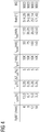
- Fig. 4 a table with exemplary measurement parameters, which are calculated according to an embodiment of the method according to the invention, in particular according to the first, second, third and fourth formula.
- Fig. 4 shows that the tube current I tube is indirectly proportional to the respiratory correlated parameter T cycle .
- Fig. 4 2 shows that the layer-effective tube current time product I red corresponds to a product from the tube current I tube of the computed tomography scanner 10 during the imaging examination and from the minimum tube rotation cycle time T rot of the computer tomograph 10 during the imaging examination.
- Fig. 4 shows that the reconstruction angle interval theta for the at least one z position is directly proportional to a product of the relative time resolution ⁇ T rel and the respiratory correlated parameter T cycle .
- Fig. 4 shows that the reconstruction angle interval theta for the at least one z position is directly proportional to the respiratory correlated parameter T cycle .
- Fig. 4 shows that a product BQ from the reconstruction angle interval theta is constant for the at least one z-position and from the slice-effective tube current-time product I red of the computed tomograph in the imaging examination.
Landscapes
- Health & Medical Sciences (AREA)
- Engineering & Computer Science (AREA)
- Life Sciences & Earth Sciences (AREA)
- Medical Informatics (AREA)
- Physics & Mathematics (AREA)
- General Health & Medical Sciences (AREA)
- Heart & Thoracic Surgery (AREA)
- Public Health (AREA)
- Radiology & Medical Imaging (AREA)
- Veterinary Medicine (AREA)
- Nuclear Medicine, Radiotherapy & Molecular Imaging (AREA)
- Animal Behavior & Ethology (AREA)
- Surgery (AREA)
- Molecular Biology (AREA)
- Biomedical Technology (AREA)
- Biophysics (AREA)
- Pathology (AREA)
- High Energy & Nuclear Physics (AREA)
- Optics & Photonics (AREA)
- Theoretical Computer Science (AREA)
- Computer Vision & Pattern Recognition (AREA)
- General Physics & Mathematics (AREA)
- Pulmonology (AREA)
- Physiology (AREA)
- Multimedia (AREA)
- Quality & Reliability (AREA)
- Apparatus For Radiation Diagnosis (AREA)
Priority Applications (3)
| Application Number | Priority Date | Filing Date | Title |
|---|---|---|---|
| EP17178749.2A EP3363363B1 (fr) | 2017-06-29 | 2017-06-29 | Procédé d'exécution d'un examen pour l'imagerie |
| US16/018,216 US10803587B2 (en) | 2017-06-29 | 2018-06-26 | Method for performing an imaging examination |
| CN201810669382.5A CN109199423B (zh) | 2017-06-29 | 2018-06-26 | 执行成像检查的方法 |
Applications Claiming Priority (1)
| Application Number | Priority Date | Filing Date | Title |
|---|---|---|---|
| EP17178749.2A EP3363363B1 (fr) | 2017-06-29 | 2017-06-29 | Procédé d'exécution d'un examen pour l'imagerie |
Publications (2)
| Publication Number | Publication Date |
|---|---|
| EP3363363A1 true EP3363363A1 (fr) | 2018-08-22 |
| EP3363363B1 EP3363363B1 (fr) | 2021-11-17 |
Family
ID=59258095
Family Applications (1)
| Application Number | Title | Priority Date | Filing Date |
|---|---|---|---|
| EP17178749.2A Active EP3363363B1 (fr) | 2017-06-29 | 2017-06-29 | Procédé d'exécution d'un examen pour l'imagerie |
Country Status (3)
| Country | Link |
|---|---|
| US (1) | US10803587B2 (fr) |
| EP (1) | EP3363363B1 (fr) |
| CN (1) | CN109199423B (fr) |
Cited By (1)
| Publication number | Priority date | Publication date | Assignee | Title |
|---|---|---|---|---|
| CN111599474A (zh) * | 2019-02-20 | 2020-08-28 | 西门子医疗有限公司 | 检查基于x射线的医学成像应用的流程的特征参数的方法 |
Families Citing this family (2)
| Publication number | Priority date | Publication date | Assignee | Title |
|---|---|---|---|---|
| DE102020211944A1 (de) | 2020-09-23 | 2022-03-24 | Siemens Healthcare Gmbh | Verfahren zur Ansteuerung eines medizinischen Bildgebungsgeräts |
| EP4560568B1 (fr) | 2023-11-21 | 2026-04-15 | Siemens Healthineers AG | Procédé d'évaluation de l'exploitabilité de données d'image de tomographie 4d, produit de programme informatique et dispositif de balayage |
Citations (3)
| Publication number | Priority date | Publication date | Assignee | Title |
|---|---|---|---|---|
| EP0370341A2 (fr) * | 1988-11-23 | 1990-05-30 | General Electric Company | Méthode pour réduire les artefacts dus au mouvement d'image en imagerie de projection |
| US20110286574A1 (en) * | 2010-05-21 | 2011-11-24 | Tatsuro Suzuki | X-ray computed tomography apparatus |
| US20120014499A1 (en) * | 2010-07-16 | 2012-01-19 | Siemens Aktiengesellschaft | CT System |
Family Cites Families (6)
| Publication number | Priority date | Publication date | Assignee | Title |
|---|---|---|---|---|
| US7177386B2 (en) * | 2004-03-15 | 2007-02-13 | Varian Medical Systems Technologies, Inc. | Breathing synchronized computed tomography image acquisition |
| US7515678B2 (en) * | 2005-11-23 | 2009-04-07 | General Electric Company | Method and system for performing CT image reconstruction with motion artifact correction |
| EP2696755A4 (fr) * | 2011-04-12 | 2015-07-01 | Orsan Medical Technologies Ltd | Dispositifs et procédés pour surveiller la pression intracrânienne et des paramètres hémodynamiques intracrâniens supplémentaires |
| DE102011079496A1 (de) * | 2011-07-20 | 2013-01-24 | Siemens Aktiengesellschaft | Verfahren zur Erzeugung kombinierter tomographischer Emissions- und Transmissions-Darstellungen sowie kombiniertes Emissionsstrahlungs- und Transmissionsstrahlungs-Untersuchungssystem |
| JP6274394B2 (ja) * | 2012-08-31 | 2018-02-07 | キヤノンメディカルシステムズ株式会社 | X線コンピュータ断層撮影装置、高電圧発生装置、及び放射線画像診断装置 |
| CN104939861B (zh) * | 2015-06-25 | 2018-11-20 | 苏州海斯菲德信息科技有限公司 | 基于分段调速的活体ct扫描控制方法 |
-
2017
- 2017-06-29 EP EP17178749.2A patent/EP3363363B1/fr active Active
-
2018
- 2018-06-26 US US16/018,216 patent/US10803587B2/en active Active
- 2018-06-26 CN CN201810669382.5A patent/CN109199423B/zh active Active
Patent Citations (3)
| Publication number | Priority date | Publication date | Assignee | Title |
|---|---|---|---|---|
| EP0370341A2 (fr) * | 1988-11-23 | 1990-05-30 | General Electric Company | Méthode pour réduire les artefacts dus au mouvement d'image en imagerie de projection |
| US20110286574A1 (en) * | 2010-05-21 | 2011-11-24 | Tatsuro Suzuki | X-ray computed tomography apparatus |
| US20120014499A1 (en) * | 2010-07-16 | 2012-01-19 | Siemens Aktiengesellschaft | CT System |
Cited By (3)
| Publication number | Priority date | Publication date | Assignee | Title |
|---|---|---|---|---|
| CN111599474A (zh) * | 2019-02-20 | 2020-08-28 | 西门子医疗有限公司 | 检查基于x射线的医学成像应用的流程的特征参数的方法 |
| US11829195B2 (en) | 2019-02-20 | 2023-11-28 | Siemens Healthcare Gmbh | Method for checking a characteristic variable of an application procedure of an X-ray based medical imaging application |
| CN111599474B (zh) * | 2019-02-20 | 2024-02-13 | 西门子医疗有限公司 | 检查基于x射线的医学成像应用的流程的特征参数的方法 |
Also Published As
| Publication number | Publication date |
|---|---|
| EP3363363B1 (fr) | 2021-11-17 |
| US10803587B2 (en) | 2020-10-13 |
| US20190005647A1 (en) | 2019-01-03 |
| CN109199423A (zh) | 2019-01-15 |
| CN109199423B (zh) | 2022-10-11 |
Similar Documents
| Publication | Publication Date | Title |
|---|---|---|
| DE102016207291B4 (de) | Ermittlung mindestens eines Protokollparameters für ein kontrastmittelunterstütztes Bildgebungsverfahren | |
| EP3437559B1 (fr) | Détermination d'un paramètre fonctionnel relatif à une fonction tissulaire locale pour des régions tissulaires multiples | |
| DE102014203463B3 (de) | Patientenabhängige Optimierung der Kontrastmittelmenge | |
| EP3210537B1 (fr) | Fabrication d'une image tridimensionnelle d'une partie de corps au moyen d'un appareil à rayons x | |
| DE202017106016U1 (de) | Medizinische Informationsverarbeitungsvorrichtung, Röntgen-CT-Vorrichtung und computerlesbares Speichermedium mit einem Programm für ein medizinisches Informationsverarbeitungsverfahren | |
| DE102012201798A1 (de) | Verfahren und Vorrichtung zur Planung einer Röntgenbildgebung mit geringer Strahlenbelastung | |
| DE102007040078A1 (de) | System und Verfahren zur adaptiven Bildverarbeitung unter Verwendung von Erfassungsdaten und Kalibrierungs/Modelldaten | |
| DE10347971B3 (de) | Verfahren und Vorrichtung zur Bestimmung des Flüssigkeitstyps einer Flüssigkeitsansammlung in einem Objekt | |
| DE102011076885A1 (de) | Verfahren zur Steuerung eines medizinischen Gerätes, Einrichtung mit einem medizinischen Gerät und Datenträger | |
| DE102017221276A1 (de) | Medizinische Bildverarbeitungsvorrichtung, Röntgen-CT Vorrichtung und medizinisches Bildverarbeitungsverfahren | |
| DE102011076880A1 (de) | Verfahren zur Steuerung eines medizinischen Gerätes, Einrichtung mit einem medizinischen Gerät und Datenträger | |
| DE102007059602A1 (de) | Bewegungskorrektur von tomographischen medizinischen Bilddaten eines Patienten | |
| DE102010000146A1 (de) | Verfahren und Vorrichtung zur Reduktion von Bildartefakten | |
| DE102011083854B4 (de) | Zeitaufgelöste Tomosynthesebildgebung | |
| DE102008010006B4 (de) | Verfahren zur dreidimensionalen Darstellung einer bewegten Struktur durch ein tomographisches Verfahren | |
| DE102015218819A1 (de) | Verfahren und System zur Bestimmung einer Atemphase | |
| DE102009043633A1 (de) | Verbesserte Abtastung eines zyklisch bewegten Untersuchungsobjektes unter Einsatz eines Kontrastmittels im Rahmen einer Voruntersuchung mittels eines CT-Gerätes | |
| DE102020210192A1 (de) | Bereitstellen einer synthetisch kontrastierten Szene | |
| DE102020204515B3 (de) | Automatisiertes Ermitteln eines Röntgenröhrenstromprofils | |
| DE10310127A1 (de) | Gating-Verfahren, Gating-Gerät sowie Therapieeinrichtung | |
| DE102012101791A1 (de) | Verfahren zur Reduktion einer durch ein Bildgebungssystem gelieferten Strahlungsdosis | |
| WO2014072153A1 (fr) | Prise de vue ct réduisant la dose de rayons, par collimation dynamique | |
| EP3363363B1 (fr) | Procédé d'exécution d'un examen pour l'imagerie | |
| WO2017092835A1 (fr) | Système tomographique et procédé de génération d'une séquence d'images en volume d'un système vasculaire | |
| EP3412208B1 (fr) | Fourniture d'une image médicale |
Legal Events
| Date | Code | Title | Description |
|---|---|---|---|
| PUAI | Public reference made under article 153(3) epc to a published international application that has entered the european phase |
Free format text: ORIGINAL CODE: 0009012 |
|
| STAA | Information on the status of an ep patent application or granted ep patent |
Free format text: STATUS: THE APPLICATION HAS BEEN PUBLISHED |
|
| STAA | Information on the status of an ep patent application or granted ep patent |
Free format text: STATUS: REQUEST FOR EXAMINATION WAS MADE |
|
| AK | Designated contracting states |
Kind code of ref document: A1 Designated state(s): AL AT BE BG CH CY CZ DE DK EE ES FI FR GB GR HR HU IE IS IT LI LT LU LV MC MK MT NL NO PL PT RO RS SE SI SK SM TR |
|
| AX | Request for extension of the european patent |
Extension state: BA ME |
|
| 17P | Request for examination filed |
Effective date: 20180719 |
|
| RBV | Designated contracting states (corrected) |
Designated state(s): AL AT BE BG CH CY CZ DE DK EE ES FI FR GB GR HR HU IE IS IT LI LT LU LV MC MK MT NL NO PL PT RO RS SE SI SK SM TR |
|
| GRAP | Despatch of communication of intention to grant a patent |
Free format text: ORIGINAL CODE: EPIDOSNIGR1 |
|
| STAA | Information on the status of an ep patent application or granted ep patent |
Free format text: STATUS: GRANT OF PATENT IS INTENDED |
|
| INTG | Intention to grant announced |
Effective date: 20210715 |
|
| GRAS | Grant fee paid |
Free format text: ORIGINAL CODE: EPIDOSNIGR3 |
|
| GRAA | (expected) grant |
Free format text: ORIGINAL CODE: 0009210 |
|
| STAA | Information on the status of an ep patent application or granted ep patent |
Free format text: STATUS: THE PATENT HAS BEEN GRANTED |
|
| AK | Designated contracting states |
Kind code of ref document: B1 Designated state(s): AL AT BE BG CH CY CZ DE DK EE ES FI FR GB GR HR HU IE IS IT LI LT LU LV MC MK MT NL NO PL PT RO RS SE SI SK SM TR |
|
| REG | Reference to a national code |
Ref country code: GB Ref legal event code: FG4D Free format text: NOT ENGLISH |
|
| REG | Reference to a national code |
Ref country code: IE Ref legal event code: FG4D Free format text: LANGUAGE OF EP DOCUMENT: GERMAN |
|
| REG | Reference to a national code |
Ref country code: DE Ref legal event code: R096 Ref document number: 502017012022 Country of ref document: DE |
|
| REG | Reference to a national code |
Ref country code: AT Ref legal event code: REF Ref document number: 1447414 Country of ref document: AT Kind code of ref document: T Effective date: 20211215 |
|
| REG | Reference to a national code |
Ref country code: LT Ref legal event code: MG9D |
|
| REG | Reference to a national code |
Ref country code: NL Ref legal event code: MP Effective date: 20211117 |
|
| PG25 | Lapsed in a contracting state [announced via postgrant information from national office to epo] |
Ref country code: RS Free format text: LAPSE BECAUSE OF FAILURE TO SUBMIT A TRANSLATION OF THE DESCRIPTION OR TO PAY THE FEE WITHIN THE PRESCRIBED TIME-LIMIT Effective date: 20211117 Ref country code: LT Free format text: LAPSE BECAUSE OF FAILURE TO SUBMIT A TRANSLATION OF THE DESCRIPTION OR TO PAY THE FEE WITHIN THE PRESCRIBED TIME-LIMIT Effective date: 20211117 Ref country code: FI Free format text: LAPSE BECAUSE OF FAILURE TO SUBMIT A TRANSLATION OF THE DESCRIPTION OR TO PAY THE FEE WITHIN THE PRESCRIBED TIME-LIMIT Effective date: 20211117 Ref country code: BG Free format text: LAPSE BECAUSE OF FAILURE TO SUBMIT A TRANSLATION OF THE DESCRIPTION OR TO PAY THE FEE WITHIN THE PRESCRIBED TIME-LIMIT Effective date: 20220217 |
|
| PG25 | Lapsed in a contracting state [announced via postgrant information from national office to epo] |
Ref country code: IS Free format text: LAPSE BECAUSE OF FAILURE TO SUBMIT A TRANSLATION OF THE DESCRIPTION OR TO PAY THE FEE WITHIN THE PRESCRIBED TIME-LIMIT Effective date: 20220317 Ref country code: SE Free format text: LAPSE BECAUSE OF FAILURE TO SUBMIT A TRANSLATION OF THE DESCRIPTION OR TO PAY THE FEE WITHIN THE PRESCRIBED TIME-LIMIT Effective date: 20211117 Ref country code: PT Free format text: LAPSE BECAUSE OF FAILURE TO SUBMIT A TRANSLATION OF THE DESCRIPTION OR TO PAY THE FEE WITHIN THE PRESCRIBED TIME-LIMIT Effective date: 20220317 Ref country code: PL Free format text: LAPSE BECAUSE OF FAILURE TO SUBMIT A TRANSLATION OF THE DESCRIPTION OR TO PAY THE FEE WITHIN THE PRESCRIBED TIME-LIMIT Effective date: 20211117 Ref country code: NO Free format text: LAPSE BECAUSE OF FAILURE TO SUBMIT A TRANSLATION OF THE DESCRIPTION OR TO PAY THE FEE WITHIN THE PRESCRIBED TIME-LIMIT Effective date: 20220217 Ref country code: NL Free format text: LAPSE BECAUSE OF FAILURE TO SUBMIT A TRANSLATION OF THE DESCRIPTION OR TO PAY THE FEE WITHIN THE PRESCRIBED TIME-LIMIT Effective date: 20211117 Ref country code: LV Free format text: LAPSE BECAUSE OF FAILURE TO SUBMIT A TRANSLATION OF THE DESCRIPTION OR TO PAY THE FEE WITHIN THE PRESCRIBED TIME-LIMIT Effective date: 20211117 Ref country code: HR Free format text: LAPSE BECAUSE OF FAILURE TO SUBMIT A TRANSLATION OF THE DESCRIPTION OR TO PAY THE FEE WITHIN THE PRESCRIBED TIME-LIMIT Effective date: 20211117 Ref country code: GR Free format text: LAPSE BECAUSE OF FAILURE TO SUBMIT A TRANSLATION OF THE DESCRIPTION OR TO PAY THE FEE WITHIN THE PRESCRIBED TIME-LIMIT Effective date: 20220218 Ref country code: ES Free format text: LAPSE BECAUSE OF FAILURE TO SUBMIT A TRANSLATION OF THE DESCRIPTION OR TO PAY THE FEE WITHIN THE PRESCRIBED TIME-LIMIT Effective date: 20211117 |
|
| PG25 | Lapsed in a contracting state [announced via postgrant information from national office to epo] |
Ref country code: SM Free format text: LAPSE BECAUSE OF FAILURE TO SUBMIT A TRANSLATION OF THE DESCRIPTION OR TO PAY THE FEE WITHIN THE PRESCRIBED TIME-LIMIT Effective date: 20211117 Ref country code: SK Free format text: LAPSE BECAUSE OF FAILURE TO SUBMIT A TRANSLATION OF THE DESCRIPTION OR TO PAY THE FEE WITHIN THE PRESCRIBED TIME-LIMIT Effective date: 20211117 Ref country code: RO Free format text: LAPSE BECAUSE OF FAILURE TO SUBMIT A TRANSLATION OF THE DESCRIPTION OR TO PAY THE FEE WITHIN THE PRESCRIBED TIME-LIMIT Effective date: 20211117 Ref country code: EE Free format text: LAPSE BECAUSE OF FAILURE TO SUBMIT A TRANSLATION OF THE DESCRIPTION OR TO PAY THE FEE WITHIN THE PRESCRIBED TIME-LIMIT Effective date: 20211117 Ref country code: DK Free format text: LAPSE BECAUSE OF FAILURE TO SUBMIT A TRANSLATION OF THE DESCRIPTION OR TO PAY THE FEE WITHIN THE PRESCRIBED TIME-LIMIT Effective date: 20211117 Ref country code: CZ Free format text: LAPSE BECAUSE OF FAILURE TO SUBMIT A TRANSLATION OF THE DESCRIPTION OR TO PAY THE FEE WITHIN THE PRESCRIBED TIME-LIMIT Effective date: 20211117 |
|
| REG | Reference to a national code |
Ref country code: DE Ref legal event code: R097 Ref document number: 502017012022 Country of ref document: DE |
|
| PLBE | No opposition filed within time limit |
Free format text: ORIGINAL CODE: 0009261 |
|
| STAA | Information on the status of an ep patent application or granted ep patent |
Free format text: STATUS: NO OPPOSITION FILED WITHIN TIME LIMIT |
|
| 26N | No opposition filed |
Effective date: 20220818 |
|
| PG25 | Lapsed in a contracting state [announced via postgrant information from national office to epo] |
Ref country code: AL Free format text: LAPSE BECAUSE OF FAILURE TO SUBMIT A TRANSLATION OF THE DESCRIPTION OR TO PAY THE FEE WITHIN THE PRESCRIBED TIME-LIMIT Effective date: 20211117 |
|
| PG25 | Lapsed in a contracting state [announced via postgrant information from national office to epo] |
Ref country code: SI Free format text: LAPSE BECAUSE OF FAILURE TO SUBMIT A TRANSLATION OF THE DESCRIPTION OR TO PAY THE FEE WITHIN THE PRESCRIBED TIME-LIMIT Effective date: 20211117 |
|
| PG25 | Lapsed in a contracting state [announced via postgrant information from national office to epo] |
Ref country code: MC Free format text: LAPSE BECAUSE OF FAILURE TO SUBMIT A TRANSLATION OF THE DESCRIPTION OR TO PAY THE FEE WITHIN THE PRESCRIBED TIME-LIMIT Effective date: 20211117 |
|
| REG | Reference to a national code |
Ref country code: CH Ref legal event code: PL |
|
| REG | Reference to a national code |
Ref country code: BE Ref legal event code: MM Effective date: 20220630 |
|
| PG25 | Lapsed in a contracting state [announced via postgrant information from national office to epo] |
Ref country code: LU Free format text: LAPSE BECAUSE OF NON-PAYMENT OF DUE FEES Effective date: 20220629 Ref country code: LI Free format text: LAPSE BECAUSE OF NON-PAYMENT OF DUE FEES Effective date: 20220630 Ref country code: IE Free format text: LAPSE BECAUSE OF NON-PAYMENT OF DUE FEES Effective date: 20220629 Ref country code: CH Free format text: LAPSE BECAUSE OF NON-PAYMENT OF DUE FEES Effective date: 20220630 |
|
| PG25 | Lapsed in a contracting state [announced via postgrant information from national office to epo] |
Ref country code: IT Free format text: LAPSE BECAUSE OF FAILURE TO SUBMIT A TRANSLATION OF THE DESCRIPTION OR TO PAY THE FEE WITHIN THE PRESCRIBED TIME-LIMIT Effective date: 20211117 Ref country code: BE Free format text: LAPSE BECAUSE OF NON-PAYMENT OF DUE FEES Effective date: 20220630 |
|
| REG | Reference to a national code |
Ref country code: AT Ref legal event code: MM01 Ref document number: 1447414 Country of ref document: AT Kind code of ref document: T Effective date: 20220629 |
|
| PG25 | Lapsed in a contracting state [announced via postgrant information from national office to epo] |
Ref country code: AT Free format text: LAPSE BECAUSE OF NON-PAYMENT OF DUE FEES Effective date: 20220629 |
|
| REG | Reference to a national code |
Ref country code: DE Ref legal event code: R081 Ref document number: 502017012022 Country of ref document: DE Owner name: SIEMENS HEALTHINEERS AG, DE Free format text: FORMER OWNER: SIEMENS HEALTHCARE GMBH, MUENCHEN, DE |
|
| PG25 | Lapsed in a contracting state [announced via postgrant information from national office to epo] |
Ref country code: HU Free format text: LAPSE BECAUSE OF FAILURE TO SUBMIT A TRANSLATION OF THE DESCRIPTION OR TO PAY THE FEE WITHIN THE PRESCRIBED TIME-LIMIT; INVALID AB INITIO Effective date: 20170629 |
|
| PG25 | Lapsed in a contracting state [announced via postgrant information from national office to epo] |
Ref country code: MK Free format text: LAPSE BECAUSE OF FAILURE TO SUBMIT A TRANSLATION OF THE DESCRIPTION OR TO PAY THE FEE WITHIN THE PRESCRIBED TIME-LIMIT Effective date: 20211117 Ref country code: CY Free format text: LAPSE BECAUSE OF FAILURE TO SUBMIT A TRANSLATION OF THE DESCRIPTION OR TO PAY THE FEE WITHIN THE PRESCRIBED TIME-LIMIT Effective date: 20211117 |
|
| PG25 | Lapsed in a contracting state [announced via postgrant information from national office to epo] |
Ref country code: MT Free format text: LAPSE BECAUSE OF FAILURE TO SUBMIT A TRANSLATION OF THE DESCRIPTION OR TO PAY THE FEE WITHIN THE PRESCRIBED TIME-LIMIT Effective date: 20211117 |
|
| PGFP | Annual fee paid to national office [announced via postgrant information from national office to epo] |
Ref country code: FR Payment date: 20250613 Year of fee payment: 9 |
|
| PGFP | Annual fee paid to national office [announced via postgrant information from national office to epo] |
Ref country code: DE Payment date: 20250820 Year of fee payment: 9 |
|
| PGFP | Annual fee paid to national office [announced via postgrant information from national office to epo] |
Ref country code: GB Payment date: 20250710 Year of fee payment: 9 |
|
| PG25 | Lapsed in a contracting state [announced via postgrant information from national office to epo] |
Ref country code: TR Free format text: LAPSE BECAUSE OF FAILURE TO SUBMIT A TRANSLATION OF THE DESCRIPTION OR TO PAY THE FEE WITHIN THE PRESCRIBED TIME-LIMIT Effective date: 20211117 |