EP3437559B1 - Détermination d'un paramètre fonctionnel relatif à une fonction tissulaire locale pour des régions tissulaires multiples - Google Patents
Détermination d'un paramètre fonctionnel relatif à une fonction tissulaire locale pour des régions tissulaires multiples Download PDFInfo
- Publication number
- EP3437559B1 EP3437559B1 EP17184699.1A EP17184699A EP3437559B1 EP 3437559 B1 EP3437559 B1 EP 3437559B1 EP 17184699 A EP17184699 A EP 17184699A EP 3437559 B1 EP3437559 B1 EP 3437559B1
- Authority
- EP
- European Patent Office
- Prior art keywords
- tissue
- recording
- function
- image
- computer
- Prior art date
- Legal status (The legal status is an assumption and is not a legal conclusion. Google has not performed a legal analysis and makes no representation as to the accuracy of the status listed.)
- Active
Links
Images


Classifications
-
- A—HUMAN NECESSITIES
- A61—MEDICAL OR VETERINARY SCIENCE; HYGIENE
- A61B—DIAGNOSIS; SURGERY; IDENTIFICATION
- A61B6/00—Apparatus or devices for radiation diagnosis; Apparatus or devices for radiation diagnosis combined with radiation therapy equipment
- A61B6/02—Arrangements for diagnosis sequentially in different planes; Stereoscopic radiation diagnosis
- A61B6/03—Computed tomography [CT]
- A61B6/032—Transmission computed tomography [CT]
-
- A—HUMAN NECESSITIES
- A61—MEDICAL OR VETERINARY SCIENCE; HYGIENE
- A61N—ELECTROTHERAPY; MAGNETOTHERAPY; RADIATION THERAPY; ULTRASOUND THERAPY
- A61N5/00—Radiation therapy
- A61N5/10—X-ray therapy; Gamma-ray therapy; Particle-irradiation therapy
- A61N5/103—Treatment planning systems
- A61N5/1031—Treatment planning systems using a specific method of dose optimization
-
- A—HUMAN NECESSITIES
- A61—MEDICAL OR VETERINARY SCIENCE; HYGIENE
- A61B—DIAGNOSIS; SURGERY; IDENTIFICATION
- A61B5/00—Measuring for diagnostic purposes; Identification of persons
- A61B5/45—For evaluating or diagnosing the musculoskeletal system or teeth
- A61B5/4504—Bones
-
- A—HUMAN NECESSITIES
- A61—MEDICAL OR VETERINARY SCIENCE; HYGIENE
- A61B—DIAGNOSIS; SURGERY; IDENTIFICATION
- A61B6/00—Apparatus or devices for radiation diagnosis; Apparatus or devices for radiation diagnosis combined with radiation therapy equipment
- A61B6/50—Apparatus or devices for radiation diagnosis; Apparatus or devices for radiation diagnosis combined with radiation therapy equipment specially adapted for specific body parts; specially adapted for specific clinical applications
- A61B6/505—Apparatus or devices for radiation diagnosis; Apparatus or devices for radiation diagnosis combined with radiation therapy equipment specially adapted for specific body parts; specially adapted for specific clinical applications for diagnosis of bone
-
- A—HUMAN NECESSITIES
- A61—MEDICAL OR VETERINARY SCIENCE; HYGIENE
- A61B—DIAGNOSIS; SURGERY; IDENTIFICATION
- A61B6/00—Apparatus or devices for radiation diagnosis; Apparatus or devices for radiation diagnosis combined with radiation therapy equipment
- A61B6/52—Devices using data or image processing specially adapted for radiation diagnosis
- A61B6/5211—Devices using data or image processing specially adapted for radiation diagnosis involving processing of medical diagnostic data
- A61B6/5217—Devices using data or image processing specially adapted for radiation diagnosis involving processing of medical diagnostic data extracting a diagnostic or physiological parameter from medical diagnostic data
-
- A—HUMAN NECESSITIES
- A61—MEDICAL OR VETERINARY SCIENCE; HYGIENE
- A61B—DIAGNOSIS; SURGERY; IDENTIFICATION
- A61B8/00—Diagnosis using ultrasonic, sonic or infrasonic waves
- A61B8/08—Clinical applications
- A61B8/0875—Clinical applications for diagnosis of bone
-
- A—HUMAN NECESSITIES
- A61—MEDICAL OR VETERINARY SCIENCE; HYGIENE
- A61B—DIAGNOSIS; SURGERY; IDENTIFICATION
- A61B8/00—Diagnosis using ultrasonic, sonic or infrasonic waves
- A61B8/52—Devices using data or image processing specially adapted for diagnosis using ultrasonic, sonic or infrasonic waves
- A61B8/5215—Devices using data or image processing specially adapted for diagnosis using ultrasonic, sonic or infrasonic waves involving processing of medical diagnostic data
- A61B8/5223—Devices using data or image processing specially adapted for diagnosis using ultrasonic, sonic or infrasonic waves involving processing of medical diagnostic data for extracting a diagnostic or physiological parameter from medical diagnostic data
-
- A—HUMAN NECESSITIES
- A61—MEDICAL OR VETERINARY SCIENCE; HYGIENE
- A61N—ELECTROTHERAPY; MAGNETOTHERAPY; RADIATION THERAPY; ULTRASOUND THERAPY
- A61N5/00—Radiation therapy
- A61N5/10—X-ray therapy; Gamma-ray therapy; Particle-irradiation therapy
- A61N5/103—Treatment planning systems
- A61N5/1039—Treatment planning systems using functional images, e.g. PET or MRI
-
- A—HUMAN NECESSITIES
- A61—MEDICAL OR VETERINARY SCIENCE; HYGIENE
- A61N—ELECTROTHERAPY; MAGNETOTHERAPY; RADIATION THERAPY; ULTRASOUND THERAPY
- A61N5/00—Radiation therapy
- A61N5/10—X-ray therapy; Gamma-ray therapy; Particle-irradiation therapy
- A61N5/1048—Monitoring, verifying, controlling systems and methods
- A61N5/1049—Monitoring, verifying, controlling systems and methods for verifying the position of the patient with respect to the radiation beam
-
- G—PHYSICS
- G06—COMPUTING OR CALCULATING; COUNTING
- G06T—IMAGE DATA PROCESSING OR GENERATION, IN GENERAL
- G06T7/00—Image analysis
- G06T7/0002—Inspection of images, e.g. flaw detection
- G06T7/0012—Biomedical image inspection
- G06T7/0014—Biomedical image inspection using an image reference approach
-
- G—PHYSICS
- G06—COMPUTING OR CALCULATING; COUNTING
- G06T—IMAGE DATA PROCESSING OR GENERATION, IN GENERAL
- G06T7/00—Image analysis
- G06T7/10—Segmentation; Edge detection
- G06T7/12—Edge-based segmentation
-
- G—PHYSICS
- G16—INFORMATION AND COMMUNICATION TECHNOLOGY [ICT] SPECIALLY ADAPTED FOR SPECIFIC APPLICATION FIELDS
- G16H—HEALTHCARE INFORMATICS, i.e. INFORMATION AND COMMUNICATION TECHNOLOGY [ICT] SPECIALLY ADAPTED FOR THE HANDLING OR PROCESSING OF MEDICAL OR HEALTHCARE DATA
- G16H50/00—ICT specially adapted for medical diagnosis, medical simulation or medical data mining; ICT specially adapted for detecting, monitoring or modelling epidemics or pandemics
- G16H50/30—ICT specially adapted for medical diagnosis, medical simulation or medical data mining; ICT specially adapted for detecting, monitoring or modelling epidemics or pandemics for calculating health indices; for individual health risk assessment
-
- A—HUMAN NECESSITIES
- A61—MEDICAL OR VETERINARY SCIENCE; HYGIENE
- A61B—DIAGNOSIS; SURGERY; IDENTIFICATION
- A61B6/00—Apparatus or devices for radiation diagnosis; Apparatus or devices for radiation diagnosis combined with radiation therapy equipment
- A61B6/48—Diagnostic techniques
- A61B6/481—Diagnostic techniques involving the use of contrast agents
-
- A—HUMAN NECESSITIES
- A61—MEDICAL OR VETERINARY SCIENCE; HYGIENE
- A61B—DIAGNOSIS; SURGERY; IDENTIFICATION
- A61B8/00—Diagnosis using ultrasonic, sonic or infrasonic waves
- A61B8/08—Clinical applications
- A61B8/0833—Clinical applications involving detecting or locating foreign bodies or organic structures
- A61B8/085—Clinical applications involving detecting or locating foreign bodies or organic structures for locating body or organic structures, e.g. tumours, calculi, blood vessels, nodules
-
- A—HUMAN NECESSITIES
- A61—MEDICAL OR VETERINARY SCIENCE; HYGIENE
- A61N—ELECTROTHERAPY; MAGNETOTHERAPY; RADIATION THERAPY; ULTRASOUND THERAPY
- A61N5/00—Radiation therapy
- A61N5/10—X-ray therapy; Gamma-ray therapy; Particle-irradiation therapy
- A61N5/1048—Monitoring, verifying, controlling systems and methods
- A61N5/1049—Monitoring, verifying, controlling systems and methods for verifying the position of the patient with respect to the radiation beam
- A61N2005/1052—Monitoring, verifying, controlling systems and methods for verifying the position of the patient with respect to the radiation beam using positron emission tomography [PET] single photon emission computer tomography [SPECT] imaging
-
- A—HUMAN NECESSITIES
- A61—MEDICAL OR VETERINARY SCIENCE; HYGIENE
- A61N—ELECTROTHERAPY; MAGNETOTHERAPY; RADIATION THERAPY; ULTRASOUND THERAPY
- A61N5/00—Radiation therapy
- A61N5/10—X-ray therapy; Gamma-ray therapy; Particle-irradiation therapy
- A61N5/1048—Monitoring, verifying, controlling systems and methods
- A61N5/1049—Monitoring, verifying, controlling systems and methods for verifying the position of the patient with respect to the radiation beam
- A61N2005/1058—Monitoring, verifying, controlling systems and methods for verifying the position of the patient with respect to the radiation beam using ultrasound imaging
-
- A—HUMAN NECESSITIES
- A61—MEDICAL OR VETERINARY SCIENCE; HYGIENE
- A61N—ELECTROTHERAPY; MAGNETOTHERAPY; RADIATION THERAPY; ULTRASOUND THERAPY
- A61N5/00—Radiation therapy
- A61N5/10—X-ray therapy; Gamma-ray therapy; Particle-irradiation therapy
- A61N5/1048—Monitoring, verifying, controlling systems and methods
- A61N5/1049—Monitoring, verifying, controlling systems and methods for verifying the position of the patient with respect to the radiation beam
- A61N2005/1061—Monitoring, verifying, controlling systems and methods for verifying the position of the patient with respect to the radiation beam using an x-ray imaging system having a separate imaging source
-
- G—PHYSICS
- G06—COMPUTING OR CALCULATING; COUNTING
- G06T—IMAGE DATA PROCESSING OR GENERATION, IN GENERAL
- G06T2207/00—Indexing scheme for image analysis or image enhancement
- G06T2207/10—Image acquisition modality
- G06T2207/10072—Tomographic images
- G06T2207/10088—Magnetic resonance imaging [MRI]
-
- G—PHYSICS
- G06—COMPUTING OR CALCULATING; COUNTING
- G06T—IMAGE DATA PROCESSING OR GENERATION, IN GENERAL
- G06T2207/00—Indexing scheme for image analysis or image enhancement
- G06T2207/30—Subject of image; Context of image processing
- G06T2207/30004—Biomedical image processing
- G06T2207/30008—Bone
Definitions
- classic tumor therapy includes chemotherapy, in which cytotoxins or cytostatics are administered to a patient, which are intended to cause targeted tumor cell death or to prevent tumor growth. It also includes radiation therapy, in which a tumor is treated with ionizing or particle radiation, which should also lead to the death of tumor cells.
- So-called RECIST criteria are often used to determine the response behavior of a tumor. Common RECIST criteria are, for example, a maximum diameter (LAD) of a tumor or its short axis diameter (SAD), i.e. essentially information about the size of the tumor, measured at a certain point in time.
- LAD maximum diameter
- SAD short axis diameter
- the RECIST criteria are used to determine whether the tumor has disappeared or shrunk over a treatment period, in particular without new lesions appearing, this is a sign of a complete (CR-Complete Response) or partial (PR-Partial Response) Tumor response.
- the tumor responds to the therapy used.
- the treatment is successful. If a constant tumor size is determined over the treatment period, this is a sign of an unchanged disease (SD stable disease), which can also correspond to the success of the treatment. If the tumor grows during treatment or new lesions appear, this is a sign of a progressive disease (PD-Progressive Disease) and ultimately of a failure of the chosen treatment method.
- SD stable disease an unchanged disease
- PD-Progressive Disease progressive disease
- Anti-angiogenesis is designed to medicamentally curb the blood supply to tumor tissue by preventing vascularization is suppressed within the tumor.
- immunotherapy the body's immune system is supported to destroy the tumor cells, which would otherwise elude the body's immune response.
- the classic treatment methods cause the tumor size to remain the same or to decrease.
- new types of treatment often have no effect on the size of the tumor, at least for a transitional period, or can even cause the tumor to grow. The desired reduction only becomes apparent when the treatment duration is longer.
- newer forms of therapy can cause structural changes in the tumor tissue, for example necrosis inside the tumor.
- CT computer tomography
- dual energy dual energy
- dynamic perfusion CT for example by displaying the accumulated in the tumor tissue
- Amount of contrast medium is determined.
- the document EP 2810598 A1 discloses a method of supporting an operation in which an abnormal region is extracted in a target tissue region and an excision method is determined for the abnormal region.
- ROIs areas of interest
- the present invention relates to a method for determining a local tissue function of a tissue in a body region of interest of an examination object.
- the process comprises a number of steps.
- the method according to the invention aims to determine a tissue function of a tissue in a body region of interest of an examination object.
- the method pays particular attention to the individual determination of this tissue function for local areas of the tissue.
- the method allows a representation of local differences in the tissue function.
- a tissue function in the sense of this invention denotes a physical, chemical, functional and / or structural property of a tissue, such as, for example, a material density, a substance or substance content or proportion, a substance enrichment rate (in dynamic measurements) or the like.
- a tissue is to be understood to mean a large number of similarly formed, generally connected body cells which perform the same or similar function.
- a tissue corresponds to a tissue type or a tissue type, the tissue being a tumor tissue.
- a tissue in the sense of the invention can also comprise or form a lesion of unknown nature with suspicion of tumor tissue, that is to say an abnormal change.
- a fabric in the sense of the invention can comprise only one or more types of fabric.
- the tissue is in a region of the body of interest.
- this corresponds to the body part or body region to be examined, for example the abdomen or the skull.
- the body region to be examined corresponds to the part of an examination object which is to be examined or imaged by means of a medical imaging system, for example a computer tomograph.
- a patient is assumed as the object to be examined without restricting generality, and it is usually a human being. In principle, the patient can also be an animal. In the following, the two terms "examination object” and “patient” are therefore used synonymously.
- the object under examination can alternatively be a plant or a non-living object, e.g. a historical artifact or the like.
- the segmentation of an outer contour of the tissue on the basis of at least one medical image recording representing the body region of interest of the examination object comprising the tissue corresponds to a first method step.
- a medical image recording is a multidimensional, in particular a two-dimensional or three-dimensional, image of sensor data belonging to the region of interest that is used by means of a medical imaging system were generated.
- Medical imaging can be computer tomography, MRI, x-ray, positron emission tomography (PET), x-ray C-arm, ultrasound, single photon emission tomography (SPECT) Recording or the like.
- the medical image acquisition can be a single exposure, but it can also be a partial object of a multilayer exposure series.
- the dimension of the image recordings indicates whether a selected layer with a defined layer thickness (2D) of the region of interest or a selected volume (3D) is shown.
- the imaging of the tissue can be three- or four-dimensional, then with or without a time course (3D or 4D).
- Medical image recordings with a time course are usually functional or dynamic image recordings, for example a perfusion measurement.
- individual recordings of different energy spectra can also be included.
- a medical image recording can also be a constructed or composed image which has been composed of several individual images, in particular from different recording techniques, or which comprises image information from several individual images.
- a medical image recording can be designed as at least one recording from the following group of recordings: a two-dimensional, tomographic image, a three-dimensional image, a four-dimensional image or a multi-spectral image.
- the image acquisition can correlate in time with the implementation of the method according to the invention.
- the medical image recording can have been recorded at any time before the method is carried out.
- the outer contour of the tissue under consideration is segmented in the medical image recording.
- the tissue to be examined is delimited from the outside and its outer contour is defined.
- the outer contour can be a depending on the size of the the image capture selected field of view (FoV) be open or closed line (2D) or area (3D). This separates the tissue from surrounding structures.
- the segmentation can be carried out automatically, semi-automatically on the basis of segmentation algorithms known in specialist circles or manually in the image acquisition by a user.
- a second step of the method according to the invention is the subdivision of the segmented tissue into at least two tissue areas.
- at least two subregions are formed for the tissue and arranged within the segmented outer contour of the tissue, which thus represent a substructure of the tissue.
- the number and the arrangement of the tissue areas with respect to one another are variable and can depend in particular on the size or type of the tissue examined. Alternatively or additionally, the number and position of the tissue areas can be determined depending on the choice of the functional parameter to be determined below.
- the subdivision is preferably automatic, but can also be semi-automatic, for example by first displaying a suggestion or a selection of suggestions to the user, which the user can confirm or select.
- the division is further preferably based on experience or reference values.
- standard subdivisions of the segmented tissue for certain types of tissue, tissue sizes, considered functional parameters and / or the like can be stored in a database, for example, which flow into the subdivision as reference or reference values or are taken into account in the process. If known, information, empirical values or assumptions about a spatial distribution of the functional parameter to be determined below preferably flow into the definition of the position and / or size of the tissue areas. In this way, the method according to the invention advantageously ensures that local fluctuations in the tissue function are displayed.
- a function parameter relating to the tissue function is determined for each of the at least two tissue areas.
- the function parameter is a measure, a representative or a parameter for the tissue function.
- a function parameter identifies a specific tissue function or allows conclusions to be drawn about this tissue function.
- the determination comprises a quantitative and / or qualitative evaluation of image information contained in the medical image recordings, in each case based on the individual tissue areas. For example, an evaluation of the brightness values of the image elements (HU values) can be carried out for each tissue area when a CT image is taken with contrast agent, so that a conclusion can be drawn about a (medium) blood flow to the tissue in each of the tissue areas.
- more than just one functional parameter can be determined per tissue area.
- several tissue functions can be examined in parallel with the method according to the invention.
- the inventors have thus recognized that the method according to the invention enables spatial fluctuations or differences of a tissue function to be represented by a function parameter characteristic of a tissue function being determined individually for different, ie at least two tissue areas.
- the local values of the functional parameter determined in this way for the various tissue areas thus serve as a measure of the homogeneity or inhomogeneity of a tissue function across the tissue examined.
- the method according to the invention is therefore suitable for a large number of different applications in medicine.
- the method according to the invention is particularly suitable for use in examining the response behavior of tumor diseases, for monitoring any other course of the disease, such as, for example, osteoporosis, or more generally for carrying out control examinations at a later point in time, because the subdivision of the segmented tissue and the determination of the functional parameter according to the invention, as was carried out during an initial initial examination, can be transferred robustly and reproducibly to image recordings of a later control measurement and thus provides data comparable to the initial examination.
- the described method steps can be pixel-based and / or voxel-based.
- the at least two fabric areas are arranged in layers such that a first fabric area completely surrounds a second fabric area.
- the tissue areas are arranged in a ring-like or nested manner, so that only an outer tissue area lies on the outside of the segmented outer contour of the tissue or encloses it and internally encompasses the other tissue areas.
- This arrangement corresponds to an onion-layer-like structure. It is particularly suitable for the examination of tissue functions of tumor tissue or of lesions of unknown nature with presumption of tumor tissue, because these often show both structural and functional differences between the core and peripheral areas. These differences can be taken into account or resolved particularly advantageously with the proposed arrangement of the tissue areas.
- the outer contour of the at least two tissue areas each has the same shape as the outer contour of the segmented tissue on.
- the shape of the outer contour of each tissue area is morphologically identical to the shape of the outer contour of the segmented tissue. Only the size of the outer contours differs.
- the segmentation of the segmented tissue is carried out using a morphological operation.
- Morphological operations are known in the art as image processing mechanisms. These are operations that are usually applied to the shape of an object with the aim of changing this shape, eliminating disturbances such as those that occur after segmentation, calculating certain shape features or certain shapes in one Detect image.
- the object corresponds to the examined tissue, the outer contour of which has already been segmented.
- the segmented outer contour corresponds to the shape of the object.
- the object should advantageously be available as a binary image for processing using a morphological operator.
- an erosion operator can be used in the present case to determine the courses of the outer contours of a large number of tissue areas in order to achieve a desired reduction in the size of the segmented outer contour.
- the contour profiles generated in this way for the individual tissue areas can then be transferred or integrated into the medical image recording, for example by means of image addition or superimposition.
- the outer contours of the at least two tissue areas are at a distance from one another which is in the range from 0.2 mm to 2.0 mm.
- the outer contours of two adjacently arranged tissue areas are each spaced apart as indicated above.
- the specific position and size of the tissue areas depends on various factors of the individual examination object, such as, for example, size of the tissue, structure of the tissue, question of the examination, functional parameters to be evaluated or the like.
- tissue areas with a radial expansion in a range between 0.2 mm and 2.0 mm provide particularly good results with regard to a spatial inhomogeneity of a tissue function.
- the segmented tissue comprises a medical lesion.
- the segmented tissue can include healthy and tissue from a lesion.
- only one lesion forms the segmented tissue.
- the segmented tissue does not include a lesion.
- a lesion is to be understood as a spatially extensive structure which is conspicuous in the medical sense, such as, for example, atypical or unexpected imaging properties in medical imaging and / or an atypical function, for example a changed metabolic activity.
- a lesion can in particular be a tumor, but other and in particular pathological lesions are also included.
- the functional parameter is at least one of the following Group of parameters: blood flow, iodine accumulation, iron accumulation, calcium density, fat percentage.
- the first three functional parameters mentioned can be determined particularly well if a CT perfusion image is used as the medical image image.
- CT perfusion the blood flow to tissue, for example the brain, liver or heart, is measured with the aid of contrast agents, in particular contrast agents containing iodine or iron, and special post-processing software.
- contrast agents in particular contrast agents containing iodine or iron, and special post-processing software.
- the area of interest is repeatedly scanned over a certain period of time, e.g. 40 s, during CT perfusion after the intravenous injection of contrast medium, which results in a 4-dimensional image data set that provides information about the inflow and outflow of the contrast medium.
- blood flow which indicates how much volume of blood (ml) flows per mass of tissue (g) per time (min)
- other parameters such as blood volume, i.e. how much volume of blood (ml) per mass of tissue, can also be measured (g) is to be found, or a tissue permeability, i.e. how much volume of blood (ml) per mass of tissue (g) per time (min) reaches the tissue of interest, evaluated and, for example, displayed in color in a color map.
- Functional parameters such as calcium density or fat content can preferably be derived based on spectrally resolved medical images using the method of material decomposition known in specialist circles.
- Spectrally resolved image recordings can be generated in particular by means of multispectral computer tomography, for example using dual source tomography.
- the method according to the invention provides in particular information about the spatial distribution and inhomogeneity of a blood supply to the examined tissue, which in particular provides information about the evaluation of one of the function parameters mentioned Allows conclusions to be drawn about a response or reaction of the tissue to a therapy measure carried out.
- the functional parameter is at least one parameter typical of texture analysis from the following group of parameters: average density, moment, heterogeneity, entropy, fractal dimension.
- Texture analysis in the sense of the invention generally includes a characterization of certain regions or areas of a medical image recording according to their texture content, in other words a texture analysis evaluates a function of a spatial distribution or change of pixel or voxel intensity values.
- a texture analysis corresponds to a quantitative measurement of quantities such as entropy, curvature or skewness, which are derived from the aforementioned intensity values with respect to a surface shown in the image.
- textures in medical image recordings can be, for example, different (average) tissue densities, different tissues or tissue types, different surface properties or the like.
- Texture analysis is typically carried out in at least one color channel, for example a grayscale image or a red, green and / or blue color channel of a medical image recording.
- a color channel can in particular characterize a special recording technique, for example different spectra of detected radiation.
- the function parameter determined according to this aspect can be a texture metric in the sense of a pixel intensity metric, a pixel variance metric, a pixel correlation metric, a metric with regard to a spatial change and / or a metric with regard to a frequency change.
- the determination of a functional parameter comprises a texture analysis, which in turn comprises, based on intensity values of image elements of the medical image acquisition determine a feature vector, apply a machine learning method to the feature vector and generate the functional parameter as a result of the machine learning method.
- the method according to the invention provides for machine learning algorithms to be applied to image elements, that is to say pixels or voxels of medical image recording, in order to determine the functional parameter.
- an algorithm can be trained before use using training images with or without the support of a user in order to recognize special features and to derive a function parameter.
- the machine learning method can be, for example, an artificial neural network known in specialist circles, a deep belief method or the like.
- medical image recording is an image recording from the following group of image recordings: single-energy recording, multi-spectral computer tomography recording, perfusion computer tomography recording, ultrasound recording, magnetic resonance recording and perfusion magnetic resonance recording or the like.
- contrast agent administration can be involved in the data acquisition.
- the invention further relates to a computing unit for determining a tissue function of a tissue in a region of interest of an examination object, comprising means for carrying out the method according to the invention.
- the invention also relates to a medical imaging system with a computing unit according to the invention.
- the computing unit is advantageously integrated into the medical imaging system.
- the computing unit can also be arranged remotely or remotely.
- the computing unit can be designed, in particular the step of determining a function parameter relating to the tissue function for everyone of at least two tissue areas, but also the entire method according to the invention, for a medical imaging system or for a large number of systems, for example in a radiology center or hospital, comprising a plurality of magnetic resonance systems.
- the invention further relates to a computer program with program code in order to carry out the method according to the invention for determining a tissue function of a tissue in a region of interest of an examination object when the computer program is executed on a computer.
- the invention further relates to a computer-readable data carrier with program code of a computer program, for the method according to the invention for determining a tissue function of a tissue in a region of interest of an examination object when the computer program is executed on a computer.
- the determination of a function parameter relating to the tissue function for each of at least two tissue areas can advantageously be carried out on a computer, for example in a computing unit of a medical imaging system.
- Figure 1 shows a medical imaging system in the form of a computer tomograph.
- the computer tomograph shown here has a recording unit 17, comprising an x-ray radiation source 8 and an x-ray radiation detector 9.
- the recording unit 17 rotates around a system axis 5 during the recording of x-ray projections, and the x-ray source 8 emits x-rays 2 during the recording.
- a patient 3 lies on a patient bed 6 when recording x-ray projections.
- the patient bed 6 is connected to a bed base 4 in such a way that he carries the patient bed 6 with the patient 3.
- the patient couch 6 is designed to move the patient 3 along a recording direction through the opening 10 of the recording unit 17.
- the recording direction is generally given by the system axis 5 about which the recording unit 17 rotates when recording X-ray projections.
- the body axis of the patient 3 is equal to the system axis 5.
- the patient bed 6 is moved continuously through the opening 10, while the recording unit 17 rotates around the patient 3 and records X-ray projections.
- the X-rays 2 thus describe a spiral on the surface of the patient 3.
- the computer tomograph has a data processing device 12 in the form of a computer, which is equipped with a display unit 11, for example for the graphic display of medical image recordings, here in the form of computer tomography recordings and an input unit 7.
- the display unit 11 can be, for example, an LCD, plasma or OLED screen. It can also be a touch-sensitive screen, which is also designed as an input unit 7. Such a touch-sensitive screen can be integrated into the imaging device or can be designed as part of a mobile device.
- the input unit 7 is, for example, a keyboard, a mouse, a so-called "touch screen" or a microphone for voice input.
- the input unit 7 can also be set up to recognize movements of a user and to translate them into corresponding commands.
- a user can confirm a segmentation of an outer contour of a tissue in a region of interest that is automatically performed by computer 12.
- a user can also confirm or adapt an automatically carried out subdivision of the segmented tissue into a plurality of tissue regions or manually subdivide it into tissue regions.
- the computer 12 is connected to the rotatable receiving unit 17 for data exchange.
- control signals for data acquisition from the computer 12 are transmitted to the recording unit 17 via the connection 14, on the other hand, projection data recorded for the patient 3 can be transmitted to the computer 12 for image reconstruction by means of common reconstruction methods.
- the connection 14 is implemented in a known manner in a wired or wireless manner.
- the data processing device 12 in the form of the computer according to this exemplary embodiment has a locally arranged computing unit 16.
- the computing unit 16 is designed as an image or image data processing unit. It is set up in particular, all in relation to the invention To carry out method-related computing steps on a medical image recorded with the recording unit 17.
- the medical image recording can also be made available to the computing unit 16 by another medical imaging system and does not have to have been acquired by the computing unit 16 directly before further processing of the image data set.
- the image data record can be supplied to the computing unit 16 in a manner known per se via a mobile, computer-readable data carrier known per se, via a hospital or radiology information system (HIS or RIS) or via the Internet.
- HIS radiology information system
- the computing unit 16 comprises a segmentation unit 21 for segmenting the outer contour of the tissue to be examined in the medical image recording.
- the segmentation takes place automatically or semi-automatically, but it can also be carried out completely manually by a user, for example depending on the quality of the medical image recording or depending on the function parameter to be determined.
- the computing unit 16 also comprises a subdivision unit 23, which is set up to subdivide the segmented tissue into at least two tissue areas.
- the subdivision unit can have a data connection to the computer 12 in order to receive information about the measurement protocol for the data acquisition or the underlying medical question, in order to automatically determine the number, size and / or position or the like for the tissue areas.
- the computing unit 16 also includes a determination unit 22, which is set up to determine a function parameter relating to the local tissue function for each of the tissue areas.
- the determination unit 22 evaluates image information, in particular intensity values, contained on the pixel level, that is to say pixel- or voxel-based, and converts this for each of the tissue areas into a value for the functional parameter under consideration.
- a different one can be used Analysis rule or analysis function for evaluation in a memory, in particular a memory of the data processing system 12 (not shown), to which the determination unit 22 has access and selects the appropriate rule for the image analysis.
- the computing unit 16 can interact with a computer-readable data carrier 13, in particular in order to carry out a method according to the invention by means of a computer program with program code. Furthermore, the computer program can be stored on the machine-readable carrier so that it can be called up.
- the machine-readable carrier can be a CD, DVD, Blu-Ray disc, a memory stick or a hard disk.
- the computing unit 16, and thus also its sub-components, can be designed in the form of hardware or in the form of software.
- the computing unit 16 is designed as a so-called FPGA (acronym for the English-language "Field Programmable Gate Array") or comprises an arithmetic logic unit. Individual or all sub-components can alternatively be arranged decentrally, e.g. individual calculation steps of the method can be carried out in a central data center of a medical service facility, e.g. a hospital, or run in the cloud. Data and patient protection in particular must be taken into account when exchanging data.
- At least one computer program is stored in a memory of the data processing system 12, which carries out all method steps of the method according to the invention when the computer program is executed on the computer 12.
- the computer program for executing the method steps of the method according to the invention comprises program code.
- the computer program can be designed as an executable file and / or can be stored on a computer system other than the computer 12.
- the computer tomograph can be designed such that the computer 12 executes the computer program of the method according to the invention loads into its internal working memory via an intranet or via the Internet.
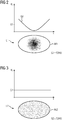
- FIG 2 shows an example of a lesion L1 in a medical image recording according to the prior art.
- the lesion can be tumor-like tissue, for example.
- the medical image depicting the lesion L1 corresponds to a computed tomography slice, which was acquired with the addition of iodine-containing contrast medium.
- Figure 3 shows an example of a lesion L2, which does not differ in shape and size from the lesion L1 and which has been imaged with the same recording technique and procedure for the sake of simplicity and for the purpose of illustration.
- Both lesions L1 and L2 were segmented using known segmentation algorithms, the outer contour AK1, AK2 is known in each case. Surrounding tissue is not shown for simplicity.
- an x-ray absorption representing G1, G2 (in Houndsfield Units HU) can be determined for each lesion, taking into account all image element values encompassed by the outer contour AK1, AK2, or here pixel entries. This corresponds to an average iodine contrast or an average iodine content for each of the lesions. It can be seen that lesion L1 has a darker area in its center, which continuously increases in brightness towards the outside. This spatial grayscale distribution GV is in Figure 2A exemplified in the diagram above the lesion L1.
- lesion L2 shows an essentially homogeneous gray scale distribution, which indicates an even contrast medium distribution.
- the mean grayscale values G1, G2 determined can be the same or almost the same for both lesions L1 and L2.
- a profound differentiation of a blood flow state or a contrast medium distribution of the two lesions L1 and L2 is based on the known Approach not possible or only possible qualitatively.
- a dedicated decision as to whether, for example, lesion L1 responds to a selected form of therapy, is not possible using the known procedure described, since local differences in the gray level are 'averaged out' within the lesion.
- Figures 4 and 5 show, by way of example, lesions L3 and L4, each in a medical image recording according to exemplary embodiments of the invention.
- the medical image recordings depicting the lesions L3, L4 also correspond to computer tomography slice recordings which were acquired with the addition of iodine-containing contrast medium.
- Lesion L4 is also identical in shape and size to lesion L3.
- lesion L3 Corresponding to lesion L1, lesion L3 has a darker area at its center, which, comparable to the situation with lesion L1, is an indicator of a reduced contrast agent uptake.
- This grayscale distribution GV is exemplified in the diagram over lesion L3 in Figure 3A shown.
- Lesion L4, like lesion L2, shows an even grayscale distribution.
- the outer contours AK3, AK4 are also known by means of segmentation. Surrounding tissue is not shown for simplicity. According to the invention, the lesions L3, L4 are divided into five tissue areas GB31, GB32, GB33, GB34, GB35 and GB41, GB42, GB43, GB44, GB45.
- the subdivision that is to say the definition of size, position, arrangement and / or the number of tissue areas, is preferably carried out automatically and / or taking into account size, position or, if known, the type or presumed type of lesion.
- a functional parameter FP to be determined below or the image recording technology used can influence the subdivision.
- an onion-layer-like division in which a tissue area located further outwards, for example GB31, completely encloses an inner tissue area, for example GB32.
- the outer contours of the individual tissue areas have been chosen to be morphologically identical to the outer contours AK3, AK4 of the lesions L3, L4.
- a functional parameter FP for each of the individual tissue areas GB31, GB32, GB33, GB34, GB35 and GB41, GB42, GB43, GB44, GB45 derived from medical imaging.
- the pixel entries included in each case give an average gray scale value or iodine contrast G31, G32, G33, G34, G35 or G41, G42, G43, G44, G45 determined.
- the averaged iodine contrast values provide information about a local tissue function GF or give a measure of the tissue function GF, in this case tissue perfusion, illustrated by the gray scale distribution GF. In other words, an increase in the spatial resolution of a tissue function GF is achieved according to the invention.
- the image recording on which the evaluation according to the invention is based can correspond to a representation of a region of interest obtained using any recording technique, or it can be composed of several individual images. According to the invention, several image recordings can also be evaluated, in particular if functional information is available in the image recordings.
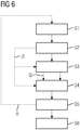
- Step S1 comprises receiving at least one medical image B of a region of interest of an examination object 3, for example a computed tomography image or an MRI image in a computing unit 16.
- the medical image B can be recorded in a time correlated manner with the image evaluation according to the invention or, if it has already been acquired beforehand, can be loaded into the computing unit 16 from a local or remote memory, for example a PACS system of a hospital.
- the medical image recording B can be generated by combining image information from at least two different images of the region of interest.
- Common image processing methods such as material decomposition, image correction methods such as noise suppression, registration methods or the like can be used, if necessary.
- an outer contour of a tissue to be examined, preferably a lesion, contained in the medical image recording is segmented in the computing unit 16, in particular the segmentation unit 21.
- individual image elements are divided into the tissue to be examined or the surrounding tissue.
- Image element or edge-based segmentation methods which are known per se, are preferably used.
- additional information ZI can be recorded for the segmentation or before the segmentation by the computing unit 16 about the question underlying the examination or an initial suspicion or about the measurement protocol used for the image data acquisition in order to decide which body region of interest and thus which tissue types have been imaged and / or which structure or which tissue and / or which quality is to be segmented in the medical image recording.
- the segmentation algorithm used can be selected, for example. Alternatively, a corresponding entry regarding the additional information ZI are made available to the computing unit 16 by a user on request or on the initiative via user interface 7, 11.
- the size that is the length extension in different spatial directions, the volume, the shape of the outer contour AK or the like of the tissue to be examined is known.
- This size information GI can also be used in subsequent steps.
- the segmented tissue is subdivided into a plurality of tissue regions GB. More than two, in particular four to five, tissue regions GB are preferably defined. The greater the number of tissue areas GB, the greater the meaningfulness of the function parameter FP determined below via a local tissue function GF within the segmented tissue.
- size information GI can also be used for step S4 flow into the definition of the individual tissue areas GB via the size, volume and / or shape and / or the like of the tissue to be examined.
- a subdivision unit 23 comprised by the computing unit 16 can be in data exchange with the segmentation unit 21 and / or the data processing device 12, which also carries out the control of a medical imaging system.
- the individual tissue areas GB are preferably nested in one another or, to put it another way, have a layered structure and / or each have the same outer contour as the segmented tissue.
- the subdivision unit 23 can define the individual tissue areas by means of morphological operations, such as an erosion and / or dilation operator.
- the tissue areas have a distance from the outer contour to the outer contour or a radial extension of 5 mm.
- a function parameter FP relating to a tissue function GF is determined for each of the individual tissue areas GB.
- a value for the function parameter FP is derived individually, that is locally for each tissue area GB.
- This evaluation can result in different function parameter values for the individual tissue areas, which when viewed together represent a spatial distribution of a tissue function, it being possible to resolve local differences in the tissue function.
- the determination of the function parameter is carried out by the determination unit 22. This is set up to individually extract and analyze image information contained in the medical image acquisition for each of the individual tissue areas.
- Functional parameters can be: blood flow, iodine enrichment, iron enrichment, calcium density, fat content.
- Function parameters can also be used for texture analysis, such as parameters such as mean density, moments, heterogeneity, entropy, fractal dimension.
- the determination step can include a texture analysis.
- the determination unit 22 can preferably determine a feature vector based on intensity values of the image elements in each individual tissue area.
- the determination unit 22 can furthermore preferably use machine learning methods in order to generate at least one function parameter based on picture element entries and / or the feature vector.
- a computing model can be stored in a memory, locally or centrally, which can be trained by using a machine learning algorithm before the method according to the invention is carried out.
- the training takes place, for example, by means of test images and / or inputs by a user, so that the algorithm is then able to independently detect features in image recordings or to derive function parameters.
- Step S6 may thus include creating a texture metric for each tissue area GB.
- the texture analysis includes to analyze pixel intensity values for the tissue area under consideration and to derive a spatial distribution of the same.
- the texture metric represents a measure of this spatial distribution, for example the texture metric is different moments of these picture element intensities.
- step S5 can include carrying out a texture analysis based on the multiple recordings or the multiple image recordings, in particular in cases in which different medical image recordings correspond to different recording aspects, for example temporal phases, that is to say different time phases of an image recording, or else detect different ones Energy bands.
- the various image recordings may or may not have been acquired with the addition of contrast medium.
- a feature vector can comprise a plurality of texture metrics for a plurality of different parts or areas of the tissue to be examined, in particular of several tissue areas.
- a texture metric in the sense of the invention can comprise: an average, maximum, minimum picture element intensity, a uniformity measure, an entropy in the sense of an irregularity via a gray value histogram, a standard deviation of a gray value histogram, a skewness in the sense of an asymmetry of a Gray value histogram, a curvature or flatness of a gray value histogram, an energy measure or an area moment of inertia (e.g. pixel repetition rate and / or an order measure), correlation (e.g. a measure for a linear dependence on gray values), run length matrix (e.g. a pixel texture in a certain spatial direction), contrast, roughness (for example as a measure for an edge density), heterogeneity (for example as a measure for the presence of edges) or the like.
- the texture metric is preferably determined for each of the tissue areas GB in the medical image recording.
- Image element in the sense of the invention should include both pixels and voxels.
- the method described can optionally branch off according to the invention into a repetition loop R, the method being repeated with steps S1 to S5 until all medical image recordings necessary for deriving the local tissue function of the tissue to be examined are used Are available, were evaluated.
- a comparison of function parameters determined according to the invention can take place, the values for the function parameters for medical image recordings being determined, which were recorded at different times.
- the same functional parameter for medical image recordings of an initial examination can be compared to medical recordings of a follow-up examination at a later point in time.
- the comparison makes it possible to identify local, even the smallest, changes in the examined tissue over the observation period.
- the comparison can preferably be made using machine learning methods.
- the described method is particularly suitable for monitoring the course of tissue conditions, e.g. the fat content or bone density and in particular for Follow-up of a new type of tumor therapy without using previous evaluation criteria such as RECIST.
Landscapes
- Health & Medical Sciences (AREA)
- Engineering & Computer Science (AREA)
- Life Sciences & Earth Sciences (AREA)
- Medical Informatics (AREA)
- Biomedical Technology (AREA)
- General Health & Medical Sciences (AREA)
- Public Health (AREA)
- Pathology (AREA)
- Nuclear Medicine, Radiotherapy & Molecular Imaging (AREA)
- Veterinary Medicine (AREA)
- Physics & Mathematics (AREA)
- Radiology & Medical Imaging (AREA)
- Animal Behavior & Ethology (AREA)
- Surgery (AREA)
- Molecular Biology (AREA)
- Heart & Thoracic Surgery (AREA)
- Biophysics (AREA)
- High Energy & Nuclear Physics (AREA)
- Computer Vision & Pattern Recognition (AREA)
- Optics & Photonics (AREA)
- Theoretical Computer Science (AREA)
- Orthopedic Medicine & Surgery (AREA)
- Physiology (AREA)
- General Physics & Mathematics (AREA)
- Rheumatology (AREA)
- Dentistry (AREA)
- Oral & Maxillofacial Surgery (AREA)
- Pulmonology (AREA)
- Quality & Reliability (AREA)
- Primary Health Care (AREA)
- Epidemiology (AREA)
- Databases & Information Systems (AREA)
- Data Mining & Analysis (AREA)
- Apparatus For Radiation Diagnosis (AREA)
- Magnetic Resonance Imaging Apparatus (AREA)
Claims (13)
- Procédé de détermination d'une fonction (GF) tissulaire locale d'un tissu dans une région du corps à laquelle on s'intéresse d'un objet (3) à examiner, le tissu étant un tissu tumoral ou comprenant une lésion de nature inconnue avec suspection de tissu tumoral, comprenant les stades :- segmentation (S3) d'un contour (AK3, AK4) extérieur du tissu à l'aide d'au moins un enregistrement d'images médicales représentant la région du corps à laquelle on s'intéresse de l'objet à examiner comprenant le tissu ;- subdivision (S4) du tissu segmenté en au moins deux régions (GB ; GB31 à GB35, GB41 à GB45) de tissu ; et- détermination (S5) d'un paramètre (FP ; G31 à G35, G41 à G45) fonctionnel concernant la fonction du tissu pour chacune des au moins deux régions du tissu, les au moins deux régions du tissu étant disposées en couche, de manière à ce qu'une première région (GB31, GB32) du tissu entoure complètement une deuxième région (GB32, GB42) du tissu.
- Procédé suivant la revendication 1, dans lequel les au moins deux régions du tissu ont chacune la même forme que le contour (AK3, AK4) extérieur du tissu segmenté.
- Procédé suivant la revendication 1 ou 2, dans lequel les contours extérieurs des au moins deux régions du tissu sont à une distance les uns des autres qui est dans la plage de 0, 2 mm à 2, 0 mm.
- Procédé suivant l'une des revendications précédentes, dans lequel la subdivision du tissu segmenté s'effectue en utilisant une opération morphologique.
- Procédé suivant l'une des revendications précédentes, dans lequel le tissu segmenté comprend une lésion (L3, L4) médicale.
- Procédé suivant l'une des revendications précédentes, dans lequel le paramètre fonctionnel est au moins un paramètre choisi dans le groupe suivant de paramètres : flux sanguin, enrichissement en iode, enrichissement en fer, teneur en calcium, proportion de graisse.
- Procédé suivant l'une des revendications précédentes, dans lequel le paramètre fonctionnel est au moins un paramètre typique pour l'analyse de texture choisi dans le groupe suivant de paramètres : masse volumique moyenne, couple, hétérogénéité, entropie, dimension fractale.
- Procédé suivant la revendication 7, dans lequel la détermination d'un paramètre fonctionnel comprend une analyse de texture, l'analyse de texture comprenant les stades suivants :- détermination d'un vecteur de caractéristique reposant sur des valeurs d'intensité d'éléments d'image de l'enregistrement d'image médicale,- application d'un procédé d'apprentissage automatique au vecteur de caractéristique, et- production du paramètre fonctionnel en résultat du procédé d'apprentissage automatique.
- Procédé suivant l'une des revendications précédentes, dans lequel l'enregistrement d'image médicale est un enregistrement choisi dans le groupe suivant d'images médicales : enregistrement single-energy, enregistrement de tomographie par ordinateur multi-spectrale, enregistrement de tomographie par ordinateur de perfusion, enregistrement par ultrasons, enregistrement par résonance magnétique et enregistrement par résonance magnétique par perfusion.
- Unité (12) informatique de détermination d'une fonction (GF) tissulaire d'un tissu dans une région à laquelle on s'intéresse d'un objet (3) à examiner, comportant des moyens (21, 22, 23) pour effectuer un procédé suivant l'une des revendications 1 à 9.
- Installation d'imagerie médicale ayant une unité (12) informatique suivant la revendication 10.
- Programme d'ordinateur ayant des codes de programme pour effectuer le procédé suivant l'une des revendications 1 à 9, lorsque le programme d'ordinateur est exécuté sur un ordinateur.
- Support (13) de données déchiffrables par ordinateur ayant un code d'un programme d'ordinateur pour effectuer le procédé suivant l'une des revendications 1 à 9, lorsque le programme d'ordinateur est réalisé sur un ordinateur.
Priority Applications (3)
| Application Number | Priority Date | Filing Date | Title |
|---|---|---|---|
| EP17184699.1A EP3437559B1 (fr) | 2017-08-03 | 2017-08-03 | Détermination d'un paramètre fonctionnel relatif à une fonction tissulaire locale pour des régions tissulaires multiples |
| CN201810859391.0A CN109381805B (zh) | 2017-08-03 | 2018-07-31 | 确定组织的局部的组织功能的方法,计算单元,医学成像设备和计算机可读的数据载体 |
| US16/051,606 US10959685B2 (en) | 2017-08-03 | 2018-08-01 | Ascertaining a function parameter relating to a local tissue function for plurality of tissue regions |
Applications Claiming Priority (1)
| Application Number | Priority Date | Filing Date | Title |
|---|---|---|---|
| EP17184699.1A EP3437559B1 (fr) | 2017-08-03 | 2017-08-03 | Détermination d'un paramètre fonctionnel relatif à une fonction tissulaire locale pour des régions tissulaires multiples |
Publications (2)
| Publication Number | Publication Date |
|---|---|
| EP3437559A1 EP3437559A1 (fr) | 2019-02-06 |
| EP3437559B1 true EP3437559B1 (fr) | 2020-06-24 |
Family
ID=59649478
Family Applications (1)
| Application Number | Title | Priority Date | Filing Date |
|---|---|---|---|
| EP17184699.1A Active EP3437559B1 (fr) | 2017-08-03 | 2017-08-03 | Détermination d'un paramètre fonctionnel relatif à une fonction tissulaire locale pour des régions tissulaires multiples |
Country Status (3)
| Country | Link |
|---|---|
| US (1) | US10959685B2 (fr) |
| EP (1) | EP3437559B1 (fr) |
| CN (1) | CN109381805B (fr) |
Families Citing this family (18)
| Publication number | Priority date | Publication date | Assignee | Title |
|---|---|---|---|---|
| AU2017348111B2 (en) | 2016-10-27 | 2023-04-06 | Progenics Pharmaceuticals, Inc. | Network for medical image analysis, decision support system, and related graphical user interface (GUI) applications |
| US12602772B2 (en) | 2018-01-08 | 2026-04-14 | Progenics Pharmaceuticals, Inc. | Systems and methods for rapid neural network-based image segmentation and radiopharmaceutical uptake determination |
| US10973486B2 (en) | 2018-01-08 | 2021-04-13 | Progenics Pharmaceuticals, Inc. | Systems and methods for rapid neural network-based image segmentation and radiopharmaceutical uptake determination |
| US11227390B2 (en) | 2018-09-26 | 2022-01-18 | Shanghai United Imaging Intelligence Co., Ltd. | Systems and methods for image processing |
| US11426131B2 (en) * | 2018-12-17 | 2022-08-30 | Siemens Medical Solutions Usa, Inc. | Automated motion correction in PET imaging |
| EP4597424A3 (fr) | 2019-01-07 | 2025-10-29 | Exini Diagnostics AB | Systèmes et procédés de segmentation d'image de corps entier agnostique de plateforme |
| BR112021021011A2 (pt) | 2019-04-24 | 2021-12-14 | Exini Diagnostics Ab | Sistemas e métodos para análise automatizada e interativa de imagens de varredura óssea para detecção de metástases |
| DE102019210473A1 (de) * | 2019-07-16 | 2021-01-21 | Siemens Healthcare Gmbh | Verfahren und Vorrichtung zur Bildgebung in der Computertomographie |
| US11900597B2 (en) | 2019-09-27 | 2024-02-13 | Progenics Pharmaceuticals, Inc. | Systems and methods for artificial intelligence-based image analysis for cancer assessment |
| US12417533B2 (en) | 2019-09-27 | 2025-09-16 | Progenics Pharmaceuticals, Inc. | Systems and methods for artificial intelligence-based image analysis for cancer assessment |
| US11564621B2 (en) | 2019-09-27 | 2023-01-31 | Progenies Pharmacenticals, Inc. | Systems and methods for artificial intelligence-based image analysis for cancer assessment |
| US11386988B2 (en) | 2020-04-23 | 2022-07-12 | Exini Diagnostics Ab | Systems and methods for deep-learning-based segmentation of composite images |
| US11321844B2 (en) | 2020-04-23 | 2022-05-03 | Exini Diagnostics Ab | Systems and methods for deep-learning-based segmentation of composite images |
| US11721428B2 (en) | 2020-07-06 | 2023-08-08 | Exini Diagnostics Ab | Systems and methods for artificial intelligence-based image analysis for detection and characterization of lesions |
| JP7732749B2 (ja) * | 2020-11-05 | 2025-09-02 | キヤノンメディカルシステムズ株式会社 | 医用画像処理装置及び医用画像処理プログラム |
| CN112690813B (zh) * | 2021-01-12 | 2022-09-02 | 范宁 | 基于分形理论的肿瘤异质性分析方法、系统、终端及介质 |
| JP7653815B2 (ja) * | 2021-03-19 | 2025-03-31 | 大阪瓦斯株式会社 | 給湯器の故障推定システム |
| CA3231578A1 (fr) | 2021-10-08 | 2023-04-13 | Exini Diagnostics Ab | Systemes et procedes d'identification et de classification automatisees de lesions dans des ganglions lymphatiques locaux et des metastases distantes |
Family Cites Families (33)
| Publication number | Priority date | Publication date | Assignee | Title |
|---|---|---|---|---|
| US6424732B1 (en) * | 1998-12-01 | 2002-07-23 | The Board Of Trustees Of The Leland Stanford Junior University | Object segregation in images |
| US7209587B1 (en) * | 2000-03-21 | 2007-04-24 | Hsu Shin-Yi | Amorphous region and boundary/chain topology for an object recognition system |
| US6785409B1 (en) * | 2000-10-24 | 2004-08-31 | Koninklijke Philips Electronics, N.V. | Segmentation method and apparatus for medical images using diffusion propagation, pixel classification, and mathematical morphology |
| GB0117187D0 (en) | 2001-07-13 | 2001-09-05 | Isis Innovations Ltd | Magnetic resonance imaging |
| US7177453B2 (en) * | 2002-11-26 | 2007-02-13 | General Electric Company | Method and apparatus for partitioning a volume |
| US7283652B2 (en) * | 2002-11-27 | 2007-10-16 | General Electric Company | Method and system for measuring disease relevant tissue changes |
| US7639855B2 (en) * | 2003-04-02 | 2009-12-29 | Ziosoft, Inc. | Medical image processing apparatus, and medical image processing method |
| US7657073B2 (en) * | 2004-11-29 | 2010-02-02 | The Board Of Trustees Of The Leland Stanford Junior University | Registration system and method for tracking lung nodules in medical images |
| GB0602739D0 (en) * | 2006-02-10 | 2006-03-22 | Ccbr As | Breast tissue density measure |
| US8878838B2 (en) | 2009-01-29 | 2014-11-04 | Koninklijke Philips N.V. | Visualizing a time-variant parameter in a biological structure |
| US20120052010A1 (en) | 2009-04-30 | 2012-03-01 | Ge Healthcare Limited | Imaging tumor perfusion, oxidative metabolism using dynamic ace pet in patients with head and neck cancer during radiotherapy |
| US8478029B2 (en) * | 2009-05-26 | 2013-07-02 | Tandent Vision Science, Inc. | Multi-resolution analysis in image segregation |
| WO2011073820A1 (fr) * | 2009-12-16 | 2011-06-23 | Koninklijke Philips Electronics N.V. | Utilisation de collection de plans pour developper de nouveaux objectifs d'optimisation |
| CN101779956A (zh) * | 2010-01-29 | 2010-07-21 | 中国人民解放军国防科学技术大学 | 一种早期肿瘤探测方法 |
| AU2012275114A1 (en) * | 2011-06-29 | 2014-01-16 | The Regents Of The University Of Michigan | Analysis of temporal changes in registered tomographic images |
| KR20140063809A (ko) * | 2011-09-13 | 2014-05-27 | 아쿠이타스 메디컬 리미티드 | 알츠하이머병 및 관련 병리의 평가를 위한 자기공명 기반 방법 |
| JP5797124B2 (ja) * | 2012-01-31 | 2015-10-21 | 富士フイルム株式会社 | 手術支援装置、手術支援方法および手術支援プログラム |
| US10433740B2 (en) * | 2012-09-12 | 2019-10-08 | Heartflow, Inc. | Systems and methods for estimating ischemia and blood flow characteristics from vessel geometry and physiology |
| CN103720474B (zh) * | 2012-10-12 | 2017-03-01 | 西门子公司 | 计算检查参数 |
| KR102042202B1 (ko) * | 2013-02-25 | 2019-11-08 | 삼성전자주식회사 | 의료영상에서 병변을 분할하는 장치 및 방법 |
| US8824752B1 (en) * | 2013-03-15 | 2014-09-02 | Heartflow, Inc. | Methods and systems for assessing image quality in modeling of patient anatomic or blood flow characteristics |
| US9629615B1 (en) * | 2013-09-06 | 2017-04-25 | University Of Louisville Research Foundation, Inc. | Combined B-mode / tissue doppler approach for improved cardiac motion estimation in echocardiographic images |
| DE102013218047B3 (de) | 2013-09-10 | 2015-01-29 | Siemens Aktiengesellschaft | Verfahren zur automatischen Anzeige und/oder Messung von Knochenveränderungen in medizinischen Bilddaten, sowie medizinisches Bildgebungsgerät und elektronisch lesbarer Datenträger |
| WO2015049324A1 (fr) | 2013-10-01 | 2015-04-09 | Koninklijke Philips N.V. | Système et procédé pour la caractérisation d'une pathologie d'irrigation du myocarde |
| US10413253B2 (en) * | 2014-11-21 | 2019-09-17 | Samsung Electronics Co., Ltd. | Method and apparatus for processing medical image |
| CA2969140A1 (fr) * | 2014-12-01 | 2016-06-09 | Quikflo Technologies Inc. | Outil d'aide a la decision pour patients atteints d'accident vasculaire cerebral |
| DE102015221877A1 (de) * | 2015-11-06 | 2017-05-11 | jung diagnostics GmbH | Bildbasierter Biomarker zur Charakterisierung der Struktur oder Funktion von menschlichemoder tierischem Hirngewebe und zugehörige Verwendungen und Verfahren |
| US10350434B2 (en) * | 2015-12-11 | 2019-07-16 | Siemens Healthcare Gmbh | Patient-specific radiation dose assessment in medical therapy |
| EP3389497B1 (fr) | 2015-12-17 | 2020-12-09 | Koninklijke Philips N.V. | Procédé d'optimisation d'un procédé de traitement d'image médicale |
| EP3471625A4 (fr) * | 2016-06-17 | 2020-05-06 | Children's National Medical Center | Quantification d'anatomie médicale : outil de diagnostic assisté par ordinateur |
| DE102016226230B4 (de) * | 2016-12-27 | 2018-07-12 | Siemens Healthcare Gmbh | Automatisierte Bildprüfung in der Röntgenbildgebung |
| EP3568059B1 (fr) * | 2017-01-11 | 2023-10-11 | University of Miami | Procédé et système de cartographie de l'épaisseur tridimensionnelle des micro-couches cornéennes et diagnostics cornéens |
| WO2018144636A1 (fr) * | 2017-02-01 | 2018-08-09 | Intuitive Surgical Operations, Inc. | Systèmes et procédés d'enregistrement pour procédures guidée par l'image |
-
2017
- 2017-08-03 EP EP17184699.1A patent/EP3437559B1/fr active Active
-
2018
- 2018-07-31 CN CN201810859391.0A patent/CN109381805B/zh active Active
- 2018-08-01 US US16/051,606 patent/US10959685B2/en active Active
Non-Patent Citations (1)
| Title |
|---|
| None * |
Also Published As
| Publication number | Publication date |
|---|---|
| CN109381805A (zh) | 2019-02-26 |
| US20190038239A1 (en) | 2019-02-07 |
| US10959685B2 (en) | 2021-03-30 |
| EP3437559A1 (fr) | 2019-02-06 |
| CN109381805B (zh) | 2021-03-12 |
Similar Documents
| Publication | Publication Date | Title |
|---|---|---|
| EP3437559B1 (fr) | Détermination d'un paramètre fonctionnel relatif à une fonction tissulaire locale pour des régions tissulaires multiples | |
| DE102017221276B4 (de) | Medizinische Bildverarbeitungsvorrichtung, Röntgen-CT Vorrichtung und medizinisches Bildverarbeitungsverfahren | |
| DE102011087127B4 (de) | Bestimmung von Aufnahmeparametern bei einer Dual-Energy Tomosynthese | |
| EP3332710B1 (fr) | Caractérisation de plaque | |
| DE102016205507B4 (de) | Verfahren zur Ermittlung einer den Blutfluss in Kollateralen beschreibenden Kollateralinformationen, medizinische Bildaufnahmeeinrichtung, Computerprogramm und elektronisch lesbarer Datenträger | |
| DE202017107196U1 (de) | Medizinische Bildverarbeitungsvorrichtung, Röntgen-CT Vorrichtung und Computerprogrammprodukt | |
| DE102016207367A1 (de) | Festlegen von Scanparametern einer CT-Bildaufnahme mit Hilfe einer Außenbildaufnahme | |
| DE112009001676T5 (de) | Diagnosesystem für Gehirnerkrankungen | |
| DE102017217599A1 (de) | Medizinische Informationsverarbeitungsvorrichtung, Röntgen-CT-Vorrichtung und medizinisches Informationsverarbeitungsverfahren | |
| DE102006012015A1 (de) | Verfahren und Systeme zur Überwachung einer Tumorbelastung | |
| DE102015226400A1 (de) | Automatisierte Ermittlung von Konturen auf Basis einer iterativen Rekonstruktion | |
| WO2014195077A1 (fr) | Procédé et système de détermination d'un instant de début de mesure | |
| DE102019210473A1 (de) | Verfahren und Vorrichtung zur Bildgebung in der Computertomographie | |
| EP3379487B1 (fr) | Reproduction à contraste renforcé de données d'imagerie scanner spectrale | |
| DE102009015386A1 (de) | Verfahren und Vorrichtung zur Erzeugung eines funktionellen Datensatzes eines perfundierten Bereichs des menschlichen oder tierischen Körpers | |
| DE102016219887A1 (de) | Verfahren und System zur Nutzung von Messdaten | |
| DE102018105327A1 (de) | Systeme und Verfahren zur Farbvisualisierung von CT-Bildern | |
| DE112017001335T5 (de) | System und Verfahren für progressive Abbildung | |
| EP3797692B1 (fr) | Procédé et dispositif de commande d'un appareil d'imagerie médicale | |
| DE102013220018A1 (de) | Berechnung eines Untersuchungsparameters | |
| DE102015210912A1 (de) | Rekonstruktion eines Ergebnisbildes unter Berücksichtigung von Kontur-Signifikanz-Daten | |
| DE102014210218A1 (de) | Verfahren zum Generieren von medizinischen Bilddatensätzen | |
| DE102016224717A1 (de) | Verfahren zum Ermitteln von Gewebeeigenschaften von Tumoren | |
| DE102012217940A1 (de) | Rekonstruktion von Bilddaten | |
| EP3363363A1 (fr) | Procédé d'exécution d'un examen pour l'imagerie |
Legal Events
| Date | Code | Title | Description |
|---|---|---|---|
| PUAI | Public reference made under article 153(3) epc to a published international application that has entered the european phase |
Free format text: ORIGINAL CODE: 0009012 |
|
| STAA | Information on the status of an ep patent application or granted ep patent |
Free format text: STATUS: THE APPLICATION HAS BEEN PUBLISHED |
|
| AK | Designated contracting states |
Kind code of ref document: A1 Designated state(s): AL AT BE BG CH CY CZ DE DK EE ES FI FR GB GR HR HU IE IS IT LI LT LU LV MC MK MT NL NO PL PT RO RS SE SI SK SM TR |
|
| AX | Request for extension of the european patent |
Extension state: BA ME |
|
| STAA | Information on the status of an ep patent application or granted ep patent |
Free format text: STATUS: REQUEST FOR EXAMINATION WAS MADE |
|
| 17P | Request for examination filed |
Effective date: 20190805 |
|
| RBV | Designated contracting states (corrected) |
Designated state(s): AL AT BE BG CH CY CZ DE DK EE ES FI FR GB GR HR HU IE IS IT LI LT LU LV MC MK MT NL NO PL PT RO RS SE SI SK SM TR |
|
| RIC1 | Information provided on ipc code assigned before grant |
Ipc: A61B 6/00 20060101AFI20191213BHEP Ipc: A61B 5/05 20060101ALN20191213BHEP Ipc: A61B 8/00 20060101ALN20191213BHEP Ipc: A61B 6/03 20060101ALN20191213BHEP |
|
| GRAP | Despatch of communication of intention to grant a patent |
Free format text: ORIGINAL CODE: EPIDOSNIGR1 |
|
| STAA | Information on the status of an ep patent application or granted ep patent |
Free format text: STATUS: GRANT OF PATENT IS INTENDED |
|
| INTG | Intention to grant announced |
Effective date: 20200124 |
|
| GRAS | Grant fee paid |
Free format text: ORIGINAL CODE: EPIDOSNIGR3 |
|
| GRAA | (expected) grant |
Free format text: ORIGINAL CODE: 0009210 |
|
| STAA | Information on the status of an ep patent application or granted ep patent |
Free format text: STATUS: THE PATENT HAS BEEN GRANTED |
|
| AK | Designated contracting states |
Kind code of ref document: B1 Designated state(s): AL AT BE BG CH CY CZ DE DK EE ES FI FR GB GR HR HU IE IS IT LI LT LU LV MC MK MT NL NO PL PT RO RS SE SI SK SM TR |
|
| REG | Reference to a national code |
Ref country code: GB Ref legal event code: FG4D Free format text: NOT ENGLISH |
|
| REG | Reference to a national code |
Ref country code: CH Ref legal event code: EP |
|
| REG | Reference to a national code |
Ref country code: DE Ref legal event code: R096 Ref document number: 502017005827 Country of ref document: DE |
|
| REG | Reference to a national code |
Ref country code: AT Ref legal event code: REF Ref document number: 1283083 Country of ref document: AT Kind code of ref document: T Effective date: 20200715 |
|
| REG | Reference to a national code |
Ref country code: IE Ref legal event code: FG4D Free format text: LANGUAGE OF EP DOCUMENT: GERMAN |
|
| PG25 | Lapsed in a contracting state [announced via postgrant information from national office to epo] |
Ref country code: LT Free format text: LAPSE BECAUSE OF FAILURE TO SUBMIT A TRANSLATION OF THE DESCRIPTION OR TO PAY THE FEE WITHIN THE PRESCRIBED TIME-LIMIT Effective date: 20200624 Ref country code: SE Free format text: LAPSE BECAUSE OF FAILURE TO SUBMIT A TRANSLATION OF THE DESCRIPTION OR TO PAY THE FEE WITHIN THE PRESCRIBED TIME-LIMIT Effective date: 20200624 Ref country code: NO Free format text: LAPSE BECAUSE OF FAILURE TO SUBMIT A TRANSLATION OF THE DESCRIPTION OR TO PAY THE FEE WITHIN THE PRESCRIBED TIME-LIMIT Effective date: 20200924 Ref country code: GR Free format text: LAPSE BECAUSE OF FAILURE TO SUBMIT A TRANSLATION OF THE DESCRIPTION OR TO PAY THE FEE WITHIN THE PRESCRIBED TIME-LIMIT Effective date: 20200925 Ref country code: FI Free format text: LAPSE BECAUSE OF FAILURE TO SUBMIT A TRANSLATION OF THE DESCRIPTION OR TO PAY THE FEE WITHIN THE PRESCRIBED TIME-LIMIT Effective date: 20200624 |
|
| REG | Reference to a national code |
Ref country code: LT Ref legal event code: MG4D |
|
| PG25 | Lapsed in a contracting state [announced via postgrant information from national office to epo] |
Ref country code: LV Free format text: LAPSE BECAUSE OF FAILURE TO SUBMIT A TRANSLATION OF THE DESCRIPTION OR TO PAY THE FEE WITHIN THE PRESCRIBED TIME-LIMIT Effective date: 20200624 Ref country code: HR Free format text: LAPSE BECAUSE OF FAILURE TO SUBMIT A TRANSLATION OF THE DESCRIPTION OR TO PAY THE FEE WITHIN THE PRESCRIBED TIME-LIMIT Effective date: 20200624 Ref country code: BG Free format text: LAPSE BECAUSE OF FAILURE TO SUBMIT A TRANSLATION OF THE DESCRIPTION OR TO PAY THE FEE WITHIN THE PRESCRIBED TIME-LIMIT Effective date: 20200924 Ref country code: RS Free format text: LAPSE BECAUSE OF FAILURE TO SUBMIT A TRANSLATION OF THE DESCRIPTION OR TO PAY THE FEE WITHIN THE PRESCRIBED TIME-LIMIT Effective date: 20200624 |
|
| REG | Reference to a national code |
Ref country code: NL Ref legal event code: MP Effective date: 20200624 |
|
| PG25 | Lapsed in a contracting state [announced via postgrant information from national office to epo] |
Ref country code: AL Free format text: LAPSE BECAUSE OF FAILURE TO SUBMIT A TRANSLATION OF THE DESCRIPTION OR TO PAY THE FEE WITHIN THE PRESCRIBED TIME-LIMIT Effective date: 20200624 Ref country code: NL Free format text: LAPSE BECAUSE OF FAILURE TO SUBMIT A TRANSLATION OF THE DESCRIPTION OR TO PAY THE FEE WITHIN THE PRESCRIBED TIME-LIMIT Effective date: 20200624 |
|
| PG25 | Lapsed in a contracting state [announced via postgrant information from national office to epo] |
Ref country code: RO Free format text: LAPSE BECAUSE OF FAILURE TO SUBMIT A TRANSLATION OF THE DESCRIPTION OR TO PAY THE FEE WITHIN THE PRESCRIBED TIME-LIMIT Effective date: 20200624 Ref country code: CZ Free format text: LAPSE BECAUSE OF FAILURE TO SUBMIT A TRANSLATION OF THE DESCRIPTION OR TO PAY THE FEE WITHIN THE PRESCRIBED TIME-LIMIT Effective date: 20200624 Ref country code: ES Free format text: LAPSE BECAUSE OF FAILURE TO SUBMIT A TRANSLATION OF THE DESCRIPTION OR TO PAY THE FEE WITHIN THE PRESCRIBED TIME-LIMIT Effective date: 20200624 Ref country code: PT Free format text: LAPSE BECAUSE OF FAILURE TO SUBMIT A TRANSLATION OF THE DESCRIPTION OR TO PAY THE FEE WITHIN THE PRESCRIBED TIME-LIMIT Effective date: 20201026 Ref country code: IT Free format text: LAPSE BECAUSE OF FAILURE TO SUBMIT A TRANSLATION OF THE DESCRIPTION OR TO PAY THE FEE WITHIN THE PRESCRIBED TIME-LIMIT Effective date: 20200624 Ref country code: EE Free format text: LAPSE BECAUSE OF FAILURE TO SUBMIT A TRANSLATION OF THE DESCRIPTION OR TO PAY THE FEE WITHIN THE PRESCRIBED TIME-LIMIT Effective date: 20200624 Ref country code: SM Free format text: LAPSE BECAUSE OF FAILURE TO SUBMIT A TRANSLATION OF THE DESCRIPTION OR TO PAY THE FEE WITHIN THE PRESCRIBED TIME-LIMIT Effective date: 20200624 |
|
| PG25 | Lapsed in a contracting state [announced via postgrant information from national office to epo] |
Ref country code: PL Free format text: LAPSE BECAUSE OF FAILURE TO SUBMIT A TRANSLATION OF THE DESCRIPTION OR TO PAY THE FEE WITHIN THE PRESCRIBED TIME-LIMIT Effective date: 20200624 Ref country code: SK Free format text: LAPSE BECAUSE OF FAILURE TO SUBMIT A TRANSLATION OF THE DESCRIPTION OR TO PAY THE FEE WITHIN THE PRESCRIBED TIME-LIMIT Effective date: 20200624 Ref country code: IS Free format text: LAPSE BECAUSE OF FAILURE TO SUBMIT A TRANSLATION OF THE DESCRIPTION OR TO PAY THE FEE WITHIN THE PRESCRIBED TIME-LIMIT Effective date: 20201024 |
|
| REG | Reference to a national code |
Ref country code: DE Ref legal event code: R097 Ref document number: 502017005827 Country of ref document: DE |
|
| PG25 | Lapsed in a contracting state [announced via postgrant information from national office to epo] |
Ref country code: MC Free format text: LAPSE BECAUSE OF FAILURE TO SUBMIT A TRANSLATION OF THE DESCRIPTION OR TO PAY THE FEE WITHIN THE PRESCRIBED TIME-LIMIT Effective date: 20200624 |
|
| REG | Reference to a national code |
Ref country code: CH Ref legal event code: PL |
|
| PG25 | Lapsed in a contracting state [announced via postgrant information from national office to epo] |
Ref country code: LU Free format text: LAPSE BECAUSE OF NON-PAYMENT OF DUE FEES Effective date: 20200803 Ref country code: LI Free format text: LAPSE BECAUSE OF NON-PAYMENT OF DUE FEES Effective date: 20200831 Ref country code: DK Free format text: LAPSE BECAUSE OF FAILURE TO SUBMIT A TRANSLATION OF THE DESCRIPTION OR TO PAY THE FEE WITHIN THE PRESCRIBED TIME-LIMIT Effective date: 20200624 Ref country code: CH Free format text: LAPSE BECAUSE OF NON-PAYMENT OF DUE FEES Effective date: 20200831 |
|
| PLBE | No opposition filed within time limit |
Free format text: ORIGINAL CODE: 0009261 |
|
| STAA | Information on the status of an ep patent application or granted ep patent |
Free format text: STATUS: NO OPPOSITION FILED WITHIN TIME LIMIT |
|
| REG | Reference to a national code |
Ref country code: BE Ref legal event code: MM Effective date: 20200831 |
|
| 26N | No opposition filed |
Effective date: 20210325 |
|
| PG25 | Lapsed in a contracting state [announced via postgrant information from national office to epo] |
Ref country code: SI Free format text: LAPSE BECAUSE OF FAILURE TO SUBMIT A TRANSLATION OF THE DESCRIPTION OR TO PAY THE FEE WITHIN THE PRESCRIBED TIME-LIMIT Effective date: 20200624 Ref country code: IE Free format text: LAPSE BECAUSE OF NON-PAYMENT OF DUE FEES Effective date: 20200803 Ref country code: BE Free format text: LAPSE BECAUSE OF NON-PAYMENT OF DUE FEES Effective date: 20200831 |
|
| PG25 | Lapsed in a contracting state [announced via postgrant information from national office to epo] |
Ref country code: TR Free format text: LAPSE BECAUSE OF FAILURE TO SUBMIT A TRANSLATION OF THE DESCRIPTION OR TO PAY THE FEE WITHIN THE PRESCRIBED TIME-LIMIT Effective date: 20200624 Ref country code: MT Free format text: LAPSE BECAUSE OF FAILURE TO SUBMIT A TRANSLATION OF THE DESCRIPTION OR TO PAY THE FEE WITHIN THE PRESCRIBED TIME-LIMIT Effective date: 20200624 Ref country code: CY Free format text: LAPSE BECAUSE OF FAILURE TO SUBMIT A TRANSLATION OF THE DESCRIPTION OR TO PAY THE FEE WITHIN THE PRESCRIBED TIME-LIMIT Effective date: 20200624 |
|
| PG25 | Lapsed in a contracting state [announced via postgrant information from national office to epo] |
Ref country code: MK Free format text: LAPSE BECAUSE OF FAILURE TO SUBMIT A TRANSLATION OF THE DESCRIPTION OR TO PAY THE FEE WITHIN THE PRESCRIBED TIME-LIMIT Effective date: 20200624 |
|
| REG | Reference to a national code |
Ref country code: AT Ref legal event code: MM01 Ref document number: 1283083 Country of ref document: AT Kind code of ref document: T Effective date: 20220803 |
|
| PG25 | Lapsed in a contracting state [announced via postgrant information from national office to epo] |
Ref country code: AT Free format text: LAPSE BECAUSE OF NON-PAYMENT OF DUE FEES Effective date: 20220803 |
|
| REG | Reference to a national code |
Ref country code: DE Ref legal event code: R081 Ref document number: 502017005827 Country of ref document: DE Owner name: SIEMENS HEALTHINEERS AG, DE Free format text: FORMER OWNER: SIEMENS HEALTHCARE GMBH, MUENCHEN, DE |
|
| PGFP | Annual fee paid to national office [announced via postgrant information from national office to epo] |
Ref country code: GB Payment date: 20250902 Year of fee payment: 9 |
|
| PGFP | Annual fee paid to national office [announced via postgrant information from national office to epo] |
Ref country code: FR Payment date: 20250827 Year of fee payment: 9 |
|
| PGFP | Annual fee paid to national office [announced via postgrant information from national office to epo] |
Ref country code: DE Payment date: 20251020 Year of fee payment: 9 |