EP3377647B1 - Acides nucléiques et procédés de détection de l'état de méthylation - Google Patents
Acides nucléiques et procédés de détection de l'état de méthylation Download PDFInfo
- Publication number
- EP3377647B1 EP3377647B1 EP16805259.5A EP16805259A EP3377647B1 EP 3377647 B1 EP3377647 B1 EP 3377647B1 EP 16805259 A EP16805259 A EP 16805259A EP 3377647 B1 EP3377647 B1 EP 3377647B1
- Authority
- EP
- European Patent Office
- Prior art keywords
- mips
- sequence
- sequencing
- nucleic acid
- methylation
- Prior art date
- Legal status (The legal status is an assumption and is not a legal conclusion. Google has not performed a legal analysis and makes no representation as to the accuracy of the status listed.)
- Active
Links
- 238000000034 method Methods 0.000 title claims description 369
- 230000011987 methylation Effects 0.000 title claims description 300
- 238000007069 methylation reaction Methods 0.000 title claims description 300
- 150000007523 nucleic acids Chemical class 0.000 title claims description 249
- 102000039446 nucleic acids Human genes 0.000 title claims description 235
- 108020004707 nucleic acids Proteins 0.000 title claims description 235
- 239000000523 sample Substances 0.000 claims description 451
- 102000040430 polynucleotide Human genes 0.000 claims description 250
- 108091033319 polynucleotide Proteins 0.000 claims description 250
- 239000002157 polynucleotide Substances 0.000 claims description 250
- 230000008685 targeting Effects 0.000 claims description 238
- 108091029430 CpG site Proteins 0.000 claims description 225
- 238000012163 sequencing technique Methods 0.000 claims description 215
- 239000000203 mixture Substances 0.000 claims description 136
- 208000037265 diseases, disorders, signs and symptoms Diseases 0.000 claims description 128
- 201000010099 disease Diseases 0.000 claims description 127
- 108091093088 Amplicon Proteins 0.000 claims description 112
- 238000006243 chemical reaction Methods 0.000 claims description 93
- OPTASPLRGRRNAP-UHFFFAOYSA-N cytosine Chemical class NC=1C=CNC(=O)N=1 OPTASPLRGRRNAP-UHFFFAOYSA-N 0.000 claims description 80
- 230000000295 complement effect Effects 0.000 claims description 69
- 210000004369 blood Anatomy 0.000 claims description 68
- 239000008280 blood Substances 0.000 claims description 68
- LSNNMFCWUKXFEE-UHFFFAOYSA-M Bisulfite Chemical compound OS([O-])=O LSNNMFCWUKXFEE-UHFFFAOYSA-M 0.000 claims description 60
- 125000003729 nucleotide group Chemical group 0.000 claims description 54
- 239000002773 nucleotide Substances 0.000 claims description 52
- 230000027455 binding Effects 0.000 claims description 43
- 238000003199 nucleic acid amplification method Methods 0.000 claims description 29
- 230000003321 amplification Effects 0.000 claims description 27
- 238000001514 detection method Methods 0.000 claims description 22
- 238000002844 melting Methods 0.000 claims description 11
- 230000008018 melting Effects 0.000 claims description 11
- 210000002966 serum Anatomy 0.000 claims description 11
- 238000009396 hybridization Methods 0.000 claims description 10
- 230000002441 reversible effect Effects 0.000 claims description 10
- 238000007481 next generation sequencing Methods 0.000 claims description 7
- 230000001419 dependent effect Effects 0.000 claims description 2
- 108020004414 DNA Proteins 0.000 description 194
- 210000001519 tissue Anatomy 0.000 description 137
- 206010028980 Neoplasm Diseases 0.000 description 105
- 238000012360 testing method Methods 0.000 description 102
- 230000000875 corresponding effect Effects 0.000 description 71
- 238000003752 polymerase chain reaction Methods 0.000 description 66
- 201000011510 cancer Diseases 0.000 description 55
- 210000002381 plasma Anatomy 0.000 description 43
- 208000003200 Adenoma Diseases 0.000 description 39
- 238000004458 analytical method Methods 0.000 description 39
- 206010001233 Adenoma benign Diseases 0.000 description 38
- 230000001174 ascending effect Effects 0.000 description 36
- 230000008569 process Effects 0.000 description 36
- 210000004027 cell Anatomy 0.000 description 35
- 210000000349 chromosome Anatomy 0.000 description 33
- 238000003556 assay Methods 0.000 description 28
- 230000001605 fetal effect Effects 0.000 description 26
- 230000008774 maternal effect Effects 0.000 description 26
- 241000894007 species Species 0.000 description 24
- 108060002716 Exonuclease Proteins 0.000 description 23
- 102000013165 exonuclease Human genes 0.000 description 23
- 230000000869 mutational effect Effects 0.000 description 23
- 238000012217 deletion Methods 0.000 description 21
- 208000009956 adenocarcinoma Diseases 0.000 description 20
- 210000004534 cecum Anatomy 0.000 description 20
- 238000004891 communication Methods 0.000 description 20
- 230000037430 deletion Effects 0.000 description 19
- 230000006870 function Effects 0.000 description 19
- 238000003780 insertion Methods 0.000 description 18
- 241000282414 Homo sapiens Species 0.000 description 17
- 210000001124 body fluid Anatomy 0.000 description 16
- 230000037431 insertion Effects 0.000 description 16
- 208000036878 aneuploidy Diseases 0.000 description 15
- 210000000056 organ Anatomy 0.000 description 15
- 238000005516 engineering process Methods 0.000 description 14
- 210000003754 fetus Anatomy 0.000 description 14
- 210000004185 liver Anatomy 0.000 description 13
- 108090000623 proteins and genes Proteins 0.000 description 13
- GNFTZDOKVXKIBK-UHFFFAOYSA-N 3-(2-methoxyethoxy)benzohydrazide Chemical compound COCCOC1=CC=CC(C(=O)NN)=C1 GNFTZDOKVXKIBK-UHFFFAOYSA-N 0.000 description 12
- 108090000364 Ligases Proteins 0.000 description 12
- 102000003960 Ligases Human genes 0.000 description 12
- 231100001075 aneuploidy Toxicity 0.000 description 12
- 208000031448 Genomic Instability Diseases 0.000 description 11
- 108091081062 Repeated sequence (DNA) Proteins 0.000 description 11
- 230000004075 alteration Effects 0.000 description 11
- 108020004999 messenger RNA Proteins 0.000 description 11
- 238000002493 microarray Methods 0.000 description 11
- 238000012545 processing Methods 0.000 description 11
- YTAHJIFKAKIKAV-XNMGPUDCSA-N [(1R)-3-morpholin-4-yl-1-phenylpropyl] N-[(3S)-2-oxo-5-phenyl-1,3-dihydro-1,4-benzodiazepin-3-yl]carbamate Chemical compound O=C1[C@H](N=C(C2=C(N1)C=CC=C2)C1=CC=CC=C1)NC(O[C@H](CCN1CCOCC1)C1=CC=CC=C1)=O YTAHJIFKAKIKAV-XNMGPUDCSA-N 0.000 description 10
- 208000001333 Colorectal Neoplasms Diseases 0.000 description 9
- 230000002068 genetic effect Effects 0.000 description 9
- 230000008901 benefit Effects 0.000 description 8
- 238000003745 diagnosis Methods 0.000 description 8
- 239000012530 fluid Substances 0.000 description 8
- 238000011160 research Methods 0.000 description 8
- 208000031404 Chromosome Aberrations Diseases 0.000 description 7
- 206010009944 Colon cancer Diseases 0.000 description 7
- 108091028043 Nucleic acid sequence Proteins 0.000 description 7
- 108091034117 Oligonucleotide Proteins 0.000 description 7
- 208000037280 Trisomy Diseases 0.000 description 7
- 238000013500 data storage Methods 0.000 description 7
- 239000003814 drug Substances 0.000 description 7
- 238000001914 filtration Methods 0.000 description 7
- 230000002974 pharmacogenomic effect Effects 0.000 description 7
- 230000035935 pregnancy Effects 0.000 description 7
- 230000007067 DNA methylation Effects 0.000 description 6
- 102000016928 DNA-directed DNA polymerase Human genes 0.000 description 6
- 108010014303 DNA-directed DNA polymerase Proteins 0.000 description 6
- 239000003153 chemical reaction reagent Substances 0.000 description 6
- 230000007423 decrease Effects 0.000 description 6
- 238000010586 diagram Methods 0.000 description 6
- 230000000694 effects Effects 0.000 description 6
- 230000035772 mutation Effects 0.000 description 6
- 102000004169 proteins and genes Human genes 0.000 description 6
- 238000000926 separation method Methods 0.000 description 6
- RWQNBRDOKXIBIV-UHFFFAOYSA-N thymine Chemical compound CC1=CNC(=O)NC1=O RWQNBRDOKXIBIV-UHFFFAOYSA-N 0.000 description 6
- 206010008805 Chromosomal abnormalities Diseases 0.000 description 5
- 102000004190 Enzymes Human genes 0.000 description 5
- 108090000790 Enzymes Proteins 0.000 description 5
- 238000007792 addition Methods 0.000 description 5
- 238000001369 bisulfite sequencing Methods 0.000 description 5
- 230000008859 change Effects 0.000 description 5
- 230000002759 chromosomal effect Effects 0.000 description 5
- 238000013461 design Methods 0.000 description 5
- 238000003205 genotyping method Methods 0.000 description 5
- 210000004072 lung Anatomy 0.000 description 5
- 238000012986 modification Methods 0.000 description 5
- 230000003287 optical effect Effects 0.000 description 5
- 238000005457 optimization Methods 0.000 description 5
- 210000005059 placental tissue Anatomy 0.000 description 5
- 201000011461 pre-eclampsia Diseases 0.000 description 5
- 210000000664 rectum Anatomy 0.000 description 5
- 238000012216 screening Methods 0.000 description 5
- 239000007787 solid Substances 0.000 description 5
- 238000012070 whole genome sequencing analysis Methods 0.000 description 5
- QEBNVGLWXKVRJR-UHFFFAOYSA-N 2-(4,5-dimethyl-1,3-diselenol-2-ylidene)-5,6-dihydro-[1,3]dithiolo[4,5-b][1,4]dithiine Chemical compound [Se]1C(C)=C(C)[Se]C1=C1SC(SCCS2)=C2S1 QEBNVGLWXKVRJR-UHFFFAOYSA-N 0.000 description 4
- 108700028369 Alleles Proteins 0.000 description 4
- 206010006187 Breast cancer Diseases 0.000 description 4
- 208000026310 Breast neoplasm Diseases 0.000 description 4
- 102000053602 DNA Human genes 0.000 description 4
- 108020004446 Long Interspersed Nucleotide Elements Proteins 0.000 description 4
- ISAKRJDGNUQOIC-UHFFFAOYSA-N Uracil Chemical class O=C1C=CNC(=O)N1 ISAKRJDGNUQOIC-UHFFFAOYSA-N 0.000 description 4
- 238000013459 approach Methods 0.000 description 4
- 238000004422 calculation algorithm Methods 0.000 description 4
- 210000001072 colon Anatomy 0.000 description 4
- 238000004590 computer program Methods 0.000 description 4
- 229940104302 cytosine Drugs 0.000 description 4
- 238000006481 deamination reaction Methods 0.000 description 4
- 238000004925 denaturation Methods 0.000 description 4
- 230000036425 denaturation Effects 0.000 description 4
- 229940079593 drug Drugs 0.000 description 4
- 239000000839 emulsion Substances 0.000 description 4
- 239000012634 fragment Substances 0.000 description 4
- 238000012165 high-throughput sequencing Methods 0.000 description 4
- 210000003734 kidney Anatomy 0.000 description 4
- 230000004048 modification Effects 0.000 description 4
- 238000012544 monitoring process Methods 0.000 description 4
- 230000037361 pathway Effects 0.000 description 4
- 210000002826 placenta Anatomy 0.000 description 4
- 238000002360 preparation method Methods 0.000 description 4
- 238000004393 prognosis Methods 0.000 description 4
- 230000035945 sensitivity Effects 0.000 description 4
- 239000004055 small Interfering RNA Substances 0.000 description 4
- 239000007790 solid phase Substances 0.000 description 4
- 210000003932 urinary bladder Anatomy 0.000 description 4
- 108010063905 Ampligase Proteins 0.000 description 3
- 238000012935 Averaging Methods 0.000 description 3
- 108091061744 Cell-free fetal DNA Proteins 0.000 description 3
- 108091029523 CpG island Proteins 0.000 description 3
- 229910019142 PO4 Inorganic materials 0.000 description 3
- 206010060862 Prostate cancer Diseases 0.000 description 3
- 208000000236 Prostatic Neoplasms Diseases 0.000 description 3
- 210000001766 X chromosome Anatomy 0.000 description 3
- JLCPHMBAVCMARE-UHFFFAOYSA-N [3-[[3-[[3-[[3-[[3-[[3-[[3-[[3-[[3-[[3-[[3-[[5-(2-amino-6-oxo-1H-purin-9-yl)-3-[[3-[[3-[[3-[[3-[[3-[[5-(2-amino-6-oxo-1H-purin-9-yl)-3-[[5-(2-amino-6-oxo-1H-purin-9-yl)-3-hydroxyoxolan-2-yl]methoxy-hydroxyphosphoryl]oxyoxolan-2-yl]methoxy-hydroxyphosphoryl]oxy-5-(5-methyl-2,4-dioxopyrimidin-1-yl)oxolan-2-yl]methoxy-hydroxyphosphoryl]oxy-5-(6-aminopurin-9-yl)oxolan-2-yl]methoxy-hydroxyphosphoryl]oxy-5-(6-aminopurin-9-yl)oxolan-2-yl]methoxy-hydroxyphosphoryl]oxy-5-(6-aminopurin-9-yl)oxolan-2-yl]methoxy-hydroxyphosphoryl]oxy-5-(6-aminopurin-9-yl)oxolan-2-yl]methoxy-hydroxyphosphoryl]oxyoxolan-2-yl]methoxy-hydroxyphosphoryl]oxy-5-(5-methyl-2,4-dioxopyrimidin-1-yl)oxolan-2-yl]methoxy-hydroxyphosphoryl]oxy-5-(4-amino-2-oxopyrimidin-1-yl)oxolan-2-yl]methoxy-hydroxyphosphoryl]oxy-5-(5-methyl-2,4-dioxopyrimidin-1-yl)oxolan-2-yl]methoxy-hydroxyphosphoryl]oxy-5-(5-methyl-2,4-dioxopyrimidin-1-yl)oxolan-2-yl]methoxy-hydroxyphosphoryl]oxy-5-(6-aminopurin-9-yl)oxolan-2-yl]methoxy-hydroxyphosphoryl]oxy-5-(6-aminopurin-9-yl)oxolan-2-yl]methoxy-hydroxyphosphoryl]oxy-5-(4-amino-2-oxopyrimidin-1-yl)oxolan-2-yl]methoxy-hydroxyphosphoryl]oxy-5-(4-amino-2-oxopyrimidin-1-yl)oxolan-2-yl]methoxy-hydroxyphosphoryl]oxy-5-(4-amino-2-oxopyrimidin-1-yl)oxolan-2-yl]methoxy-hydroxyphosphoryl]oxy-5-(6-aminopurin-9-yl)oxolan-2-yl]methoxy-hydroxyphosphoryl]oxy-5-(4-amino-2-oxopyrimidin-1-yl)oxolan-2-yl]methyl [5-(6-aminopurin-9-yl)-2-(hydroxymethyl)oxolan-3-yl] hydrogen phosphate Polymers Cc1cn(C2CC(OP(O)(=O)OCC3OC(CC3OP(O)(=O)OCC3OC(CC3O)n3cnc4c3nc(N)[nH]c4=O)n3cnc4c3nc(N)[nH]c4=O)C(COP(O)(=O)OC3CC(OC3COP(O)(=O)OC3CC(OC3COP(O)(=O)OC3CC(OC3COP(O)(=O)OC3CC(OC3COP(O)(=O)OC3CC(OC3COP(O)(=O)OC3CC(OC3COP(O)(=O)OC3CC(OC3COP(O)(=O)OC3CC(OC3COP(O)(=O)OC3CC(OC3COP(O)(=O)OC3CC(OC3COP(O)(=O)OC3CC(OC3COP(O)(=O)OC3CC(OC3COP(O)(=O)OC3CC(OC3COP(O)(=O)OC3CC(OC3COP(O)(=O)OC3CC(OC3COP(O)(=O)OC3CC(OC3COP(O)(=O)OC3CC(OC3CO)n3cnc4c(N)ncnc34)n3ccc(N)nc3=O)n3cnc4c(N)ncnc34)n3ccc(N)nc3=O)n3ccc(N)nc3=O)n3ccc(N)nc3=O)n3cnc4c(N)ncnc34)n3cnc4c(N)ncnc34)n3cc(C)c(=O)[nH]c3=O)n3cc(C)c(=O)[nH]c3=O)n3ccc(N)nc3=O)n3cc(C)c(=O)[nH]c3=O)n3cnc4c3nc(N)[nH]c4=O)n3cnc4c(N)ncnc34)n3cnc4c(N)ncnc34)n3cnc4c(N)ncnc34)n3cnc4c(N)ncnc34)O2)c(=O)[nH]c1=O JLCPHMBAVCMARE-UHFFFAOYSA-N 0.000 description 3
- 230000001594 aberrant effect Effects 0.000 description 3
- 230000003322 aneuploid effect Effects 0.000 description 3
- 239000011324 bead Substances 0.000 description 3
- 229910052799 carbon Inorganic materials 0.000 description 3
- 230000030833 cell death Effects 0.000 description 3
- 230000002596 correlated effect Effects 0.000 description 3
- 230000009615 deamination Effects 0.000 description 3
- 238000011033 desalting Methods 0.000 description 3
- 238000011161 development Methods 0.000 description 3
- 230000018109 developmental process Effects 0.000 description 3
- 239000000975 dye Substances 0.000 description 3
- 238000001502 gel electrophoresis Methods 0.000 description 3
- 238000007031 hydroxymethylation reaction Methods 0.000 description 3
- 230000003993 interaction Effects 0.000 description 3
- 238000004949 mass spectrometry Methods 0.000 description 3
- 238000012775 microarray technology Methods 0.000 description 3
- 208000030454 monosomy Diseases 0.000 description 3
- 238000013188 needle biopsy Methods 0.000 description 3
- 210000001672 ovary Anatomy 0.000 description 3
- 210000000496 pancreas Anatomy 0.000 description 3
- 230000032696 parturition Effects 0.000 description 3
- NBIIXXVUZAFLBC-UHFFFAOYSA-K phosphate Chemical compound [O-]P([O-])([O-])=O NBIIXXVUZAFLBC-UHFFFAOYSA-K 0.000 description 3
- 239000010452 phosphate Substances 0.000 description 3
- 102000054765 polymorphisms of proteins Human genes 0.000 description 3
- 238000000746 purification Methods 0.000 description 3
- 230000028327 secretion Effects 0.000 description 3
- 210000002784 stomach Anatomy 0.000 description 3
- 238000003786 synthesis reaction Methods 0.000 description 3
- 210000002700 urine Anatomy 0.000 description 3
- 210000004291 uterus Anatomy 0.000 description 3
- MJEQLGCFPLHMNV-UHFFFAOYSA-N 4-amino-1-(hydroxymethyl)pyrimidin-2-one Chemical compound NC=1C=CN(CO)C(=O)N=1 MJEQLGCFPLHMNV-UHFFFAOYSA-N 0.000 description 2
- RYVNIFSIEDRLSJ-UHFFFAOYSA-N 5-(hydroxymethyl)cytosine Chemical compound NC=1NC(=O)N=CC=1CO RYVNIFSIEDRLSJ-UHFFFAOYSA-N 0.000 description 2
- FHSISDGOVSHJRW-UHFFFAOYSA-N 5-formylcytosine Chemical compound NC1=NC(=O)NC=C1C=O FHSISDGOVSHJRW-UHFFFAOYSA-N 0.000 description 2
- 206010069754 Acquired gene mutation Diseases 0.000 description 2
- 108091023043 Alu Element Proteins 0.000 description 2
- 241000972773 Aulopiformes Species 0.000 description 2
- 241000282472 Canis lupus familiaris Species 0.000 description 2
- 108010078791 Carrier Proteins Proteins 0.000 description 2
- 108010077544 Chromatin Proteins 0.000 description 2
- 102100024458 Cyclin-dependent kinase inhibitor 2A Human genes 0.000 description 2
- 102100024902 Cytochrome P450 4F2 Human genes 0.000 description 2
- 230000005778 DNA damage Effects 0.000 description 2
- 231100000277 DNA damage Toxicity 0.000 description 2
- 230000030933 DNA methylation on cytosine Effects 0.000 description 2
- 241000282326 Felis catus Species 0.000 description 2
- 108020004996 Heterogeneous Nuclear RNA Proteins 0.000 description 2
- 101000909122 Homo sapiens Cytochrome P450 4F2 Proteins 0.000 description 2
- 101000642815 Homo sapiens Protein SSXT Proteins 0.000 description 2
- 101000643895 Homo sapiens Ubiquitin carboxyl-terminal hydrolase 6 Proteins 0.000 description 2
- 206010061218 Inflammation Diseases 0.000 description 2
- 206010058467 Lung neoplasm malignant Diseases 0.000 description 2
- 241000124008 Mammalia Species 0.000 description 2
- 206010033128 Ovarian cancer Diseases 0.000 description 2
- 208000037273 Pathologic Processes Diseases 0.000 description 2
- 208000006399 Premature Obstetric Labor Diseases 0.000 description 2
- 206010036790 Productive cough Diseases 0.000 description 2
- 102100035586 Protein SSXT Human genes 0.000 description 2
- 238000012300 Sequence Analysis Methods 0.000 description 2
- 108020004682 Single-Stranded DNA Proteins 0.000 description 2
- 108020003224 Small Nucleolar RNA Proteins 0.000 description 2
- 102000042773 Small Nucleolar RNA Human genes 0.000 description 2
- 108091027967 Small hairpin RNA Proteins 0.000 description 2
- 108020004459 Small interfering RNA Proteins 0.000 description 2
- 108020004566 Transfer RNA Proteins 0.000 description 2
- 102100021015 Ubiquitin carboxyl-terminal hydrolase 6 Human genes 0.000 description 2
- 230000002159 abnormal effect Effects 0.000 description 2
- 230000005856 abnormality Effects 0.000 description 2
- 230000007172 age related pathology Effects 0.000 description 2
- 230000032683 aging Effects 0.000 description 2
- 230000001640 apoptogenic effect Effects 0.000 description 2
- 210000003567 ascitic fluid Anatomy 0.000 description 2
- 239000000090 biomarker Substances 0.000 description 2
- 238000001574 biopsy Methods 0.000 description 2
- 230000015572 biosynthetic process Effects 0.000 description 2
- 238000005251 capillar electrophoresis Methods 0.000 description 2
- 230000001413 cellular effect Effects 0.000 description 2
- 238000005119 centrifugation Methods 0.000 description 2
- 210000001175 cerebrospinal fluid Anatomy 0.000 description 2
- 210000003483 chromatin Anatomy 0.000 description 2
- 201000010989 colorectal carcinoma Diseases 0.000 description 2
- 239000002299 complementary DNA Substances 0.000 description 2
- 230000002559 cytogenic effect Effects 0.000 description 2
- 230000006378 damage Effects 0.000 description 2
- 238000012350 deep sequencing Methods 0.000 description 2
- 230000003831 deregulation Effects 0.000 description 2
- 235000005911 diet Nutrition 0.000 description 2
- 230000037213 diet Effects 0.000 description 2
- 230000029087 digestion Effects 0.000 description 2
- 238000006073 displacement reaction Methods 0.000 description 2
- 238000009826 distribution Methods 0.000 description 2
- 238000009509 drug development Methods 0.000 description 2
- 230000008482 dysregulation Effects 0.000 description 2
- 238000001493 electron microscopy Methods 0.000 description 2
- 238000001839 endoscopy Methods 0.000 description 2
- 230000007613 environmental effect Effects 0.000 description 2
- 230000001973 epigenetic effect Effects 0.000 description 2
- 230000007717 exclusion Effects 0.000 description 2
- 208000021045 exocrine pancreatic carcinoma Diseases 0.000 description 2
- 238000007672 fourth generation sequencing Methods 0.000 description 2
- 230000002496 gastric effect Effects 0.000 description 2
- 210000001035 gastrointestinal tract Anatomy 0.000 description 2
- 238000012252 genetic analysis Methods 0.000 description 2
- 230000007614 genetic variation Effects 0.000 description 2
- 239000010931 gold Substances 0.000 description 2
- 229910052737 gold Inorganic materials 0.000 description 2
- 230000003301 hydrolyzing effect Effects 0.000 description 2
- 238000003384 imaging method Methods 0.000 description 2
- 230000002779 inactivation Effects 0.000 description 2
- 238000010348 incorporation Methods 0.000 description 2
- 238000011534 incubation Methods 0.000 description 2
- 208000015181 infectious disease Diseases 0.000 description 2
- 230000004054 inflammatory process Effects 0.000 description 2
- 238000011835 investigation Methods 0.000 description 2
- 150000002500 ions Chemical class 0.000 description 2
- 230000003902 lesion Effects 0.000 description 2
- 230000000670 limiting effect Effects 0.000 description 2
- 201000007270 liver cancer Diseases 0.000 description 2
- 208000014018 liver neoplasm Diseases 0.000 description 2
- 238000007403 mPCR Methods 0.000 description 2
- 239000003550 marker Substances 0.000 description 2
- 238000005259 measurement Methods 0.000 description 2
- 230000001404 mediated effect Effects 0.000 description 2
- 239000002679 microRNA Substances 0.000 description 2
- 238000010208 microarray analysis Methods 0.000 description 2
- 230000001338 necrotic effect Effects 0.000 description 2
- 230000003647 oxidation Effects 0.000 description 2
- 238000007254 oxidation reaction Methods 0.000 description 2
- 208000008443 pancreatic carcinoma Diseases 0.000 description 2
- 239000012188 paraffin wax Substances 0.000 description 2
- 230000009054 pathological process Effects 0.000 description 2
- 230000003169 placental effect Effects 0.000 description 2
- 230000036470 plasma concentration Effects 0.000 description 2
- 210000004910 pleural fluid Anatomy 0.000 description 2
- 238000009598 prenatal testing Methods 0.000 description 2
- 238000002203 pretreatment Methods 0.000 description 2
- 238000012175 pyrosequencing Methods 0.000 description 2
- 238000011002 quantification Methods 0.000 description 2
- 239000002096 quantum dot Substances 0.000 description 2
- 238000003753 real-time PCR Methods 0.000 description 2
- 239000013074 reference sample Substances 0.000 description 2
- 230000004044 response Effects 0.000 description 2
- 108020004418 ribosomal RNA Proteins 0.000 description 2
- 235000019515 salmon Nutrition 0.000 description 2
- 239000004065 semiconductor Substances 0.000 description 2
- 238000007841 sequencing by ligation Methods 0.000 description 2
- 230000037439 somatic mutation Effects 0.000 description 2
- 210000003802 sputum Anatomy 0.000 description 2
- 208000024794 sputum Diseases 0.000 description 2
- 238000000528 statistical test Methods 0.000 description 2
- 230000007704 transition Effects 0.000 description 2
- 230000005945 translocation Effects 0.000 description 2
- 238000011144 upstream manufacturing Methods 0.000 description 2
- 238000012800 visualization Methods 0.000 description 2
- 229960005080 warfarin Drugs 0.000 description 2
- PJVWKTKQMONHTI-UHFFFAOYSA-N warfarin Chemical compound OC=1C2=CC=CC=C2OC(=O)C=1C(CC(=O)C)C1=CC=CC=C1 PJVWKTKQMONHTI-UHFFFAOYSA-N 0.000 description 2
- HWPZZUQOWRWFDB-UHFFFAOYSA-N 1-methylcytosine Chemical compound CN1C=CC(N)=NC1=O HWPZZUQOWRWFDB-UHFFFAOYSA-N 0.000 description 1
- 102100025007 14-3-3 protein epsilon Human genes 0.000 description 1
- UEJJHQNACJXSKW-UHFFFAOYSA-N 2-(2,6-dioxopiperidin-3-yl)-1H-isoindole-1,3(2H)-dione Chemical compound O=C1C2=CC=CC=C2C(=O)N1C1CCC(=O)NC1=O UEJJHQNACJXSKW-UHFFFAOYSA-N 0.000 description 1
- DIDGPCDGNMIUNX-UUOKFMHZSA-N 2-amino-9-[(2r,3r,4s,5r)-5-(dihydroxyphosphinothioyloxymethyl)-3,4-dihydroxyoxolan-2-yl]-3h-purin-6-one Chemical compound C1=2NC(N)=NC(=O)C=2N=CN1[C@@H]1O[C@H](COP(O)(O)=S)[C@@H](O)[C@H]1O DIDGPCDGNMIUNX-UUOKFMHZSA-N 0.000 description 1
- NMUSYJAQQFHJEW-UHFFFAOYSA-N 5-Azacytidine Natural products O=C1N=C(N)N=CN1C1C(O)C(O)C(CO)O1 NMUSYJAQQFHJEW-UHFFFAOYSA-N 0.000 description 1
- XAUDJQYHKZQPEU-KVQBGUIXSA-N 5-aza-2'-deoxycytidine Chemical compound O=C1N=C(N)N=CN1[C@@H]1O[C@H](CO)[C@@H](O)C1 XAUDJQYHKZQPEU-KVQBGUIXSA-N 0.000 description 1
- NMUSYJAQQFHJEW-KVTDHHQDSA-N 5-azacytidine Chemical compound O=C1N=C(N)N=CN1[C@H]1[C@H](O)[C@H](O)[C@@H](CO)O1 NMUSYJAQQFHJEW-KVTDHHQDSA-N 0.000 description 1
- LRSASMSXMSNRBT-UHFFFAOYSA-N 5-methylcytosine Chemical class CC1=CNC(=O)N=C1N LRSASMSXMSNRBT-UHFFFAOYSA-N 0.000 description 1
- CKOMXBHMKXXTNW-UHFFFAOYSA-N 6-methyladenine Chemical compound CNC1=NC=NC2=C1N=CN2 CKOMXBHMKXXTNW-UHFFFAOYSA-N 0.000 description 1
- GFFGJBXGBJISGV-UHFFFAOYSA-N Adenine Chemical compound NC1=NC=NC2=C1N=CN2 GFFGJBXGBJISGV-UHFFFAOYSA-N 0.000 description 1
- 229930024421 Adenine Natural products 0.000 description 1
- 208000010507 Adenocarcinoma of Lung Diseases 0.000 description 1
- 208000024827 Alzheimer disease Diseases 0.000 description 1
- 208000008884 Aneurysmal Bone Cysts Diseases 0.000 description 1
- 102100021569 Apoptosis regulator Bcl-2 Human genes 0.000 description 1
- 102100021631 B-cell lymphoma 6 protein Human genes 0.000 description 1
- 239000005552 B01AC04 - Clopidogrel Substances 0.000 description 1
- 108091012583 BCL2 Proteins 0.000 description 1
- 108700020463 BRCA1 Proteins 0.000 description 1
- 102000036365 BRCA1 Human genes 0.000 description 1
- 101150072950 BRCA1 gene Proteins 0.000 description 1
- 102000052609 BRCA2 Human genes 0.000 description 1
- 108700020462 BRCA2 Proteins 0.000 description 1
- KWIUHFFTVRNATP-UHFFFAOYSA-N Betaine Natural products C[N+](C)(C)CC([O-])=O KWIUHFFTVRNATP-UHFFFAOYSA-N 0.000 description 1
- 208000020925 Bipolar disease Diseases 0.000 description 1
- 206010005003 Bladder cancer Diseases 0.000 description 1
- 241000283690 Bos taurus Species 0.000 description 1
- 101150008921 Brca2 gene Proteins 0.000 description 1
- 108010083123 CDX2 Transcription Factor Proteins 0.000 description 1
- 102000006277 CDX2 Transcription Factor Human genes 0.000 description 1
- 102100038700 Calcium-responsive transactivator Human genes 0.000 description 1
- 241000283707 Capra Species 0.000 description 1
- OKTJSMMVPCPJKN-UHFFFAOYSA-N Carbon Chemical compound [C] OKTJSMMVPCPJKN-UHFFFAOYSA-N 0.000 description 1
- 108010022366 Carcinoembryonic Antigen Proteins 0.000 description 1
- 102100025475 Carcinoembryonic antigen-related cell adhesion molecule 5 Human genes 0.000 description 1
- 208000005623 Carcinogenesis Diseases 0.000 description 1
- 108020004635 Complementary DNA Proteins 0.000 description 1
- 108010009392 Cyclin-Dependent Kinase Inhibitor p16 Proteins 0.000 description 1
- 206010011732 Cyst Diseases 0.000 description 1
- 108010015742 Cytochrome P-450 Enzyme System Proteins 0.000 description 1
- 102000003849 Cytochrome P450 Human genes 0.000 description 1
- 108010061982 DNA Ligases Proteins 0.000 description 1
- 102000012410 DNA Ligases Human genes 0.000 description 1
- 102100031866 DNA excision repair protein ERCC-5 Human genes 0.000 description 1
- 108010035476 DNA excision repair protein ERCC-5 Proteins 0.000 description 1
- 238000000018 DNA microarray Methods 0.000 description 1
- 230000033616 DNA repair Effects 0.000 description 1
- 102100024607 DNA topoisomerase 1 Human genes 0.000 description 1
- 230000004568 DNA-binding Effects 0.000 description 1
- 201000010374 Down Syndrome Diseases 0.000 description 1
- 102100039577 ETS translocation variant 5 Human genes 0.000 description 1
- 201000006360 Edwards syndrome Diseases 0.000 description 1
- 241000283086 Equidae Species 0.000 description 1
- LFQSCWFLJHTTHZ-UHFFFAOYSA-N Ethanol Chemical compound CCO LFQSCWFLJHTTHZ-UHFFFAOYSA-N 0.000 description 1
- 102100022462 Eukaryotic initiation factor 4A-II Human genes 0.000 description 1
- 108010007577 Exodeoxyribonuclease I Proteins 0.000 description 1
- 102100029075 Exonuclease 1 Human genes 0.000 description 1
- 208000001362 Fetal Growth Retardation Diseases 0.000 description 1
- 206010016654 Fibrosis Diseases 0.000 description 1
- 208000001914 Fragile X syndrome Diseases 0.000 description 1
- 102100033452 GMP synthase [glutamine-hydrolyzing] Human genes 0.000 description 1
- 101710071060 GMPS Proteins 0.000 description 1
- 208000034951 Genetic Translocation Diseases 0.000 description 1
- 102100032610 Guanine nucleotide-binding protein G(s) subunit alpha isoforms XLas Human genes 0.000 description 1
- 102100039121 Histone-lysine N-methyltransferase MECOM Human genes 0.000 description 1
- 241000282412 Homo Species 0.000 description 1
- 101000760079 Homo sapiens 14-3-3 protein epsilon Proteins 0.000 description 1
- 101000971234 Homo sapiens B-cell lymphoma 6 protein Proteins 0.000 description 1
- 101000957728 Homo sapiens Calcium-responsive transactivator Proteins 0.000 description 1
- 101000830681 Homo sapiens DNA topoisomerase 1 Proteins 0.000 description 1
- 101000813745 Homo sapiens ETS translocation variant 5 Proteins 0.000 description 1
- 101001044475 Homo sapiens Eukaryotic initiation factor 4A-II Proteins 0.000 description 1
- 101001014590 Homo sapiens Guanine nucleotide-binding protein G(s) subunit alpha isoforms XLas Proteins 0.000 description 1
- 101001014594 Homo sapiens Guanine nucleotide-binding protein G(s) subunit alpha isoforms short Proteins 0.000 description 1
- 101001033728 Homo sapiens Histone-lysine N-methyltransferase MECOM Proteins 0.000 description 1
- 101000981546 Homo sapiens LHFPL tetraspan subfamily member 6 protein Proteins 0.000 description 1
- 101001003687 Homo sapiens Lipoma-preferred partner Proteins 0.000 description 1
- 101001013158 Homo sapiens Myeloid leukemia factor 1 Proteins 0.000 description 1
- 101001014610 Homo sapiens Neuroendocrine secretory protein 55 Proteins 0.000 description 1
- 101000579484 Homo sapiens Period circadian protein homolog 1 Proteins 0.000 description 1
- 101000605639 Homo sapiens Phosphatidylinositol 4,5-bisphosphate 3-kinase catalytic subunit alpha isoform Proteins 0.000 description 1
- 101000596046 Homo sapiens Plastin-2 Proteins 0.000 description 1
- 101000728236 Homo sapiens Polycomb group protein ASXL1 Proteins 0.000 description 1
- 101001126582 Homo sapiens Post-GPI attachment to proteins factor 3 Proteins 0.000 description 1
- 101000797903 Homo sapiens Protein ALEX Proteins 0.000 description 1
- 101000932478 Homo sapiens Receptor-type tyrosine-protein kinase FLT3 Proteins 0.000 description 1
- 101000742859 Homo sapiens Retinoblastoma-associated protein Proteins 0.000 description 1
- 101000984753 Homo sapiens Serine/threonine-protein kinase B-raf Proteins 0.000 description 1
- 101000740519 Homo sapiens Syndecan-4 Proteins 0.000 description 1
- 101000762938 Homo sapiens TOX high mobility group box family member 4 Proteins 0.000 description 1
- 101000819111 Homo sapiens Trans-acting T-cell-specific transcription factor GATA-3 Proteins 0.000 description 1
- 101000979190 Homo sapiens Transcription factor MafB Proteins 0.000 description 1
- 101000687905 Homo sapiens Transcription factor SOX-2 Proteins 0.000 description 1
- 101000835093 Homo sapiens Transferrin receptor protein 1 Proteins 0.000 description 1
- 101000785690 Homo sapiens Zinc finger protein 521 Proteins 0.000 description 1
- 101000772560 Homo sapiens Zinc finger transcription factor Trps1 Proteins 0.000 description 1
- 208000017924 Klinefelter Syndrome Diseases 0.000 description 1
- 102100024116 LHFPL tetraspan subfamily member 6 protein Human genes 0.000 description 1
- 102000006835 Lamins Human genes 0.000 description 1
- 108010047294 Lamins Proteins 0.000 description 1
- 102100026358 Lipoma-preferred partner Human genes 0.000 description 1
- 241001424413 Lucia Species 0.000 description 1
- 101150113681 MALT1 gene Proteins 0.000 description 1
- 238000007476 Maximum Likelihood Methods 0.000 description 1
- 208000037196 Medullary thyroid carcinoma Diseases 0.000 description 1
- ABSPRNADVQNDOU-UHFFFAOYSA-N Menaquinone 1 Natural products C1=CC=C2C(=O)C(CC=C(C)C)=C(C)C(=O)C2=C1 ABSPRNADVQNDOU-UHFFFAOYSA-N 0.000 description 1
- 208000024556 Mendelian disease Diseases 0.000 description 1
- 241001465754 Metazoa Species 0.000 description 1
- 108700011259 MicroRNAs Proteins 0.000 description 1
- 108700026676 Mucosa-Associated Lymphoid Tissue Lymphoma Translocation 1 Proteins 0.000 description 1
- 102100038732 Mucosa-associated lymphoid tissue lymphoma translocation protein 1 Human genes 0.000 description 1
- 102100029691 Myeloid leukemia factor 1 Human genes 0.000 description 1
- KWIUHFFTVRNATP-UHFFFAOYSA-O N,N,N-trimethylglycinium Chemical compound C[N+](C)(C)CC(O)=O KWIUHFFTVRNATP-UHFFFAOYSA-O 0.000 description 1
- ZDZOTLJHXYCWBA-VCVYQWHSSA-N N-debenzoyl-N-(tert-butoxycarbonyl)-10-deacetyltaxol Chemical compound O([C@H]1[C@H]2[C@@](C([C@H](O)C3=C(C)[C@@H](OC(=O)[C@H](O)[C@@H](NC(=O)OC(C)(C)C)C=4C=CC=CC=4)C[C@]1(O)C3(C)C)=O)(C)[C@@H](O)C[C@H]1OC[C@]12OC(=O)C)C(=O)C1=CC=CC=C1 ZDZOTLJHXYCWBA-VCVYQWHSSA-N 0.000 description 1
- BAWFJGJZGIEFAR-NNYOXOHSSA-O NAD(+) Chemical compound NC(=O)C1=CC=C[N+]([C@H]2[C@@H]([C@H](O)[C@@H](COP(O)(=O)OP(O)(=O)OC[C@@H]3[C@H]([C@@H](O)[C@@H](O3)N3C4=NC=NC(N)=C4N=C3)O)O2)O)=C1 BAWFJGJZGIEFAR-NNYOXOHSSA-O 0.000 description 1
- 208000034176 Neoplasms, Germ Cell and Embryonal Diseases 0.000 description 1
- 241000208125 Nicotiana Species 0.000 description 1
- 235000002637 Nicotiana tabacum Nutrition 0.000 description 1
- 108091092724 Noncoding DNA Proteins 0.000 description 1
- 101710163270 Nuclease Proteins 0.000 description 1
- 206010061535 Ovarian neoplasm Diseases 0.000 description 1
- 108090000854 Oxidoreductases Proteins 0.000 description 1
- 102000004316 Oxidoreductases Human genes 0.000 description 1
- 238000012408 PCR amplification Methods 0.000 description 1
- 201000009928 Patau syndrome Diseases 0.000 description 1
- 241001494479 Pecora Species 0.000 description 1
- 102100028293 Period circadian protein homolog 1 Human genes 0.000 description 1
- 102100038332 Phosphatidylinositol 4,5-bisphosphate 3-kinase catalytic subunit alpha isoform Human genes 0.000 description 1
- 102100029799 Polycomb group protein ASXL1 Human genes 0.000 description 1
- ZLMJMSJWJFRBEC-UHFFFAOYSA-N Potassium Chemical compound [K] ZLMJMSJWJFRBEC-UHFFFAOYSA-N 0.000 description 1
- 102000007066 Prostate-Specific Antigen Human genes 0.000 description 1
- 108010072866 Prostate-Specific Antigen Proteins 0.000 description 1
- 238000011529 RT qPCR Methods 0.000 description 1
- 102100020718 Receptor-type tyrosine-protein kinase FLT3 Human genes 0.000 description 1
- 102100038042 Retinoblastoma-associated protein Human genes 0.000 description 1
- 101100495925 Schizosaccharomyces pombe (strain 972 / ATCC 24843) chr3 gene Proteins 0.000 description 1
- 102100027103 Serine/threonine-protein kinase B-raf Human genes 0.000 description 1
- 241000282887 Suidae Species 0.000 description 1
- 102100037220 Syndecan-4 Human genes 0.000 description 1
- 102100026749 TOX high mobility group box family member 4 Human genes 0.000 description 1
- 102100021386 Trans-acting T-cell-specific transcription factor GATA-3 Human genes 0.000 description 1
- 102100023234 Transcription factor MafB Human genes 0.000 description 1
- 102100024270 Transcription factor SOX-2 Human genes 0.000 description 1
- 102100026144 Transferrin receptor protein 1 Human genes 0.000 description 1
- 206010044686 Trisomy 13 Diseases 0.000 description 1
- 208000006284 Trisomy 13 Syndrome Diseases 0.000 description 1
- 208000007159 Trisomy 18 Syndrome Diseases 0.000 description 1
- 102000015098 Tumor Suppressor Protein p53 Human genes 0.000 description 1
- 108010078814 Tumor Suppressor Protein p53 Proteins 0.000 description 1
- 208000026928 Turner syndrome Diseases 0.000 description 1
- 208000007097 Urinary Bladder Neoplasms Diseases 0.000 description 1
- 210000002593 Y chromosome Anatomy 0.000 description 1
- 102100026302 Zinc finger protein 521 Human genes 0.000 description 1
- 206010000210 abortion Diseases 0.000 description 1
- 231100000176 abortion Toxicity 0.000 description 1
- 206010000269 abscess Diseases 0.000 description 1
- 239000000654 additive Substances 0.000 description 1
- 230000000996 additive effect Effects 0.000 description 1
- 229960000643 adenine Drugs 0.000 description 1
- 238000001042 affinity chromatography Methods 0.000 description 1
- 102000013529 alpha-Fetoproteins Human genes 0.000 description 1
- 108010026331 alpha-Fetoproteins Proteins 0.000 description 1
- 210000004381 amniotic fluid Anatomy 0.000 description 1
- 206010002224 anaplastic astrocytoma Diseases 0.000 description 1
- 238000000137 annealing Methods 0.000 description 1
- 230000003409 anti-rejection Effects 0.000 description 1
- 230000006907 apoptotic process Effects 0.000 description 1
- 210000001742 aqueous humor Anatomy 0.000 description 1
- 238000003149 assay kit Methods 0.000 description 1
- 230000001363 autoimmune Effects 0.000 description 1
- 229960002756 azacitidine Drugs 0.000 description 1
- 229960003237 betaine Drugs 0.000 description 1
- 238000003766 bioinformatics method Methods 0.000 description 1
- 239000013060 biological fluid Substances 0.000 description 1
- 239000012472 biological sample Substances 0.000 description 1
- 210000000601 blood cell Anatomy 0.000 description 1
- 210000000988 bone and bone Anatomy 0.000 description 1
- 210000004958 brain cell Anatomy 0.000 description 1
- 210000000481 breast Anatomy 0.000 description 1
- 201000008275 breast carcinoma Diseases 0.000 description 1
- 238000010804 cDNA synthesis Methods 0.000 description 1
- 238000004364 calculation method Methods 0.000 description 1
- 230000036952 cancer formation Effects 0.000 description 1
- 231100000504 carcinogenesis Toxicity 0.000 description 1
- 239000000969 carrier Substances 0.000 description 1
- 230000024245 cell differentiation Effects 0.000 description 1
- 230000010261 cell growth Effects 0.000 description 1
- 108091092259 cell-free RNA Proteins 0.000 description 1
- 108091092356 cellular DNA Proteins 0.000 description 1
- 238000012512 characterization method Methods 0.000 description 1
- 239000003795 chemical substances by application Substances 0.000 description 1
- 238000000546 chi-square test Methods 0.000 description 1
- 201000002797 childhood leukemia Diseases 0.000 description 1
- 238000004587 chromatography analysis Methods 0.000 description 1
- 231100000244 chromosomal damage Toxicity 0.000 description 1
- 230000008711 chromosomal rearrangement Effects 0.000 description 1
- 231100000005 chromosome aberration Toxicity 0.000 description 1
- 235000019504 cigarettes Nutrition 0.000 description 1
- 230000007882 cirrhosis Effects 0.000 description 1
- 208000019425 cirrhosis of liver Diseases 0.000 description 1
- 238000003776 cleavage reaction Methods 0.000 description 1
- GKTWGGQPFAXNFI-HNNXBMFYSA-N clopidogrel Chemical compound C1([C@H](N2CC=3C=CSC=3CC2)C(=O)OC)=CC=CC=C1Cl GKTWGGQPFAXNFI-HNNXBMFYSA-N 0.000 description 1
- 229960003009 clopidogrel Drugs 0.000 description 1
- 238000002052 colonoscopy Methods 0.000 description 1
- 238000010835 comparative analysis Methods 0.000 description 1
- 238000002591 computed tomography Methods 0.000 description 1
- 238000012790 confirmation Methods 0.000 description 1
- 238000001218 confocal laser scanning microscopy Methods 0.000 description 1
- 238000010276 construction Methods 0.000 description 1
- 239000013068 control sample Substances 0.000 description 1
- 238000007796 conventional method Methods 0.000 description 1
- 238000012937 correction Methods 0.000 description 1
- 230000001186 cumulative effect Effects 0.000 description 1
- 230000001351 cycling effect Effects 0.000 description 1
- 208000031513 cyst Diseases 0.000 description 1
- 238000004163 cytometry Methods 0.000 description 1
- 230000003247 decreasing effect Effects 0.000 description 1
- 239000012649 demethylating agent Substances 0.000 description 1
- 230000017858 demethylation Effects 0.000 description 1
- 238000010520 demethylation reaction Methods 0.000 description 1
- 230000002074 deregulated effect Effects 0.000 description 1
- 206010012601 diabetes mellitus Diseases 0.000 description 1
- 230000000741 diarrhetic effect Effects 0.000 description 1
- 238000007865 diluting Methods 0.000 description 1
- 238000010790 dilution Methods 0.000 description 1
- 239000012895 dilution Substances 0.000 description 1
- 238000004821 distillation Methods 0.000 description 1
- 229960003668 docetaxel Drugs 0.000 description 1
- 230000036267 drug metabolism Effects 0.000 description 1
- 230000009977 dual effect Effects 0.000 description 1
- 230000005684 electric field Effects 0.000 description 1
- 238000001962 electrophoresis Methods 0.000 description 1
- 238000000295 emission spectrum Methods 0.000 description 1
- 230000037149 energy metabolism Effects 0.000 description 1
- 230000002255 enzymatic effect Effects 0.000 description 1
- 230000005284 excitation Effects 0.000 description 1
- 238000000695 excitation spectrum Methods 0.000 description 1
- 230000029142 excretion Effects 0.000 description 1
- 230000001747 exhibiting effect Effects 0.000 description 1
- 108010052305 exodeoxyribonuclease III Proteins 0.000 description 1
- 210000001808 exosome Anatomy 0.000 description 1
- 238000002474 experimental method Methods 0.000 description 1
- 238000000605 extraction Methods 0.000 description 1
- 208000030941 fetal growth restriction Diseases 0.000 description 1
- 238000011049 filling Methods 0.000 description 1
- 238000000684 flow cytometry Methods 0.000 description 1
- 238000000799 fluorescence microscopy Methods 0.000 description 1
- 239000007850 fluorescent dye Substances 0.000 description 1
- 238000013467 fragmentation Methods 0.000 description 1
- 238000006062 fragmentation reaction Methods 0.000 description 1
- 238000004108 freeze drying Methods 0.000 description 1
- 238000007710 freezing Methods 0.000 description 1
- 230000008014 freezing Effects 0.000 description 1
- 206010017758 gastric cancer Diseases 0.000 description 1
- 208000010749 gastric carcinoma Diseases 0.000 description 1
- 210000004392 genitalia Anatomy 0.000 description 1
- 238000011331 genomic analysis Methods 0.000 description 1
- 210000004602 germ cell Anatomy 0.000 description 1
- 229910001385 heavy metal Inorganic materials 0.000 description 1
- 210000003958 hematopoietic stem cell Anatomy 0.000 description 1
- 210000003494 hepatocyte Anatomy 0.000 description 1
- 230000013632 homeostatic process Effects 0.000 description 1
- 206010020488 hydrocele Diseases 0.000 description 1
- 230000006607 hypermethylation Effects 0.000 description 1
- 230000028993 immune response Effects 0.000 description 1
- 238000009169 immunotherapy Methods 0.000 description 1
- 238000007901 in situ hybridization Methods 0.000 description 1
- 230000002452 interceptive effect Effects 0.000 description 1
- 210000002011 intestinal secretion Anatomy 0.000 description 1
- 238000002955 isolation Methods 0.000 description 1
- 210000005053 lamin Anatomy 0.000 description 1
- 210000000265 leukocyte Anatomy 0.000 description 1
- 210000005229 liver cell Anatomy 0.000 description 1
- 210000005228 liver tissue Anatomy 0.000 description 1
- 244000144972 livestock Species 0.000 description 1
- 201000005249 lung adenocarcinoma Diseases 0.000 description 1
- 201000005202 lung cancer Diseases 0.000 description 1
- 201000005296 lung carcinoma Diseases 0.000 description 1
- 208000020816 lung neoplasm Diseases 0.000 description 1
- 230000002934 lysing effect Effects 0.000 description 1
- 238000002595 magnetic resonance imaging Methods 0.000 description 1
- 208000030883 malignant astrocytoma Diseases 0.000 description 1
- 238000007726 management method Methods 0.000 description 1
- 238000004519 manufacturing process Methods 0.000 description 1
- 238000013507 mapping Methods 0.000 description 1
- 239000000463 material Substances 0.000 description 1
- 208000023356 medullary thyroid gland carcinoma Diseases 0.000 description 1
- 230000002503 metabolic effect Effects 0.000 description 1
- MYWUZJCMWCOHBA-VIFPVBQESA-N methamphetamine Chemical compound CN[C@@H](C)CC1=CC=CC=C1 MYWUZJCMWCOHBA-VIFPVBQESA-N 0.000 description 1
- 125000002496 methyl group Chemical group [H]C([H])([H])* 0.000 description 1
- 108091070501 miRNA Proteins 0.000 description 1
- 238000002156 mixing Methods 0.000 description 1
- 201000006417 multiple sclerosis Diseases 0.000 description 1
- 238000007837 multiplex assay Methods 0.000 description 1
- 210000003205 muscle Anatomy 0.000 description 1
- 239000003471 mutagenic agent Substances 0.000 description 1
- 231100000707 mutagenic chemical Toxicity 0.000 description 1
- 230000003505 mutagenic effect Effects 0.000 description 1
- 239000011807 nanoball Substances 0.000 description 1
- 238000002663 nebulization Methods 0.000 description 1
- 230000017074 necrotic cell death Effects 0.000 description 1
- 230000004770 neurodegeneration Effects 0.000 description 1
- 208000015122 neurodegenerative disease Diseases 0.000 description 1
- 239000002547 new drug Substances 0.000 description 1
- 208000002154 non-small cell lung carcinoma Diseases 0.000 description 1
- 238000003203 nucleic acid sequencing method Methods 0.000 description 1
- 231100000590 oncogenic Toxicity 0.000 description 1
- 230000002246 oncogenic effect Effects 0.000 description 1
- 230000008520 organization Effects 0.000 description 1
- 201000003738 orofaciodigital syndrome VIII Diseases 0.000 description 1
- 230000002611 ovarian Effects 0.000 description 1
- 230000007170 pathology Effects 0.000 description 1
- 230000002093 peripheral effect Effects 0.000 description 1
- 239000000575 pesticide Substances 0.000 description 1
- 230000026731 phosphorylation Effects 0.000 description 1
- 238000006366 phosphorylation reaction Methods 0.000 description 1
- SHUZOJHMOBOZST-UHFFFAOYSA-N phylloquinone Natural products CC(C)CCCCC(C)CCC(C)CCCC(=CCC1=C(C)C(=O)c2ccccc2C1=O)C SHUZOJHMOBOZST-UHFFFAOYSA-N 0.000 description 1
- 235000019175 phylloquinone Nutrition 0.000 description 1
- 239000011772 phylloquinone Substances 0.000 description 1
- 230000035790 physiological processes and functions Effects 0.000 description 1
- 229960001898 phytomenadione Drugs 0.000 description 1
- 238000002600 positron emission tomography Methods 0.000 description 1
- 229910052700 potassium Inorganic materials 0.000 description 1
- 239000011591 potassium Substances 0.000 description 1
- 238000001556 precipitation Methods 0.000 description 1
- 230000002028 premature Effects 0.000 description 1
- 230000002265 prevention Effects 0.000 description 1
- 230000037452 priming Effects 0.000 description 1
- 238000000513 principal component analysis Methods 0.000 description 1
- 238000007639 printing Methods 0.000 description 1
- 238000003672 processing method Methods 0.000 description 1
- 210000002307 prostate Anatomy 0.000 description 1
- 238000003762 quantitative reverse transcription PCR Methods 0.000 description 1
- 230000005855 radiation Effects 0.000 description 1
- 239000011541 reaction mixture Substances 0.000 description 1
- 230000008707 rearrangement Effects 0.000 description 1
- 230000002829 reductive effect Effects 0.000 description 1
- 230000022983 regulation of cell cycle Effects 0.000 description 1
- 230000001105 regulatory effect Effects 0.000 description 1
- 230000003252 repetitive effect Effects 0.000 description 1
- 230000000717 retained effect Effects 0.000 description 1
- 238000012502 risk assessment Methods 0.000 description 1
- 210000003296 saliva Anatomy 0.000 description 1
- 201000000980 schizophrenia Diseases 0.000 description 1
- 230000007017 scission Effects 0.000 description 1
- 210000000582 semen Anatomy 0.000 description 1
- 238000002864 sequence alignment Methods 0.000 description 1
- 239000000779 smoke Substances 0.000 description 1
- 239000000243 solution Substances 0.000 description 1
- 230000000392 somatic effect Effects 0.000 description 1
- 238000000527 sonication Methods 0.000 description 1
- 238000010561 standard procedure Methods 0.000 description 1
- 238000007619 statistical method Methods 0.000 description 1
- 201000000498 stomach carcinoma Diseases 0.000 description 1
- 238000003860 storage Methods 0.000 description 1
- 239000000126 substance Substances 0.000 description 1
- 238000006467 substitution reaction Methods 0.000 description 1
- 230000004083 survival effect Effects 0.000 description 1
- 239000000725 suspension Substances 0.000 description 1
- 210000004243 sweat Anatomy 0.000 description 1
- 208000024891 symptom Diseases 0.000 description 1
- 230000002195 synergetic effect Effects 0.000 description 1
- 201000000596 systemic lupus erythematosus Diseases 0.000 description 1
- 210000001138 tear Anatomy 0.000 description 1
- 210000001550 testis Anatomy 0.000 description 1
- 229960003433 thalidomide Drugs 0.000 description 1
- 238000002560 therapeutic procedure Methods 0.000 description 1
- 229940113082 thymine Drugs 0.000 description 1
- 210000001685 thyroid gland Anatomy 0.000 description 1
- 208000013818 thyroid gland medullary carcinoma Diseases 0.000 description 1
- 239000003053 toxin Substances 0.000 description 1
- 231100000765 toxin Toxicity 0.000 description 1
- 108700012359 toxins Proteins 0.000 description 1
- 238000002054 transplantation Methods 0.000 description 1
- 230000032258 transport Effects 0.000 description 1
- 238000009966 trimming Methods 0.000 description 1
- 206010053884 trisomy 18 Diseases 0.000 description 1
- 210000004881 tumor cell Anatomy 0.000 description 1
- 230000005748 tumor development Effects 0.000 description 1
- 230000005751 tumor progression Effects 0.000 description 1
- 208000029729 tumor suppressor gene on chromosome 11 Diseases 0.000 description 1
- 229940035893 uracil Drugs 0.000 description 1
- 201000005112 urinary bladder cancer Diseases 0.000 description 1
- 230000000007 visual effect Effects 0.000 description 1
- 238000001262 western blot Methods 0.000 description 1
- 238000007482 whole exome sequencing Methods 0.000 description 1
Images









Classifications
-
- C—CHEMISTRY; METALLURGY
- C12—BIOCHEMISTRY; BEER; SPIRITS; WINE; VINEGAR; MICROBIOLOGY; ENZYMOLOGY; MUTATION OR GENETIC ENGINEERING
- C12Q—MEASURING OR TESTING PROCESSES INVOLVING ENZYMES, NUCLEIC ACIDS OR MICROORGANISMS; COMPOSITIONS OR TEST PAPERS THEREFOR; PROCESSES OF PREPARING SUCH COMPOSITIONS; CONDITION-RESPONSIVE CONTROL IN MICROBIOLOGICAL OR ENZYMOLOGICAL PROCESSES
- C12Q1/00—Measuring or testing processes involving enzymes, nucleic acids or microorganisms; Compositions therefor; Processes of preparing such compositions
- C12Q1/68—Measuring or testing processes involving enzymes, nucleic acids or microorganisms; Compositions therefor; Processes of preparing such compositions involving nucleic acids
- C12Q1/6813—Hybridisation assays
- C12Q1/6827—Hybridisation assays for detection of mutation or polymorphism
-
- C—CHEMISTRY; METALLURGY
- C07—ORGANIC CHEMISTRY
- C07H—SUGARS; DERIVATIVES THEREOF; NUCLEOSIDES; NUCLEOTIDES; NUCLEIC ACIDS
- C07H21/00—Compounds containing two or more mononucleotide units having separate phosphate or polyphosphate groups linked by saccharide radicals of nucleoside groups, e.g. nucleic acids
- C07H21/04—Compounds containing two or more mononucleotide units having separate phosphate or polyphosphate groups linked by saccharide radicals of nucleoside groups, e.g. nucleic acids with deoxyribosyl as saccharide radical
-
- C—CHEMISTRY; METALLURGY
- C12—BIOCHEMISTRY; BEER; SPIRITS; WINE; VINEGAR; MICROBIOLOGY; ENZYMOLOGY; MUTATION OR GENETIC ENGINEERING
- C12P—FERMENTATION OR ENZYME-USING PROCESSES TO SYNTHESISE A DESIRED CHEMICAL COMPOUND OR COMPOSITION OR TO SEPARATE OPTICAL ISOMERS FROM A RACEMIC MIXTURE
- C12P19/00—Preparation of compounds containing saccharide radicals
- C12P19/26—Preparation of nitrogen-containing carbohydrates
- C12P19/28—N-glycosides
- C12P19/30—Nucleotides
- C12P19/34—Polynucleotides, e.g. nucleic acids, oligoribonucleotides
-
- C—CHEMISTRY; METALLURGY
- C12—BIOCHEMISTRY; BEER; SPIRITS; WINE; VINEGAR; MICROBIOLOGY; ENZYMOLOGY; MUTATION OR GENETIC ENGINEERING
- C12Q—MEASURING OR TESTING PROCESSES INVOLVING ENZYMES, NUCLEIC ACIDS OR MICROORGANISMS; COMPOSITIONS OR TEST PAPERS THEREFOR; PROCESSES OF PREPARING SUCH COMPOSITIONS; CONDITION-RESPONSIVE CONTROL IN MICROBIOLOGICAL OR ENZYMOLOGICAL PROCESSES
- C12Q1/00—Measuring or testing processes involving enzymes, nucleic acids or microorganisms; Compositions therefor; Processes of preparing such compositions
- C12Q1/68—Measuring or testing processes involving enzymes, nucleic acids or microorganisms; Compositions therefor; Processes of preparing such compositions involving nucleic acids
- C12Q1/6811—Selection methods for production or design of target specific oligonucleotides or binding molecules
-
- C—CHEMISTRY; METALLURGY
- C12—BIOCHEMISTRY; BEER; SPIRITS; WINE; VINEGAR; MICROBIOLOGY; ENZYMOLOGY; MUTATION OR GENETIC ENGINEERING
- C12Q—MEASURING OR TESTING PROCESSES INVOLVING ENZYMES, NUCLEIC ACIDS OR MICROORGANISMS; COMPOSITIONS OR TEST PAPERS THEREFOR; PROCESSES OF PREPARING SUCH COMPOSITIONS; CONDITION-RESPONSIVE CONTROL IN MICROBIOLOGICAL OR ENZYMOLOGICAL PROCESSES
- C12Q1/00—Measuring or testing processes involving enzymes, nucleic acids or microorganisms; Compositions therefor; Processes of preparing such compositions
- C12Q1/68—Measuring or testing processes involving enzymes, nucleic acids or microorganisms; Compositions therefor; Processes of preparing such compositions involving nucleic acids
- C12Q1/6876—Nucleic acid products used in the analysis of nucleic acids, e.g. primers or probes
- C12Q1/6883—Nucleic acid products used in the analysis of nucleic acids, e.g. primers or probes for diseases caused by alterations of genetic material
-
- G—PHYSICS
- G16—INFORMATION AND COMMUNICATION TECHNOLOGY [ICT] SPECIALLY ADAPTED FOR SPECIFIC APPLICATION FIELDS
- G16B—BIOINFORMATICS, i.e. INFORMATION AND COMMUNICATION TECHNOLOGY [ICT] SPECIALLY ADAPTED FOR GENETIC OR PROTEIN-RELATED DATA PROCESSING IN COMPUTATIONAL MOLECULAR BIOLOGY
- G16B25/00—ICT specially adapted for hybridisation; ICT specially adapted for gene or protein expression
- G16B25/20—Polymerase chain reaction [PCR]; Primer or probe design; Probe optimisation
-
- G—PHYSICS
- G16—INFORMATION AND COMMUNICATION TECHNOLOGY [ICT] SPECIALLY ADAPTED FOR SPECIFIC APPLICATION FIELDS
- G16B—BIOINFORMATICS, i.e. INFORMATION AND COMMUNICATION TECHNOLOGY [ICT] SPECIALLY ADAPTED FOR GENETIC OR PROTEIN-RELATED DATA PROCESSING IN COMPUTATIONAL MOLECULAR BIOLOGY
- G16B30/00—ICT specially adapted for sequence analysis involving nucleotides or amino acids
- G16B30/10—Sequence alignment; Homology search
-
- G—PHYSICS
- G16—INFORMATION AND COMMUNICATION TECHNOLOGY [ICT] SPECIALLY ADAPTED FOR SPECIFIC APPLICATION FIELDS
- G16B—BIOINFORMATICS, i.e. INFORMATION AND COMMUNICATION TECHNOLOGY [ICT] SPECIALLY ADAPTED FOR GENETIC OR PROTEIN-RELATED DATA PROCESSING IN COMPUTATIONAL MOLECULAR BIOLOGY
- G16B35/00—ICT specially adapted for in silico combinatorial libraries of nucleic acids, proteins or peptides
-
- G—PHYSICS
- G16—INFORMATION AND COMMUNICATION TECHNOLOGY [ICT] SPECIALLY ADAPTED FOR SPECIFIC APPLICATION FIELDS
- G16C—COMPUTATIONAL CHEMISTRY; CHEMOINFORMATICS; COMPUTATIONAL MATERIALS SCIENCE
- G16C20/00—Chemoinformatics, i.e. ICT specially adapted for the handling of physicochemical or structural data of chemical particles, elements, compounds or mixtures
- G16C20/60—In silico combinatorial chemistry
-
- C—CHEMISTRY; METALLURGY
- C12—BIOCHEMISTRY; BEER; SPIRITS; WINE; VINEGAR; MICROBIOLOGY; ENZYMOLOGY; MUTATION OR GENETIC ENGINEERING
- C12N—MICROORGANISMS OR ENZYMES; COMPOSITIONS THEREOF; PROPAGATING, PRESERVING, OR MAINTAINING MICROORGANISMS; MUTATION OR GENETIC ENGINEERING; CULTURE MEDIA
- C12N15/00—Mutation or genetic engineering; DNA or RNA concerning genetic engineering, vectors, e.g. plasmids, or their isolation, preparation or purification; Use of hosts therefor
-
- C—CHEMISTRY; METALLURGY
- C12—BIOCHEMISTRY; BEER; SPIRITS; WINE; VINEGAR; MICROBIOLOGY; ENZYMOLOGY; MUTATION OR GENETIC ENGINEERING
- C12Q—MEASURING OR TESTING PROCESSES INVOLVING ENZYMES, NUCLEIC ACIDS OR MICROORGANISMS; COMPOSITIONS OR TEST PAPERS THEREFOR; PROCESSES OF PREPARING SUCH COMPOSITIONS; CONDITION-RESPONSIVE CONTROL IN MICROBIOLOGICAL OR ENZYMOLOGICAL PROCESSES
- C12Q2523/00—Reactions characterised by treatment of reaction samples
- C12Q2523/10—Characterised by chemical treatment
- C12Q2523/125—Bisulfite(s)
-
- C—CHEMISTRY; METALLURGY
- C12—BIOCHEMISTRY; BEER; SPIRITS; WINE; VINEGAR; MICROBIOLOGY; ENZYMOLOGY; MUTATION OR GENETIC ENGINEERING
- C12Q—MEASURING OR TESTING PROCESSES INVOLVING ENZYMES, NUCLEIC ACIDS OR MICROORGANISMS; COMPOSITIONS OR TEST PAPERS THEREFOR; PROCESSES OF PREPARING SUCH COMPOSITIONS; CONDITION-RESPONSIVE CONTROL IN MICROBIOLOGICAL OR ENZYMOLOGICAL PROCESSES
- C12Q2531/00—Reactions of nucleic acids characterised by
- C12Q2531/10—Reactions of nucleic acids characterised by the purpose being amplify/increase the copy number of target nucleic acid
- C12Q2531/113—PCR
-
- C—CHEMISTRY; METALLURGY
- C12—BIOCHEMISTRY; BEER; SPIRITS; WINE; VINEGAR; MICROBIOLOGY; ENZYMOLOGY; MUTATION OR GENETIC ENGINEERING
- C12Q—MEASURING OR TESTING PROCESSES INVOLVING ENZYMES, NUCLEIC ACIDS OR MICROORGANISMS; COMPOSITIONS OR TEST PAPERS THEREFOR; PROCESSES OF PREPARING SUCH COMPOSITIONS; CONDITION-RESPONSIVE CONTROL IN MICROBIOLOGICAL OR ENZYMOLOGICAL PROCESSES
- C12Q2600/00—Oligonucleotides characterized by their use
- C12Q2600/154—Methylation markers
-
- C—CHEMISTRY; METALLURGY
- C12—BIOCHEMISTRY; BEER; SPIRITS; WINE; VINEGAR; MICROBIOLOGY; ENZYMOLOGY; MUTATION OR GENETIC ENGINEERING
- C12Q—MEASURING OR TESTING PROCESSES INVOLVING ENZYMES, NUCLEIC ACIDS OR MICROORGANISMS; COMPOSITIONS OR TEST PAPERS THEREFOR; PROCESSES OF PREPARING SUCH COMPOSITIONS; CONDITION-RESPONSIVE CONTROL IN MICROBIOLOGICAL OR ENZYMOLOGICAL PROCESSES
- C12Q2600/00—Oligonucleotides characterized by their use
- C12Q2600/156—Polymorphic or mutational markers
Definitions
- This disclosure relates to systems and methods for determining, inter alia , whether a subject has a predisposition to a disease state that is associated with the methylation state of a nucleic acid.
- This disclosure also relates to systems and methods for diagnosing a disease or condition in a subject, where the disease or condition is associated with the methylation state of a nucleic acid.
- This disclosure also relates to systems and methods for detecting the state of a disease or condition in a subject, where the disease or condition is associated with the methylation state of a nucleic acid.
- methylation state of an individual's genome can be correlated to diseases or conditions associated with methylation state.
- current methods are both labor-intensive and expensive.
- whole genome bisulfite sequencing (WGBS) requires a large amount of input DNA for library preparation, and further requires deep sequencing for a high level of genomic coverage.
- targeted DNA methylation methods require an extensive, multiplex design that introduces bias and has limited breadth of genomic coverage.
- current methods provide a picture of only the methylation state, ignoring other parameters, such as mutational landscape and genomic instability, which can be used in combination to diagnose an individual or determine whether the individual is predisposed to a disease or condition. Accordingly, there is a need for developing comprehensive genomic and epigenomic tests that are cost-effective, efficient, and have high sensitivities and specificities.
- This disclosure provides a system and method for detecting diseases or conditions.
- There is a need for informative, non-invasive tools facilitating improved diagnosis, prognosis, and surveillance of human disease.
- Several complex diseases including carcinogenesis display altered CpG methylation at specific loci and more broadly across the genome.
- Both targeted and whole genome bisulfite sequencing methods have been used to discern these changes in tissue and in cell-free DNA, but have significant cost and complexity drawbacks.
- the inventors have developed a single-probe capture method for sequencing ready libraries from input of DNA as low as 200 pg of tissue or circulating genetic material. This method simultaneously assesses >200,000 sites across the genome, which can be analyzed simultaneously to determine one or more of methylation status, genomic instability (e.g. copy number variation), or mutational landscape. Further, the inventors have developed methods to identify methylated regions and patterns, areas of genomic instability, and mutational landscapes that are significantly different between sample types.
- DNA methylation refers to the addition of a methyl group to the 5' carbon of cytosine residues (i.e. 5-methylcytosines) among CpG dinucleotides.
- DNA methylation may occur in cytosines in other contexts, for example CHG and CHH, where H is adenine, cytosine or thymine. Cytosine methylation may also be in the form of 5-hydroxymethylcytosine.
- DNA methylation may include non-cytosine methylation such as N6-methyladenine.
- methylation state refers to the state of a nucleic acid molecule or population of nucleic acid molecules with respect to the methylation of certain nucleotides.
- genomic DNA is methylated at certain sites (e.g., CpG sites) at cytosine nucleotides.
- the methylation state of a nucleic acid may refer to the ratio of CpG sites in a genome that is methylated, or the ratio of CpG sites in a genome that is unmethylated.
- methylation score refers to a ratio calculated from the number of cytosine sites (C's) observed at CpG sites. It may also be referred to as a "test ratio” or “reference ratio”.
- the methylation score provides the ratio of unconverted (i.e., methylated) cytosine nucleotides at one or more CpG sites usually across a region or the entire genome.
- the methylation score can be calculated using the following ratio: Methylated C's at CpG site / (Methylated C's at CpG site + Unmethylated C's at CpG site).
- compositions and methods described herein provide a methylation score for a subset of the total diploid genomes within a selected sample.
- the binary methylation status of a collection of single CpG sites is being summed over the population of cells to give a methylation score across many CpG sites in a sample.
- the methylation score of a region can be calculated as a median, mean or average of the individual site ratios.
- the methylation score can also be expressed as a ratio or a percentage.
- the methylation score is calculated from the nucleic acid sequence information contained in sequencing reads comprising CpG sites.
- a methylation score can be thought of as the proportion of sequence reads showing methylation at CpG sites over the total number of reads covering the CpG sites - whether methylated or not.
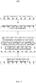
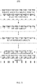
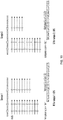
- a single read can generate multiple counts if it comprises multiple CpG sites. For example in FIG.
- the number of CpG sites covered by the assay does not necessarily dictate the methylation score. If that was the case, as illustrated in FIG. 10 , the three CpG sites in both Samples 1 and 2 would be methylated and the methylation index would be 100% for both samples.
- the methylation score can be used to determine the methylation state of a single CpG site, or a series of individual CpG sites.
- CpG sites can be selectively filtered out of the analysis or the CpG sites can be grouped and the methylation density can be calculated.
- the methylation score can also be the average of the methylation score at individual CpG sites in order to correct for different sequencing coverage at each individual site.
- a CpG with 100x coverage might be weighted more in the methylation score than a site with 20x coverage.
- every cytosine with a methylation score is given a methylation score regardless of sequencing coverage.
- the methylation score can be expressed as the "methylation density", which is the methylation score for the CpG sites in a defined region (e.g., a particular CpG site, CpG sites within a CpG island, or a larger region such as a block).
- the methylation density for a 1Mb bin in the human genome can be determined from the number of counts showing CpG methylation divided by the total number of counts covering CpG sites in the 1Mb region. This analysis can also be performed for other bin sizes, e.g. 50 kb, 100 kb, 200kb, 250 kb, 300kb, 400 kb, 500 kb, 750 kb, etc.
- the methylation score can be expressed as the "proportion of methylated cytosines", which includes cytosines outside of the CpG context in the region.
- the methylation score can be expressed as a "global methylation score” or "global methylation index".
- the global methylation index refers to the methylation score for all of the CpG sites interrogated by the compositions and methods described herein, which includes CpG sites across the genome (e.g., greater than 50,000, 60,000, 70,000, 80,000, 100,000, 150,000, 200,000, 300,000, 400,000, 500,000 or more CpG sites distributed throughout the genome); thus allowing one to generate a global methylation index with a single assay.
- the skilled worker will appreciate that not every CpG site in a genome needs to be studied to determine the global methylation index.
- the methylation score of a subset of CpG sites may be determined as an indication for the global methylation state of the entire genome, and given as a "global methylation index".
- a methylation score is determined for a test subject, sample, tissue or portion thereof, in which case it is referred to as a "test methylation score” or “test ratio”.
- a test ratio can be compared to a "reference methylation score” or “reference ratio” from a corresponding known (reference) subject, sample or tissue.
- the methylation score from a population of cells e.g., from a tumor, or from a particular tissue type
- multiple or mixed populations of cells e.g., maternal and fetal cells
- multiple subjects e.g., smoker vs. non-smoker
- the regions with methylation differences between test samples and reference samples are referred to as "differentially methylated regions" (DMRs), which are regions or blocks having different methylation scores.
- DMRs differentially methylated regions
- a differentially methylated region e.g., block, chromosome, gene, island, etc. is identified by a difference in the methylation score between a test and reference sample across a sufficient number of samples to be significant.
- site refers to a single site, which may be a single base position or a group of correlated base positions, e.g., a CpG site; whereas a "block” or “region” refers to a portion of the genome that includes multiple sites.
- a block may include one or more CpG islands, genes, chromatin regions such as large organized chromatin lysine-modifications, or nuclear organization regions such as lamin associated domains.
- a block may contain one or more repeat elements.
- compositions and methods described are able to assay the regions of the genome believed to be pathologically important for cell differentiation and disease. Furthermore, in the case of cancer, there is evidence that epigenetic dysregulation is occurring early in cancer - even before full cancer development (see Timp et al. Genome Medicine 2014, 6:61 ) and is more likely to occur at CpG sites that reside in Alu repeat elements (see Luo et al. BioMed research international 2014 ); thereby adding to the clinical utility of the methods and compositions described herein.
- methylome refers to the amount or pattern of methylation at different sites or regions within a population of cells. Thus, methylome can be thought of as the methylation score for a particular population of cells.
- a disease state may have a methylome, such as the healthy liver methylome versus the necrotic liver.
- a tissue type may have a methylome, such as a liver methylome versus a blood methylome.
- a cellular phenotype may have a methylome, such as senescent cells versus dividing cells.
- the methylome may correspond to all of the genome, a subset of the genome (e.g., repeat elements in the genome), or a portion of the subset (e.g., those areas found to be associated with disease).
- a "fetal methylome” corresponds to a methylome of a fetus of a pregnant female. The fetal methylome can be determined using a variety of fetal tissues or sources of fetal DNA, including placental tissues and cell-free fetal DNA in maternal plasma.
- a "tumor methylome” corresponds to a methylome of a tumor of an organism (e.g., a human). The tumor methylome can be determined using tumor tissue or cell-free tumor DNA .
- cell-free tumor DNA is present in plasma.
- the fetal methylome and the tumor methylome are examples of a methylome of interest.
- Other examples of methylomes of interest are the methylomes of organs (e.g. methylomes of the liver, lungs, prostate, gastrointestinal tract, bladder etc.) that can contribute DNA into a bodily fluid (e.g. plasma, serum, sweat, saliva, urine, genital secretions, semen, stools fluid, diarrheal fluid, cerebrospinal fluid, secretions of the gastrointestinal tract, ascitic fluid, pleural fluid, intraocular fluid, fluid from a hydrocele (e.g.
- a methylome from plasma may be referred to a "plasma methylome".
- the plasma methylome is an example of a cell-free methylome since plasma and serum include cell-free DNA (cfDNA).
- the plasma methylome is also an example of a mixed population methylome since it is a mixture of fetal/maternal methylome or tumor/non-tumor methylome or DNA derived from different tissues or organs.
- read refers to the raw or processed output of sequencing systems, such as massively parallel sequencing.
- the output of the methods and compositions described herein is reads.
- these reads may need to be trimmed, filtered, and aligned, resulting in raw reads, trimmed reads, aligned reads.
- count refers to a uniquely aligned read within a target sequence of interest. In the context of the methylation score, a count will correspond to the information retrieved from the reads (methylated or unmethylated) at the CpG sites. Therefore, if a read encompassed multiple CpG site, this read can produce multiple counts.
- the methods may be used to detect copy number variations.
- a "copy number variation” generally is a class or type of genetic variation or chromosomal aberration.
- copy number variations refer to changes in copy number in germline cells
- copy number alterations/aberrations CNAs
- copy number variations include copy number alterations/aberrations.
- a copy number variation can be a deletion (e.g. micro-deletion), duplication (e.g., a micro-duplication), or insertion (e.g., a micro-insertion).
- the prefix "micro” as used herein may refer to a segment of a nucleic acid less than 5 base pairs in length.
- a copy number variation can include one or more deletions (e.g. micro-deletion), duplications and/or insertions (e.g., a micro-duplication, micro-insertion) of a segment of a chromosome.
- a duplication comprises an insertion.
- an insertion is a duplication.
- an insertion is not a duplication. For example, a duplication of a sequence in a portion increases the counts for a portion in which the duplication is found. Often a duplication of a sequence in a portion increases the elevation or level.
- a duplication present in portions making up a first elevation or level increases the elevation or level relative to a second elevation or level where a duplication is absent.
- an insertion increases the counts of a portion and a sequence representing the insertion is present (i.e., duplicated) at another location within the same portion.
- an insertion does not significantly increase the counts of a portion or elevation or level and the sequence that is inserted is not a duplication of a sequence within the same portion.
- an insertion is not detected or represented as a duplication and a duplicate sequence representing the insertion is not present in the same portion.
- a copy number variation is a fetal copy number variation.
- a fetal copy number variation is a copy number variation in the genome of a fetus.
- a copy number variation is a maternal and/or fetal copy number variation.
- a maternal and/or fetal copy number variation is a copy number variation within the genome of a pregnant female (e.g., a female subject bearing a fetus), a female subject that gave birth or a female capable of bearing a fetus.
- a copy number variation can be a heterozygous copy number variation where the variation (e.g., a duplication or deletion) is present on one allele of a genome.
- a copy number variation can be a homozygous copy number variation where the variation is present on both alleles of a genome.
- a copy number variation is a heterozygous or homozygous fetal copy number variation.
- a copy number variation is a heterozygous or homozygous maternal and/or fetal copy number variation.
- a copy number variation sometimes is present in a maternal genome and a fetal genome, a maternal genome and not a fetal genome, or a fetal genome and not a maternal genome.
- aneuploidy refers to a chromosomal abnormality characterized by an abnormal variation in chromosome number, e.g., a number of chromosomes that is not an exact multiple of the haploid number of chromosomes.
- a euploid individual will have a number of chromosomes equaling 2n, where n is the number of chromosomes in the haploid individual. In humans, the haploid number is 23. Thus, a diploid individual will have 46 chromosomes.
- An aneuploid individual may contain an extra copy of a chromosome (trisomy of that chromosome) or lack a copy of the chromosome (monosomy of that chromosome).
- the abnormal variation is with respect to each individual chromosome.
- an individual with both a trisomy and a monosomy is aneuploid despite having 46 chromosomes.
- aneuploidy diseases or conditions include, but are not limited to, Down syndrome (trisomy of chromosome 21), Edwards syndrome (trisomy of chromosome 18), Patau syndrome (trisomy of chromosome 13), Turner syndrome (monosomy of the X chromosome in a female), and Klinefelter syndrome (an extra copy of the X chromosome in a male).
- non-aneuploid chromosomal abnormalities include translocation (wherein a segment of a chromosome has been transferred to another chromosome), deletion (wherein a piece of a chromosome has been lost), and other types of chromosomal damage (e.g., Fragile X syndrome, which is caused by an X chromosome that is abnormally susceptible to damage).
- subject and patient refer to any animal, such as a dog, a cat, a bird, livestock, and particularly a mammal, and preferably a human.
- reference subject and “reference patients” refer to any subject or patient that exhibits known genotypes (e.g., known euploidy or aneuploidy), phenotypes, or age, or to a subject or patient that is known to have a disease or condition, or known to not have a disease or condition, or known to have a particular state of a disease or condition, or known to have a predisposition to a disease or condition, or known to have been exposed to drugs, toxins, a particular diet, or an agent or conditions suspected of causing methylation changes.
- the subject can be any human.
- the subject is a pregnant female.
- the blood sample may be a maternal plasma or serum sample.
- the subject is an organ transplant recipient, and the subject's methylation state may be indicative of organ rejection.
- the methylation state of a population of target sequences of interest e.g., of fetal, tumor, or disease origin
- a background of target sequences of interest e.g., maternal, non-tumor, or disease-free origin.
- the background target sequences of interest may serve as a reference, wherein differences from the reference are indicative of a disease or condition, or to identify a nucleic acid species.
- mutational landscape is the cumulative frequency of a collection of mutations that generally span the genome.
- the types of mutations that make up a mutational landscape include but are not limited to, single nucleotide variations, deletions, and insertions, and the type of mutations also inform a given mutational landscape or pattern.
- Examples of specific mutational landscapes associated with diseases or conditions include an increased frequency of C>A transversions associated with cigarette smoke exposure ( Ding, L. et al. Somatic mutations affect key pathways in lung adenocarcinoma.
- tissue-of-origin refers to the tissue source of nucleic acids in a sample, where "tissue” is used to describe a group or population of cells of a same type. Some tissue may have multiple cell types, for example hepatocytes, alveolar cells or blood cells, while other tissue may originate from different organisms, for example, mother and fetus, or from healthy vs. disease tissue. Likewise, “reference tissues” may be used to determine methylation-specific tissue patterns or levels. For example, reference tissues from multiple subjects may be used to determine the tissue components of a test sample. In some embodiments, the DNA molecules of differing origin are DNA molecules of maternal origin and DNA molecules of fetal origin.
- the DNA molecules of differing origin are DNA molecules of a first tissue origin and DNA molecules of a second tissue origin or of leukocyte origin.
- determining the tissue-of-origin may be indicative of the presence of disease (e.g., cancer), or may be used to determine the relative or absolute amount of DNA from a particular tissue (e.g., fetal cfDNA in a maternal sample).
- the compositions and methods described herein can be used to differentiate and identify the tissue source of DNA. Given an unknown source of tissue, DNA can be extracted using standard methods, the compositions and methods described herein can be used to generate tissue-specific data, and the data can be fit into the most likely tissue reference bin as described further in the Examples.
- nucleic acid and nucleic acid molecules
- DNA molecules e.g., cDNA or genomic DNA
- RNA molecules e.g., mRNA
- DNA-RNA hybrids DNA-RNA hybrids
- analogs of the DNA or RNA generated using nucleotide analogs are used interchangeably and refer to DNA molecules (e.g., cDNA or genomic DNA), RNA molecules (e.g., mRNA), DNA-RNA hybrids, and analogs of the DNA or RNA generated using nucleotide analogs.
- the nucleic acid molecule can be a nucleotide, oligonucleotide, double-stranded DNA, single-stranded DNA, multi-stranded DNA, complementary DNA, genomic DNA, non-coding DNA, messenger RNA (mRNAs), microRNA (miRNAs), small nucleolar RNA (snoRNAs), ribosomal RNA (rRNA), transfer RNA (tRNA), small interfering RNA (siRNA), heterogeneous nuclear RNAs (hnRNA), or small hairpin RNA (shRNA).
- the methods can be performed on a nucleic acid sample such as DNA or RNA, e.g., genomic DNA.
- the nucleic acid molecule may be cell-free DNA (cfDNA).
- Cell-free DNA is thought to result from cellular necrosis or apoptosis, wherein genomic cellular DNA is digested and becomes fragmented, extracellular DNA.
- Cell-free DNA of apoptotic origin may be from a non-host (e.g., transplanted organ or tissue), fetus (e.g., from the placenta resulting in cell-free fetal DNA), or a diseased tissue (e.g., from a tumor resulting in circulating tumor DNA).
- Cell-free DNA can be detected in a range of samples including, but not limited to, blood, plasma and urine.
- the nucleic acid molecules are associated with exosomes, which are microvesicles released from a variety of different cells, including cancer cells.
- the compositions and methods described herein may be able to differentiate cfDNA of necrotic and apoptotic origin based on its methylation status.
- a nucleic acid sample may be isolated in any manner known to a person of ordinary skill in the art (e.g., by centrifugation).
- sample refers to a sample typically derived from a biological fluid, cell, tissue, organ, or organism, comprising a nucleic acid or a mixture of nucleic acids comprising at least one nucleic acid sequence that is to be screened for, e.g., cancer or aneuploidy.
- a sample is a blood sample such as a whole blood sample, a serum sample, or a plasma sample.
- the sample comprises at least one nucleic acid sequence whose genome is suspected of having undergone variation.
- Such samples include, but are not limited to sputum/oral fluid, amniotic fluid, blood, a blood fraction, or fine needle biopsy samples (e.g., surgical biopsy, core needle biopsy, fine needle biopsy, etc.) urine, stool, peritoneal fluid, pleural fluid, cerebro-spinal fluid, gastrointestinal fluid, cell lines, tissue embedded in paraffin, fresh frozen tissue, and the like.
- fine needle biopsy samples e.g., surgical biopsy, core needle biopsy, fine needle biopsy, etc.
- the assays can be used to detect a disease or condition, or detect the state of a disease or condition, or determine whether a subject has a predisposition to a disease or condition, in samples from any mammal, including, but not limited to dogs, cats, horses, goats, sheep, cattle, pigs, etc.
- the sample may be used directly as obtained from the biological source or following a pretreatment to modify the character of the sample.
- pretreatment may include preparing plasma from blood, diluting viscous fluids and so forth.
- Methods of pretreatment may also involve, but are not limited to, bisulfite conversion, filtration, precipitation, dilution, distillation, mixing, centrifugation, freezing, lyophilization, concentration, amplification, nucleic acid fragmentation, inactivation of interfering components, the addition of reagents, lysing, etc. If such methods of pretreatment are employed with respect to the sample, such pretreatment methods are typically such that the nucleic acid(s) of interest remain in the test sample, preferably at a concentration proportional to that in an untreated test sample (e.g., namely, a sample that is not subjected to any such pretreatment method(s)).
- an untreated test sample e.g., namely, a sample that is not subjected to any such pretreatment method(s)
- additional processing and/or purification steps may be performed to obtain nucleic acid fragments of a desired purity or size, using processing methods including but not limited to sonication, nebulization, gel purification, PCR purification systems, nuclease cleavage, size-specific capture or exclusion, targeted capture or a combination of these methods.
- cell-free DNA may be isolated from the sample prior to further analysis.
- the sample is from the subject whose disease or condition is to be determined by the systems and methods of the disclosure, also referred as "a test sample.”
- MIP refers to a molecular inversion probe (also known as a circular capture probe).
- the term “primer”, “probe”, or “capture probe” also may refer to a MIP in the context of their ability to selectively bind to nucleic acid molecules.
- Molecular inversion probes are nucleic acid molecules that contain two targeting polynucleotide arms, one or more unique molecular tags (also known as unique molecular identifiers (UMID's)), and a polynucleotide linker (e.g., a universal backbone linker).
- a polynucleotide linker can range from 10, 20, 30, 40, 50, 60, 70, 80, 90, 100, 125, 150, 175, 200, 225, 250, 275, 300, 400, 500, 1000, 1500, 2000 or more bases. See, for example, FIG. 5 .
- a MIP may comprise more than one unique molecular tags, such as, two unique molecular tags, three unique molecular tags, or more.
- the polynucleotide arms in each MIP are located at the 5' and 3' ends of the MIP, while the unique molecular tag(s) and the polynucleotide linker are located in the middle.
- the MIPs comprise in sequence the following components: first targeting polynucleotide arm - first unique molecular tag - polynucleotide linker - second unique molecular tag - second targeting polynucleotide arm.
- the polynucleotide linker (or the backbone linker) in the MIPs are universal in all the MIPs used in a method of the invention.
- the MIPs may not comprise any unique molecular tags.
- the polynucleotide arms are designed to hybridize upstream and downstream of target sequences (or sites) in a genomic nucleic acid sample. These polynucleotide arms are substantially complementary to one or more repeat sequences in a genomic nucleic acid sample that flank a target sequence (herein referred to as a "gap sequence” or a "unique gap sequence".
- the gap sequences are 1, 2, 3, 4, 5, 6, 7, 8, 9, 10, 11, 12, 13, 14, 15, 16, 17, 18, 19, 20, 25, 30, 40, 50, 60, 70, 80, 90, 100, 100, 125, 150, 175, 200, 225, 250, 275, 300, 400, 500, 1000, 1500, 2000 bases or greater in length.
- the gap sequences are generally less than 150 or 200 bases in length.
- the targeting polynucleotide arms comprise a ligation sequence and an extension sequence.
- a MIP may comprise targeting polynucleotide arms that are substantially complementary to a plurality of repeat sequences in a DNA sample. For example, a MIP can hybridize to tens, hundreds, thousands, hundreds of thousands, or millions of target sequences of interest in a DNA sample (e.g., a sample comprising a human genome).
- a MIP targets, for example, greater than 1,000, greater than 10,000, greater than 20,000, greater than 30,000, greater than 40,000, greater than 50,000, greater than 60,000, greater than 70,000, greater than 80,000, greater than 90,000, greater than 100,000, greater than 200,000, greater than 300,000, greater than 400,000, greater than 500,000, greater than 600,000, greater than 700,000, greater than 800,000, greater than 900,000, and/or greater than 1,000,000 target sequences of interest.
- substantially complementary refers to 0 mismatches in both arms, or at most 1 mismatch in only one arm (e.g., when the targeting polynucleotide arms hybridize to the first and second regions in the nucleic acid that, respectively, flank a site of interest). In some embodiments, “substantially complementary” refers to at most a small number of mismatches in both arms, such as 1, 2, 3, 3, 5, 6, 7, or 8.
- target sequence refers to the sequence bound or captured by the primes or probes of the disclosure and comprise one or more CpG sites.
- the target sequences of interest are selected to, among other considerations, comprise at least one CpG site; however, not every nucleic acid sequence captured by the primers or probes of the disclosure comprises a CpG site.
- target sequences of interest may include the repeat sequences to which the targeting polynucleotide arms hybridize, as well as the gap sequences flanked by the repeat sequences.
- the repeat sequences have 0, 1, 2, 3, 4, or more mismatches in hybridizing with the targeting polynucleotide arms.
- the same repeat sequences are found in a target sequence of interest, in which case the target polynucleotide arms are identical.
- two different repeat sequences e.g., repeat A and repeat B
- are found in a target sequence of interest in which case the target polynucleotide arms are not identical.
- the repeat sequences have 0 or 1 mismatches in hybridizing with the targeting polynucleotide arms.
- a MIP binds to Alu repeats.
- a MIP does not bind long interspersed nucleotide elements (LINE) in the genome.
- the unique molecular tags are short nucleotide sequences that are randomly generated. In certain embodiments, the unique molecular tags are not designed to hybridize to any sequence or site located on a genomic nucleic acid fragment or in a genomic nucleic acid sample. In certain embodiments, the unique molecular tag is any tag with a suitable detectable label that can be incorporated into or attached to a nucleic acid (e.g., a polynucleotide) that allows detection and/or identification of nucleic acids that comprise or attach to the tag. In certain embodiments, unique molecular tags of sufficient length are introduced at concentrations to ensure that each MIP comprises a unique combination of molecular tags, thereby making each capture event distinct.
- a nucleic acid e.g., a polynucleotide
- the tag is incorporated into or attached to a nucleic acid during a sequencing method (e.g., by a polymerase).
- Non-limiting examples of tags include nucleic acid tags, nucleic acid indexes or barcodes, a radiolabel (e.g., an isotope), metallic label, a fluorescent label, a chemiluminescent label, a phosphorescent label, a fluorophore quencher, a dye, a protein (e.g., an enzyme, an antibody or part thereof, a linker, a member of a binding pair), the likes or combinations thereof.
- the tag e.g., a nucleic acid index or barcode
- the tag is a unique, known and/or identifiable sequence of nucleotides or nucleotide analogues.
- tags or UMID's help reduce or remove amplification errors and sequencing errors by allowing for the identification of unique molecules during bioinformatics analysis.
- tags are four, five, or six or more contiguous nucleotides.
- Unique molecular identifiers comprising oligonucleotides are described in U.S. Patent Application No. 11/186,636 , which published as US20070020640A1 , and the use of oligonucleotide unique molecular identifiers with MIPs is described in U.S. Patent Application No. 12/027,039 , which published as US20080269068A1 .
- a multitude of fluorophores are available with a variety of different excitation and emission spectra.
- any suitable type and/or number of fluorophores can be used as a tag.
- one or two types of tags are linked to each nucleic acid in a library.
- chromosome-specific tags are used to make chromosomal counting faster or easier. Detection and/or quantification of a tag can be performed by a suitable method, machine or apparatus, non-limiting examples of which include flow cytometry, quantitative polymerase chain reaction (qPCR), gel electrophoresis, a luminometer, a fluorometer, a spectrophotometer, a suitable gene-chip or microarray analysis, Western blot, mass spectrometry, chromatography, cytofluorimetric analysis, fluorescence microscopy, a suitable fluorescence or digital imaging method, confocal laser scanning microscopy, laser scanning cytometry, affinity chromatography, manual batch mode separation, electric field suspension, a suitable nucleic acid sequencing method and/or nucleic acid sequencing apparatus, the like and combinations thereof.
- the tag is suitable for use with microarra
- the MIPs are introduced to nucleic acids (e.g., nucleic acid fragments) to perform capture of target sequences or sites located on a nucleic acid sample (e.g., a genomic DNA).
- a nucleic acid sample e.g., a genomic DNA
- fragmenting may aid in capture of target nucleic acid by molecular inversion probes.
- the captured target may further be subjected to an enzymatic gap-filling and ligation step, such that a copy of the target sequence is incorporated into a circle, which is herein referred to as a replicon.
- Capture efficiency of the MIP to the target sequence on the nucleic acid fragment can be improved by lengthening the hybridization and gap-filing incubation periods. (See, e.g., Turner E H, et al., Nat Methods. 2009 Apr. 6:1-2 .).
- MIP technology may be used to detect or amplify particular nucleic acid sequences in complex mixtures.
- One of the advantages of using the MIP technology is in its capacity for a high degree of multiplexing, which allows thousands of target sequences to be captured in a single reaction containing thousands of MIPs.
- MIP technology has also been applied to the identification of new drug-related biomarkers. See, e.g., Caldwell et al., "CYP4F2 genetic variant alters required warfarin dose," Blood, 111(8): 4106-4112 (2008 ); and McDonald et al., "CYP4F2 Is a Vitamin K1 Oxidase: An Explanation for Altered Warfarin Dose in Carriers of the V433M Variant," Molecular Pharmacology, 75: 1337-1346 (2009 ).
- Other MIP applications include drug development and safety research.
- capture refers to the binding or hybridization reaction between a primer or probe (e.g., molecular inversion probe) and the corresponding targeting site.
- sensitivity refers to a statistical measure of performance of an assay (e.g., method, test), calculated by dividing the number of true positives by the sum of the true positives and the false negatives.
- the term "specificity”, as used herein, refers to a statistical measure of performance of an assay (e.g., method, test), calculated by dividing the number of true negatives by the sum of true negatives and false positives.
- amplicon refers to a nucleic acid generated via capturing reactions or amplification reactions.
- the amplicon is a single-stranded nucleic acid molecule.
- the amplicon is a single-stranded circular nucleic acid molecule.
- the amplicon is a double-stranded nucleic acid molecule.
- a MIP captures or hybridizes to a target sequence or site.
- a ligation/extension mixture is introduced to extend and ligate the gap region between the two targeting polynucleotide arms to form a single-stranded circular nucleotide molecule, i.e., a MIP replicon.
- the gap-filled sequence in the replicon can be thought of as an "insert” or "insert sequence”.
- the MIP replicon may be amplified through a polymerase chain reaction (PCR) to produce a plurality of MIP amplicons, which are double-stranded nucleotide molecules.
- MIP replicons and amplicons can be produced from a first plurality of target sequences of interest (e.g., a sequence containing known or suspected CpG sites) and a second plurality of target sequences of interest (e.g., target sequences distributed throughout the genome).
- a first plurality of target sequences of interest e.g., a sequence containing known or suspected CpG sites
- second plurality of target sequences of interest e.g., target sequences distributed throughout the genome
- sequence is used in a broad sense and may refer to any technique known in the art that allows the order of at least some consecutive nucleotides in at least part of a nucleic acid to be identified, including without limitation at least part of an extension product or a vector insert. Sequencing also may refer to a technique that allows the detection of differences between nucleotide bases in a nucleic acid sequence.
- Exemplary sequencing techniques include targeted sequencing, single molecule real-time sequencing, electron microscopy-based sequencing, transistor-mediated sequencing, direct sequencing, random shotgun sequencing, Sanger dideoxy termination sequencing, targeted sequencing, exon sequencing, whole-genome sequencing, sequencing by hybridization (e.g., in an array such as a microarray), pyrosequencing, capillary electrophoresis, gel electrophoresis, duplex sequencing, cycle sequencing, single-base extension sequencing, solid-phase sequencing, high-throughput sequencing, massively parallel signature sequencing, emulsion PCR, co-amplification at lower denaturation temperature-PCR (COLD-PCR), multiplex PCR, sequencing by reversible dye terminator, paired-end sequencing, near-term sequencing, exonuclease sequencing, sequencing by ligation, short-read sequencing, single-molecule sequencing, sequencing-by-synthesis, real-time sequencing, reverse-terminator sequencing, ion semiconductor sequencing, nanoball sequencing, nanopore sequencing, 454 sequencing, Solexa Genome Analyzer sequencing, mi
- sequencing comprises detecting the sequencing product using an instrument, for example but not limited to an ABI PRISM® 377 DNA Sequencer, an ABI PRISM® 310, 3100, 3100-Avant, 3730, or 373OxI Genetic Analyzer, an ABI PRISM® 3700 DNA Analyzer, or an Applied Biosystems SOLiDTM System (all from Applied Biosystems), a Genome Sequencer 20 System (Roche Applied Science), or a mass spectrometer.
- sequencing comprises emulsion PCR.
- sequencing comprises a high throughput sequencing technique, for example but not limited to, massively parallel sequencing (MPS).
- MPS massively parallel sequencing
- MIPs may alternatively employ microarray technology to quantify MIPs products.
- "Microarray” or “array” refers to a solid phase support having a surface, preferably but not exclusively a planar or substantially planar surface, which carries an array of sites containing nucleic acids such that each site of the array comprises substantially identical or identical copies of oligonucleotides or polynucleotides and is spatially defined and not overlapping with other member sites of the array; that is, the sites are spatially discrete.
- the array or microarray can also comprise a non-planar interrogatable structure with a surface such as a bead or a well.
- the oligonucleotides or polynucleotides of the array may be covalently bound to the solid support, or may be non-covalently bound.
- Conventional microarray technology is reviewed in, e.g., Schena, Ed., Microarrays: A Practical Approach, IRL Press, Oxford (2000 ).
- "Array analysis”, “analysis by array” or “analysis by microarray” refers to analysis, such as, e.g., sequence analysis, of one or more biological molecules using a microarray.
- each sample is hybridized individually to a single microarray.
- processing through-put can be enhanced by physically connecting multiple microarrays onto a single multi-microarray plate for convenient high-throughput handling.
- custom DNA microarrays for example from Affymetrix Inc. (Santa Clara,Calif., USA), can be manufactured to specifically quantify products of the MIPs assay.
- compositions and methods described herein may be adapted and modified as is appropriate for the application being addressed and that the compositions and methods described herein may be employed in other suitable applications, and that such other additions and modifications will not depart from the scope hereof.
- Embodiments of the present invention provide a solution to the problems of existing methylation detection methods. These embodiments replace previous library preparations with a capture method using a small number of oligonucleotide MIPs comprising targeting polynucleotide arms that hybridize to repeat sequences, said arms being arms attached to high performance universal backbone structures. These MIPs are designed to flank and incorporate uniquely aligning sequences over the entire human genome, but are enriched for targets pertinent to methylation (i.e., targets containing CpG sites).
- CpG sites are sites located throughout the genome where methylation occurs at the cytosine nucleotide of the site.
- hydrolytic deamination and desulphonation e.g., bisulfite conversion, or simply deamination
- unmethylated cytosines are converted to uracils.
- methylated cytosines are protected from this reaction, and, thus, remain cytosines. Therefore, by performing a bisulfite conversion or an alternative procedure that preserves methylated cytosines, and subsequently sequencing CpG sites, the embodiments described herein provide a method of detecting whether a CpG site was methylated or not.
- Contemplated methods of selecting capture molecules enable the selection of unique sequences in a desired area for quantitation, and do not rely on the presence of some unique sequences in the amplification of convenient repeat sequences.
- the use of repeat sequences in the optimized capture method allows dense tiling of a target area with little or no interference of similar sequences in the production of barcoded targets for single molecule kinetics during library preparation.
- the method has economic benefits over previous methods. In particular, these methods provide savings from the use of a small number of capture reagents (primers or probes) that still are capable of surveying genome-wide indices.
- the capture reagents can provide information not only about methylation status, but also more generally about the sequences of the target sites. This information can be used to determine, for example, copy number variation or mutational landscape.
- This information also can be used to detect chromosomal abnormalities, e.g., aneuploidies such as trisomy, or tissue-specific methylation scores and patterns along with disease or condition-specific mutational landscapes or patterns, e.g., the presence of tissue-specific circulating tumor DNA (ctDNA) in blood.
- chromosomal abnormalities e.g., aneuploidies such as trisomy, or tissue-specific methylation scores and patterns along with disease or condition-specific mutational landscapes or patterns, e.g., the presence of tissue-specific circulating tumor DNA (ctDNA) in blood.
- ctDNA tissue-specific circulating tumor DNA
- the methods also provide a rapid analysis with a low read count in an assay that is easily multiplexed.
- multiple layers of unique molecular tags and/or barcodes can be used within the methods to identify specific primer species as well as to deconvolute multiplex data to trace signals back to individual samples.
- a first population of MIPs can be used to obtain methylation status (and optionally, sequence information), while a second population of MIPs provides sequence information.
- the methods can be used in ultra low coverage applications such as detecting trisomies in a 100% fetal sample, such as a product of conception, or a non-fetal diagnostic sample.
- a sample can be mixed (e.g., fetal vs. maternal or diseased vs.
- the methods also are fast as compared to whole genome sequencing, whole exome sequencing, and targeted sequencing.
- the methods described herein also offer the advantage of requiring relatively small amounts of input DNA as compared to whole genome bisulfite sequencing, which suffers from input DNA loss during the harsh bisulfite conversion process, whereas the methods described herein allow for the capture of bisulfite converted DNA after the conversion step thereby preserving input DNA and reducing bias. More specifically, most library prep kits require double-stranded input DNA for an adapter ligation step.
- the bisulfite conversion step needs to be performed after the adapter ligation step, but before PCR.
- the harsh bisulfite conversion can compromise some of the ligated molecules, and thereby make them unusable.
- the ligation adapters need to be methylated, otherwise the cytosines will be converted, which adds additional cost.
- the methods are related to the field of genetic analysis. In general, these methods can be used as a rapid and economical means to detect and quantify methylation status. Because the methylation status can be determined by sequencing, the sequence information obtained by the methods described herein allows for detection of mutations as well as detection of deletions and duplications of genetic features in a range extending from complete chromosomes and arms of chromosomes to microscopic deletions and duplications, submicroscopic deletions and deletions, and even single nucleotide features including single nucleotide polymorphisms, deletions, and insertions. In certain embodiments, these methods can be used to detect sub-chromosomal genetic lesions, e.g., microdeletions.
- the methods can be used to determine mutations or other sequence elements that correlate to a disease or condition (e.g., by detecting a SNP or SNPs). Because the methods provide different types of information in a single assay, they are simpler, more efficient, and less expensive than current methods. In certain embodiments, the methods also provide a maximum likelihood estimate (k) which will allow for increased accuracy and an estimation of the probe capture efficiency and reduces need for extraneous sequencing during copy number variation (CNV) detection. This may result in a low coefficient of variation (CV) due to probe uniformity because a small number of probes (e.g., one, two or more) is used. These capture probes allow the combination of additional probes with no interference or cross assay reactions. Combining the information from several probes and their unique reads greatly reduces error in the system. Indeed, targeted probe addition can greatly enhance assay utility while reducing cost.
- k maximum likelihood estimate
- CV coefficient of variation
- the methods provided by some embodiments have particular advantages as compared to targeted sequencing.
- the methods described herein use a simultaneous recognition of two sequence elements at the point of capture, and the two arms are limited by proximity.
- a typical targeted sequencing method will allow a polymerase to initiate at a single site.
- the run on-product created by typical sequencing produces inefficiency, but may also produce internal or "off-target priming" with the second primer.
- the inherent "dual recognition" of the nucleic acids of some embodiments increases stringency, an effect which carries over into the quantitation by the molecular identifier element in the MIP structure.
- a unique molecular tag may be placed at one site in the MIP backbone, but in standard targeted sequencing using a molecular identifier, a random sequence is used in both primers. Also, the methods allow for lower reagent costs since coverage across the genome can be achieved with very few MIPs compared to the hundreds or thousands of multiplexed, PCR primers required for targeted sequencing. Nevertheless, the methods enjoy most, if not all, of the economic and performance advantages that targeted sequencing displays over shotgun methods.
- the methods and nucleic acids of some embodiments offer clear advantages over previously described genetic methods.
- whole genome sequencing and massively parallel signature sequencing generally require costly analysis of large, non-informative portions of the genome; whereas the present methods can produce similar answers using a fraction of the genome, thereby reducing assay costs and time.
- Other approaches rely on selectively assaying informative portions of the genome.
- the methods use a novel, comprehensive approach for identifying repeat, primer-binding sites that allow for greater assay design parameters (sequence agnostic - for example, not limited to repeat line elements), more candidate primers (e.g., because all potential primers are enumerated), simple, lower cost assays that are specific and sensitive enough for clinical utility, and a greater ability to multiplex.
- compositions and methods described herein can be used to assemble methylomes through the sequence analysis of plasma DNA.
- the ability to determine the placental or fetal methylome from maternal plasma provides a noninvasive method to determine, detect and monitor the aberrant methylation profiles associated with pregnancy-related conditions such as preeclampsia, intrauterine growth restriction, preterm labor and others.
- pregnancy-related conditions such as preeclampsia, intrauterine growth restriction, preterm labor and others.
- the detection of a disease-specific aberrant methylation signature allows the screening, diagnosis and monitoring of such pregnancy-associated conditions.
- the measuring of the maternal plasma methylation level allows the screening, diagnosis and monitoring of such pregnancy-associated conditions.
- the approach could be applied to other areas of medicine where plasma DNA analysis is of interest.
- the methylomes of cancers could be determined from plasma DNA of cancer patients.
- Cancer methylome analysis from plasma is potentially a synergistic technology to cancer genomic analysis from plasma (e.g., the detection of well-known cancer-associated somatic mutations).
- the determination of a methylation state could be used to screen for cancer.
- the methylation test ratio of a plasma sample shows aberrant levels compared with healthy controls (reference ratio)
- cancer may be suspected.
- further confirmation and assessment of the type of cancer or tissue-of-origin of the cancer may be performed.
- the compositions and methods described herein also allow for the detection of tumor-associated copy number aberrations, chromosomal translocations and single nucleotide variants across the genome (mutational landscape).
- radiological and imaging investigations e.g. computed tomography, magnetic resonance imaging, positron emission tomography
- endoscopy e.g. upper gastrointestinal endoscopy or colonoscopy
- the determination of a methylation score of a plasma (or other biologic) sample can be used in conjunction with other modalities for cancer screening or detection such as prostate specific antigen measurement (e.g. for prostate cancer), carcinoembryonic antigen (e.g. for colorectal carcinoma, gastric carcinoma, pancreatic carcinoma, lung carcinoma, breast carcinoma, medullary thyroid carcinoma), alpha fetoprotein (e.g. for liver cancer or germ cell tumors), CA125 (e.g. for ovarian and breast cancer) and CA19-9 (e.g. for pancreatic carcinoma).
- prostate specific antigen measurement e.g. for prostate cancer
- carcinoembryonic antigen e.g. for colorectal carcinoma, gastric carcinoma, pancreatic carcinoma, lung carcinoma, breast carcinoma, medullary thyroid carcinoma
- alpha fetoprotein e.g. for liver cancer or germ cell tumors
- CA125 e.g. for ovarian and breast cancer
- CA19-9 e.g. for pancreatic carcinoma
- liver tissue can be analyzed to determine a methylation pattern specific to the liver, which may be used to identify liver pathologies.
- Other tissues which can also be analyzed include brain cells, bones, the lungs, the heart, the muscles and the kidneys, etc.
- the methylation profiles of various tissues may change from time to time, e.g. as a result of development, aging, disease processes (e.g. inflammation or cirrhosis or autoimmune processes (such as in systemic lupus erythematosus)) or treatment (e.g. treatment with demethylating agents such as 5-azacytidine and 5-azadeoxycytidine).
- DNA methylation makes such analysis potentially valuable for monitoring of physiological and pathological processes. For example, if one detects a change in the plasma methylome of an individual compared to a baseline value obtained when they were healthy, one could then detect disease processes in organs that contribute plasma DNA.
- the methylomes of transplanted organs could be determined from plasma DNA of organ transplantation recipients.
- plasma DNA is generally regarded as a marker of cell death
- an increase in the plasma level of DNA released from a transplanted organ could be used as a marker for increased cell death from that organ, such as a rejection episode or other pathologic processes involving that organ (e.g. infection or abscess).
- the plasma level of DNA released by the transplanted organ will be expected to decrease.
- Exemplary applications of the methods include the detection, diagnosis, prognosis, recurrence, minimum residual risk assessment of methylation-associated diseases and conditions.
- applications might include a method of determining whether a subject has a predisposition to a disease or condition that is associated with the methylation state of a nucleic acid; a method of diagnosing a disease or condition in a subject, said disease or condition being associated with the methylation state of a nucleic acid; a method of detecting the state of a disease or condition in a subject, said disease or condition being associated with the methylation state of a nucleic acid.
- Particular diseases and conditions include, for example, cancers. A hallmark of cancer cells is that they divide more rapidly than non-cancer cells.
- cancer cells and non-cancer cells will have different methylation patterns.
- the embodiments and methods described herein provide an assay for methylation, CNV and mutational landscape status in tumor biopsy or circulating tumor DNA.
- the embodiments and methods can be used to provide a diagnosis, prognosis, staging, and/or likelihood of developing cancers such as, for example, prostate cancer, colorectal cancer, lung cancer, breast cancer, liver cancer, or bladder cancer.
- Certain embodiments provide a diagnosis, or staging or prognostic information about a cancer, or to inform a treatment decision, or to assess minimum residual risk and recurrence.
- Conditions known to be affected by methylation, or known to affect methylation include but are not limited to, aging, diet, lifestyle, ethnicity, development, bipolar disorder, multiple sclerosis, diabetes, schizophrenia, cancer, neurodegenerative diseases, inflammation, lesion, infection, immune response, exposure to: drugs, alcohol, tobacco, pesticides, heavy metals, radiation, UV other environmental factors.
- the methods provided herein may provide the methylation status and sequence information of circulating cell-free fetal DNA, for example, as a noninvasive prenatal test.
- a noninvasive prenatal test using the methods described herein can be used, for example, to determine a risk for preeclampsia or preterm parturition.
- Additional tests using the methods described herein include pediatric diagnosis of aneuploidy, testing for product of conception or risk of premature abortion, noninvasive prenatal testing (both qualitative and quantitative genetic testing, such as detecting Mendelian disorders, insertions/deletions, and chromosomal imbalances), testing preimplantation genetics, tumor characterization, postnatal testing including cytogenetics, and mutagen effect monitoring.
- Another exemplary application of the methods includes a method of differentiating nucleic acid species originating from a subject and one or more additional individuals, said subject and one or more additional individuals having differing methylation states of a nucleic acid.
- the subject may be a pregnant female and the one or more additional individuals may be an unborn fetus.
- the blood sample may be maternal plasma or maternal serum.
- the subject may be a tissue transplant recipient, and the one or more additional individuals may be a tissue transplant donor.
- Another exemplary application of the methods includes a method of differentiating nucleic acid species originating from different tissues in a subject.
- the methods of the invention may be used to differentiate between a tissue of interest (e.g., a tissue of fetal, tumor, or disease origin) and other tissues (e.g., maternal, non-tumor, or disease-free origin).
- a tissue of interest e.g., a tissue of fetal, tumor, or disease origin
- other tissues e.g., maternal, non-tumor, or disease-free origin
- the subject may be a pregnant female.
- the blood sample may be maternal plasma or maternal serum.
- the subject may be a tissue transplant recipient or a cancer patient.
- Another exemplary application of the methods includes a method of determining the age or "bio-age” or "methylation age” of a subject or group of subjects. More specifically, an individual's genetic material is known to change over time, and the methods described herein allow for the methylation-based age determination of genetic material from an individual by determining the methylation status of thousands or hundreds of thousands of CpG sites in a single assay. This has utility both for forensic purposes and for age-related pathologies such as Alzheimer's disease. The methods are also useful for determining the age of a specific tissue. As described further in the Examples, the methods are useful for determining the "bio-age" for specific tissues such as colorectal tissue or the gestational age of a fetus.
- the capture primers and probes in some embodiments also have the benefit of increased binding stability as compared to conventional PCR primer pairs that are not part of the same molecule.
- the exact targeting arm sequences are somewhat short for PCR primers, and hence will have very low melting temperatures in a PCR context.
- the primers will enhance binding specificity by cooperating to stabilize the interaction. If one arm has a high binding efficiency, the capture is enhanced even if the opposite arm has a lower efficiency.
- the additive length of the pair improves the "on/off" equilibrium for capture because the lower efficiency arm is more often in proximity of its target in a MIP than it would be as a free PCR primer.
- a method for determining whether a subject has a predisposition to a disease or condition that is associated with the methylation state of a nucleic acid.
- the disclosure provides a method for diagnosing a disease or condition in a subject, said disease or condition being associated with the methylation state of a nucleic acid.
- the disclosure provides a method for detecting the state of a disease or condition in a subject, said disease or condition being associated with the methylation state of a nucleic acid. In certain embodiments, these methods comprise:
- a method is provided of differentiating nucleic acid species originating from a subject and one or more additional individuals, said subject and one or more additional individuals having differing methylation states of a nucleic acid, the method comprising:
- a method is provided of differentiating nucleic acid species originating from a first tissue and one or more additional tissues in a subject, said first tissue and one or more additional tissues having differing methylation states of a nucleic acid, the method comprising:
- a method of determining the methylation state of a nucleic acid and detecting copy number variation in a subject, the method comprising:
- targeting molecular tags are used to remove duplicates and thereby improve analysis.
- the number of unique MIP amplicon sequences is determined for defined regions to determine read density.
- a method of determining the methylation age of a subject, the method comprising:
- a method of determining the methylation age of a tissue in a subject, the method comprising:
- the method is a method of determining whether a subject has a predisposition to a disease or condition that is associated with the methylation state of a nucleic acid, the method comprising:
- the method is a method of diagnosing a disease or condition in a subject, the method comprising:
- the method is a method of detecting the state of a disease or condition in a subject, said disease or condition being associated with the methylation state of a nucleic acid, the method comprising:
- the MIPs of the disclosure may not comprise any unique molecular tags. It is possible to determine the methylation score, copy number variation, mutational landscape, etc. using MIPs that do not contain unique molecular tags, according to the methods of the disclosure.
- the bisulfite conversion of any of the methods described herein may be replaced by another type of deamination reaction.
- the above methods may also be used to detect aneuploidy in a fetal or non-fetal subject.
- the methods can be used to detect and quantify deletions and duplications of genetic features in arms of chromosomes, as well as microscopic deletions and duplications, submicroscopic deletions and deletions, and single nucleotide features including single nucleotide polymorphisms, deletions, and insertions.
- the methods of the disclosure use a single species of MIP.
- the methods are useful with 2, 3, 4, 5, 6, 7, 8, 9, 10, 11, 12, 13, 14, 15, 16, 17, 18, 19, 20, 21, 22, 23, 24, 25, 26, 27, 28, 29, 30, 31, 32, 33, 34, 35, 36, 37, 38, 39, 40, 41, 42, 43, 44, 45, 46, 47, 48, 49, 50, or more species of MIPs.
- multiple species of MIPs can be used to detect different diseases or conditions (e.g., cancer, pregnancy-related conditions such as preeclampsia or preterm parturition, or chromosomal abnormalities such as aneuploidy) in a single sample.
- a single MIP can be used to detect different diseases or conditions (e.g., cancer, pregnancy-related conditions such as preeclampsia or preterm parturition, or chromosomal abnormalities such as aneuploidy) in a single sample.
- diseases or conditions e.g., cancer, pregnancy-related conditions such as preeclampsia or preterm parturition, or chromosomal abnormalities such as aneuploidy
- first and second targeting polynucleotide arms can be varied as appropriate to provide efficient hybridization between the targeting polynucleotide and the nucleic acid sample.
- first and/or second targeting polynucleotide arms can be between 14 and 30 bases, e.g., 18-21 bases.
- the length of the first and/or second targeting polynucleotide arms is 14, 15, 16, 17, 18, 19, 20, 21, 22, 23, 24, 25, 26, 27, 28, 29, or 30 bases.
- the targeting polynucleotide arms have a melting temperature (T M ) between 45 °C and 80 °C (e.g., 45 °C, 46 °C, 47 °C, 48 °C, 49 °C, 50 °C, 51 °C, 52 °C, 53 °C, 54 °C, 55 °C, 56 °C, 57 °C, 58 °C, 59 °C, 60 °C, 61 °C, 62 °C, 63 °C, 64 °C, or 65°C) and/or a GC content between 10% and 80% (e.g., approximately 10%, 11%, 12%, 13%, 14%, 15%, 16%, 17%, 18%, 19%, 20%, 25%, 30%, 35%, 40%, 45 %, 50%, 55%, 60%, 65%, 70%, 75%, or 80%).
- T M melting temperature
- the targeting polynucleotide arms have a T M between 45 °C and 55 °C and/or a GC content between 15% and 25%. In certain embodiments, the targeting polynucleotide arms have a T M between 45 °C and 66 °C and/or a GC content between 10% and 40%. In some embodiments, the sequence of the first targeting polynucleotide arm is CACTACACTCCAACCTAA (SEQ ID NO:4) or TTCTCCTACCTCAACCTC (SEQ ID NO:5).
- the sequence of the second targeting polynucleotide arm is CAAAAAACTAAAACAAAA (SEQ ID NO:6) or CCAAACTAAAATACAATA (SEQ ID NO:7).
- the targeting polynucleotide arms target, for example, greater than 1,000, greater than 10,000, greater than 20,000, greater than 30,000, greater than 40,000, greater than 50,000, greater than 60,000, greater than 70,000, greater than 80,000, greater than 90,000, greater than100,000, greater than 200,000, greater than 300,000, greater than 400,000, greater than 500,000, greater than 600,000, greater than 700,000, greater than 800,000, greater than 900,000, and/or greater than 1,000,000 sequences of interest.
- a MIP does not bind long interspersed nucleotide elements (LINE) in the genome.
- a MIP may comprise one or more unique molecular tags, e.g., 1, 2, 3, 4, or 5 unique molecular tags.
- the length of the first and/or second unique molecular tag is between 4 and 15 bases, e.g., 4, 5, 6, 7, 8, 9, 10, 11, 12, 13, 14, or 15 bases.
- a polynucleotide linker bridges the gap between the two targeting polynucleotide arms.
- the polynucleotide linker is located directly between the first and second unique molecular tags.
- the polynucleotide linker is not substantially complementary to any genomic region of the subject.
- the polynucleotide linker has a length of between 20 and 1,000 bases (e.g., 20, 25, 30, 35, 40, 45, 50, 55, 60 or 65 bases) and/or a melting temperature of between 45 °C and 85 °C (e.g., 45 °C, 50 °C, 55 °C, 60 °C, 65°C, 70°C, 75°C, 80°C, or 85°C) and/or a GC content between 10% and 80% (e.g., approximately 10%, 15%, 20%, 30%, 35%, 40%, 45 %, 50%, 55%, 60%, 65%, 70%, 75%, or 80%).
- bases e.g., 20, 25, 30, 35, 40, 45, 50, 55, 60 or 65 bases
- a melting temperature e.g., 45 °C, 50 °C, 55 °C, 60 °C, 65°C, 70°C, 75°C, 80°C, or 85°C
- a GC content between 10% and
- the polynucleotide linker comprises at least one amplification primer binding site, e.g., a forward amplification primer binding site.
- the linker includes a reverse amplification primer binding site.
- the reverse primer binding site will be generated after the first extension of the PCR with a forward amplification primer.
- the sequence of the forward amplification primer can comprise the nucleotide sequence of CCGTAATCGGGAAGCTGAAG (SEQ ID NO:1) and/or the sequence of the reverse amplification primer can comprise the nucleotide sequence of GCACGATCCGACGGTAGTGT (SEQ ID NO:2).
- the nucleotide sequence of the polynucleotide linker can comprise the nucleotide sequence of CTTCAGCTTCCCGATTACGGGCACGATCCGACGGTAGTGT (SEQ ID NO:3).
- the MIP comprises the nucleotide sequence of CACTACACTCCAACCTAA(N1) CTTCAGCTTCCCGATTACGGGCACGATCCGACGGTAGTGT(N2) CAAAAAACTAAAACAAAA (SEQ ID NO:8), wherein (N1) represents the first unique molecular tag and (N2) represents the second unique molecular tag.
- the MIP comprises the nucleotide sequence of TTCTCCTACCTCAACCTC(N1) CTTCAGCTTCCCGATTACGGGCACGATCCGACGGTAGTGT(N2) CCAAACTAAAATACAATA (SEQ ID NO:9), wherein (N1) represents the first unique molecular tag and (N2) represents the second unique molecular tag.
- the MIP comprises a 5' phosphate to facilitate ligation.
- the population of MIPs has a concentration between 10 fM and 100 nM, for example, 0.5 nM.
- the concentration of MIPs used will vary with the number of sequences being targeted, e.g., as calculated by multiplying the number of target sequences of interest by the number of genomic equivalents in a reaction (the "total target number").
- the approximate ratio of the number of MIP molecules to the total target number is 1:50, 1:100, 1:150, 1:200, 1:250, 1:300, 1:350, 1:400, 1:450, 1:500, 1:550, 1:600, 1:650, 1:700, 1:750, 1:800, 1:850, 1:900, 1:950, or 1:1,000.
- each of the MIPs replicons and/or amplicons is a single-stranded circular nucleic acid molecule.
- the MIPs replicons are produced by: i) the first and second targeting polynucleotide arms, respectively, hybridizing to the first and second regions in the nucleic acid sample, respectively, wherein the first and second regions flank a target sequence of interest; and ii) after the hybridization, using a ligation/extension mixture to extend and ligate the gap region between the two targeting polynucleotide arms to form single-stranded circular nucleic acid molecules.
- a MIP amplicon is produced by amplifying a MIP replicon, e.g., through PCR.
- the sequencing step comprises a next generation sequencing method, for example, a massively parallel sequencing method, or a short read sequencing method.
- sequencing may be by any method known in the art, for example, targeted sequencing, single molecule real-time sequencing, electron microscopy-based sequencing, transistor-mediated sequencing, direct sequencing, random shotgun sequencing, Sanger dideoxy termination sequencing, targeted sequencing, exon sequencing, whole-genome sequencing, sequencing by hybridization, pyrosequencing, capillary electrophoresis, gel electrophoresis, duplex sequencing, cycle sequencing, single-base extension sequencing, solid-phase sequencing, high-throughput sequencing, massively parallel signature sequencing, emulsion PCR, co-amplification at lower denaturation temperature-PCR (COLD-PCR), multiplex PCR, sequencing by reversible dye terminator, paired-end sequencing, near-term sequencing, exonuclease sequencing, sequencing by ligation, short-read sequencing, single-molecule sequencing, sequencing-by-synthesis, real-time sequencing, reverse-termin
- sequencing comprises an detecting the sequencing product using an instrument, for example but not limited to an ABI PRISM® 377 DNA Sequencer, an ABI PRISM® 310, 3100, 3100-Avant, 3730, or 373OxI Genetic Analyzer, an ABI PRISM® 3700 DNA Analyzer, or an Applied Biosystems SOLiDTM System (all from Applied Biosystems), a Genome Sequencer 20 System (Roche Applied Science), or a mass spectrometer.
- sequencing comprises emulsion PCR.
- sequencing comprises a high throughput sequencing technique, for example but not limited to, massively parallel signature sequencing (MPSS).
- MPSS massively parallel signature sequencing
- a sequencing technique that can be used in various embodiments includes, for example, Illumina® sequencing.
- Illumina® sequencing is based on the amplification of DNA on a solid surface using fold-back PCR and anchored primers. Genomic DNA is fragmented, and adapters are added to the 5' and 3' ends of the fragments. DNA fragments that are attached to the surface of flow cell channels are extended and bridge amplified. The fragments become double stranded, and the double stranded molecules are denatured. Multiple cycles of the solid-phase amplification followed by denaturation can create several million clusters of approximately 1,000 copies of single-stranded DNA molecules of the same template in each channel of the flow cell.
- Primers, DNA polymerase and four fluorophore-labeled, reversibly terminating nucleotides are used to perform sequential sequencing. After nucleotide incorporation, a laser is used to excite the fluorophores, and an image is captured and the identity of the first base is recorded. The 3' terminators and fluorophores from each incorporated base are removed and the incorporation, detection and identification steps are repeated. Sequencing according to this technology is described in U.S. Pat. No. 7,960,120 ; U.S. Pat. No. 7,835,871 ; U.S. Pat. No. 7,232,656 ; U.S. Pat. No. 7,598,035 ; U.S. Pat. No. 6,911,345 ; U.S.
- Some embodiments comprise, before sequencing (e.g., the sequencing step of d) as described above), a PCR reaction to amplify the MIPs amplicons for sequencing.
- This PCR reaction may be an indexing PCR reaction.
- the indexing PCR reaction introduces into each of the MIPs amplicons the following components: a pair of indexing primers comprising a unique sample barcode and a pair of sequencing adaptors.
- the barcoded targeting MIPs amplicons comprise in sequence the following components in a 5' to 3' orientation: a first sequencing adaptor - a first sequencing primer binding site - the first unique targeting molecular tag - the first targeting polynucleotide arm - captured nucleic acid - the second targeting polynucleotide arm - the second unique targeting molecular tag - a second sequencing primer binding site - a unique sample barcode - a second sequencing adaptor.
- the target sequences of interest are on a single chromosome. In alternative embodiments, the target sequences of interest are on multiple chromosomes. In particular embodiments, the target sequences of interest are selected at particular sites where methylation status correlates with a disease or condition. Because a single MIP sequence can be used to target sequences of interest across an entire genome, in certain embodiments the methods of the invention provide the benefit of being able to detect methylation status of more than one chromosome at a time. By selecting MIPs that provide ample coverage across the genome, the methylation status of the sequences of interest may serve as a proxy for the methylation status of a genome. Moreover, because the MIPs provide sequence information as well as methylation status, the MIPs may be used to detect 1, 2, 3, 4, 5, 6, 7, 8, 9, 10, or more conditions associated with methylation status and/or chromosomal or other sequence abnormalities.
- the methods provided include a method of selecting a molecular inversion probe (MIP) from a plurality of candidate MIPs for using to detect methylation in a subject, the method comprising:
- the MIP at step c) may be selected such that a sum of the seventh number (I) and the eighth number (J) is smaller than the corresponding sum for a remaining set of the candidate MIPs.
- a first sum is a sum of the first number (A) and the second number (C)
- a second sum is a sum of the third number (E), the fourth number (G), the fifth number (F), and the sixth number (H); and the MIP at step c) is selected such that a ratio between the first sum and the second sum is larger than the ratio for a remaining set of the candidate MIPs.
- a third sum is a sum of the third number (E) and the fourth number (G); a fourth sum is a sum of the third number (E), the fourth number (G), the fifth number (F), and the sixth number (H); and the MIP at step c) is selected such that a ratio between the third sum and the fourth sum is larger than the ratio for a remaining set of the candidate MIPs.
- the MIP at step c) is selected based on a ratio (K e ) of an average capture coefficient of one mismatch sites (K 1 ) on the binding arm sequence and an average capture coefficient of zero mismatch sites (K 0 ): and wherein the ratio (K e ) is experimentally estimated.
- the performance metric at step b) includes a factor corresponding to a weighted sum of the first number (A) and the second number (C). In certain embodiments, the weighted sum corresponds to A + Ke ⁇ C. In certain embodiments, the performance metric at step b) includes a factor corresponding to a weighted sum of the third number (E) and the fourth number (G). In certain embodiments, the weighted sum corresponds to E + Ke ⁇ G. In certain embodiments, the MIP at step c) is selected such that a product between a first weighted sum of A + Ke ⁇ C and a second weighted sum of E + Ke ⁇ G is larger than the product for a remaining set of the candidate MIPs.
- a nucleic acid molecule comprising a nucleotide sequence of CACTACACTCCAACCTAA(N1) CTTCAGCTTCCCGATTACGGGCACGATCCGACGGTAGTGT(N2) CAAAAAACTAAAACAAAA (SEQ ID NO:8), wherein (N1) represents a first unique molecular tag and (N2) represents a second unique molecular tag, is provided.
- a nucleic acid molecule comprising a nucleotide sequence of TTCTCCTACCTCAACCTC(N1) CTTCAGCTTCCCGATTACGGGCACGATCCGACGGTAGTGT(N2) CCAAACTAAAATACAATA (SEQ ID NO:9), wherein (N1) represents the first unique molecular tag and (N2) represents the second unique molecular tag, is provided.
- Additional MIP molecules of the disclosure include the following:
- BB refers to a backbone sequence.
- the skilled artisan will understand that any generic backbone sequence may be applicable here.
- a) the length of the first unique molecular tag is between 4 and 15 bases; and/or b) the length of the second unique molecular tag is between 4 and 15 bases.
- FIG. 1 is a block diagram of a computing device 100 for performing any of the processes described herein, including processes 200, 300, and 400.
- processor or “computing device” refers to one or more computers, microprocessors, logic devices, servers, or other devices configured with hardware, firmware, and software to carry out one or more of the computerized techniques described herein.
- Processors and processing devices may also include one or more memory devices for storing inputs, outputs, and data which is currently being processed.
- the computing device 100 may include a "user interface," which may include, without limitation, any suitable combination of one or more input devices (e.g ., keypads, touch screens, trackballs, voice recognition systems, etc .) and/or one or more output devices (e.g ., visual displays, speakers, tactile displays, printing devices, etc .).
- the computing device 100 may include, without limitation, any suitable combination of one or more devices configured with hardware, firmware, and software to carry out one or more of the computerized techniques described herein.
- Each of the components described herein may be implemented on one or more computing devices 100. In certain aspects, a plurality of the components of these systems may be included within one computing device 100. In certain embodiments, a component and a storage device may be implemented across several computing devices 100.
- the computing device 100 comprises at least one communications interface unit 108, an input/output controller 110, system memory, and one or more data storage devices.
- the system memory includes at least one random access memory (RAM 102) and at least one read-only memory (ROM 104). All of these elements are in communication with a central processing unit (CPU 106) to facilitate the operation of the computing device 100.
- the computing device 100 may be configured in many different ways. For example, the computing device 100 may be a conventional standalone computer or alternatively, the functions of computing device 100 may be distributed across multiple computer systems and architectures. In FIG. 1 , the computing device 100 is linked, via network or local network, to other servers or systems.
- the computing device 100 may be configured in a distributed architecture, wherein databases and processors are housed in separate units or locations. Some units perform primary processing functions and contain at a minimum a general controller or a processor and a system memory. In distributed architecture embodiments, each of these units may be attached via the communications interface unit 108 to a communications hub or port (not shown) that serves as a primary communication link with other servers, client or user computers and other related devices.
- the communications hub or port may have minimal processing capability itself, serving primarily as a communications router.
- a variety of communications protocols may be part of the system, including, but not limited to: Ethernet, SAP, SASTM, ATP, BLUETOOTHTM, GSM and TCP/IP.
- the CPU 106 comprises a processor, such as one or more conventional microprocessors and one or more supplementary co-processors such as math co-processors for offloading workload from the CPU 106.
- the CPU 106 is in communication with the communications interface unit 108 and the input/output controller 110, through which the CPU 106 communicates with other devices such as other servers, user terminals, or devices.
- the communications interface unit 108 and the input/output controller 110 may include multiple communication channels for simultaneous communication with, for example, other processors, servers or client terminals.
- the CPU 106 is also in communication with the data storage device.
- the data storage device may comprise an appropriate combination of magnetic, optical or semiconductor memory, and may include, for example, RAM 102, ROM 104, flash drive, an optical disc such as a compact disc or a hard disk or drive.
- the CPU 106 and the data storage device each may be, for example, located entirely within a single computer or other computing device; or connected to each other by a communication medium, such as a USB port, serial port cable, a coaxial cable, an Ethernet cable, a telephone line, a radio frequency transceiver or other similar wireless or wired medium or combination of the foregoing.
- the CPU 106 may be connected to the data storage device via the communications interface unit 108.
- the CPU 106 may be configured to perform one or more particular processing functions.
- the data storage device may store, for example, (i) an operating system 112 for the computing device 100; (ii) one or more applications 114 (e.g., computer program code or a computer program product) adapted to direct the CPU 106 in accordance with the systems and methods described here, and particularly in accordance with the processes described in detail with regard to the CPU 106; or (iii) database(s) 116 adapted to store information that may be utilized to store information required by the program.
- applications 114 e.g., computer program code or a computer program product
- the operating system 112 and applications 114 may be stored, for example, in a compressed, an uncompiled and an encrypted format, and may include computer program code.
- the instructions of the program may be read into a main memory of the processor from a computer-readable medium other than the data storage device, such as from the ROM 104 or from the RAM 102. While execution of sequences of instructions in the program causes the CPU 106 to perform the process steps described herein, hard-wired circuitry may be used in place of, or in combination with, software instructions for embodiment of the processes of the present invention. Thus, the systems and methods described are not limited to any specific combination of hardware and software.
- Suitable computer program code may be provided for performing one or more functions as described herein.
- the program also may include program elements such as an operating system 112, a database management system and "device drivers" that allow the processor to interface with computer peripheral devices (e.g ., a video display, a keyboard, a computer mouse, etc .) via the input/output controller 110.
- computer peripheral devices e.g ., a video display, a keyboard, a computer mouse, etc .
- Non-volatile media include, for example, optical, magnetic, or opto-magnetic disks, or integrated circuit memory, such as flash memory.
- Volatile media include dynamic random access memory (DRAM), which typically constitutes the main memory.
- Computer-readable media include, for example, a floppy disk, a flexible disk, hard disk, magnetic tape, any other magnetic medium, a CD-ROM, DVD, any other optical medium, punch cards, paper tape, any other physical medium with patterns of holes, a RAM, a PROM, an EPROM or EEPROM (electronically erasable programmable read-only memory), a FLASH-EEPROM, any other memory chip or cartridge, or any other non-transitory medium from which a computer can read.
- a floppy disk a flexible disk, hard disk, magnetic tape, any other magnetic medium, a CD-ROM, DVD, any other optical medium, punch cards, paper tape, any other physical medium with patterns of holes, a RAM, a PROM, an EPROM or EEPROM (electronically erasable programmable read-only memory), a FLASH-EEPROM, any other memory chip or cartridge, or any other non-transitory medium from which a computer can read.
- Various forms of computer readable media may be involved in carrying one or more sequences of one or more instructions to the CPU 106 (or any other processor of a device described herein) for execution.
- the instructions may initially be borne on a magnetic disk of a remote computer (not shown).
- the remote computer can load the instructions into its dynamic memory and send the instructions over an Ethernet connection, cable line, or even telephone line using a modem.
- a communications device local to a computing device 100 e.g ., a server
- the system bus carries the data to main memory, from which the processor retrieves and executes the instructions.
- the instructions received by main memory may optionally be stored in memory either before or after execution by the processor.
- instructions may be received via a communication port as electrical, electromagnetic or optical signals, which are exemplary forms of wireless communications or data streams that carry various types of information.
- FIG. 2 is a flowchart of a process 200 for designing and selecting a probe (e.g., a MIP), according to an illustrative embodiment.
- the process 200 includes the steps of determining a set of constraints (step 202), identifying primers (e.g., target polynucleotide arms) using the set of constraints (step 204), performing an optimization technique to minimize a number of CpG sites on the extension and ligation arms of the MIP, maximize a total number of captured CpG sites, and maximize a number of uniquely mappable sites (step 206), and selecting a probe based on the optimization technique (step 208).
- “primer” can refer to the hybridizing portion of a capture probe such as a molecular inversion probe.
- “primer” can refer to the targeting polynucleotide arm or arms of a MIP.
- a set of constraints is determined.
- the set of constraints may be determined, for example, by CPU 106 using software or application(s) implemented thereon.
- the software or application(s) may also be used by CPU 106 to perform any one or more of the subsequent steps in process 200.
- the software and application(s) may be used by CPU 106 to find abundant primer pairs in a given reference genome (e.g., HG19) based on the determined constraints, and to automatically create suffix-array-based index for the genome file.
- the set of constraints may alternatively be referred to as algorithm flags.
- the constraints may include a length of the left primer and right primers (or ligation and extension arms, respectively), a minimum frequency of the primer-pair, a maximum distance between primers (e.g., amplicon length), a minimum and/or maximum total frequency of the primer, a minimum GC-content per primer in percent, a minimum amount of non-identical amplicons in percent, a distribution of primers in genome, or any suitable combination thereof.
- the following set of constraints may be used in designing primer pairs:
- a set of primers are identified using the set of constraints determined at step 202.
- any combination of the following parameters may be provided: the left primer sequences (e.g., as well as the number of their occurrences on the positive and negative strands of the genome), the right primer sequences (e.g., as well as the number of their occurrences on the positive and negative strands of the genome), the frequency of the pair (e.g., the left primer sequence and the right primer sequence paired together with the amplicon length limited by a constraint) including both unique and non-unique pairs, the frequency and percentage of the uniquely occurring amplicons, and the amplicon sequences from unique and non-unique pairs.
- each primer pair may be able to amplify multiple regions on the genome (e.g., more than hundreds, more than thousands, more than tens of thousands, more than hundreds of thousands, or more than millions).
- the predicted primer pairs are converted to target bisulfite converted genome.
- the generated primer pairs may identify or predict amplicon sites without allowing for any mismatches to occur in either the left primer sequence or in the right primer sequence (i.e., the left or right arms) on bisulfite converted genome.
- a small number of mismatches may be allowed, such as allowing for:
- the amplicon prediction scheme described above provides the genomic coordinates of the predicted amplicons in the bisulfite converted genome.
- the scheme may be divided into two parts. In a first part, the amplicon sites are identified without allowing for any mismatches to occur, and the genomic coordinates of the identified amplicon sites are not provided.
- the amplicon sites that include a small number of mismatches are identified, and the genomic coordinates of these amplicon sites are provided, as well as the genomic coordinate of the no-mismatch amplicon sites.
- Splitting up the scheme into these two modular parts may save computational complexity. However, in general, it will be understood that the two parts may be combined to provide the set of no-mismatch amplicon sites, mismatch amplicon sites, and their genomic coordinates in a single function.
- one or more of the amplicon sites identified at step 204 may be removed (e.g., by a filtering operation).
- arm sequences containing CG dinucleotides are removed.
- the amplicon sites of those primers that passed the filtering operation should target multiple regions of the reference genome (e.g., typically 2500 or more).
- both the left and right arm sequences of the candidate primers have melting temperatures (T M ) ranging from 40° C to the high 60°s C as computed by the nearest neighbor model of DNA binding stability, wherein empirical stability parameters are summed according to the nucleic acid sequence. See, e.g., Santa Lucia and Hicks 2004.
- the remaining amplicon sites will be further processed in order to generate a set of parameter values for each candidate primer.
- the number of CpGs and the total number of amplicon sites that have passed the filtering operation will be calculated.
- the enrichment information e.g., the calculated proportion
- the associated amplicon sites information may be saved in a database, such as database 116.
- an optimization technique is performed to identify a primer with an optimal predicted performance.
- the optimization technique involves evaluating an objective function for each candidate primer.
- the objective function for each candidate MIP may, in some embodiments, be established based on the following matrices:
- rows labeled as "0 mismatch” indicates MIPs with perfect matches in both arms, and rows labeled as "1 mismatch” indicates primers that tolerates at most 1 mismatch in one of its arms in reference to bisulfite converted genome.
- Several intuitive objective functions can be readily deduced from these probe matrices. For example, an objective function that minimizes I+J (e.g., the sum of I and J is 0) would ensure that probe performance is not a function of an individual's methylation status (e.g., because there are CpG sites present in the arm sequence binding sites.
- an objective function that maximizes (A+C)/(E+F+G+H) may produce reads that specifically target CpG sites.
- an objective function that maximizes (E+G)/(E+F+G+H) selects primers that have significantly more unique capture sites than non-unique capture sites on bisulfite converted genome. To further illustrate this concept, three exemplary objective functions are explained in detail below.
- a primer is selected from the set of candidate primers based on the optimization technique performed at step 206.
- Process 200 may be used with any other embodiment of this disclosure.
- steps and descriptions described in relation to FIG. 2 may be done in alternative orders or in parallel to further the purpose of this disclosure.
- each of these steps may be performed in any order or in parallel or substantially simultaneously to reduce lag or increase the speed of the system or method.
- Process 200 may be carried out using computing device 100, and more particularly, CPU 106 of computing device 100.
- FIG. 3 is a flowchart of a process 300 for predicting a disease state in a test subject, according to an illustrative embodiment.
- the process 300 includes the steps of receiving sequencing data for a test subject (step 302), computing a methylation ratio for the test subject (step 304), receiving methylation ratios for a set of reference subjects (step 306), and predicting a disease state in the test subject based on comparison of the methylation ratio for the test subject to methylation ratios for the reference subjects (step 308).
- the methylation score is calculated from the information contained in the sequencing reads encompassing CpG sites. Every time a CpG site is covered by a read, the information retrieved is considered as a count (methylated or unmethylated) in the formula below. A single read can generate multiple counts if it encompasses multiple CpG sites.
- sequencing data for a test subject is received.
- the test subject may have a disease state that is unknown, or a predisposition to a particular disease state.
- the received sequencing data is obtained by obtaining a nucleic acid sample from the test subject, treating the sample with bisulfite conversion, and using a population of primers, such as molecular inversion probes (MIPs), to capture a set of sites in the nucleic acid sample.
- MIPs molecular inversion probes
- each MIP includes in sequence a first targeting polynucleotide arm, a first unique targeting molecular tag, a polynucleotide linker, a second unique targeting molecular tag, and a second targeting polynucleotide arm.
- the first and second targeting polynucleotide arms are the same across the MIPs in the population, while the first and second unique targeting molecular tags are distinct across the MIPs in the population.
- MIPs amplicons result from the capture of the sites, and the amplicons are sequenced to obtain the sequencing data.
- a methylation ratio is computed for the test subject by evaluating a ratio between a number of methylated cytosine nucleotides within the target regions and a total number of known CpG sites.
- the process of bisulfite-conversion converts un-methylated cytosine nucleotides to uracil nucleotides (which are subsequently converted to thymine nucleotides during PCR), and does not have an effect on methylated cytosine nucleotides.
- the presence of remaining cytosine nucleotides at a CpG site indicates that those cytosine nucleotides are methylated.
- the methylation ratio provides a proportional measure of the methylated cytosine nucleotides, compared to a total number of CpG sites.
- a set of methylation ratios for a set of reference subjects is received.
- the reference subjects may correspond to a group of people that exhibit a known disease state or a known predisposition to have a disease.
- the methylation ratios for the reference subjects are computed in the same manner as was described in relation to step 304, but for each reference subject.
- the methylation ratio for the test subject (computed at step 304) is compared to the methylation ratios for the reference subjects (obtained at step 306), and the disease state or predisposition for a particular disease of the test subject is predicted based on this comparison.
- a statistical test may be used to compare the test methylation ratio to the population of reference methylation ratios, and determine whether the test methylation ratio belongs in any cluster of reference methylation ratios associated with the same disease state or predisposition.
- FIG. 4 is a flowchart of a process 400 for predicting a disease state of a test subject, according to an illustrative embodiment.
- the process 400 may be used to implement the steps 304 and 308 of the process 300 shown and described in relation to FIG. 3 .
- a methylation ratio may be used to predict a disease state in a test subject that has an unknown disease state or a predisposition for a disease.
- the process 400 includes the steps of receiving sequencing data recorded from a sample that was treated with bisulfite-conversion (step 402), filtering the sequencing reads to remove known artifacts (step 406), aligning the reads to the bisulfite converted human genome (step 408), setting a CpG site iteration parameter k to 1 (step 412), and determining whether a cytosine nucleotide is present a the k-th CpG site (step 414).
- the process 400 further includes the steps of computing a sum S of the numbers of cytosine nucleotides determined at step 414 (step 420), computing a methylation ratio S/K for the test sample (step 422, where K corresponds to a total number of CpG sites), and selecting a disease state for the test sample by comparing the methylation ratio for the test sample to a set of reference methylation ratios (step 424).
- the test subject has an unknown disease state.
- the sample may be a nucleic acid sample isolated from the test subject and treated with bisulfite-conversion.
- the data may include sequencing data obtained from the nucleic acid samples.
- the sequencing data is obtained by using a population of MIPs to amplify a set of sites in the nucleic acid sample to produce a set of MIPs amplicons.
- the MIPs amplicons may then be sequenced to obtain the sequencing data received at step 402.
- the sequencing reads for the test sample are filtered to remove known artifacts.
- the data received at step 402 may be processed to remove an effect of probe-to-probe interaction.
- the ligation and extension targeting arms of all MIPs are matched to the paired-end sequence reads. Reads that failed to match both arms of the MIPs are determined to be invalid and discarded. In some implementations, at most one base pair mismatch in each arm is allowed, but any reads that have more mismatches may be discarded. The arm sequences for the remaining valid reads are removed, and the molecular tags from both ligation and extension ends may be also removed from the reads.
- the resulting trimmed reads are aligned to the human genome.
- an alignment tool may be used to align the reads to a reference human genome.
- an alignment score may be assessed for representing how well does a specific read align to the reference.
- Reads with alignment scores above a threshold may be referred to herein as primary alignments, and are retained.
- reads with alignment scores below the threshold may be referred to herein as secondary alignments, and are discarded. Any reads that aligned to multiple locations along the reference genome may be referred to herein as multi-alignments, and are discarded.
- a CpG site iteration parameter k is initialized to one.
- the numbers and positions of CpG sites are known.
- the k-th CpG site is examined to determine whether a cytosine nucleotide is present.
- the process of bisulfite-conversion converts un-methylated cytosine nucleotides to uracil nucleotides (which are later converted to thymine nucleotides during PCR), but does not have an effect on methylated cytosine nucleotides. Accordingly, after a sample has been treated with bisulfite-conversion, the presence of remaining cytosine nucleotides at a CpG site indicates that those cytosine nucleotides are methylated.
- the CpG site iteration parameter k is incremented at step 418 until all K CpG sites have been considered.
- the process 400 proceeds to step 420 to compute a sum S of the cytosine nucleotides for the test sample.
- a methylation ratio S/K is computed for the test sample.
- the methylation ratio corresponds to the total number of cytosine nucleotides present at the K CpG sites, normalized by K, and provides a proportional measure of the methylated cytosine nucleotides, compared to a total number of CpG sites.
- the methylation ratio for the test sample is then compared to a set of reference methylation ratios (that have been computed from reference subjects that have known disease states), and a statistical test is performed to select a predicted disease state for the test subject.
- the methylation ratio can be calculated, in some embodiments, by filtering out or isolating targets close to key elements of the genome. For example, to increase the sensitivity of detection of hypomethylation in cancer samples, the targets in proximity to CpG islands can be filtered out since they tend to become hypermethylated. In a second instance, the methylation ratio may be calculated with targets contained in the intergenic regions since they are known to show higher levels of hypomethylation.
- the level of hypomethylation of a test sample can be determined by comparing its methylation density to a set of control samples (5, 10, 50, 100, 500, 1000, 10,000 or more control samples).
- the methylation density is defined as the average percentage of methylated C in a CpG context for a defined region or for a defined bin size (1,000, 10,000, 100,000, 1,000,000, 10,000,000 or more bases).
- a Z-score is calculated as follow and the percentage of Z meth over a defined threshold is determined.
- Z meth MD test ⁇ MD controls MD SD-controls where:
- CNVs including CNAs
- CNAs can be calculated the same way by replacing methylation density with read density.
- Methylkit is a R package for DNA methylation analysis ( Altuna Akalin, Matthias Kormaksson, Sheng Li, Francine E. Garrett-Bakelman, Maria E. Figueroa, Ari Melnick, Christopher E. Mason. (2012). "methylKit: A comprehensive R package for the analysis of genome-wide DNA methylation profiles.” Genome Biology, 13:R87 .) Methylkit can be used to perform sample correlation and clustering, as well as, differential methylation analysis. CpG sites with differential methylation between the test group and the control group can be identified.
- Some CpG sites may show differential methylation status only in a subset of the test samples. Therefore, identifying a combination of CpG sites with a defined "weight" may be more appropriate to generate an algorithm allowing to evaluate if an unknown samples belong to the tested groups.
- compositions and methods described herein may be adapted and modified as is appropriate for the application being addressed and that the compositions and methods described herein may be employed in other suitable applications, and that such other additions and modifications will not depart from the scope hereof.
- Example 1 MIP design and method for capturing target sequences of interest
- a single targeted capture probe is created for a semi-redundant site in the genome pertaining to any repeat regions. Additional criteria are designed to target short ( ⁇ 160bp) circulating nucleic acids and have the number of said repeat sites per bisulfite converted human genome of >150,000 sites with either exact primer match or 1 mismatch.
- the probe arm melting temperature is between 42°C and 50°C.
- a single oligonucleotide ranging in size between 80-105bp (depending on the length of the repeat-targeting sequences) is synthesized such as that seen in FIG. 5 and FIG. 11A
- DNA can be extracted from a variety of sources depending on the downstream use, including genomic DNA from whole blood, fragmented plasma DNA or DNA extracted from formalin-fixed paraffin embedded (FFPE) tissues.
- FFPE formalin-fixed paraffin embedded
- the DNA is bisulfite converted via a standard workflow such as: Input DNA ⁇ Denature DNA ⁇ Incubate ⁇ Bisulfite Conversion ⁇ Incubate ⁇ Column-based sulphonation- hydrolytic deamination - desulphonatin ⁇ Elute to desired concentrations. Because the probe may be designed such that it is only amplifiable to the bisulfite converted genome, it allows for the capture of representative sites across the genome that have been bisulfite converted.
- the capture probe (at an empirically determined concentration) is mixed with 10- 20 ng bisulfite treated DNA extracted from a variety of sources.
- the mixture is incubated in a thermocycler at temperatures that have been optimized for annealing of probes to the template (98 °C for 5 min ⁇ touchdown from 98 °C to 50 °C for 24 min ⁇ 50 °C for 120 min).
- the probe molecules anneal to the DNA template at specific chromosomal locations that are complementary to the probe sequence.
- the most easily predicted sites are those with sequences that are exactly complementary to the probe arms (invariant sites), but sites that have one or more variants in either arm are also targeted at somewhat lower efficiency.
- the optimal amount of probe for each reaction is dependent on four main considerations: 1) the number of genomes used as template, which can vary between samples, 2) the overall number of sites targeted by the specific probe, 3) the ratio of invariant sites to variant sites, and 4) the diversity of genomic CpG sites desired.
- DNA polymerase preferably a polymerase with no strand displacement properties
- DNA polymerase fills in the gap and the probe is covalently circularized by a DNA ligase using the phosphate at the 5' of the ligation arm ( FIG. 6 ). Only probes that were annealed to the template and extended are circularized during this step, and linear probes are not amplified because of the gap between the primer binding sites. These linear probes are subsequently digested in a later step.
- a cocktail of exonucleases is added. This removes remaining probes and genomic DNA by means of the exonuclease digestion. The exonucleases are then heat inactivated.
- the purified PCR products are pooled into a library.
- the library is sequenced using either single-end or paired-end sequencing, using 75-100 cycles in order to determine the full sequence of the site-specific gap. If single-end sequencing is used, the read will consist of the ligation arm followed by the molecular tag and the unique gap sequence that was filled in during the extension/ligation step. Sequencing into the extension arm is unnecessary because the sequence is known from the probe. For example, massively parallel sequencing is used to identify both the methylated pattern in this area (hypo or hyper), the type and amount of DNA damage (e.g., mutational landscape) incurred, and the count of the sites to assay for large chromosomal abnormalities or genomic instability.
- DNA damage e.g., mutational landscape
- Reads from the sequencer are aligned to an in silico-converted genome to determine positions where C nucleotides are observed instead of the expected T (the bisulfite-conversion produces a U nucleotide, which is read out as a T nucleotide by the sequencing methods).
- the methylation ratio is calculated as the number of C's observed in CpG sites, divided by the total number CpG dinucleotides in the target sequences of interest. This identifies the ratio of unconverted (i.e., methylated) cytosine nucleotides in the target region. The average methylation ratio of the sample is then reported.
- Described below is an example of the importance of identifying and removing PCR duplicate previous to site specific methylation analysis.
- the use of unique molecular identifiers allows for each capture event to be characterized. More specifically, these identifiers are used to bin reads resulting from the same capture event, remove duplicates and report a single consensus read.
- a mixture of human genomic DNA (gDNA) extracted from the blood of twelve individuals was obtained from Promega (cat #G3041).
- One, five, ten or twenty nano grams of blood gDNA and two hundred nano grams of salmon sperm DNA carrier were bisulfite converted using the EZ-96 DNA Methylation-Gold following the vendor's manual.
- the MIP capture was performed as described herein.
- the subsequent PCR was also performed as described in the present Example with the exception that 40ul of the exonuclease reaction was added instead of 10ul, and the PCR was assembled in a final volume of 100ul instead of 50ul.
- the libraries were pooled and sequenced on a HiSEQ 2500 using paired-end reagent V2 chemistry. The sequencing data was processed according to the pipeline described in FIG.
- the pipeline included the step of condensing the unique molecular identifiers to remove PCR duplicates after Bismark alignment (1312).
- the bam files were imported into the Bismark Methylation Extractor, and the Bismark coverage files were uploaded into SeqMonk for further analysis (1314).
- the differences in the methylation at CpG sites from the different input gDNA libraries were determined by comparing them to the 20ng library control. First, the CpG sites with under 20x coverage were filtered out and the number of sites covered over 20x were determined (see Tables 4 and 5 below).
- Table 4 Differential methylation observed between samples without PCR duplicate removal Input gDNA # CpG covered >20x reads #CpG shared at >20x reads with the 20ng control ⁇ 100% ⁇ 80% ⁇ 60% ⁇ 50% ⁇ 40% ⁇ 30% 1ng 119,691 118,496 0 1 16 90 437 2062 5ng 251,595 243,108 0 0 1 10 109 766 10ng 289,955 271,608 0 0 0 3 66 438 20ng 243,189 243,189 0 0 0 3 37 398 20ng 295,235 control Table 5 : Differential methylation observed between samples without PCR duplicate removal Input gDNA # CpG covered >20x reads # CpG shared at >20x reads with the 20ng control ⁇ 100% ⁇ 80% ⁇ 60% ⁇ 50% ⁇ 40% ⁇ 30% 1ng 14,865 14,863 0 0 0 4 13 85 5ng 193,
- Table 4 shows the data without PCR duplicate removal and Table 5 shows the data after duplicate removal.
- the PCR duplicate removal slightly improved the reproducibility of the assay by about 2 times.
- removing PCR duplicates becomes a key element of the analysis.
- the number of CpG sites with discordant methylation status with the 20ng control library was brought down by removing duplicates (top vs bottom table).
- eliminating PCR duplicates also greatly reduced the number of CpG sites with over 20x coverage in the low input DNA libraries.
- detecting differential methylation in low input DNA can be improved by removing PCR duplicates prior to analysis, which can reduce the number of false positives.
- the PCR products generated using the compositions and methods described herein can be sequenced, and the sequence information can be informative of methylation status, CNV status and also provide a mutational landscape of the genome. All three of these measures can be hallmarks of different diseases and conditions, including cancer.
- Cancer is a disease of deregulated cell growth caused by damage or alteration to a cell's DNA. As a cell evolves away from a state of regulated homeostasis, it acquires DNA alterations that disrupt key control pathways such as cell cycle regulation, cell death, and energy metabolism.
- a more recently appreciated hallmark of cancer is the deregulation of genome stability and DNA repair processes.
- Deregulation of genome stability can occur via multiple pathways with different cancers having distinctive patterns of instability termed "mutational landscape".
- mutational landscape For example, a colorectal tumor in one individual may have a different mutational landscape than a colorectal tumor from someone else.
- This landscape includes the summation of all single nucleotide substitutions or variations, small insertions and deletions and larger aneuploidies and chromosomal rearrangements.
- This pattern of mutational landscape has been proposed to differentiate treatment effectiveness in response to immunotherapies. See Rizvi et al. "Mutational landscape determines sensitivity to PD-1 blockade in non-small cell lung cancer” Science; 03 Apr 2015: Vol. 348, Issue 6230, pp. 124-128 .
- compositions and methods described herein are particularly useful for determining mutational landscape. For example, after capturing and sequencing the DNA of interest, one can align the DNA to the genome using standard or custom methods, and proceed to apply a general variant caller. After application of the variant caller and additional methods to filter variants, the number of transitions, transversions, deletions and insertions can be binned into respective categories and enumerated per megabase of DNA analyzed. Since the location of DNA damage is spread across the genome, one does not need to focus on predetermined, targeted locations. Instead, a technology like that described herein that assays many repeat regions across the genome allows one to elucidate the mutational landscape in a single assay.
- Raw sequencing data must be processed in order for it to be useful in detecting methylation status.
- sequencing reads are filtered to remove known artifacts such as probe-to-probe interaction, backbone sequences or adapter sequences.
- the ligation and extension arms of the MIP i.e., the first and second targeting polynucleotide arms
- the ligation and extension arms of the MIP are then matched to the sequence reads, allowing a maximum of one base pair mismatch in each arm. Reads that fail to meet this criterion are treated as invalid and discarded.
- the molecular tags from both the ligation and the extension ends are kept separately for counting of the capture events in a later step - although in some embodiments the tags are kept together.
- the trimmed reads are aligned to the bisulfite-converted human genome (hg19) with the Bismark Bisulfite Mapper by Babraham Bioinformatics.
- the uniquely aligned reads (in sam/bam format files) are examined to count the unique molecular tags for each targeted site with a unique probe gap sequence. These counts are the initial number of probe-to-target hybridization events that are sequenced in the Next Generation Sequencing (NGS) platform (e.g., an Illumina HiSeq 2500 flowcell). Alternatively or additionally, massively parallel sequencing or sequencing by synthesis may be used.
- NGS Next Generation Sequencing
- massively parallel sequencing or sequencing by synthesis may be used.
- the uniquely aligned reads (in bam format files) are finally run through the Bismark's Methylation Extractor to determine the methylation status of the sample.
- the repeat methylation ratio (or score) is reported as the ratio of methylated C's in CpG context throughout the sample. For example, the number of methylated C's in CpG can be divided by the sum of the methylated C's in CpG plus unmethylated C's in CpG.
- Targets or regions that display an aberrantly high level of either technical variation or population baseline variation are screened depending on the disease or condition to give a lower coefficient of variation than could be obtained by other random methods of capture and sequencing.
- Example 3 Determination of methylation status in cancer cell lines
- FIG. 8 depicts a graphical representation of the data obtained; the inner two tracks represent the ratio of unique capture events in each 1Mb bin between the averaged male samples and the respective cancer cell line, which can be used to analyze copy number variations in the samples.
- the cancer cell line DNA and the non-cancer blood derived DNA samples in terms of their methylation status across the genome.
- the blood-derived DNA exists in a stable range of overall methylation as interrogated by the compositions and methods described herein.
- the MIPs described herein can be used to determine methylation across the genome or at desired intervals and presented as a score (index, ratio, percentage, density, etc.), which may be useful to clinicians comparing healthy tissue to diseased tissue as in the case of delineating cancerous versus non-cancerous specimens, aggressively growing cancer tissue versus non-aggressively growing tissue, or aiding in the diagnosis of diseases in which various types of non-normal tissue can be integrated in plasma DNA, including prediction of preterm labor, preeclampsia, and as a screening method for earlier cancer detection from a blood sample.
- a MIP to detect from a blood draw the presence of, for example, colorectal cancer.
- Small fragmented DNA from tumors exists in the plasma component of blood, and this DNA can be isolated. After isolation of this DNA from the blood, the DNA can be interrogated by the methods described herein.
- a population-derived baseline can be calculated using information obtained after interrogating a variety of non-cancerous individuals.
- a second disease state score can be established.
- cancer e.g., colorectal cancer
- methylation status is given as a repeat methylation score, which corresponds to the methylation ratio.
- a chromosomal aneuploidy score or another score may be used.
- cancer includes genomic instability (e.g., copy number variation) and global hypomethylation, especially in repeat regions as opposed to non-repeat regions that may contain context-specific hyper methylation.
- genomic instability e.g., copy number variation
- global hypomethylation especially in repeat regions as opposed to non-repeat regions that may contain context-specific hyper methylation.
- Using the methods described herein is advantageous because they reduce the noise of targeting the whole genome, resulting in a lower sample-to-sample variation within the specific repeat targeted by the MIP while simultaneously using less reads and lowering costs.
- compositions and methods of the invention to measure the methylation status of adenoma and adenocarcinoma isolated from the colon or the rectum.
- the same compositions and methods were also used to identify common CpG sites hypomethylated in the cancer samples. Finally, copy number alteration was detected from the same data set.
- hgDNA Human genomic DNA was extracted from fresh frozen tissues from fourteen adenomas, and four adenocarcinomas as well as their corresponding normal adjacent tissues. Allprep DNA/RNA/Protein Mini Kit from Qiagen was used to extract hgDNA following the vendor's manual. The extracted hgDNA was quantified using a NanoDropTM spectrophotometer and 10ng of the samples and 200ng of salmon sperm carrier were used for bisulfite conversion using the EZ-96 DNA Methylation-Gold following the vendor's manual.
- the bisulfite converted DNA was added to the first target capture reaction (described in Figures 11AB ).
- a specially designed molecular inversion probe (MIP) was used to capture repetitive elements rich in CpG sites from bisulfite converted genome as described herein.
- MIP molecular inversion probe
- the reaction was assembled in a final volume of 20ul comprising a final concentration of 1x Ampligase buffer and 1 pmol of probe.
- the following cycling program was run: 98°C for 5 minutes, touchdown (decrease of 1°C every 30 seconds to reach 50°C), 50°C for 120 minutes.
- the annealed probe was extended at its 3' end by a high fidelity DNA polymerase (see FIG. 11A ). The extension was stopped when the newly synthetized DNA met with the ligation arm of the MIP since the DNA polymerase lacks strand displacement activity. The new 3' end was ligated to the 5' end of the probe using the energy of the phosphate modification at this position creating a single-stranded circular molecule (or replicon).
- the extension/ligation reaction was assembled in a final volume of 40ul including the entire capture reaction and a final concentration of 1x Ampligase buffer, 150nM of dNTP, 1mM NAD+, 375mM of Betaine, 10 units of Ampligase and 1 unit of Phusion® High-Fidelity DNA Polymerase. The reaction was incubated at 50°C for 60 minutes, 72°C for 20 minutes.
- the unligated probes and the gDNA were digested by exonucleases enzymes. This step is performed to avoid the formation of potential undesired products in the subsequent PCR amplification reaction.
- the exonuclease reaction was assembled in a final volume of 50ul comprising 40ul of the extension ligation reaction and final concentrations of 1x NEBuffer1 as well as 40 units of exonuclease I and 200 units of exonuclease III. This reaction was incubated at 37°C for 55 minutes. Exonuclease enzyme were then inactivated at 90°C for 40 minutes and held at 4°C or -20°C until processing to the PCR reaction.
- the PCR reaction was assembled in a final volume of 50ul using 10ul of the exonuclease reaction and a final concentration of 1x Phusion® High-Fidelity buffer, 200nM dNTP, 2 units of Phusion® Hot start flex DNA polymerase, 500nM of PCR_Forward_Primer and 500nM of a unique PCR_Reverse_Indexing_Primer.
- the PCR was conducted as follow: 1 cycle of 95°C for 2 minutes follow by 21 cycles of 98°C 15 seconds, 65°C 15 seconds, 72°C 15 seconds and 1 cycle of 72°C 5 minutes and hold at 4°C.
- the PCR product was cleaned-up using AMPure XP beads at a ratio of 1.2x following the vendor's recommendations.
- the amplified libraries were quantified using Qubit® dsDNA HS Assay Kit. The libraries were pooled at an equimolar ratio at a final concentration of 4nM
- the libraries were sequenced on a HiSeq2500 using fast run mode and custom primers for read 1 and 2, as well as for indexing read at a final concentration of 570nM.
- a paired-end reads of 106 cycles were performed for read 1 and read 2 as well as an indexing read of 8 cycles. (See FIG. 12 ).
- Read 1 is defined as the read sequencing from the ligation arm toward the extension arm. The index is read after the read 1.
- read 2 sequences the reverse complement of the extension arm toward the reverse complement of the ligation arm ( Figure 12 ).
- the extension arm sequence was trimmed off using a custom pipeline.
- the ligation arm sequence was trimmed off using a custom pipeline (1308).
- Alignment was performed using Bismark, a three letter aligner, (Felix Krueger, Babraham institute) with the bowtie2 option to generate SAM files, then BAM files using Samtools (1310) ( Li H.*, Handsaker B.*, Wysoker A., Fennell T., Ruan J., Homer N., Marth G., Abecasis G., Durbin R. and 1000 Genome Project Data Processing Subgroup (2009) The Sequence alignment/map (SAM) format and SAMtools. Bioinformatics, 25, 2078-9 ).
- SAM Sequence alignment/map
- the bowtie2 output file contains uniquely aligned reads.
- a read (or read pair) align uniquely if the alignment has a unique best alignment score. In other words, the reads with multiple best alignment scores are discarded.
- Bismark minimum alignment score was defined by the option: --score-min L,0,-0.4, where L is the read length ( ⁇ 75bases after trimming).
- Bowtie 2 ( Langmead B, Salzberg S. Fast gapped-read alignment with Bowtie 2. Nature Methods. 2012, 9:357-359 ).
- Bismark Methylation Extractor was used with the following options: -p, -- no_overlap, --ignore 18, --ignore_r2 18, --comprehensive, -bedGraph,cytosine_report, where -p : paired-end analysis, --no_overlap: cytosine covered by read 1 and 2 are only reported once even if sequenced twice, --ignore 18: the sequence from the MIP in read 1 is excluded from the analysis, --ignore_r2 18: the sequence from the MIP in read 2 is excluded from the analysis, --comprehensive: display methylation status at CpG, CHG and CHH contexts merging all four possible strand-specific methylation info.
- the coverage output had the following features: ⁇ chromosome> ⁇ start position> ⁇ end position> ⁇ methylation percentage> ⁇ count methylated> ⁇ count unmethylated> and use a 1-based chromosome coordinates and report CpG context only.
- the BAM and the cov files were imported into Seqmonk (Simon Andrews, Babraham institute) for visualization of the methylation status and for further statistical analysis.
- % methylation at CpG 100 * methylated Cs at CpG / (methylated Cs at CpG + unmethylated Cs at CpG).
- Table 1 shows the % methylation at CpG for the tumor and corresponding normal adjacent samples.
- the level of hypomethylation of the tumor samples was determined by comparing the methylation density of tumor samples and normal samples.
- the methylation density is defined as the average percentage of methylated C in a CpG context for a defined one megabase bin.
- the coverage files obtained from the Bismark Methylation Extractor were imported into SeqMonk.
- the methylation density was determined by averaging the methylation status at every megabase bins with a minimum of 25 different counts. Every time a CpG site was covered by a read, the information retrieved was considered as a count. A single read can generate multiple counts if it encompasses multiple CpG. On average, 2 CpG sites were covered per read.
- the bins on the Y chromosome were filtered out and a total of 2852 bins were analyzed. For each bin, Zmeth was calculated as follows:
- Circos plots can also be used to visualized the hypomethylated state of a sample.
- the circos plot on the left (normal) shows a high level of methylation across the genome with most of the bins (dots) in the red area.
- the plot on the right (tumor) displays a significant amount of bins (dots) with lower methylation in the grey and green zones.
- Thirteen tumor samples out of eighteen showed some level of hypomethylation.
- Seqmonk the sites commonly hypomethylated in those tumor samples were identified. First, the CpG sites with a coverage of less than 30x were filtered out.
- Table 8 Common hypomethylated sites in thirteen samples with significant global hypomethylation ID Tissue type Location Age % C methylated at chr8:116,160,339 % C methylated chr8:116,369,046 Normal Tumor Normal Tumor 815 Adenocarcinoma Cecum 74 83 17 78 28 861 Adenocarcinoma Ascending 76 74 44 90 47 G1-21 Adenocarcinoma Ascending 57 89 59 91 56 781 Adenocarcinoma Descending 52 74 47 88 41 227 Adenoma Cecum 50 84 43 92 59 469 Adenoma Cecum 86 82 39 78 29 282 Adenoma Ascending 66 75 33 88 51 735 Adenoma Ascending 72 88 34 94 62 575 Adenoma Ascending 73 87 26 95 27 300 Adenoma Transverse 59 89 54 100 52 772 Adenoma Rectum 79 83 40 80 42
- Copy number alterations present in the adenoma and the adenocarcinoma samples were determined by comparing the read density (RD) from tumor and normal samples.
- the read density is defined here as the total number of reads found in bins of one megabase.
- the reads were normalized for total number of reads to the samples with the highest total number of reads.
- a total of 3114 bins were created from the human genome hg19 (3,137,161,264 bases).
- the bins with less than 50 reads were removed from the analysis.
- a total of 2874 bins were preserved.
- the bins removed were mostly corresponding to the unknown bases in the hg19 assembly marked by 'N' (7.6 %) (UCSC).
- FIG. 15 shows CNAs in one of the adenocarcinoma samples (sample 781). Gains are present at chr3q, chr10p, chr13q, chr20q and losses are visible in chr5q, chr17p and chr18q.
- Nexus 8.0 software shows in detail the CNAs events as well as identified cancer related genes positioned at CNAs events in sample 781 (See Table 4).
- the genome instability can be reported as the percentage of bins with significant CNAs (gains or losses). This genome instability index is calculated by first determining the reads density at every megabase bin for the tumor and the normal samples as described above. For each bin, ZCNA is calculated as follow:
- Different methylation status at specific bases can also be assessed between the tumor and the normal samples. For example, coverage files were imported into Seqmonk and the methylation status were analyzed for CpG sites with a coverage of at least 30x for the normal and the tumor samples. A total of 191,490 CpG sites met that criteria for sample 781. A total of 811 CpG sites exhibited significant difference of at least 45% between the normal and the tumor sample. Also the 811 sites were less methylated in the tumor sample than the normal sample. No site was more methylated in the tumor sample than the normal sample for a ⁇ 45%. Interestingly, most of the sites exhibiting hypomethylation in the tumor sample co-localized with the sites with a gain in copy number. The percentage of CpG sites with a ⁇ 45% between the normal and the tumor were plotted for every chromosome arms with at least one hundred CpG sites analyzed. See FIG. 16 .
- cfDNA cell-free DNA
- tissue-of-origin for cfDNA Existing methods for determining the tissue-of-origin for cfDNA include the use of tissue-specific RNA expression patterns or tissue-specific methylation patterns. For example, Winston Koh et al. showed the RNA expression patterns of cell-free RNA in plasma can be correlated to certain tissue types (see Koh, et al. PNAS 2014 111 (20) 7361-7366 ). However, RNA is notoriously unstable, so when measured by RNASeq or RT-qPCR, it has so far proved and non-reliable for clinical use. Using tissue-specific methylation patterns to determine tissue-of-origin has previously relied on whole genome bisulfite sequencing. (See Sun et al. PNAS 2015 112 (40 ). This has shown tissue-of-origin can be determined from the methylation patterns of the cfDNA at specific loci in the genome. However, in order to get the required signal from whole genome bisulfite sequencing, deep sequencing is required which is time-
- compositions and methods described herein one can determine the contributions of different tissues to a biological sample that includes a mixture of cell-free DNA from different tissues types, whereby one can analyze the methylation patterns of the DNA mixture (e.g., methylation levels at repeat sites in the genome) and determine the fractional makeup of various tissue types to the DNA mixture.
- the methylation patterns of the tissue types that potentially contribute to the DNA mixture can be determined.
- the methylation pattern of the DNA mixture of interest is determined. For example, methylation levels can be computed at various sites. Since the DNA mixture is composed of the DNA from the candidate tissues, the composition of the DNA mixture can be determined by comparing the methylation patterns of the DNA mixture and the candidate tissue types.
- a separation value in a contribution percentage of a particular tissue type in the DNA relative to a reference value can indicate a disease state.
- the reference value may correspond to a contribution percentage determined in a healthy individual, and a separation value greater than a threshold can determine a disease state, as the diseased tissue releases more cell-free DNA molecules than healthy tissue.
- tissue-of-origin the pathway of analysis to determine tissue-of-origin is similar independent of biology or measurement. See FIG. 17 .
- a reference library is generated from an assay that has tissue-specific signals.
- a deconvolution algorithm is used to interpret unknown samples and provide a percentage estimate of the unknown sample.
- FIG. 19 shows, in order from left to right: liver (6), kidney(2), uterus(3), ovary (3), placenta (1), pancreas(3), colon(3), stomach (3), lung(3), bladder(3).
- the inventors were able to distinguish between the different tissue types. See the dendrogram provided in FIG. 19 , which shows all samples from the same tissue source grouped together. The following MIP was used:
- Methylation status is known to change over time with particular tissues becoming hyper- or hypomethylated at different rates depending on a range of factors, including exposure to environmental factors or the presence of disease. Therefore, determining the methylation status as described herein can provide a measure of "bio age", which may provide an early indication of the presence of age-related pathologies. For example, using the compositions and methods described herein with the tissue samples described in Example 4, it was shown that overall methylation decreases with age of the subject. See FIG. 20 . Regional summaries of methylation were computed using a wavelet based sliding window analysis, and coefficients were examined for correlation to age of the subject. For each genomic region, the correlation between the region coefficient and sample age, as well as the associated p-value were computed. The p-values were corrected for multiple hypotheses using the Benjamini-Hochberg method, and significant correlations after correction were illustrated.
- GMI Global Methylation Index
- GMI global methylation index
- 5'hydroxymethylcytosine originates from the oxidation of 5' methylcytosine (5mC).
- the conversion of 5mC to 5hmC is an intermediate step in the active demethylation process. In cells, this reaction is catalyzed by the ten-eleven translocation enzyme family (TET). 5'hydroxymethylcytosine level is often dysregulated in cancer and may contribute to tumor development and progression.
- TET ten-eleven translocation enzyme family
- Bisulfite conversion does not distinguish between 5hmC and 5mC. Both modifications prevent the conversion of cytosines to uracils.
- gDNA is extracted from a sample and divided into 2 reactions: 1) regular bisulfite conversion and 2) denaturation of gDNA follow by oxidation of 5hmC to 5-formylcytosine using potassium perruthenate, followed by conversion of 5-formylcytosine to uracil with bisulfite.
- reaction 1 both 5hmC and 5mC are found as cytosines, whereas unmethylated cytosines are found as thymines.
- reaction 2 5mC are found as cytosines but the 5hmC and unmethylated cytosines are found as thymines.
- the hydroxymethylation status, as well as the hydroxymethylation density can be calculated as described in Example 4.
Landscapes
- Life Sciences & Earth Sciences (AREA)
- Chemical & Material Sciences (AREA)
- Health & Medical Sciences (AREA)
- Engineering & Computer Science (AREA)
- Organic Chemistry (AREA)
- Physics & Mathematics (AREA)
- General Health & Medical Sciences (AREA)
- Biotechnology (AREA)
- Bioinformatics & Cheminformatics (AREA)
- Molecular Biology (AREA)
- Proteomics, Peptides & Aminoacids (AREA)
- Biophysics (AREA)
- Genetics & Genomics (AREA)
- Zoology (AREA)
- Wood Science & Technology (AREA)
- Biochemistry (AREA)
- Analytical Chemistry (AREA)
- Bioinformatics & Computational Biology (AREA)
- Theoretical Computer Science (AREA)
- Spectroscopy & Molecular Physics (AREA)
- Evolutionary Biology (AREA)
- Medical Informatics (AREA)
- Microbiology (AREA)
- General Engineering & Computer Science (AREA)
- Immunology (AREA)
- Chemical Kinetics & Catalysis (AREA)
- Pathology (AREA)
- Medicinal Chemistry (AREA)
- Crystallography & Structural Chemistry (AREA)
- Computing Systems (AREA)
- Library & Information Science (AREA)
- General Chemical & Material Sciences (AREA)
- Measuring Or Testing Involving Enzymes Or Micro-Organisms (AREA)
Claims (15)
- Procédé mis en œuvre par ordinateur consistant à sélectionner une sonde d'inversion moléculaire (MIP) parmi une pluralité de MIP candidates destinée à être utilisée pour détecter la méthylation chez un sujet, le procédé comprenant :a) la réception de séquences d'acide nucléique de la pluralité de MIP candidates, chacune des MIP de la pluralité de MIP candidates comprenant dans l'ordre les composants suivants : premier bras polynucléotidique de ciblage - premier marqueur moléculaire unique - lieur polynucléotidique - second marqueur moléculaire unique - second bras polynucléotidique de ciblage ;b) pour chaque MIP respective de la pluralité de MIP candidates,i) le calcul par ordinateur d'un premier nombre (A) d'îlots CpG uniques prédits, sans mésappariement sur la séquence de bras de liaison, être capturés par la MIP respective ;ii) le calcul par ordinateur d'un deuxième nombre (C) d'îlots CpG uniques prédits, avec un mésappariement sur la séquence de bras de liaison, être capturés par la MIP respective ;iii) le calcul par ordinateur d'un troisième nombre (E) de sites uniques prédits, sans mésappariement sur la séquence de bras de liaison, être capturés par la MIP respective sur toute l'étendue d'un génome ;iv) le calcul par ordinateur d'un quatrième nombre (G) de sites uniques prédits, avec un mésappariement sur la séquence de bras de liaison, être capturés par la MIP respective sur toute l'étendue du génome ;v) le calcul par ordinateur d'un cinquième nombre (F) de sites non uniques prédits, sans mésappariement sur la séquence de bras de liaison, être capturés par la MIP respective sur toute l'étendue du génome ;vi) le calcul par ordinateur d'un sixième nombre (H) de sites non uniques prédits, avec un mésappariement sur la séquence de bras de liaison, être capturés par la MIP respective sur toute l'étendue du génome ;vii) le calcul par ordinateur d'un septième nombre (I) d'îlots CpG présents sur le premier bras polynucléotidique de ciblage ;viii) le calcul par ordinateur d'un huitième nombre (J) d'îlots CpG présents sur le second bras polynucléotidique de ciblage ;ix) le calcul par ordinateur d'une mesure de performance pour la MIP respective sur la base au moins en partie des premier, deuxième, troisième, quatrième, cinquième, sixième, septième et huitième nombres ;c) la sélection d'une MIP, sur la base au moins en partie de la mesure de performance calculée par ordinateur dans l'étape b)ix) pour chaque MIP de la pluralité de MIP candidates, et dans lequel- la MIP à l'étape c) est sélectionnée de façon telle qu'une somme du septième nombre (I) et du huitième nombre (J) est plus petite que la somme correspondante pour un ensemble restant des MIP candidates et/ou- une première somme est une somme du premier nombre (A) et du deuxième nombre (C) ;
une deuxième somme est une somme du troisième nombre (E), du quatrième nombre (G), du cinquième nombre (F) et du sixième nombre (H) ; et
la MIP à l'étape c) est sélectionnée de façon telle qu'un rapport entre la première somme et la deuxième somme est plus grand que le rapport pour un ensemble restant des MIP candidates et dans lequel de préférence :une troisième somme est une somme du troisième nombre (E) et du quatrième nombre (G) ;une quatrième somme est une somme du troisième nombre (E), du quatrième nombre (G), du cinquième nombre (F) et du sixième nombre (H) ; etla MIP à l'étape c) est sélectionnée de façon telle qu'un rapport entre la troisième somme et la quatrième somme est plus grand que le rapport pour un ensemble restant des MIP candidates. - Procédé de détermination de l'état de méthylation d'un acide nucléique chez un sujet, le procédé comprenant :a) l'obtention d'un échantillon d'acide nucléique isolé à partir d'un échantillon de sang provenant du sujet, l'échantillon de sang étant de préférence un échantillon de sang total, un échantillon de plasma ou un échantillon de sérum, de préférence encore un échantillon de plasma ;b) la mise en œuvre d'une conversion au bisulfite de l'échantillon d'acide nucléique ;c) la capture d'une pluralité de séquences présentant de l'intérêt cibles dans l'échantillon d'acide nucléique obtenu dans l'étape b) pour produire une pluralité de réplicons par la sélection d'une ou plusieurs populations de sondes d'inversion moléculaires (MIP) selon le procédé de la revendication 1 et par l'utilisation des MIP sélectionnées en une quantité qui est fonction du nombre de génomes utilisés en tant que matrice, du nombre global de sites, ciblés par la sonde spécifique, du rapport des sites invariants aux sites variants et de la diversité des îlots CpG génomiques souhaitée,chacune des MIP de la population de MIP comprenant dans l'ordre les composants suivants : premier bras polynucléotidique de ciblage - premier marqueur moléculaire unique - lieur polynucléotidique - second marqueur moléculaire unique - second bras polynucléotidique de ciblage ;le premier bras polynucléotidique de ciblage dans chacune des MIP étant en grande partie complémentaire à une première région de répétition et le second bras polynucléotidique de ciblage dans chacune des MIP étant en grande partie complémentaire à une seconde région de répétition, en outre les première et seconde régions de répétition, respectivement, flanquant chaque séquence de la pluralité de séquences présentant de l'intérêt cibles ;les premier et second marqueurs moléculaires de ciblage uniques dans chacune des MIP en association étant distincts dans chacune des MIP ;la séquence présentant de l'intérêt cible de la pluralité de séquences présentant l'intérêt cibles ayant un ou plusieurs îlots CpG ;chaque îlot CpG ayant une position connue correspondante au sein de la séquence présentant de l'intérêt cible ;d) le séquençage d'une pluralité d'amplicons des MIP qui sont amplifiés à partir des réplicons obtenus dans l'étape c) ; ete) la détermination du nombre d'occurrences de nucléotides de cytosine au niveau de chaque îlot CpG correspondant au sein des amplicons des MIP séquencés à l'étape d) pour déterminer l'état de méthylation d'un acide nucléique.
- Procédé selon la revendication 2, dans lequel la longueur du premier bras polynucléotidique de ciblage et/ou du second bras polynucléotidique de ciblage est comprise entre 14 et 30 bases ; dans lequel chacun des bras polynucléotidiques de ciblage a de préférence une température de fusion comprise entre 45 °C et 66 °C et/ou chacun des bras polynucléotidiques de ciblage a une teneur en GC comprise entre 10 % et 40 %.
- Procédé selon l'une quelconque des revendications 2-3, dans lequel la longueur du premier marqueur moléculaire unique et/ou du second marqueur moléculaire unique est comprise entre 4 et 15 bases.
- Procédé selon l'une quelconque des revendications 2-4, dans lequel le lieur polynucléotidique :- n'est pas en grande partie complémentaire à une quelconque région génomique du sujet et/ou- a une longueur comprise entre 14 et 50 bases et/ou- a une température de fusion comprise entre 45 °C et 85 °C et/ou- a une teneur en GC comprise entre 30 % et 66 %.
- Procédé selon l'une quelconque des revendications 2-5, dans lequel le lieur polynucléotidique comprend au moins un site de liaison d'amorce d'amplification, de préférence un site de liaison d'amorce d'amplification sens.
- Procédé selon la revendication 6, dans lequel la séquence de l'amorce d'amplification sens comprend la séquence de nucléotides CCGTAATCGGGAAGCTGAAG (SEQ ID n° : 1).
- Procédé selon l'une quelconque des revendications 2-7, dans lequel :- la séquence d'une amorce d'amplification antisens comprend la séquence de nucléotides GCACGATCCGACGGTAGTGT (SEQ ID n° : 2) ; et/ou- le lieur polynucléotidique comprend la séquence de nucléotides CTTCAGCTTCCCGATTACGGGCACGATCCGACGGTAGTGT (SEQ ID n° : 3) ; et/ou- le premier bras polynucléotidique de ciblage comprend la séquence de nucléotides CACTACACTCCAACCTAA (SEQ ID n° : 4) ou TTCTCCTACCTCAACCTC (SEQ ID n° : 5) ; et/ou- le second bras polynucléotidique de ciblage comprend la séquence de nucléotides CAAAAAACTAAAACAAAA (SEQ ID n° : 6) ou CCAAACTAAAATACAATA (SEQ ID n° : 7).
- Procédé selon l'une quelconque des revendications 1-8, dans lequel la MIP comprend la séquence de nucléotides CACTACACTCCAACCTAA-(N1)-CTTCAGCTTCCCGATTACGGGCACGATCCGACGGTAGTGT-(N2)-CAAAAAACTAAAACAAAA (SEQ ID n° : 8) ou TTCTCCTACCTCAACCTC-(N1)-CTTCAGCTTCCCGATTACGGGCACGATCCGACGGTAGTGT-(N2)-CCAAACTAAAATACAATA (SEQ ID n° : 9), (N1) représentant la séquence de nucléotides du premier marqueur moléculaire unique et (N2) représentant la séquence de nucléotides du second marqueur moléculaire unique.
- Procédé selon l'une quelconque des revendications 2-9, dans lequel chacun des réplicons des MIP obtenus dans l'étape c) est produit par :i) l'hybridation des premier et second bras polynucléotidiques de ciblage, respectivement, aux première et seconde régions de répétition présentes dans l'échantillon d'acide nucléique, respectivement, les première et seconde régions de répétitions flanquant une séquence présentant de l'intérêt cible ; etii) après l'hybridation, l'utilisation d'un mélange de ligature/extension pour allonger et ligaturer la région de brèche entre les deux bras polynucléotidiques de ciblage pour former des molécules d'acide nucléique circulaire simple brin.
- Procédé selon l'une quelconque des revendications 2-10, dans lequel l'étape de séquençage de d) comprend un procédé de séquençage de nouvelle génération, de préférence un procédé de séquençage massivement parallèle.
- Procédé selon l'une quelconque des revendications 2-11, le procédé comprenant, avant l'étape de séquençage de d), une réaction de PCR pour amplifier les réplicons des MIP pour le séquençage ; la réaction de PCR étant de préférence une réaction de PCR à indexation qui introduit dans chacun des amplicons des MIP les composants suivants : une paire d'adaptateurs de séquençage comprenant un marqueur moléculaire unique pour le séquençage multiplex, de préférence encore, les amplicons des MIP portant des codes à barres comprenant dans l'ordre les composants suivants :
un premier adaptateur de séquençage - un premier site de liaison d'amorce de séquençage - le premier marqueur moléculaire de ciblage unique - le premier bras polynucléotidique de ciblage - l'acide nucléique capturé - le second bras polynucléotidique de ciblage - le second marqueur moléculaire de ciblage unique - un second site de liaison d'amorce de séquençage - un code à barres d'échantillon unique - un second adaptateur de séquençage. - Procédé selon l'une quelconque des revendications 2-12, ledit procédé comprenant en outre l'obtention d'informations de séquençage à partir de la ou les populations de MIP, dans lequel lesdites informations de séquençage sont de préférence utilisées dans la détermination de l'état de méthylation d'un acide nucléique chez un sujet, ou utilisées dans la détermination du fait de savoir si le sujet a la prédisposition à la maladie ou affection, ou utilisées en diagnostic de la maladie ou affection, ou utilisées en détection du stade de la maladie ou affection, ou utilisées en différenciation des espèces d'acide nucléique provenant du sujet et du ou des individus supplémentaires, ou utilisées en différenciation des espèces d'acide nucléique provenant du premier tissu et du ou des tissus supplémentaires, ou utilisées dans la détermination de l'état de méthylation d'un acide nucléique et la détection d'une variation de nombre de copies chez le sujet, ou utilisées dans la détermination de l'âge de méthylation du sujet, ou utilisées dans la détermination de l'âge de méthylation du tissu, respectivement.
- Molécule d'acide nucléique comprenant une séquence de nucléotides CACTACACTCCAACCTAA-(N1)-CTTCAGCTTCCCGATTACGGGCACGATCCGACGGTAGTGT-(N2)-CAAAAAACTAAAACAAAA (SEQ ID n° : 8) ou TTCTCCTACCTCAACCTC-(N1)-CTTCAGCTTCCCGATTACGGGCACGATCCGACGGTAGTGT-(N2)- CCAAACTAAAATACAATA (SEQ ID n° : 9), dans laquelle (N1) représente une séquence de nucléotides de premier marqueur moléculaire unique et (N2) représente une séquence de nucléotides de second marqueur moléculaire unique.
- Acide nucléique selon la revendication 14, dans lequel la longueur du premier marqueur moléculaire unique est comprise entre 4 et 15 bases et/ou dans lequel la longueur du second marqueur moléculaire unique est comprise entre 4 et 15 bases.
Applications Claiming Priority (2)
| Application Number | Priority Date | Filing Date | Title |
|---|---|---|---|
| US201562255947P | 2015-11-16 | 2015-11-16 | |
| PCT/US2016/062339 WO2017087560A1 (fr) | 2015-11-16 | 2016-11-16 | Acides nucléiques et procédés de détection de l'état de méthylation |
Publications (2)
| Publication Number | Publication Date |
|---|---|
| EP3377647A1 EP3377647A1 (fr) | 2018-09-26 |
| EP3377647B1 true EP3377647B1 (fr) | 2020-03-11 |
Family
ID=57460619
Family Applications (1)
| Application Number | Title | Priority Date | Filing Date |
|---|---|---|---|
| EP16805259.5A Active EP3377647B1 (fr) | 2015-11-16 | 2016-11-16 | Acides nucléiques et procédés de détection de l'état de méthylation |
Country Status (5)
| Country | Link |
|---|---|
| US (1) | US20170298427A1 (fr) |
| EP (1) | EP3377647B1 (fr) |
| CN (1) | CN108779487A (fr) |
| CA (1) | CA3005101A1 (fr) |
| WO (1) | WO2017087560A1 (fr) |
Families Citing this family (38)
| Publication number | Priority date | Publication date | Assignee | Title |
|---|---|---|---|---|
| US11111543B2 (en) | 2005-07-29 | 2021-09-07 | Natera, Inc. | System and method for cleaning noisy genetic data and determining chromosome copy number |
| US9424392B2 (en) | 2005-11-26 | 2016-08-23 | Natera, Inc. | System and method for cleaning noisy genetic data from target individuals using genetic data from genetically related individuals |
| US11408031B2 (en) | 2010-05-18 | 2022-08-09 | Natera, Inc. | Methods for non-invasive prenatal paternity testing |
| US12152275B2 (en) | 2010-05-18 | 2024-11-26 | Natera, Inc. | Methods for non-invasive prenatal ploidy calling |
| US12655483B2 (en) | 2010-05-18 | 2026-06-16 | Natera, Inc. | Methods for simultaneous amplification of target loci |
| US9677118B2 (en) | 2014-04-21 | 2017-06-13 | Natera, Inc. | Methods for simultaneous amplification of target loci |
| US11322224B2 (en) | 2010-05-18 | 2022-05-03 | Natera, Inc. | Methods for non-invasive prenatal ploidy calling |
| US10316362B2 (en) | 2010-05-18 | 2019-06-11 | Natera, Inc. | Methods for simultaneous amplification of target loci |
| US12221653B2 (en) | 2010-05-18 | 2025-02-11 | Natera, Inc. | Methods for simultaneous amplification of target loci |
| US20190010543A1 (en) | 2010-05-18 | 2019-01-10 | Natera, Inc. | Methods for simultaneous amplification of target loci |
| US11939634B2 (en) | 2010-05-18 | 2024-03-26 | Natera, Inc. | Methods for simultaneous amplification of target loci |
| JP6153874B2 (ja) | 2011-02-09 | 2017-06-28 | ナテラ, インコーポレイテッド | 非侵襲的出生前倍数性呼び出しのための方法 |
| US20140100126A1 (en) | 2012-08-17 | 2014-04-10 | Natera, Inc. | Method for Non-Invasive Prenatal Testing Using Parental Mosaicism Data |
| US20180173845A1 (en) | 2014-06-05 | 2018-06-21 | Natera, Inc. | Systems and Methods for Detection of Aneuploidy |
| EP4428863A3 (fr) | 2015-05-11 | 2024-12-11 | Natera, Inc. | Procédés et compositions pour déterminer la ploïdie |
| JP7280044B2 (ja) | 2016-04-15 | 2023-05-23 | ナテラ, インコーポレイテッド | 肺癌の検出方法 |
| GB201618485D0 (en) | 2016-11-02 | 2016-12-14 | Ucl Business Plc | Method of detecting tumour recurrence |
| WO2018094031A1 (fr) * | 2016-11-16 | 2018-05-24 | Progenity, Inc. | Dosage multimodal pour la détection d'aberrations de l'acide nucléique |
| WO2019118926A1 (fr) | 2017-12-14 | 2019-06-20 | Tai Diagnostics, Inc. | Évaluation de la compatibilité d'une greffe pour la transplantation |
| CN109979534B (zh) * | 2017-12-28 | 2021-07-09 | 浙江安诺优达生物科技有限公司 | 一种c位点提取方法及装置 |
| US12590326B2 (en) * | 2018-01-10 | 2026-03-31 | Guardant Health, Inc. | Methods for fragmentome profiling of cell-free nucleic acids |
| EP3737773B1 (fr) | 2018-01-12 | 2023-04-12 | Natera, Inc. | Nouvelles amorces et utilisations correspondantes |
| WO2019161244A1 (fr) | 2018-02-15 | 2019-08-22 | Natera, Inc. | Procédés pour isoler des acides nucléiques avec sélection de taille |
| CA3095134A1 (fr) * | 2018-03-28 | 2019-10-03 | Board Of Regents, The University Of Texas System | Identification d'alterations epigenetiques dans de l'adn isole a partir d'exosomes |
| DK3781714T3 (da) | 2018-04-14 | 2026-03-30 | Natera Inc | Fremgangsmåder til cancerpåvisning og -overvågning ved personlig påvisning af cirkulerende tumor-dna |
| SG11202012585YA (en) | 2018-06-22 | 2021-01-28 | Bluestar Genomics Inc | Hydroxymethylation analysis of cell-free nucleic acid samples for assigning tissue of origin, and related methods of use |
| US12234509B2 (en) | 2018-07-03 | 2025-02-25 | Natera, Inc. | Methods for detection of donor-derived cell-free DNA |
| US20200109449A1 (en) * | 2018-10-09 | 2020-04-09 | Tai Diagnostics, Inc. | Cell lysis assay for cell-free dna analysis |
| US11931674B2 (en) | 2019-04-04 | 2024-03-19 | Natera, Inc. | Materials and methods for processing blood samples |
| WO2020254364A1 (fr) * | 2019-06-17 | 2020-12-24 | Vib Vzw | Prédiction de lésion d'allogreffe chronique par méthylation d'adn liée à l'âge |
| CN110734966B (zh) * | 2019-09-19 | 2021-06-25 | 广州医科大学附属肿瘤医院 | 一种用于检测sav1基因启动子区甲基化位点的检测体系及试剂盒 |
| WO2021108654A1 (fr) * | 2019-11-27 | 2021-06-03 | Grail, Inc. | Systèmes et procédés pour évaluer des données de caractéristique biologique longitudinale |
| US20230170052A1 (en) * | 2020-04-30 | 2023-06-01 | The University Of Sydney | Method and system for processing genomic data |
| CN119193839A (zh) * | 2020-05-09 | 2024-12-27 | 广州燃石医学检验所有限公司 | 癌症预后方法 |
| EP4450639B1 (fr) * | 2020-06-25 | 2025-10-15 | 10X Genomics, Inc. | Analyse spatiale de la méthylation de l'adn |
| WO2022061305A1 (fr) | 2020-09-21 | 2022-03-24 | Progenity, Inc. | Compositions et procédés d'isolement d'adn acellulaire |
| US12497651B2 (en) | 2020-11-23 | 2025-12-16 | Pleno, Inc. | Encoded dual-probe endonuclease assays |
| CN117766028B (zh) * | 2022-09-16 | 2025-03-11 | 深圳吉因加医学检验实验室 | 一种基于甲基化差异预测样本来源的方法及装置 |
Family Cites Families (31)
| Publication number | Priority date | Publication date | Assignee | Title |
|---|---|---|---|---|
| US5866337A (en) | 1995-03-24 | 1999-02-02 | The Trustees Of Columbia University In The City Of New York | Method to detect mutations in a nucleic acid using a hybridization-ligation procedure |
| US5750341A (en) | 1995-04-17 | 1998-05-12 | Lynx Therapeutics, Inc. | DNA sequencing by parallel oligonucleotide extensions |
| US6852487B1 (en) | 1996-02-09 | 2005-02-08 | Cornell Research Foundation, Inc. | Detection of nucleic acid sequence differences using the ligase detection reaction with addressable arrays |
| SE9601676D0 (sv) | 1996-04-30 | 1996-04-30 | Ulf Landegren | Improved probing of specific mucleic acids |
| AU730633B2 (en) | 1996-05-29 | 2001-03-08 | Phillip Belgrader | Detection of nucleic acid sequence differences using coupled ligase detection and polymerase chain reactions |
| US5817921A (en) | 1996-07-12 | 1998-10-06 | Advanced Technology Materials, Inc. | Piezoelectric enviromental fluid monitoring assembly and method |
| GB9620209D0 (en) | 1996-09-27 | 1996-11-13 | Cemu Bioteknik Ab | Method of sequencing DNA |
| US6054276A (en) | 1998-02-23 | 2000-04-25 | Macevicz; Stephen C. | DNA restriction site mapping |
| WO1999049079A1 (fr) | 1998-03-25 | 1999-09-30 | Ulf Landegren | Replication en cercle roulant destinee a des sondes cadenas |
| US6787308B2 (en) | 1998-07-30 | 2004-09-07 | Solexa Ltd. | Arrayed biomolecules and their use in sequencing |
| GB9901475D0 (en) | 1999-01-22 | 1999-03-17 | Pyrosequencing Ab | A method of DNA sequencing |
| US6818395B1 (en) | 1999-06-28 | 2004-11-16 | California Institute Of Technology | Methods and apparatus for analyzing polynucleotide sequences |
| WO2001023610A2 (fr) | 1999-09-29 | 2001-04-05 | Solexa Ltd. | Sequençage de polynucleotides |
| DE60131903T2 (de) | 2000-10-24 | 2008-11-27 | The Board of Trustees of the Leland S. Stanford Junior University, Palo Alto | Direkte multiplex charakterisierung von genomischer dna |
| GB2378245A (en) | 2001-08-03 | 2003-02-05 | Mats Nilsson | Nucleic acid amplification method |
| ATE510024T1 (de) | 2003-10-31 | 2011-06-15 | Advanced Genetic Analysis Corp Ab | Verfahren zur herstellung eines gepaarten tag aus einer nukleinsäuresequenz sowie verfahren zur verwendung davon |
| WO2007145612A1 (fr) | 2005-06-06 | 2007-12-21 | 454 Life Sciences Corporation | Séquençage d'extrémités appariées |
| US20070020640A1 (en) | 2005-07-21 | 2007-01-25 | Mccloskey Megan L | Molecular encoding of nucleic acid templates for PCR and other forms of sequence analysis |
| US7329860B2 (en) * | 2005-11-23 | 2008-02-12 | Illumina, Inc. | Confocal imaging methods and apparatus |
| US7754429B2 (en) | 2006-10-06 | 2010-07-13 | Illumina Cambridge Limited | Method for pair-wise sequencing a plurity of target polynucleotides |
| US7835871B2 (en) | 2007-01-26 | 2010-11-16 | Illumina, Inc. | Nucleic acid sequencing system and method |
| US20080269068A1 (en) | 2007-02-06 | 2008-10-30 | President And Fellows Of Harvard College | Multiplex decoding of sequence tags in barcodes |
| WO2011019874A1 (fr) * | 2009-08-12 | 2011-02-17 | President And Fellows Of Harvard College | Procédés et compositions de biodétection |
| CN102690809B (zh) * | 2011-03-24 | 2013-12-04 | 深圳华大基因科技服务有限公司 | Dna标签及其在构建和测序配对末端标签文库中的应用 |
| CN102373287B (zh) * | 2011-11-30 | 2013-05-15 | 盛司潼 | 一种检测肺癌易感基因的方法及试剂盒 |
| US20130210659A1 (en) * | 2012-02-10 | 2013-08-15 | Andrew Watson | Molecular diagnostic screening assay |
| CA2901138A1 (fr) * | 2013-03-12 | 2014-10-09 | Counsyl, Inc. | Systemes et methodes d'analyse genetique prenatale |
| EP2979168A4 (fr) * | 2013-03-29 | 2016-11-23 | Univ Washington Ct Commerciali | Systèmes, algorithmes et logiciels de conception de sonde d'inversion moléculaire (mip) |
| CN104745679B (zh) * | 2013-12-31 | 2018-06-15 | 杭州贝瑞和康基因诊断技术有限公司 | 一种无创检测egfr基因突变的方法及试剂盒 |
| WO2017020024A2 (fr) * | 2015-07-29 | 2017-02-02 | Progenity, Inc. | Systèmes et procédés d'analyse génétique |
| CN108350500A (zh) * | 2015-07-29 | 2018-07-31 | 普罗格尼迪公司 | 用于检测染色体异常的核酸和方法 |
-
2016
- 2016-11-16 EP EP16805259.5A patent/EP3377647B1/fr active Active
- 2016-11-16 CA CA3005101A patent/CA3005101A1/fr not_active Abandoned
- 2016-11-16 CN CN201680078973.4A patent/CN108779487A/zh active Pending
- 2016-11-16 US US15/353,670 patent/US20170298427A1/en not_active Abandoned
- 2016-11-16 WO PCT/US2016/062339 patent/WO2017087560A1/fr not_active Ceased
Non-Patent Citations (1)
| Title |
|---|
| None * |
Also Published As
| Publication number | Publication date |
|---|---|
| US20170298427A1 (en) | 2017-10-19 |
| EP3377647A1 (fr) | 2018-09-26 |
| CA3005101A1 (fr) | 2017-05-26 |
| CN108779487A (zh) | 2018-11-09 |
| WO2017087560A1 (fr) | 2017-05-26 |
Similar Documents
| Publication | Publication Date | Title |
|---|---|---|
| US20170298427A1 (en) | Nucleic acids and methods for detecting methylation status | |
| US10947595B2 (en) | Nucleic acids and methods for detecting chromosomal abnormalities | |
| JP7704792B2 (ja) | 肝細胞癌の検出 | |
| USRE50621E1 (en) | Detecting breast cancer | |
| US12612664B2 (en) | Detection and analysis of methylated DNA | |
| US12435373B2 (en) | Identification of polymorphic sequences in mixtures of genomic DNA | |
| US20190309352A1 (en) | Multimodal assay for detecting nucleic acid aberrations | |
| CN110603329B (zh) | 用于诊断肝细胞癌和肺癌的甲基化标志物 | |
| EP4619550A1 (fr) | Compositions et procédés de détection d'un cancer urologique | |
| KR20240095538A (ko) | 미소위성체 마커 | |
| HK40103516A (en) | Detecting hepatocellular carcinoma | |
| CN115772567A (zh) | 用于辅助检测肺癌体细胞tp53基因突变的甲基化位点及其应用 | |
| HK1256543B (en) | Nucleic acids and methods for detecting chromosomal abnormalities |
Legal Events
| Date | Code | Title | Description |
|---|---|---|---|
| STAA | Information on the status of an ep patent application or granted ep patent |
Free format text: STATUS: UNKNOWN |
|
| STAA | Information on the status of an ep patent application or granted ep patent |
Free format text: STATUS: THE INTERNATIONAL PUBLICATION HAS BEEN MADE |
|
| PUAI | Public reference made under article 153(3) epc to a published international application that has entered the european phase |
Free format text: ORIGINAL CODE: 0009012 |
|
| STAA | Information on the status of an ep patent application or granted ep patent |
Free format text: STATUS: REQUEST FOR EXAMINATION WAS MADE |
|
| 17P | Request for examination filed |
Effective date: 20180611 |
|
| AK | Designated contracting states |
Kind code of ref document: A1 Designated state(s): AL AT BE BG CH CY CZ DE DK EE ES FI FR GB GR HR HU IE IS IT LI LT LU LV MC MK MT NL NO PL PT RO RS SE SI SK SM TR |
|
| AX | Request for extension of the european patent |
Extension state: BA ME |
|
| DAV | Request for validation of the european patent (deleted) | ||
| DAX | Request for extension of the european patent (deleted) | ||
| REG | Reference to a national code |
Ref country code: DE Ref legal event code: R079 Ref document number: 602016031677 Country of ref document: DE Free format text: PREVIOUS MAIN CLASS: C12Q0001680000 Ipc: C12Q0001681100 |
|
| RIC1 | Information provided on ipc code assigned before grant |
Ipc: C12Q 1/6827 20180101ALI20190725BHEP Ipc: C12Q 1/6811 20180101AFI20190725BHEP Ipc: C07H 21/04 20060101ALI20190725BHEP Ipc: C12Q 1/6883 20180101ALI20190725BHEP |
|
| GRAP | Despatch of communication of intention to grant a patent |
Free format text: ORIGINAL CODE: EPIDOSNIGR1 |
|
| STAA | Information on the status of an ep patent application or granted ep patent |
Free format text: STATUS: GRANT OF PATENT IS INTENDED |
|
| INTG | Intention to grant announced |
Effective date: 20191018 |
|
| GRAS | Grant fee paid |
Free format text: ORIGINAL CODE: EPIDOSNIGR3 |
|
| GRAA | (expected) grant |
Free format text: ORIGINAL CODE: 0009210 |
|
| STAA | Information on the status of an ep patent application or granted ep patent |
Free format text: STATUS: THE PATENT HAS BEEN GRANTED |
|
| AK | Designated contracting states |
Kind code of ref document: B1 Designated state(s): AL AT BE BG CH CY CZ DE DK EE ES FI FR GB GR HR HU IE IS IT LI LT LU LV MC MK MT NL NO PL PT RO RS SE SI SK SM TR |
|
| REG | Reference to a national code |
Ref country code: GB Ref legal event code: FG4D |
|
| REG | Reference to a national code |
Ref country code: CH Ref legal event code: EP |
|
| REG | Reference to a national code |
Ref country code: AT Ref legal event code: REF Ref document number: 1243194 Country of ref document: AT Kind code of ref document: T Effective date: 20200315 |
|
| REG | Reference to a national code |
Ref country code: IE Ref legal event code: FG4D |
|
| REG | Reference to a national code |
Ref country code: DE Ref legal event code: R096 Ref document number: 602016031677 Country of ref document: DE |
|
| PG25 | Lapsed in a contracting state [announced via postgrant information from national office to epo] |
Ref country code: RS Free format text: LAPSE BECAUSE OF FAILURE TO SUBMIT A TRANSLATION OF THE DESCRIPTION OR TO PAY THE FEE WITHIN THE PRESCRIBED TIME-LIMIT Effective date: 20200311 Ref country code: NO Free format text: LAPSE BECAUSE OF FAILURE TO SUBMIT A TRANSLATION OF THE DESCRIPTION OR TO PAY THE FEE WITHIN THE PRESCRIBED TIME-LIMIT Effective date: 20200611 Ref country code: FI Free format text: LAPSE BECAUSE OF FAILURE TO SUBMIT A TRANSLATION OF THE DESCRIPTION OR TO PAY THE FEE WITHIN THE PRESCRIBED TIME-LIMIT Effective date: 20200311 |
|
| REG | Reference to a national code |
Ref country code: NL Ref legal event code: MP Effective date: 20200311 |
|
| PG25 | Lapsed in a contracting state [announced via postgrant information from national office to epo] |
Ref country code: LV Free format text: LAPSE BECAUSE OF FAILURE TO SUBMIT A TRANSLATION OF THE DESCRIPTION OR TO PAY THE FEE WITHIN THE PRESCRIBED TIME-LIMIT Effective date: 20200311 Ref country code: SE Free format text: LAPSE BECAUSE OF FAILURE TO SUBMIT A TRANSLATION OF THE DESCRIPTION OR TO PAY THE FEE WITHIN THE PRESCRIBED TIME-LIMIT Effective date: 20200311 Ref country code: HR Free format text: LAPSE BECAUSE OF FAILURE TO SUBMIT A TRANSLATION OF THE DESCRIPTION OR TO PAY THE FEE WITHIN THE PRESCRIBED TIME-LIMIT Effective date: 20200311 Ref country code: GR Free format text: LAPSE BECAUSE OF FAILURE TO SUBMIT A TRANSLATION OF THE DESCRIPTION OR TO PAY THE FEE WITHIN THE PRESCRIBED TIME-LIMIT Effective date: 20200612 Ref country code: BG Free format text: LAPSE BECAUSE OF FAILURE TO SUBMIT A TRANSLATION OF THE DESCRIPTION OR TO PAY THE FEE WITHIN THE PRESCRIBED TIME-LIMIT Effective date: 20200611 |
|
| REG | Reference to a national code |
Ref country code: LT Ref legal event code: MG4D |
|
| PG25 | Lapsed in a contracting state [announced via postgrant information from national office to epo] |
Ref country code: NL Free format text: LAPSE BECAUSE OF FAILURE TO SUBMIT A TRANSLATION OF THE DESCRIPTION OR TO PAY THE FEE WITHIN THE PRESCRIBED TIME-LIMIT Effective date: 20200311 |
|
| PG25 | Lapsed in a contracting state [announced via postgrant information from national office to epo] |
Ref country code: IS Free format text: LAPSE BECAUSE OF FAILURE TO SUBMIT A TRANSLATION OF THE DESCRIPTION OR TO PAY THE FEE WITHIN THE PRESCRIBED TIME-LIMIT Effective date: 20200711 Ref country code: CZ Free format text: LAPSE BECAUSE OF FAILURE TO SUBMIT A TRANSLATION OF THE DESCRIPTION OR TO PAY THE FEE WITHIN THE PRESCRIBED TIME-LIMIT Effective date: 20200311 Ref country code: RO Free format text: LAPSE BECAUSE OF FAILURE TO SUBMIT A TRANSLATION OF THE DESCRIPTION OR TO PAY THE FEE WITHIN THE PRESCRIBED TIME-LIMIT Effective date: 20200311 Ref country code: SM Free format text: LAPSE BECAUSE OF FAILURE TO SUBMIT A TRANSLATION OF THE DESCRIPTION OR TO PAY THE FEE WITHIN THE PRESCRIBED TIME-LIMIT Effective date: 20200311 Ref country code: EE Free format text: LAPSE BECAUSE OF FAILURE TO SUBMIT A TRANSLATION OF THE DESCRIPTION OR TO PAY THE FEE WITHIN THE PRESCRIBED TIME-LIMIT Effective date: 20200311 Ref country code: LT Free format text: LAPSE BECAUSE OF FAILURE TO SUBMIT A TRANSLATION OF THE DESCRIPTION OR TO PAY THE FEE WITHIN THE PRESCRIBED TIME-LIMIT Effective date: 20200311 Ref country code: SK Free format text: LAPSE BECAUSE OF FAILURE TO SUBMIT A TRANSLATION OF THE DESCRIPTION OR TO PAY THE FEE WITHIN THE PRESCRIBED TIME-LIMIT Effective date: 20200311 Ref country code: PT Free format text: LAPSE BECAUSE OF FAILURE TO SUBMIT A TRANSLATION OF THE DESCRIPTION OR TO PAY THE FEE WITHIN THE PRESCRIBED TIME-LIMIT Effective date: 20200805 |
|
| REG | Reference to a national code |
Ref country code: AT Ref legal event code: MK05 Ref document number: 1243194 Country of ref document: AT Kind code of ref document: T Effective date: 20200311 |
|
| REG | Reference to a national code |
Ref country code: DE Ref legal event code: R097 Ref document number: 602016031677 Country of ref document: DE |
|
| PLBE | No opposition filed within time limit |
Free format text: ORIGINAL CODE: 0009261 |
|
| STAA | Information on the status of an ep patent application or granted ep patent |
Free format text: STATUS: NO OPPOSITION FILED WITHIN TIME LIMIT |
|
| PG25 | Lapsed in a contracting state [announced via postgrant information from national office to epo] |
Ref country code: AT Free format text: LAPSE BECAUSE OF FAILURE TO SUBMIT A TRANSLATION OF THE DESCRIPTION OR TO PAY THE FEE WITHIN THE PRESCRIBED TIME-LIMIT Effective date: 20200311 Ref country code: ES Free format text: LAPSE BECAUSE OF FAILURE TO SUBMIT A TRANSLATION OF THE DESCRIPTION OR TO PAY THE FEE WITHIN THE PRESCRIBED TIME-LIMIT Effective date: 20200311 Ref country code: DK Free format text: LAPSE BECAUSE OF FAILURE TO SUBMIT A TRANSLATION OF THE DESCRIPTION OR TO PAY THE FEE WITHIN THE PRESCRIBED TIME-LIMIT Effective date: 20200311 Ref country code: IT Free format text: LAPSE BECAUSE OF FAILURE TO SUBMIT A TRANSLATION OF THE DESCRIPTION OR TO PAY THE FEE WITHIN THE PRESCRIBED TIME-LIMIT Effective date: 20200311 |
|
| PGFP | Annual fee paid to national office [announced via postgrant information from national office to epo] |
Ref country code: GB Payment date: 20201127 Year of fee payment: 5 Ref country code: DE Payment date: 20201127 Year of fee payment: 5 Ref country code: FR Payment date: 20201125 Year of fee payment: 5 |
|
| 26N | No opposition filed |
Effective date: 20201214 |
|
| PG25 | Lapsed in a contracting state [announced via postgrant information from national office to epo] |
Ref country code: PL Free format text: LAPSE BECAUSE OF FAILURE TO SUBMIT A TRANSLATION OF THE DESCRIPTION OR TO PAY THE FEE WITHIN THE PRESCRIBED TIME-LIMIT Effective date: 20200311 Ref country code: SI Free format text: LAPSE BECAUSE OF FAILURE TO SUBMIT A TRANSLATION OF THE DESCRIPTION OR TO PAY THE FEE WITHIN THE PRESCRIBED TIME-LIMIT Effective date: 20200311 |
|
| REG | Reference to a national code |
Ref country code: DE Ref legal event code: R082 Ref document number: 602016031677 Country of ref document: DE Representative=s name: MURGITROYD & COMPANY, DE |
|
| PG25 | Lapsed in a contracting state [announced via postgrant information from national office to epo] |
Ref country code: MC Free format text: LAPSE BECAUSE OF FAILURE TO SUBMIT A TRANSLATION OF THE DESCRIPTION OR TO PAY THE FEE WITHIN THE PRESCRIBED TIME-LIMIT Effective date: 20200311 |
|
| REG | Reference to a national code |
Ref country code: CH Ref legal event code: PL |
|
| PG25 | Lapsed in a contracting state [announced via postgrant information from national office to epo] |
Ref country code: LU Free format text: LAPSE BECAUSE OF NON-PAYMENT OF DUE FEES Effective date: 20201116 |
|
| REG | Reference to a national code |
Ref country code: BE Ref legal event code: MM Effective date: 20201130 |
|
| PG25 | Lapsed in a contracting state [announced via postgrant information from national office to epo] |
Ref country code: CH Free format text: LAPSE BECAUSE OF NON-PAYMENT OF DUE FEES Effective date: 20201130 Ref country code: LI Free format text: LAPSE BECAUSE OF NON-PAYMENT OF DUE FEES Effective date: 20201130 |
|
| PG25 | Lapsed in a contracting state [announced via postgrant information from national office to epo] |
Ref country code: IE Free format text: LAPSE BECAUSE OF NON-PAYMENT OF DUE FEES Effective date: 20201116 |
|
| PG25 | Lapsed in a contracting state [announced via postgrant information from national office to epo] |
Ref country code: TR Free format text: LAPSE BECAUSE OF FAILURE TO SUBMIT A TRANSLATION OF THE DESCRIPTION OR TO PAY THE FEE WITHIN THE PRESCRIBED TIME-LIMIT Effective date: 20200311 Ref country code: MT Free format text: LAPSE BECAUSE OF FAILURE TO SUBMIT A TRANSLATION OF THE DESCRIPTION OR TO PAY THE FEE WITHIN THE PRESCRIBED TIME-LIMIT Effective date: 20200311 Ref country code: CY Free format text: LAPSE BECAUSE OF FAILURE TO SUBMIT A TRANSLATION OF THE DESCRIPTION OR TO PAY THE FEE WITHIN THE PRESCRIBED TIME-LIMIT Effective date: 20200311 |
|
| REG | Reference to a national code |
Ref country code: DE Ref legal event code: R119 Ref document number: 602016031677 Country of ref document: DE |
|
| PG25 | Lapsed in a contracting state [announced via postgrant information from national office to epo] |
Ref country code: MK Free format text: LAPSE BECAUSE OF FAILURE TO SUBMIT A TRANSLATION OF THE DESCRIPTION OR TO PAY THE FEE WITHIN THE PRESCRIBED TIME-LIMIT Effective date: 20200311 Ref country code: AL Free format text: LAPSE BECAUSE OF FAILURE TO SUBMIT A TRANSLATION OF THE DESCRIPTION OR TO PAY THE FEE WITHIN THE PRESCRIBED TIME-LIMIT Effective date: 20200311 |
|
| GBPC | Gb: european patent ceased through non-payment of renewal fee |
Effective date: 20211116 |
|
| PG25 | Lapsed in a contracting state [announced via postgrant information from national office to epo] |
Ref country code: BE Free format text: LAPSE BECAUSE OF NON-PAYMENT OF DUE FEES Effective date: 20201130 |
|
| PG25 | Lapsed in a contracting state [announced via postgrant information from national office to epo] |
Ref country code: GB Free format text: LAPSE BECAUSE OF NON-PAYMENT OF DUE FEES Effective date: 20211116 Ref country code: DE Free format text: LAPSE BECAUSE OF NON-PAYMENT OF DUE FEES Effective date: 20220601 |
|
| PG25 | Lapsed in a contracting state [announced via postgrant information from national office to epo] |
Ref country code: FR Free format text: LAPSE BECAUSE OF NON-PAYMENT OF DUE FEES Effective date: 20211130 |