EP3399028B1 - Procédé de culture de cellules souches limbiques à l'aide d'un support formé d'une coupe de membrane amniotique - Google Patents
Procédé de culture de cellules souches limbiques à l'aide d'un support formé d'une coupe de membrane amniotique Download PDFInfo
- Publication number
- EP3399028B1 EP3399028B1 EP16881915.9A EP16881915A EP3399028B1 EP 3399028 B1 EP3399028 B1 EP 3399028B1 EP 16881915 A EP16881915 A EP 16881915A EP 3399028 B1 EP3399028 B1 EP 3399028B1
- Authority
- EP
- European Patent Office
- Prior art keywords
- limbal
- stem cells
- amniotic membrane
- tissue
- cells
- Prior art date
- Legal status (The legal status is an assumption and is not a legal conclusion. Google has not performed a legal analysis and makes no representation as to the accuracy of the status listed.)
- Active
Links
Images





Classifications
-
- C—CHEMISTRY; METALLURGY
- C12—BIOCHEMISTRY; BEER; SPIRITS; WINE; VINEGAR; MICROBIOLOGY; ENZYMOLOGY; MUTATION OR GENETIC ENGINEERING
- C12N—MICROORGANISMS OR ENZYMES; COMPOSITIONS THEREOF; PROPAGATING, PRESERVING, OR MAINTAINING MICROORGANISMS; MUTATION OR GENETIC ENGINEERING; CULTURE MEDIA
- C12N5/00—Undifferentiated human, animal or plant cells, e.g. cell lines; Tissues; Cultivation or maintenance thereof; Culture media therefor
- C12N5/06—Animal cells or tissues; Human cells or tissues
- C12N5/0602—Vertebrate cells
- C12N5/0618—Cells of the nervous system
- C12N5/0621—Eye cells, e.g. cornea, iris pigmented cells
-
- A—HUMAN NECESSITIES
- A61—MEDICAL OR VETERINARY SCIENCE; HYGIENE
- A61L—METHODS OR APPARATUS FOR STERILISING MATERIALS OR OBJECTS IN GENERAL; DISINFECTION, STERILISATION OR DEODORISATION OF AIR; CHEMICAL ASPECTS OF BANDAGES, DRESSINGS, ABSORBENT PADS OR SURGICAL ARTICLES; MATERIALS FOR BANDAGES, DRESSINGS, ABSORBENT PADS OR SURGICAL ARTICLES
- A61L27/00—Materials for grafts or prostheses or for coating grafts or prostheses
- A61L27/36—Materials for grafts or prostheses or for coating grafts or prostheses containing ingredients of undetermined constitution or reaction products thereof, e.g. transplant tissue, natural bone, extracellular matrix
- A61L27/3604—Materials for grafts or prostheses or for coating grafts or prostheses containing ingredients of undetermined constitution or reaction products thereof, e.g. transplant tissue, natural bone, extracellular matrix characterised by the human or animal origin of the biological material, e.g. hair, fascia, fish scales, silk, shellac, pericardium, pleura, renal tissue, amniotic membrane, parenchymal tissue, fetal tissue, muscle tissue, fat tissue, enamel
-
- A—HUMAN NECESSITIES
- A61—MEDICAL OR VETERINARY SCIENCE; HYGIENE
- A61L—METHODS OR APPARATUS FOR STERILISING MATERIALS OR OBJECTS IN GENERAL; DISINFECTION, STERILISATION OR DEODORISATION OF AIR; CHEMICAL ASPECTS OF BANDAGES, DRESSINGS, ABSORBENT PADS OR SURGICAL ARTICLES; MATERIALS FOR BANDAGES, DRESSINGS, ABSORBENT PADS OR SURGICAL ARTICLES
- A61L27/00—Materials for grafts or prostheses or for coating grafts or prostheses
- A61L27/36—Materials for grafts or prostheses or for coating grafts or prostheses containing ingredients of undetermined constitution or reaction products thereof, e.g. transplant tissue, natural bone, extracellular matrix
- A61L27/38—Materials for grafts or prostheses or for coating grafts or prostheses containing ingredients of undetermined constitution or reaction products thereof, e.g. transplant tissue, natural bone, extracellular matrix containing added animal cells
- A61L27/3804—Materials for grafts or prostheses or for coating grafts or prostheses containing ingredients of undetermined constitution or reaction products thereof, e.g. transplant tissue, natural bone, extracellular matrix containing added animal cells characterised by specific cells or progenitors thereof, e.g. fibroblasts, connective tissue cells, kidney cells
-
- A—HUMAN NECESSITIES
- A61—MEDICAL OR VETERINARY SCIENCE; HYGIENE
- A61L—METHODS OR APPARATUS FOR STERILISING MATERIALS OR OBJECTS IN GENERAL; DISINFECTION, STERILISATION OR DEODORISATION OF AIR; CHEMICAL ASPECTS OF BANDAGES, DRESSINGS, ABSORBENT PADS OR SURGICAL ARTICLES; MATERIALS FOR BANDAGES, DRESSINGS, ABSORBENT PADS OR SURGICAL ARTICLES
- A61L27/00—Materials for grafts or prostheses or for coating grafts or prostheses
- A61L27/36—Materials for grafts or prostheses or for coating grafts or prostheses containing ingredients of undetermined constitution or reaction products thereof, e.g. transplant tissue, natural bone, extracellular matrix
- A61L27/38—Materials for grafts or prostheses or for coating grafts or prostheses containing ingredients of undetermined constitution or reaction products thereof, e.g. transplant tissue, natural bone, extracellular matrix containing added animal cells
- A61L27/3804—Materials for grafts or prostheses or for coating grafts or prostheses containing ingredients of undetermined constitution or reaction products thereof, e.g. transplant tissue, natural bone, extracellular matrix containing added animal cells characterised by specific cells or progenitors thereof, e.g. fibroblasts, connective tissue cells, kidney cells
- A61L27/3834—Cells able to produce different cell types, e.g. hematopoietic stem cells, mesenchymal stem cells, marrow stromal cells, embryonic stem cells
-
- C—CHEMISTRY; METALLURGY
- C12—BIOCHEMISTRY; BEER; SPIRITS; WINE; VINEGAR; MICROBIOLOGY; ENZYMOLOGY; MUTATION OR GENETIC ENGINEERING
- C12N—MICROORGANISMS OR ENZYMES; COMPOSITIONS THEREOF; PROPAGATING, PRESERVING, OR MAINTAINING MICROORGANISMS; MUTATION OR GENETIC ENGINEERING; CULTURE MEDIA
- C12N5/00—Undifferentiated human, animal or plant cells, e.g. cell lines; Tissues; Cultivation or maintenance thereof; Culture media therefor
-
- A—HUMAN NECESSITIES
- A61—MEDICAL OR VETERINARY SCIENCE; HYGIENE
- A61L—METHODS OR APPARATUS FOR STERILISING MATERIALS OR OBJECTS IN GENERAL; DISINFECTION, STERILISATION OR DEODORISATION OF AIR; CHEMICAL ASPECTS OF BANDAGES, DRESSINGS, ABSORBENT PADS OR SURGICAL ARTICLES; MATERIALS FOR BANDAGES, DRESSINGS, ABSORBENT PADS OR SURGICAL ARTICLES
- A61L2430/00—Materials or treatment for tissue regeneration
- A61L2430/16—Materials or treatment for tissue regeneration for reconstruction of eye parts, e.g. intraocular lens, cornea
-
- C—CHEMISTRY; METALLURGY
- C12—BIOCHEMISTRY; BEER; SPIRITS; WINE; VINEGAR; MICROBIOLOGY; ENZYMOLOGY; MUTATION OR GENETIC ENGINEERING
- C12N—MICROORGANISMS OR ENZYMES; COMPOSITIONS THEREOF; PROPAGATING, PRESERVING, OR MAINTAINING MICROORGANISMS; MUTATION OR GENETIC ENGINEERING; CULTURE MEDIA
- C12N2500/00—Specific components of cell culture medium
- C12N2500/30—Organic components
- C12N2500/46—Amines, e.g. putrescine
-
- C—CHEMISTRY; METALLURGY
- C12—BIOCHEMISTRY; BEER; SPIRITS; WINE; VINEGAR; MICROBIOLOGY; ENZYMOLOGY; MUTATION OR GENETIC ENGINEERING
- C12N—MICROORGANISMS OR ENZYMES; COMPOSITIONS THEREOF; PROPAGATING, PRESERVING, OR MAINTAINING MICROORGANISMS; MUTATION OR GENETIC ENGINEERING; CULTURE MEDIA
- C12N2500/00—Specific components of cell culture medium
- C12N2500/90—Serum-free medium, which may still contain naturally-sourced components
-
- C—CHEMISTRY; METALLURGY
- C12—BIOCHEMISTRY; BEER; SPIRITS; WINE; VINEGAR; MICROBIOLOGY; ENZYMOLOGY; MUTATION OR GENETIC ENGINEERING
- C12N—MICROORGANISMS OR ENZYMES; COMPOSITIONS THEREOF; PROPAGATING, PRESERVING, OR MAINTAINING MICROORGANISMS; MUTATION OR GENETIC ENGINEERING; CULTURE MEDIA
- C12N2501/00—Active agents used in cell culture processes, e.g. differentation
- C12N2501/10—Growth factors
- C12N2501/11—Epidermal growth factor [EGF]
-
- C—CHEMISTRY; METALLURGY
- C12—BIOCHEMISTRY; BEER; SPIRITS; WINE; VINEGAR; MICROBIOLOGY; ENZYMOLOGY; MUTATION OR GENETIC ENGINEERING
- C12N—MICROORGANISMS OR ENZYMES; COMPOSITIONS THEREOF; PROPAGATING, PRESERVING, OR MAINTAINING MICROORGANISMS; MUTATION OR GENETIC ENGINEERING; CULTURE MEDIA
- C12N2501/00—Active agents used in cell culture processes, e.g. differentation
- C12N2501/30—Hormones
- C12N2501/33—Insulin
-
- C—CHEMISTRY; METALLURGY
- C12—BIOCHEMISTRY; BEER; SPIRITS; WINE; VINEGAR; MICROBIOLOGY; ENZYMOLOGY; MUTATION OR GENETIC ENGINEERING
- C12N—MICROORGANISMS OR ENZYMES; COMPOSITIONS THEREOF; PROPAGATING, PRESERVING, OR MAINTAINING MICROORGANISMS; MUTATION OR GENETIC ENGINEERING; CULTURE MEDIA
- C12N2533/00—Supports or coatings for cell culture, characterised by material
- C12N2533/90—Substrates of biological origin, e.g. extracellular matrix, decellularised tissue
- C12N2533/92—Amnion; Decellularised dermis or mucosa
Definitions
- the present invention relates to a method for culturing limbal stem cells using an amniotic membrane. More particularly, the present invention relates to a method for effectively increasing/culturing a proportion of stem cells in a limbal tissue-derived epithelial cell sheet by culturing limbal tissues on an amniotic membrane.
- the cornea is a structure located at the most anterior part of the eyeball, accounts for approximately 1/6 th of the anterior surface area of the eyeball, and has a size of approximately 11 mm and a thickness of generally 0.55 mm.
- the cornea is thinnest in the center and becomes thicker toward the periphery.
- the cornea is transparent tissue, which plays a major role in the refraction and transmission of light and responds sensitively to a foreign substance due to well-developed nerves.
- the cornea consists of five layers such as the corneal epithelium, Bowman's membrane, corneal stroma, posterior limiting membrane (Descemet's membrane) and corneal endothelium.
- There are no blood vessels in a normal cornea and oxygen is provided from air via tears, and nutrients are provided from front aqueous humor and limbus behind the cornea.
- the corneal epithelium accounts for approximately 10% of the total thickness of the cornea, extends to the limbal and conjunctival epithelium, and consists of 5 to 7 layers of cells. Bottom cells of the lowermost layer are proliferated and come out of the surface of the cornea, and after 7 to 14 days, the cells are detached.
- the Bowman's membrane is a membrane consisting of acellular, colorless and transparent fibrils. When once damaged, this membrane is not regenerated.
- the corneal stroma accounts for approximately 90% of the corneal thickness, mostly consists of collagen fibers, has a uniform size and direction, and is transparent.
- the posterior limiting membrane is a thick basement membrane secreted in endothelial cells, and when a person becomes older, the thickness of this membrane is increased.
- the corneal endothelial cells are flat cuboidal monolayer cells, which are not regenerated after birth.
- limbal stem cells are monofunctional adult stem cells that can differentiate only into the corneal epithelium.
- limbal stem cell deficiency the reduction in migration of basal corneal epithelial cells to the surface and migration of peripheral corneal epithelial cells to the center and the increase in detachment of the corneal epithelial cells, rather than the proliferation thereof, prevent the regeneration of the corneal epithelium.
- the conjunctival epithelial cells penetrate the cornea through the limbus, which is called conjunctivalization.
- conjunctival epithelial cells maintain a unique phenotype, and corneal neovascularization occurs, resulting in corneal opacity and blindness.
- the cornea becomes opaque resulting from an inability to maintain transparency due to trauma, severe inflammation or congenital reasons, although all other functions of the eye including the optic nerve are normal, a patient has serious visual impairment.
- the cornea needs to be removed and then replaced with a transparent cornea obtained from a donated eye to facilitate the entry of light into the eye, which is accomplished by corneal transplantation.
- vision recovery caused by such a lesion of the cornea itself is achieved by corneal transplantation, and in the case of conjunctivalization and opacity of the cornea caused by limbal stem cell deficiency, sight restoration can only be expected by successful transplantation of limbal stem cells. Therefore, in this case, since there is no alternative but stem cell transplantation, the corneal opacity and blindness caused by limbal stem cell deficiency are classified as an intractable disease.
- a limbal stem cell-deficient disease is a disease that is caused by extensive damage to the limbus due to genetic factors, or acquired factors such as trauma, infection, UV damage, surgical damage, complications caused by wearing of contact lenses and systemic diseases, and thus deficiency of limbal stem cells that can continuously regenerate the corneal epithelium, and the number of patients with limbal stem cell deficiency is continuously increasing because of unreasonable wearing of contact lenses including unapproved cosmetic colored contact lenses, increased prevalence and severity of dry eye, and increased use of toxic eyedrops, in addition to the above-listed causes.
- Autologous or allogeneic limbal tissue transplantation has been used as a method for treating limbal stem cell deficiency.
- a method for transplanting the same size of limbal tissue extracted from the opposite eye has been used, but a limbal stem cell-deficient disease may occur in the opposite eye.
- allogeneic limbal tissue transplantation donated from a corpse patients are suffering from side effects due to the continuous use of a systemic immunosuppressant, and even when a systemic immunosuppressant is used, there is a frequently-occurring problem in that a considerable proportion of stem cells die due to rejection of limbal grafts.
- the limbal stem cell-deficient disease is continuously increasing, but effective and less complicated treatment methods for patients are not properly developed, and therefore there is a need to develop new technologies that increase the success rate of treatment and achieve optimal therapeutic effects.
- limbal stem cells present in the limbus which is the contact site of the cornea and conjunctiva, play a pivotal role in regenerating the corneal epithelium while maintaining and repairing the corneal epithelium.
- the number of the limbal stem cells is less than 1%, and the cells are buried in the basal membrane of the multilayer limbal epithelium, it is very difficult to isolate the cells.
- Korean Patent No. 10-0931887 Korean Patent No. 10-0931887
- WO 2005/079145 A2 relates to a tissue system with self-regenerating limbal stem cells, wherein the limbal stem cells are primarily undifferentiated stem cells (USCs).
- USCs undifferentiated stem cells
- the present invention is directed to providing a method for effectively increasing the proportion of limbal stem cells in a limbal tissue-derived epithelial cell sheet, so that the success rate of transplantation is maximized by transplanting the limbal tissue-derived epithelial cell sheet into a patient with reduced vision and blindness due to limbal stem cell deficiency.
- the present invention provides a method for culturing limbal stem cells from limbal tissue, comprising: covering a slide glass with an epithelial cell-removed amniotic membrane to obtain a slide glass scaffold for fixation; and culturing limbal tissue on the fixed amniotic membrane, wherein the limbal tissue is cultured in DMEM/F12(1:1) supplemented with human serum added at 4 to 6%(v/v) of the entire medium, an epithelial cell growth factor (EGF), dimethyl sulfoxide (DMSO), insulin transferrin selenium (ITS) and O-phosphoethanolamine.
- EGF epithelial cell growth factor
- DMSO dimethyl sulfoxide
- ITS insulin transferrin selenium
- O-phosphoethanolamine O-phosphoethanolamine
- each of the width and the length of the amniotic membrane is 28 to 35 mm.
- epithelial cells of the amniotic membrane were removed using 4 to 6 M urea.
- each of the width and the length of the slide glass is 18 to 28 mm.
- the limbal tissue is cultured for 10 to 14 days.
- the limbal tissue-derived epithelial cell sheet is grown to 85 to 95% of the area of the scaffold, and then is classified as a transplant for a patient.
- the human serum is human AB serum.
- the proportion of limbal stem cells in a limbal tissue-derived epithelial cell sheet can be stable and rapidly increased. Therefore, the success rate of transplantation can be increased when the limbal tissue-derived epithelial cell sheet is transplanted into a limbal stem cell-deficient patient.
- the proportion of limbal stem cells effective for compatibility in transplantation can be confirmed.
- the present invention relates to a method for effectively proliferating and culturing the proportion of limbal stem cells in a limbal tissue-derived epithelial cell sheet by culturing limbal tissues on a slide scaffold fixed to an amniotic membrane under a specific culture condition.
- the limbus is a circular region having a width of approximately 1 mm between the cornea and the sclera, includes many blood vessels in a matrix underlying multilayer epithelial cells, and is involved in metabolism and integration of the cornea.
- limbal stem cells ABCG2 positive and p63 ⁇ positive
- corneal epithelial cells are differentiated from limbal stem cells distributed in a limbal epithelium basal layer and migrate to the corneal center, thereby forming a multilayer corneal epithelium.
- the inventors had conducted a study on a method for increasing a transplantation success rate by dividing circular limbal tissue of a donor, which remains after corneal transplantation, into 12 pieces, and culturing limbal stem cells in a limbal tissue-derived epithelial cell sheet to contain the limbal stem cells at a predetermined ratio or more from limbal tissue using a slide with an amniotic membrane scaffold, resulting in completion of the present invention.
- limbal tissue is preferably cultured on an amniotic membrane scaffold, following the division of the circular limbus of a donor into 12 pieces by surgery to so as to have a size of 2 ⁇ 3 mm (major axis ⁇ length).
- the amniotic membrane scaffold is prepared by covering a fixable scaffold with an amniotic membrane. Any plate-type scaffold that can fix the amniotic membrane may be used, and particularly, a slide glass is preferably used.
- the amniotic membrane may have a size of 28 to 35 mm (width ⁇ length), and particularly, an amniotic membrane in which each of the width and length is 20 mm or more is most preferable because, considering the size of a human cornea (approximately 12 ⁇ 12 mm), limbal tissue with a most suitable size for transplantation can be cultured thereon.
- the slide glass having a size of 20 mm ⁇ 26 mm (width ⁇ length) is most suitable for being stably fixed to the center of a 30 mm culture dish after being covered with the preferable size of amniotic membrane.
- the amniotic membrane slide scaffold is located in the center of the 30 mm culture dish, limbal tissue is settled in the middle of the amniotic membrane slide scaffold, and incubated in a culture dish containing a culture medium in a CO 2 incubator at 37°C.
- the limbal tissue is divided into 12 pieces each having a major axis length of 2 to 3 mm, which is preferable to increase the proportion of the limbal stem cells in the limbal tissue-derived epithelial cell sheet when being cultured in the middle of the amniotic membrane scaffold.
- a culture medium is DMEM/F12(1:1) supplemented with human serum added at 4 to 6% (v/v) of the entire medium, an epithelial cell growth factor (EGF), dimethyl sulfoxide (DMSO), insulin transferrin selenium (ITS) and O-phosphoethanolamine.
- EGF epithelial cell growth factor
- DMSO dimethyl sulfoxide
- ITS insulin transferrin selenium
- O-phosphoethanolamine O-phosphoethanolamine
- a culture medium of the limbal tissue is preferably changed every 2 to 3 days, and the limbal tissue is grown until a limbal tissue-derived epithelial cell sheet formed through cell division accounts for 85 to 95% of the amniotic membrane scaffold to reach 250 mm 2 to 300 mm 2 , which is suitable in consideration of the size of the human cornea.
- a period in which the limbal tissue-derived epithelial cell sheet accounts for 85 to 95% of the amniotic membrane scaffold is approximately 10 to 14 days, and generally a 12-day culture period is needed.
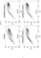
- the proportion of limbal stem cells may be confirmed as a 5,5',6,6'-tetrachloro-1,1',3,3'-tetraethylbenzimidazol-carbocyanine iodide (JC-1) low population
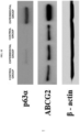
- sternness of the limbal stem cells may be identified by colony forming efficiency (CFE) and western blotting for limbal stem cells-positive markers (p63 ⁇ and ABCG2), but the present invention is not limited thereto.
- Example 2 Culture of limbal stem cells using amniotic membrane slide scaffold
- the limbal tissue was divided into 12 pieces each having a size of approximately 2 mm ⁇ 3 mm using a surgical tool, and the peripheral cornea and conjunctiva were dissected and removed. Afterward, the extracted limbus was put on the amniotic membrane slide scaffold prepared in Example 1 and cultured for approximately 12 days at 37°C in a CO 2 incubator. In the incubation, a culture medium was changed every 2 to 3 days, and when a limbal tissue-derived epithelial cell sheet accounted for 85 to 90% or more of the slide area, it was transplanted into a patient.
- the culture medium was a supplemented human epithelium medium (SHEM) which is conventionally and generally used to culture limbal tissue, and its composition includes EGF, DMSO, ITS, O-phosphoethanolamine, CT and FBS in DMEM:F-12(1:1).
- SHEM human epithelium medium
- Example 3 Comparative analysis of proportions of limbal stem cells on amniotic membrane slide scaffold and transwell
- limbal stem cells grown to 90% or more on an amniotic membrane slide scaffold and a transwell (control group) were treated with 2 mg/ml of a dispase II solution at 4°C overnight.
- the limbal tissue-derived epithelial cell sheet was isolated from the amniotic membrane using fine forceps, treated with 1 ml of trypLE for 5 minutes, and centrifuged at 1000 rpm for 5 minutes, thereby obtaining a cell precipitate.
- the cell precipitate was suspended in SHEM, and then cells were counted to measure a JC-1 low population and CFE, and seeded in a 6-well plate.
- the cells were seeded at 1.0 ⁇ 10 5 cells/ml, cultured in SHEM for 24 hours and treated with a JC-1 dye for 1 hour, followed by measuring a JC-1 low cell population, which are side population cells, using FACS. Meanwhile, to confirm colony formation, the cells were cultured in a CNT50 medium (Cellntec) for 14 days, and the number of colonies was counted, thereby confirming CFE.
- CNT50 medium Cellntec
- Example 5 Comparative analysis of proportions of limbal stem cells in various scaffolds
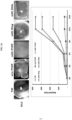
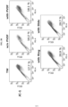
- FIGS. 3A to 3C when the amniotic membrane slide scaffold prepared according to the present invention was used, compared with the control groups, a cell proliferation rate was considerably increased ( FIG. 3A ), and as a result of observing a JC-1 low population and flow cytometry for a limbal stem cell marker (p63 ⁇ (+))( FIGS. 3B and 3C ), it can be seen that the amniotic membrane slide scaffold was most preferable.
- a method for culturing limbal stem cells using an amniotic membrane slide scaffold according to the present invention may allow the proportion of the limbal stem cells in a limbal tissue-derived epithelial cell sheet to be stably and rapidly increased, and thus this method is expected to be useful in treatment of a patient with limbal stem cell deficiency through limbal stem cell transplantation.
Landscapes
- Health & Medical Sciences (AREA)
- Engineering & Computer Science (AREA)
- Life Sciences & Earth Sciences (AREA)
- Biomedical Technology (AREA)
- Chemical & Material Sciences (AREA)
- Zoology (AREA)
- General Health & Medical Sciences (AREA)
- Cell Biology (AREA)
- Wood Science & Technology (AREA)
- Genetics & Genomics (AREA)
- Bioinformatics & Cheminformatics (AREA)
- Biotechnology (AREA)
- Organic Chemistry (AREA)
- Veterinary Medicine (AREA)
- Botany (AREA)
- Animal Behavior & Ethology (AREA)
- Transplantation (AREA)
- Public Health (AREA)
- Oral & Maxillofacial Surgery (AREA)
- Medicinal Chemistry (AREA)
- Urology & Nephrology (AREA)
- Epidemiology (AREA)
- Dermatology (AREA)
- Chemical Kinetics & Catalysis (AREA)
- Microbiology (AREA)
- Biochemistry (AREA)
- General Engineering & Computer Science (AREA)
- Neurology (AREA)
- Neurosurgery (AREA)
- Molecular Biology (AREA)
- Ophthalmology & Optometry (AREA)
- Developmental Biology & Embryology (AREA)
- Hematology (AREA)
- Micro-Organisms Or Cultivation Processes Thereof (AREA)
- Materials For Medical Uses (AREA)
- Medicines Containing Material From Animals Or Micro-Organisms (AREA)
Claims (7)
- Procédé de culture de cellules souches limbiques provenant de tissu limbique, comportant les étapes consistant à :recouvrir une lame de verre d'une membrane amniotique dont les cellules épithéliales ont été retirées pour obtenir un support de lame de verre en vue d'une fixation ; etmettre en culture du tissu limbique sur la membrane amniotique fixée,dans lequel le tissu limbique est mis en culture dans du DMEM/F12 (1:1) complété par du sérum humain ajouté à 4 à 6 % (v/v) du milieu complet, un facteur de croissance des cellules épithéliales (EGF), du sulfoxyde de diméthyle (DMSO), de l'insuline-transferrine-sélénium (ITS) et de l'O-phosphoéthanolamine.
- Procédé selon la revendication 1, dans lequel chaque dimension parmi la largeur et la longueur de la membrane amniotique est de 28 à 35 mm.
- Procédé selon la revendication 1, dans lequel des cellules épithéliales de la membrane amniotique sont retirées en utilisant de l'urée 4 à 6 M.
- Procédé selon la revendication 1, dans lequel chaque dimension parmi la largeur et la longueur de la lame de verre est de 18 à 28 mm.
- Procédé selon la revendication 4, dans lequel le tissu limbique est mis en culture pendant 10 à 14 jours.
- Procédé selon la revendication 5, dans lequel, lorsque le tissu limbique a proliféré sur 85 à 95 % de la surface du support, le tissu limbique est classé comme un transplant destiné à un patient.
- Procédé selon la revendication 1, dans lequel le sérum humain est le sérum AB humain.
Applications Claiming Priority (2)
| Application Number | Priority Date | Filing Date | Title |
|---|---|---|---|
| KR1020150190629A KR101645901B1 (ko) | 2015-12-31 | 2015-12-31 | 양막 슬라이드 지지체를 이용한 윤부줄기세포 배양방법 |
| PCT/KR2016/008003 WO2017115962A1 (fr) | 2015-12-31 | 2016-07-22 | Procédé de culture de cellules souches limbiques à l'aide d'un support formé d'une coupe de membrane amniotique |
Publications (4)
| Publication Number | Publication Date |
|---|---|
| EP3399028A1 EP3399028A1 (fr) | 2018-11-07 |
| EP3399028A4 EP3399028A4 (fr) | 2019-07-24 |
| EP3399028C0 EP3399028C0 (fr) | 2024-10-23 |
| EP3399028B1 true EP3399028B1 (fr) | 2024-10-23 |
Family
ID=56709628
Family Applications (1)
| Application Number | Title | Priority Date | Filing Date |
|---|---|---|---|
| EP16881915.9A Active EP3399028B1 (fr) | 2015-12-31 | 2016-07-22 | Procédé de culture de cellules souches limbiques à l'aide d'un support formé d'une coupe de membrane amniotique |
Country Status (6)
| Country | Link |
|---|---|
| US (1) | US20190010454A1 (fr) |
| EP (1) | EP3399028B1 (fr) |
| JP (2) | JP6703130B2 (fr) |
| KR (1) | KR101645901B1 (fr) |
| CN (1) | CN108699526B (fr) |
| WO (1) | WO2017115962A1 (fr) |
Families Citing this family (1)
| Publication number | Priority date | Publication date | Assignee | Title |
|---|---|---|---|---|
| CN115068504A (zh) * | 2021-03-12 | 2022-09-20 | 广州康睿生物医药科技股份有限公司 | 一种角膜修复材料及其制备方法和用途 |
Family Cites Families (11)
| Publication number | Priority date | Publication date | Assignee | Title |
|---|---|---|---|---|
| EP1456354A4 (fr) * | 2001-11-09 | 2005-11-09 | Artecel Sciences Inc | Cellules stromales derivees d'un tissu adipeux pour la reparation de defauts corneens et inter-orbitaux ainsi que leurs utilisations |
| US20050186672A1 (en) * | 2004-01-27 | 2005-08-25 | Reliance Life Sciences Pvt. Ltd. | Tissue system with undifferentiated stem cells derived from corneal limbus |
| WO2005084119A2 (fr) * | 2004-02-26 | 2005-09-15 | Reliance Life Sciences Pvt. Ltd. | Cellules souches de type embryonnaire multipotentes derivee de limbe, leurs procedes d'isolation et d'utilisation |
| US20060216821A1 (en) * | 2004-02-26 | 2006-09-28 | Reliance Life Sciences Pvt. Ltd. | Pluripotent embryonic-like stem cells derived from corneal limbus, methods of isolation and uses thereof |
| CN1590541A (zh) * | 2004-05-27 | 2005-03-09 | 天津医科大学眼科中心 | 角膜缘干细胞组织工程复合体及其制备方法 |
| NZ570746A (en) * | 2006-02-24 | 2012-02-24 | Reliance Life Sciences Pvt Ltd | Conjunctival tissue system |
| CN102166374B (zh) * | 2010-11-03 | 2013-12-04 | 山东省眼科研究所 | 羊膜复合角膜缘干细胞膜片的制备方法 |
| CN102586175A (zh) * | 2011-09-30 | 2012-07-18 | 上海交通大学附属第一人民医院 | 一种培养人角膜缘干细胞植片的方法 |
| KR20170020527A (ko) * | 2014-06-27 | 2017-02-22 | 더 리젠츠 오브 더 유니버시티 오브 캘리포니아 | 배양된 포유동물 윤부 줄기세포, 이를 생성하는 방법, 및 이의 용도 |
| CN109749997B (zh) * | 2018-05-11 | 2020-03-17 | 中山大学中山眼科中心 | 一种角膜缘干细胞无血清培养基及其培养方法 |
| CN111560348A (zh) * | 2020-07-16 | 2020-08-21 | 北京昱龙盛世生物科技有限公司 | 一种角膜缘上皮干细胞分离培养方法 |
-
2015
- 2015-12-31 KR KR1020150190629A patent/KR101645901B1/ko active Active
-
2016
- 2016-07-22 CN CN201680076831.4A patent/CN108699526B/zh active Active
- 2016-07-22 EP EP16881915.9A patent/EP3399028B1/fr active Active
- 2016-07-22 WO PCT/KR2016/008003 patent/WO2017115962A1/fr not_active Ceased
- 2016-07-22 JP JP2018553030A patent/JP6703130B2/ja active Active
- 2016-07-22 US US16/066,389 patent/US20190010454A1/en active Pending
-
2020
- 2020-03-13 JP JP2020044028A patent/JP2020108393A/ja not_active Withdrawn
Also Published As
| Publication number | Publication date |
|---|---|
| JP6703130B2 (ja) | 2020-06-03 |
| WO2017115962A1 (fr) | 2017-07-06 |
| US20190010454A1 (en) | 2019-01-10 |
| CN108699526B (zh) | 2023-09-29 |
| EP3399028C0 (fr) | 2024-10-23 |
| JP2019501669A (ja) | 2019-01-24 |
| JP2020108393A (ja) | 2020-07-16 |
| EP3399028A4 (fr) | 2019-07-24 |
| KR101645901B1 (ko) | 2016-08-04 |
| CN108699526A (zh) | 2018-10-23 |
| EP3399028A1 (fr) | 2018-11-07 |
Similar Documents
| Publication | Publication Date | Title |
|---|---|---|
| Satake et al. | Long-term outcome of cultivated oral mucosal epithelial sheet transplantation in treatment of total limbal stem cell deficiency | |
| Hirayama et al. | Transplantation of cultivated oral mucosal epithelium prepared in fibrin-coated culture dishes | |
| US7347876B2 (en) | Method for expansion of epithelial stem cells | |
| EP1980274B1 (fr) | Préparation endothéliale cornéenne qui permet aux cellules de croître in vivo | |
| CA2642947A1 (fr) | Systeme de tissu conjonctif | |
| Choe et al. | Ocular surface reconstruction using circumferentially-trephined autologous oral mucosal graft transplantation in limbal stem cell deficiency | |
| US7611895B2 (en) | Method for growth of human conjunctival tissue equivalents for research, clinical ocular surface transplantation and tissue engineering | |
| EP1276431B1 (fr) | Methode de developpement de cellules epitheliales | |
| AU2001253802A1 (en) | Method for expansion of epithelial stem cells | |
| CN115671398A (zh) | 一种3d打印仿生角膜缘移植物及其制备方法和用途 | |
| Parmar et al. | Ocular surface restoration using non-surgical transplantation of tissue-cultured human amniotic epithelial cells | |
| EP3399028A1 (fr) | Procédé de culture de cellules souches limbiques à l'aide d'un support formé d'une coupe de membrane amniotique | |
| CA2637856A1 (fr) | Cellule nourriciere derivant d'une cellule souche de tissu | |
| EP0572364A2 (fr) | Cultures de cellules différenciées de surface épithéliales oculaires, leur méthode de préparation et support à cet usage | |
| Wang et al. | Migration and proliferation of retinal pigment epithelium on extracellular matrix ligands | |
| RU2609253C1 (ru) | Способ лечения глубоких дефектов роговицы | |
| Dekaris et al. | Cultivated Limbal Epithelial Stem Cell | |
| Javadi et al. | Early results of autologous cultivated limbal stem cell transplantation in total limbal stem cell deficiency | |
| Barequet | Induction of Ocular Surface Regeneration | |
| Dobrowolski et al. | Clinical Study Cultivated Oral Mucosa Epithelium in Ocular Surface Reconstruction in Aniridia Patients | |
| Ponzin et al. | The Use of Allograft Corneas and Cells in Ophthalmic Surgery |
Legal Events
| Date | Code | Title | Description |
|---|---|---|---|
| STAA | Information on the status of an ep patent application or granted ep patent |
Free format text: STATUS: THE INTERNATIONAL PUBLICATION HAS BEEN MADE |
|
| PUAI | Public reference made under article 153(3) epc to a published international application that has entered the european phase |
Free format text: ORIGINAL CODE: 0009012 |
|
| STAA | Information on the status of an ep patent application or granted ep patent |
Free format text: STATUS: REQUEST FOR EXAMINATION WAS MADE |
|
| 17P | Request for examination filed |
Effective date: 20180723 |
|
| AK | Designated contracting states |
Kind code of ref document: A1 Designated state(s): AL AT BE BG CH CY CZ DE DK EE ES FI FR GB GR HR HU IE IS IT LI LT LU LV MC MK MT NL NO PL PT RO RS SE SI SK SM TR |
|
| AX | Request for extension of the european patent |
Extension state: BA ME |
|
| DAV | Request for validation of the european patent (deleted) | ||
| DAX | Request for extension of the european patent (deleted) | ||
| A4 | Supplementary search report drawn up and despatched |
Effective date: 20190624 |
|
| RIC1 | Information provided on ipc code assigned before grant |
Ipc: C12N 5/00 20060101ALI20190617BHEP Ipc: C12N 5/0797 20100101ALI20190617BHEP Ipc: C12N 5/079 20100101AFI20190617BHEP Ipc: A61L 27/36 20060101ALI20190617BHEP |
|
| STAA | Information on the status of an ep patent application or granted ep patent |
Free format text: STATUS: EXAMINATION IS IN PROGRESS |
|
| 17Q | First examination report despatched |
Effective date: 20200817 |
|
| REG | Reference to a national code |
Ref country code: DE Ref legal event code: R079 Free format text: PREVIOUS MAIN CLASS: C12N0005079000 Ipc: C12N0005000000 Ref document number: 602016089981 Country of ref document: DE |
|
| GRAP | Despatch of communication of intention to grant a patent |
Free format text: ORIGINAL CODE: EPIDOSNIGR1 |
|
| STAA | Information on the status of an ep patent application or granted ep patent |
Free format text: STATUS: GRANT OF PATENT IS INTENDED |
|
| RIC1 | Information provided on ipc code assigned before grant |
Ipc: A61L 27/36 20060101ALI20240527BHEP Ipc: A61L 27/38 20060101ALI20240527BHEP Ipc: C12N 5/0797 20100101ALI20240527BHEP Ipc: C12N 5/079 20100101ALI20240527BHEP Ipc: C12N 5/00 20060101AFI20240527BHEP |
|
| INTG | Intention to grant announced |
Effective date: 20240624 |
|
| GRAS | Grant fee paid |
Free format text: ORIGINAL CODE: EPIDOSNIGR3 |
|
| GRAA | (expected) grant |
Free format text: ORIGINAL CODE: 0009210 |
|
| STAA | Information on the status of an ep patent application or granted ep patent |
Free format text: STATUS: THE PATENT HAS BEEN GRANTED |
|
| AK | Designated contracting states |
Kind code of ref document: B1 Designated state(s): AL AT BE BG CH CY CZ DE DK EE ES FI FR GB GR HR HU IE IS IT LI LT LU LV MC MK MT NL NO PL PT RO RS SE SI SK SM TR |
|
| REG | Reference to a national code |
Ref country code: GB Ref legal event code: FG4D |
|
| REG | Reference to a national code |
Ref country code: CH Ref legal event code: EP |
|
| REG | Reference to a national code |
Ref country code: DE Ref legal event code: R096 Ref document number: 602016089981 Country of ref document: DE |
|
| REG | Reference to a national code |
Ref country code: IE Ref legal event code: FG4D |
|
| U01 | Request for unitary effect filed |
Effective date: 20241122 |
|
| U07 | Unitary effect registered |
Designated state(s): AT BE BG DE DK EE FI FR IT LT LU LV MT NL PT RO SE SI Effective date: 20241129 |
|
| PG25 | Lapsed in a contracting state [announced via postgrant information from national office to epo] |
Ref country code: IS Free format text: LAPSE BECAUSE OF FAILURE TO SUBMIT A TRANSLATION OF THE DESCRIPTION OR TO PAY THE FEE WITHIN THE PRESCRIBED TIME-LIMIT Effective date: 20250223 Ref country code: HR Free format text: LAPSE BECAUSE OF FAILURE TO SUBMIT A TRANSLATION OF THE DESCRIPTION OR TO PAY THE FEE WITHIN THE PRESCRIBED TIME-LIMIT Effective date: 20241023 |
|
| PG25 | Lapsed in a contracting state [announced via postgrant information from national office to epo] |
Ref country code: ES Free format text: LAPSE BECAUSE OF FAILURE TO SUBMIT A TRANSLATION OF THE DESCRIPTION OR TO PAY THE FEE WITHIN THE PRESCRIBED TIME-LIMIT Effective date: 20241023 |
|
| PG25 | Lapsed in a contracting state [announced via postgrant information from national office to epo] |
Ref country code: NO Free format text: LAPSE BECAUSE OF FAILURE TO SUBMIT A TRANSLATION OF THE DESCRIPTION OR TO PAY THE FEE WITHIN THE PRESCRIBED TIME-LIMIT Effective date: 20250123 |
|
| PG25 | Lapsed in a contracting state [announced via postgrant information from national office to epo] |
Ref country code: GR Free format text: LAPSE BECAUSE OF FAILURE TO SUBMIT A TRANSLATION OF THE DESCRIPTION OR TO PAY THE FEE WITHIN THE PRESCRIBED TIME-LIMIT Effective date: 20250124 |
|
| PG25 | Lapsed in a contracting state [announced via postgrant information from national office to epo] |
Ref country code: PL Free format text: LAPSE BECAUSE OF FAILURE TO SUBMIT A TRANSLATION OF THE DESCRIPTION OR TO PAY THE FEE WITHIN THE PRESCRIBED TIME-LIMIT Effective date: 20241023 |
|
| PG25 | Lapsed in a contracting state [announced via postgrant information from national office to epo] |
Ref country code: RS Free format text: LAPSE BECAUSE OF FAILURE TO SUBMIT A TRANSLATION OF THE DESCRIPTION OR TO PAY THE FEE WITHIN THE PRESCRIBED TIME-LIMIT Effective date: 20250123 |
|
| PG25 | Lapsed in a contracting state [announced via postgrant information from national office to epo] |
Ref country code: SM Free format text: LAPSE BECAUSE OF FAILURE TO SUBMIT A TRANSLATION OF THE DESCRIPTION OR TO PAY THE FEE WITHIN THE PRESCRIBED TIME-LIMIT Effective date: 20241023 |
|
| PG25 | Lapsed in a contracting state [announced via postgrant information from national office to epo] |
Ref country code: SK Free format text: LAPSE BECAUSE OF FAILURE TO SUBMIT A TRANSLATION OF THE DESCRIPTION OR TO PAY THE FEE WITHIN THE PRESCRIBED TIME-LIMIT Effective date: 20241023 |
|
| PG25 | Lapsed in a contracting state [announced via postgrant information from national office to epo] |
Ref country code: CZ Free format text: LAPSE BECAUSE OF FAILURE TO SUBMIT A TRANSLATION OF THE DESCRIPTION OR TO PAY THE FEE WITHIN THE PRESCRIBED TIME-LIMIT Effective date: 20241023 |
|
| PLBE | No opposition filed within time limit |
Free format text: ORIGINAL CODE: 0009261 |
|
| STAA | Information on the status of an ep patent application or granted ep patent |
Free format text: STATUS: NO OPPOSITION FILED WITHIN TIME LIMIT |
|
| U20 | Renewal fee for the european patent with unitary effect paid |
Year of fee payment: 10 Effective date: 20250729 |
|
| 26N | No opposition filed |
Effective date: 20250724 |
|
| PGFP | Annual fee paid to national office [announced via postgrant information from national office to epo] |
Ref country code: GB Payment date: 20250724 Year of fee payment: 10 |
|
| REG | Reference to a national code |
Ref country code: CH Ref legal event code: H13 Free format text: ST27 STATUS EVENT CODE: U-0-0-H10-H13 (AS PROVIDED BY THE NATIONAL OFFICE) Effective date: 20260224 |
|
| PG25 | Lapsed in a contracting state [announced via postgrant information from national office to epo] |
Ref country code: CH Free format text: LAPSE BECAUSE OF NON-PAYMENT OF DUE FEES Effective date: 20250731 |