EP3429543B1 - Dispositifs d'analyse biologique à ventilation sélective et procédés associés - Google Patents
Dispositifs d'analyse biologique à ventilation sélective et procédés associés Download PDFInfo
- Publication number
- EP3429543B1 EP3429543B1 EP17767339.9A EP17767339A EP3429543B1 EP 3429543 B1 EP3429543 B1 EP 3429543B1 EP 17767339 A EP17767339 A EP 17767339A EP 3429543 B1 EP3429543 B1 EP 3429543B1
- Authority
- EP
- European Patent Office
- Prior art keywords
- reaction
- sample
- amplification
- selective venting
- reaction chambers
- Prior art date
- Legal status (The legal status is an assumption and is not a legal conclusion. Google has not performed a legal analysis and makes no representation as to the accuracy of the status listed.)
- Active
Links
Images


Classifications
-
- B—PERFORMING OPERATIONS; TRANSPORTING
- B01—PHYSICAL OR CHEMICAL PROCESSES OR APPARATUS IN GENERAL
- B01L—CHEMICAL OR PHYSICAL LABORATORY APPARATUS FOR GENERAL USE
- B01L3/00—Containers or dishes for laboratory use, e.g. laboratory glassware; Droppers
- B01L3/50—Containers for the purpose of retaining a material to be analysed, e.g. test tubes
- B01L3/502—Containers for the purpose of retaining a material to be analysed, e.g. test tubes with fluid transport, e.g. in multi-compartment structures
- B01L3/5027—Containers for the purpose of retaining a material to be analysed, e.g. test tubes with fluid transport, e.g. in multi-compartment structures by integrated microfluidic structures, i.e. dimensions of channels and chambers are such that surface tension forces are important, e.g. lab-on-a-chip
- B01L3/502738—Containers for the purpose of retaining a material to be analysed, e.g. test tubes with fluid transport, e.g. in multi-compartment structures by integrated microfluidic structures, i.e. dimensions of channels and chambers are such that surface tension forces are important, e.g. lab-on-a-chip characterised by integrated valves
-
- A—HUMAN NECESSITIES
- A61—MEDICAL OR VETERINARY SCIENCE; HYGIENE
- A61J—CONTAINERS SPECIALLY ADAPTED FOR MEDICAL OR PHARMACEUTICAL PURPOSES; DEVICES OR METHODS SPECIALLY ADAPTED FOR BRINGING PHARMACEUTICAL PRODUCTS INTO PARTICULAR PHYSICAL OR ADMINISTERING FORMS; DEVICES FOR ADMINISTERING FOOD OR MEDICINES ORALLY; BABY COMFORTERS; DEVICES FOR RECEIVING SPITTLE
- A61J1/00—Containers specially adapted for medical or pharmaceutical purposes
- A61J1/14—Details; Accessories therefor
-
- B—PERFORMING OPERATIONS; TRANSPORTING
- B01—PHYSICAL OR CHEMICAL PROCESSES OR APPARATUS IN GENERAL
- B01L—CHEMICAL OR PHYSICAL LABORATORY APPARATUS FOR GENERAL USE
- B01L7/00—Heating or cooling apparatus; Heat insulating devices
- B01L7/52—Heating or cooling apparatus; Heat insulating devices with provision for submitting samples to a predetermined sequence of different temperatures, e.g. for treating nucleic acid samples
-
- G—PHYSICS
- G01—MEASURING; TESTING
- G01J—MEASUREMENT OF INTENSITY, VELOCITY, SPECTRAL CONTENT, POLARISATION, PHASE OR PULSE CHARACTERISTICS OF INFRARED, VISIBLE OR ULTRAVIOLET LIGHT; COLORIMETRY; RADIATION PYROMETRY
- G01J3/00—Spectrometry; Spectrophotometry; Monochromators; Measuring colours
- G01J3/46—Measurement of colour; Colour measuring devices, e.g. colorimeters
- G01J3/52—Measurement of colour; Colour measuring devices, e.g. colorimeters using colour charts
- G01J3/524—Calibration of colorimeters
-
- B—PERFORMING OPERATIONS; TRANSPORTING
- B01—PHYSICAL OR CHEMICAL PROCESSES OR APPARATUS IN GENERAL
- B01L—CHEMICAL OR PHYSICAL LABORATORY APPARATUS FOR GENERAL USE
- B01L2200/00—Solutions for specific problems relating to chemical or physical laboratory apparatus
- B01L2200/14—Process control and prevention of errors
- B01L2200/141—Preventing contamination, tampering
-
- B—PERFORMING OPERATIONS; TRANSPORTING
- B01—PHYSICAL OR CHEMICAL PROCESSES OR APPARATUS IN GENERAL
- B01L—CHEMICAL OR PHYSICAL LABORATORY APPARATUS FOR GENERAL USE
- B01L2300/00—Additional constructional details
- B01L2300/04—Closures and closing means
- B01L2300/046—Function or devices integrated in the closure
- B01L2300/048—Function or devices integrated in the closure enabling gas exchange, e.g. vents
-
- B—PERFORMING OPERATIONS; TRANSPORTING
- B01—PHYSICAL OR CHEMICAL PROCESSES OR APPARATUS IN GENERAL
- B01L—CHEMICAL OR PHYSICAL LABORATORY APPARATUS FOR GENERAL USE
- B01L2300/00—Additional constructional details
- B01L2300/04—Closures and closing means
- B01L2300/046—Function or devices integrated in the closure
- B01L2300/049—Valves integrated in closure
-
- B—PERFORMING OPERATIONS; TRANSPORTING
- B01—PHYSICAL OR CHEMICAL PROCESSES OR APPARATUS IN GENERAL
- B01L—CHEMICAL OR PHYSICAL LABORATORY APPARATUS FOR GENERAL USE
- B01L2300/00—Additional constructional details
- B01L2300/08—Geometry, shape and general structure
- B01L2300/0861—Configuration of multiple channels and/or chambers in a single devices
- B01L2300/0864—Configuration of multiple channels and/or chambers in a single devices comprising only one inlet and multiple receiving wells, e.g. for separation, splitting
-
- B—PERFORMING OPERATIONS; TRANSPORTING
- B01—PHYSICAL OR CHEMICAL PROCESSES OR APPARATUS IN GENERAL
- B01L—CHEMICAL OR PHYSICAL LABORATORY APPARATUS FOR GENERAL USE
- B01L2300/00—Additional constructional details
- B01L2300/18—Means for temperature control
- B01L2300/1805—Conductive heating, heat from thermostatted solids is conducted to receptacles, e.g. heating plates, blocks
- B01L2300/1822—Conductive heating, heat from thermostatted solids is conducted to receptacles, e.g. heating plates, blocks using Peltier elements
-
- B—PERFORMING OPERATIONS; TRANSPORTING
- B01—PHYSICAL OR CHEMICAL PROCESSES OR APPARATUS IN GENERAL
- B01L—CHEMICAL OR PHYSICAL LABORATORY APPARATUS FOR GENERAL USE
- B01L2300/00—Additional constructional details
- B01L2300/18—Means for temperature control
- B01L2300/1805—Conductive heating, heat from thermostatted solids is conducted to receptacles, e.g. heating plates, blocks
- B01L2300/1827—Conductive heating, heat from thermostatted solids is conducted to receptacles, e.g. heating plates, blocks using resistive heater
-
- B—PERFORMING OPERATIONS; TRANSPORTING
- B01—PHYSICAL OR CHEMICAL PROCESSES OR APPARATUS IN GENERAL
- B01L—CHEMICAL OR PHYSICAL LABORATORY APPARATUS FOR GENERAL USE
- B01L2400/00—Moving or stopping fluids
- B01L2400/04—Moving fluids with specific forces or mechanical means
- B01L2400/0475—Moving fluids with specific forces or mechanical means specific mechanical means and fluid pressure
- B01L2400/0487—Moving fluids with specific forces or mechanical means specific mechanical means and fluid pressure fluid pressure, pneumatics
- B01L2400/049—Moving fluids with specific forces or mechanical means specific mechanical means and fluid pressure fluid pressure, pneumatics vacuum
-
- B—PERFORMING OPERATIONS; TRANSPORTING
- B01—PHYSICAL OR CHEMICAL PROCESSES OR APPARATUS IN GENERAL
- B01L—CHEMICAL OR PHYSICAL LABORATORY APPARATUS FOR GENERAL USE
- B01L2400/00—Moving or stopping fluids
- B01L2400/06—Valves, specific forms thereof
- B01L2400/0694—Valves, specific forms thereof vents used to stop and induce flow, backpressure valves
Definitions
- Document US 2004/052689 A1 discloses a self-sealing material comprising a hydrogel adhered to pore walls of a porous substrate, this self-sealing material may be implemented e.g. to form a self-sealing pipette tip, or a vent in a cap for a bottle.
- Document US 2014/188089 A1 discloses a device for covering or venting at least a portion of a medical device, the device comprising a porous cap or a porous vent comprising a sintered porous media, wherein the sintered porous plastic cap has a self-sealing capability, thus allowing gas to pass through while blocking aqueous solutions from passing through.
- Document US 2009/071911 A1 discloses an extracorporeal medical fluid circuit component, comprising: a vent assembly comprising: a micro-porous membrane; and a vent structure adjacent to the micro-porous membrane, wherein the vent structure comprises a porous material capable of swelling when moistened.
- Document US 2013/130232 A1 discloses a self-loading microfluidic device comprising a porous organic polymer and a reaction well, an inlet port, a vacuum well, a main channel, and a side channel.
- the methods include contacting a sample liquid with the selective venting element and thereby making the selective venting element impermeable to fluid. Also, where desired, the methods include reacting the sample with the modifying reagent and generating a reaction product. Furthermore, in some instances, the methods include detecting a characteristic of the reaction product, wherein such detection can be performed by an un-assisted human eye.
- colorimetry and “colorimetric” refers to techniques of quantifying or otherwise observing colored compound concentrations in solution.
- Colorimetric detection refers to any method of detecting such colored compounds and/or the change in color of the compounds in solution. Methods can include visual observation, absorbance measurements, or fluorescence measurements, among others.
- sufficient amount means an amount sufficient to produce a desired effect, e.g., an amount sufficient to modulate protein aggregation in a cell.
- the phrase "passively tunable porosity,” as used herein, refers to the ability of having a first conformation in which one or more gasses, e.g., air, can pass therethrough, e.g., through pores, and a second conformation in which fluids including the one or more gasses and liquids, such as liquids including a biological sample, are prevented from passing therethrough, e.g., through the pores, and proceeding automatically from the first to the second conformation upon contact with a liquid. Also, in the second conformation, the selective venting elements prevent evaporation of the liquids therethrough, e.g., through the pores.
- one or more gasses e.g., air
- fluids including the one or more gasses and liquids such as liquids including a biological sample
- a thickness of a selective venting element body can also range for example, from 10 cm to 50 microns, such as 5 cm to 50 microns, such as 2 cm to 50 microns, from 1 cm to 50 microns, such as 5 mm to 50 microns, or from 5 mm to .1 mm, such as 2 mm to .1 mm, inclusive.
- “inclusive” refers to a provided range including each of the listed numbers. Unless otherwise indicated, all provided ranges are inclusive.
- a length and/or width of a body can also range from 1 mm to 40 cm, such as from 1 cm to 30 m, such as from 1 cm to 10 cm, such as from 1 cm to 5 cm, or from 1 mm to 5 cm, from 1 mm to 3 cm, from 1 mm to 1 cm or from 1 mm to 5 mm.
- a body of a selective venting element can have three edges, four edges, or more than four edges which define the area of the body. In various embodiments, the edges meet at corners, e.g., three, four, five, or ten or more corners.
- a first edge of an adhesive layer is opposite a second edge of an adhesive layer and adjacent to a third and/or fourth edge of an adhesive layer. In such an embodiment, the third edge can be opposite a fourth edge and the fourth edge can be adjacent to the first and/or second edge.
- a selective venting element includes only a body and does not include protrusions extending therefrom.
- a selective venting element can include 20000 or less, 15000 or less, 10000 or less, 5000 or less, 1000 or less, 100 or less, 50 or less, such as 20 or less, such as 15 or less, such as 10 or less, such as 5 or less protrusions.
- a selective venting element can include from 1 to 15000, 1 to 10000, 1 to 5000, 1 to 1000, 1 to 25, such as from 1 to 20, such as from 1 to 15, such as from 1 to 10 such as from 1 to 5, protrusions, or from 2 to 20, such as from 2 to 15, such as from 5 to 15 protrusions, wherein each range is inclusive.
- a selective venting element can include 1, 2, 3, 4, 5, 6, 7, 8, 9, 10, 11, 12, 13, 14, 15, 16, 17, 18, 19, or 20 or more protrusions.
- a selective venting element of a device can have a number of protrusions equal to the number of reaction chambers in the device.
- protrusions are shaped as a cylinder, they can have a height, e.g., a distance from a surface of a venting element body to a sealing surface at an end of the protrusion, ranging from .1 mm to 5 cm, such as .1 mm to 1 cm, such as .1 mm to 5 mm, such as .1 mm to 1 mm, or 1 mm to 5 mm, inclusive.
- a protrusion can also have a height of 15 cm or less, 10 cm or less, 5 cm or less, such as 3 cm or less, such as 1 cm or less, such as 5 mm or less, such as 3 mm or less, such as 1 mm or less.
- a protrusion can also have a height of .1 mm o more, such as 1 mm or more, such as 3 mm or more, such as 5 mm or more, such as 1 cm or more, such as 3 cm or more, such as 5 cm or more, such as 10 cm or more.
- Such a protrusion can also have a diameter ranging from .1 mm to 10 cm, such as .1 mm to 5 cm, such as .1 mm to 3 cm, such as .1 mm to 1 cm, such as .1 mm to 5 mm, such as .1 mm to 1 mm, or 1 mm to 1 cm, or 1 cm to 3 cm, each inclusive.
- Protrusions can also have a diameter of 5 cm or less, such as 3 cm or less, such as 1 cm or less, such as 5 mm or less, such as 3 mm or less, such as 1 mm or less, such as .5 mm or less.
- a protrusion can also have a diameter of .1 mm or more, such as 1 mm or more, such as 3 mm or more, such as 5 mm or more, such as 1 cm or more.
- the optical property modifying reagent is or includes an enzyme-linked immunosorbent assay (ELISA) reagent.
- ELISA enzyme-linked immunosorbent assay
- the ELISA reagent is selected from the group consisting of alkaline phosphatase, horseradish peroxidase, ⁇ -galactosidase, BCIP/NBT (5-bromo-4-chloro-3-indolyl-phosphate/nitrobluetetrazolium), TMB (3,3',5,5' tetramethylbenzidine), DAB (3,3',4,4' diaminobenzidine), 4CN (4-chloro-1-naphthol).
- performing an optical property modification includes changing the pH of reaction chamber contents by performing a reaction.
- An optical property modifying reagent can produce a modification based on the location and extent of such a pH change.
- Each reaction chamber can include, such as contain within a chamber, one or more nucleic acid amplification composition.
- nucleic acid amplification composition can include, for example, one or more primers, deoxynucleotides (dNTPs), and/or polymerases, Trizma pre-set crystals (Tris buffer, pH 8.8; Sigma, cat. no. T9443), Potassium chloride (KCl; Wako Pure Chemicals, cat. no. 163-03545), Magnesium sulfate heptahydrate (MgSO4; Wako Pure Chemicals, cat. no. 137-00402), Ammonium sulfate ((NH4)2SO4; Kanto Chemical, cat. no.
- a nucleic acid amplification composition can be stored in a sample receiving cartridge in dry, e.g., lyophilized, form.
- moving a biological sample e.g., a fluid biological sample, into a reaction chamber can include mixing the biological sample and the nucleic acid amplification composition and/or hydrating the nucleic acid amplification composition.
- the amplification according to the subject embodiments is reverse transcriptase loop-mediated amplification (RT-LAMP).
- RT-LAMP is an isothermal gene amplification procedure in which the reaction can be processed at a constant temperature, e.g., 63°C, by one type of enzyme, e.g., Bst polymerase, in a single step.
- two enzymes are used with a reverse transcriptase and/or a polymerase, e.g., Bst polymerase, e.g., BST 2.0.
- RT-LAMP in various aspects, uses six primers that recognize eight regions on a target nucleic acid.
- the sensitivities and specificities of the RT-LAMP technique is higher than those associated with performing a polymerase chain reaction (PCR).
- the RT-LAMP method is also fast, producing a signal from a few copies of RNA or DNA in 60 minutes, or less, 45 minutes or less, 30 minutes or less, or 15 minutes or less.
- RT-LAMP can also not require any special reagents.
- a "detection" according to the subject embodiments is a detection of one or more aspects, such as specific pathogenic genetic markers in samples. Amplification according to the subject embodiments can also be performed by applying PCR.
- the sample receiving cartridges also include one or more conduits operatively, e.g., fluidically, connecting each or any combination of the one or more reaction chambers with one another and/or with a sample inlet.
- Each of the one or more conduits can be shaped as a cylinder or a quadrilateral prism and can have dimensions including a length of 10 m or less, such as 1 m or less, such as 10 cm or less, such as 1mm or less, and/or have a diameter, width and/or height of 100 mm or less, such as 10 mm or less, such as 1mm or less, such as .1 mm or less, such as 10 micrometers or less.
- the sample receiving cartridges or portions thereof, e.g., substrates are composed of one or more materials including, for example, polymeric materials (e.g., materials having one or more polymers including, for example, plastic and/or rubber) and/or metallic materials.
- any of the device components including sample receiving cartridges or portions thereof described herein can be composed include, but are not limited to: polymeric materials, e.g., plastics, such as polytetrafluoroethene or polytetrafluoroethylene (PFTE), including expanded polytetrafluoroethylene (e-PFTE), polyethylene, polyester (DacronTM), nylon, polypropylene, polyethylene, high-density polyethylene (HDPE), polyurethane, polydimethylsiloxane (PDMS), etc., metals and metal alloys, e.g., titanium, chromium, aluminum, stainless steel, etc., and the like.
- the materials are transparent materials and as such, allow light within the visible spectrum to efficiently pass therethrough.
- the subject devices include a substrate.
- a substrate can be operatively coupled to the sample receiving cartridge via, for example, an adhesive layer.
- the substrate in some instances, can be a circuit board, e.g., a printed circuit board, composed, for example, of a layer of Silicon and/or Copper and/or Gold and/or Aluminum contacts therein or thereon.
- Substrates can be printed circuit boards composed, for example, of a layer, e.g., a silicon layer, having thereon metallic contacts affixed thereto with one or more adhesive, e.g., epoxy.
- the substrates can include one or more heating elements.
- Heating elements are elements and/or one or more reactants that are configured to generate thermal energy and can be proximate to one or more reaction chambers. By “proximate” is meant close to.
- heating elements may be configured for heating one or more reaction chambers and contents thereof, e.g., a biological sample and/or an optical property modifying reagent and/or a nucleic acid amplification composition.
- thermoelectric heating elements e.g., thermoelectric heating elements that include resistive conductors, e.g., thermistors, Peltier devices, or other elements that generate heat.
- heating elements are or include one or more heat-generating reactants, e.g., liquid reactants, that cause an exothermic/exothermal reaction when exposed to one another or one or more of the compositions and/or reagents disclosed herein, e.g., water.
- the methods include adding to contents of a device as disclosed herein, e.g., contents including a biological sample, one or more heating reagents which, when mixed, cause an exothermal reaction.
- a reaction can, for example, heat a sample for lysis or produce a colorimetric change as described herein.
- Exothermal reactions can generate heat and/or gas.
- Exothermal reactions can include the hydration of a mixture composed of encapsulated and/or non-encapsulated oxides such as calcium oxide and/or magnesium oxide and dehydrated and/or hydrated zeolite, or any combinations thereof.
- a process can be coupled with control of pH of the mixture through compounds such as Citric acid, or combination exothermic mixes, such as Cao and Mg-Fe.
- Modulation can include timed/controlled release from encapsulated reactants and can include particles with tailored size distribution and different burn characteristics.
- Phase change materials PCM
- PCMs include, for example, organics (paraffins, non paraffins and fatty acids) and inorganics (salt hydrates).
- the reagents applied in exothermal reactions or other gas-producing reagents may also be applied to produce gas inside one or more of the chambers, e.g., sealed chambers, of the devices disclosed herein and thereby increase pressure in the one or more container.
- Such a temperature can be maintained at, for example, 59 °C, 60 °C, 61 °C, 62 °C, 63 °C, 64 °C, 65 °C, 66 °C, or 67 °C and/or within a range from 50-75 °C, such as 60-70 °C, such as 60-66 °C. Maintaining such a temperature can be performed by applying a thermistor as a heating sensing element and/or can be based on sensor feedback to a control unit.
- Heating elements can be configured to elevate the temperature of a reaction chamber and/or contents thereof, repeatedly, e.g., heat the contents a first time and then a second time.
- the subject heating elements also can heat the contents of a reaction chamber so that an optical property modification and/or nucleic acid amplification occurs.
- the subject heating elements also can heat contents to perform thermo-cycling for amplification reactions, such as PCR.
- the subject substrates include one or more power sources.
- a power source can be operatively connected to one or more heating elements.
- power source is meant a device that supplies electric power to an electrical load.
- power sources can include, for example, one or more battery, direct current (DC) power supply, alternating current (AC) power supply, linear regulated power supply, switched-mode power supply, programmable power supply, uninterruptible power supply, high-voltage power supply and/or a voltage multiplier.
- a power source can, in some aspects, be one or more battery, e.g., a portable and/or self-contained and/or replaceable battery, such as one or two AA batteries, an outlet, or another source of electrical power.
- a power source can include one or more electrical cords, e.g., cords configured to operatively connect a device to an outlet. Cords of power sources can be configured to removably connect to a device and/or an outlet.
- Versions of power sources include power sources configured to turn on to provide electrical power to another component and/or turn off to stop providing electrical power to another component.
- Such power sources can be configured to be turned on and/or off, for example, by operation of a switch, button, timer or other component operatively connected to or included in the power source, such as a control unit.
- Power sources can also, in certain aspects, be operatively connected to one or more components of the disclosed systems, e.g., a control unit.
- embodiments of power sources include electrical connections from a power source to components of the disclosed systems.
- Such electrical connections can include one or more lengths of electrically conductive material, e.g., contacts, traces, and/or wires.
- Substrates can include one or more control unit, e.g., a central processing unit (CPU) or a field-programmable gate array (FPGA).
- a unit can include a memory and/or a processor, e.g., a microprocessor, configured to generate one or more outputs, e.g., electrical signals, based on one or more sets of inputs, e.g., inputs from a user and/or a sensor, and/or a timer, and/or instructions stored in the memory.
- a device can also include a user interface for receiving an input and operatively coupled to the control unit.
- a control unit is configured to perform an optical property modification and/or colorimetric analysis of a biological sample in the one or more reaction chambers.
- a control unit can be configured to determine, based on an input from one or more sensors, whether a change in an optical property, e.g., color, of one or more contents of a reaction chamber, has occurred. Based on the determination, the control unit can be configured to generate an output, such as an output to a user via a display, wherein the output reflects to the user whether a change has occurred.
- substrates according to the subject embodiments include one or more light source configured to emit light.
- Such light sources can be operatively coupled to the one or more sensors and/or control units such that when a sensor detects a liquid, e.g., a biological sample, in the one or more reaction chambers, the light source emits light.
- Such light sources can also be operatively coupled to the one or more sensors and/or control units such that when an optical property modification occurs or does not occur in the one or more reaction chambers, the light source emits light.
- Light sources according to the subject embodiments can also include one or more light emitting diode (LED).
- LED light emitting diode
- the subject devices can also include a housing.
- a housing can include a first portion and a second portion operatively coupleable, e.g., mateable, e.g., snapedly coupleable, with the first portion to encapsulate the sample receiving cartridge, substrate and adhesive layer.
- a second portion is substantially flat and a first portion is composed of five walls separated by edges and configured to contain, e.g., fully contain, one or more other components of a device, such as by retaining the components between at least two portions, e.g., opposite walls, thereof.
- a second portion makes up a bottom surface of the housing and the housing includes an inlet opening in a top surface of the housing opposite the bottom surface.
- An adhesive layer and/or a portion thereof e.g., a sheet and/or an adhesive material can define an end of a reaction chamber and/or sealably contain one or more solid and/or fluid media, e.g., a biological sample and/or a modifying reagent and/or an amplification composition within the reaction chamber.
- an adhesive layer can be operatively coupled to a sample receiving cartridge such that the adhesive layer fluidically seals one or more openings, e.g., an opening at an end, of one or more reaction chambers of the cartridge.
- a sheet can have a length, a width and a height, also referred to as a thickness.
- a sheet can be shaped as a rectangular box with the width and length being substantially greater than the thickness.
- a thickness of an adhesive layer and/or a sheet e.g., a thickness between a first surface and a second surface opposite the first surface, can be 5 mm or less, 3 mm or less, 1 mm or less, .5 mm or less, 0.1 mm or less, or 50 microns or less.
- a thickness of an adhesive layer and/or a sheet thereof can also range for example, from 5 mm to 50 microns, such as 3 mm to .1 mm, such as 1 mm to .1 mm, inclusive.
- a length and/or width of an adhesive layer and/or a sheet can also range from 1 mm to 2 m, such as from 1 cm to 1 m, such as from 1 cm to 10 cm, such as from 1 cm to 5 cm.
- Adhesive layers can each be composed of a variety of materials and can be composed of the same or different materials.
- the sample receiving modules and/or caps or portions thereof can be composed of polymeric materials, e.g., materials having one or more polymers including, for example, plastic and/or rubber.
- Such materials can have characteristics of flexibility and/or high strength (e.g., resistant to wear) and/or high fatigue resistance (e.g., able to retain its physical properties for long periods of time regardless of the amount of use or environment).
- materials of interest of which adhesive layers or portions thereof described herein can be composed include, but are not limited to: polymeric materials, e.g., plastics, such as polytetrafluoroethene or polytetrafluoroethylene (PFTE), including expanded polytetrafluoroethylene (e-PFTE), polyester (DacronTM), nylon, polypropylene, polyethylene, high-density polyethylene (HDPE), polyurethane, one or more acrylic adhesive, silicone adhesive, epoxy adhesive, or any combination thereof.
- PFTE polytetrafluoroethene or polytetrafluoroethylene
- e-PFTE expanded polytetrafluoroethylene
- HDPE high-density polyethylene
- each of such materials can include coatings or layers of adhesive materials, e.g., acrylic adhesive materials, on one or more surface thereof.
- each of such materials can include coatings or layers of adhesive materials, e.g., acrylic adhesive materials, on one or more surface thereof.
- an adhesive layer, or a portion thereof, such as a first and/or second laminated layer does not include an acid.
- an adhesive layer, or a portion thereof, e.g., such as a first and/or second laminated layer is opaque and/or white. Where an adhesive layer or a portion thereof is white, the white layer provides a uniform background of visual inspection of one or more reaction chambers.
- a layer, e.g., a first layer and/or second layer and/or an adhesive layer is opaque and/or a color complementary to a reaction start color, e.g., red, orange, yellow, green, blue, indigo, violet, black, gold, silver, brown, or any combination thereof.
- an adhesive layer, or a portion thereof is transparent to light, e.g., visible light.
- an adhesive layer, or a portion thereof is reflective, e.g., entirely or substantially reflective to light, e.g., visible light.
- an adhesive layer can include a first layer laminated with a second layer. In such embodiments, for example, a first layer does not include an acid and/or a second layer is opaque and/or white.
- a biological sample can be collected from a subject and include one or more cells, such as tissue cells of the subject.
- tissue refers to one or more aggregates of cells in a subject (e.g., a living organism, such as a mammal, such as a human) that have a similar function and structure or to a plurality of different types of such aggregates.
- Tissue can include, for example, organ tissue, muscle tissue (e.g., cardiac muscle; smooth muscle; and/or skeletal muscle), connective tissue, nervous tissue and/or epithelial tissue.
- Tissue can, in some versions, include cells from the inside of a subject's cheek and/or cells in a subject's saliva.
- a biological sample can be collected from a subject.
- a subject is a "mammal” or a “mammalian” subject, where these terms are used broadly to describe organisms which are within the class mammalia, including the orders carnivore (e.g., dogs and cats), rodentia (e.g., mice, guinea pigs, and rats), and primates (e.g., humans, chimpanzees, and monkeys).
- the subject is a human.
- humans can include human subjects of both genders and at any stage of development (e.g., fetal, neonates, infant, juvenile, adolescent, and adult), where in certain embodiments the human subject is a juvenile, adolescent or adult. While the devices and methods described herein can be applied in association with a human subject, it is to be understood that the subject devices and methods can also be applied in association with other subjects, that is, on “non-human subjects.”
- a biological sample is an environmental and/or agricultural sample.
- biological samples are collected from one or more farming aspects, e.g., animals and/or plants, and/or from natural organic sources such as organisms, such as animals, e.g., wild animals, plants, e.g., wild plants, bacteria, viruses, or any combination thereof.
- a biological sample can in some versions be a prepared biological sample.
- a prepared biological assay sample is a biological assay sample which has been processed for example by exposing the sample to a preparation solution, such as a solution including a lysing agent, such as a detergent. Accordingly, in some embodiments, a biological sample is a lysate.
- a preparation solution such as a solution including a lysing agent, such as a detergent.
- a biological sample is a lysate.
- Such preparation can enable the prepared biological sample to react, for example, with an amplification composition and/or an optical property modifying reagent upon exposure thereto.
- the exposure can include lysing cells of the sample with a lysing agent of the preparation solution and/or extracting nucleic acids therefrom. Such extracted nucleic acids can be released into a resulting prepared sample solution.
- a step of extracting genomic deoxyribonucleic acid (DNA) from a biological sample is included.
- the preparation solution is a nucleic acid amplification preparation solution and exposure to the solution prepares nucleic acids of the sample for amplification, e.g., isothermal amplification.
- optical property modifying devices can also include one or more code, e.g., a quick response (QR) code, on an exterior of a housing thereof.
- QR quick response
- Such a code can include an identification of assay type, expiration date for the device, serial number, or any combination thereof.
- a sample analyzer can be configured to read and/or recognize such a code so that a proper identification of the device can be made and the device used accordingly.
- the methods include introducing a biological sample into a biological assay device by flowing the sample into one or more reaction chambers of a sample receiving cartridge of the device via one or more sample receiving openings.
- a sample can first be introduced to an inlet operatively coupled to the one or more reaction chambers by contacting the sample with the inlet and then flowed from the inlet into the reaction chambers.
- flowing the sample into the one or more reaction includes flowing a gas through a selective venting element of the device and/or contacting the sample with one or more modifying reagent in a reaction chamber.
- a selective venting element can form a wall of each of the one or more reaction chambers, and the one or more reaction chambers can each include a modifying reagent.
- the methods include contacting the sample liquid with the selective venting element and thereby making the selective venting element impermeable to fluid.
- the methods include contacting the sample liquid with the selective venting element and thereby advancing the selective venting element from a first conformation to a second conformation, as described herein.
- the methods include flowing an amount, e.g., a small amount, of a liquid, e.g., biological sample, water and/or buffer, into a selective venting elements, or a portion thereof, e.g., a sealing surface, by contacting the element with the liquid.
- the presence of the liquid within the element seals pores of the element and/or expands the element so that further liquid and/or gas cannot pass into or through the element.
- the methods include sealing the selective venting element and preventing further passage of liquid or gas, such as by evaporation, into or through the element.
- the methods also include reacting the sample with the modifying reagent and generating a reaction product.
- a reaction product can include, for example, one or more compositions, e.g., an aspect of a biological sample, e.g., protons, which, when reacted with an optical property modifying reagent, result in a modification of one or more optical property.
- the methods also include detecting a characteristic of the reaction product, wherein such detection can be performed by an un-assisted human eye.
- human can include human users or subjects of both genders and at any stage of development, e.g., fetal, neonates, infant, juvenile, adolescent, adult, where in certain embodiments the human subject or user is a juvenile, adolescent or adult.
- an un-assisted human eye refers to a human eye that is not enhanced by one or more devices which enhance or modify visual ability. Such devices might include a camera, optical magnifier, microscope, or optimized, e.g., filtered, e.g., polarized, glasses or contacts, etc.
- detecting a characteristic of the reaction product can include visually inspecting the one or more reaction chambers to detect a modified optical property. Also, in some aspects, detecting a characteristic of the reaction product includes detecting presence or absence of a nucleic acid amplification.
- the methods also can include modifying an optical property in a biological sample assay.
- modifying an optical property in a biological sample assay can be performed on a biological sample, or an aspect associated therewith, such as a reaction mixture or a reaction product.
- a modification of an optical property can be performed with a selectively vented biological assay device, as such devices are described herein.
- modifying an optical property refers to changing one or more optically-recognizable characteristics of an aspect, e.g., a sample, such as a characteristic resulting from wavelength and/or frequency of radiation, e.g., light, emitted from an aspect, such as color, fluorescence, phosphorescence, etc.
- the optical property is color and modifying the optical property includes changing the color.
- such an optical property modification e.g., color change, is detectable by an un-assisted human eye under, for example ambient light, and the subject methods include making such detection with an un-assisted human eye.
- the methods include transmitting a biological sample into one or more reaction chambers of a sample receiving cartridge of a device.
- Transmitting a sample can include moving, e.g., flowing, a sample, to a particular location, such as one or more reaction chambers.
- Transmitting can include flowing the sample through a sample inlet and/or one or more conduits operatively connecting each of the one or more reaction chambers.
- Such flowing can include biasing, e.g., pumping, the sample to move through the inlet and/or conduits.
- the flowing can also include flowing the sample into an opening in the sample inlet through a receptacle opening in the housing of a device.
- transmitting a biological sample into one or more reaction chambers includes operatively coupling a selectively vented biological assay device with a sample preparation device and flowing a prepared biological sample from the sample preparation device into the selectively vented biological assay device.
- Operatively coupling such devices can include coupling reciprocating connectors, e.g., fluidic connectors, e.g., luer connectors, of each device.
- the methods include applying the subject devices for removing bubbles from microfluidic systems.
- one or more one or more reaction chambers of a device can include one or more modifying reagent, e.g., an optical property modifying reagent.
- transmitting a biological sample into one or more reaction chambers can include mixing a biological sample with the one or more optical property modifying reagent and thereby generating a reaction mixture including the biological sample and optical property modifying reagent.
- a reaction mixture is a mixture which can be employed in one or more reactions as designated herein.
- a reaction mixture can also include, for example, an amount of buffer, water, and/or other compositions such as a biological sample, e.g., a prepared biological sample, an amplification composition, e.g., a nucleic acid amplification composition, and/or one or more optical property modifying reagent, or any combination thereof.
- a biological sample e.g., a prepared biological sample
- an amplification composition e.g., a nucleic acid amplification composition
- optical property modifying reagent e.g., optical property modifying reagent, or any combination thereof.
- Embodiments of the methods also include heating a reaction mixture with a heating element of a device.
- heating includes transferring thermal energy to one or more reaction chambers via an adhesive layer. Heating the reaction mixture in turn can generate a reaction product, e.g., a reaction product including a plurality of protons.
- a heating element is operatively coupled to a substrate, e.g., a circuit board, such as a printed circuit board, of a device.
- a substrate can also include and/or be operatively coupled to one or more sensors and/or a control unit and/or a power source, and/or one or more light source.
- transmitting a biological sample into one or more reaction chambers includes detecting a sample, e.g., a liquid, in one or more reaction chambers with one or more sensors.
- the sensors can be, for example, electrochemical sensors.
- the sensors can be configured to send and/or receive electrical energy to and/or from one or more reaction chambers via, in some versions, an adhesive layer and/or one or more electrical contacts. Such sensors can be configured to detect the presence and/or absence of liquid in one or more reaction chambers. Also, in some variations wherein a substrate is operatively coupled to a light source, transmitting a biological sample into one or more reaction chambers can include activating the light source to emit light and/or deactivating the light source to stop emitting light. In some versions of the subject devices, the sensors, control unit and/or heating element are operatively connected such that when liquid enters a reaction chamber, the sensor senses the liquid and the heating element begins heating the reaction chamber automatically, such as without a particular user action required.
- modifying an optical property of the biological sample can include performing an optical property, e.g., colorimetric, analysis of a sample in the one or more reaction chambers with the control unit. Such an analysis can be performed on a reaction product after reacting it with the optical property modifying reagent.
- Performing an optical property, e.g., colorimetric, analysis can include determining, based on an input, e.g., an input from one or more sensors, whether a change in an optical property, e.g., color, of one or more contents of a reaction chamber, has occurred.
- performing the analysis can include generating an output, such as an output to a user via a display, wherein the output reflects to the user whether a modification has occurred.
- performing an optical property, e.g., colorimetric, analysis can also be performed by a user without employing a control unit, such as by using an analyzing device or by making a determination based on a visual inspection.
- performing an optical property, e.g., colorimetric, analysis can also include obtaining image data, e.g., photo and/or video, of an optical property modification or lack thereof with, for example, a camera, such as a camera on a mobile phone, and evaluating the data visually or with an analyzing device, such as a mobile phone.
- detecting a characteristic of the reaction product can include inspecting the one or more reaction chambers with a mobile electronic device to detect a modified optical property with the device. Such a step can be performed by taking one or more photo of a modified optical property and/or lack thereof with the device and analyzing the one or more photo with the device to produce an output to the user indicating a result.
- the subject methods include transferring electrical energy from one or more elements of a substrate, e.g., a control unit and/or a sensor, to one or more reaction chambers via an adhesive layer.
- the methods can also include transferring electrical energy from one or more reaction chambers to one or more elements of a substrate, e.g., a control unit and/or a sensor, via an adhesive layer.
- performing an optical property modification analysis requires such electrical energy to be transmitted.
- the sample receiving cartridge is transparent, and performing an optical property, e.g., colorimetric, analysis includes detecting, visualizing, one or more characteristics of light, e.g., color or opacity, transmitted through the sample receiving cartridge.
- an optical property modifying device also includes an adhesive layer, an opaque and/or white adhesive layer, operatively connected to the sample receiving cartridge.
- the methods can include performing an optical property analysis, such as by visually inspecting the chambers to detect a modified optical property, of the reaction product after reacting it with an optical property modifying reagent.
- the subject devices can also be manufactured according to the subject methods by operatively coupling a sample receiving cartridge and/or a substrate with the adhesive layer. Such coupling can be performed by placing an adhesive layer against a sample receiving cartridge and/or a substrate and attaching, such as by adhesively binding and/or melting the components to one another.
- the methods include contacting an adhesive layer directly with a substrate, e.g., a printed circuit board, and binding, e.g., adhesively binding, or laminating the two together.
- an adhesive layer has a first side and a second side opposite the first side.
- manufacturing a device by operatively coupling a sample receiving cartridge and substrate can include adhesively attaching the sample receiving cartridge to the first side and the substrate to the second side.
- Such manufacturing can be performed manually or automatically, such as with an electronic manufacturing device, such as a manufacturing device which can be programmed to perform one or more manufacturing steps.
- the reaction chambers each include an amplification composition, e.g., a nucleic acid amplification composition.
- one or more one or more reaction chambers of a device can each include an amplification composition, e.g., a nucleic acid amplification composition.
- transmitting a biological sample into one or more reaction chambers can include mixing a biological sample with the one or more amplification composition. Such mixing can include causing a chemical reaction between the two.
- heating a reaction mixture with a heating element includes accelerating a nucleic acid amplification reaction between, for example, nucleic acids of a biological sample and one or more aspects of an amplification composition, e.g., a nucleic acid amplification composition.
- the reaction generates one or more amplified nucleic acid.
- Such a reaction can also generate a reaction product.
- Such a reaction product can be or include a plurality of protons and/or one or more amplified nucleic acid.
- the subject methods also can include reacting the reaction product, or an aspect thereof, such as one or more protons and/or one or more amplified nucleic acid, with an optical property modifying reagent.
- reacting can be performed, for example, by placing the reaction product, or an aspect thereof, such as one or more protons and/or one or more amplified nucleic acid, in contact with an optical property modifying reagent, such as by mixing them in one or more container, e.g., one or more reaction chambers.
- Reacting the reaction product, or an aspect thereof, with an optical property modifying reagent can include chemically modifying the reaction product and/or the optical property modifying reagent, such as by bonding the one or more protons to the optical property modifying reagent, so that one or the other displays one or more different optical property, such as a color and/or opacity.
- reaction product or an aspect thereof, such as one or more protons and/or one or more amplified nucleic acid
- an optical property modifying reagent in various embodiments, sufficiently modifies an optical property, e.g., color and/or opacity, of the optical property modifying reagent to allow detection of the modified optical property by an un-assisted human eye.
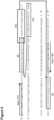
- the methods include introducing a biological sample into a biological assay device 200 by flowing the sample into one or more reaction chambers 202 of a sample receiving cartridge of the device via one or more sample receiving openings 210.
- flowing the sample into the one or more reaction chambers 202 includes flowing a gas, such as air, through a selective venting element 207 of the device, wherein the selective venting element forms a wall of each of the one or more reaction chambers 202, and wherein the one or more reaction chambers each include a modifying reagent 301.
- the methods can also include contacting the sample liquid with the selective venting element 207 and thereby making the selective venting element 207 impermeable to fluid.
- the methods include preventing further flow of fluid though a device. As such, after such flow is stopped, diffusion is the only method for transporting any contaminants in and/or out of the reaction chamber. Therefore if the inlet and/or conduits have a sufficient length, the contaminant diffusion time is substantially longer than the reaction and/or readout time and a result is not affected by contaminants.
- the subject methods include collecting a biological sample, such as collecting a sample with a sample collector.
- a biological sample can include, for example, human saliva, urine, human mucus, blood, or a solid tissue such as buccal tissue.
- a sample can also include bacteria or spores.
- Collecting can include contacting, e.g., rubbing and/or scraping, the sample collector against one or more surfaces of a subject and/or surfaces of a biological sample of a subject, such as a liquid, e.g., saliva and/or blood, sample extracted from the subject.
- collecting includes extracting one or more biological samples from the subject.
- collecting the biological sample can include instructing a subject to produce a biological sample, such as by spitting onto and/or into a sample collector.
- Collecting the biological sample can also include retaining a biological sample or a portion thereof, e.g., one or more cells, on the sample collector while, for example transferring the sample collector to an assay device.
- a sample collector is a swab and collecting the biological sample includes swabbing the inside of a subject's mouth and/or nose to obtain the biological sample on the collector.
- sample collectors are nasopharyngeal, mid turbinate and/or nasal swabs.
- a device is manufactured by encapsulating within a housing a selective venting element, sample receiving cartridge, adhesive layer, and/or substrate, or any combination thereof, by contacting them together in a single concerted step.
- the methods do not include manufacturing a device for example, by performing a first step of patterning a substrate layer, such as a glass, silicon and/or polymer layer, and/or binding a patterned, e.g., binding it chemically and/or physically, to a non-patterned layer, e.g., a sealing layer, and a second subsequent step of integrating the bound and/or sealed layer into a housing or cassette that provides additional functionality to employ the fluidic device.
- a substrate layer such as a glass, silicon and/or polymer layer
- the methods of manufacturing the subject devices include substantially preserving the functionality, e.g., chemical functionality, of reaction chamber contents, such as optical property modifying reagents and/or amplification compositions, while the contents are contained in the reaction chambers during manufacturing. This is achieved as the manufacturing process does not expose reagents to extreme temperature or chemical environments.
- the methods include manufacturing a device by operatively coupling an adhesive layer and a substrate while reaction chamber contents, such as optical property modifying reagents and/or amplification compositions, are retained within the reaction chambers.
- operatively coupling an adhesive layer and a substrate does not include heating the adhesive layer, substrate, or environment surrounding either.
- the methods include a step of inserting the optical property modifying reagent into each the one or more reaction chambers and storing the optical property modifying reagent therein while retaining functionality of the optical property modifying reagent.
- the amplification reaction amplifies nucleotides from a nucleic acid template.
- the amplification reaction is an isothermal amplification reaction, such as a strand displacement reaction.
- a strand displacement reaction is provided by a polymerase with strand displacement activity under reaction conditions such that strand displacement is possible. Examples of strand displacement reactions include strand displacement amplification (SDA), multiple displacement amplification (MDA), rolling circle amplification (RCA) or loop mediated isothermal amplification (LAMP).
- the amplification reaction includes other non-isothermal amplification reactions such as polymerase chain reaction (PCR).
- the amplification reaction performed is LAMP.
- a LAMP reaction a double- or single-stranded DNA template in dynamic equilibrium at an elevated temperature is amplified using two or three pairs of primers.
- the primers are designed based on the DNA template, using primer design software such as PrimerExplorer (Eiken).
- PrimerExplorer Eiken
- the F2 region of the FIP Forward Inner Primer
- F2c complementary
- a polymerase with strand displacement activity incorporates dNTPs along the template from the 3' end of F2. The incorporation of nucleotides releases protons, reducing the pH of the reaction mix.
- the F3 forward primer anneals to the F3c region upstream of the F2 region and on the template.
- the F3 forward primer begins amplifying the template strand, which releases further protons and displaces the FIP-incorporated strand that was synthesized previously.
- This single strand contains an F1 sequence (within the target sequence) along with its complementary F1c sequence (within the FIP).
- F1c anneals to F1 at the 5' end.
- the BIP Backward Inner Primer
- anneals to the other end of the strand and nucleotides extend from B2, releasing more protons.
- the backward primer B3 then binds to the B3c region, downstream of the B2 region, displaces the BIP-amplified strands and promotes extension to create the double strand.
- This displaced strand now contains a B1 sequence (within the target sequence) along with its complementary B1c sequence (within the BIP), forming another stem loop in the 3' end.
- the structure now has two stem-loop structures at each end from which continuous displacement and extension occur to amplify the template.
- the LAMP reaction can be amplified by adding further Forward and Backward Loop primers to produce more amplicons with stem loop structures.
- the LAMP procedure can take place at a fixed temperature, minimizing the need for any expensive thermocycling equipment.
- isothermal methods require a set temperature, which is determined by the selected reagents.
- enzymes function best between 60-65°C in LAMP methods.
- Colorimetric detection of the nucleic acid amplification reaction product can be performed in real-time throughout the amplification reaction, or after the performance of the amplification reaction. Detection of the colorimetric change of the reaction mix can be associated with a digital indication of a presence or absence of the amplification reaction product. In other words, a visual observation of the color change of the reaction mix can provide information regarding whether the amplification reaction product is present or absent. In certain embodiments, detection of a colorimetric change of the reaction mix indicates that the exponential or plateau phase of the amplification reaction has been obtained.
- detection of the amplification reaction product is accelerated relative to an amplification reaction that uses a reaction mix without a halochromic agent.
- the colorimetric change of the reaction mix is detected in less than 60 minutes from a starting time of the amplification reaction. Accelerated detection of the amplification reaction product is obtained because the halochromic agent (a weak acid or base) in the reaction mix absorbs protons generated during the amplification reaction, and recombination of the free protons acts to accelerate the detection of the amplification reaction.
- the reaction can be designed so that minimal amplification is required to generate a pH transition sufficient for the halochromic agent to change color.
- the amplification reaction product is detected visually by observation of a color change of the reaction mix.
- the human eye is used for the visual detection.
- a camera, a computer, or some other optical device is used for the visual detection or for imaging the reaction mix. Imaging programs include Photoshop (Adobe, San Jose CA), ImageJ (National Institutes of Health, Bethesda MD), and MATLAB (MathWorks, Natick MA).
- the amplification reaction product is detected by measuring fluorescence of the reaction mix, using fluorescence spectroscopy methods.
- the amplification reaction product is detected by measuring absorbance of the reaction mix, using absorption spectroscopy methods.
- the endpoint or overall change in absorbance or fluorescence of the reaction mix is measured at a given wavelength or set of wavelengths.
- compositions and methods for accelerated and efficient colorimetric detection of nucleic acid amplification reaction products are disclosed herein.
- a colorimetric assay is used to visually detect the presence of an amplified nucleic acid product, which eliminates the need for expensive and sophisticated instrumentation.
- the colorimetric detection of amplification products is achieved by amplifying a target nucleic acid template molecule to obtain the amplification reaction product.
- the amplification reaction includes a reaction mix.
- the reaction mix includes a nucleic acid template molecule, one or more enzymes for catalyzing the amplification reaction, and one or more halochromic agents for colorimetric detection.
- the reaction mix also includes a buffer having a buffering capacity equivalent to Tris buffer at a concentration between 1 mM-19 mM in a solution having a starting pH of 8.0.
- the reaction mix also includes a plurality of nucleic acid primers, deoxynucleotide triphosphates (dNTPs), suitable salts for the enzyme, and other non-buffered chemicals that enable nucleic acid amplification.
- dNTPs deoxynucleotide triphosphates
- the amplification reaction changes the starting pH of the reaction mix to cause a detectable colorimetric change of the halochromic agent, thereby indicating the presence of the target nucleic acid, and if the target nucleic acid is not present, the amplification reaction does not generate a sufficient number of protons to change the starting pH of the reaction mix sufficient to cause a detectable colorimetric change of the halochromic agent, thereby indicating that the amplification reaction product has not been produced.
- the halochromic agent (or pH indicator) in the reaction mix has a transition pH range for a colorimetric change of the halochromic agent that is narrower than an expected pH change between (1) a starting pH of the reaction mix before the amplification reaction is performed, and (2) an ending pH of the reaction mix after the amplification reaction has been performed.
- the halochromic agent is a colorimetric agent or a fluorescent agent.
- Suitable halochromic agents include phenol red, bromocresol purple, bromothymol blue, neutral red, naphtholphthalein, cresol red, cresolphthalein, phenolphthalein, methyl red, and thymolphthalein, among others.
- a wide range of concentrations of these halochromic agents can be used in the reaction mix.
- Different halochromic agents have different transition pH ranges.
- the halochromic agent has a transition pH range between pH 5-10, between pH 6-9, or between pH 6.5-8.8.
- the halochromic agent is at a concentration between 25-100 ⁇ M in the reaction mix.
- the halochromic agent is at a concentration between 50-260 ⁇ M.
- a combination of two or more halochromic agents is used in the reaction mix, which increases the normalized color contrast change of the reaction mix by being of complementary colors at the beginning and similar colors at the end of the amplification reaction.
- the combination of halochromic agents comprises phenol red and bromothymol blue.
- the combination of halochromic agents comprises cresol red and bromothymol blue.
- Phenol red is a halochromic agent that has a transition pH range from around 6.4-8.0. At the upper limit of the transition pH range, phenol red is red, and at the lower limit of the transition pH range, phenol red is yellow. A reaction mix containing phenol red will change color from red to yellow throughout the amplification reaction, as long as the starting pH of the reaction mix is around or above 8.0, and the ending pH of the reaction mix is within the transition pH range or around or below 6.4.
- the starting pH of the reaction mix is set by adding an acid or a base to the reaction mix until the desired starting pH is reached.
- the ending pH of the reaction mix is determined by performing a sample amplification reaction and measuring the ending pH (for example, with a micro-pH electrode).
- the halochromic agent for an amplification reaction is selected so that the transition pH range lies in between the starting pH and ending pH.
- the halochromic agent is selected so that the transition pH range is nearer to the starting pH than the ending pH.
- the halochromic agent can also be selected based on the particular enzyme used for catalyzing the amplification reaction.
- the enzyme in the reaction mix terminates polymerization of the amplification reaction as the pH decreases to unfavorable H+ concentrations.
- additional hydronium ions or hydronium ion equivalents are added to the reaction mix via the sample.
- additional hydronium ions or hydronium ion equivalents are added to the reaction mix via the sample.
- additional hydronium ion equivalents per 10 ⁇ l reaction mix can be tolerated for the amplification reaction to proceed.
- between 4.8 x 10 -10 and 4.8 x 10 -18 , 4.8 x 10 -12 and 4.8 x 10 -18 , or 4.8 x 10 -15 and 4.8 x 10 -18 can be tolerated.
- the enzyme will catalyze amplification reactions within a pH range that encompasses or is close to the transition pH range of the selected halochromic agent.
- Various enzymes can be used for the reaction, and different enzymes catalyze amplification reactions at different pH ranges.
- Bst polymerase is believed to catalyze amplification reactions within the pH range of 6.6-9.0.
- the preferred starting pH for Bst polymerase is greater than 7, more preferably greater than 8.2, and more preferably at 8.8.
- Other examples of a preferred starting pH for Bst polymerase are found in U.S. Pat. No. 5,830,714, filed April 17, 1996 .
- phenol red is coupled with Bst polymerase in a reaction mix, since the pH range at which Bst polymerase is active (6.6-9.0) encompasses the transition pH range of phenol red (6.4-8.0).
- methyl red is coupled with U exo-Klenow fragment (polymerase for Helicase Dependent Amplification, HDA) in a reaction mix, since a starting pH at which U exo-Klenow fragment is active (around 7.5) is higher than the transition pH range of methyl red (4.8-6.2).
- an isothermal amplification reaction uses an enzyme that is a strand displacement polymerase, such as phi29-DNA-Polymerase, Klenow DNA-Polymerase, Vent DNA Polymerase, Deep Vent DNA Polymerase, Bst DNA Polymerase, 9oNm(TM) DNA Polymerase, U exo-Klenow fragment, or mutants and variants thereof.
- suitable salts for the enzyme are also added to the reaction mix.
- the starting pH of the reaction mix is set based on an optimal pH for the specific enzyme used for catalyzing the amplification reaction.
- the pH of the entire DNA sample is between pH 3 and pH 11.
- a fluorescent halochromic agent is used to detect protons released during amplification.
- the halochromic agent can change optical properties (such as amplitude and emitted wavelength) as the pH of the reaction mix changes during the amplification reaction.
- Fluorescent halochromic agents include fluorescein, pyranine, and pHrodo dye (Life Technologies, Carlsbad CA).
- the base and/or acid added to the reaction mix maintains the starting pH of the reaction mix around or above an upper limit of the transition pH range of the halochromic agent.
- an acid such as hydrochloric acid (HCl) or sulfuric acid (H2SO4), or a base such as sodium hydroxide (NaOH) or potassium hydroxide (KOH), can be added to the reaction mix.
- the acid or base sets the starting pH of the reaction mix between pH 6-10, between pH 7-8, or between pH 8-8.6.
- the reaction mix is capable of offsetting the starting pH of the reaction mix by less than 0.1 pH units.
- the reaction mix has a starting pH lower than 2 pH units above the upper limit of the transition pH range of the halochromic agent. In further embodiments, the reaction mix has a starting pH lower than 1 pH unit, 0.5 pH units, or 0.1 pH units above the upper limit of the transition pH range of the halochromic agent. In a further embodiment, noise from non-specific amplification is minimized by setting the pH transition range sufficiently separated from the starting pH of the reaction mix, so that any color change is only achieved by a specific and sustained amplification.
- the reaction mix does not require any additional buffering agent for the amplification reaction, since a buffering agent could prevent large changes in pH from occurring during the amplification reaction.
- the reaction mix contains a minimal amount of buffering agent, such that the buffering capacity of the reaction mixture is less than the expected change in pH during amplification.
- the buffer is at a concentration between 1 mM and 3 mM. In a further embodiment, the buffer is at a concentration of 1 mM.
- the buffer used is Tris buffer (formulated to pH 8.8), HEPES (pH 7-9), or TAPS (pH 7-9).
- the buffer used is a buffer having a buffering capacity equivalent to a Tris buffer at a concentration between 1 mM-19 mM in a solution having a starting pH of 8.0.
- This broad range of suitable buffer concentrations allows the reaction mix to resist unwanted starting pH changes during reaction setup, unlike reaction setups with minimal ( ⁇ 1mM) Tris buffer equivalents (see US 13/799,995, filed March 13, 2013 ). These unwanted changes in pH come about due to hydronium or hydroxide ion equivalents added to the reaction via the sample reagents.
- colorimetric detection and enzyme kinetics depend on the starting pH, the presence of buffer capacity in the reaction mix high enough to avoid starting pH change, but low enough to allow color change upon amplification, become important.
- the pH of the reaction mix is between pH 7.5-8.8.
- Table 1 shows various buffers having buffering capacities equivalent to a Tris buffer at a concentration between 1 mM-19 mM in a solution having a starting pH of 8.0.
- the buffer capacity of 1 mM - 19 mM Tris (in a solution having a starting pH of 8.0) was found to range from 0.000575 to 0.010873.
- the starting pH of the buffer was considered to be in the range of 7.5 - 8.8 to be compatible with the reaction biochemistry (polymerase function, nucleic acid melting, etc.).
- the buffer has a buffering capacity equivalent to a Tris buffer at a concentration between 1.5 mM - 19 mM, 2 mM - 19 mM, 3 mM - 19 mM, 4 mM - 19 mM, 5 mM - 19 mM, 6 mM - 19 mM, 7 mM - 19 mM, or otherwise, in a solution having a starting pH of 8.0.
- the buffer has a buffering capacity equivalent to a Tris buffer at a concentration between 1.92 mM - 36.29 mM, 3 mM - 36.29 mM, 4 mM - 36.29 mM, 5 mM - 36.29 mM, or otherwise, in a solution having a starting pH of 8.8.
- the buffer has a buffering capacity equivalent to a Tris buffer at a concentration between 1.48 mM - 27.92 mM, 2 mM - 27.92 mM, 3 mM - 27.92 mM, 4 mM - 27.92 mM, 5 mM - 27.92 mM, or otherwise, in a solution having a starting pH of 7.5.
- Table 1 Buffer Capacity Table Buffer Full Chemical Name pKa at 25°C Starting Reaction pH Min Conc (mM) Max Conc (mM) Tris tris(hydroxymethyl)meth ylamine 8.06 8.8 1.92 36.29 8.0 1.00 19.00 7.5 1.48 27.92 TAPS N-Tris(hydroxymethyl)meth yl-3-aminopropanesulfonic acid 8.43 8.8 1.19 22.55 8.0 1.27 23.94 7.5 2.66 50.25 Bicine N,N-bis(2-hydroxyethyl)glycine 8.35 8.8 1.29 24.46 8.0 1.17 22.15 7.5 2.31 43.59 Tricine N-tris(hydroxymethyl) methylglycine 8.15 8.8 1.67 31.63 8.0 1.03 19.48 7.5 1.67 31.63 TAPSO 3-[N-Tris(hydroxymethyl)meth ylamino]-2-hydroxypropanesulfonic acid 7.635 8.8 4.17 78.90 8.0 1.19 22.45 7.5
- a magnesium compound is added to the reaction mix, because magnesium promotes nucleotide incorporation into the template and influences the activity of the polymerase.
- concentration of a magnesium compound (such as magnesium sulfate) in the reaction mix is at least 0.5 mM, at least 1 mM, at least 2 mM, or at least 4 mM.
- concentration of added magnesium ion is dependent on the concentration of dNTPs, nucleic acid template, and primers.
- the ratio of dNTPs to magnesium sulphate in the reaction mix is less than 1:2, less than 1:3, less than 1:4 or less than 1:5.
- the concentrations of other reagents of the reaction mix are kept at amounts as generally used in amplification reactions. See Notomi T et. al. Nucleic Acids Res. 2000 Jun 15; 28(12): E63 ; Nature Protocols 2008, Loop-mediated isothermal amplification (LAMP) of gene sequences and simple visual detection of products, 2008 3(5): pg 880 .
- the Bst or Bst 2.0 enzyme is used, and the amount of enzyme is at least 0.8 Unit per microliter of combined fluid.
- Betaine is also present in the reaction mix at a concentration between 0-1.5 M or 0.8M-1 M, and the total concentration of primers is between 3.6 ⁇ M and 6.2 ⁇ M.
- any of the following reagents is present in the reaction mix: Tris buffer (pH 8.8) at 20 mM, KCl at 10 mM, MgSO4 at 8 mM, (NH4)2SO4 at 10 mM, Tween 20 at 0.1%, Betaine at 0.8 M, dNTPs at 1.4 mM each, MnCl2 at 0.5 mM, FIP at 1.6 ⁇ M, F3 at 0.2 ⁇ M, B3 at 0.2 ⁇ M, primers at a total concentration of 5.2 ⁇ M (2 x (1.6+0.8+0.2)), and Bst / Bst 2.0 at 8 U per 10 ⁇ L.
- the concentrations of reaction mix reagents depend on the enzyme selection. In further embodiments, guidance regarding appropriate reagent concentrations is available from the enzyme manufacturers. In an embodiment, the ratio of the sample volume to the reaction mix volume is such that the sample is diluted between 5% and 40% when the reaction mix is added.
- amplification reaction reagents are stored separately before being added to a reaction mix, since some reagents have specific required conditions for stability.
- the enzyme can be stored long term in a moderately buffered solution separate from the other reagents to ensure stability of the enzyme.
- the buffering agent Upon mixing with the remaining reagents in the reaction mix, the buffering agent becomes sufficiently diluted so as not to significantly mask a pH change.
- primers for specific genes of interest can be provided in a separate solution or in a lyophilized form.
- the amplification reaction is performed within a microtube. In other embodiments, the amplification reaction is performed within a fluidic or microfluidic structure.
- the fluidic or microfluidic structure is a well, chamber, or channel that receives the reagents and the nucleic acid sample separately, and then mixes the components together.
- the fluidic or microfluidic structure is a well, chamber, or channel that receives the pre-mixed reaction mix.
- the fluidic or microfluidic structure possesses a long optical path for colorimetric observation, or a fluorescent/ absorbance excitation source and detector.
- the fluidic or microfluidic structure receives the reagents in a lyophilized form, and subsequently receives the nucleic acid sample and hydration solution.
- a chamber fluidic or microfluidic structure has a channel depth ranging between 50 ⁇ m-400 ⁇ m or greater.
- colorimetric observation is accomplished for channel depths (path length) of 50 ⁇ m, 50 ⁇ m-400 ⁇ m, or 50 ⁇ m or greater.
- kits for colorimetric detection of an amplification product can include one or more halochromic agents, one or more enzymes for catalyzing an amplification reaction, and instructions for contacting a sample with a reaction mix including the buffer and the enzyme and the halochromic agent under conditions that an amplification reaction occurs and produces an amplification reaction product if the sample contains a target nucleic acid template molecule, the reaction mix having a starting pH, and if the target nucleic acid template molecule is present, the amplification reaction changes the starting pH of the reaction mix to cause a detectable colorimetric change of the halochromic agent, thereby indicating the presence of the target nucleic acid, and if the target nucleic acid template molecule is not present, the amplification reaction does not generate a sufficient number of protons to change the starting pH of the reaction mix sufficient to cause a detectable colorimetric change of the halochromic agent, thereby indicating that the amplification reaction product has not been produced.
- the instructions are for contacting a nucleic acid template molecule with the halochromic agent and enzyme in a reaction mix, under conditions that result in (1) an amplification reaction that amplifies the nucleic acid template molecule to produce an amplification reaction product, and (2) generation of a sufficient number of protons so that an ending pH of the reaction mix is sufficiently low to produce a detectable colorimetric change of the halochromic agent, thereby indicating that the amplification reaction product has been produced.
- the kit also includes an acid or base, dNTPs, primers, and monovalent cations.
- the kit includes the following reagents at the following concentrations:
- kits including the subject devices and which can be used according to the subject methods.
- the subject kits can include two or more, e.g., a plurality, three or less, four or less, five or less, ten or less, or fifteen or less, or fifteen or more, selectively vented biological assay devices or components thereof, according to any of the embodiments described herein, or any combinations thereof.
- kits can include one or more compositions and/or reagents, such as any of those described herein, e.g., optical property modifying reagents, amplification compositions, preparation solutions and/or buffers, which can be stored in the kits in containers separate from the devices.
- the kits can include any device or other element which can facilitate the operation of any aspect of the kits.
- a kit can include one or more devices for preparing a sample and/or analyzing one or more characteristics of a sample, e.g., a prepared sample. Kits can also include packaging, e.g., packaging for shipping the devices without breaking.
- kits which are disclosed herein include instructions, such as instructions for using devices.
- the instructions for using devices are, in some aspects, recorded on a suitable recording medium.
- the instructions can be printed on a substrate, such as paper or plastic, etc.
- the instructions can be present in the kits as a package insert, in the labeling of the container of the kit or components thereof (i.e., associated with the packaging or subpackaging etc.).
- the instructions are present as an electronic storage data file present on a suitable computer readable storage medium, e.g., Portable Flash drive, CD-ROM, diskette, on the cloud, etc.
- the instructions may be storable and/or reproducible within one or more programs, such as computer applications.
- the instructions can take any form, including complete instructions for how to use the devices or as a website address with which instructions posted on the world wide web can be accessed.
- the subject devices and methods are directed to performing biological assays by effectively evaluating one or more characteristics of biological samples such as, by modifying optical properties of biological samples or aspects thereof.
- the devices and methods provide, for example, effective sample aliquoting protocols.
- Aliquoting a sample is a procedure applied in biochemical analyses for multiple tests or downstream processing.
- Once challenge is how a sample can be accurately aliqoted into multiple sites.
- One way to do this is to route the sample through bifurcating channels and into multiple reaction chambers.
- the aliquots have to be isolated so that there is no cross talk between the reactions.
- Such isolation can be achieved using input and/or output valves positioned between each chamber. The valves, for example, allow for an aliquot to enter a chamber and simultaneously evacuate any fluid present in the chamber. Additionally, the valves seal the chamber off from any cross talk.
- valves Although using multiple valves works to some extent, such a protocol imposes requirements to actively control the opening and closing of the valves, which in return requires energy and infrastructure to implement, and thus complicating the system design. Also some valve structures work best when primed and as such, require the microfluidic system to be filled with an initial priming liquid. Such a priming step in turn complicates the system workflow. As such, according to versions of the subject methods, the methods do not include priming.
- the subject devices and methods sufficiently provide automatic fluid flow control by passive aliquoting through one or more portions of a device such that an assay can be performed.
- one or more fluids e.g., air and/or biological sample
- Passive sealing of the device or portions thereof eliminates the need for active control and minimizes the complexity of the full device and the user steps required to run the device.
- the subject disclosure provides simple and easy to use assay devices.
- the subject devices and methods do not require valves or complicated valve control protocols.
- the subject disclosure provides a simple and robust implementation of an on-chip aliquoting function with no moving parts.
- aliquot volumes and numbers are controlled by channel and chamber geometries.
- the system can operate with imprecise loading mechanisms while maintaining very precise aliquot numbers and volumes. In other words, there is no need control the loading pressure, or sample volumes beyond a simple threshold. As such, the aliquoting precision is obtained by the chamber manufacturing precision.
- the self-sealing characteristic of the system that allows for gas and liquids to pass through until the pores of a selective venting element are sealed can be effectively applied for the prevention of evaporation.
- evaporation though the self-sealing pores is minimized.
- the subject devices and methods also minimize dead volume as compared to a mechanical valve that requires contact surface area. They also allow for the filling of multiple chambers without channel resistance matching.
- the devices and methods disclosed herein also protect from washing out any reagents or dry material in the reaction chamber during reaction loading.
- the devices and methods in some versions modify an optical property to allow detection of the modified optical property by an un-assisted human eye.
- the content of the subject disclosure eliminates a need for complex evaluation techniques or equipment to read or interpret a signal generated by a biological assay. Because a user can recognize a modified optical property with a user's eye, performing an assay with the subject methods can reduce time and expense compared to performing such an assay using other equipment or methods.
- the subject devices can also be finely tuned to provide efficient energy conduction, e.g., heat or electrical energy, into a fluidic network and/or specific variations in optical properties such as adhesive color.
- methods of assembling biological assay devices have included patterning a substrate layer, e.g., a layer of glass, silicon or polymer, and then bonding it to a non-patterned sealing layer using chemical or physical bonds.
- a substrate layer e.g., a layer of glass, silicon or polymer
- bonding it to a non-patterned sealing layer using chemical or physical bonds Once the fluidic device was assembled, e.g., assembled by being bonded and sealed, then is has been integrated into a housing or cassette that provides additional functionality required to utilize the fluidic system.
- many microfluidic device bonding techniques have had the potential to damage any fragile pre-loaded reagents.
- the adhesive layer can be employed for simultaneously sealing the microfluidic system and integrating into the final assembly while preserving reagent functionality, such as functionality of reagents pre-loaded into reaction chambers.
- the subject methods and devices simplify the operation of such devices, as well as the manufacturing of such devices while improving effectiveness in generating an assay result.
- the reagent mix was adjusted to an initial pH of 8.8 to enable efficient initial polymerization.
- the reagent mix was autoclaved for 1 hour for sterilization.
- the following ingredients were then added (in a sterile form) to the reagent mix to generate the reaction mix:
- Primer F3 has the sequence: GATCTGAATCCGACCAACCG (SEQ ID NO: 1); primer B3 has the sequence: AACGCCCACGCTCTCGCA (SEQ ID NO: 2); the primer FIP has the sequence: AAATCCGTCCAGTGGTTTTTTTGAAAATCGTTGTATCTCCG (SEQ ID NO: 3); and the primer BIP has the sequence: CCGAAACCACTGGACGGATTTTTATTTTTAATCTAAAACAAACATC (SEQ ID NO: 4).
- the nucleic acid template molecule was purified from Schistosoma mansoni.
- FIG. 4 shows the SM1-7 target region of the nucleic acid template molecule ( see Hamburger et al, Detection of Schistosoma mansoni and Schistosoma haematobium DNA by Loop-Mediated Isothermal Amplification: Identification of infected Snails from Early Prepatency, Am J Trop Med Hyg, 2010 ).
- the positive test reactions contained template DNA, and the negative control reactions contained water.
- the reaction mixes had a starting pH in the range of 7.5 - 8.5.
- the reaction mixes were heated in micro-tubes to 63°C on a thermocycler to allow template amplification. After a predetermined reaction period of 45 minutes, during which sufficient template amplification occurred, the resultant color of the reaction mix was visually observed.
- FIG. 5 is a graph showing the pH measurements for repeated positive (test) and negative (negative control) amplification reactions.
- the halochromic agent used was Phenol red, which has a transition pH range of 6.8 - 8.2. Phenol red changes color over this transition pH range from red to yellow (when the pH is lowered from the upper pH limit to the lower pH limit).
- the reaction mix changed color from red (at pH 8.0) to yellow (at pH 6.6) in response to the pH change during nucleic acid amplification.
- FIG. 1 is a graph showing the pH measurements for repeated positive (test) and negative (negative control) amplification reactions.
- the halochromic agent used was Phenol red, which has a transition pH range of 6.8 - 8.2. Phenol red changes color over this transition pH range from red to yellow (when the pH is lowered from the upper pH limit to the lower pH limit).
- the reaction mix changed color from red (at pH 8.0) to yellow (at pH 6.6) in response to the pH
- FIG. 6 is a graph showing the difference in contrast value using HSV (hue, saturation, value) of images of the reaction mixes of a positive and negative amplification reaction at the reaction endpoints.
- HSV hue, saturation, value
- the color change is quantitatively demonstrated in the hue variable.
- endpoint reactions were analyzed using gel electrophoresis to verify the presence of amplicons ( FIG. 7 ).
- amplification of a DNA template can be easily observed, either at the reaction end-point or in real-time throughout the reaction, by visually observing the color change in the reaction mix, or by measuring the absorbance or fluorescence of the reaction mix.
- This mechanism generates much larger contrast in comparison to other colorimetric detection techniques and can be imaged without the need of expensive optical instrumentation.
- LAMP reactions were performed with a reaction mix comprising of: 10 mM (NH4) 2 SO4, 15 mM KCl, 0.1 mM EDTA, 0.1 mM DTT, 0.01 % Triton X-100 (v/v), 5 % Glycerol, 8 mM MgSO 4 , 1.4 mM each dNTPs, 0.1% v/v Tween-20, 0.8 M Betaine.
- Three primer pairs, specific to different targets, were added to a final concentration of 1.6 ⁇ M each for FIP/BIP, 0.2 ⁇ M each for F3 /B3, 0.4 ⁇ M each for LoopB/F.
- the final reaction volume is 10 ⁇ L and was held at 63 °C for different incubation times.
- the final Tris buffer concentration of the reaction mix was varied from 0.34 mM to 19 mM (by varying amount of Tris buffer formulated to pH 8.8).
- Reactions were performed with primers for lambda phage DNA, 5 ng of lambda DNA (New England Biolabs), 0.8 U/ ⁇ l Bst 2.0 DNA polymerase (New England Biolabs) and 0.2 mM Neutral Red (Sigma Aldrich).
- the reaction tubes were then imaged and the Normalized Hue value was calculated for the color of the reaction mix.
- the Normalized Hue value was defined as the difference in Hue values between a positive and a no-template negative reaction.
- FIGS. 10A-10D the compatibility of different pH indicators and amplification targets with visual detection of LAMP amplification was evaluated.
- the reactions were performed with final Tris buffer concentration in the range of 1.2 - 1.3 mM and 0.8 U/ ⁇ L Bst DNA polymerase.
- Three different indicator were tested with 5 ng lambda DNA target: 50 ⁇ M Phenol Red, 260 ⁇ M Cresol Red and 160 ⁇ M Bromothymol Blue ( FIG. 10A ). High contrast change in the normalized hue value was observed for all indicators tested.
- FIG. 11 the compatibility of different polymerases with visual detection of LAMP amplification was evaluated.
- the reactions were performed with 1.3 mM final Tris buffer concentration, 5 ng lambda DNA target and 0.2 mM Neutral Red. 0.8 U/ ⁇ l of two different polymerases, Bst 2.0 and Gspm 2.0 (OptiGene), were used. High contrast color change was observed for both polymerases after 60 minutes of incubation ( FIG. 11 ).
- Table 2 Sequences Used Lambda FIP SEQ ID NO: 5 Lambda BIP SEQ ID NO: 6 Lambda F3 SEQ ID NO: 7 Lambda B3 SEQ ID NO: 8 Lambda Loop F SEQ ID NO: 9 Lambda Loop B SEQ ID NO: 10 Schistosoma F3 SEQ ID NO: 1 Schistosoma B3 SEQ ID NO: 2 Schistosoma FIP SEQ ID NO: 3 Schistosoma BIP SEQ ID NO: 4 GAPDH F3 SEQ ID NO: 11 GAPDH B3 SEQ ID NO: 12 GAPDH FIP SEQ ID NO: 13 GAPDH BIP SEQ ID NO: 14 GAPDH Loop F SEQ ID NO: 15 GAPDH Loop B SEQ ID NO: 16
- LAMP reactions were performed as in Example 1 with 1.3 mM final Tris buffer concentration (buffer formulated to pH 8.8), 0.8 U/ ⁇ l of Bst 2.0 DNA Polymerase, 5 ng lambda DNA template and 0.2 mM Neutral Red or 160 ⁇ M Bromothymol Blue. Both the positive and the no-template negative reactions were added after amplification to flow chambers with varying channel depths ( FIG. 12A for Neutral Red and FIG. 12B for Bromothymol Blue). These flow chambers were machined in acrylic with channel depths ranging from 50 ⁇ m to 400 ⁇ m. High contrast color difference (above the visual detection threshold; dotted line) between the positive and the negative reactions was observed for channel depths of 50 ⁇ m and above.
- reaction chambers with sub-millimeter path lengths (depths) and above.
- Such reaction chambers can be used to reduce the amount of reagents used and to allow multiple reactions to take place in a certain footprint (e.g. in a microfluidic cartridge).
- LAMP reactions were performed as in Example 1 with 1.6 mM final Tris buffer concentration (buffer formulated to pH 8.8), 0.8 U/ ⁇ l of Bst 2.0 DNA Polymerase, 5 ng lambda DNA template, and Phenol Red and Bromothymol Blue at 50 ⁇ M and 160 ⁇ M concentrations respectively.
- the solution was loaded into a fluidic device with reaction chambers consisting of a sample receiving input and a vent outlet.
- the vent outlet of each reaction chambers was sealed with a selective venting element, e.g., a self-sealing element. Alternating chambers had lambda primers dried in them.
- the sample receiving inputs are all connected to a bus channel connected to the device inlet.
- the reaction chambers were heated to 63 O C for 1 hour. The color change in the chambers was measured with a camera and the data is shown in FIG. 17 .
- SDA reactions were performed using a reaction mix comprising of: 1.3 mM final Tris buffer concentration (buffer formulated to pH 8.8), 10 mM (NH4) 2 SO4, 50 mM KCl (adjusted to pH 8.5), 8 mM MgSO 4 , 4.4 mM each dATP, dGTP, dTTP, 0.8 mM dCTP- ⁇ S (TriLink Biotechnologies), 0.1% v/v Tween-20, 0.8 M Betaine, 0.32 U/ ⁇ l Bst DNA polymerase (New England Biolabs), 0.2U/uL BSoBI (New England Biolabs) and 0.2 mM Neutral Red halochromic agent.
- PCR reactions were performed using a reaction mix comprising of: 50 mM KCl and 2 mM MgCl 2 (pH adjusted 8.5), 0.5 mM each dNTP, 5U Taq DNA polymerase (New England Biolabs) and 0.2 mM Neutral Red halochromic agent.
- Total carry-over Tris-HCl concentration from enzyme storage buffer and primers (Forward: SEQ ID NO: 21; Reverse: SEQ ID NO: 22) was 1.15 mM in the final reaction mix.
- Primers were designed for Escherichia coli 16s rRNA gene and added to the reaction at 0.5 ⁇ M final concentration each. 10 ng of E.
- coli gDNA was added to a final reaction volume of 25 ⁇ L and was initially held at 95 °C hold for 2 min, followed by 50 cycles of 95 °C for 10 sec, 55 °C for 30 sec, 68 °C for 30 sec.
- a change in Normalized Hue value over time indicates that this visual detection chemistry works with PCR.
- Example 7 Increase in Visual Detection Contrast with Combination of Halochromic Agents
- LAMP reactions were performed as in Example 1 with 1.3 mM final Tris buffer concentration (buffer formulated to pH 8.8), 0.8 U/ ⁇ l of Bst 2.0 DNA Polymerase and 5 ng lambda DNA template.
- the color change contrast was evaluated for Phenol Red at 50 ⁇ M concentration and combination of Phenol Red and Bromothymol Blue at 50 ⁇ M and 160 ⁇ M concentrations respectively ( FIG. 15A ).
- the color change contrast was also evaluated for Cresol Red at 260 ⁇ M concentration and combination of Cresol Red and Bromothymol Blue at 260 ⁇ M and 160 ⁇ M concentrations respectively ( FIG. 15B ).
- the contrast values were calculated from the RGB values of images of the reaction mix using the formula: 0.299R + 0.587G + 0.114B.
- the normalized contrast change was defined as the difference between positive and negative reaction contrast values normalized to the background. The increase in the normalized contrast change with the use of the halochromic agent combination demonstrates the utility of such combinations.
- Example 8 Real-time Color Monitoring of Amplification for Quantification Using Visual Halochromic Agents
- LAMP reactions were performed as in Example 1 with 1.3 mM final Tris buffer concentration (buffer formulated to pH 8.8), 0.8 U/ ⁇ l of Bst 2.0 DNA Polymerase, Phenol Red and Bromothymol Blue at 50 ⁇ M and 160 ⁇ M concentrations respectively and varying lambda DNA template concentrations. Color change contrast was evaluated for lambda DNA target at 0.5 fg/ ⁇ l, 0.05 pg/ ⁇ l and 0.5 pg/ ⁇ l final concentrations. The contrast values were calculated from the RGB values of images of the reaction mix as described in Example 5. The results ( FIG. 16 ) indicate that the higher DNA concentrations led to a detectable change in visual contrast earlier than the lower DNA concentrations. Hence, we demonstrate the ability to distinguish between different target concentrations with the real-time color monitoring of this chemistry.
Landscapes
- Health & Medical Sciences (AREA)
- Chemical & Material Sciences (AREA)
- General Health & Medical Sciences (AREA)
- Chemical Kinetics & Catalysis (AREA)
- Clinical Laboratory Science (AREA)
- Life Sciences & Earth Sciences (AREA)
- Physics & Mathematics (AREA)
- Spectroscopy & Molecular Physics (AREA)
- Analytical Chemistry (AREA)
- Molecular Biology (AREA)
- Biochemistry (AREA)
- Dispersion Chemistry (AREA)
- Hematology (AREA)
- Pharmacology & Pharmacy (AREA)
- Animal Behavior & Ethology (AREA)
- Public Health (AREA)
- Veterinary Medicine (AREA)
- General Physics & Mathematics (AREA)
- Apparatus Associated With Microorganisms And Enzymes (AREA)
- Measuring Or Testing Involving Enzymes Or Micro-Organisms (AREA)
- Automatic Analysis And Handling Materials Therefor (AREA)
Claims (15)
- Dispositif (200) d'essai biologique à ventilation sélective, le dispositif (200) comprenant :a. une cartouche de réception d'échantillon (201) comprenant une entrée à échantillon (212) et une ou plusieurs chambres de réaction (202), la chambre ou chacune des chambres de réaction (202) : (i) comprenant une ouverture de réception d'échantillon (210) fonctionnellement raccordée à l'entrée à échantillon (212), et (ii) comportant un réactif de modification (301) ; et caractérisé en ce que le dispositif (200) d'essai biologique à ventilation sélective comprend en outreb. un ou plusieurs éléments de ventilation sélective (207) ayant une porosité passivement réglable et recouvrant une ou plusieurs ouvertures de ventilation de la cartouche de réception d'échantillon (201),dans lequel le ou les éléments de ventilation sélective sont poreux et conçus pour passer, lors d'un contact avec un liquide, d'une première conformation, dans laquelle un ou plusieurs gaz peuvent passer à travers, à une deuxième conformation qui réduit la perméabilité des fluides à travers ceux-ci, la première conformation permettant un écoulement depuis l'entrée à échantillon (212) à l'intérieur de la ou des chambres de réaction (202), la réduction de la perméabilité étant suffisante pour rendre le ou les éléments de ventilation sélective imperméables aux fluides ;dans lequel, quand un liquide échantillonné s'écoule à l'intérieur de la ou des chambres de réaction par le biais des ouvertures de réception d'échantillon, du gaz s'écoule à travers le ou les éléments de ventilation sélective jusqu'à ce que le liquide échantillonné entre en contact avec le ou les éléments de ventilation sélective, faisant ainsi passer le ou les éléments de ventilation sélective de la première conformation à la deuxième conformation et empêchant un écoulement supplémentaire de fluide à l'intérieur de la chambre de réaction.
- Dispositif selon la revendication 1, dans lequel l'élément ou chacun des éléments de ventilation sélective (207) comprend un corps (101) et une ou plusieurs saillies (102) s'étendant depuis le corps (101) pour recouvrir l'ouverture ou chacune des ouvertures de ventilation, éventuellement dans lequel chaque saillie (102) a la forme d'un cylindre.
- Dispositif selon l'une quelconque des revendications 1 à 2, dans lequel l'élément ou chacun des éléments de ventilation sélective (207) comprend une matrice polymère poreuse et un hydrogel.
- Dispositif selon l'une quelconque des revendications 1 à 3, comprenant en outre un substrat (203) fonctionnellement couplé à la cartouche de réception d'échantillon (201), le substrat (203) comprenant un élément chauffant (204), éventuellement dans lequel chaque chambre de réaction (202) comprend en outre une ouverture supplémentaire, dans lequel l'ouverture ou chacune des ouvertures de ventilation se trouve sur un premier côté de la cartouche de réception d'échantillon (201) et chacune des ouvertures supplémentaires se trouve sur un deuxième côté de la cartouche de réception d'échantillon (201), dans lequel le premier côté est opposé au deuxième côté.
- Dispositif selon la revendication 4, comprenant en outre un boîtier comprenant une première partie (208) et une deuxième partie (209) assemblable à la première partie pour encapsuler la cartouche de réception d'échantillon (201) et le ou les éléments de ventilation sélective (207).
- Procédé de réalisation d'un essai biologique avec un dispositif (200) d'essai biologique à ventilation sélective, le procédé comprenant :a. l'introduction d'un liquide échantillonné dans un dispositif (200) d'essai biologique par écoulement du liquide échantillonné à l'intérieur d'une ou plusieurs chambres de réaction (202) d'une cartouche de réception d'échantillon (201) du dispositif (200) par le biais d'une ou plusieurs ouvertures de réception d'échantillon (210), la chambre ou au moins une des chambres de réaction (202) comprenant un réactif de modification (301) ;i. dans lequel l'écoulement du liquide échantillonné à l'intérieur de la ou des chambres de réaction (202) comprend l'écoulement d'un gaz à travers un ou plusieurs éléments de ventilation sélective (207) du dispositif (200), dans lequel l'élément ou chacun des éléments de ventilation sélective (207) forme une paroi d'une ou plusieurs des chambres de réaction (202), dans lequel le ou les éléments de ventilation sélective sont poreux et sont conçus pour passer d'une première conformation, dans laquelle un ou plusieurs gaz peuvent passer à travers, à une deuxième conformation qui réduit la perméabilité des fluides à travers ceux-ci, et dans lequel l'écoulement d'un liquide échantillonné à l'intérieur de la ou des chambres de réaction comporte la mise en contact du liquide échantillonné avec le ou les éléments de ventilation sélective, faisant ainsi passer le ou les éléments de ventilation sélective d'une première conformation à une deuxième conformation, dans lequel la réduction de la perméabilité est suffisante pour rendre le ou les éléments de ventilation sélective imperméable aux fluides ;b. la mise en contact du liquide échantillonné avec le ou les éléments de ventilation sélective, permettant ainsi à l'élément ou aux éléments de ventilation sélective (207) de passer de la première conformation à la deuxième conformation ;c. la réaction du liquide échantillonné avec le réactif de modification (301) et la génération d'un produit de réaction ; etd. la détection d'une caractéristique du produit de réaction.
- Procédé selon la revendication 6, dans lequel le réactif de modification (301) comprend un réactif d'amplification d'acide nucléique, et éventuellement dans lequel la réaction du liquide échantillonné avec le réactif de modification (301) comprend la mise en contact du liquide échantillonné avec le réactif d'amplification d'acide nucléique dans la ou les chambres de réaction (302) dans des conditions qui se soldent par l'amplification de l'acide nucléique si l'acide nucléique est présent dans le liquide échantillonné.
- Procédé selon la revendication 6, dans lequel le réactif de modification (301) comprend un réactif d'essai d'immuno-absorption enzymatique (ELISA), le réactif ELISA étant choisi dans le groupe constitué par la phosphatase alcaline, la peroxydase de raifort, la β-galactosidase, le BCIP/NBT (phosphate de 5-bromo-4-chloro-3-indolyle/ nitro-bleu de tétrazolium), la TMB (3,3',5,5'-tétra-méthylbenzidine), la DAB (3,3',4,4'-diaminobenzidine), le 4CN (4-chloro-1-naphtol), la TMB (substrat à double fonction), l'ABTS (sulfonate de 2,2'-azino-di[3-éthyl-benzthiazoline]), l'OPD (o-phénylènediamine), le MUG (4-méthyllumbelliféryl-galactoside), le HPA (acide hydroxy-phénylacétique), et le HPPA (acide 3-p-hydroxyphényl-propionique).
- Procédé selon la revendication 7, dans lequel la détection d'une caractéristique du produit de réaction comprend la détection de la présence ou de l'absence de l'amplification.
- Procédé selon l'une quelconque des revendications 6 à 9, dans lequel l'élément ou chacun des éléments de ventilation sélective (207) comprend une matrice polymère poreuse et un hydrogel.
- Procédé selon l'une quelconque des revendications 6 à 10, le dispositif (200) comprenant un capteur et le procédé comprenant la détection de la présence ou de l'absence du liquide échantillonné dans la cartouche de réception d'échantillon (201) avec le capteur.
- Procédé selon la revendication 11, le dispositif (200) comprenant en outre un élément chauffant (204), et le procédé comprenant en outre le chauffage du liquide échantillonné dans la ou les chambres de réaction (202) quand le capteur détecte le liquide échantillonné.
- Procédé selon l'une quelconque des revendications 6 à 12, dans lequel la détection d'une caractéristique du produit de réaction comprend l'inspection visuelle de la ou des chambres de réaction (202) pour détecter une propriété optique modifiée du produit de réaction.
- Procédé selon l'une quelconque des revendications 11 à 12, dans lequel la détection d'une caractéristique du produit de réaction comprend la détection d'une propriété optique modifiée du produit de réaction au moyen du capteur.
- Procédé selon la revendication 12, dans lequel l'élément chauffant (204) comprend un ou plusieurs réactifs générant de la chaleur qui produisent de la chaleur quand ils sont mélangés les uns avec les autres ou avec le liquide échantillonné, et dans lequel la génération du produit de réaction comprend le chauffage du liquide échantillonné et du réactif de modification avec l'élément chauffant (204) par mélange du ou des réactifs générant de la chaleur les uns avec les autres ou avec le liquide échantillonné.
Priority Applications (1)
| Application Number | Priority Date | Filing Date | Title |
|---|---|---|---|
| EP24208925.8A EP4477316A3 (fr) | 2016-03-14 | 2017-03-14 | Dispositifs d'analyse biologique à ventilation sélective et procédés associés |
Applications Claiming Priority (2)
| Application Number | Priority Date | Filing Date | Title |
|---|---|---|---|
| US201662307886P | 2016-03-14 | 2016-03-14 | |
| PCT/US2017/022306 WO2017160840A1 (fr) | 2016-03-14 | 2017-03-14 | Dispositifs d'analyse biologique à ventilation sélective et procédés associés |
Related Child Applications (1)
| Application Number | Title | Priority Date | Filing Date |
|---|---|---|---|
| EP24208925.8A Division EP4477316A3 (fr) | 2016-03-14 | 2017-03-14 | Dispositifs d'analyse biologique à ventilation sélective et procédés associés |
Publications (3)
| Publication Number | Publication Date |
|---|---|
| EP3429543A1 EP3429543A1 (fr) | 2019-01-23 |
| EP3429543A4 EP3429543A4 (fr) | 2019-11-06 |
| EP3429543B1 true EP3429543B1 (fr) | 2024-11-20 |
Family
ID=59851861
Family Applications (2)
| Application Number | Title | Priority Date | Filing Date |
|---|---|---|---|
| EP17767339.9A Active EP3429543B1 (fr) | 2016-03-14 | 2017-03-14 | Dispositifs d'analyse biologique à ventilation sélective et procédés associés |
| EP24208925.8A Withdrawn EP4477316A3 (fr) | 2016-03-14 | 2017-03-14 | Dispositifs d'analyse biologique à ventilation sélective et procédés associés |
Family Applications After (1)
| Application Number | Title | Priority Date | Filing Date |
|---|---|---|---|
| EP24208925.8A Withdrawn EP4477316A3 (fr) | 2016-03-14 | 2017-03-14 | Dispositifs d'analyse biologique à ventilation sélective et procédés associés |
Country Status (8)
| Country | Link |
|---|---|
| US (3) | US11291995B2 (fr) |
| EP (2) | EP3429543B1 (fr) |
| JP (3) | JP2019509740A (fr) |
| AU (3) | AU2017232344B2 (fr) |
| CA (1) | CA3015378C (fr) |
| DK (1) | DK3429543T3 (fr) |
| PT (1) | PT3429543T (fr) |
| WO (1) | WO2017160840A1 (fr) |
Cited By (1)
| Publication number | Priority date | Publication date | Assignee | Title |
|---|---|---|---|---|
| EP4477316A3 (fr) * | 2016-03-14 | 2025-02-19 | Pfizer Inc. | Dispositifs d'analyse biologique à ventilation sélective et procédés associés |
Families Citing this family (25)
| Publication number | Priority date | Publication date | Assignee | Title |
|---|---|---|---|---|
| EP3594360B1 (fr) | 2014-04-24 | 2021-06-23 | Lucira Health, Inc. | Détection colorimétrique de l'amplification d'acides nucléiques |
| AU2017232340B2 (en) | 2016-03-14 | 2022-04-28 | Pfizer Inc. | Systems and methods for performing biological assays |
| ES2927062T3 (es) | 2016-03-14 | 2022-11-02 | Lucira Health Inc | Dispositivos y procedimientos para modificar propiedades ópticas |
| JP6949864B2 (ja) | 2016-03-14 | 2021-10-13 | ルシラ ヘルス インコーポレイテッド | 生物学的アッセイ試料調製および送り出しのための装置および方法 |
| US10987674B2 (en) | 2016-04-22 | 2021-04-27 | Visby Medical, Inc. | Printed circuit board heater for an amplification module |
| WO2017197040A1 (fr) | 2016-05-11 | 2017-11-16 | Click Diagnostics, Inc. | Compositions et méthodes d'extraction d'acides nucléiques |
| MX2018015889A (es) | 2016-06-29 | 2019-05-27 | Click Diagnostics Inc | Dispositivos y metodos para la deteccion de moleculas usando una celda de flujo. |
| US11080848B2 (en) | 2017-04-06 | 2021-08-03 | Lucira Health, Inc. | Image-based disease diagnostics using a mobile device |
| US10549275B2 (en) | 2017-09-14 | 2020-02-04 | Lucira Health, Inc. | Multiplexed biological assay device with electronic readout |
| CN113777011B (zh) | 2017-09-14 | 2024-11-19 | 辉瑞公司 | 具有电子读出的复用生物测定装置 |
| SG11202002931VA (en) | 2017-11-09 | 2020-04-29 | Visby Medical Inc | Portable molecular diagnostic device and methods for the detection of target viruses |
| KR102050161B1 (ko) * | 2018-05-31 | 2019-11-28 | 목포대학교산학협력단 | 유전체 분자 진단용 휴대용 램프 pcr 장치 및 그 장치를 이용한 유전체 분자 진단 방법 |
| KR20210081335A (ko) | 2018-09-03 | 2021-07-01 | 비스비 메디컬, 인코포레이티드 | 항생제 감수성 시험을 위한 장치 및 방법 |
| USD910200S1 (en) | 2018-12-21 | 2021-02-09 | Lucira Health, Inc. | Test tube |
| JPWO2020218439A1 (fr) * | 2019-04-25 | 2020-10-29 | ||
| JP2022530323A (ja) | 2019-04-28 | 2022-06-29 | ビスビュー メディカル,インコーポレイテッド | デジタル検出能力および無線接続性を有する分子診断デバイス |
| CA3139147A1 (fr) * | 2019-05-06 | 2020-11-12 | University Of Prince Edward Island | Procede et appareil portable de test sur le terrain |
| KR102368556B1 (ko) * | 2019-11-21 | 2022-02-28 | 주식회사 코사이언스 | 유전체 분자 진단용 휴대용 램프 pcr 장치 |
| US11352675B2 (en) | 2020-01-03 | 2022-06-07 | Visby Medical, Inc. | Devices and methods for antibiotic susceptability testing |
| USD953561S1 (en) | 2020-05-05 | 2022-05-31 | Lucira Health, Inc. | Diagnostic device with LED display |
| USD962470S1 (en) | 2020-06-03 | 2022-08-30 | Lucira Health, Inc. | Assay device with LCD display |
| USD1064314S1 (en) | 2021-08-13 | 2025-02-25 | Visby Medical, Inc. | Molecular diagnostic device |
| EP4442824A4 (fr) * | 2021-12-02 | 2025-04-09 | Denso Corporation | Substance de liaison et procédé d'analyse |
| JPWO2023100486A1 (fr) * | 2021-12-02 | 2023-06-08 | ||
| US20230256438A1 (en) * | 2022-02-14 | 2023-08-17 | Lifeos Genomics Corporation | Microfluidic cartridge |
Citations (1)
| Publication number | Priority date | Publication date | Assignee | Title |
|---|---|---|---|---|
| US20130130267A1 (en) * | 2010-05-19 | 2013-05-23 | Gerd Lüdke | Reaction vessel for pcr device and method of performing pcr |
Family Cites Families (248)
| Publication number | Priority date | Publication date | Assignee | Title |
|---|---|---|---|---|
| USD244555S (en) | 1975-03-25 | 1977-05-31 | Greiner Electronic Ag | Covered test tube |
| US4310488A (en) | 1980-05-19 | 1982-01-12 | Hoffmann-La Roche Inc. | Sample or reagent container for analyzers |
| EP0056241B1 (fr) | 1981-01-12 | 1986-04-16 | MetPath, Inc. | Appareil pour le prélèvement de salive |
| US4379848A (en) | 1981-09-28 | 1983-04-12 | Eastman Kodak Company | Method of analyzing an aqueous liquid for hexacyanoferrates |
| US4624929A (en) | 1984-12-03 | 1986-11-25 | Syntex (U.S.A.) Inc. | Sample collector and assay device and method for its use |
| US4859610A (en) | 1985-06-05 | 1989-08-22 | Synbiotics Corporation | Immunoassay incubation device |
| JPS62151198A (ja) | 1985-12-24 | 1987-07-06 | Terumo Corp | Dna利用能検査用培地組成物 |
| US4849340A (en) | 1987-04-03 | 1989-07-18 | Cardiovascular Diagnostics, Inc. | Reaction system element and method for performing prothrombin time assay |
| US4936682A (en) | 1987-08-11 | 1990-06-26 | Associates Of Cape Cod, Inc. | Instrument for independently and kinetically measuring light transpassion through a plurality of samples |
| AR244884A1 (es) | 1991-06-25 | 1993-11-30 | Saliva Diagnostic Systems Inc | Dispositivo tomador de muestras y disposicion medidora de muestras. |
| USD334065S (en) | 1991-12-05 | 1993-03-16 | Miles Inc. | Diagnostic reflectance photometer |
| US5837546A (en) | 1993-08-24 | 1998-11-17 | Metrika, Inc. | Electronic assay device and method |
| EP0722563A4 (fr) | 1993-08-24 | 1998-03-04 | Metrika Lab Inc | Nouveau dispositif electronique d'analyse jetable |
| US5888826A (en) | 1994-06-30 | 1999-03-30 | Dade Behring Inc. | Combination reagent holding and test device |
| US6071394A (en) | 1996-09-06 | 2000-06-06 | Nanogen, Inc. | Channel-less separation of bioparticles on a bioelectronic chip by dielectrophoresis |
| DE19500915A1 (de) | 1995-01-13 | 1996-07-18 | Sarstedt Walter Geraete | Verfahren und Vorrichtung zum Entnehmen und Ausstreichen von Flüssigkeiten |
| AU713409B2 (en) | 1995-07-12 | 1999-12-02 | Charm Sciences, Inc. | Test apparatus, system and method for the detection of test samples |
| US5662866A (en) | 1995-09-25 | 1997-09-02 | Steris Corporation | Two compartment cup for powdered sterilant reagent components |
| AU7394696A (en) | 1995-10-02 | 1997-04-28 | Analyte Diagnostics, Inc. | Sample collection, recovery and dispensing device for saliva |
| US5830714A (en) | 1996-04-17 | 1998-11-03 | Molecular Biology Resources, Inc. | Biologically active fragment of bacillus stearothermophilus DNA polymerase |
| EP0990135A4 (fr) | 1996-04-30 | 2000-04-26 | Metrika Inc | Procede et dispositif de mesure du rayonnement optique reflechi |
| US5958349A (en) | 1997-02-28 | 1999-09-28 | Cepheid | Reaction vessel for heat-exchanging chemical processes |
| DE69837839T2 (de) | 1997-03-07 | 2007-12-13 | Clare Chemical Research LLC, Denver | Fluorometrischer Nachweis mit sichtbarem Light |
| US6426050B1 (en) | 1997-05-16 | 2002-07-30 | Aurora Biosciences Corporation | Multi-well platforms, caddies, lids and combinations thereof |
| US5922604A (en) | 1997-06-05 | 1999-07-13 | Gene Tec Corporation | Thin reaction chambers for containing and handling liquid microvolumes |
| AU745989B2 (en) | 1997-08-13 | 2002-04-11 | Cepheid | Microstructures for the manipulation of fluid samples |
| ES2192344T3 (es) | 1997-11-28 | 2003-10-01 | Provalis Diagnostics Ltd | Dispositivo y aparato para realizar un analisis. |
| DK1179585T3 (da) * | 1997-12-24 | 2008-11-10 | Cepheid | Indretning og fremgangsmåde til lysis |
| US6074606A (en) | 1998-10-19 | 2000-06-13 | Sayles; Philip W. | One-step test device |
| US6352838B1 (en) | 1999-04-07 | 2002-03-05 | The Regents Of The Universtiy Of California | Microfluidic DNA sample preparation method and device |
| US6336900B1 (en) | 1999-04-12 | 2002-01-08 | Agilent Technologies, Inc. | Home hub for reporting patient health parameters |
| JP4078073B2 (ja) | 1999-05-28 | 2008-04-23 | シーフィード | 流体試料分析装置および方法 |
| US6878540B2 (en) | 1999-06-25 | 2005-04-12 | Cepheid | Device for lysing cells, spores, or microorganisms |
| WO2001012746A1 (fr) * | 1999-08-17 | 2001-02-22 | Porex Technologies Corporation | Matieres auto-obturantes et dispositifs comprenant ces dernieres |
| EP1080785A1 (fr) | 1999-09-04 | 2001-03-07 | F. Hoffmann-La Roche Ag | Système de thermocyclage pour des fluides en cartouches |
| US6699384B1 (en) | 1999-09-21 | 2004-03-02 | Battelle Memorial Institute | Compact electrochemical sensor system and method for field testing for metals in saliva or other fluids |
| US6900059B1 (en) | 1999-11-26 | 2005-05-31 | Associates Of Cape Cod, Inc. | Reader for conducting assays |
| US7156809B2 (en) | 1999-12-17 | 2007-01-02 | Q-Tec Systems Llc | Method and apparatus for health and disease management combining patient data monitoring with wireless internet connectivity |
| US6699713B2 (en) | 2000-01-04 | 2004-03-02 | The Regents Of The University Of California | Polymerase chain reaction system |
| US6548018B2 (en) | 2000-03-31 | 2003-04-15 | Neogen Corporation | Apparatus for chemiluminescent assays |
| EP1201304B1 (fr) | 2000-10-25 | 2006-08-16 | Boehringer Ingelheim microParts GmbH | Plateforme microstructurée pour l'examen d'un liquide |
| US6817256B2 (en) | 2001-02-27 | 2004-11-16 | Alfa Wassermann, Inc. | Pipette sampling system |
| US6565808B2 (en) | 2001-05-18 | 2003-05-20 | Acon Laboratories | Line test device and methods of use |
| USD456082S1 (en) | 2001-06-25 | 2002-04-23 | Bayer Corporation | Lateral flow immunoassay cassette |
| CA2453253A1 (fr) | 2001-07-13 | 2003-01-23 | The University Of British Columbia | Procedes et appareils de cyclage thermique |
| USD631553S1 (en) | 2001-09-28 | 2011-01-25 | Orasure Technologies, Inc. | Test device |
| US7879293B2 (en) | 2001-09-28 | 2011-02-01 | Orasure Technologies, Inc. | Sample collector and test device |
| US20030082632A1 (en) | 2001-10-25 | 2003-05-01 | Cytoprint, Inc. | Assay method and apparatus |
| US6599098B2 (en) | 2001-12-31 | 2003-07-29 | Industrial Technology Research Institute | Thermolysis reaction actuating pump |
| US20040018634A1 (en) | 2002-07-29 | 2004-01-29 | Kiamars Hajizadeh | Sample-collection and preparation device and method |
| WO2004020590A2 (fr) | 2002-08-27 | 2004-03-11 | Vanderbilt University | Bioreacteurs a chambres multiples |
| WO2004024892A2 (fr) | 2002-09-12 | 2004-03-25 | Chondrogene Inc. | Identification de sequences particulierement utiles au diagnostic et a l'identification de cibles therapeutiques pour l'osteo-arthrite |
| US6564968B1 (en) | 2002-10-03 | 2003-05-20 | Brian Terrell | Automated fluid dispenser |
| JP4262466B2 (ja) | 2002-10-28 | 2009-05-13 | アークレイ株式会社 | 分析用具および分析装置 |
| US7010964B2 (en) | 2002-10-31 | 2006-03-14 | Nanostream, Inc. | Pressurized microfluidic devices with optical detection regions |
| CA101498S (en) | 2002-12-06 | 2004-01-20 | Dna Genotek Inc | Saliva collection tube |
| EP1608952B1 (fr) | 2002-12-20 | 2016-08-10 | Life Technologies Corporation | Appareil et procede de dosage utilisant des reseaux microfluidiques |
| US7122153B2 (en) | 2003-01-08 | 2006-10-17 | Ho Winston Z | Self-contained microfluidic biochip and apparatus |
| US20050221281A1 (en) | 2003-01-08 | 2005-10-06 | Ho Winston Z | Self-contained microfluidic biochip and apparatus |
| US7419638B2 (en) * | 2003-01-14 | 2008-09-02 | Micronics, Inc. | Microfluidic devices for fluid manipulation and analysis |
| DE10315074A1 (de) | 2003-04-02 | 2004-10-14 | Clondiag Chip Technologies Gmbh | Vorrichtung zur Vervielfältigung und zum Nachweis von Nukleinsäuren |
| US20030157503A1 (en) | 2003-04-04 | 2003-08-21 | Mcgarry Mark W | Compositions and methods for performing biological reactions |
| US7025935B2 (en) | 2003-04-11 | 2006-04-11 | Illumina, Inc. | Apparatus and methods for reformatting liquid samples |
| JP2007524816A (ja) | 2003-06-20 | 2007-08-30 | エフ ホフマン−ラ ロッシュ アクチェン ゲゼルシャフト | 細い均一な試薬ストリップの製造方法およびその試薬 |
| US6843281B1 (en) | 2003-07-30 | 2005-01-18 | Agilent Techinologies, Inc. | Methods and apparatus for introducing liquids into microfluidic chambers |
| US20060194207A1 (en) | 2003-07-30 | 2006-08-31 | Riken | Kit for detecting nucleic acids |
| EP1687620B1 (fr) | 2003-11-14 | 2014-04-23 | Alere Switzerland GmbH | Récipient de prélèvement d'échantillon avec système d'analyse d'échantillon integré |
| USD567961S1 (en) | 2004-05-13 | 2008-04-29 | Koganei Corporation | Tube for chemical supply pump |
| US7785535B2 (en) | 2004-06-07 | 2010-08-31 | Iquum, Inc. | Sample multiprocessing |
| EP2752874A1 (fr) | 2004-06-07 | 2014-07-09 | Fluidigm Corporation | Procédé et apparat pour la prise d'image d'un dispositif sous contrôle de temperature. |
| US20060094004A1 (en) | 2004-10-28 | 2006-05-04 | Akihisa Nakajima | Micro-reactor, biological material inspection device, and microanalysis system |
| WO2006116616A2 (fr) | 2005-04-26 | 2006-11-02 | Applera Corporation | Systemes et procedes de detection multiple d'analytes |
| US20060245977A1 (en) | 2005-05-02 | 2006-11-02 | Saliva Diagnostic Systems Inc. | Collection device for a fluid specimen |
| KR100984963B1 (ko) * | 2005-09-29 | 2010-10-05 | 지멘스 메디컬 솔루션즈 유에스에이, 인크. | 양전자 방출 단층촬영에 의한 인체 영상화에 적합한 규모의방사성 표지 분자를 합성할 수 있는 마이크로유체 칩 |
| JP4187259B2 (ja) | 2005-10-04 | 2008-11-26 | キヤノン株式会社 | 構造体の加圧支持機構 |
| CN100478671C (zh) | 2005-10-25 | 2009-04-15 | 艾康生物技术(杭州)有限公司 | 用于液体样本的检测装置和方法 |
| US8133741B2 (en) | 2005-10-26 | 2012-03-13 | General Electric Company | Methods and systems for delivery of fluidic samples to sensor arrays |
| US7723120B2 (en) | 2005-10-26 | 2010-05-25 | General Electric Company | Optical sensor array system and method for parallel processing of chemical and biochemical information |
| US20080038713A1 (en) | 2005-11-02 | 2008-02-14 | Affymetrix, Inc. | System and Method for Biological Assay |
| CA113861S (en) | 2005-12-09 | 2007-08-22 | Dna Genotek Inc | Vial |
| JP5030110B2 (ja) | 2005-12-21 | 2012-09-19 | サムスン エレクトロニクス カンパニー リミテッド | バイオメモリディスクドライブ装置及びそれを用いた分析方法 |
| US7976795B2 (en) | 2006-01-19 | 2011-07-12 | Rheonix, Inc. | Microfluidic systems |
| US7794656B2 (en) | 2006-01-23 | 2010-09-14 | Quidel Corporation | Device for handling and analysis of a biological sample |
| US8124015B2 (en) | 2006-02-03 | 2012-02-28 | Institute For Systems Biology | Multiplexed, microfluidic molecular assay device and assay method |
| USD560812S1 (en) | 2006-03-01 | 2008-01-29 | Home Access Health Corporation | Specimen collection device |
| US7998708B2 (en) | 2006-03-24 | 2011-08-16 | Handylab, Inc. | Microfluidic system for amplifying and detecting polynucleotides in parallel |
| ES2692380T3 (es) * | 2006-03-24 | 2018-12-03 | Handylab, Inc. | Método para realizar PCR con un cartucho con varias pistas |
| WO2007119067A1 (fr) | 2006-04-19 | 2007-10-25 | It-Is International Ltd | Surveillance de réactions |
| USD561905S1 (en) | 2006-05-02 | 2008-02-12 | Metrika, Inc. | Body fluid sample test meter |
| USD559996S1 (en) | 2006-05-26 | 2008-01-15 | Fujifilm Corporation | Component extractor for extracting nucleic acids |
| EP2046940A4 (fr) | 2006-06-26 | 2011-08-10 | Life Technologies Corp | Procédés et technologie de couvercle chauffé |
| WO2008030433A2 (fr) | 2006-09-06 | 2008-03-13 | Canon U.S. Life Sciences, Inc. | Configuration de modèle de puce et de cartouche permettant de réaliser des analyses microfluidiques |
| WO2008060604A2 (fr) | 2006-11-14 | 2008-05-22 | Handylab, Inc. | Système microfluidique utilisé pour amplifier et détecter des polynucléotides en parallèle |
| KR100800436B1 (ko) | 2006-12-27 | 2008-02-01 | 이금필 | 바이오 센서 |
| JP2008173218A (ja) | 2007-01-17 | 2008-07-31 | Aloka Co Ltd | 採血管 |
| KR100844781B1 (ko) | 2007-02-23 | 2008-07-07 | 삼성에스디아이 주식회사 | 유기 전계 발광표시장치 및 그 구동방법 |
| EP1964927A1 (fr) | 2007-02-27 | 2008-09-03 | F. Hoffmann-La Roche AG | Quinone en tant que médiateurs pour tests photométriques |
| EP2251435B1 (fr) | 2007-03-02 | 2013-10-16 | DNA Electronics Ltd | Appareil de mesure pour suivre l'amplification des acides nucléiques, utilisant un transistor à effet de champ sensible aux ions (ISFET) pour determiner le pH. |
| ES2440572T3 (es) | 2007-03-02 | 2014-01-29 | Dna Electronics Ltd | Aparato de detección para controlar la amplificación de ácido nucleico, usando un transistor de efecto de campo sensible a iones (ISFET) para detectar el pH |
| US20080233015A1 (en) | 2007-03-23 | 2008-09-25 | Bioinnovations Oy | Device and method for use in analysis |
| US20080293156A1 (en) | 2007-05-22 | 2008-11-27 | 3M Innovative Properties Company | Devices and methods for dispensing reagents into samples |
| EP2153295B1 (fr) | 2007-06-06 | 2014-11-05 | Path | Contrôle d'une température chimique |
| GB0715171D0 (en) | 2007-08-03 | 2007-09-12 | Enigma Diagnostics Ltd | Sample processor |
| CA2698545C (fr) | 2007-09-07 | 2014-07-08 | Fluidigm Corporation | Determination de la variation du nombre de copies, procedes et systemes |
| EP2214753B1 (fr) * | 2007-09-19 | 2014-09-03 | Fresenius Medical Care Holdings, Inc. | Structure d'aération de sécurité pour circuit extracorporel |
| US8038886B2 (en) * | 2007-09-19 | 2011-10-18 | Fresenius Medical Care North America | Medical hemodialysis container including a self sealing vent |
| USD602599S1 (en) | 2007-10-04 | 2009-10-20 | Inverness Medical Switzerland Gmbh | Testing device |
| USD608885S1 (en) | 2007-10-16 | 2010-01-26 | Methven Limited | Dispenser cartridge |
| EP2090997A1 (fr) | 2008-02-08 | 2009-08-19 | ISEEU Global Limited | Transport de données médicales en temps réel |
| US7850922B2 (en) | 2008-02-13 | 2010-12-14 | Capitol Vial Inc. | Fluid sample collection system |
| WO2009113010A1 (fr) | 2008-03-13 | 2009-09-17 | Nxp B.V. | Dispositif de capteur et procédé de détection de composés, de particules ou de complexes |
| GB0806771D0 (en) | 2008-04-12 | 2008-05-14 | Spd Swiss Prec Diagnostics Gmb | Assay devices comprising bubble-forming means |
| WO2009152094A2 (fr) | 2008-06-09 | 2009-12-17 | Accumetrics, Inc. | Dispositif à double canule de type moyeu pour des systèmes d'échantillonnage de contenant fermé |
| US20110151432A1 (en) | 2008-07-16 | 2011-06-23 | Boston Microfluidics | Methods and systems to collect and prepare samples, to implement, initiate and perform assays, and to control and manage fluid flow |
| JP2010061383A (ja) | 2008-09-03 | 2010-03-18 | Nikon Corp | 自動レシピ作成装置およびプログラム |
| USD618351S1 (en) | 2008-12-24 | 2010-06-22 | Panasonic Electric Works Co., Ltd. | Blood pressure measuring apparatus |
| CN102348986B (zh) | 2009-01-13 | 2015-05-06 | Fio公司 | 与电子设备和快速诊断测试中的测试盒结合使用的手持诊断测试设备和方法 |
| EP2394152B1 (fr) | 2009-02-03 | 2019-05-01 | ANDE Corporation | Purification d'acide nucléique |
| EP2230435A1 (fr) | 2009-03-16 | 2010-09-22 | Caterpillar Work Tools B. V. | Coupleur fluide extensible |
| US9415392B2 (en) | 2009-03-24 | 2016-08-16 | The University Of Chicago | Slip chip device and methods |
| CN104237539A (zh) | 2009-04-07 | 2014-12-24 | 连接Dx股份有限公司 | 用于读取救护点测试结果的手持扫描仪系统和方法 |
| RU2525425C2 (ru) | 2009-04-15 | 2014-08-10 | Конинклейке Филипс Электроникс Н.В. | Безгазовая камера для текучих сред |
| WO2010132453A2 (fr) | 2009-05-11 | 2010-11-18 | Nexus Dx, Inc. | Procédés et compositions pour une détection d'analyte |
| JP2012529268A (ja) | 2009-06-05 | 2012-11-22 | インテジェンクス,インコーポレイテッド | ユニバーサルサンプル調製システムおよび統合解析システムの使用方法 |
| JP5426937B2 (ja) | 2009-06-19 | 2014-02-26 | 興和株式会社 | 光学的反応測定装置および光学的反応測定方法 |
| JP5823389B2 (ja) | 2009-07-01 | 2015-11-25 | ボード・オブ・リージエンツ,ザ・ユニバーシテイ・オブ・テキサス・システム | サンプルにおける検体の存在および/または濃度を決定する方法 |
| WO2011005778A1 (fr) | 2009-07-06 | 2011-01-13 | Sony Corporation | Dispositif microfluidique |
| US9144799B2 (en) | 2009-11-24 | 2015-09-29 | Lawrence Livermore National Security, Llc | Modular microfluidic system for biological sample preparation |
| CN102811754B (zh) | 2010-01-13 | 2017-05-17 | 第七感生物系统有限公司 | 流体的快速输送和/或抽出 |
| EP2528687B1 (fr) | 2010-01-29 | 2018-08-22 | Micronics, Inc. | Système comprenant un instrument-hôte et une cartouche microfluidique pour essais |
| KR20160088958A (ko) | 2010-02-23 | 2016-07-26 | 루미넥스 코포레이션 | 일체화된 샘플 제조, 반응 및 검출을 위한 장치 및 방법 |
| GB201004147D0 (en) | 2010-03-12 | 2010-04-28 | Dna Electronics Ltd | Method and apparatus for sensing methylation |
| WO2011119711A1 (fr) | 2010-03-23 | 2011-09-29 | Arcxis Biotechnologies Inc. | Préparation automatisée de matières cellulaires |
| SG174649A1 (en) | 2010-03-30 | 2011-10-28 | Clearbridge Bioloc Pte Ltd | Assay |
| US9315858B2 (en) | 2010-05-25 | 2016-04-19 | Lawrence Livermore National Security, Llc | Apparatus for point-of-care detection of nucleic acid in a sample |
| US9104082B2 (en) | 2011-06-29 | 2015-08-11 | Hewlett-Packard Development Company, L.P. | Electronic paper with porous standoff layer |
| US9816131B2 (en) | 2010-08-02 | 2017-11-14 | Dxna Llc | Pressurizable cartridge for polymerase chain reactions |
| KR20130094321A (ko) | 2010-10-08 | 2013-08-23 | 디엔에이 일렉트로닉스 엘티디 | 이온성 신호 강화 |
| US8804114B2 (en) | 2010-11-03 | 2014-08-12 | Pocared Diagnostics Ltd. | Optical cup |
| US8968585B2 (en) | 2010-12-23 | 2015-03-03 | California Institute Of Technology | Methods of fabrication of cartridges for biological analysis |
| EP4024029A3 (fr) | 2011-01-21 | 2022-09-14 | Labrador Diagnostics LLC | Systèmes et procédés de maximisation d'utilisation d'échantillon |
| USD675335S1 (en) | 2011-10-03 | 2013-01-29 | Biocartis Sa | Diagnosis apparatus for laboratories |
| US9469871B2 (en) * | 2011-04-14 | 2016-10-18 | Corporos Inc. | Methods and apparatus for point-of-care nucleic acid amplification and detection |
| USD659848S1 (en) | 2011-04-22 | 2012-05-15 | Streck, Inc. | Sample tube |
| WO2013066404A1 (fr) | 2011-05-10 | 2013-05-10 | Aperia Technologies | Mécanisme de commande pour un système sous pression |
| USD686311S1 (en) | 2011-05-10 | 2013-07-16 | Fujifilm Corporation | Cartridge for immunochromatographic test device |
| GB201112140D0 (en) | 2011-07-14 | 2011-08-31 | Dna Electronics | Nucleic acid amplification |
| WO2013010178A1 (fr) | 2011-07-14 | 2013-01-17 | Brigham And Women's Hospital, Inc. | Système et procédé destinés à l'intégration de l'imagerie d'un dispositif mobile à elisa sur micropuce |
| KR101267274B1 (ko) | 2011-08-25 | 2013-05-23 | 전남대학교산학협력단 | 세포 또는 세포기반 마이크로로봇의 주화성 분석장치 및 방법 |
| US20140335505A1 (en) | 2011-09-25 | 2014-11-13 | Theranos, Inc. | Systems and methods for collecting and transmitting assay results |
| US20140308661A1 (en) | 2011-09-25 | 2014-10-16 | Theranos, Inc. | Systems and methods for multi-analysis |
| US20160084863A1 (en) | 2011-09-25 | 2016-03-24 | Theranos, Inc. | Systems and methods for multi-analysis |
| US9250229B2 (en) | 2011-09-25 | 2016-02-02 | Theranos, Inc. | Systems and methods for multi-analysis |
| US10865440B2 (en) | 2011-10-21 | 2020-12-15 | IntegenX, Inc. | Sample preparation, processing and analysis systems |
| USD687564S1 (en) | 2011-10-31 | 2013-08-06 | Personal Diagnostix, Inc. | Combined sample container and processor with screw cap |
| KR101306338B1 (ko) | 2011-11-09 | 2013-09-06 | 삼성전자주식회사 | 미세유동장치 및 이를 포함하는 미세유동시스템 |
| US9050593B2 (en) * | 2011-11-23 | 2015-06-09 | Wisconsin Alumni Research Foundation | Self-loading microfluidic device and methods of use |
| ITMI20112177A1 (it) | 2011-11-29 | 2013-05-30 | Genefast S R L | Metodo per rilevare la sintesi e/o l'amplificazione di un acido nucleico |
| USD669375S1 (en) | 2011-12-20 | 2012-10-23 | Industrial Technology Research Institute | Gas detector |
| JP6139565B2 (ja) | 2012-02-06 | 2017-05-31 | ザ リージェンツ オブ ザ ユニバーシティ オブ カリフォルニアThe Regents Of The University Of California | 携帯型迅速診断テストリーダ |
| US9604213B2 (en) | 2012-02-13 | 2017-03-28 | Neumodx Molecular, Inc. | System and method for processing and detecting nucleic acids |
| USD736403S1 (en) | 2012-02-29 | 2015-08-11 | Agilent Technologies, Inc. | Modular dried matrix spotting device |
| DK2825309T3 (en) | 2012-03-16 | 2018-07-30 | Stat Diagnostica & Innovation Sl | Sample cartridge with integrated transfer module |
| GB2500658A (en) | 2012-03-28 | 2013-10-02 | Dna Electronics Ltd | Biosensor device and system |
| USD690827S1 (en) | 2012-04-12 | 2013-10-01 | Becton Dickinson And Company | Cap portion of a vial |
| USD838379S1 (en) | 2012-04-20 | 2019-01-15 | Stratec Biomedical Ag | Sheath for a test tube |
| US9808798B2 (en) | 2012-04-20 | 2017-11-07 | California Institute Of Technology | Fluidic devices for biospecimen preservation |
| JP2013228350A (ja) | 2012-04-24 | 2013-11-07 | Masayasu Enomoto | 検体抽出液及び該抽出液を用いる検査方法 |
| US9546358B2 (en) | 2012-06-04 | 2017-01-17 | New England Biolabs, Inc. | Compositions and methods for reducing background DNA amplification |
| US9074249B2 (en) | 2012-06-04 | 2015-07-07 | New England Biolabs, Inc. | Detection of amplification products |
| EP2867645B1 (fr) | 2012-06-27 | 2019-06-05 | Advanced Liquid Logic, Inc. | Techniques et conceptions de dispositif de commande de gouttelette permettant de réduire la formation de bulles |
| US9074243B2 (en) | 2012-07-27 | 2015-07-07 | New England Biolabs, Inc. | Detection of amplification products |
| WO2014018828A1 (fr) | 2012-07-27 | 2014-01-30 | New England Biolabs, Inc. | Détection de produits d'amplification |
| WO2014020326A2 (fr) | 2012-07-30 | 2014-02-06 | University Of Huddersfield | Système indicateur et procédé d'utilisation associé |
| DE102012213756A1 (de) | 2012-08-03 | 2014-02-06 | Robert Bosch Gmbh | Reagenzgefäß-Einsetzteil, Einsetzkomponente und Reagenzgefäß |
| EP2882534A4 (fr) | 2012-08-07 | 2016-04-13 | California Inst Of Techn | Thermocycleur ultra rapide |
| CN104969068B (zh) | 2012-08-08 | 2019-06-18 | 思勘度股份有限公司 | 用于在自动校准环境中执行及量化由特定浓度的生物分析物诱发的色彩改变的方法及设备 |
| NL2017959B1 (en) | 2016-12-08 | 2018-06-19 | Illumina Inc | Cartridge assembly |
| US9034606B2 (en) | 2012-08-23 | 2015-05-19 | New England Biolabs, Inc. | Detection of an amplification reaction product using pH-sensitive dyes |
| GB201217390D0 (en) | 2012-09-28 | 2012-11-14 | Agplus Diagnostics Ltd | Test device and sample carrier |
| CN104870652A (zh) | 2012-10-05 | 2015-08-26 | 加州理工学院 | 用于微流体成像和分析的方法和系统 |
| USD683642S1 (en) | 2012-10-31 | 2013-06-04 | Thermo Scientific Portable Analytical Instruments Inc. | Analytical instrument |
| EP3549674B1 (fr) * | 2012-12-21 | 2020-08-12 | PerkinElmer Health Sciences, Inc. | Films à faible élasticité pour utilisation micro-fluidique |
| EP2938374B1 (fr) * | 2012-12-28 | 2018-10-03 | Porex Corporation | Bouchons et évents polymères poreux frittés pour composants de dispositifs médicaux |
| CN105164514A (zh) | 2013-01-21 | 2015-12-16 | 康奈尔大学 | 基于智能手机的装置和用于获取可重复的定量比色测量的方法 |
| US9399125B2 (en) | 2013-02-13 | 2016-07-26 | Becton, Dickinson And Company | Needleless connector and access port disinfection cleaner and antimicrobial protection cap |
| AU2014248813B2 (en) | 2013-03-11 | 2016-05-19 | Cue Health Inc. | Systems and methods for detection and quantification of analytes |
| US10545161B2 (en) | 2013-03-11 | 2020-01-28 | Cue Health Inc. | Systems and methods for detection and quantification of analytes |
| US9623409B2 (en) | 2013-03-11 | 2017-04-18 | Cue Inc. | Cartridges, kits, and methods for enhanced mixing for detection and quantification of analytes |
| US9481903B2 (en) | 2013-03-13 | 2016-11-01 | Roche Molecular Systems, Inc. | Systems and methods for detection of cells using engineered transduction particles |
| US20160016171A1 (en) | 2013-03-15 | 2016-01-21 | Nanobiosym, Inc. | Systems and Methods for Mobile Device Analysis of Nucleic Acids and Proteins |
| JP1489862S (fr) | 2013-03-26 | 2017-01-30 | ||
| CA2918243A1 (fr) | 2013-06-28 | 2014-12-31 | Life Technologies Corporation | Procedes et systemes permettant de visualiser la qualite de donnees |
| KR101439982B1 (ko) | 2013-07-29 | 2014-09-12 | 한국과학기술원 | 회전 방식 바이오 칩 및 이를 이용한 롤링 써클 증폭 방법 |
| JP2015054002A (ja) | 2013-09-11 | 2015-03-23 | 株式会社日立システムズ | 疲労・ストレス検診システム |
| WO2015038717A1 (fr) | 2013-09-11 | 2015-03-19 | Wellumina Health, Inc. | Système pour le test diagnostique d'échantillons liquides |
| USD787682S1 (en) | 2013-10-11 | 2017-05-23 | Immunetics, Inc. | LED assay reader |
| US8719989B1 (en) | 2013-10-24 | 2014-05-13 | Ahmd Abdallah Al-Jassem Qanaei | Chemical pigging apparatus for pipelines |
| US9540675B2 (en) | 2013-10-29 | 2017-01-10 | GeneWeave Biosciences, Inc. | Reagent cartridge and methods for detection of cells |
| JP6194777B2 (ja) | 2013-11-29 | 2017-09-13 | 富士通株式会社 | 動作判定方法、動作判定装置および動作判定プログラム |
| WO2015103326A1 (fr) * | 2013-12-31 | 2015-07-09 | Canon U.S. Life Sciences, Inc. | Système microfluidique à premiers résultats rapides de petit format pouvant être déployé sur le terrain |
| JP6704851B2 (ja) | 2014-01-20 | 2020-06-03 | アボゲン,インコーポレイティド | サンプル収集のためのデバイス、溶液、および方法 |
| EP3594360B1 (fr) | 2014-04-24 | 2021-06-23 | Lucira Health, Inc. | Détection colorimétrique de l'amplification d'acides nucléiques |
| USD749420S1 (en) | 2014-04-24 | 2016-02-16 | Hans Kahlau | Packaging tube |
| EP3134210A4 (fr) | 2014-04-25 | 2017-06-28 | Siemens Healthcare Diagnostics Inc. | Unité de prélèvement d'échantillon |
| USD745423S1 (en) | 2014-05-12 | 2015-12-15 | Cue Inc. | Automated analyzer test cartridge and sample collection device for analyte detection |
| USD743571S1 (en) | 2014-05-27 | 2015-11-17 | Dna Genotek Inc. | Sample collection device |
| EP3149151B1 (fr) | 2014-05-30 | 2021-08-04 | Pressure Biosciences, Inc. | Dispositifs et procédés de préparation d'échantillon |
| KR102604091B1 (ko) | 2015-07-17 | 2023-11-22 | 큐 헬스 인코퍼레이티드 | 분석물질의 증진된 검출 및 정량을 위한 시스템 및 방법 |
| USD829336S1 (en) | 2015-07-23 | 2018-09-25 | Meso Scale Technologies, Llc. | Automated assay system |
| USD879994S1 (en) | 2015-07-23 | 2020-03-31 | Meso Scale Technologies, Llc. | Automated assay system |
| CN114405561B (zh) | 2015-07-24 | 2024-03-15 | 新型微装置有限责任公司(Dba 新型装置) | 微流体样品处理设备和制备生物样品的方法 |
| USD791952S1 (en) | 2015-12-18 | 2017-07-11 | Silicon Biodevices, Inc. | Analysis device |
| EP3394293B1 (fr) | 2015-12-22 | 2021-05-26 | Canon U.S. Life Sciences, Inc. | Système d'échantillon-à-réponse pour la détection de micro-organismes permettant l'enrichissement, l'amplification et la détection de cibles |
| EP3429543B1 (fr) | 2016-03-14 | 2024-11-20 | Pfizer Inc. | Dispositifs d'analyse biologique à ventilation sélective et procédés associés |
| AU2017232340B2 (en) | 2016-03-14 | 2022-04-28 | Pfizer Inc. | Systems and methods for performing biological assays |
| JP6949864B2 (ja) | 2016-03-14 | 2021-10-13 | ルシラ ヘルス インコーポレイテッド | 生物学的アッセイ試料調製および送り出しのための装置および方法 |
| ES2927062T3 (es) * | 2016-03-14 | 2022-11-02 | Lucira Health Inc | Dispositivos y procedimientos para modificar propiedades ópticas |
| CA2978737C (fr) | 2016-04-01 | 2018-10-02 | Chromedx Corp. | Systeme de test au point de soin destine aux gaz sanguins et co-oxymetrie |
| USD854703S1 (en) | 2016-04-15 | 2019-07-23 | Abbott Diagnostics Technologies AS | Multi-assay analyzer device |
| USD800912S1 (en) | 2016-06-17 | 2017-10-24 | Abreos Biosciences, Inc. | Point-of-care diagnostic device |
| USD823047S1 (en) | 2016-06-27 | 2018-07-17 | National Presto Industires, Inc. | Griddle |
| USD808833S1 (en) | 2016-07-25 | 2018-01-30 | Now Group Uk Ltd | Testing apparatus |
| USD865212S1 (en) | 2016-08-15 | 2019-10-29 | Kobe Bio Robotix Co., Ltd | Sample storage |
| GB2554377A (en) | 2016-09-23 | 2018-04-04 | Dnanudge Ltd | Method and apparatus for analysing a biological sample |
| USD840049S1 (en) | 2016-11-11 | 2019-02-05 | Robert Bosch Gmbh | Cartridge for an analysis unit |
| USD860472S1 (en) | 2016-12-27 | 2019-09-17 | Illumina, Inc. | Library tube |
| US11237161B2 (en) | 2017-01-25 | 2022-02-01 | Cue Health Inc. | Systems and methods for enhanced detection and quantification of analytes |
| JP1588984S (fr) | 2017-02-07 | 2017-10-30 | ||
| US11080848B2 (en) | 2017-04-06 | 2021-08-03 | Lucira Health, Inc. | Image-based disease diagnostics using a mobile device |
| US10146909B2 (en) | 2017-04-06 | 2018-12-04 | Diassess Inc. | Image-based disease diagnostics using a mobile device |
| USD829337S1 (en) | 2017-04-17 | 2018-09-25 | Neogen Corporation | Assay reader |
| USD874677S1 (en) | 2017-04-20 | 2020-02-04 | Biomerieux, Inc. | Testing apparatus set |
| US10843185B2 (en) | 2017-07-12 | 2020-11-24 | Ca Casyso Gmbh | Autoplatelet cartridge device |
| CN113777011B (zh) | 2017-09-14 | 2024-11-19 | 辉瑞公司 | 具有电子读出的复用生物测定装置 |
| US10549275B2 (en) | 2017-09-14 | 2020-02-04 | Lucira Health, Inc. | Multiplexed biological assay device with electronic readout |
| USD859683S1 (en) | 2017-09-21 | 2019-09-10 | Becton, Dickinson And Company | Collection device |
| USD875963S1 (en) | 2017-11-08 | 2020-02-18 | EUROIMMUN Medizinsche Labordiagnostika AG | Diagnostic instrument for medical or laboratory purposes |
| USD865218S1 (en) | 2017-11-10 | 2019-10-29 | Life Technologies Corporation | Thermal cycler housing |
| USD923797S1 (en) | 2018-05-15 | 2021-06-29 | Galil Medical Inc. | Medical device console |
| USD883515S1 (en) | 2018-07-11 | 2020-05-05 | Hologic, Inc. | Tube printer |
| US10717080B2 (en) | 2018-09-05 | 2020-07-21 | Paratus Diagnostics, LLC | Systems and method for metering and timing of fluid flow in a point-of-care diagnostic cartridge |
| USD886901S1 (en) | 2018-10-23 | 2020-06-09 | Talis Biomedical Corporation | Cartridge |
| USD907232S1 (en) | 2018-12-21 | 2021-01-05 | Lucira Health, Inc. | Medical testing device |
| CA3130782A1 (fr) | 2019-03-05 | 2020-09-10 | Lucira Health, Inc. | Remplissage de liquide sans bulles de chambres fluidiques |
| US11717202B2 (en) | 2019-04-10 | 2023-08-08 | Foothold Labs Inc. | Mobile lab-on-a-chip diagnostic system |
| CA3185490A1 (fr) | 2020-06-04 | 2021-12-09 | Detect, Inc. | Ecouvillon de prelevement d'echantillon |
| USD928341S1 (en) | 2020-10-09 | 2021-08-17 | Freestyle Partners, LLC | Handheld ultraviolet sterilization device |
-
2017
- 2017-03-14 EP EP17767339.9A patent/EP3429543B1/fr active Active
- 2017-03-14 PT PT177673399T patent/PT3429543T/pt unknown
- 2017-03-14 WO PCT/US2017/022306 patent/WO2017160840A1/fr not_active Ceased
- 2017-03-14 CA CA3015378A patent/CA3015378C/fr active Active
- 2017-03-14 US US16/081,802 patent/US11291995B2/en active Active
- 2017-03-14 AU AU2017232344A patent/AU2017232344B2/en active Active
- 2017-03-14 DK DK17767339.9T patent/DK3429543T3/da active
- 2017-03-14 JP JP2018548379A patent/JP2019509740A/ja active Pending
- 2017-03-14 EP EP24208925.8A patent/EP4477316A3/fr not_active Withdrawn
-
2021
- 2021-11-12 JP JP2021184487A patent/JP7359826B2/ja active Active
-
2022
- 2022-02-22 US US17/677,999 patent/US12023671B2/en active Active
- 2022-11-03 AU AU2022263544A patent/AU2022263544B2/en active Active
-
2023
- 2023-09-28 JP JP2023166594A patent/JP2023169393A/ja not_active Ceased
-
2024
- 2024-05-01 US US18/652,012 patent/US20240299941A1/en not_active Abandoned
- 2024-11-26 AU AU2024266909A patent/AU2024266909A1/en active Pending
Patent Citations (1)
| Publication number | Priority date | Publication date | Assignee | Title |
|---|---|---|---|---|
| US20130130267A1 (en) * | 2010-05-19 | 2013-05-23 | Gerd Lüdke | Reaction vessel for pcr device and method of performing pcr |
Cited By (1)
| Publication number | Priority date | Publication date | Assignee | Title |
|---|---|---|---|---|
| EP4477316A3 (fr) * | 2016-03-14 | 2025-02-19 | Pfizer Inc. | Dispositifs d'analyse biologique à ventilation sélective et procédés associés |
Also Published As
| Publication number | Publication date |
|---|---|
| JP2022033770A (ja) | 2022-03-02 |
| WO2017160840A1 (fr) | 2017-09-21 |
| EP3429543A4 (fr) | 2019-11-06 |
| AU2022263544A1 (en) | 2022-12-22 |
| JP7359826B2 (ja) | 2023-10-11 |
| PT3429543T (pt) | 2025-02-25 |
| US11291995B2 (en) | 2022-04-05 |
| AU2024266909A1 (en) | 2024-12-19 |
| EP4477316A2 (fr) | 2024-12-18 |
| AU2017232344B2 (en) | 2022-08-04 |
| DK3429543T3 (da) | 2025-01-27 |
| US12023671B2 (en) | 2024-07-02 |
| JP2019509740A (ja) | 2019-04-11 |
| EP3429543A1 (fr) | 2019-01-23 |
| CA3015378A1 (fr) | 2017-09-21 |
| US20240299941A1 (en) | 2024-09-12 |
| AU2017232344A1 (en) | 2018-08-30 |
| AU2022263544B2 (en) | 2024-09-26 |
| US20200030798A1 (en) | 2020-01-30 |
| US20220280939A1 (en) | 2022-09-08 |
| JP2023169393A (ja) | 2023-11-29 |
| EP4477316A3 (fr) | 2025-02-19 |
| CA3015378C (fr) | 2023-09-26 |
Similar Documents
| Publication | Publication Date | Title |
|---|---|---|
| AU2022263544B2 (en) | Selectively vented biological assay devices and associated methods | |
| US20240269672A1 (en) | Devices and Methods for Modifying Optical Properties | |
| AU2022209255B2 (en) | Systems and methods for performing biological assays | |
| CA3015368C (fr) | Systemes et procedes pour effectuer des tests biologiques |
Legal Events
| Date | Code | Title | Description |
|---|---|---|---|
| STAA | Information on the status of an ep patent application or granted ep patent |
Free format text: STATUS: THE INTERNATIONAL PUBLICATION HAS BEEN MADE |
|
| PUAI | Public reference made under article 153(3) epc to a published international application that has entered the european phase |
Free format text: ORIGINAL CODE: 0009012 |
|
| STAA | Information on the status of an ep patent application or granted ep patent |
Free format text: STATUS: REQUEST FOR EXAMINATION WAS MADE |
|
| 17P | Request for examination filed |
Effective date: 20181005 |
|
| AK | Designated contracting states |
Kind code of ref document: A1 Designated state(s): AL AT BE BG CH CY CZ DE DK EE ES FI FR GB GR HR HU IE IS IT LI LT LU LV MC MK MT NL NO PL PT RO RS SE SI SK SM TR |
|
| AX | Request for extension of the european patent |
Extension state: BA ME |
|
| DAV | Request for validation of the european patent (deleted) | ||
| DAX | Request for extension of the european patent (deleted) | ||
| RAP1 | Party data changed (applicant data changed or rights of an application transferred) |
Owner name: LUCIRA HEALTH, INC. |
|
| REG | Reference to a national code |
Ref country code: DE Ref legal event code: R079 Free format text: PREVIOUS MAIN CLASS: A61J0001140000 Ipc: B01L0003000000 |
|
| A4 | Supplementary search report drawn up and despatched |
Effective date: 20191004 |
|
| RIC1 | Information provided on ipc code assigned before grant |
Ipc: B01L 7/00 20060101ALI20190927BHEP Ipc: B01L 3/00 20060101AFI20190927BHEP |
|
| REG | Reference to a national code |
Ref country code: HK Ref legal event code: DE Ref document number: 40001688 Country of ref document: HK |
|
| STAA | Information on the status of an ep patent application or granted ep patent |
Free format text: STATUS: EXAMINATION IS IN PROGRESS |
|
| 17Q | First examination report despatched |
Effective date: 20210311 |
|
| P01 | Opt-out of the competence of the unified patent court (upc) registered |
Effective date: 20230529 |
|
| RAP3 | Party data changed (applicant data changed or rights of an application transferred) |
Owner name: LUCIRA HEALTH, INC. |
|
| RAP1 | Party data changed (applicant data changed or rights of an application transferred) |
Owner name: PFIZER INC. |
|
| GRAP | Despatch of communication of intention to grant a patent |
Free format text: ORIGINAL CODE: EPIDOSNIGR1 |
|
| STAA | Information on the status of an ep patent application or granted ep patent |
Free format text: STATUS: GRANT OF PATENT IS INTENDED |
|
| INTG | Intention to grant announced |
Effective date: 20240612 |
|
| GRAS | Grant fee paid |
Free format text: ORIGINAL CODE: EPIDOSNIGR3 |
|
| GRAA | (expected) grant |
Free format text: ORIGINAL CODE: 0009210 |
|
| STAA | Information on the status of an ep patent application or granted ep patent |
Free format text: STATUS: THE PATENT HAS BEEN GRANTED |
|
| AK | Designated contracting states |
Kind code of ref document: B1 Designated state(s): AL AT BE BG CH CY CZ DE DK EE ES FI FR GB GR HR HU IE IS IT LI LT LU LV MC MK MT NL NO PL PT RO RS SE SI SK SM TR |
|
| REG | Reference to a national code |
Ref country code: GB Ref legal event code: FG4D |
|
| REG | Reference to a national code |
Ref country code: CH Ref legal event code: EP |
|
| REG | Reference to a national code |
Ref country code: NL Ref legal event code: FP |
|
| REG | Reference to a national code |
Ref country code: DE Ref legal event code: R096 Ref document number: 602017086247 Country of ref document: DE |
|
| REG | Reference to a national code |
Ref country code: IE Ref legal event code: FG4D |
|
| REG | Reference to a national code |
Ref country code: SE Ref legal event code: TRGR |
|
| REG | Reference to a national code |
Ref country code: DK Ref legal event code: T3 Effective date: 20250124 |
|
| REG | Reference to a national code |
Ref country code: PT Ref legal event code: SC4A Ref document number: 3429543 Country of ref document: PT Date of ref document: 20250225 Kind code of ref document: T Free format text: AVAILABILITY OF NATIONAL TRANSLATION Effective date: 20250219 |
|
| REG | Reference to a national code |
Ref country code: LT Ref legal event code: MG9D |
|
| PG25 | Lapsed in a contracting state [announced via postgrant information from national office to epo] |
Ref country code: HR Free format text: LAPSE BECAUSE OF FAILURE TO SUBMIT A TRANSLATION OF THE DESCRIPTION OR TO PAY THE FEE WITHIN THE PRESCRIBED TIME-LIMIT Effective date: 20241120 Ref country code: IS Free format text: LAPSE BECAUSE OF FAILURE TO SUBMIT A TRANSLATION OF THE DESCRIPTION OR TO PAY THE FEE WITHIN THE PRESCRIBED TIME-LIMIT Effective date: 20250320 |
|
| PG25 | Lapsed in a contracting state [announced via postgrant information from national office to epo] |
Ref country code: FI Free format text: LAPSE BECAUSE OF FAILURE TO SUBMIT A TRANSLATION OF THE DESCRIPTION OR TO PAY THE FEE WITHIN THE PRESCRIBED TIME-LIMIT Effective date: 20241120 |
|
| PG25 | Lapsed in a contracting state [announced via postgrant information from national office to epo] |
Ref country code: BG Free format text: LAPSE BECAUSE OF FAILURE TO SUBMIT A TRANSLATION OF THE DESCRIPTION OR TO PAY THE FEE WITHIN THE PRESCRIBED TIME-LIMIT Effective date: 20241120 |
|
| PG25 | Lapsed in a contracting state [announced via postgrant information from national office to epo] |
Ref country code: ES Free format text: LAPSE BECAUSE OF FAILURE TO SUBMIT A TRANSLATION OF THE DESCRIPTION OR TO PAY THE FEE WITHIN THE PRESCRIBED TIME-LIMIT Effective date: 20241120 |
|
| PG25 | Lapsed in a contracting state [announced via postgrant information from national office to epo] |
Ref country code: LV Free format text: LAPSE BECAUSE OF FAILURE TO SUBMIT A TRANSLATION OF THE DESCRIPTION OR TO PAY THE FEE WITHIN THE PRESCRIBED TIME-LIMIT Effective date: 20241120 Ref country code: GR Free format text: LAPSE BECAUSE OF FAILURE TO SUBMIT A TRANSLATION OF THE DESCRIPTION OR TO PAY THE FEE WITHIN THE PRESCRIBED TIME-LIMIT Effective date: 20250221 |
|
| PG25 | Lapsed in a contracting state [announced via postgrant information from national office to epo] |
Ref country code: PL Free format text: LAPSE BECAUSE OF FAILURE TO SUBMIT A TRANSLATION OF THE DESCRIPTION OR TO PAY THE FEE WITHIN THE PRESCRIBED TIME-LIMIT Effective date: 20241120 |
|
| PG25 | Lapsed in a contracting state [announced via postgrant information from national office to epo] |
Ref country code: RS Free format text: LAPSE BECAUSE OF FAILURE TO SUBMIT A TRANSLATION OF THE DESCRIPTION OR TO PAY THE FEE WITHIN THE PRESCRIBED TIME-LIMIT Effective date: 20250220 |
|
| PG25 | Lapsed in a contracting state [announced via postgrant information from national office to epo] |
Ref country code: SM Free format text: LAPSE BECAUSE OF FAILURE TO SUBMIT A TRANSLATION OF THE DESCRIPTION OR TO PAY THE FEE WITHIN THE PRESCRIBED TIME-LIMIT Effective date: 20241120 |
|
| PG25 | Lapsed in a contracting state [announced via postgrant information from national office to epo] |
Ref country code: EE Free format text: LAPSE BECAUSE OF FAILURE TO SUBMIT A TRANSLATION OF THE DESCRIPTION OR TO PAY THE FEE WITHIN THE PRESCRIBED TIME-LIMIT Effective date: 20241120 |
|
| PG25 | Lapsed in a contracting state [announced via postgrant information from national office to epo] |
Ref country code: RO Free format text: LAPSE BECAUSE OF FAILURE TO SUBMIT A TRANSLATION OF THE DESCRIPTION OR TO PAY THE FEE WITHIN THE PRESCRIBED TIME-LIMIT Effective date: 20241120 |
|
| PG25 | Lapsed in a contracting state [announced via postgrant information from national office to epo] |
Ref country code: SK Free format text: LAPSE BECAUSE OF FAILURE TO SUBMIT A TRANSLATION OF THE DESCRIPTION OR TO PAY THE FEE WITHIN THE PRESCRIBED TIME-LIMIT Effective date: 20241120 |
|
| PG25 | Lapsed in a contracting state [announced via postgrant information from national office to epo] |
Ref country code: CZ Free format text: LAPSE BECAUSE OF FAILURE TO SUBMIT A TRANSLATION OF THE DESCRIPTION OR TO PAY THE FEE WITHIN THE PRESCRIBED TIME-LIMIT Effective date: 20241120 |
|
| REG | Reference to a national code |
Ref country code: DE Ref legal event code: R097 Ref document number: 602017086247 Country of ref document: DE |
|
| PLBE | No opposition filed within time limit |
Free format text: ORIGINAL CODE: 0009261 |
|
| STAA | Information on the status of an ep patent application or granted ep patent |
Free format text: STATUS: NO OPPOSITION FILED WITHIN TIME LIMIT |
|
| REG | Reference to a national code |
Ref country code: DE Ref legal event code: R119 Ref document number: 602017086247 Country of ref document: DE |
|
| PG25 | Lapsed in a contracting state [announced via postgrant information from national office to epo] |
Ref country code: MC Free format text: LAPSE BECAUSE OF FAILURE TO SUBMIT A TRANSLATION OF THE DESCRIPTION OR TO PAY THE FEE WITHIN THE PRESCRIBED TIME-LIMIT Effective date: 20241120 |
|
| 26N | No opposition filed |
Effective date: 20250821 |
|
| REG | Reference to a national code |
Ref country code: CH Ref legal event code: H13 Free format text: ST27 STATUS EVENT CODE: U-0-0-H10-H13 (AS PROVIDED BY THE NATIONAL OFFICE) Effective date: 20251023 |
|
| REG | Reference to a national code |
Ref country code: DK Ref legal event code: EBP Effective date: 20250331 |
|
| REG | Reference to a national code |
Ref country code: SE Ref legal event code: EUG |
|
| REG | Reference to a national code |
Ref country code: NL Ref legal event code: MM Effective date: 20250401 |
|
| PG25 | Lapsed in a contracting state [announced via postgrant information from national office to epo] |
Ref country code: LU Free format text: LAPSE BECAUSE OF NON-PAYMENT OF DUE FEES Effective date: 20250314 |
|
| GBPC | Gb: european patent ceased through non-payment of renewal fee |
Effective date: 20250314 |
|
| REG | Reference to a national code |
Ref country code: BE Ref legal event code: MM Effective date: 20250331 |
|
| PG25 | Lapsed in a contracting state [announced via postgrant information from national office to epo] |
Ref country code: NL Free format text: LAPSE BECAUSE OF NON-PAYMENT OF DUE FEES Effective date: 20250401 |
|
| PG25 | Lapsed in a contracting state [announced via postgrant information from national office to epo] |
Ref country code: DE Free format text: LAPSE BECAUSE OF NON-PAYMENT OF DUE FEES Effective date: 20251001 |
|
| PG25 | Lapsed in a contracting state [announced via postgrant information from national office to epo] |
Ref country code: GB Free format text: LAPSE BECAUSE OF NON-PAYMENT OF DUE FEES Effective date: 20250314 |
|
| PG25 | Lapsed in a contracting state [announced via postgrant information from national office to epo] |
Ref country code: NO Free format text: LAPSE BECAUSE OF NON-PAYMENT OF DUE FEES Effective date: 20250331 |
|
| PG25 | Lapsed in a contracting state [announced via postgrant information from national office to epo] |
Ref country code: PT Free format text: LAPSE BECAUSE OF NON-PAYMENT OF DUE FEES Effective date: 20251216 |
|
| PG25 | Lapsed in a contracting state [announced via postgrant information from national office to epo] |
Ref country code: IT Free format text: LAPSE BECAUSE OF NON-PAYMENT OF DUE FEES Effective date: 20250314 Ref country code: FR Free format text: LAPSE BECAUSE OF NON-PAYMENT OF DUE FEES Effective date: 20250331 |
|
| PG25 | Lapsed in a contracting state [announced via postgrant information from national office to epo] |
Ref country code: BE Free format text: LAPSE BECAUSE OF NON-PAYMENT OF DUE FEES Effective date: 20250331 |
|
| PG25 | Lapsed in a contracting state [announced via postgrant information from national office to epo] |
Ref country code: CH Free format text: LAPSE BECAUSE OF NON-PAYMENT OF DUE FEES Effective date: 20250331 |
|
| PG25 | Lapsed in a contracting state [announced via postgrant information from national office to epo] |
Ref country code: IE Free format text: LAPSE BECAUSE OF NON-PAYMENT OF DUE FEES Effective date: 20250314 Ref country code: SE Free format text: LAPSE BECAUSE OF NON-PAYMENT OF DUE FEES Effective date: 20250315 |
|
| PG25 | Lapsed in a contracting state [announced via postgrant information from national office to epo] |
Ref country code: DK Free format text: LAPSE BECAUSE OF NON-PAYMENT OF DUE FEES Effective date: 20250331 |