EP3441459B1 - Verfahren zur expansion embryonaler stammzellen in einer suspensionskultur - Google Patents
Verfahren zur expansion embryonaler stammzellen in einer suspensionskultur Download PDFInfo
- Publication number
- EP3441459B1 EP3441459B1 EP18186556.9A EP18186556A EP3441459B1 EP 3441459 B1 EP3441459 B1 EP 3441459B1 EP 18186556 A EP18186556 A EP 18186556A EP 3441459 B1 EP3441459 B1 EP 3441459B1
- Authority
- EP
- European Patent Office
- Prior art keywords
- cells
- culture
- embryonic stem
- stem cells
- medium
- Prior art date
- Legal status (The legal status is an assumption and is not a legal conclusion. Google has not performed a legal analysis and makes no representation as to the accuracy of the status listed.)
- Active
Links
Images


Classifications
-
- C—CHEMISTRY; METALLURGY
- C12—BIOCHEMISTRY; BEER; SPIRITS; WINE; VINEGAR; MICROBIOLOGY; ENZYMOLOGY; MUTATION OR GENETIC ENGINEERING
- C12N—MICROORGANISMS OR ENZYMES; COMPOSITIONS THEREOF; PROPAGATING, PRESERVING, OR MAINTAINING MICROORGANISMS; MUTATION OR GENETIC ENGINEERING; CULTURE MEDIA
- C12N5/00—Undifferentiated human, animal or plant cells, e.g. cell lines; Tissues; Cultivation or maintenance thereof; Culture media therefor
- C12N5/06—Animal cells or tissues; Human cells or tissues
- C12N5/0602—Vertebrate cells
- C12N5/0603—Embryonic cells ; Embryoid bodies
- C12N5/0606—Pluripotent embryonic cells, e.g. embryonic stem cells [ES]
-
- C—CHEMISTRY; METALLURGY
- C12—BIOCHEMISTRY; BEER; SPIRITS; WINE; VINEGAR; MICROBIOLOGY; ENZYMOLOGY; MUTATION OR GENETIC ENGINEERING
- C12N—MICROORGANISMS OR ENZYMES; COMPOSITIONS THEREOF; PROPAGATING, PRESERVING, OR MAINTAINING MICROORGANISMS; MUTATION OR GENETIC ENGINEERING; CULTURE MEDIA
- C12N5/00—Undifferentiated human, animal or plant cells, e.g. cell lines; Tissues; Cultivation or maintenance thereof; Culture media therefor
- C12N5/0018—Culture media for cell or tissue culture
- C12N5/0031—Serum-free culture media
-
- C—CHEMISTRY; METALLURGY
- C12—BIOCHEMISTRY; BEER; SPIRITS; WINE; VINEGAR; MICROBIOLOGY; ENZYMOLOGY; MUTATION OR GENETIC ENGINEERING
- C12N—MICROORGANISMS OR ENZYMES; COMPOSITIONS THEREOF; PROPAGATING, PRESERVING, OR MAINTAINING MICROORGANISMS; MUTATION OR GENETIC ENGINEERING; CULTURE MEDIA
- C12N5/00—Undifferentiated human, animal or plant cells, e.g. cell lines; Tissues; Cultivation or maintenance thereof; Culture media therefor
- C12N5/0018—Culture media for cell or tissue culture
- C12N5/0037—Serum-free medium, which may still contain naturally-sourced components
-
- C—CHEMISTRY; METALLURGY
- C12—BIOCHEMISTRY; BEER; SPIRITS; WINE; VINEGAR; MICROBIOLOGY; ENZYMOLOGY; MUTATION OR GENETIC ENGINEERING
- C12N—MICROORGANISMS OR ENZYMES; COMPOSITIONS THEREOF; PROPAGATING, PRESERVING, OR MAINTAINING MICROORGANISMS; MUTATION OR GENETIC ENGINEERING; CULTURE MEDIA
- C12N5/00—Undifferentiated human, animal or plant cells, e.g. cell lines; Tissues; Cultivation or maintenance thereof; Culture media therefor
- C12N5/0018—Culture media for cell or tissue culture
- C12N5/0043—Medium free of human- or animal-derived components
-
- C—CHEMISTRY; METALLURGY
- C12—BIOCHEMISTRY; BEER; SPIRITS; WINE; VINEGAR; MICROBIOLOGY; ENZYMOLOGY; MUTATION OR GENETIC ENGINEERING
- C12N—MICROORGANISMS OR ENZYMES; COMPOSITIONS THEREOF; PROPAGATING, PRESERVING, OR MAINTAINING MICROORGANISMS; MUTATION OR GENETIC ENGINEERING; CULTURE MEDIA
- C12N2501/00—Active agents used in cell culture processes, e.g. differentation
- C12N2501/10—Growth factors
- C12N2501/115—Basic fibroblast growth factor (bFGF, FGF-2)
-
- C—CHEMISTRY; METALLURGY
- C12—BIOCHEMISTRY; BEER; SPIRITS; WINE; VINEGAR; MICROBIOLOGY; ENZYMOLOGY; MUTATION OR GENETIC ENGINEERING
- C12N—MICROORGANISMS OR ENZYMES; COMPOSITIONS THEREOF; PROPAGATING, PRESERVING, OR MAINTAINING MICROORGANISMS; MUTATION OR GENETIC ENGINEERING; CULTURE MEDIA
- C12N2501/00—Active agents used in cell culture processes, e.g. differentation
- C12N2501/10—Growth factors
- C12N2501/15—Transforming growth factor beta (TGF-β)
-
- C—CHEMISTRY; METALLURGY
- C12—BIOCHEMISTRY; BEER; SPIRITS; WINE; VINEGAR; MICROBIOLOGY; ENZYMOLOGY; MUTATION OR GENETIC ENGINEERING
- C12N—MICROORGANISMS OR ENZYMES; COMPOSITIONS THEREOF; PROPAGATING, PRESERVING, OR MAINTAINING MICROORGANISMS; MUTATION OR GENETIC ENGINEERING; CULTURE MEDIA
- C12N2501/00—Active agents used in cell culture processes, e.g. differentation
- C12N2501/20—Cytokines; Chemokines
- C12N2501/23—Interleukins [IL]
- C12N2501/2306—Interleukin-6 (IL-6)
-
- C—CHEMISTRY; METALLURGY
- C12—BIOCHEMISTRY; BEER; SPIRITS; WINE; VINEGAR; MICROBIOLOGY; ENZYMOLOGY; MUTATION OR GENETIC ENGINEERING
- C12N—MICROORGANISMS OR ENZYMES; COMPOSITIONS THEREOF; PROPAGATING, PRESERVING, OR MAINTAINING MICROORGANISMS; MUTATION OR GENETIC ENGINEERING; CULTURE MEDIA
- C12N2501/00—Active agents used in cell culture processes, e.g. differentation
- C12N2501/20—Cytokines; Chemokines
- C12N2501/23—Interleukins [IL]
- C12N2501/235—Leukemia inhibitory factor [LIF]
-
- C—CHEMISTRY; METALLURGY
- C12—BIOCHEMISTRY; BEER; SPIRITS; WINE; VINEGAR; MICROBIOLOGY; ENZYMOLOGY; MUTATION OR GENETIC ENGINEERING
- C12N—MICROORGANISMS OR ENZYMES; COMPOSITIONS THEREOF; PROPAGATING, PRESERVING, OR MAINTAINING MICROORGANISMS; MUTATION OR GENETIC ENGINEERING; CULTURE MEDIA
- C12N2533/00—Supports or coatings for cell culture, characterised by material
- C12N2533/10—Mineral substrates
- C12N2533/12—Glass
-
- C—CHEMISTRY; METALLURGY
- C12—BIOCHEMISTRY; BEER; SPIRITS; WINE; VINEGAR; MICROBIOLOGY; ENZYMOLOGY; MUTATION OR GENETIC ENGINEERING
- C12N—MICROORGANISMS OR ENZYMES; COMPOSITIONS THEREOF; PROPAGATING, PRESERVING, OR MAINTAINING MICROORGANISMS; MUTATION OR GENETIC ENGINEERING; CULTURE MEDIA
- C12N2533/00—Supports or coatings for cell culture, characterised by material
- C12N2533/90—Substrates of biological origin, e.g. extracellular matrix, decellularised tissue
Definitions
- the present disclosure relates to a method of expanding and maintaining embryonic stem cells (ESCs) in an undifferentiated state in a suspension culture, and more particularly, to methods of using such ESCs for the generation of lineage-specific cells which can be used for cell-based therapy.
- ESCs embryonic stem cells
- hESCs Human embryonic stem cells
- hESCs Human embryonic stem cells
- hESCs cultures should be scaled-up and optimized.
- culturing of hESCs on any of the currently available 2-dimensional (2-D) culturing systems limits the expansion capacity of the cells.
- 2-D culturing systems i.e., feeder layers or feeder-free matrices
- the cells loose their undifferentiated state and rapidly differentiate (Thomson et al., 1998).
- culturing of hESCs in suspension in Petri dishes usually results in the formation of aggregates containing differentiating cells termed embryoid bodies (EBs) [Itskovitz-Eldor et al, 2000].
- EBs embryoid bodies
- Fok and Zandstra Fok EY, and Zandstra PW, Stem Cells. 2005, 23: 1333-42 ) developed stirred-suspension cultures in which the ESCs are attached to glass microcarriers.
- ESCs cultured under such conditions exhibited typical ESC expression patterns and retained the developmental potential of the starting cell population, the technical difficulties associated with adherence and dissociation of the ESCs from the microcarrier surface limit the robustness potential of such a culturing method.
- PCT/IL03/01017 Another study by Gerecht-Nir and Itskovitz-Eldor (disclosed in PCT/IL03/01017 ) describes a dynamic culturing system for differentiating embryoid bodies or expanding ESCs under non-differentiation conditions. In this system, ESCs are seeded in a bioreactor designed to exert random gravity forces. However, PCT/IL03/01017 does not teach non-dynamic suspension culture systems.
- Another study by Cormier J. et al. (Tissue engineering 12: 3233-3245, 2006 ) describes culturing for 6 days of mouse embryonic stem cells (mESCs) in a suspension culture in the presence of leukemia inhibitory factor (LIF) and bovine serum under constant agitation.
- LIF leukemia inhibitory factor
- WO 2004055155 relates to methods of establishing and propagating human embryonic stem cell lines using feeder cells-free, xeno-free culture systems and stem cells which are capable of being maintained in an undifferentiated, pluripotent and proliferative state in culture which is free of xeno contaminants and feeder cells.
- WO 0244343 provides a system for overcoming HLA mismatch between an allograft derived from stem cells, and a patient being treated for tissue regeneration.
- a state of specific immune tolerance is induced in the patient, by administering a population of tolerizing cells derived from the stem cells. This allows the patient to accept an allograft of differentiated cells derived from the same source. It allows a single line of stem cells to act as a universal donor source for tissue regeneration in any patient, regardless of tissue type.
- WO 2004031369 relates to a method for determining the effect of a plurality of culture conditions on a cell, comprising the steps of: a) providing a first set of groups of cell units each comprising one or more cells, and exposing said groups to desired culture conditions; (b) pooling two or more of said groups to form at least one second pool; (c) subdividing the second pool to create a further set of groups of cell units; (d) exposing said further groups to desired culture conditions; (e) optionally, repeating steps (b) - (d) iteratively as required; and (f) optionally assessing the effect on a given cell unit of the culture conditions to which it has been exposed.
- MARGOT VAN DER JEUGHT ET AL relates to "Application Of Small Molecules Favoring Naive Pluripotency during Human Embryonic Stem Cell Derivation", CELLULAR REPROGRAMMING, US, (20150601), vol. 17, no. 3, doi:10.1089/cell.2014.0085, ISSN 2152-4971, pages 170 - 180 .
- AMIT MICHAL ET AL relates to "Dynamic suspension culture for scalable expansion of undifferentiated human pluripotent stem cells", NATURE PROTOCOLS, NATURE PUBLISHING GROUP, GB, vol. 6, no. 5, doi:10.1038/NPROT.2011.325, ISSN 1750-2799, (20110501), pages 572 - 579 .
- AMIT M ET AL relates to "Feeder Layer- and Serum-Free Culture of Human Embryonic Stem Cells", BIOLOGY OF REPRODUCTION, NEW YORK, NY [U.A.] : ACADEM. PRESS, US, (20040101), vol. 70, no. 3, doi:10.1095/BIOLREPROD.103.021147, ISSN 0006-3363, pages 837 - 845
- the present invention relates to a method of expanding and maintaining human embryonic stem cells in an undifferentiated state, the method comprising culturing the human embryonic stem cells in a suspension culture in the culture medium comprising at least 2000 units per milliliter (u/ml) leukemia inhibitor factor (LIF) and basic fibroblast growth factor (bFGF) under culturing conditions devoid of substrate adherence and which allow expansion of the human embryonic stem cells in the undifferentiated state, thereby expanding and maintaining the human embryonic stem cells in the undifferentiated state.
- LIF leukemia inhibitor factor
- bFGF basic fibroblast growth factor
- the present invention relates also to a method of generating lineage-specific cells from human embryonic stem cells, the method comprising:
- the present invention relates also to a method of generating embryoid bodies from human embryonic stem cells, the method comprising:
- the present invention relates also to a method of generating lineage-specific cells from embryonic stem cells, the method comprising:
- said culturing is effected under conditions devoid of substrate adherence.
- said culturing is effected under xeno-free conditions.
- said bFGF is present at a concentration of at least 2 ng/ml.
- said bFGF is present at a concentration of at least 4 ng/ml.
- the present disclosure is of a method of expanding and maintaining embryonic stem cells (ESCs) in the undifferentiated state in a suspension culture.
- the present disclosure is of methods of generating lineage-specific cells from ESCs which were expanded by the method of the present disclosure and which can be used cell-based therapy.
- hESCs cultures should be scaled-up and optimized.
- culturing of hESCs on any of the currently available 2-dimensional (2-D) culturing systems limits the expansion capacity of the cells.
- 2-D culturing systems i.e., feeder layers or feeder-free matrices
- the cells loose their undifferentiated state and rapidly differentiate (Thomson et al., 1998).
- Fok and Zandstra Fok EY, and Zandstra PW, Stem Cells. 2005, 23: 1333-42 ) developed stirred-suspension cultures in which the ESCs are attached to glass microcarriers.
- ESCs cultured under such conditions exhibited typical ESC expression patterns and retained the developmental potential of the starting cell population, the technical difficulties associated with adherence and dissociation of the ESCs from the microcarrier surface limits the robustness potential of such a culturing method.
- PCT/IL03/01017 Another study by Gerecht-Nir and Itskovitz-Eldor (disclosed in PCT/IL03/01017 ) describes a dynamic culturing system for differentiating embryoid bodies or expanding ESCs under non-differentiation conditions. In this system ESCs are seeded in a bioreactor designed to exert random gravity forces. However, PCT/IL03/01017 does not teach non-dynamic suspension culture systems.
- Another study by Cormier J. et al. (Tissue engineering 12: 3233-3245, 2006 ) describes culturing for 6 days of mouse embryonic stem cells (mESCs) in a suspension culture in the presence of leukemia inhibitory factor (LIF) and bovine serum under constant agitation.
- LIF leukemia inhibitory factor
- hESCs can be cultured in the undifferentiated state in a suspension culture devoid of substrate adherence and that cells cultured in such conditions maintain all typical hESC characteristics including unlimited proliferation in the undifferentiated state while preserving the pluripotent capacity.
- hESCs cultured in a suspension culture devoid of substrate adherence in the presence of a TGF-beta [ ⁇ ]-containing media e.g., the D1, D2 or HA19 medium
- the IL6RIL6 chimera-containing medium e.g., CM100F or HACM100
- soluble IL6 receptor and IL6 e.g., the yFIL25 medium
- leukemia inhibitory factor e.g., the yFL1, yFL2 or yFL3 media
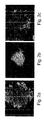
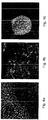
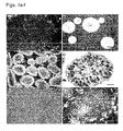
- exhibited typical hESC morphology e.g., round cells with large nuclei; for example Figures 5a-g , 18a-e
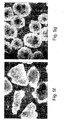
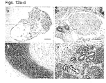
- hESCs cultured in the suspension cultures maintained their pluripotent capacity as evidenced by their ability to form embryoid bodies (EBs) or teratomas containing representative tissues of all three embryonic germ layers ( Figures 12a-d and data not shown).
- EBs embryoid bodies
- teratomas containing representative tissues of all three embryonic germ layers Figures 12a-d and data not shown.
- a method of expanding and maintaining embryonic stem cells in an undifferentiated state is effected by culturing the embryonic stem cells in a suspension culture under culturing conditions devoid of substrate adherence and which allow expansion of the embryonic stem cells in the undifferentiated state, thereby expanding and maintaining the embryonic stem cells in the undifferentiated state.
- embryonic stem cells refers to embryonic cells which are capable of differentiating into cells of all three embryonic germ layers (i.e., endoderm, ectoderm and mesoderm), or remaining in an undifferentiated state.
- Preferred embryonic stem cells according to this aspect of the present disclosure are of a human or primate (e.g., monkey) origin.
- embryonic stem cells can be obtained using well-known cell-culture methods.
- human embryonic stem cells can be isolated from human blastocysts. Human blastocysts are typically obtained from human in vivo preimplantation embryos or from in vitro fertilized (IVF) embryos. Alternatively, a single cell human embryo can be expanded to the blastocyst stage.
- ICM inner cell mass
- the ICM is then plated in a tissue culture flask containing the appropriate medium which enables its outgrowth. Following 9 to 15 days, the ICM derived outgrowth is dissociated into clumps either by a mechanical dissociation or by an enzymatic degradation and the cells are then re-plated on a fresh tissue culture medium. Colonies demonstrating undifferentiated morphology are individually selected by micropipette, mechanically dissociated into clumps, and re-plated. Resulting ES cells are then routinely split every 4-7 days. For further details on methods of preparing human ES cells see Thomson et al., (U.S. Pat. No. 5,843,780 ; Science 282: 1145, 1998 ; Curr. Top. Dev.
- human ESCs can be purchased from the NIH human embryonic stem cells registry (http://escr.nih.gov).
- Non-limiting examples of commercially available embryonic stem cell lines are BG01, BG02, BG03, BG04, CY12, CY30, CY92, CY10, TE03 and TE32.
- EBCs extended blastocyst cells
- the zona pellucida Prior to culturing the blastocyst, the zona pellucida is digested [for example by Tyrode's acidic solution (Sigma Aldrich, St Louis, MO, USA)] so as to expose the inner cell mass.
- the blastocysts are then cultured in vitro as whole embryos for at least nine and no more than fourteen days post fertilization (i.e., prior to the gastrulation event) using standard embryonic stem cell culturing methods.
- WO2006/040763 to the present inventors.
- EG cells are prepared from the primordial germ cells obtained from fetuses of about 8-11 weeks of gestation (in the case of a human fetus) using laboratory techniques known to anyone skilled in the arts.
- the genital ridges are dissociated and cut into small chunks which are thereafter disaggregated into cells by mechanical dissociation.
- the EG cells are then grown in tissue culture flasks with the appropriate medium.
- the cells are cultured with daily replacement of medium until a cell morphology consistent with EG cells is observed, typically after 7-30 days or 1-4 passages.
- Shamblott et al., Proc. Natl. Acad. Sci. USA 95: 13726, 1998 and U.S. Pat. No. 6,090,622 are additional details on methods of preparation human EG cells.
- embryonic stem cells in an undifferentiated state are of a distinct morphology, which is clearly distinguishable by the skilled in the art from that of differentiated cells of embryo or adult origin.
- undifferentiated embryonic stem cells typically have high nuclear/cytoplasmic ratios, prominent nucleoli and compact colony formation with poorly discernable cell junctions. Additional features of the undifferentiated state of the embryonic stem cells are further described hereinunder.
- expanding embryonic stem cells refers to obtaining a plurality of embryonic stem cells from a single or a population of embryonic stem cells.
- expanding embryonic stem cells refers also to increasing the number of embryonic stem cells over the culturing period. It will be appreciated that the number of cells which can be obtained from a single embryonic stem cell depends on the proliferation capacity of the cell.
- the proliferation capacity of an embryonic stem cell can be calculated by the doubling time of the cell (i.e., the time needed for a cell to undergo a mitotic division in the culture) and the period the stem cell can be maintained in the undifferentiated state while in culture (which is equivalent to the number of passages multiplied by the days between each passage).
- hESCs could be maintained in the suspension culture of the present disclosure for at least 80 days while being subjected to 17 serial passaging (culture splitting) which occurred every 4-6 days. Given that the hESCs cultured in suspension exhibited a doubling time of 36 hours (e.g., when cultured on the CM100F medium), a single hESC cultured under these conditions could be expanded to give rise to 2 45 hESCs (i.e., 3.5 x 10 13 hESCs).
- the method according to this aspect of the present disclosure is effected by culturing the embryonic stem cells in a suspension culture under culturing conditions devoid of substrate adherence and which allow expansion of the embryonic stem cells in the undifferentiated state.
- suspension culture refers to a culture in which the embryonic stem cells are suspended in a medium rather than adhering to a surface.
- the culture of the present disclosure is "devoid of substrate adherence" in which the embryonic stem cells are capable of expanding without adherence to an external substrate such as components of extracellular matrix, a glass microcarrier or beads.
- Culturing according to this aspect of the present disclosure is effected by plating the stem cells in a culture vessel at a cell density which promotes cell survival and proliferation but limits differentiation. Typically, a plating density of between about 5 x 10 4 - 2 x 10 5 cells per ml is used. It will be appreciated that although single-cell suspensions of stem cells are usually seeded, small clusters such as 10-200 cells may also be used.
- the culture medium can be replaced on a daily basis, or, at a pre-determined schedule such as every 2-3 days.
- replacement of the culture medium can be performed by subjecting the ESC suspension culture to centrifugation for about 3 minutes at 80 g, and resuspension of the formed ESC pellet in a fresh medium.
- a culture system in which the culture medium is subject to constant filtration or dialysis so as to provide a constant supply of nutrients or growth factors to the ESCs may be employed.
- the formed ESC clumps are dissociated every 5-7 days and the single cells or small clumps of cells are either split into additional culture vessels (i.e., passaged) or remained in the same culture vessel yet with additional culture medium.
- a pellet of ESCs which may be achieved by centrifugation as described hereinabove
- an isolated ESC clump can be subject to enzymatic digestion and/or mechanical dissociation.
- Enzymatic digestion of ESC clump(s) can be performed by subjecting the clump(s) to an enzyme such as type IV Collagenase (Worthington biochemical corporation, Lakewood, NJ, USA) and/or Dispase (Invitrogen Corporation products, Grand Island NY, USA).
- an enzyme such as type IV Collagenase (Worthington biochemical corporation, Lakewood, NJ, USA) and/or Dispase (Invitrogen Corporation products, Grand Island NY, USA).
- the time of incubation with the enzyme depends on the size of cell clumps present in the suspension culture. Typically, when hESC cell clumps are dissociated every 5-7 days while in the suspension culture, incubation of 20-60 minutes with 1.5 mg/ml type IV Collagenase results in small cell clumps which can be further cultured in the undifferentiated state.
- ESC clumps can be subjected to incubation of about 25 minutes with 1.5 mg/ml type IV Collagenase followed by five minutes incubation with 1 mg/ml Dispase, essentially as described under "General Materials and Experimental Methods" of the Examples section which follows. It should be noted that passaging of human ESCs with trypsin may result in chromosomal instability and abnormalities (see for example, Mitalipova MM., et al., Nature Biotechnology, 23: 19-20, 2005 and Cowan CA et al., N. Engl. J. of Med. 350: 1353-1356, 2004 ), and therefore should be avoided.
- Mechanical dissociation of large ESC clumps can be performed using a device designed to break the clumps to a predetermined size. Such a device can be obtained from CellArtis Goteborg, Sweden. Additionally or alternatively, mechanical dissociation can be manually performed using a needle such as a 27g needle (BD Microlance, Drogheda, Ireland) while viewing the clumps under an inverted microscope.
- a needle such as a 27g needle (BD Microlance, Drogheda, Ireland) while viewing the clumps under an inverted microscope.
- the dissociated ESC clumps are further broken to small clumps using 200 ⁇ l Gilson pipette tips (e.g., by pipetting up and down the cells).
- the culture vessel used for culturing the ESC in suspension according to the method of this aspect of the present disclosure can be any tissue culture vessel (e.g., with a purity grade suitable for culturing ESCs) having an internal surface designed such that ESC cultured therein are unable to adhere or attach to such a surface (e.g., non-tissue culture treated cells, to prevent attachment or adherence to the surface).
- culturing according to this aspect of the present disclosure is effected using a controlled culturing system (preferably a computer-controlled culturing system) in which culture parameters such as temperature, agitation, pH, and pO 2 is automatically performed using a suitable device. Once the culture parameters are recorded, the system is set for automatic adjustment of culture parameters as needed for ESCs expansion.
- culturing according to the method of this aspect of the present disclosure can be performed under dynamic conditions (i.e., under conditions in which the ESCs are subject to constant movement while in the suspension culture) or under non-dynamic conditions (i.e ., a static culture).
- the ESCs can be cultured in uncoated 58 mm petri dishes (Greiner, Frickenhausen, Germany).
- the ESCs can be cultured in spinner flasks [e.g., of 200 ml to 1000 ml, for example 250 ml which can be obtained from CellSpin of Integra Biosciences, Fernwald, Germany; of 100 ml which can be obtained from Bellco,Vineland, NJ; or in 125 ml Erlenmeyer (Corning Incorporated, Corning NY, USA)] which can be connected to a control unit and thus present a controlled culturing system.
- spinner flasks e.g., of 200 ml to 1000 ml, for example 250 ml which can be obtained from CellSpin of Integra Biosciences, Fernwald, Germany; of 100 ml which can be obtained from Bellco,Vineland, NJ; or in 125 ml Erlenmeyer (Corning Incorporated, Corning NY, USA)
- the medium used to culture the ESCs in suspension according to the method of this aspect of the present disclosure can be any culture medium capable of supporting the growth of ESCs while maintaining them in an undifferentiated state.
- a culture medium can be a water-based medium which includes a combination of substances such as salts, nutrients, minerals, vitamins, amino acids, nucleic acids, proteins such as cytokines, growth factors and hormones, all of which are needed for cell proliferation and are capable of maintaining the ESCs in an undifferentiated state.
- a culture medium according to this aspect of the present disclosure can be a synthetic tissue culture medium such as Ko-DMEM (Gibco-Invitrogen Corporation products, Grand Island, NY, USA), DMEM/F12 (Biological Industries, Biet Haemek, Israel), Mab ADCB medium (HyClone, Utah, USA) or DMEM/F12 (Biological Industries, Biet Haemek, Israel) supplemented with the necessary additives as is further described hereinunder.
- all ingredients included in the culture medium of the present disclosure are substantially pure, with a tissue culture grade.
- the culture medium used by the method of this aspect of the present disclosure should be well-defined (i.e ., with known and constant components) and xeno-free (i.e., devoid of xeno contaminants).
- the culture medium used by the method of this aspect of the present disclosure is serum-free, serum replacement-free, xeno-free, feeder-free (i.e., devoid of feeder cells) and protein carrier-free.
- Serum or serum replacement are usually added to most culture media which are designed for culturing stem cells, and particularly, embryonic stem cells, in order to provide the cells with the optimal environment, similar to that present in vivo (i.e., within the organism from which the cells are derived, e.g., a blastocyst of an embryo or an adult tissue of a postnatal individual).
- serum which is derived from either an animal source (e.g., bovine serum) or a human source (human serum) is limited by the significant variations in serum components between individuals and the risk of having xeno contaminants (in case of an animal serum is used)
- the use of the more defined composition such as the currently available serum replacementTM (Gibco-Invitrogen Corporation, Grand Island, NY USA) may be limited by the presence of Albumax (Bovine serum albumin enriched with lipids) which is from an animal source within the composition (International Patent Publication No. WO 98/30679 to Price, P.J. et al ).
- a protein carrier refers to a protein which acts in the transfer of proteins or nutrients (e.g., minerals such as zinc) to the cells in the culture.
- protein carriers can be, for example, albumin (e.g., bovine serum albumin), Albumax (lipid enriched albumin) or plasmanate (human plasma isolated proteins). Since these carriers are derived from either human or animal sources their use in hESCs cultures is limited by batch-specific variations and/or exposure to pathogens. On the other hand, the recombinant human albumin, which is substantially pure and devoid of animal contaminants is highly expensive, thus not commonly used in hESCs cultures. Thus, a culture medium which is devoid of a protein carrier is highly advantageous since it enables a truly defined medium that can be manufacture from recombinant or synthetic materials.
- a culture medium which is serum-free, serum replacement-free, xeno-free, feeder-free and protein carrier-free can be a culture medium which comprises a TGF ⁇ isoform (for non-limiting examples see the D1, D2, HA16 or HA19 culture media which are described in Examples 1 and 2 of the Examples section which follows).
- TGF ⁇ isoform refers to any isoform of the transforming growth factor beta ( ⁇ ) including TGF ⁇ 1 (e.g., homo sapiens TGF ⁇ 1, GenBank Accession No. NP_000651), TGF ⁇ 2 (e.g., homo sapiens TGF ⁇ 2, GenBank Accession No. NP_003229) and TGF ⁇ 3 (e.g., homo sapiens TGF ⁇ 3, GenBank Accession No. NP_003230) which function through the same receptor signaling system in the control of proliferation, differentiation, and other functions in many cell types.
- TGF ⁇ acts in inducing transformation and also acts as a negative autocrine growth factor.
- the TGF ⁇ isoform which is included in the culture medium of the present disclosure is TGF ⁇ 1 or TGF ⁇ 3 .
- TGF ⁇ isoforms can be obtained from various commercial sources such as R&D Systems Minneapolis MN, USA.
- TGF ⁇ 1 e.g., the D1 medium which contains 0.12 ng/ml TGF ⁇ 1
- TGF ⁇ 3 e.g., the D2 medium, the HA16 medium or the HA19 medium which contain 2 ng/ml TGF ⁇ 3
- TGF ⁇ 1 which is included in the culture medium of this aspect of the present disclosure is present at a concentration of at least 0.06 ng/ml, more preferably, at least 0.07 ng/ml, more preferably, at least 0.08 ng/ml, more preferably, at least 0.09 ng/ml, more preferably, at least 0.1 ng/ml, more preferably, at least 0.11 ng/ml, even more preferably, at least 0.12 ng/ml.
- TGF ⁇ 3 which is included in the culture medium of this aspect of the present disclosure is present at a concentration of at least 0.5 ng/ml, more preferably, at least 0.6 ng/ml, more preferably, at least 0.8 ng/ml, more preferably, at least 0.9 ng/ml, more preferably, at least 1 ng/ml, more preferably, at least 1.2 ng/ml, more preferably, at least 1.4 ng/ml, more preferably, at least 1.6 ng/ml, more preferably, at least 1.8 ng/ml, even more preferably, at least 2 ng/ml.
- the TGF ⁇ -containing culture medium of this aspect of the present disclosure further includes basic fibroblast growth factor (bFGF).
- bFGF can be obtained from any commercial supplier of tissue culture ingredients such as Invitrogen Corporation products, Grand Island NY, USA.
- the bFGF which is included in TGF ⁇ -containing culture medium of this aspect of the present disclosure is present at a concentration of at least 2 ng/ml, at least 3 ng, at least 4 ng/ml, at least 5 ng/ml, at least 6 ng/ml, at least 7 ng, at least 8 ng/ml, at least 9 ng/ml, at least 10 ng/ml.
- a culture medium which is based on the IL6RIL6 chimera and is serum or serum replacement-free, xeno-free and protein carrier-free can be also used along with the method of this aspect of the present disclosure.
- IL6RIL6 refers to a chimeric polypeptide which comprises the soluble portion of interleukin-6 receptor (IL-6-R, e.g., the human IL-6-R as set forth by GenBank Accession No. AAH89410) (e.g., a portion of the soluble IL6 receptors as set forth by amino acids 112-355 of GenBank Accession No. AAH89410) and the interleukin-6 (IL6) (e.g., human IL-6 as set forth by GenBank Accession No. CAG29292) or a biologically active fraction thereof (e.g., a receptor binding domain).
- the IL6RIL6 chimera used by the method according to this aspect of the present disclosure is capable of supporting the undifferentiated growth of human embryonic stem cells, while maintaining their pluripotent capacity.
- the two functional portions i.e., the IL6 and its receptor
- the two functional portions can be directly fused (e.g., attached or translationally fused, i.e., encoded by a single open reading frame) to each other or conjugated (attached or translationally fused) via a suitable linker (e.g., a polypeptide linker).
- the IL6RIL6 chimeric polypeptide exhibits a similar amount and pattern of glycosylation as the naturally occurring IL6 and IL6 receptor.
- a suitable IL6RIL6 chimera is as set forth in SEQ ID NO:31 and in Figure 11 of WO 99/02552 to Revel M., et al.
- the IL6RIL6 chimera which is included in the culture medium of this aspect of the present disclosure is present at a concentration of at least 25 ng/ml, preferably at least 50 ng/ml, preferably, at least 100 ng/ml, preferably, at least 200 ng/ml, preferably, at least 300 ng/ml.
- concentration of the IL6RIL6 chimera can vary depending on the purity of the chimeric polypeptide following its synthesis or recombinant expression and those of skills in the art are capable of adjusting the optimal concentration depending on such purity.
- the IL6RIL6-containing culture medium of this aspect of the present disclosure includes at least 2 ng/ml bFGF, at least 3 ng/ml, at least 4 ng/ml, at least 5 ng/ml, at least 6 ng/ml, at least 7 ng, at least 8 ng/ml, at least 9 ng/ml, at least 10 ng/ml bFGF.
- a suitable IL6RIL6-containing culture medium which can be used for culturing the ESC in a suspension culture can be the HACM100 culture medium described under the "General Materials and Experimental Methods" and Example 2 of the Examples section which follows, which was shown capable of maintaining hESCs in an undifferentiated state for at least 1-2 passages.
- a culture medium which is based on soluble interleukin-6 receptor (sIL6R) [e.g., GenBank Accession No. NM_000565.2, NM_181359.1, NP_000556.1, NP_852004.1] and soluble interleukin-6 (IL6) [e.g., GenBank Accession No. NM_000600.1, NP_000591.1] (separately) can be also used along with the method of the present disclosure.
- sIL6R soluble interleukin-6 receptor
- IL6R soluble interleukin-6 receptor
- a culture medium such as the yFIL25 which comprises 25 ng IL6 and 25 ng sIL6R can be used to culture, expand and maintain human ESCs in a pluripotent, proliferative and undifferentiated state for at least 19 passages.
- human ESCs cultured in such a culture medium expressed markers characteristics of the undifferentiated state, exhibited normal chromosomal karyotype (as tested after 14 passages) and were capable of forming EBs which included all three embryonic germ layers (pluripotent).
- the sIL6R is present at a concentration of at least 10 nanogram per milliliter (ng/ml), more preferably, at least 15 ng/ml, more preferably, at least 20 ng/ml, e.g., at least 22 ng/ml, 25 ng/ml, 27 ng/ml, or 30 ng/ml.
- ng/ml nanogram per milliliter
- sIL6R can be present at a concentration of 15-30 ng/ml, e.g., 25 ng/ml.
- sIL6R and IL6 can be obtained, for example, from R&D systems, Minneapolis, MN, USA.
- a culture medium which is based on leukemia inhibitory factor [e.g., GenBank Accession No. NM_002309.2 (mRNA) or NP_002300.1 (protein)] can be also used along with the method of the present disclosure.
- LIF leukemia inhibitory factor
- a culture medium such as the yFL1, yFL2, or yFL3 can be used to culture, expand and maintain human ESCs in a pluripotent, proliferative and undifferentiated state for at least 18 passages.
- human ESCs cultured in such a culture medium expressed markers characteristics of the undifferentiated state, exhibited normal chromosomal karyotype (as tested after 14 passages) and were capable of forming EBs which included all three embryonic germ layers (pluripotent).
- LIF is present at a concentration of at least 1000 units/ml, more preferably, at least 2000 units/ml, more preferably, at least 3000 units/ml.
- Human recombinant leukemia inhibitory factor (hrLIF) can be obtained from R&D Systems Minneapolis MN, USA.
- a culture medium which is based on leukemia inhibitory factor (LIF) [e.g., GenBank Accession No. NM_002309.2 (mRNA) or NP_002300.1 (protein)] and TGF ⁇ 1 can be used along with the method of the present disclosure.
- LIF leukemia inhibitory factor
- a culture medium such as the TLF medium can be used to culture, expand and maintain human ESCs in a pluripotent, proliferative and undifferentiated state for at least 31 passages.
- human ESCs cultured in such a culture medium expressed markers characteristics of the undifferentiated state, exhibited normal chromosomal karyotype (as tested after 18 passages) and were capable of forming EBs which included all three embryonic germ layers (pluripotent).
- any of the proteinaceous factors used in the culture medium of the present disclosure can be recombinantly expressed or biochemically synthesized.
- naturally occurring proteinaceous factors such as bFGF and TGF ⁇ can be purified from biological samples (e.g., from human serum, cell cultures) using methods well known in the art.
- Biochemical synthesis of the proteinaceous factors of the present disclosure can be performed using standard solid phase techniques. These methods include exclusive solid phase synthesis, partial solid phase synthesis methods, fragment condensation and classical solution synthesis.
- Recombinant expression of the proteinaceous factors of the present disclosure can be generated using recombinant techniques such as described by Bitter et al., (1987) Methods in Enzymol. 153:516-544 , Studier et al. (1990) Methods in Enzymol. 185:60-89 , Brisson et al. (1984) Nature 310:511-514 , Takamatsu et al. (1987) EMBO J. 10-157-311 , Coruzzi et al. (1984) EMBO J. 3:1671-1680 , Brogli et al., (1984) Science 224:838-843 , Gurley et al.
- IL6RIL6 chimera can be generated as described in PCT publication WO 99/02552 to Revel M., et al. and Chebath J,, et al., 1997.
- a polynucleotide sequence encoding the IL6RIL6 chimera (e.g., the polypeptide set forth by SEQ ID NO:31) is preferably ligated into a nucleic acid construct suitable for expression in a host cell [ i.e., a cell in which the polynucleotide encoding the polypeptide-of-choice (e.g., the IL6RIL6 chimera) is expressed].
- the host cell employed is a eukaryotic host cell, more preferably a mammalian host cell such as human cell or CHO cell).
- mammalian expression vectors For expression in mammalian cells [e.g., CHO cells, human HEK 293 cells (ATCC CRL 1573)] a number of mammalian expression vectors can be used. Examples include, but are not limited to, pcDNA3, pcDNA3.1(+/-), pGL3, pZeoSV2(+/-), pSecTag2, pDisplay, pEF/myc/cyto, pCMV/myc/cyto, pCR3.1, pSinRep5, DH26S, DHBB, pNMT1, pNMT41, pNMT81, which are available from Invitrogen, pCI which is available from Promega, pMbac, pPbac, pBK-RSV and pBK-CMV which are available from Strategene, pTRES which is available from Clontech, and their derivatives.
- mammalian expression vectors include, but are not limited to,
- Expression vectors containing regulatory elements from eukaryotic viruses such as retroviruses can be also used.
- SV40 vectors include pSVT7 and pMT2.
- Vectors derived from bovine papilloma virus include pBV-1MTHA, and vectors derived from Epstein Bar virus include pHEBO, and p2O5.
- exemplary vectors include pMSG, pAV009/A + , pMTO10/A + , pMAMneo-5, baculovirus pDSVE, and any other vector allowing expression of proteins under the direction of the SV-40 early promoter, SV-40 later promoter, metallothionein promoter, murine mammary tumor virus promoter, Rous sarcoma virus promoter, polyhedrin promoter, or other promoters shown effective for expression in eukaryotic cells.
- Transformed cells are cultured under effective conditions, which allow for the expression of high amounts of the recombinant polypeptide (e.g., the IL6RIL6 chimera). Following a predetermined time in culture, recovery of the recombinant polypeptide is effected.
- the phrase "recovery of the recombinant polypeptide" used herein refers to collecting the whole fermentation medium containing the polypeptide and need not imply additional steps of separation or purification.
- polypeptides of the present disclosure can be purified using a variety of standard protein purification techniques, such as, but not limited to, affinity chromatography, ion exchange chromatography, filtration, electrophoresis, hydrophobic interaction chromatography, gel filtration chromatography, reverse phase chromatography, concanavalin A chromatography, chromatofocusing and differential solubilization.
- standard protein purification techniques such as, but not limited to, affinity chromatography, ion exchange chromatography, filtration, electrophoresis, hydrophobic interaction chromatography, gel filtration chromatography, reverse phase chromatography, concanavalin A chromatography, chromatofocusing and differential solubilization.
- the polypeptide of the present disclosure (e.g., the IL6RIL6 chimera) is preferably retrieved in "substantially pure” form.
- the phrase “substantially pure” refers to a purity that allows for the effective use of the polypeptide of the present disclosure (e.g., the IL6RIL6 chimera) in maintaining the human embryonic stem cells in an undifferentiated state while in culture.
- the culture media which comprise the IL6RIL6 chimera but also include serum or serum replacementTM (e.g., the CM100F medium or similar media with other concentrations of the IL6RIL6 chimera such as 200 or 300 ng/ml as described in the Examples section which follows) can be used by the method of this aspect of the present disclosure.
- the serum e.g., human serum
- serum replacementTM can be provided at various concentrations, such as a concentration of at least 10 %, e.g., a concentration of at least 15 %, at least 20 %, at least 25 % or at least 30 %.
- Serum replacementTM includes albumin or albumin substitutes, amino acids, vitamins, transferrins or transferrin substitutes, antioxidants, insulin or insulin substitutes, collagen precursors and trace elements (International Patent Publication No. WO 98/30679 to Price, P.J. et al ).
- albumin or albumin substitutes are preferably derived from a human source and/or are prepared using recombinant techniques in host cells as described hereinabove.
- embryonic stem cell growth is monitored to determine their differentiation state.
- the differentiation state can be determined using various approaches including, for example, morphological evaluation (e.g., as shown in Figures 5a-g ) and/or detection of the expression pattern of typical markers of the undifferentiated state using immunological techniques such as flow cytometry for membrane-bound markers, immunohistochemistry or immunofluorescence for extracellular and intracellular markers and enzymatic immunoassay, for secreted molecular markers.
- immunofluorescence employed on hESCs cultured according to the method of this aspect of the present disclosure revealed the expression of Oct4, stage-specific embryonic antigen (SSEA) 4, the tumour-rejecting antigen (TRA)-1-60 and TRA-1-81 ( Figures 4a-c and data not shown).
- SSEA stage-specific embryonic antigen
- TRA tumour-rejecting antigen
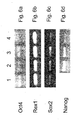
- the level of transcripts of specific undifferentiation markers e.g., Oct 4, Nanog, Sox2 and Rexl as shown in Figures 6a-d
- differentiation markers e.g., albumin, glucagons, ⁇ -cardiac actin, ⁇ -globulin, Flk1, AC133 and neurofilament
- RNA-based techniques such as RT-PCR analysis and/or cDNA microarray analysis.
- Determination of ES cell differentiation can be also effected via measurements of alkaline phosphatase activity.
- Undifferentiated human ES cells have alkaline phosphatase activity which can be detected by fixing the cells with 4 % paraformaldehyde and developing with the Vector Red substrate kit according to manufacturer's instructions (Vector Laboratories, Burlingame, California, USA).
- the embryonic stem cells cultured in any of the suspension culture media described hereinabove exhibit normal chromosomal karyotype following at least 1 passage, preferably, following at least 2 passages, preferably, following at least 3 passages, preferably, following at least 4 passages, preferably, following at least 5 passages, preferably, following at least 7 passages, preferably, following at least 10 passages, preferably, following at least 12 passages, preferably, following at least 15 passages, preferably, following at least 20 passages, preferably, following at least 25 passages, preferably, following at least 30 passages (e.g., hESCs exhibited normal karyotype following at least 14, 18, 23 or 36 passages), thus representing genetically stable human ESC lines.
- hESCs exhibited normal karyotype following at least 14, 18, 23 or 36 passages
- the embryonic stem cells cultured in any of the suspension culture media described hereinabove exhibit a doubling time of at least 20 hours, more preferably, a doubling time which is between 20 to 40 hours (e.g., about 36 hours), thus representing a non-tumorigenic, genetically stable human ESCs.
- the present disclosure provides, for the first time, a cell culture which comprises embryonic stem cells and a culture media which comprises the soluble interleukin-6 receptor (sIL6R) and interleukin-6 (IL6) (separately) wherein the soluble IL6R is present at a concentration of at least 10 (ng/ml) (e.g., 25 ng/ml), and whereas the culture medium being capable of maintaining the embryonic stem cells in an undifferentiated state for at least 5 passages (see Example 4 of the Examples section which follows, which demonstrates undifferentiated hESCs following 19 passages).
- sIL6R soluble interleukin-6 receptor
- IL6 interleukin-6
- the present disclosure provides, for the first time, a cell culture which comprises human embryonic stem cells and a culture media which comprises LIF (at a concentration of at least 1000 u/ml), wherein the culture medium being capable of maintaining the human ESCs in an undifferentiated state for at least 18 passages (see Example 4 of the Examples section which follows).
- a method of deriving an embryonic stem cell line is effected by (a) obtaining an embryonic stem cell from a pre-implantation stage blastocyst, post-implantation stage blastocyst and/or a genital tissue of a fetus; and (b) culturing the embryonic stem cell in a suspension culture, under culturing conditions which allow expansion of the embryonic stem cells in an undifferentiated state, thereby deriving the embryonic stem cell line.
- the term "deriving” as used herein refers to generating an embryonic stem cell line from at least one embryonic stem cell.
- embryonic stem cell line refers to embryonic stem cells which are derived from a single or a group of embryonic stem cells of a single organism (e.g., a single human blastocyst), and which are characterized by the ability to proliferate in culture while maintaining the undifferentiated state and the pluripotent capacity.
- obtaining an embryonic stem cell from a pre-implantation stage blastocyst, post-implantation stage blastocyst and/or a genital tissue of a fetus can be performed using methods known in the art, as described hereinabove and in the "General Materials and Experimental Methods" of the Examples section which follows.
- the zona pellucida is removed from a 5-7 day-old blastocyst using Tyrode's acidic solution (Sigma, St Louis MO, USA), the trophoblast layer is specifically removed either by immunosurgery or mechanically using 27g needles and the exposed ICM is either directly cultured in a suspension culture with a suitable culture medium (e.g., the CM100F, HA16 or D2 medium) for 4-10 days (in case a preimplantation blastocyst is used) or subject to in vitro implantation by culturing the ICM for 6-8 days (to obtain cells of a 13 day-old blastocyst in case a post-implantation/pre-gastrulation blastocyst is used) on feeder layers or a feeder-free culturing system which allow implantation of the blastocyst to the surface, following which the implanted cells are isolated and further cultured in suspension as described hereinunder.
- a suitable culture medium e.g., the CM100F
- the genital ridges are dissociated and cut into small chunks which are thereafter disaggregated into cells by mechanical dissociation.
- the single cell EG cells are then cultured in suspension culture with a suitable culture medium (e.g., the CM100F, HA16 or D2 medium) for 4-10 days.
- a suitable culture medium e.g., the CM100F, HA16 or D2 medium
- the ESCs are further cultured in suspension under conditions which allow expansion of the embryonic stem cells in the undifferentiated state, essentially as described hereinabove.
- the cell culture of the present disclosure is characterized by at least 40 %, at least 50 %, at least 60 %, more preferably at least 70 %, more preferably at least 80 %, most preferably at least 85 % of undifferentiated embryonic stem cells.
- an established embryonic stem cell line can be subject to freeze/thaw cycles without hampering the proliferative capacity of the cells in the undifferentiated state while preserving their pluriptent capacity.
- hESCs were successfully frozen and thawed.
- hESCs which were expanded and maintained in the suspension culture described hereinabove are pluripotent (i.e., capable of differentiating into all cell types of the three embryonic germ layers, the ectoderm, the endoderm and the mesoderm) as evidenced in vitro (by the formation of EBs).
- hESCs cultured according to the teachings of the present disclosure can be used as a source for generating differentiated, lineage-specific cells.
- Such cells can be obtained directly from the ESCs by subjecting the ESCs to various differentiation signals (e.g., cytokines, hormones, growth factors) or indirectly, via the formation of embryoid bodies and the subsequent differentiation of cells of the EBs to lineage-specific cells.
- various differentiation signals e.g., cytokines, hormones, growth factors
- a method of generating embryoid bodies from embryonic stem cells is effected by (a) culturing the embryonic stem cells in a suspension culture under culturing conditions which allow expansion of the embryonic stem cells in an undifferentiated state to thereby obtain expanded, undifferentiated embryonic stem cells; and (b) subjecting the expanded, undifferentiated embryonic stem cells to culturing conditions suitable for differentiating the embryonic stem cells to embryoid bodies; thereby generating the embryoid bodies from the embryonic stem cells.
- embryonic bodies refers to morphological structures comprised of a population of ESCs, extended blastocyst cells (EBCs) and/or embryonic germ cells (EGCs) which have undergone differentiation.
- EBCs extended blastocyst cells
- EECs embryonic germ cells
- EBs formation initiates following the removal of differentiation blocking factors from ES cell cultures.
- ESCs proliferate into small masses of cells which then proceed with differentiation.
- a layer of endodermal cells is formed on the outer layer of the small mass, resulting in "simple EBs”.
- complex EBs are formed. Complex EBs are characterized by extensive differentiation of ectodermal and mesodermal cells and derivative tissues.
- the method of this aspect of the present disclosure involves the culturing of ESCs in a suspension culture using any of the culture media described hereinabove in order to obtain expanded, undifferentiated embryonic stem cells and then subjecting the expanded, undifferentiated ESCs to culturing conditions suitable for differentiating the ESCs to embryoid bodies.
- Such culturing conditions are substantially devoid of differentiation inhibitory factors which were employed during step (a), e.g., a TGF ⁇ isoform or the IL6RIL6 chimera.
- a culture medium suitable for EBs formation may include a basic culture medium (e.g., Ko-DMEM or DMEM/F12) supplemented with 20 % FBSd (HyClone, Utah, USA), 1 mM L-glutamine, 0.1 mM ⁇ -mercaptoethanol, and 1 % non-essential amino acid stock.
- EBs can be effected by morphological evaluations (e.g., histological staining as described in Example 2) and determination of expression of differentiation-specific markers [using e.g., immunological techniques or RNA-based analysis (e.g., RT-PCR, cDNA microarray)].
- differentiation-specific markers of all three embryonic germ layers include albumin and glucagon (typical of the embryonic endoderm), ⁇ -cardiac actin, ⁇ -globulin and Flk1 (typical of the embryonic mesoderm), and AC133 and neurofilament (NFH) (typical of the embryonic ectoderm).
- cells of the EBs can be further subjected to culturing conditions suitable for lineage-specific cells.
- the method of this aspect of the present disclosure further includes step (c): subjecting cells of the embryoid bodies to culturing conditions suitable for differentiating and/or expanding lineage specific cells; thereby generating the lineage-specific cells from the embryonic stem cells.
- culturing conditions suitable for differentiating and/or expanding lineage specific cells refers to a combination of a culture system, e.g., feeder cell layers, feeder-free matrix or a suspension culture and a culture medium which are suitable for the differentiation and/or expansion of specific cell lineages derived from cells of the EBs.
- a culture system e.g., feeder cell layers, feeder-free matrix or a suspension culture and a culture medium which are suitable for the differentiation and/or expansion of specific cell lineages derived from cells of the EBs.
- the method of this aspect of the present disclosure further includes isolating lineage specific cells following step (b).
- the phrase "isolating lineage specific cells” refers to the enrichment of a mixed population of cells in a culture with cells predominantly displaying at least one characteristic associated with a specific lineage phenotype. It will be appreciated that all cell lineages are derived from the three embryonic germ layers. Thus, for example, hepatocytes and pancreatic cells are derived from the embryonic endoderm, osseous, cartilaginous, elastic, fibrous connective tissues, myocytes, myocardial cells, bone marrow cells, vascular cells (namely endothelial and smooth muscle cells), and hematopoietic cells are differentiated from embryonic mesoderm and neural, retina and epidermal cells are derived from the embryonic ectoderm.
- isolating is effected by sorting of cells of the EBs via fluorescence activated cell sorter (FACS).
- FACS fluorescence activated cell sorter
- EBs are disaggregated using a solution of Trypsin and EDTA (0.025 % and 0.01 %, respectively), washed with 5 % fetal bovine serum (FBS) in phosphate buffered saline (PBS) and incubated for 30 minutes on ice with fluorescently-labeled antibodies directed against cell surface antigens characteristics to a specific cell lineage.
- FBS fetal bovine serum
- PBS phosphate buffered saline
- endothelial cells are isolated by attaching an antibody directed against the platelet endothelial cell adhesion molecule-1 (PECAM1) such as the fluorescently-labeled PECAM1 antibodies (30884X) available from PharMingen (PharMingen, Becton Dickinson Bio Sciences, San Jose, CA, USA) as described in Levenberg, S. et al., (Endothelial cells derived from human embryonic stem cells. Proc. Natl. Acad. Sci. USA. 2002. 99: 4391-4396 ).
- PECAM1 platelet endothelial cell adhesion molecule-1
- Hematopoietic cells are isolated using fluorescently-labeled antibodies such as CD34-FITC, CD45-PE, CD31-PE, CD38-PE, CD90-FITC, CD117-PE, CD15-FITC, class I-FITC, all of which IgG1 are available from PharMingen, CD133/1-PE (IgG1) (available from Miltenyi Biotec, Auburn, CA), and glycophorin A-PE (IgG1), available from Immunotech (Miami, FL). Live cells ( i.e. , without fixation) are analyzed on a FACScan (Becton Dickinson Bio Sciences) by using propidium iodide to exclude dead cells with either the PC-LYSIS or the CELLQUEST software.
- fluorescently-labeled antibodies such as CD34-FITC, CD45-PE, CD31-PE, CD38-PE, CD90-FITC, CD117-PE, CD15-FITC, class I-FITC, all of which IgG1
- isolated cells can be further enriched using magnetically-labeled second antibodies and magnetic separation columns (MACS, Miltenyi) as described by Kaufman, D.S. et al., (Hematopoietic colony-forming cells derived from human embryonic stem cells. Proc. Natl. Acad. Sci. USA. 2001, 98: 10716-10721 ).
- MCS magnetically-labeled second antibodies and magnetic separation columns
- isolating is effected by a mechanical separation of cells, tissues and/or tissue-like structures contained within the EBs.
- beating cardiomyocytes can be isolated from EBs as disclosed in U.S. Pat. Appl. No. 20030022367 to Xu et al.
- Four-day-old EBs of the present disclosure are transferred to gelatin-coated plates or chamber slides and are allowed to attach and differentiate.
- Spontaneously contracting cells which are observed from day 8 of differentiation, are mechanically separated and collected into a 15-mL tube containing low-calcium medium or PBS.
- Cells are dissociated using Collagenase B digestion for 60-120 minutes at 37 °C, depending on the Collagenase activity.
- Dissociated cells are then resuspended in a differentiation KB medium (85 mM KCI, 30 mM K 2 HPO 4 , 5 mM MgSO 4 , 1 mM EGTA, 5 mM creatine, 20 mM glucose, 2 mM Na 2 ATP, 5 mM pyruvate, and 20 mM taurine, buffered to pH 7.2, Maltsev et al., Circ. Res. 75:233, 1994 ) and incubated at 37 °C for 15-30 minutes. Following dissociation cells are seeded into chamber slides and cultured in the differentiation medium to generate single cardiomyocytes capable of beating.
- a differentiation KB medium 85 mM KCI, 30 mM K 2 HPO 4 , 5 mM MgSO 4 , 1 mM EGTA, 5 mM creatine, 20 mM glucose, 2 mM Na 2 ATP, 5 mM pyruvate, and 20 mM taurine
- isolating is effected by subjecting the EBs to differentiation factors to thereby induce differentiation of the EBs into lineage specific differentiated cells.
- EBs of the present disclosure are cultured for 5-12 days in tissue culture dishes including DMEM/F-12 medium with 5 mg/ml insulin, 50 mg/ml transferrin, 30 nM selenium chloride, and 5 mg/ml fibronectin (ITSFn medium, Okabe, S. et al., 1996, Mech. Dev. 59: 89-102 ).
- the resultant neural precursors can be further transplanted to generate neural cells in vivo ( Brüstle, O. et al., 1997. In vitro-generated neural precursors participate in mammalian brain development. Proc. Natl. Acad. Sci. USA. 94: 14809-14814 ). It will be appreciated that prior to their transplantation, the neural precursors are trypsinized and triturated to single-cell suspensions in the presence of 0.1 % DNase.
- EBs of the present disclosure can differentiate to oligodendrocytes and myelinate cells by culturing the cells in modified SATO medium, i.e., DMEM with bovine serum albumin (BSA), pyruvate, progesterone, putrescine, thyroxine, triiodothryonine, insulin, transferrin, sodium selenite, amino acids, neurotrophin 3, ciliary neurotrophic factor and Hepes ( Bottenstein, J. E. & Sato, G. H., 1979, Proc. Natl. Acad. Sci. USA 76, 514-517 ; Raff, M. C., Miller, R.
- modified SATO medium i.e., DMEM with bovine serum albumin (BSA), pyruvate, progesterone, putrescine, thyroxine, triiodothryonine, insulin, transferrin, sodium selenite, amino acids, neurotrophin 3, ciliary neurotrophic
- EBs are dissociated using 0.25 % Trypsin/EDTA (5 min at 37 °C) and triturated to single cell suspensions. Suspended cells are plated in flasks containing SATO medium supplemented with 5 % equine serum and 5 % fetal calf serum (FCS). Following 4 days in culture, the flasks are gently shaken to suspend loosely adhering cells (primarily oligodendrocytes), while astrocytes are remained adhering to the flasks and further producing conditioned medium. Primary oligodendrocytes are transferred to new flasks containing SATO medium for additional two days.
- FCS fetal calf serum
- oligospheres are either partially dissociated and resuspended in SATO medium for cell transplantation, or completely dissociated and a plated in an oligosphere-conditioned medium which is derived from the previous shaking step [ Liu, S. et al., (2000). Embryonic stem cells differentiate into oligodendrocytes and myelinate in culture and after spinal cord transplantation. Proc. Natl. Acad. Sci. USA. 97: 6126-6131 ].
- two-week-old EBs of the present disclosure are transferred to tissue culture dishes including DMEM medium supplemented with 10 % FCS, 2 mM L-glutamine, 100 units/ml penicillin, 100 mg/ml streptomycin, 20 % (v/v) WEHI-3 cell-conditioned medium and 50 ng/ml recombinant rat stem cell factor (rrSCF, Tsai, M. et al., 2000.
- rrSCF recombinant rat stem cell factor
- hemato-lymphoid cells from the EBs of the present disclosure, 2-3 days-old EBs are transferred to gas-permeable culture dishes in the presence of 7.5 % CO 2 and 5 % O 2 using an incubator with adjustable oxygen content. Following 15 days of differentiation, cells are harvested and dissociated by gentle digestion with Collagenase (0.1 unit/mg) and Dispase (0.8 unit/mg), both are available from F.Hoffman-La Roche Ltd, Basel, Switzerland. CD45-positive cells are isolated using anti-CD45 monoclonal antibody (mAb) M1/9.3.4.HL.2 and paramagnetic microbeads (Miltenyi) conjugated to goat anti-rat immunoglobulin as described in Potocnik, A.J.
- mAb monoclonal antibody
- Miltenyi paramagnetic microbeads
- the isolated CD45-positive cells can be further enriched using a single passage over a MACS column (Miltenyi).
- the culturing conditions suitable for the differentiation and expansion of the isolated lineage specific cells include various tissue culture media, growth factors, antibiotic, amino acids and the like and it is within the capability of one skilled in the art to determine which conditions should be applied in order to expand and differentiate particular cell types and/or cell lineages.
- lineage specific cells can be obtained by directly inducing the expanded, undifferentiated ESCs to culturing conditions suitable for the differentiation of specific cell lineage.
- EBs of the present disclosure can be used to generate lineage-specific cell lines which are capable of unlimited expansion in culture.
- Cell lines of the present disclosure can be produced by immortalizing the EB-derived cells by methods known in the art, including, for example, expressing a telomerase gene in the cells ( Wei, W. et al., 2003. Mol Cell Biol. 23: 2859-2870 ) or co-culturing the cells with NIH 3T3 hph-HOX11 retroviral producer cells ( Hawley, R.G. et al., 1994. Oncogene 9: 1-12 ).
- lineage-specific cells or cell lines obtained according to the teachings of the present disclosure are developed by differentiation processes similar to those naturally occurring in the human embryo they can be further used for human cell-based therapy and tissue regeneration.
- the present disclosure envisages the use of the expanded and/or differentiated lineage-specific cells or cell lines of the present disclosure for treating a disorder requiring cell replacement therapy.
- oligodendrocyte precursors can be used to treat myelin disorders (Repair of myelin disease: Strategies and progress in animal models. Molecular Medicine Today. 1997. pp. 554-561 ), chondrocytes or mesenchymal cells can be used in treatment of bone and cartilage defects ( U.S. Pat. No. 4,642,120 ) and cells of the epithelial lineage can be used in skin regeneration of a wound or burn ( U.S. Pat. No. 5,716,411 ).
- ESC-derived cells are preferably manipulated to over-express the mutated gene prior to their administration to the individual. It will be appreciated that for other disorders, the ESC-derived cells should be manipulated to exclude certain genes.
- Over-expression or exclusion of genes can be effected using knock-in and/or knock-out constructs [see for example, Fukushige, S. and Ikeda, J. E.: Trapping of mammalian promoters by Cre-lox site-specific recombination. DNA Res 3 (1996) 73-50 ; Bedell, M. A., Jerkins, N. A. and Copeland, N. G.: Mouse models of human disease. Part I: Techniques and resources for genetic analysis in mice. Genes and Development 11 (1997) 1-11 ; Bermingham, J. J., Scherer, S. S., O'Connell, S., Arroyo, E., Kalla, K. A., Powell, F. L. and Rosenfeld, M. G.: Tst-1/Oct-6/SCIP regulates a unique step in peripheral myelination and is required for normal respiration. Genes Dev 10 (1996) 1751-62 ].
- the lineage specific cells of the present disclosure can also be utilized to prepare a cDNA library.
- mRNA is prepared by standard techniques from the lineage specific cells and is further reverse transcribed to form cDNA.
- the cDNA preparation can be subtracted with nucleotides from embryonic fibroblasts and other cells of undesired specificity, to produce a subtracted cDNA library by techniques known in the art.
- the lineage specific cells of the present disclosure can be used to screen for factors (such as small molecule drugs, peptides, polynucleotides, and the like) or conditions (such as culture conditions or manipulation) that affect the differentiation of lineage precursor to terminally differentiated cells.
- factors such as small molecule drugs, peptides, polynucleotides, and the like
- conditions such as culture conditions or manipulation
- growth affecting substances, toxins or potential differentiation factors can be tested by their addition to the culture medium.
- ESC culture - Human embryonic stem cell (hESC) lines 1-6, 14 and 1-3 [Amit&Itskovitz-Eldor, 2002] were cultured with inactivated mouse embryonic fibroblasts (MEFs) for 40-60 passages in a "basic hESC culture medium" consisting of 85 % DMEM/F12 (Biological Industries, Biet Haemek, Israel) supplemented with 15 % serum replacement (SR), 2 mM L-glutamine, 0.1 mM ⁇ -mercaptoethanol, 1 % non-essential amino acid stock, and 4 ng/ml basic fibroblast growth factor (bFGF) (all but mentioned are from Gibco Invitrogen Corporation products, Grand Island NY, USA).
- This basic culture medium was used for the routine culture of hESCs in 2D culture with MEFs as control.
- Tested media on the feeder layer, feeder-free or suspension cultures - The tested medium were as follows:
- Feeder layers or feeder-free culturing systems To test the ability of various culture media to support the growth of hESC in an undifferentiated yet pluripotent state the hESCs were transferred to several culture systems:
- hESCs were cultured in suspension in 58 mm petri dishes (Greiner, Frickenhausen, Germany) in a cell density of 5 x 10 4 - 2 x 10 5 cells/ml.
- the HA16 medium was supplemented with 0.1 % F68 (Sigma, St. Louis, MO, USA) for the suspended culture.
- the culture medium in the suspension culture was changed on a daily basis.
- the basic media used for culturing hESCs in suspension (which can be further supplemented with the additive and growth factors as described hereinabove) were DMEM, ko-DMEM, DMEM/F12, MabADCB or NCTC medium.
- RT PCR analysis Total RNA was isolated from hESCs grown for 10-15 passages in the suspension culture using Tri-Reagent (Sigma, St. Louis MO, USA), according to the manufacturer's instructions. cDNA was synthesized from 1 ⁇ g total RNA using MMLV reverse transcriptase RNase H minus (Promega, Madison WI, USA). PCR reactions included denaturation for 5 minutes at 94 °C followed by repeated cycles of 94 °C for 30 seconds, annealing for 30 seconds at an annealing temperature as specified in Table 1, hereinbelow and extension at 72 °C for 30 seconds. PCR primers and reaction conditions used are described in Table 1, hereinbelow.
- Karyotype analysis was performed on at least 20 cells from each sample, two samples per test, as previously described [Amit et al, 2003].
- Karyotypes were analyzed and reported according to the "International System for Human Cytogenetic Nomenclature" (ISCN).
- hESCs cultured in suspension - For the formation of EBs, one to three 58 mm petri dishes (Greiner, Frickenhausen, Germany) containing ESCs in suspension cultures were transferred to new 58 mm petri dishes containing EBs-differentiation medium consisting of 80 % DMEM/F12 (Biological Industries, Beit Haemek, Israel), supplemented with 20 % FBSd (HyClone, Utah, USA), and 1 mM L-glutamine (Invitrogen Corporation, Grand Island NY, USA).
- 80 % DMEM/F12 Biological Industries, Beit Haemek, Israel
- FBSd HyClone, Utah, USA
- 1 mM L-glutamine Invitrogen Corporation, Grand Island NY, USA.
- the ESCs were subject to treatment with 1 mg/ml type IV collagenase and further broken into small clumps using 1000 ⁇ l Gilson pipette tips. 10 day-old EBs were harvested for RNA isolation and histological examination.
- 2-D feeder free or feeder layers
- ESCs were removed from their culture dish using 1 mg/ml type IV collagenase, further broken into small clumps using 1000 ⁇ l Gilson pipette tips, and cultured in suspension in 58 mm petri dishes (Greiner, Frickenhausen, Germany).
- EBs were grown in differentiation medium consisting of 80 % DMEM/F12 (Biological Industries, Beit Haemek, Israel), supplemented with 20 % FBSd (HyClone, Utah, USA), and 1 mM L-glutamine (Invitrogen Corporation, Grand Island NY, USA).
- Teratoma formation For teratoma formation, cells cultured in the offered culture methods for more than 15 passages, were injected into the rear leg muscle of 4-week-old male SCID-beige mice (two mice for each tested culture system). Cell numbers ranged from 5 x 10 6 cells to 10 7 cells per injection. Three to eight to 12 weeks after injection the mice were sacrificed and the resulting teratomas examined histologically.
- Blastocyst cultivation - Zygotes were donated by couples undergoing pre-implantation genetic diagnosis (PGD) or in vitro fertilization (IVF) at Cornell Medical College, NY, who signed informed consent forms. The couples underwent the traditional IVF procedure after ovarian stimulation with gonadotropins and oocyte retrieval. Zygotes were cultured to the blastocyst stage according to IVF laboratory standard protocol: under oil using specialized C1/C2 media for insemination, growth and blastocyst development (Cornell).
- hESC lines in a suspension culture - Following the removal of the zona pellucida using Tyrode's acidic solution (Sigma, St Louis MO, USA), the trophoblast layer is specifically removed either by immunosurgery or mechanically using 27g needles.
- the exposed ICM is further cultured in suspension culture with a suitable culture medium (e.g., the CM100F, HA16 or D2) for 4-10 days. Initially, the cells are mechanically split using 27g needles.
- a suitable culture medium e.g., the CM100F, HA16 or D2
- hESC lines on foreskin fibroblasts After digestion of the zona pellucida by Tyrode's acidic solution (Sigma, St Louis MO, USA) or its mechanical removal, the exposed blastocysts were placed in whole on a mitotically inactivated foreskin fibroblasts feeder layer (line F21 which was cultured in an animal free medium since its derivation until used). For the derivation and initial passages, cells were grown in the D2 or HA16 culture medium. The cells were initially passaged mechanically every four to ten days.
- the basic medium, D1 or D2 is a commercial medium design for industrial and clinical proposes for the culture of hybridomas in suspension.
- the medium is free from animal, serum products and proteins.
- HA16 and HA19 are based on defined materials only.
- the CM100 medium contains the IL6RIL6 chimera and serum replacement.
- TGF ⁇ TGF ⁇ 1 and TGF ⁇ 3
- TGF ⁇ 3 TGF ⁇ 1 and TGF ⁇ 3
- the culture system used were: (1) feeder layer-free method based on fibronectin or MatrigelTM which are the most used matrices; (2) MEF, and (3) foreskin fibroblast. Based on these two parameters, the media supplemented with TGF ⁇ 3 , D2, HA16 and HA19, were found to be the most suitable to support undifferentiated hESC proliferation in all tested culture methods.
- D1 medium on a feeder layer-free system is capable of maintaining all hESCs features along with high proliferation rate -
- the hESCs maintained all hESCs features including high proliferation rates.
- the hESCs demonstrated a relatively high background differentiation rate of 20 % and low proliferation abilities as compared to hESCs cultured at the same feeder layers systems with the D2 HA19 or HA16 medium.
- D1 , D2 and HA16 media in feeder layer-free are capable of maintaining hESCs in a proliferative, undifferentiated state, with chromosomal stability and pluripotency - Human ESCs grown in the presence of the D1, D2 or HA16 medium in feeder-layer free conditions were cultured continuously for up to 53, 24 or 10 passages, respectively, while maintaining their ESC features, including undifferentiated proliferation, chromosomal stability (as test by karyotype analysis, not shown) and pluripotency.
- the background differentiation rates were found to be less than 10 %, which is similar to the differentiation rates occurring when hESCs are cultured in the traditional culture system based on MEFs as the feeder layer and medium supplemented with serum replacement and 4 ng/ml bFGF [Amit et al, 2000].
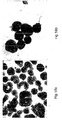
- Examples of undifferentiated colonies cultured with D1, D2 or HA16 medium in feeder-layer free conditions and with the D2 or HA16 medium with the tested feeder layers are illustrated in Figures 1a-d .
- the D1, D2 or HA16 media are capable of maintaining hESCs with normal population doubling - Similar to cells grown on MEFs, cells cultured with D2 or HA16 medium in all tested culture methods, and the D1 medium in the feeder layer-free systems, were passaged routinely every four to six days, at the same ratio of 1/2 or 1/3, indicating a similar population doubling time as of hESCs grown on MEFs. The cells were passage at the same seeding efficiency of about 1 million cells per 10 cm 2 , with the same viability rate of over 95 %. Using 40 % human serum and 10 % DMSO, cells were successfully frozen and thawed.
- Karyotype analysis revealed normal karyotype of hESCs grown with the D1, D2, CM100 or HA16 media - 15 passages and more after transferring the cells into the tested environments, karyotype analysis was performed by Giemsa banding on two separate cultures, representing the four medium conditions, D1, D2, CM100 and HA16 at the different culture methods. At least 20 cells were tested from each sample, 40 cells from each medium combination. All examined cells were found to sustain normal karyotype of 46,XX for cell lines 13 and 14 and 46,XY for cell line 16 (data not shown). Overall, these results suggest that the cells' karyotype remains stable in the tested conditions, similarly to ESCs grown with MEFs using traditional methods (Amit et al, 2000).
- hESCs cultured with the D1, D2 or HA16 express typical cell surface markers -
- Several surface markers typical of primate undifferentiated ES cells were examined using immunofluorescent staining (Thomson et al, 1995, 1996, 1998).
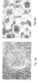
- hESCs cultured with the D1, D2 or HA16 medium for more than 20 passages, while using the tested culture conditions, were found to be strongly positive to surface markers TRA-1-60 ( Figure 2a ), SSEA4 ( Figure 2b ), TRA-1-81 ( Figure 2c ) and Oct 4 (data not shown).
- staining with SSEA3 was weak and negative for SSEA1 (data not shown).
- hESCs cultured with the D1, D2 or HA16 medium are pluripotent as tested by EBs formation in vitro -
- the developmental potential of the cells after prolonged culture in the tested culture methods was examined in vitro by the formation of embryoid bodies (EBs).
- EBs embryoid bodies
- stem cells differentiated into cell types representative of the three embryonic germ layers as described for EBs formed from hESCs cultured on other culture systems (Itskovitz-Eldor et al, 2000).
- EBs formed from the hESCs cultured on the D1, D2 or HA16 medium are capable of differentiating into the ectoderm, endoderm and mesoderm cell lineages - While undifferentiated cells cultured in the tested medium, feeder layers and matrices, expressed undifferentiated genetic markers such as Oct 4, Nanog, Sox2, Rexl, Cx43 and FGF4 (not shown) [Bhattacharya et al, 2004], cells harvested from 10 day-old EBs expressed genes such as albumin and glucagon (endoderm), ⁇ -cardiac actin, ⁇ -globulin and Flk1 (mesoderm), and AC133 and neurofilament (ectoderm) as demonstrated by RT-PCR analysis (data not shown).
- hESCs cultured with the D1, D2 or HA16 medium are pluripotent as tested by teratomas formation in vivo - The cells pluripotency was also tested in vivo by teratomas formation.
- hESCs cultured for over 12 passages in the HA16, D1 or D2 medium, in the tested culture systems formed teratomas following their injection into SCID-Beige mice.
- hESCs differentiated to representative tissues of the three embryonic germ layers including; cartilage, muscle, bone and fat (mesoderm), stratified epithelium, melanin containing epithelium (ectoderm), and kidney like structure (endoderm and mesoderm), and epithelium of endoderm origin (data not shown).
- Teratomas formation rates of 90 %, and the number of injected cells were identical to those demonstrated by cells cultured using traditional methods (Amit et al, 2000).
- the HA16 and D2 media are suitable for derivation of hESC line on foreskin fibroblast feeder layers in a complete xeno-free system -
- the medium combinations of the present disclosure were also tested for the ability to support hESC line derivation.
- new hESC lines were successfully derived and maintained for at least 2 passages (in the presence of the D2 medium) or at least 18 passages (in the presence of the HA16 medium).
- the hESC line derived on foreskin in the presence of the HA16 culture medium demonstrated stem cells morphology at passage 18 (and the culture is still ongoing), normal XY karyotype and pluripotency as evidenced by the formation of EBs ( Figures 3a-b and data not shown).
- the growth and success rates were similar to those obtained while using traditional culture methods. Since the used foreskin fibroblasts line, F21, were derived without any animal products, this new hESC lines were derived under complete xeno-free conditions. Thus, the new hESC lines exhibit typical hESC morphology and proliferation rates, normal karyotype and pluripotency as evidenced by the formation of EBs.
- hESCs cells subjected to prolonged culture in the tested culture systems demonstrated all hESCs features including; pluripotency, chromosomal stability, expression of specific genes and surface markers and indefinite proliferation as undifferentiated cells.
- the new TGF ⁇ -containing culture medium which is described in Example 1, hereinabove, is designed for massive cell culture (low protein content), suitable for industrial and clinical proposes cell production
- D1, D2, HA19 and HA16 media to support suspension culture of undifferentiated hESCs
- a medium containing the IL6RIL6 chimera such as the CM100F or HACM100 medium (as described in the General Materials and Experimental Methods) to support a suspension culture of hESCs was also examined.
- CM100F contains serum replacement
- the HACM100 medium is serum or serum replacement-free and thus presents a well-defined culture, xeno-free culture medium.
- CM100F, HA16, D1, D2 and HA19 media are suitable for culturing hESCs in suspension - hESCs were cultured in suspension using the newly developed TGF ⁇ -containing medium types which are devoid of serum, serum replacement and albumin.
- TGF ⁇ -containing medium types which are devoid of serum, serum replacement and albumin.
- the highest passage of hESCs grown in suspension in the tested medium types were 3 passages in the D1 medium, 7 passages in the D2 medium, 10 passages in the HA19 medium and 17 passages in the CM100F medium. All hESCs exhibited undifferentiated morphology at these passages and can be further cultured in these media and maintain hESCs features.
- Histological sections of the hESCs clumps formed in the suspension cultures illustrated homogeneous cell population, of round cells with large nucleus ( Figures 5a-g ).
- the cells were plated back on MEFs, they created colonies with typical hESCs morphology ( Figures 5b-e ), and if returned to suspension cultures, they continued proliferation as undifferentiated cells (data not shown).
- hESCs were cultured in a suspension culture in the presence of the serum or serum replacement-free, IL6RIL6-containing HACM100 medium, the cells were expanded and maintained in the undifferentiated state for at least 1-2 passages (data not shown).
- hESCs cultured in suspension in the presence of the D1, D2, HA19 or CM100F media express markers of undifferentiated hESCs - Cells cultured in suspension in the presence of the D2 medium for 3 passages as small clumps of 200-1500 cells expressed stem cells markers such as Oct 4 ( Figure 4a ), TRA-1-60 ( Figure 4b ), TRA-1-81 ( Figure 4c ) and SSEA4 (data not shown). Similar results were obtained with the CM100F, D1 or D2 medium at passage 5 (p-5) (data not shown).
- CM100F or the HA19 medium When cultured in suspension culture in the presence of the CM100F or the HA19 medium the cells expressed high levels of typical stem cells markers such as Oct 4 ( Figure 6a ), Rexl ( Figure 6b ), Sox2 ( Figure 6c ), Nanog ( Figure 6d ) and FGF4 (data not shown) as demonstrated by RT-PCR analysis.
- typical stem cells markers such as Oct 4 ( Figure 6a ), Rexl ( Figure 6b ), Sox2 ( Figure 6c ), Nanog ( Figure 6d ) and FGF4 (data not shown) as demonstrated by RT-PCR analysis.
- ESCs cultured in suspension are capable of forming EBs - When removed from the D1, D2 or HA16 medium and transferred to EBs medium (80 % DMEM/F12 supplemented with 20 % FBSd and 1 mM L-glutamine), the cells formed EBs containing representative tissues of three embryonic germ layers (as evidenced by histological analysis, data not shown).
- EBs medium 80 % DMEM/F12 supplemented with 20 % FBSd and 1 mM L-glutamine
- Rhesus ESCs can be also cultured in the suspension cultures of the present disclosure - Similar results with Rhesus ESCs (monkey embryonic stem cells, line R366.4, University of Wisconsin, primate center, Thomson lab, Madison, Wisconsin), which are regarded as good candidate for transgenic model to human diseases, were obtained when the Rhesus ESCs were cultured in suspension in the HA16, D1 and D2 TGF ⁇ -containing culture media (data not shown).
- the new TGF ⁇ -containing medium which is devoid of serum, serum replacement and albumin, or the IL6RIL6-containing medium are capable of supporting the undifferentiated culture of hESCs, while maintaining hESCs characteristics, and provide methods for massive culture of these cells for industrial and clinical purposes.
- hESCs like mouse ES cells, are traditionally cultured with MEFs, which may expose them to animal pathogens.
- MEFs which may expose them to animal pathogens.
- the present inventors have demonstrated, for the first time, a defined animal, serum and feeder layer-free culture system for hESCs, based on the use of commercial medium supplemented with either TGF ⁇ 3 or TGF ⁇ 1 and bFGF, and human fibronectin matrix as substitute. This medium is designed for massive cultivation of cells in GMP for industrial or clinical purposes. All medium types of the present disclosure (with TGF ⁇ 3 or TGF ⁇ 1 ) support hESCs culture. When using the culture medium with TGF ⁇ isoform 3 the results are better; less background differentiation.
- All media types of the present disclosure support the culture with feeders as good as with the regular serum containing media.
- Cells retained the same proliferation rates and the same background differentiation percentages as hESCs cultured with MEFs using traditional culture methods.
- the medium can also be used for massive suspended culture of undifferentiated hESCs.
- TGF ⁇ 3 and TGF ⁇ 1 Two isoforms of TGF ⁇ , TGF ⁇ 3 and TGF ⁇ 1 , were tested for their ability to maintain hESCs in an undifferentiated state using various culture conditions.
- TGF ⁇ 3 D2 and HA16 media
- ESCs cultured while using these media maintained all the characteristics of ESCs. After prolonged culture of more than 20 passages, the cells remained undifferentiated, as demonstrated by the colony and single cell morphology, and by the expression of markers typical of undifferentiated primate ESCs [Thomson et al, 1995, 1996, 1998; Reubinoff et al, 2000].
- hESCs expressed specific markers for the undifferentiated stage such as Oct 4, Sox 2, Rex1 and Nanog, as demonstrated by RT-PCR.
- the cells' pluripotency was examined in vitro.
- Cells cultured in the tested culture systems for more than 10 passages formed EBs similar to those created when grown on MEFs [Itskovitz-Eldor et al, 2000].
- RT-PCR analysis demonstrated that cells within these EBs differentiated into different cell types representative of the three germ layers.
- hESCs cultured in the presence of the D1 and D2 media formed teratomas containing a multitude of tissues types.
- hESCs cultured in the presence of the HA16 medium also formed teratomas and their histological evaluation is in process.
- the teratoma formation rates were identical to those of cells cultured with MEFs.
- the pluripotency of the cells culture continuously in the tested culture methods remained intact.
- hESCs can be maintained as undifferentiated cells in the proposed defined animal- and serum-free medium combination, without any feeder cells (D1, D2 and HA16) or alternatively, with commonly used acceptable feeder layers (D2 and HA16).
- these media can facilitate hESCs culture for research, industrial and clinical purposes.
- this novel culture media was found to support suspended culture of undifferentiated hESCs, the first and primary step in developing a massive culture system for their growth and scale-up, a crucial step for any industrial and clinical uses.
- hESCs Future clinical uses of hESCs will require a reproducible, well-defined and xeno-free culture system.
- the well-defined media demonstrated in the present study are suitable for culturing hESCs and may be advantageous for undertaking research on the mechanisms of ESC self-maintenance, especially of the possible roles of the TGF ⁇ pathway.
- Other studies using hESCs, such as the research on differentiation pathways and mechanisms, will benefit from the availability of a well-defined and reproducible culture system.
- hESC lines I-3, I-4 and I-6 [Amit & Itskovitz-Eldor, 2002] were cultured with inactivated MEF for 54-89 passages as previously described [Amit et al, 2000]. The following culture medium combinations were tested for their ability to support the suspended culture of hESCs:
- the IL6RIL6 chimera but not low concentrations of the IL6, soluble IL6 or LIF, are capable of maintaining hESCs in an undifferentiated state when cultured on fibronectin (2-D) -
- Three key cytokines of the IL6 family (LIF, IL6 and IL6RIL6 chimera) were tested for their ability to support the culture of undifferentiated hESCs in suspension.
- the ability of the various cytokines to maintain hESC self-renewal was first evaluated in the 2-D feeder layer-free culture system using fibronectin as a matrix. While the background differentiation of hESCs cultured in the presence of LIF or IL6 at the used concentrations (i.e.
- the IL6RIL6 chimera but not low concentrations of the IL6 and soluble IL6, are capable of maintaining cultures of hESCs in suspension in an undifferentiated state -
- the cytokines' ability to support the culture of undifferentiated hESCs in suspension was then examined.
- medium CM6* medium supplementation of 100 ng/ml IL6 and 0.5 ng/ml IL6 soluble receptor (medium CM6*) the cells formed either EBs or neurosphere-like structures within five to seven days of culture (data not shown).
- the neurosphere-like structures demonstrated round morphology when cultured in suspension, and several days after being plated on fibronectin neuron-like structures positively stained with either nestin or ⁇ -tubulin were observed (data not shown).
- hESCs cultured in suspension in the presence of the IL6RIL6 chimera exhibit normal karyotype - Karyotype analyses by Giemsa banding was carried out on each cell line after 23, 18 and 36 passages in suspension, for I-6, I-3 and I-4, respectively, and were found to be normal (data not shown). Similar results were obtained when the cells were re-cultured on MEFs; all samples but one of I-3 (passage 12 on MEFs after 10 passages in suspension) demonstrated normal karyotype. The original culture of 1-3 that remained in suspension demonstrated normal karyotype. Thus the karyotype of the suspended cell culture remained stable.
- hESCs cultured in suspension in the presence of the IL6RIL6 chimera exhibit expression pattern of undifferentiated state -
- CM100F medium for 42 and 43 passages were found to be strongly positive to surface markers SSEA4, TRA-1-60 and TRA-1-81 and Oct 4 ( Figures 8a-d ).
- SSEA3 was weak and negative for SSEA1.
- Similar results were obtained for the cells cultured in suspension and returned to the MEFs. The former were further tested for typical undifferentiation markers using RT-PCR.
- hESCs cultured in suspension in the presence of the ILRIL6 chimera preserve their pluripotency as demonstrated by EBs' formation -
- the developmental potential of the cells after prolonged culture in suspension was examined in vitro by the formation of EBs.
- hESCs formed cystic EBs similar to those created by hESCs grown with MEFs ( Figures 12a-b ).
- stem cells differentiated into cell types representative of the three embryonic germ layers [Itskovitz-Eldor et al, 2000].
- hESCs cultured in suspension in the presence of the ILRIL6 chimera preserve their pluripotency as demonstrated by teratoma formation - Cell pluripotency in vivo was demonstrated by teratoma formation.
- Cells cultured in the presence of the CM100F medium in suspension for 9, 10, 14 or 26 passages were injected into SCID Beige mice, representing the three tested cell lines, and after 10 weeks tumors formed in all four injected mice. Within these teratomas tissues representative of the three germ layers were observed ( Figures 12a-b ). Similar results were obtained when cells cultured for at least 10 passages in suspension were returned to the MEFs and cultured for additional 5-10 passages (data not shown).
- the total sphere number increased from 1.33 x 10 4 ⁇ 461 on the seeding day to 3.5 x 10 5 ⁇ 2.8x10 4 after 10-11 days in the dynamic culture, a 25-fold increase, and the total cell number increased from 5 x 10 8 to 1.31 x 10 10 .
- Figures 17a-b depict examples of differentiated ( Figure 17b ) and undifferentiated ( Figure 17a ) hESC clumps in the presence of the anti-gp 130 antibody.
- yFIL25 + - 85 % DMEM/F12 (Biological Industries, Biet Haemek, Israel), containing 15 % knockout serum replacement (SR), 2 mM L-glutamine, 0.1 mM ⁇ -mercaptoethanol, 1 % non-essential amino acid stock, 4 ng/ml bFGF (all but mentioned are Invitrogen Corporation products, Grand Island NY, USA), 25 ng/ml IL6 and 25 ng/ml sIL6-R (R&D systems, Minneapolis, MN, USA).
- SR knockout serum replacement
- 2 mM L-glutamine 2 mM L-glutamine
- 0.1 mM ⁇ -mercaptoethanol 1 % non-essential amino acid stock
- 4 ng/ml bFGF all but mentioned are Invitrogen Corporation products, Grand Island NY, USA
- 25 ng/ml IL6 and 25 ng/ml sIL6-R (R&D systems, Minneapolis, MN
- yFL3 - 85 % DMEM/F12 (Biological Industries, Biet Haemek, Israel), containing 15 % knockout serum replacement (SR), 2 mM L-glutamine, 0.1 mM ⁇ -mercaptoethanol, 1 % non-essential amino acid stock, 4 ng/ml bFGF (all but mentioned are Invitrogen Corporation products, Grand Island NY, USA), and 3000 u/ml human recombinant leukemia inhibitory factor (hrLIF) (R&D Systems Minneapolis MN, USA).
- SR knockout serum replacement
- 2 mM L-glutamine 0.1 mM ⁇ -mercaptoethanol
- 1 % non-essential amino acid stock 4 ng/ml bFGF (all but mentioned are Invitrogen Corporation products, Grand Island NY, USA)
- hrLIF human recombinant leukemia inhibitory factor
- yFL2 - 85 % DMEM/F12 (Biological Industries, Biet Haemek, Israel), containing 15 % knockout serum replacement (SR), 2 mM L-glutamine, 0.1 mM ⁇ -mercaptoethanol, 1 % non-essential amino acid stock, 4 ng/ml bFGF (all but mentioned are Invitrogen Corporation products, Grand Island NY, USA), and 2000 u/ml hrLIF (R&D Systems Minneapolis MN, USA).
- yFL1 - 85 % DMEM/F12 (Biological Industries, Biet Haemek, Israel), containing 15 % knockout serum replacement (SR), 2 mM L-glutamine, 0.1 mM ⁇ -mercaptoethanol, 1 % non-essential amino acid stock, 4 ng/ml bFGF (all but mentioned are Invitrogen Corporation products, Grand Island NY, USA), and 1000 u/ml hrLIF (R&D Systems Minneapolis MN, USA).
- TLF - 85 % DMEM/F12 (Biological Industries, Biet Haemek, Israel), containing 15 % knockout serum replacement (SR), 2 mM L-glutamine, 0.1 mM ⁇ -mercaptoethanol, 1 % non-essential amino acid stock, 4 ng/ml bFGF (all but mentioned are Invitrogen Corporation products, Grand Island NY, USA), 1000 u/ml hrLIF (R&D Systems Minneapolis MN, USA) and 0.12 ng/ml TGF ⁇ 1 (R&D Systems Minneapolis MN, USA).
- SR knockout serum replacement
- 2 mM L-glutamine 0.1 mM ⁇ -mercaptoethanol
- 1 % non-essential amino acid stock 4 ng/ml bFGF (all but mentioned are Invitrogen Corporation products, Grand Island NY, USA)
- 1000 u/ml hrLIF R&D Systems Minneapolis MN, USA
- 0.12 ng/ml TGF ⁇ 1 R&
- New media combinations are suitable for culturing human ESCs in suspension culture -
- Four additional culture medium were tested for the ability to support suspension culture of hESCs; TLF, yFIL25+, yFL3 and yFL1.
- the human ESCs created spheres, or disc-like structures, 24 hours after being cultured in suspension using non-dynamic culture conditions ( Figure 18a-e ).
- the clumps were split every 5-7 days while maintaining their morphology, and as of the day of writing this report reached 31, 19, 18 and 18 passages for TLF, yFIL25+, yFL3 and yFL1 respectively.
- Several surface markers typical of primate undifferentiated ESCs were examined using immunofluorescence staining according to the methods described elsewhere [Thomson et al, 1995, 1996, 1998].
- Human ESCs cultured in suspension in the presence of the TLF, yFIL25+, yFL3 or yFL1 culture media express markers of pluripotency - Human ESCs cultured in suspension under non-dynamic culture conditions were further tested for typical undifferentiation markers using RT-PCR analysis.
- cells cultured with MEFs cells cultured in suspension for 18, 16, 16 or 18 passages in the presence of medium TLF, yFIL25+, yFL3 or yFL1, respectively, expressed genetic markers of pluripotency Oct 4, Nanog, Sox2, Rex1, and FGF4 (Data not shown).
- Human ESCs cultured in suspension in the presence of the TLF, yFIL25 +, yFL3 or yFL1 are capable of forming EBs which represent all three embryonic germ layers -
- the developmental potential of the cells after prolonged culture in suspension was examined in vitro by the formation of EBs.
- hESCs formed cystic EBs (Data not shown).
- stem cells differentiated into cell types representative of the three embryonic germ layers.
- Undifferentiated hESCs are traditionally cultured in 2D on either feeder-layer cells or on acellular matrix. This study demonstrates for the first time a method of culturing these cells as free floating spheres for prolonged periods in medium consisting of serum replacement, bFGF, and IL6RIL6 chimera.
- Mouse ESCs can be cultured continuously without feeder layers provided that the culture medium is supplemented with leukemia inhibitory factor (LIF), which was found to be involved in the self-maintenance of mouse ESCs [Smith et al, 1988; Williams et al, 1988; Rose et al, 1994; Conover et al, 1993; Niwa et al, 1998].
- LIF leukemia inhibitory factor
- IL6RIL6 chimera An additional candidate for hESC self-maintenance is the IL6RIL6 chimera. While LIF acts through the heteromeric complex of LIF-receptor and gp130, the IL6RIL6 chimera requires only gp130 to activate the intracellular pathway involving activation of JAK kinases and STAT transcription factors.
- the IL6RIL6 chimera is known to be a potent inducer of the LIF/IL6 pathway, which results in a higher response compared with the effect caused by IL6 alone or even the chimera components added separately (IL6 and IL6 soluble receptors IL6R) (Chebath et al, 1997).
- the only method for deriving new mouse ESC lines in feeder layer-free conditions is based on the addition of factors from the IL6 family [Nichols et al, 1994], or the combination of LIF and BMP4 when serum-free conditions are used [Ying QL et al, 2003].
- the IL6RIL6 chimera was demonstrated as the most potent factor in supporting the feeder layer-free isolation of mouse ESC lines [Nichols et al, 1994].
- the chimera has a much higher affinity for human gp130 than the mixture of IL6 and sIL6R [Kollet et al, 1999].
- cytokines of the IL-6 family were tested for their ability to maintain hESCs in their undifferentiated state.
- Medium supplemented with 100 ng/ml of the IL6RIL6 chimera when used in conjunction with 4 ng/ml bFGF supported the culture of three hESC lines in suspension for over 40 passages and retained normal hESC features, including the expression of surface markers and genes typical of undifferentiated hESCs as detected by FACS, RT-PCR and immunostaining, normal karyotype and teratoma formation.
- hESCs self-maintain
- mouse ESCs the role of LIF and other members of the IL6 family, acting through gp130 and the JAK/STAT3 pathway, in maintaining prolonged culture of undifferentiated ESCs is well known [Smith et al, 1988; Williams et al, 1988; Rose et al, 1994; Conover et al, 1993; Niwa et al, 1998].
- the described culture system was also found to support hESCs proliferation.
- the size of each clump during passaging (5-7 days) was found to increase by 1.5 folds, and associated with low apoptosis levels and high viability rates.
- the number of cells in each sphere increased 1.66-fold during each passage (5-7 days). Taking together, the kinetic features of the newly developed culture system indicate that the system is as proficient as the 2D culture systems and could be used as a base for routine culture of undifferentiated hESCs.
- hESCs An important requirement for clinical and industrial application of hESCs is a scalable culture system capable of generating masses of cells.
- the culture system presented here can also be used as a base for the mass production of undifferentiated hESCs using dynamic systems such as spinner flasks and bioreactors.
- hESCs can be cultured continuously and maintain their typical features, while enabling a scale-up of 25 folds in 10 days. Although seeding concentrations and medium metabolites should still be optimized, the results presented here demonstrate that this system could serve as a basis for developing a controlled process for mass production of hESCs in bioreactors.
- mice and human ESCs share only some of their features; they differ in the inability to culture hESCs in 2D using the traditional medium supplemented with calf serum and LIF without a feeder layer [Thomson et al, 1998].
- the ability of the IL6RIL6 chimera to support the self-maintenance of hESCs in suspension illuminates once again the question of LIF-STAT3 pathway's possible role in hESC self-renewal.
- this culture system presented herein is a further step forward toward creating the culture conditions that will make possible to fulfill the promise of hESCs.
Landscapes
- Health & Medical Sciences (AREA)
- Life Sciences & Earth Sciences (AREA)
- Engineering & Computer Science (AREA)
- Biomedical Technology (AREA)
- Biotechnology (AREA)
- Genetics & Genomics (AREA)
- Wood Science & Technology (AREA)
- Organic Chemistry (AREA)
- Chemical & Material Sciences (AREA)
- Bioinformatics & Cheminformatics (AREA)
- Zoology (AREA)
- Microbiology (AREA)
- Cell Biology (AREA)
- Biochemistry (AREA)
- General Engineering & Computer Science (AREA)
- General Health & Medical Sciences (AREA)
- Reproductive Health (AREA)
- Developmental Biology & Embryology (AREA)
- Gynecology & Obstetrics (AREA)
- Micro-Organisms Or Cultivation Processes Thereof (AREA)
Claims (8)
- Verfahren zur Expansion und Beibehaltung humaner embryonaler Stammzellen in einem undifferenzierten Zustand, wobei das Verfahren das Kultivieren der humanen embryonalen Stammzellen in einer Suspensionskultur in dem Kulturmedium, das mindestens 2000 Einheiten pro Milliliter (U/ml) Leukämie-Inhibitor-Faktor (LIF) und basischen Fibroblasten-Wachstumsfaktor (bFGF) umfasst, unter Kulturbedingungen ohne Substrathaftung, die eine Expansion der humanen embryonalen Stammzellen im undifferenzierten Zustand ermöglichen, umfasst, wodurch die humanen embryonalen Stammzellen im undifferenzierten Zustand expandiert und beibehalten werden.
- Verfahren zum Erzeugen von Abstammungslinien-spezifischen Zellen aus humanen embryonalen Stammzellen, wobei das Verfahren Folgendes umfasst:(a) Kultivieren der humanen embryonalen Stammzellen in einer Suspensionskultur in dem Kulturmedium, das mindestens 2000 Einheiten pro Milliliter (U/ml) Leukämie-Inhibitor-Faktor (LIF) und basischen Fibroblasten-Wachstumsfaktor (bFGF) umfasst, unter Kulturbedingungen, die eine Expansion der humanen embryonalen Stammzellen in einem undifferenzierten Zustand ermöglichen, um dadurch expandierte, undifferenzierte humane embryonale Stammzellen zu erhalten; und(b) Unterwerfen der expandierten undifferenzierten humanen embryonalen Stammzellen Kulturbedingungen, die geeignet sind, Abstammungslinien-spezifische Zellen zu differenzieren und/oder zu expandieren;wodurch die Abstammungslinien-spezifischen Zellen aus den humanen embryonalen Stammzellen erzeugt werden.
- Verfahren zum Erzeugen von embryonalen Körpern aus humanen embryonalen Stammzellen, wobei das Verfahren Folgendes umfasst:(a) Kultivieren der humanen embryonalen Stammzellen in einer Suspensionskultur in dem Kulturmedium, das mindestens 2000 Einheiten pro Milliliter (U/ml) Leukämie-Inhibitor-Faktor (LIF) und basischen Fibroblasten-Wachstumsfaktor (bFGF) umfasst, unter Kulturbedingungen, die eine Expansion der humanen embryonalen Stammzellen in einem undifferenzierten Zustand ermöglichen, um dadurch expandierte, undifferenzierte humane embryonale Stammzellen zu erhalten; und(b) Unterwerfen der expandierten, undifferenzierten humanen embryonalen Stammzellen Kulturbedingungen, die zur Differenzierung der humanen embryonalen Stammzellen zu embryonalen Körpern geeignet sind;wodurch die embryonalen Körper aus den humanen embryonalen Stammzellen erzeugt werden.
- Verfahren zum Erzeugen von Abstammungslinien-spezifischen Zellen aus embryonalen Stammzellen, wobei das Verfahren Folgendes umfasst:(a) Kultivieren der humanen embryonalen Stammzellen in einer Suspensionskultur in dem Kulturmedium, das mindestens 2000 Einheiten pro Milliliter (U/ml) Leukämie-Inhibitor-Faktor (LIF) und basischen Fibroblasten-Wachstumsfaktor (bFGF) umfasst, unter Kulturbedingungen, die eine Expansion der humanen embryonalen Stammzellen in einem undifferenzierten Zustand ermöglichen, um dadurch expandierte, undifferenzierte humane embryonale Stammzellen zu erhalten;(b) Unterwerfen der expandierten, undifferenzierten humanen embryonalen Stammzellen Kulturbedingungen, die zur Differenzierung der expandierten, undifferenzierten humanen embryonalen Stammzellen zu embryonalen Körpern geeignet sind; und(c) Unterwerfen von Zellen der embryonalen Körper Kulturbedingungen, die geeignet sind, Abstammungslinien-spezifische Zellen zu differenzieren und/oder zu expandieren;wodurch die Abstammungslinien-spezifischen Zellen aus den humanen embryonalen Stammzellen erzeugt werden.
- Verfahren nach den Anspruch 2, 3 oder 4, wobei das Kultivieren unter Bedingungen ohne Substrathaftung erfolgt.
- Verfahren nach Anspruch 1, 2, 3, 4 oder 5, wobei das Kultivieren unter xenofreien Bedingungen erfolgt.
- Verfahren nach einem der Ansprüche 1-6, wobei der bFGF in einer Konzentration von mindestens 2 ng/ml vorliegt.
- Verfahren nach einem der Ansprüche 1-6, wobei der bFGF in einer Konzentration von mindestens 4 ng/ml vorliegt.
Applications Claiming Priority (5)
| Application Number | Priority Date | Filing Date | Title |
|---|---|---|---|
| US83479506P | 2006-08-02 | 2006-08-02 | |
| US84069206P | 2006-08-29 | 2006-08-29 | |
| PCT/IL2007/000970 WO2008015682A2 (en) | 2006-08-02 | 2007-08-02 | Methods of expanding embryonic stem cells in a suspension culture |
| EP13185969.6A EP2733203B1 (de) | 2006-08-02 | 2007-08-02 | Verfahren zur Expansion embryonaler Stammzellen in einer Suspensionskultur |
| EP07790025.6A EP2059586B1 (de) | 2006-08-02 | 2007-08-02 | Verfahren zur expansion embryonaler stammzellen in einer suspensionskultur |
Related Parent Applications (4)
| Application Number | Title | Priority Date | Filing Date |
|---|---|---|---|
| EP13185969.6A Division-Into EP2733203B1 (de) | 2006-08-02 | 2007-08-02 | Verfahren zur Expansion embryonaler Stammzellen in einer Suspensionskultur |
| EP13185969.6A Division EP2733203B1 (de) | 2006-08-02 | 2007-08-02 | Verfahren zur Expansion embryonaler Stammzellen in einer Suspensionskultur |
| EP13185969.6A Previously-Filed-Application EP2733203B1 (de) | 2006-08-02 | 2007-08-02 | Verfahren zur Expansion embryonaler Stammzellen in einer Suspensionskultur |
| EP07790025.6A Division EP2059586B1 (de) | 2006-08-02 | 2007-08-02 | Verfahren zur expansion embryonaler stammzellen in einer suspensionskultur |
Publications (2)
| Publication Number | Publication Date |
|---|---|
| EP3441459A1 EP3441459A1 (de) | 2019-02-13 |
| EP3441459B1 true EP3441459B1 (de) | 2021-03-17 |
Family
ID=38704894
Family Applications (3)
| Application Number | Title | Priority Date | Filing Date |
|---|---|---|---|
| EP07790025.6A Active EP2059586B1 (de) | 2006-08-02 | 2007-08-02 | Verfahren zur expansion embryonaler stammzellen in einer suspensionskultur |
| EP13185969.6A Active EP2733203B1 (de) | 2006-08-02 | 2007-08-02 | Verfahren zur Expansion embryonaler Stammzellen in einer Suspensionskultur |
| EP18186556.9A Active EP3441459B1 (de) | 2006-08-02 | 2007-08-02 | Verfahren zur expansion embryonaler stammzellen in einer suspensionskultur |
Family Applications Before (2)
| Application Number | Title | Priority Date | Filing Date |
|---|---|---|---|
| EP07790025.6A Active EP2059586B1 (de) | 2006-08-02 | 2007-08-02 | Verfahren zur expansion embryonaler stammzellen in einer suspensionskultur |
| EP13185969.6A Active EP2733203B1 (de) | 2006-08-02 | 2007-08-02 | Verfahren zur Expansion embryonaler Stammzellen in einer Suspensionskultur |
Country Status (6)
| Country | Link |
|---|---|
| US (4) | US9040297B2 (de) |
| EP (3) | EP2059586B1 (de) |
| DK (2) | DK2733203T3 (de) |
| ES (2) | ES2874223T3 (de) |
| PT (1) | PT2733203T (de) |
| WO (1) | WO2008015682A2 (de) |
Families Citing this family (40)
| Publication number | Priority date | Publication date | Assignee | Title |
|---|---|---|---|---|
| US7410798B2 (en) | 2001-01-10 | 2008-08-12 | Geron Corporation | Culture system for rapid expansion of human embryonic stem cells |
| KR20160116024A (ko) | 2005-06-22 | 2016-10-06 | 아스테리아스 바이오세라퓨틱스, 인크. | 인간 배아 줄기 세포의 현탁 배양 |
| WO2007026353A2 (en) | 2005-08-29 | 2007-03-08 | Technion Research & Development Foundation Ltd. | Media for culturing stem cells |
| EP2059586B1 (de) | 2006-08-02 | 2016-07-20 | Technion Research & Development Foundation Ltd. | Verfahren zur expansion embryonaler stammzellen in einer suspensionskultur |
| US9080145B2 (en) | 2007-07-01 | 2015-07-14 | Lifescan Corporation | Single pluripotent stem cell culture |
| CA3114827C (en) | 2007-07-31 | 2023-09-05 | Lifescan, Inc. | Differentiation of human embryonic stem cells to pancreatic endocrine |
| KR101731474B1 (ko) | 2008-02-21 | 2017-05-11 | 얀센 바이오테크 인코포레이티드 | 세포 부착, 배양 및 탈리를 위한 방법, 표면 개질 플레이트 및 조성물 |
| US8691569B2 (en) | 2008-03-17 | 2014-04-08 | Agency For Science, Technology And Research | Microcarriers for stem cell culture |
| US9458431B2 (en) | 2008-03-17 | 2016-10-04 | Agency For Science, Technology And Research | Microcarriers for stem cell culture |
| SG188918A1 (de) | 2008-03-17 | 2013-04-30 | Agency Science Tech & Res | |
| US8828720B2 (en) | 2008-03-17 | 2014-09-09 | Agency For Science, Technology And Research | Microcarriers for stem cell culture |
| PL2310492T3 (pl) | 2008-06-30 | 2015-12-31 | Janssen Biotech Inc | Różnocowanie pluripotencjalnych komórek macierzystych |
| US8895300B2 (en) | 2008-11-04 | 2014-11-25 | Viacyte, Inc. | Scalable primate pluripotent stem cell aggregate suspension culture and differentiation thereof |
| ES2642070T3 (es) | 2008-11-20 | 2017-11-15 | Janssen Biotech, Inc. | Cultivo de células madre pluripotentes en microportadores |
| ES2584053T3 (es) | 2008-11-20 | 2016-09-23 | Janssen Biotech, Inc. | Métodos y composiciones para la unión de células y cultivo en sustratos planos |
| US20110243898A1 (en) * | 2008-12-10 | 2011-10-06 | The University Of Louisville Research Foundation, Inc. | Somatic cell-derived pluripotent cells and methods of use therefor |
| US9150829B2 (en) | 2009-03-20 | 2015-10-06 | Agency For Science, Technoloy And Research | Culture of pluripotent and multipotent cells on microcarriers |
| KR102058901B1 (ko) | 2009-07-20 | 2019-12-24 | 얀센 바이오테크 인코포레이티드 | 인간 배아 줄기 세포의 분화 |
| EP3633025B1 (de) * | 2009-11-12 | 2022-09-14 | Technion Research & Development Foundation Ltd. | Kulturmedien, zellkulturen und verfahren zur kultivierung pluripotenter stammzellen in undifferenziertem zustand |
| SG181687A1 (en) | 2009-12-23 | 2012-07-30 | Centocor Ortho Biotech Inc | Differentiation of human embryonic stem cells |
| EP2542667A4 (de) | 2010-03-01 | 2013-07-31 | Janssen Biotech Inc | Verfahren zur reinigung von aus pluripotenten stammzellen gewonnenen zellen |
| CN103221536B (zh) | 2010-08-31 | 2016-08-31 | 詹森生物科技公司 | 人胚胎干细胞的分化 |
| CA2810488C (en) * | 2010-09-07 | 2025-05-06 | Technion Research & Development Foundation Limited | NEW PROCESSES AND CULTURE MEDIA FOR THE CULTURE OF MULTIPOTENTED STEM CELLS |
| US20140329317A1 (en) | 2011-11-25 | 2014-11-06 | Kyoto University | Method for culturing pluripotent stem cell |
| KR102203056B1 (ko) | 2011-12-22 | 2021-01-14 | 얀센 바이오테크 인코포레이티드 | 인간 배아 줄기 세포의 단일 인슐린 호르몬 양성 세포로의 분화 |
| AU2013271581B2 (en) | 2012-06-08 | 2018-08-09 | Janssen Biotech, Inc. | Differentiation of human embryonic stem cells into pancreatic endocrine cells |
| BR112015015770A2 (pt) | 2012-12-31 | 2017-07-11 | Janssen Biotech Inc | cultivo de células-tronco embrionárias humanas na interface ar-líquido para diferenciação em células pancreáticas endócrinas |
| US10370644B2 (en) | 2012-12-31 | 2019-08-06 | Janssen Biotech, Inc. | Method for making human pluripotent suspension cultures and cells derived therefrom |
| RU2687379C2 (ru) | 2012-12-31 | 2019-05-13 | Янссен Байотек, Инк. | Суспендирование и кластеризация плюрипотентных клеток человека с целью их дифференцировки в панкреатические эндокринные клетки |
| JP6557147B2 (ja) | 2012-12-31 | 2019-08-07 | ヤンセン バイオテツク,インコーポレーテツド | Hb9調節物を用いたヒト胚性幹細胞の膵臓内分泌細胞への分化 |
| US9738861B2 (en) | 2013-03-06 | 2017-08-22 | Kyoto University | Culture system for pluripotent stem cells and method for subculturing pluripotent stem cells |
| KR20160079072A (ko) * | 2013-11-01 | 2016-07-05 | 얀센 바이오테크 인코포레이티드 | 췌장 내분비 세포로의 분화를 위한 인간 만능 줄기세포의 현탁 및 클러스터링 |
| US10006006B2 (en) | 2014-05-16 | 2018-06-26 | Janssen Biotech, Inc. | Use of small molecules to enhance MAFA expression in pancreatic endocrine cells |
| AU2015363008B2 (en) | 2014-12-19 | 2021-12-09 | Janssen Biotech, Inc. | Suspension culturing of pluripotent stem cells |
| DK3443073T3 (da) | 2016-04-14 | 2024-09-02 | Janssen Biotech Inc | Differentiering af pluripotente stamceller til intestinale mellemtarmendodermceller |
| KR102711870B1 (ko) * | 2016-07-19 | 2024-10-02 | 아셀라타 리미티드 | 현탁 내 만능줄기세포를 배양하기 위한 배양 배지 |
| EP3865566A4 (de) | 2018-10-12 | 2022-08-03 | Nikon Corporation | Vorrichtung zur berechnung einer unterkulturstufe und verfahren zur berechnung einer unterkulturstufe sowie programm |
| CN114207118A (zh) | 2019-05-14 | 2022-03-18 | 阿利夫农场公司 | 多能细胞聚集体及其用途 |
| US11001810B1 (en) | 2019-11-11 | 2021-05-11 | Lancell AB | Serum-free human pluripotent stem cell culture medium |
| US11735303B2 (en) | 2021-06-22 | 2023-08-22 | David Haase | Apparatus and method for determining a composition of a replacement therapy treatment |
Family Cites Families (125)
| Publication number | Priority date | Publication date | Assignee | Title |
|---|---|---|---|---|
| DE69028514T2 (de) * | 1989-01-10 | 1997-02-20 | Amrad Corp Ltd | Leukämiehemmender faktor aus vieharten und seine verwendung zur verbesserung der implantation und der entwicklung von embryonalen zellen |
| US6107543A (en) * | 1992-08-20 | 2000-08-22 | Infigen, Inc. | Culture of totipotent embryonic inner cells mass cells and production of bovine animals |
| AU7243100A (en) | 1995-02-02 | 2001-05-03 | Tristem Trading (Cyprus) Limited | A method of preparing an undifferentiated cell |
| US7410773B2 (en) | 1995-02-02 | 2008-08-12 | Ghazi Jaswinder Dhoot | Method of preparing an undifferentiated cell |
| US5811297A (en) | 1996-03-07 | 1998-09-22 | Amba Biosciences, Llc | Immortalized hematopoietic cell lines, cell system thereof with stromal cells, in vitro, ex vivo and in vivo uses, & in vitro generation of dendritic cells and macrophages |
| DK0888452T3 (da) | 1996-03-15 | 2004-06-14 | Munin Corp | Human CYR61, et ekstracellulært matrix-signalmolekyle |
| US6331406B1 (en) | 1997-03-31 | 2001-12-18 | The John Hopkins University School Of Medicine | Human enbryonic germ cell and methods of use |
| WO1999001763A2 (en) | 1997-07-02 | 1999-01-14 | Board Of Regents, The University Of Texas System | p53 AS A REGULATOR OF CELL DIFFERENTIATION |
| IL122818A0 (en) * | 1997-07-10 | 1998-08-16 | Yeda Res & Dev | Chimeric interleukin-6 soluble receptor/ligand protein analogs thereof and uses thereof |
| AU729377B2 (en) | 1997-10-23 | 2001-02-01 | Asterias Biotherapeutics, Inc. | Methods and materials for the growth of primate-derived primordial stem cells in feeder-free culture |
| US6667176B1 (en) | 2000-01-11 | 2003-12-23 | Geron Corporation | cDNA libraries reflecting gene expression during growth and differentiation of human pluripotent stem cells |
| US7410798B2 (en) | 2001-01-10 | 2008-08-12 | Geron Corporation | Culture system for rapid expansion of human embryonic stem cells |
| US7413904B2 (en) | 1998-10-23 | 2008-08-19 | Geron Corporation | Human embryonic stem cells having genetic modifications |
| WO2000027995A1 (en) | 1998-11-09 | 2000-05-18 | Monash University | Embryonic stem cells |
| IL129966A (en) | 1999-05-14 | 2009-12-24 | Technion Res & Dev Foundation | ISOLATED HUMAN EMBRYOID BODIES (hEB) DERIVED FROM HUMAN EMBRYONIC STEM CELLS |
| US20030129745A1 (en) | 1999-10-28 | 2003-07-10 | Robl James M. | Gynogenetic or androgenetic production of pluripotent cells and cell lines, and use thereof to produce differentiated cells and tissues |
| US7455983B2 (en) | 2000-01-11 | 2008-11-25 | Geron Corporation | Medium for growing human embryonic stem cells |
| CA2403000C (en) | 2000-03-14 | 2015-06-23 | Es Cell International Pte Ltd | Embryonic stem cells and neural progenitor cells derived therefrom |
| US7473555B2 (en) | 2000-04-27 | 2009-01-06 | Geron Corporation | Protocols for making hepatocytes from embryonic stem cells |
| NL1017973C2 (nl) | 2000-05-10 | 2002-11-08 | Tristem Trading Cyprus Ltd | Inrichting. |
| JP5943533B2 (ja) | 2000-05-17 | 2016-07-06 | アステリアス バイオセラピューティクス インコーポレイテッド | 神経前駆細胞の集団 |
| US7250294B2 (en) | 2000-05-17 | 2007-07-31 | Geron Corporation | Screening small molecule drugs using neural cells differentiated from human embryonic stem cells |
| US20020090722A1 (en) | 2000-06-15 | 2002-07-11 | Tanja Dominko | Pluripotent mammalian cells |
| GB0025088D0 (en) | 2000-10-13 | 2000-11-29 | Univ Edinburgh | Stem cells |
| US20020086005A1 (en) * | 2000-11-22 | 2002-07-04 | Choy-Pik Chiu | Tolerizing allografts of pluripotent stem cells |
| EP1379624A2 (de) | 2000-11-27 | 2004-01-14 | Yissum Research Development Company Of The Hebrew University Of Jerusalem | Transfektion humaner embryonaler stammzellen |
| US6576464B2 (en) | 2000-11-27 | 2003-06-10 | Geron Corporation | Methods for providing differentiated stem cells |
| AU2001297880B2 (en) | 2000-11-30 | 2007-05-31 | Stemron Inc. | Isolated homozygous stem cells differentiated cells derived therefrom and materials and methods for making and using same |
| DE60232585D1 (de) | 2001-04-24 | 2009-07-23 | Dolores Baksh | Vorläuferzellpopulationen, deren expansion und die entstehung von nicht-hämatopoetischen zelltypen daraus |
| CA2447015A1 (en) | 2001-05-15 | 2002-11-21 | Rappaport Family Institute For Research In The Medical Sciences | Insulin producing cells derived from human embryonic stem cells |
| CA2351156A1 (en) * | 2001-07-04 | 2003-01-04 | Peter W. Zandstra | A bioprocess for the generation of pluripotent cell derived cells and tissues |
| WO2003004605A2 (en) | 2001-07-06 | 2003-01-16 | Geron Corporation | Mesenchymal cells and osteoblasts from human embryonic stem cell |
| WO2003006950A2 (en) | 2001-07-12 | 2003-01-23 | Geron Corporation | Cells of the cardiomyocyte lineage produced from human pluripotent stem cells |
| WO2003010303A1 (en) | 2001-07-24 | 2003-02-06 | Es Cell International Pte Ltd | Methods of inducing differentiation of stem cells |
| GB0119015D0 (en) * | 2001-08-03 | 2001-09-26 | Univ Cardiff | A fusion protein |
| WO2003014313A2 (en) | 2001-08-06 | 2003-02-20 | Bresagen, Ltd. | Alternative compositions and methods for the culture of stem cells |
| US20030211603A1 (en) | 2001-08-14 | 2003-11-13 | Earp David J. | Reprogramming cells for enhanced differentiation capacity using pluripotent stem cells |
| WO2003018780A1 (en) | 2001-08-27 | 2003-03-06 | Advanced Cell Technology, Inc. | De-differentiation and re-differentiation of somatic cells and production of cells for cell therapies |
| JP2005502356A (ja) | 2001-09-12 | 2005-01-27 | デヴェロゲン アクチエンゲゼルシャフト フュア エントヴィックルングスビオローギッシェ フォルシュング | 治療のための腸内幹細胞の分離、培養および分化方法 |
| US20050095708A1 (en) | 2001-11-09 | 2005-05-05 | Pera Martin F. | Characterization and isolation of subsets of human embryonic stem cells (HES) and cells associated or derived therefrom |
| CA2468335C (en) | 2001-12-07 | 2014-07-08 | Geron Corporation | Chondrocyte precursors derived from human embryonic stem cells |
| US20030153082A1 (en) | 2001-12-07 | 2003-08-14 | The John P. Robarts Research Institute | Hematopoietic cells from human embryonic stem cells |
| CA2470539C (en) | 2001-12-07 | 2011-10-04 | Geron Corporation | Islet cells from human embryonic stem cells |
| JP4408371B2 (ja) | 2001-12-21 | 2010-02-03 | トロンボジェニクス・ナムローゼ・フエンノートシャップ | 生殖細胞系伝達能力を有する胚性幹(es)細胞のインビトロの誘導および培養のための組成物 |
| CN1671835A (zh) | 2001-12-28 | 2005-09-21 | 塞拉提斯股份公司 | 一种建立多能人胚泡衍生的干细胞的方法 |
| IL147412A0 (en) | 2001-12-31 | 2002-08-14 | Yeda Res & Dev | The use of il6r/il6 chimera in nerve cell regeneration |
| GB0210539D0 (en) * | 2002-05-08 | 2002-06-19 | Univ Edinburgh | Control of es cell self renewal and lineage specification, and medium therefor |
| AU2009213101B2 (en) | 2002-06-05 | 2012-03-08 | Es Cell International Pte Ltd | Generation of neural stem cells from undifferentiated human embryonic stem cells |
| AU2003247514A1 (en) | 2002-06-11 | 2003-12-22 | Roy Ogle | Meningeal-derived stem cells |
| US7662625B2 (en) | 2002-07-02 | 2010-02-16 | Cancer Research Technology Limited | Methods for detecting the differentiation status of cells using 5T4 antigen expression |
| US20050101014A1 (en) | 2002-07-11 | 2005-05-12 | Keirstead Hans S. | Oligodendrocytes derived from human embryonic stem cells for remyelination and treatment of spinal cord injury |
| US7285415B2 (en) | 2002-07-11 | 2007-10-23 | The Regents Of The University Of California | Oligodendrocytes derived from human embryonic stem cells for remyelination and treatment of spinal cord injury |
| US20040077985A1 (en) | 2002-09-03 | 2004-04-22 | Donnie Rudd | Method of replenishing cells damaged by treatment for cancer |
| GB0222846D0 (en) * | 2002-10-03 | 2002-11-06 | Choo Yen | Cell culture |
| US7267981B2 (en) | 2002-10-07 | 2007-09-11 | Technion Research & Development Foundation Ltd. | Human foreskin fibroblasts for culturing ES cells |
| US20060252150A1 (en) | 2002-11-08 | 2006-11-09 | Linzhao Cheng | Human embryonic stem cell cultures, and compositions and methods for growing same |
| WO2004050826A2 (en) | 2002-11-29 | 2004-06-17 | Technion Research & Development Foundation Ltd. | Method of dynamically culturing embryonic stem cells |
| US20040110286A1 (en) | 2002-12-06 | 2004-06-10 | The John P. Robarts Research Institute | Method for making hematopoietic cells |
| ES2705683T3 (es) * | 2002-12-16 | 2019-03-26 | Technion Res & Dev Foundation | Medio de cultivo de células madre pluripotentes |
| WO2004078944A2 (en) | 2003-03-05 | 2004-09-16 | Maine Medical Center Research Institute | Thrombin, soluble jaggedi, and trap and growth of stem cells |
| US7153650B2 (en) | 2003-03-13 | 2006-12-26 | Geron Corporation | Marker system for preparing and characterizing high-quality human embryonic stem cells |
| US20060134636A1 (en) | 2003-03-13 | 2006-06-22 | Stanton Lawrence W | Standardization of growth conditions for human embryonic stem cells intended for use in regenerative medicine |
| US20030224411A1 (en) | 2003-03-13 | 2003-12-04 | Stanton Lawrence W. | Genes that are up- or down-regulated during differentiation of human embryonic stem cells |
| WO2004085631A2 (en) | 2003-03-28 | 2004-10-07 | Mount Sinai Hospital | Canine embryonic stem cells |
| US7638328B2 (en) | 2003-05-08 | 2009-12-29 | Cellartis Ab | Method for efficient transfer of human blastocyst-derived stem cells (hBS cells) from a feeder-supported to a feeder-free culture system, long-term propagation of hBS cells under feeder-free conditions and use of cultured hBS cells for applications in myocardial regeneration |
| US20040229350A1 (en) | 2003-05-12 | 2004-11-18 | Nikolai Strelchenko | Morula derived embryonic stem cells |
| US20050118713A1 (en) | 2003-05-12 | 2005-06-02 | Nikolai Strelchenko | Morula derived embryonic stem cells |
| AU2004247961B2 (en) | 2003-06-12 | 2010-06-24 | Yeda Research & Development Co.Ltd | Enhancement of oligodendrocyte differentiation |
| US20050042595A1 (en) | 2003-08-14 | 2005-02-24 | Martin Haas | Banking of multipotent amniotic fetal stem cells |
| US20070269790A1 (en) | 2003-12-01 | 2007-11-22 | Technion Research & Development | Methods of Generating Stem Cells and Embryonic Bodies Carrying Disease-Causing Mutations and Methods of Using same for Studying Genetic Disorders |
| TWI280280B (en) | 2003-12-09 | 2007-05-01 | Ind Tech Res Inst | Culture system and method for expansion and undifferentiated growth of human embryonic stem cells |
| US20050233446A1 (en) | 2003-12-31 | 2005-10-20 | Parsons Xuejun H | Defined media for stem cell culture |
| US20070298453A1 (en) | 2004-02-12 | 2007-12-27 | University Of Newcastle Upon Tyne | Stem Cells |
| US20060216821A1 (en) | 2004-02-26 | 2006-09-28 | Reliance Life Sciences Pvt. Ltd. | Pluripotent embryonic-like stem cells derived from corneal limbus, methods of isolation and uses thereof |
| US20090155218A1 (en) | 2004-03-10 | 2009-06-18 | Alberto Hayek | Compositions and methods for growth of embryonic stem cells |
| IL178006A (en) | 2004-03-19 | 2011-08-31 | Geron Corp | Method for making high purity cardiomyocyte preparations suitable for regenerative medicine |
| CN1969040B (zh) | 2004-03-19 | 2014-12-10 | 阿斯特利亚斯生物治疗股份公司 | 制备适合用于再生医学的高纯度心肌细胞的方法 |
| CA2560581C (en) | 2004-03-23 | 2013-06-11 | Toshihiro Akaike | Pluripotent stem cell growing method |
| US20050214938A1 (en) | 2004-03-26 | 2005-09-29 | Gold Joseph D | Cardiac bodies: clusters of spontaneously contracting cells for regenerating cardiac function |
| US7452718B2 (en) | 2004-03-26 | 2008-11-18 | Geron Corporation | Direct differentiation method for making cardiomyocytes from human embryonic stem cells |
| US20050227352A1 (en) | 2004-04-09 | 2005-10-13 | Stowers Institute For Medical Research | Use of BMP (BAM) signaling to control stem cell fate determination |
| US9375514B2 (en) | 2004-05-21 | 2016-06-28 | Fraunhofer-Gesellschaft Zur Forderung Der Angewandten Forschung E.V. | Multicellular tissue and organ culture systems |
| US20080038820A1 (en) | 2004-06-22 | 2008-02-14 | Rudy-Reil Diane E | Induction of pluripotent stem cells into mesodermal lineages |
| US20090136559A1 (en) | 2004-07-09 | 2009-05-28 | Athanasiou Kyriacos A | Chondrocyte Differentiation from Human Embryonic Stem Cells and Their Use in Tissue Engineering |
| WO2006017370A2 (en) | 2004-07-13 | 2006-02-16 | Geron Corporation | Medium for growing human embryonic stem cells |
| CA2504451A1 (en) | 2004-08-10 | 2006-02-10 | Geron Corporation | Dendritic cell vaccines for treating cancer made from embryonic stem cells |
| US20080311607A1 (en) | 2004-08-12 | 2008-12-18 | Board Of Regents Of The University Of Texas System | Methods and Compositions for Regulation of Stem Cell Survival, Proliferation, and Differentiation by Protein Ubiquitination |
| EP1819809A4 (de) | 2004-09-21 | 2008-08-06 | Nvr Labs Ltd | Zusammensetzungen und verfahren zur expansion und differenzierung von stammzellen |
| WO2006041910A2 (en) | 2004-10-05 | 2006-04-20 | Trustees Of The University Of Pennsylvania | Stem cells derived from uniparental embryos and methods of use thereof |
| US7641897B2 (en) | 2004-11-23 | 2010-01-05 | The Board Of Trustees Of The Leland Stanford Junior University | Feeder layer and serum independent embryonic stem cells |
| FR2878860A1 (fr) | 2004-12-08 | 2006-06-09 | Vivalis Sa | Lignees de cellules souches humaines derivees de cellules es et utilisations pour la production de vaccins et de proteines recombinantes |
| US8597947B2 (en) | 2004-12-29 | 2013-12-03 | Hadasit Medical Research Services & Development Limited | Undifferentiated stem cell culture systems |
| EP2410044B1 (de) | 2004-12-29 | 2022-06-22 | Hadasit Medical Research Services And Development Ltd. | Stammzellenkultursysteme |
| WO2006088867A2 (en) | 2005-02-15 | 2006-08-24 | Medistem Laboratories, Incorporated | Method for expansion of stem cells |
| KR20080009263A (ko) | 2005-02-23 | 2008-01-28 | 에이전시 포 사이언스, 테크놀로지 앤드 리서치 | 불멸화된 지지 세포 |
| US8012751B2 (en) | 2005-03-31 | 2011-09-06 | Wisconsin Alumni Research Foundation | Differentiation of pluripotent embryonic stem cells |
| US9446073B2 (en) | 2005-05-15 | 2016-09-20 | Biodontos, Llc | Non-lineage committed precursor cells from the dental papillary tissue of teeth |
| US7811817B2 (en) | 2005-05-17 | 2010-10-12 | Reliance Life Sciences Pvt. Ltd. | Establishment of a human embryonic stem cell line using mammalian cells |
| US20110311977A1 (en) | 2005-05-17 | 2011-12-22 | Arundhati Mandal | In Vitro Generation of Hepatocytes from Human Embryonic Stem Cells |
| WO2007002210A2 (en) | 2005-06-20 | 2007-01-04 | Bresagen, Inc. | Embryonic stem cell culture compositions and methods of use thereof |
| KR20160116024A (ko) | 2005-06-22 | 2016-10-06 | 아스테리아스 바이오세라퓨틱스, 인크. | 인간 배아 줄기 세포의 현탁 배양 |
| WO2007026353A2 (en) | 2005-08-29 | 2007-03-08 | Technion Research & Development Foundation Ltd. | Media for culturing stem cells |
| US7754483B2 (en) | 2005-11-09 | 2010-07-13 | The Penn State Research Foundation | Systems and methods for selection and maintenance of homogeneous and pluripotent human embryonic stem cells |
| US20080070303A1 (en) | 2005-11-21 | 2008-03-20 | West Michael D | Methods to accelerate the isolation of novel cell strains from pluripotent stem cells and cells obtained thereby |
| US20070249044A1 (en) | 2006-01-30 | 2007-10-25 | University Of Illinois At Chicago | Microstructures in three dimensional gel suspensions for growth of cells |
| KR101348325B1 (ko) | 2006-01-31 | 2014-01-08 | 각고호우징 게이오기주크 | 줄기세포 및 태아에서 유래된 심근세포 및 예정 심근세포의제조방법 |
| WO2007122233A1 (en) | 2006-04-25 | 2007-11-01 | Vrije Universiteit Brussel | Preparation of mesenchymal progenitor cells, particularly osteogenic progenitor cells |
| US8241908B2 (en) | 2006-05-16 | 2012-08-14 | Becton, Dickinson And Company | Extracellular matrix coated surface for culturing cells |
| US9096831B2 (en) | 2006-05-19 | 2015-08-04 | Versitech Limited | Methods to enhance cell migration and engraftment |
| GB0613756D0 (en) | 2006-07-12 | 2006-08-23 | Univ Sheffield | Cell culture medium |
| EP2059586B1 (de) * | 2006-08-02 | 2016-07-20 | Technion Research & Development Foundation Ltd. | Verfahren zur expansion embryonaler stammzellen in einer suspensionskultur |
| US20100069251A1 (en) | 2006-09-15 | 2010-03-18 | Children's Medical Center Corporation | Methods for producing embryonic stem cells from parthenogenetic embryos |
| US20100166714A1 (en) | 2006-11-02 | 2010-07-01 | The General Hospital Corporation | Cardiovascular stem cells, methods for stem cell isolation, and uses thereof |
| EP2081956B1 (de) | 2006-11-13 | 2013-03-20 | Charité - Universitätsmedizin Berlin | Zellkulturverfahren und behandlungsverfahren mit einer vepo-protein-variante |
| US8414928B2 (en) | 2006-12-06 | 2013-04-09 | George R. Dodge | Methods for the generation of cartilage-like material by mechanical loading |
| US20080182328A1 (en) | 2006-12-19 | 2008-07-31 | The Burnham Institute | Mammalian extraembryonic endoderm cells and methods of isolation |
| WO2008075378A2 (en) | 2006-12-19 | 2008-06-26 | Stempeutics Research Private Limited | Xeno-free culture conditions for human embryonic stem cells and methods thereof |
| CN101605885B (zh) | 2007-01-04 | 2013-03-27 | 卡尔·特吕格瓦松 | 能够使多能干细胞增殖的组合物及方法 |
| WO2008089351A1 (en) | 2007-01-17 | 2008-07-24 | Wisconsin Alumni Research Foundation | Improved culture of stem cells |
| AU2008206884B2 (en) | 2007-01-18 | 2012-07-05 | Glykos Finland Oy | Novel methods and reagents directed to production of cells |
| US20110033928A1 (en) | 2007-03-09 | 2011-02-10 | The Regents Of The University Of Michigan | Methods and compositions for growth of cells and embryonic tissue on a synthetic polymer matrix |
| DE102007020302B4 (de) | 2007-04-20 | 2012-03-22 | Fraunhofer-Gesellschaft zur Förderung der angewandten Forschung e.V. | Verbesserte dreidimensionale biokompatible Gerüststruktur, die Nanopartikel beinhaltet |
| US20090053182A1 (en) | 2007-05-25 | 2009-02-26 | Medistem Laboratories, Inc. | Endometrial stem cells and methods of making and using same |
| JP2008307007A (ja) | 2007-06-15 | 2008-12-25 | Bayer Schering Pharma Ag | 出生後のヒト組織由来未分化幹細胞から誘導したヒト多能性幹細胞 |
| US8815585B2 (en) | 2007-06-29 | 2014-08-26 | Cellular Dynamics International, Inc. | Automated method and apparatus for embryonic stem cell culture |
-
2007
- 2007-08-02 EP EP07790025.6A patent/EP2059586B1/de active Active
- 2007-08-02 EP EP13185969.6A patent/EP2733203B1/de active Active
- 2007-08-02 US US12/309,817 patent/US9040297B2/en active Active
- 2007-08-02 PT PT13185969T patent/PT2733203T/pt unknown
- 2007-08-02 ES ES18186556T patent/ES2874223T3/es active Active
- 2007-08-02 DK DK13185969.6T patent/DK2733203T3/en active
- 2007-08-02 WO PCT/IL2007/000970 patent/WO2008015682A2/en not_active Ceased
- 2007-08-02 DK DK18186556.9T patent/DK3441459T3/da active
- 2007-08-02 ES ES13185969T patent/ES2704401T3/es active Active
- 2007-08-02 EP EP18186556.9A patent/EP3441459B1/de active Active
-
2015
- 2015-05-25 US US14/720,853 patent/US9834749B2/en active Active
-
2017
- 2017-11-16 US US15/814,432 patent/US10968427B2/en active Active
-
2021
- 2021-02-18 US US17/178,279 patent/US12060575B2/en active Active
Non-Patent Citations (1)
| Title |
|---|
| None * |
Also Published As
| Publication number | Publication date |
|---|---|
| EP2733203B1 (de) | 2018-10-10 |
| US9040297B2 (en) | 2015-05-26 |
| EP3441459A1 (de) | 2019-02-13 |
| US9834749B2 (en) | 2017-12-05 |
| US20150252326A1 (en) | 2015-09-10 |
| DK3441459T3 (da) | 2021-06-07 |
| ES2874223T3 (es) | 2021-11-04 |
| US12060575B2 (en) | 2024-08-13 |
| ES2704401T3 (es) | 2019-03-18 |
| EP2733203A3 (de) | 2014-10-15 |
| US20180066227A1 (en) | 2018-03-08 |
| US10968427B2 (en) | 2021-04-06 |
| EP2059586B1 (de) | 2016-07-20 |
| DK2733203T3 (en) | 2019-02-04 |
| WO2008015682A3 (en) | 2008-07-31 |
| US20090311781A1 (en) | 2009-12-17 |
| WO2008015682A2 (en) | 2008-02-07 |
| EP2059586A2 (de) | 2009-05-20 |
| EP2733203A2 (de) | 2014-05-21 |
| PT2733203T (pt) | 2019-01-23 |
| US20210189331A1 (en) | 2021-06-24 |
Similar Documents
| Publication | Publication Date | Title |
|---|---|---|
| US12060575B2 (en) | Methods of expanding embryonic stem cells in a suspension culture | |
| US12391918B2 (en) | Media for culturing stem cells | |
| DK2767546T3 (en) | Lipoprotein complexes and their preparation and uses | |
| EP3633025B1 (de) | Kulturmedien, zellkulturen und verfahren zur kultivierung pluripotenter stammzellen in undifferenziertem zustand | |
| AU2015203310B2 (en) | Media for Culturing Stem Cells | |
| AU2012261726B2 (en) | Media for Culturing Stem Cells | |
| HK1259231B (en) | Media for culturing stem cells | |
| HK40026788B (en) | Culture media, cell cultures and methods of culturing pluripotent stem cells in an undifferentiated state |
Legal Events
| Date | Code | Title | Description |
|---|---|---|---|
| PUAI | Public reference made under article 153(3) epc to a published international application that has entered the european phase |
Free format text: ORIGINAL CODE: 0009012 |
|
| STAA | Information on the status of an ep patent application or granted ep patent |
Free format text: STATUS: REQUEST FOR EXAMINATION WAS MADE |
|
| 17P | Request for examination filed |
Effective date: 20180731 |
|
| AC | Divisional application: reference to earlier application |
Ref document number: 2733203 Country of ref document: EP Kind code of ref document: P Ref document number: 2059586 Country of ref document: EP Kind code of ref document: P |
|
| AK | Designated contracting states |
Kind code of ref document: A1 Designated state(s): AT BE BG CH CY CZ DE DK EE ES FI FR GB GR HU IE IS IT LI LT LU LV MC MT NL PL PT RO SE SI SK TR |
|
| STAA | Information on the status of an ep patent application or granted ep patent |
Free format text: STATUS: EXAMINATION IS IN PROGRESS |
|
| 17Q | First examination report despatched |
Effective date: 20200115 |
|
| GRAJ | Information related to disapproval of communication of intention to grant by the applicant or resumption of examination proceedings by the epo deleted |
Free format text: ORIGINAL CODE: EPIDOSDIGR1 |
|
| GRAP | Despatch of communication of intention to grant a patent |
Free format text: ORIGINAL CODE: EPIDOSNIGR1 |
|
| GRAP | Despatch of communication of intention to grant a patent |
Free format text: ORIGINAL CODE: EPIDOSNIGR1 |
|
| STAA | Information on the status of an ep patent application or granted ep patent |
Free format text: STATUS: GRANT OF PATENT IS INTENDED |
|
| INTG | Intention to grant announced |
Effective date: 20201015 |
|
| GRAS | Grant fee paid |
Free format text: ORIGINAL CODE: EPIDOSNIGR3 |
|
| GRAA | (expected) grant |
Free format text: ORIGINAL CODE: 0009210 |
|
| STAA | Information on the status of an ep patent application or granted ep patent |
Free format text: STATUS: THE PATENT HAS BEEN GRANTED |
|
| AC | Divisional application: reference to earlier application |
Ref document number: 2733203 Country of ref document: EP Kind code of ref document: P Ref document number: 2059586 Country of ref document: EP Kind code of ref document: P |
|
| AK | Designated contracting states |
Kind code of ref document: B1 Designated state(s): AT BE BG CH CY CZ DE DK EE ES FI FR GB GR HU IE IS IT LI LT LU LV MC MT NL PL PT RO SE SI SK TR |
|
| REG | Reference to a national code |
Ref country code: GB Ref legal event code: FG4D |
|
| REG | Reference to a national code |
Ref country code: CH Ref legal event code: EP |
|
| REG | Reference to a national code |
Ref country code: DE Ref legal event code: R096 Ref document number: 602007061000 Country of ref document: DE |
|
| REG | Reference to a national code |
Ref country code: IE Ref legal event code: FG4D |
|
| REG | Reference to a national code |
Ref country code: AT Ref legal event code: REF Ref document number: 1372270 Country of ref document: AT Kind code of ref document: T Effective date: 20210415 |
|
| REG | Reference to a national code |
Ref country code: DK Ref legal event code: T3 Effective date: 20210604 |
|
| REG | Reference to a national code |
Ref country code: NL Ref legal event code: FP |
|
| REG | Reference to a national code |
Ref country code: SE Ref legal event code: TRGR |
|
| REG | Reference to a national code |
Ref country code: LT Ref legal event code: MG9D |
|
| PG25 | Lapsed in a contracting state [announced via postgrant information from national office to epo] |
Ref country code: BG Free format text: LAPSE BECAUSE OF FAILURE TO SUBMIT A TRANSLATION OF THE DESCRIPTION OR TO PAY THE FEE WITHIN THE PRESCRIBED TIME-LIMIT Effective date: 20210617 Ref country code: FI Free format text: LAPSE BECAUSE OF FAILURE TO SUBMIT A TRANSLATION OF THE DESCRIPTION OR TO PAY THE FEE WITHIN THE PRESCRIBED TIME-LIMIT Effective date: 20210317 Ref country code: GR Free format text: LAPSE BECAUSE OF FAILURE TO SUBMIT A TRANSLATION OF THE DESCRIPTION OR TO PAY THE FEE WITHIN THE PRESCRIBED TIME-LIMIT Effective date: 20210618 |
|
| REG | Reference to a national code |
Ref country code: AT Ref legal event code: MK05 Ref document number: 1372270 Country of ref document: AT Kind code of ref document: T Effective date: 20210317 |
|
| PG25 | Lapsed in a contracting state [announced via postgrant information from national office to epo] |
Ref country code: LV Free format text: LAPSE BECAUSE OF FAILURE TO SUBMIT A TRANSLATION OF THE DESCRIPTION OR TO PAY THE FEE WITHIN THE PRESCRIBED TIME-LIMIT Effective date: 20210317 |
|
| PG25 | Lapsed in a contracting state [announced via postgrant information from national office to epo] |
Ref country code: LT Free format text: LAPSE BECAUSE OF FAILURE TO SUBMIT A TRANSLATION OF THE DESCRIPTION OR TO PAY THE FEE WITHIN THE PRESCRIBED TIME-LIMIT Effective date: 20210317 Ref country code: CZ Free format text: LAPSE BECAUSE OF FAILURE TO SUBMIT A TRANSLATION OF THE DESCRIPTION OR TO PAY THE FEE WITHIN THE PRESCRIBED TIME-LIMIT Effective date: 20210317 Ref country code: EE Free format text: LAPSE BECAUSE OF FAILURE TO SUBMIT A TRANSLATION OF THE DESCRIPTION OR TO PAY THE FEE WITHIN THE PRESCRIBED TIME-LIMIT Effective date: 20210317 Ref country code: AT Free format text: LAPSE BECAUSE OF FAILURE TO SUBMIT A TRANSLATION OF THE DESCRIPTION OR TO PAY THE FEE WITHIN THE PRESCRIBED TIME-LIMIT Effective date: 20210317 |
|
| PG25 | Lapsed in a contracting state [announced via postgrant information from national office to epo] |
Ref country code: SK Free format text: LAPSE BECAUSE OF FAILURE TO SUBMIT A TRANSLATION OF THE DESCRIPTION OR TO PAY THE FEE WITHIN THE PRESCRIBED TIME-LIMIT Effective date: 20210317 Ref country code: IS Free format text: LAPSE BECAUSE OF FAILURE TO SUBMIT A TRANSLATION OF THE DESCRIPTION OR TO PAY THE FEE WITHIN THE PRESCRIBED TIME-LIMIT Effective date: 20210717 Ref country code: RO Free format text: LAPSE BECAUSE OF FAILURE TO SUBMIT A TRANSLATION OF THE DESCRIPTION OR TO PAY THE FEE WITHIN THE PRESCRIBED TIME-LIMIT Effective date: 20210317 Ref country code: PL Free format text: LAPSE BECAUSE OF FAILURE TO SUBMIT A TRANSLATION OF THE DESCRIPTION OR TO PAY THE FEE WITHIN THE PRESCRIBED TIME-LIMIT Effective date: 20210317 Ref country code: PT Free format text: LAPSE BECAUSE OF FAILURE TO SUBMIT A TRANSLATION OF THE DESCRIPTION OR TO PAY THE FEE WITHIN THE PRESCRIBED TIME-LIMIT Effective date: 20210719 |
|
| PGFP | Annual fee paid to national office [announced via postgrant information from national office to epo] |
Ref country code: BE Payment date: 20210819 Year of fee payment: 15 |
|
| REG | Reference to a national code |
Ref country code: DE Ref legal event code: R097 Ref document number: 602007061000 Country of ref document: DE |
|
| PLBE | No opposition filed within time limit |
Free format text: ORIGINAL CODE: 0009261 |
|
| STAA | Information on the status of an ep patent application or granted ep patent |
Free format text: STATUS: NO OPPOSITION FILED WITHIN TIME LIMIT |
|
| 26N | No opposition filed |
Effective date: 20211220 |
|
| PG25 | Lapsed in a contracting state [announced via postgrant information from national office to epo] |
Ref country code: SI Free format text: LAPSE BECAUSE OF FAILURE TO SUBMIT A TRANSLATION OF THE DESCRIPTION OR TO PAY THE FEE WITHIN THE PRESCRIBED TIME-LIMIT Effective date: 20210317 |
|
| PG25 | Lapsed in a contracting state [announced via postgrant information from national office to epo] |
Ref country code: MC Free format text: LAPSE BECAUSE OF FAILURE TO SUBMIT A TRANSLATION OF THE DESCRIPTION OR TO PAY THE FEE WITHIN THE PRESCRIBED TIME-LIMIT Effective date: 20210317 |
|
| PG25 | Lapsed in a contracting state [announced via postgrant information from national office to epo] |
Ref country code: IS Free format text: LAPSE BECAUSE OF FAILURE TO SUBMIT A TRANSLATION OF THE DESCRIPTION OR TO PAY THE FEE WITHIN THE PRESCRIBED TIME-LIMIT Effective date: 20210717 Ref country code: LU Free format text: LAPSE BECAUSE OF NON-PAYMENT OF DUE FEES Effective date: 20210802 |
|
| PG25 | Lapsed in a contracting state [announced via postgrant information from national office to epo] |
Ref country code: IE Free format text: LAPSE BECAUSE OF NON-PAYMENT OF DUE FEES Effective date: 20210802 |
|
| REG | Reference to a national code |
Ref country code: BE Ref legal event code: MM Effective date: 20220831 |
|
| PG25 | Lapsed in a contracting state [announced via postgrant information from national office to epo] |
Ref country code: CY Free format text: LAPSE BECAUSE OF FAILURE TO SUBMIT A TRANSLATION OF THE DESCRIPTION OR TO PAY THE FEE WITHIN THE PRESCRIBED TIME-LIMIT Effective date: 20210317 |
|
| PG25 | Lapsed in a contracting state [announced via postgrant information from national office to epo] |
Ref country code: HU Free format text: LAPSE BECAUSE OF FAILURE TO SUBMIT A TRANSLATION OF THE DESCRIPTION OR TO PAY THE FEE WITHIN THE PRESCRIBED TIME-LIMIT; INVALID AB INITIO Effective date: 20070802 |
|
| PG25 | Lapsed in a contracting state [announced via postgrant information from national office to epo] |
Ref country code: BE Free format text: LAPSE BECAUSE OF NON-PAYMENT OF DUE FEES Effective date: 20220831 |
|
| PGFP | Annual fee paid to national office [announced via postgrant information from national office to epo] |
Ref country code: NL Payment date: 20230821 Year of fee payment: 17 |
|
| P01 | Opt-out of the competence of the unified patent court (upc) registered |
Effective date: 20230825 |
|
| PGFP | Annual fee paid to national office [announced via postgrant information from national office to epo] |
Ref country code: IT Payment date: 20230825 Year of fee payment: 17 Ref country code: CH Payment date: 20230902 Year of fee payment: 17 |
|
| PGFP | Annual fee paid to national office [announced via postgrant information from national office to epo] |
Ref country code: SE Payment date: 20230821 Year of fee payment: 17 Ref country code: DK Payment date: 20230823 Year of fee payment: 17 |
|
| PGFP | Annual fee paid to national office [announced via postgrant information from national office to epo] |
Ref country code: ES Payment date: 20231027 Year of fee payment: 17 |
|
| PG25 | Lapsed in a contracting state [announced via postgrant information from national office to epo] |
Ref country code: MT Free format text: LAPSE BECAUSE OF FAILURE TO SUBMIT A TRANSLATION OF THE DESCRIPTION OR TO PAY THE FEE WITHIN THE PRESCRIBED TIME-LIMIT Effective date: 20210317 |
|
| PGFP | Annual fee paid to national office [announced via postgrant information from national office to epo] |
Ref country code: DE Payment date: 20240821 Year of fee payment: 18 |
|
| PGFP | Annual fee paid to national office [announced via postgrant information from national office to epo] |
Ref country code: GB Payment date: 20240826 Year of fee payment: 18 |
|
| PGFP | Annual fee paid to national office [announced via postgrant information from national office to epo] |
Ref country code: FR Payment date: 20240829 Year of fee payment: 18 |
|
| REG | Reference to a national code |
Ref country code: DK Ref legal event code: EBP Effective date: 20240831 |
|
| REG | Reference to a national code |
Ref country code: CH Ref legal event code: PL |
|
| REG | Reference to a national code |
Ref country code: SE Ref legal event code: EUG |
|
| REG | Reference to a national code |
Ref country code: NL Ref legal event code: MM Effective date: 20240901 |
|
| PG25 | Lapsed in a contracting state [announced via postgrant information from national office to epo] |
Ref country code: CH Free format text: LAPSE BECAUSE OF NON-PAYMENT OF DUE FEES Effective date: 20240831 |
|
| PG25 | Lapsed in a contracting state [announced via postgrant information from national office to epo] |
Ref country code: NL Free format text: LAPSE BECAUSE OF NON-PAYMENT OF DUE FEES Effective date: 20240901 |
|
| PG25 | Lapsed in a contracting state [announced via postgrant information from national office to epo] |
Ref country code: DK Free format text: LAPSE BECAUSE OF NON-PAYMENT OF DUE FEES Effective date: 20240831 |
|
| PG25 | Lapsed in a contracting state [announced via postgrant information from national office to epo] |
Ref country code: IT Free format text: LAPSE BECAUSE OF NON-PAYMENT OF DUE FEES Effective date: 20240802 |
|
| REG | Reference to a national code |
Ref country code: ES Ref legal event code: FD2A Effective date: 20250926 |
|
| PG25 | Lapsed in a contracting state [announced via postgrant information from national office to epo] |
Ref country code: ES Free format text: LAPSE BECAUSE OF NON-PAYMENT OF DUE FEES Effective date: 20240803 |
|
| PG25 | Lapsed in a contracting state [announced via postgrant information from national office to epo] |
Ref country code: SE Free format text: LAPSE BECAUSE OF NON-PAYMENT OF DUE FEES Effective date: 20240803 |
|
| PG25 | Lapsed in a contracting state [announced via postgrant information from national office to epo] |
Ref country code: TR Free format text: LAPSE BECAUSE OF FAILURE TO SUBMIT A TRANSLATION OF THE DESCRIPTION OR TO PAY THE FEE WITHIN THE PRESCRIBED TIME-LIMIT Effective date: 20210317 |
|
| REG | Reference to a national code |
Ref country code: DE Ref legal event code: R119 Ref document number: 602007061000 Country of ref document: DE |