EP3441761A1 - Procédé d'immunocoloration fluorescente - Google Patents
Procédé d'immunocoloration fluorescente Download PDFInfo
- Publication number
- EP3441761A1 EP3441761A1 EP17778902.1A EP17778902A EP3441761A1 EP 3441761 A1 EP3441761 A1 EP 3441761A1 EP 17778902 A EP17778902 A EP 17778902A EP 3441761 A1 EP3441761 A1 EP 3441761A1
- Authority
- EP
- European Patent Office
- Prior art keywords
- antibody
- fluorescent
- antigen
- fluorescent nanoparticle
- primary
- Prior art date
- Legal status (The legal status is an assumption and is not a legal conclusion. Google has not performed a legal analysis and makes no representation as to the accuracy of the status listed.)
- Withdrawn
Links
Images


Classifications
-
- G—PHYSICS
- G01—MEASURING; TESTING
- G01N—INVESTIGATING OR ANALYSING MATERIALS BY DETERMINING THEIR CHEMICAL OR PHYSICAL PROPERTIES
- G01N33/00—Investigating or analysing materials by specific methods not covered by groups G01N1/00 - G01N31/00
- G01N33/48—Biological material, e.g. blood, urine; Haemocytometers
- G01N33/50—Chemical analysis of biological material, e.g. blood, urine; Testing involving biospecific ligand binding methods; Immunological testing
- G01N33/53—Immunoassay; Biospecific binding assay; Materials therefor
- G01N33/531—Production of immunochemical test materials
- G01N33/532—Production of labelled immunochemicals
- G01N33/533—Production of labelled immunochemicals with fluorescent label
-
- G—PHYSICS
- G01—MEASURING; TESTING
- G01N—INVESTIGATING OR ANALYSING MATERIALS BY DETERMINING THEIR CHEMICAL OR PHYSICAL PROPERTIES
- G01N33/00—Investigating or analysing materials by specific methods not covered by groups G01N1/00 - G01N31/00
- G01N33/48—Biological material, e.g. blood, urine; Haemocytometers
- G01N33/50—Chemical analysis of biological material, e.g. blood, urine; Testing involving biospecific ligand binding methods; Immunological testing
- G01N33/53—Immunoassay; Biospecific binding assay; Materials therefor
- G01N33/543—Immunoassay; Biospecific binding assay; Materials therefor with an insoluble carrier for immobilising immunochemicals
-
- G—PHYSICS
- G01—MEASURING; TESTING
- G01N—INVESTIGATING OR ANALYSING MATERIALS BY DETERMINING THEIR CHEMICAL OR PHYSICAL PROPERTIES
- G01N1/00—Sampling; Preparing specimens for investigation
- G01N1/28—Preparing specimens for investigation including physical details of (bio-)chemical methods covered elsewhere, e.g. G01N33/50, C12Q
- G01N1/30—Staining; Impregnating ; Fixation; Dehydration; Multistep processes for preparing samples of tissue, cell or nucleic acid material and the like for analysis
-
- G—PHYSICS
- G01—MEASURING; TESTING
- G01N—INVESTIGATING OR ANALYSING MATERIALS BY DETERMINING THEIR CHEMICAL OR PHYSICAL PROPERTIES
- G01N33/00—Investigating or analysing materials by specific methods not covered by groups G01N1/00 - G01N31/00
- G01N33/48—Biological material, e.g. blood, urine; Haemocytometers
-
- G—PHYSICS
- G01—MEASURING; TESTING
- G01N—INVESTIGATING OR ANALYSING MATERIALS BY DETERMINING THEIR CHEMICAL OR PHYSICAL PROPERTIES
- G01N33/00—Investigating or analysing materials by specific methods not covered by groups G01N1/00 - G01N31/00
- G01N33/48—Biological material, e.g. blood, urine; Haemocytometers
- G01N33/50—Chemical analysis of biological material, e.g. blood, urine; Testing involving biospecific ligand binding methods; Immunological testing
- G01N33/53—Immunoassay; Biospecific binding assay; Materials therefor
-
- G—PHYSICS
- G01—MEASURING; TESTING
- G01N—INVESTIGATING OR ANALYSING MATERIALS BY DETERMINING THEIR CHEMICAL OR PHYSICAL PROPERTIES
- G01N33/00—Investigating or analysing materials by specific methods not covered by groups G01N1/00 - G01N31/00
- G01N33/48—Biological material, e.g. blood, urine; Haemocytometers
- G01N33/50—Chemical analysis of biological material, e.g. blood, urine; Testing involving biospecific ligand binding methods; Immunological testing
- G01N33/53—Immunoassay; Biospecific binding assay; Materials therefor
- G01N33/536—Immunoassay; Biospecific binding assay; Materials therefor with immune complex formed in liquid phase
-
- G—PHYSICS
- G01—MEASURING; TESTING
- G01N—INVESTIGATING OR ANALYSING MATERIALS BY DETERMINING THEIR CHEMICAL OR PHYSICAL PROPERTIES
- G01N33/00—Investigating or analysing materials by specific methods not covered by groups G01N1/00 - G01N31/00
- G01N33/48—Biological material, e.g. blood, urine; Haemocytometers
- G01N33/50—Chemical analysis of biological material, e.g. blood, urine; Testing involving biospecific ligand binding methods; Immunological testing
- G01N33/53—Immunoassay; Biospecific binding assay; Materials therefor
- G01N33/543—Immunoassay; Biospecific binding assay; Materials therefor with an insoluble carrier for immobilising immunochemicals
- G01N33/54313—Immunoassay; Biospecific binding assay; Materials therefor with an insoluble carrier for immobilising immunochemicals the carrier being characterised by its particulate form
- G01N33/54346—Nanoparticles
-
- G—PHYSICS
- G01—MEASURING; TESTING
- G01N—INVESTIGATING OR ANALYSING MATERIALS BY DETERMINING THEIR CHEMICAL OR PHYSICAL PROPERTIES
- G01N33/00—Investigating or analysing materials by specific methods not covered by groups G01N1/00 - G01N31/00
- G01N33/48—Biological material, e.g. blood, urine; Haemocytometers
- G01N33/50—Chemical analysis of biological material, e.g. blood, urine; Testing involving biospecific ligand binding methods; Immunological testing
- G01N33/58—Chemical analysis of biological material, e.g. blood, urine; Testing involving biospecific ligand binding methods; Immunological testing involving labelled substances
- G01N33/582—Chemical analysis of biological material, e.g. blood, urine; Testing involving biospecific ligand binding methods; Immunological testing involving labelled substances with fluorescent label
-
- G—PHYSICS
- G01—MEASURING; TESTING
- G01N—INVESTIGATING OR ANALYSING MATERIALS BY DETERMINING THEIR CHEMICAL OR PHYSICAL PROPERTIES
- G01N33/00—Investigating or analysing materials by specific methods not covered by groups G01N1/00 - G01N31/00
- G01N33/48—Biological material, e.g. blood, urine; Haemocytometers
- G01N33/50—Chemical analysis of biological material, e.g. blood, urine; Testing involving biospecific ligand binding methods; Immunological testing
- G01N33/58—Chemical analysis of biological material, e.g. blood, urine; Testing involving biospecific ligand binding methods; Immunological testing involving labelled substances
- G01N33/585—Chemical analysis of biological material, e.g. blood, urine; Testing involving biospecific ligand binding methods; Immunological testing involving labelled substances with a particulate label, e.g. coloured latex
-
- B—PERFORMING OPERATIONS; TRANSPORTING
- B82—NANOTECHNOLOGY
- B82Y—SPECIFIC USES OR APPLICATIONS OF NANOSTRUCTURES; MEASUREMENT OR ANALYSIS OF NANOSTRUCTURES; MANUFACTURE OR TREATMENT OF NANOSTRUCTURES
- B82Y30/00—Nanotechnology for materials or surface science, e.g. nanocomposites
Definitions
- the present invention relates to a fluorescent immunostaining method which allows obtainment of a higher fluorescence signal than before.
- a fluorescent immunostaining method is carried out in which a sample slide is prepared from a biopsy sample of body tissues of a subject who is suspected of having a disorder and the presence of an antigen derived from the disorder (specific antigen) in the sample slide is examined by using the specific antibody.
- Patent Literature 1 a method of determining the presence or absence of a specific antigen using fluorescence, in which a primary antibody and a fluorescence labeled secondary antibody are allowed to bind in the order against a specific antigen in a sample slide, is carried out (for example, Patent Literature 1).
- Patent Literature 1 as a fluorescent immunostaining method, a method of determining the presence or absence of a specific antigen, in which anti HER2 rabbit monoclonal antibody (4B5) (primary antibody) is bound to HER2 (specific antigen) of a sample slide, biotin-labeled anti rabbit antibody (secondary antibody) is bound to the primary antibody, and then the SA part of a fluorescent dye-containing nanoparticle (fluorescent nanoparticle)-streptavidin (SA) complex is bound to the biotin of the secondary antibody, is disclosed.
- 4B5 primary antibody
- secondary antibody biotin-labeled anti rabbit antibody
- SA part of a fluorescent dye-containing nanoparticle (fluorescent nanoparticle)-streptavidin (SA) complex is bound to the biotin of the secondary antibody
- a monoclonal antibody is generally used as a primary antibody and a secondary antibody.
- Patent Literature 1 JP 2015-059806 A
- a monoclonal antibody as a primary antibody and a secondary antibody has a problem that one antigen is bound and fixed with only one primary antibody and only one fluorescent nanoparticle, respectively, so that it is difficult to detect lowly-expressed antigens.
- the present invention is devised in consideration of the above problems, and object of the present invention is to provide a fluorescent immunostaining method which allows detection of lowly-expressed antigens.
- the inventors of the present invention found that the aforementioned problems can be solved by sensitization based on fixing of two or more secondary antibodies that are fluorescence labeled (or to be subsequently fluorescence labeled) with a fluorescent nanoparticle, to one antigen molecule, by way of a primary antibody, and the present invention is completed accordingly.
- the present invention provides a fluorescent immunostaining method as follows.
- a fluorescent immunostaining method allowing detection of lowly-expressed antigens is provided so that antigens can be detected at higher sensitivity compared to the techniques of a related art. In addition, higher accuracy of protein quantification property can be obtained.
- the fluorescent immunostaining method according to the present invention is a fluorescent immunostaining method including a first reaction step of specifically binding and fixing a primary antibody to an antigen as a staining subject, a second reaction step of specifically binding a secondary antibody to the bound and fixed primary antibody, and a third reaction step of fluorescence labeling the secondary antibody with a fluorescent nanoparticle, characterized in that plural secondary antibodies are fixed to one molecule of the antigen by way of the primary antibodies.
- the antigen is a biological substance, in particular, a protein (antigen), expressed in sample slide, and it indicates a subject for immunostaining using fluorescent label to have quantification or detection mainly from the viewpoint of pathological diagnosis.
- the antigen to which the present invention can be applied include a biological substance which is expressed in the cell membranes of various cancer tissues and can be utilized as biomarkers, such as PD-L1 (Programmed cell death 1 ligand 1) of transmembrane protein which shows increased expression in cancer cells and are related to immune checkpoint, or growth factor receptors such as EGFR (HER1) (Epidermal Growth Factor Receptor), HER2 (Human Epidermal Growth Factor Receptor), HER3, HER4, VEGFR (Vasular Endothelial Growth Factor Receptor), IGFR (Insulin-like Growth Factor Receptor), or HGFR (Hepatocyte Growth Factor Receptor)), and proteins serving as immune system receptors such as PD-1 (Programmed cell death 1) which
- Examples of EGFR/HER include EGFR/HER1 (also called “ErbB1") which is overexpressed in cancer tissues such as colon cancer, EGFR2/HER2 (also called “ErbB2" or “neu”) which is overexpressed in cancer tissues such as breast cancer, EGFR3/HER3, and EGFR4/HER4.
- Examples of VEGFR include VEGFR-1 (also called “Flt-1”) and VEGFR-2 (also called “Flt-2” or “KDR”), which show enhanced expression in vascular endothelial cells of cancer tissues such as liver cancer and esophageal cancer, and VEGFR-3 (also called “Flt-4") which shows enhanced expression in lymphatic endothelial cells.
- PD-L1 is suitable as an antigen when the method of the present invention is performed in pathological diagnosis relating to cancer.
- the primary antibody is an antibody which recognizes a unique epitope in antigen, and it may be a free primary antibody or a primary antibody either directly or indirectly fixed on a surface of a nanoparticle not containing a fluorophore that is described below (non-fluorescent nanoparticle).
- a polyclonal antibody is used as primary antibody, a common polyclonal antibody that is produced by having a specific sequence region containing the epitope of an antigen molecule as an immune antigen can be used, but it is not limited thereto.
- a mixture of two or more kinds of a monoclonal antibody can be used as a substitute (equivalent) of a polyclonal antibody. In that case, it is preferable to have a combination of monoclonal antibodies in which each antibody specifically binds to a different epitope.
- the primary antibody As long as the secondary antibody can bind to the primary antibody already bound to an antigen, the primary antibody is not required to be those with full length like natural antibody, and it may be either a fragment or a derivative of an antibody.
- antibody used herein encompasses not only the full-length antibody but also an antibody fragment and a derivative like a chimeric antibody (humanized antibody or the like) and a polyfunctional antibody.
- the kind of the animal (immune animal) for producing the primary antibody is not particularly limited, and the animal may be selected from mice, rats, guinea pigs, rabbits, goats, sheep and the like as in conventional cases.
- IgG antibody can be preferably used.
- anti PD-L1 antibody can be used
- HER2 is an antigen
- anti HER2 antibody can be used.
- the primary antibody can be a primary antibody that is directly fixed on a surface of a non-fluorescent nanoparticle (to be described later) with diameter of nanometer order (mainly, 1 to 1000 nm) as described above.
- the non-fluorescent nanoparticle is a nanoparticle of nanometer order which does not contain a fluorescent substance, and it may be either an organic nanoparticle or an inorganic nanoparticle as long as a functional group capable of binding to the functional group of the primary antibody is included therein or can be introduced thereto.
- a nanoparticle made of a resin like polystyrene, melamine resin, epoxy resin, acyl resin, and urea resin can be used, for example.
- the non-fluorescent nanoparticle can be produced in the same manner as the method for producing a fluorescent nanoparticle described below.
- the average particle size of the non-fluorescent nanoparticle is preferably 50 nm to 200 nm, although it is not particularly limited as long as the sensitivity for detecting a signal is not impaired.
- emulsion polymerization in which a resin monomer is polymerized in emulsified micelles (W/O emulsion) among the polymerization methods that are generally known. That is because, according to emulsion polymerization, micelle size (that is, average particle size of non-fluorescent nanoparticle) can be suitably adjusted to a desired size according to modification of the type or amount of a surfactant to be added.
- Binding between the non-fluorescent nanoparticle and primary antibody can be carried out by using a known method by which proteins are bound to an organic substance or an inorganic substance. Mode of the binding is not particularly limited. For example, by utilizing a specific binding between biological molecules (e.g., hapten-anti hapten antibody or the like), the non-fluorescent nanoparticle and primary antibody can bind to each other, or binding based on covalent bond, ionic bond, hydrogen bond, coordination bond, physical adsorption, chemical adsorption, or the like can be employed. In case of a covalent bond, a covalent bond between SH group (antibody side)-maleimide group or the like (nanoparticle side) is preferable from the viewpoint of having easy binding.
- a covalent bond a covalent bond between SH group (antibody side)-maleimide group or the like (nanoparticle side) is preferable from the viewpoint of having easy binding.
- Examples of a functional group which can replace the maleimide group include an aldehyde group and a bromoacetamide group (iodoacetamide group and bromoacetamide group) which can undergo a binding reaction with SH group.
- the maleimide group is more preferable than those functional groups since it has favorable reactivity with SH group and the reagent required for introduction is easily obtainable.
- a method of producing a non-fluorescent nanoparticle using polystyrene as a mother compound and introducing a maleimide group to the non-fluorescent nanoparticle there is a method of introducing a maleimide group based on Cl-exchange etherification between the OH group of N-hydroxymethylmaleimide and chloromethyl group while the phenyl group of polystyrene chain is already cholomethylated, as it is described in JP 2007-23120 A .
- a maleimide group or the like can be introduced by using a coupling agent like silane coupling agent.
- Determination of the introduction of a functional group (group capable of binding to SH group or the like) to a surface of the non-fluorescent nanoparticle and also the quantification of the functional group can be carried out by a method of measuring peaks and an area at a wavelength corresponding to the binding based on FT-IR, or a kit for quantifying the functional group, for example.
- the quantification can be made by using "AmpliteTM Fluorimetric Maleimide Quantification Kit” (manufactured by Cosmo Bio Co., Ltd.).
- the quantification method using 2,4-dinitrophenylhydrazine can be mentioned. It is a method in which the aldehyde group on a surface of the non-fluorescent nanoparticle is reacted with DNPH to form a DNPH derivative, which is then subjected to high performance liquid chromatography (HPLC) to have adsorption on column followed by elution, and the aldehyde group is analyzed and quantified from the absorption amount of the absorbance (Abs. 360 nm) of the elution fraction corresponding to the DNPH derivative.
- HPLC high performance liquid chromatography
- the introduction amount can be examined, according to a known method, by quantifying the halogen which is actually generated in accordance with the reaction with a SH group.
- a suitable reducing agent examples include 2-aminoethanethiol and 2-mercaptoethanol. It is preferable to use a pH buffer (e.g., PBS) such that the reduction is carried out in neutral-range pH buffer, in which the reducing agent to be used can exhibit the reducing powder.
- a pH buffer e.g., PBS
- the reducing pH is preferably adjusted, within a pH range in which the reducing ability of a reducing agent is exhibited, such that introduction is made to have the smallest amount of SH group possible in one antibody molecule.
- the reducing pH when 2-aminoethanethiol is used, for example, by adjusting it to pH of from 6.0 to 7.5, suitable reducing power can be obtained.
- 2-mercaptoethanol when 2-mercaptoethanol is used, it is pH of from pH 7.0 to 8.5.
- Conditions for the reducing reaction include 8 to 36 hours at 4°C to 8°C. Furthermore, it is preferable that the molar concentration of the reducing agent is 100,000,000,000 to 10,000,000,000,000 mol relative to 1 mol of the antibody.
- Quantification of the SH group in antibody molecule can be carried out by a known method like a method of using a kit for SH group quantification including a known agent for SH group quantification (e.g., 5,5'-dithiobis(2-nitrobenzoic acid)), Dojindo product code: D029, Product name: DTNB), for example.
- Binding between the antibody and non-fluorescent nanoparticle is preferably achieved by a binding reaction for 1 to 12 hours at room temperature (1 to 40°C) while the non-fluorescent nanoparticle : antibody (molar ratio) is set within a range of from 1 : 100,000 to 1 : 100,000,000.
- the binding reaction is preferably carried out in a pH buffer (e.g., PBS buffer) containing a chelating agent (e.g., EDTA).
- the above example relates to a method of binding between a functional group like maleimide group and a SH group of an antibody, as a substitute, a method of reacting a non-fluorescent nanoparticle having an amino group with sulfo-SMCC followed by additional binding reaction with a SH group in the antibody to bind the primary antibody to non-fluorescent nanoparticle can be also used.
- an amino group is introduced to a non-fluorescent nanoparticle, and after further carrying out a treatment of converting the amino group to a maleimide group, binding to the primary antibody group having SH group is achieved.
- the non-fluorescent nanoparticle is made of a melamine resin
- the treatment for introducing an amino group there is an example in which the fluorescent nanoparticle is dispersed in a predetermined solvent (e.g., water), and the resulting dispersion is added with a reagent for introducing an amino group and heated and stirred for 10 to 30 minutes at 60 to 80°C.
- a predetermined solvent e.g., water
- BAEE 1,2-bis(2-aminoethoxy)ethane
- TAEA tris(2-aminoethyl)amine
- a linker which has a functional group capable of reacting with an amino group (NHS group or the like) at one end and also a functional group capable of reacting with a SH group of the primary antibody (maleimide group or the like) at the other end is reacted with an amino group of the non-fluorescent nanoparticle, a maleimide group or the like can be introduced to the non-fluorescent nanoparticle.
- the non-fluorescent nanoparticle and antibody can bind to each other.
- the binding may be achieved by using a coupling reagent.
- the coupling agent which may be used include a silane-based, a titanate-based, an aluminum-based, and a zirconium-based coupling agent, and two or more kinds of a coupling agent may be used in combination.
- a silane-based, a titanate-based, an aluminum-based, and a zirconium-based coupling agent examples include a silane-based, a titanate-based, an aluminum-based, and a zirconium-based coupling agent, and two or more kinds of a coupling agent may be used in combination.
- a condensation agent like EDC may be used, if necessary.
- Binding density of the primary antibody on a surface of the non-fluorescent nanoparticle is not particularly limited as long as the binding reactivity between the primary antibody on a surface of the non-fluorescent nanoparticle and the antigen or secondary antibody is maintained. However, it is preferably from 2 ⁇ 10 -14 mol/l mm 2 (particle surface) to 5 ⁇ 10 -7 mol/l mm 2 (particle surface) (in case of polyclonal antibody, it is a value in total).
- the detection sensitivity may be controlled (enhanced or reduced) by adjusting the binding density within a range in which the binding reactivity is maintained. For example, when the antigen cannot be detected due to low sensitivity, the detection sensitivity can be enhanced by carrying out an adjustment for increasing the binding density like increasing the number of a functional group to be introduced (SH group or the like) and vice versa.
- the binding density is calculated as follows. Amount of the primary antibody on a surface of the non-fluorescent nanoparticle after a purification treatment is measured by BCA method or the like, and the amount is divided by weight molecular weight of the primary antibody to calculate mole number.
- the surface area (mean) of one non-fluorescent nanoparticle is calculated from the equation for calculating surface area of sphere (4 ⁇ r 2 ) and then multiplied by number of the particles in a dispersion of a subject measured by using particle counter or the like to calculate a total surface area of a non-fluorescent nanoparticle, and the calculated value is divided by the mole number calculated in the above, the binding density of the primary antibody on a surface of the non-fluorescent nanoparticle can be calculated.
- the secondary antibody means an antibody which specifically recognizes an antigen non-reacting part of the antigen-fixed primary antibody (Fc, F(ab), or F(ab')) and binds to a part or whole of the primary antibody, and it is an antibody that does not bind to the antigen, and is already bound to the fluorescent nanoparticle or to be subsequently bound thereto.
- the secondary antibody will react with a non-reacted part (Fc, F(ab), or F(ab')) of a primary antibody present on a surface of the non-fluorescent nanoparticle fixed to the antigen (primary antibody not bound to the antigen).
- the secondary antibody IgG antibody or the like can be used.
- the secondary antibody is produced from an immune animal which is different from the animal of the primary antibody, and it is produced such that it can recognize the antibody (Fc region or the like) of animal species of the primary antibody.
- the kind of the animal (immune animal) used for producing the primary and secondary antibodies is not particularly limited, and the selection is preferably made from mice, rats, guinea pigs, rabbits, goats, sheep and the like as in conventional cases.
- Binding between the secondary antibody and fluorescent nanoparticle can be direct binding between functional groups as it has been described regarding the binding between the primary antibody and non-fluorescent nanoparticle. Alternatively, it can be an indirect linking using a linker and a first and a second binding group material as described below.
- the binding density of the secondary antibody to a surface of the fluorescent nanoparticle is not limited as long as the binding reactivity of the primary antibody fixed to an antigen is maintained. However, it is preferably from 2 ⁇ 10 -14 mol/l mm 2 (particle surface) to 5 ⁇ 10 -7 mol/1 mm 2 (particle surface) (same binding density range as the primary antibody).
- the binding density of the secondary antibody is the same as those described above in relation to the non-fluorescent nanoparticle.
- the binding density of the secondary antibody is preferably 1.0 to 1.5 times the binding density of the primary antibody.
- the method for having indirect binding between the secondary antibody and fluorescent nanoparticle may be a method in which a linker is bonded to each of the secondary antibody and fluorescent nanoparticle and the secondary antibody is bound to the fluorescent nanoparticle based on specific binding reaction between the first and second binding group materials that are provided at tip part of the linker, respectively.
- the first and second binding group materials include a combination of biomolecules like biotin : avidin. It is also possible to have a combination of a known substance other than biotin : avidin (e.g., hapten-anti hapten antibody).
- the first and second binding group materials do not include those reacting with the primary or secondary antibody, or the antigen.
- linker which has a functional group available for the binding and a first binding group material at both ends.
- linker is commercially available, and it can be purchased from Thermo Fisher Scientific Inc., for example.
- linker which has a maleimide group at one end and biotin at the other end and binding the maleimide group at one end to a SH group of an antibody which is formed by reduction as described above, biotin can be bonded to the secondary antibody by way of PGE.
- the linker include "Biotin-PEG6-NH-Mal” (manufactured by PurePEG LLC) and EZ-Link Maleimide-PEG2-Biotin (manufactured by Thermo Fisher Scientific Inc.).
- SM(PEG) 6 is a linker which has a NHS group at one end and a maleimide group at the other end, and therefore, after binding the NHS group at one end to an amino group introduced to the fluorescent nanoparticle, the maleimide group at the other end of a linker is bonded to a SH group of streptavidin (SA) which is formed by reduction described above, the second binding group material (in this case, SA) can be bonded to a fluorescent nanoparticle.
- SA streptavidin
- BAEE 1,2-bis(2-aminoethoxy)ethane
- TAEA tris(2-aminoethyl)amine
- the fluorescent nanoparticle is bonded to the secondary antibody to yield a fluorescence labeled secondary antibody.
- the secondary antibody can be fluorescence labeled at desired time point either before and after the second reaction step in which binding of the secondary antibody to the primary antibody is carried out.
- the binding density of the secondary antibody relative to the fluorescent nanoparticle and the use ratio of the antibody are the same as those described for the non-fluorescent nanoparticle or primary antibody.
- the secondary antibody and fluorescent nanoparticle are connected to each other by having one linker interposed therebetween without having a binding between the first and second binding group materials.
- the third reaction step is carried out before the first reaction step (before starting the immunostaining step), and the secondary antibody is bound to the fluorescent nanoparticle to be in a fluorescence labeled state.
- the fluorescent nanoparticle and secondary antibody can be bound to each other.
- the binding density of the secondary antibody relative to the fluorescent nanoparticle and the use ratio in this case are the same as those described for the direct binding of the primary antibody to a fluorescent nanoparticle.
- length of the linking part originating from the linker is preferably 15 angstrom or more and 1000 angstrom or less, and particularly preferably 30 angstrom or more and 65 angstrom or less.
- linker which may be used include a hydrophilic polymer like polyethylene glycol, polypropylene glycol, ficoll, and polyvinyl alcohol, although it is not particularly limited thereto.
- the non-fluorescent nanoparticle can be produced in the same manner as above except that a fluorescent dye is used for the fluorescent nanoparticle.
- an emulsifying agent for emulsion polymerization is first added to an aqueous dispersion medium like distilled water, and after raising the temperature to a predetermined temperature (e.g., 60 to 80°C) under heating and stirring, a resin material is added to the solution.
- a predetermined temperature e.g. 60 to 80°C
- the non-fluorescent nanoparticles are granulated by emulsion polymerization.
- non-fluorescent nanoparticles By carrying out a washing treatment to remove impurities like excess resin materials and fluorescent dyes from a dispersion of the obtained melamine-based fluorescent nanoparticles, non-fluorescent nanoparticles can be produced.
- emulsifying agent for emulsion polymerization a known emulsifying agent can be used, and examples thereof include an anionic, a non-ionic, and a cationic emulsifier.
- a (cationic) thermosetting resin having a positively charged substituent or region it is preferable to use an anionic or a non-ionic surfactant.
- an (anionic) thermosetting resin having a negatively charged substituent or region it is preferable to use a cationic or a non-ionic surfactant.
- anionic surfactant examples include sodium dodecybenzenesulfonate (product name: NEOPELEX, manufactured by Kao Corporation).
- non-ionic surfactant examples include polyoxyethylene alkyl ether-based compound (product name: EMULGEN, manufactured by Kao Corporation), polyvinyl pyrrolidone (PVP), and polyvinyl alcohol (PVA).
- cationic surfactant examples include dodecyl trimethylammonium bromide.
- the resin material a known resin monomer can be used, and examples thereof include a melamine resin material "NIKALAC MX-035" (manufactured by NIPPON CARBIDE INDUSTRIES CO., INC.).
- the surfactant a known surfactant can be used, and examples thereof include dodecybenzenesulfonate (manufactured by KANTO CHEMICAL CO., INC.).
- Particle diameter of the non-fluorescent nanoparticle and variation coefficient thereof can be obtained by performing an observation by scanning electron microscopy or the like and calculating the average particle size and variation coefficient.
- the fluorescent nanoparticle used in the present invention is preferably a "fluorescent substance-integrated nanoparticle" that is capable of emitting fluorescence with an intensity sufficient for exhibiting each antigen molecule as a bright spot.
- fluorescent substance refers to a substance whose electrons are excited when the substance is irradiated with an electromagnetic wave of a prescribed wavelength (X-ray, UV radiation or visible light) and absorbs the energy thereof and which releases an excess energy in the form of an electromagnetic wave during the transition from an excited state to the ground state, namely a substance which emits "fluorescence", and the substance can be directly or indirectly bound with the primary antibody.
- electromagnetic wave a prescribed wavelength (X-ray, UV radiation or visible light) and absorbs the energy thereof and which releases an excess energy in the form of an electromagnetic wave during the transition from an excited state to the ground state, namely a substance which emits "fluorescence", and the substance can be directly or indirectly bound with the primary antibody.
- fluorescence has a broad meaning and encompasses phosphorescence which has a long emission lifetime sustaining the emission even after the irradiation with an electromagnetic wave for excitation is terminated; and also fluorescence in a narrow sense, which has a short emission lifetime.
- the fluorescent substance-integrated nanoparticle which can be used in the present invention is a nano-sized particle having a structure in which a particle made of an organic or inorganic substance is a matrix and plural fluorescent substances (e.g., fluorescent dyes) are contained in the matrix and/or adsorbed on the surface of the matrix.
- the matrix e.g., a resin
- the fluorescent substances each include a substituent or moiety having an opposite electric charge and that an electrostatic interaction occurs therebetween.
- examples of an organic substance include resins that are generally classified into thermosetting resins, such as melamine resins, urea resins, aniline resins, guanamine resin, phenolic resins, xylene resins and furan resins; resins that are generally classified into thermoplastic resins, such as styrene resins, acrylic resins, acrylonitrile resins, AS resins (acrylonitrile-styrene copolymers) and ASA resins (acrylonitrile-styrene-methyl acrylate copolymers); other resins such as polylactic acids; and polysaccharides, and examples of an inorganic substance include silica and glass.
- thermosetting resins such as melamine resins, urea resins, aniline resins, guanamine resin, phenolic resins, xylene resins and furan resins
- resins that are generally classified into thermoplastic resins such as styrene resins, acrylic resins, acrylonitrile resin
- Semiconductor-integrated nanoparticle has a structure in which semiconductor nanoparticle as fluorophore is contained in the above-described matrix and/or adsorbed on the surface of the matrix.
- the material constituting the semiconductor nanoparticle is not particularly limited and may be a material containing a Group II-VI compound, a Group III-V compound or a Group IV element, examples of which include CdSe, CdS, CdTe, ZnSe, ZnS, ZnTe, InP, InN, InAs, InGaP, GaP, GaAs, Si, and Ge.
- the semiconductor when the semiconductor is contained in the matrix, the semiconductor is preferably dispersed in the matrix, and it may or may not be chemically bound with the matrix itself.
- Fluorescent dye-integrated nanoparticle has a structure in which a fluorescent dye is contained in the above-described matrix and/or adsorbed on the surface of the matrix.
- the fluorescent dye is not particularly limited, and examples thereof include rhodamine-based dye molecules, squarylium-based dye molecules, cyanine-based dye molecules, aromatic ring-containing dye molecules, oxazine-based dye molecules, carbopyronine-based dye molecules, and pyrromethene-based dye molecules.
- Alexa Fluor registered trademark, manufactured by Invitrogen Corp.
- BODIPY registered trademark, manufactured by Invitrogen Corp.
- Cy registered trademark, manufactured by GE Healthcare
- DY registered trademark, manufactured by Dyomics GmbH
- HiLyte registered trademark, manufactured by AnaSpec Inc.
- DyLight registered trademark, manufactured by Thermo Fisher Scientific Inc.
- ATTO registered trademark, manufactured by ATTO-TEC GmbH
- MFP registered trademark, manufactured by Mobitec Co., Ltd.
- the fluorescent dye when the fluorescent dye is contained in the matrix, the fluorescent dye is preferably dispersed in the matrix, and it may or may not be chemically bound with the matrix itself.
- Fluorescent substance-integrated nanoparticle can be produced in accordance with a known method (see, for example, JP 2013-57937 A ). More specifically, for example, fluorescent substance-containing silica particles in which silica is used as a matrix and fluorescent substances are contained therein can be produced by adding dropwise a solution, in which inorganic semiconductor nanoparticle, a fluorescent substance such as an organic fluorescent dye and a silica precursor such as tetraethoxysilane are dissolved, to a solution in which ethanol and ammonia are dissolved, and subsequently hydrolyzing the silica precursor.
- a fluorescent substance such as an organic fluorescent dye and a silica precursor such as tetraethoxysilane are dissolved
- fluorescent substance-containing resin particles in which a resin is used as a matrix and a fluorescent substance is adsorbed on the surface of the resin particles or contained in the resin particles can be produced by preparing in advance a solution of the resin or a dispersion of fine particles of the resin, adding thereto inorganic semiconductor nanoparticle and a fluorescent substance such as an organic fluorescent dye, and subsequently stirring the resultant.
- fluorescent substance-containing resin particles can also be produced by adding a fluorescent dye to a solution of a resin material and then allowing polymerization reaction to proceed.
- organic fluorescent dye-containing resin particles can be produced by heating a reaction mixture, which contains a raw material of the resin (a monomer, an oligomer or a prepolymer, such as methylolmelamine obtained by condensation of melamine and formaldehyde), an organic fluorescent dye, and preferably further a surfactant and a polymerization reaction accelerator (e.g., an acid), and thereby allowing polymerization reaction to proceed by an emulsion polymerization method.
- a reaction mixture which contains a raw material of the resin (a monomer, an oligomer or a prepolymer, such as methylolmelamine obtained by condensation of melamine and formaldehyde), an organic fluorescent dye, and preferably further a surfactant and a polymerization reaction accelerator (e.g., an acid), and thereby allowing polymerization reaction to proceed by an emulsion polymerization method.
- a reaction mixture which contains a raw material of the resin (a monomer, an oligomer or
- organic fluorescent dye-containing resin particles can be produced by heating a reaction mixture, which contains a raw material of the resin, an organic fluorescent dye (as a resin material monomer, a monomer bound with an organic fluorescent dye through a covalent bond or the like in advance may also be used) and a polymerization initiator (e.g., benzoyl peroxide or azobis-isobutyronitrile), and thereby allowing polymerization reaction to proceed by a radical polymerization method or an ionic polymerization method.
- a reaction mixture which contains a raw material of the resin, an organic fluorescent dye (as a resin material monomer, a monomer bound with an organic fluorescent dye through a covalent bond or the like in advance may also be used) and a polymerization initiator (e.g., benzoyl peroxide or azobis-isobutyronitrile), and thereby allowing polymerization reaction to proceed by a radical polymerization method or an ionic polymerization method.
- Examples of the fluorescent substance to be contained in a matrix include, in addition to the above-described semiconductor nanoparticles and fluorescent dyes, "long-afterglow fluorophors" that include Y 2 O 3 , Zn 2 SiO 4 or the like as a matrix and Mn 2+ , Eu 3+ or the like as an activator.
- the average particle size of the fluorescent substance-integrated nanoparticle is not particularly limited as long as it is suitable for immunostaining of a sample slide; however, taking into consideration, for example, the easiness of detection as bright spots, the average particle size is usually 10 to 500 nm, and preferably 50 to 200 nm. Furthermore, the variation coefficient, which represents the variation in the particle size, is usually 20% or less, and preferably 5 to 15%. Fluorescent substance-integrated nanoparticle satisfying these conditions can be produced by adjusting the production conditions.
- the particle size can be adjusted by changing the amount of a surfactant to be added and, in general, when the amount of the surfactant is relatively large with respect to the amount of the parent materials of the fluorescent substance-integrated nanoparticle, the particle size tends to be small, whereas when the amount of the surfactant is relatively small, the particle size tends to be large.
- the surfactant those described in relation to the production of the non-fluorescent nanoparticle can be also used.
- the size of a fluorescent substance-integrated nanoparticle can be determined by taking an electron micrograph thereof using a scanning electron microscope (SEM), measuring the cross-sectional area of the fluorescent substance-integrated nanoparticle and then calculating the particle size as the diameter of a circle corresponding to the thus-measured cross-sectional area with an assumption that the cross-sectional shape is a circle.
- SEM scanning electron microscope
- the average particle size and variation coefficient of a group consisting of plural fluorescent substance-integrated nanoparticles after determining the particle size for a sufficient number (e.g., 1,000) of the fluorescent substance-integrated nanoparticles in the above-described manner, the average particle size is calculated as the arithmetic mean of the thus-obtained values, and the variation coefficient is calculated using the following equation: 100 ⁇ (standard deviation of particle size)/(average particle size).
- the fluorescent immunostaining method according to the present invention is a fluorescent immunostaining method including a first reaction step of specifically binding and fixing a primary antibody to an antigen as a staining subject, a second reaction step of specifically binding a secondary antibody to the bound and fixed primary antibody, and a third reaction step of fluorescence labeling the secondary antibody with a fluorescent nanoparticle, characterized in that plural secondary antibodies are fixed to one molecule of the antigen by way of the primary antibodies.
- sample slide can be also prepared by a known method for producing a tissue specimen.
- sample slide prepared from any one of tissue specimen and cultured cells are an application subject of the present invention.
- the fluorescent immunostaining method of the first embodiment includes at least immunostaining steps in which, with regard to the sample slide, a first reaction step of binding a polyclonal antibody as a free primary antibody to an antigen, a second reaction step of binding a free monoclonal antibody as a secondary antibody to the primary antibody, and a third reaction step of fluorescence labeling the secondary antibody are carried out (see, Fig. 1(A) ).
- the third reaction step for labeling the secondary antibody with a fluorescent nanoparticle
- evaluation evaluation step or the like
- the third reaction step may be either before or after the first reaction step or the second reaction step.
- the sample slide is immersed in xylene contained in a vessel to remove paraffin.
- the temperature for this process is not particularly limited and may be room temperature.
- the immersion time is preferably 3 minutes or longer but not longer than 30 minutes. If necessary, xylene may be replaced with fresh xylene during the immersion.
- the sample slide is immersed in ethanol contained in a vessel to remove xylene.
- the temperature of this process is not particularly limited and may be room temperature.
- the immersion time is preferably 3 minutes or longer but not longer than 30 minutes. If necessary, ethanol may be replaced with fresh ethanol during the immersion.
- the sample slide is immersed in water contained in a vessel to remove ethanol.
- the temperature of this process is not particularly limited and may be room temperature.
- the immersion time is preferably 3 minutes or longer but not longer than 30 minutes. If necessary, water may be replaced with fresh water during the immersion.
- a biological substance of interest is subjected to an activation treatment.
- the activation conditions are not particularly defined here; however, as an activation liquid, for example, 0.01 M citrate buffer (pH 6.0), 1 mM EDTA solution (pH 8.0), 5% urea or 0.1 M Tris-HCl buffer can be used.
- an activation liquid for example, 0.01 M citrate buffer (pH 6.0), 1 mM EDTA solution (pH 8.0), 5% urea or 0.1 M Tris-HCl buffer can be used.
- a heating device for example, an autoclave, a microwave oven, a pressure cooker, or a water bath can be used.
- the temperature is not particularly limited, and the activation may be performed at room temperature.
- the heating can be performed at a temperature of 50 to 130°C for a time period of 5 to 30 minutes.
- the sample slide after an activation treatment is immersed and washed in PBS contained in a vessel.
- the temperature of this process is not particularly limited and may be room temperature.
- the immersion time is preferably 3 minutes or longer but not longer than 30 minutes. If necessary, PBS may be replaced with fresh PBS during the immersion.
- the primary antibodies 1a and 1b as a free polyclonal antibody are bound to epitopes ep1 and ep2 of the antigen P in sample slide.
- the same condition as the reaction temperature, reaction time, and antibody concentration for an antibody reaction of common Western blotting can be employed.
- the reaction can be carried out by incubation overnight at 4°C.
- a blocking agent for example, a blocking agent, pH buffer (TBS or the like), and surfactant (Tween20 or the like) in the same manner as the primary reaction of Western blotting.
- TBS pH buffer
- surfactant Tween20 or the like
- the primary antibody is a polyclonal antibody like the present embodiment, it is preferable that a commercially available antibody is diluted at 1 : 100 to 1 : 1,000, and used at antibody concentration of 0.2 to 2 ⁇ g/mL.
- number of the type of the primary antibody to be bound to an antigen may vary depending on the antigen epitope number or number of the type of the primary antibody prepared according to the antigen epitope number, and thus it is not limited to 2.
- Concentration of the secondary antibody 2a to be used is not particularly limited, as long as it allows the fixation. However, it is generally 0.8 to 1.2 ⁇ g/mL, and, by carrying a series of immunostaining steps with modification of dilution ratio, optimum concentration can be set. Furthermore, number of the type of the primary antibody to be bound to the antigen P is not limited to 2.
- the secondary antibody 2a is bound with the fluorescent nanoparticle 5 to have fluorescence labeling.
- the fluorescence labeling is carried out based on the binding between the first and second binding group materials (e.g., biotin 3 and the streptavidin 4).
- the fluorescent nanoparticle 5 is used in the form of liquid in which tit is dispersed at 0.005 to 0.5 nM in physiological pH buffer like PBS.
- the dispersion liquid may suitably contain a blocking agent (BSA or the like) and a pH buffer (phosphoric acid). According to incubation for 1 to 5 hours at room temperature after applying the dispersion liquid on top of a sample slide, the fluorescent nanoparticle can be bound to the secondary antibody.
- the third reaction step may be carried out either before the first reaction step, or between the first reaction step and the second reaction step, it is preferably carried out after the second reaction step since the binding reactivity of the secondary antibody to the primary antibody is not likely to be impaired.
- a washing step for washing the sample slide with PBS or the like.
- a known blocking agent such as BSA-containing PBS or a surfactant such as Tween 20.
- each step of the immunostaining such as the temperature and time of immersing the sample slide in each prescribed solution (reagent) for each of the first reaction step and the second reaction step, can be adjusted as appropriate in accordance with a conventional fluorescent immunostaining method such that appropriate signals can be obtained.
- the sample slide is preferably subjected to treatments, such as fixation-dehydration, clearing, or sealing, such that the sample slide is made suitable for observation.
- the fixation-dehydration treatment can be performed by immersing the sample slide in a fixation liquid (a cross-linking agent such as formalin, paraformaldehyde, glutaraldehyde, acetone, ethanol, or methanol).
- the clearing can be performed by immersing the thus-fixed and dehydrated sample slide in a clearing liquid (e.g., xylene).
- the sealing treatment can be performed by immersing the thus-cleared sample slide in a sealing medium.
- the conditions for performing these treatments, such as the temperature and time of immersing the sample slide in each prescribed treatment liquid, can be adjusted as appropriate in accordance with a conventional fluorescent immunostaining method such that appropriate signals can be obtained.
- a staining step for morphological observation can be incorporated so that the morphology of cells, tissues, organs and the like can be observed in a bright field.
- the staining step for morphological observation can be performed in accordance with a conventional method.
- eosin staining which stains cytoplasm, interstitial tissues, various fibers, erythrocytes and keratinocytes in red to dark red is typically employed.
- hematoxylin staining which stains cell nuclei, calcareous parts, cartilaginous tissues, bacteria and mucus in livid to light blue is also typically employed (a method of simultaneously performing these two staining processes is known as "hematoxylin-eosin staining" (HE staining)).
- HE staining hematoxylin-eosin staining
- the sample slide is irradiated with light having wavelength capable of exciting the fluorophores in the fluorescent nanoparticle, which are used in the immunostaining step, and each of the immunostained images produced by the emitted fluorescence is observed and captured.
- the excitation lights can be irradiated using, for example, a laser light source installed in a fluorescence microscope and, as required, an optical filter for excitation light which selectively transmits a prescribed wavelength.
- the immunostained images can be captured by using, for example, a digital camera mounted on a fluorescence microscope.
- immunostained images including only the desired fluorescence, from which undesired fluorescence, noise-causing exciting light and other lights are excluded, can be obtained.
- the fluorescence labeled signals corresponding to the antigen are measured based on the results of image processing, and the fluorescence labeled signals corresponding to the antigen which is present in the cell membrane region are identified.
- the fluorescence labeled signals are preferably handled in terms of the number of fluorescent bright spots.
- Examples of software that can be used for the image processing include "ImageJ" (open source).
- imageJ open source
- the use of such an image processing software enables performing a process of extracting bright spots of a prescribed wavelength (color) from the immunostained images and determining the total brightness of the bright spots and a process of measuring the number of bright spots having a brightness of not less than a prescribed value, particularly those processes for carrying out the first and second embodiments that are described below, in a semi-automatic and prompt manner.
- the secondary antibodies 2a and 2b and the fluorescent nanoparticle are bound to the primary antibodies 1a and 1b in the order to allow a detection, and thus the detection noise can be suppressed at low level. Furthermore, as two fluorescent nanoparticles are fixed on each antigen, higher detection sensitivity can be obtained. Furthermore, number of the type of the primary antibody can be increased without being limited to 2. It is also possible that, depending on the number of an epitope of the antigen P and number of the type of the primary antibody to be used, number of the fluorescent nanoparticle 5 to be bound to one antigen P (that is, detection sensitivity) is controlled.
- the immunostaining step of the first embodiment is modified in the second embodiment, explanations for other steps are omitted, and only the immunostaining step is explained hereinbelow. Furthermore, other than those described below, the immunostaining step is the same as that of the first embodiment (ditto for other embodiments).
- a solution of the primary antibody 1a as a free monoclonal antibody is applied on a sample slide, and, by carrying out an antigen and antibody reaction, the primary antibody 1a is allowed to bind only to epitope ep1 between plural epitopes ep1 and ep2 that are included in the antigen P of the sample slide.
- the second reaction step in which Fc region (part or whole) of the primary antibody 1a, or Fab region (part or whole) thereof not bound to the antigen P, that is, primary antibody part not contributing to the reaction with the antigen P, is bound with polyclonal antibodies (secondary antibody) 2a to 2c is carried out so that 2 or more of the secondary antibody are fixed to one antigen.
- each of the secondary antibodies 2a to 2c as a polyclonal antibody is bound with the fluorescent nanoparticle 5 by way of binding between the biotin 3 and the streptavidin 4 as it is shown in Fig. 1(B) .
- the fluorescent nanoparticle 5 is bound and fixed to one antigen P by way of the secondary antibodies 2a to 2c.
- the primary antibody 1a is a monoclonal antibody
- binding of the primary antibody 1a can be achieved in a state in which the specificity for the antigen P is maintained, in particular.
- plural kinds of the fluorescent nanoparticle 5 and the secondary antibodies 2a to 2c as a polyclonal antibody are bound and fixed to the primary antibody 1a, high sensitization can be achieved while the specificity is preferably maintained.
- binding between the fluorescent nanoparticle and secondary antibody is based on binding between the biotin 3 and the avidin 4
- binding of the fluorescent nanoparticle to the secondary antibody can be achieved irrespective of the type of the fluorescent nanoparticle, and thus high sensitization can be suitably obtained.
- number of the type of the secondary antibody to be used is not limited to 3, and it may be either increased or decreased depending on the necessity of sensitization, and it may be 2 types, for example.
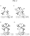
- a solution of the primary antibodies 1a and 1b as a free polyclonal antibody is applied on a sample slide, and an antigen and antibody reaction is allowed to occur. Accordingly, each of the primary antibodies 1a and 1b binds to epitopes ep1 and ep2 that are included in the antigen P of the sample slide.
- the second reaction step in which Fc region (part or whole) of the primary antibodies 1a and 1b, or Fab region (part or whole) thereof not bound to the antigen P (that is, primary antibody part not contributing to the reaction with the antigen P) is bound with the secondary antibodies 2a and 2b as a free polyclonal antibody is carried out so that 2 or more of the secondary antibody are fixed to one antigen.
- each of the secondary antibodies 2a and 2b as a polyclonal antibody is bound with the fluorescent nanoparticle 5 by way of binding between the biotin 3 and the streptavidin 4, as it is shown in Fig. 1(C) .
- the secondary antibodies 2a and 2b and fluorescent nanoparticle 5 are bound and fixed to one antigen P.
- the primary antibodies 1a and 1b are bound to plural epitopes ep1 and ep2 included in the antigen P, and also, after binding the secondary antibodies 2a and 2b to multiple sites of the primary antibodies, the fluorescent nanoparticle 5 binds to each of the secondary antibodies 2a and 2b, and thus high sensitization can be suitably obtained.
- number of the type of the secondary antibody to be used is not limited to 3, and it may be either increased or decreased depending on the necessity of sensitization, and it may be 2 types, for example. The same shall apply to the primary antibody.
- a solution of the primary antibody 1a as a free monoclonal antibody is applied on a sample slide, and an antigen and antibody reaction is allowed to occur. Accordingly, the primary antibody 1a binds only to the epitope ep1 that is included in the antigen P of the sample slide.
- the second reaction step in which Fc region (part or whole) of the primary antibody 1a or Fab region (part or whole) thereof not bound to the antigen P (that is, primary antibody part not contributing to the reaction with the antigen P) is bound with the secondary antibody 2a as a monoclonal antibody that is concentrated and directly bound in large number on a surface of the fluorescent nanoparticle 5 is carried out so that the fluorescent nanoparticle 5 is fixed on the antigen P.
- the fluorescent nanoparticle 5 is bound with plural secondary antibodies 2a as it has been described above for the second antibody.
- the primary antibody 1a is a monoclonal antibody
- binding of the primary antibody 1a can be achieved in a state in which the specificity for the antigen P is maintained, in particular.
- the secondary antibody 2a is concentrated and directly bound on a surface of the fluorescent nanoparticle 5, compared to the secondary antibody in free state, the secondary antibody 2a can be bound to the primary antibody 1a at higher probability, and, based on this binding, the fluorescent nanoparticle 5 can be bound with the secondary antibody, and thus high sensitization can be suitably obtained.
- binding density of the secondary antibody 2a on a surface of the fluorescent nanoparticle 5 and average particle size of the fluorescent nanoparticle 5 that is, number of secondary antibody per particle
- a solution of the primary antibody 1a as a free monoclonal antibody is applied on a sample slide, and an antigen and antibody reaction is allowed to occur. Accordingly, the primary antibody 1a binds only to the epitope ep1 that is included in the antigen P of the sample slide.
- the second reaction step in which Fc region (part or whole) of the primary antibody 1a or Fab region (part or whole) thereof not bound to the antigen P (that is, primary antibody part not contributing to the reaction with the antigen P) is bound with the secondary antibodies 2a and 2b as a polyclonal antibody directly bound in plurality on a surface of the fluorescent nanoparticle 5 is carried out so that the secondary antibodies 2a and 2b and the fluorescent nanoparticle 5 are fixed on the antigen P.
- plural secondary antibodies 2a and 2b are bound to the fluorescent nanoparticle 5 as it has been described above with regard to the second antibody.
- the primary antibody 1a is a monoclonal antibody
- binding of the primary antibody can be achieved in a state in which the specificity for the antigen P is maintained, in particular.
- the secondary antibodies 2a and 2b as a polyclonal antibody are concentrated and directly bound in large number on a surface of the fluorescent nanoparticle 5, compared to a case in which monoclonal antibodies are present in large number in concentrated state, linking and fixing is achieved at multiple different sites of a primary antibody (see, Fig. 2(E) ), and at even higher probability and specificity, the secondary antibody 2a and the fluorescent nanoparticle 5 can be bound and fixed on the primary antibody, and thus high sensitization can be suitably obtained.
- each of the primary antibodies 1a and 1b binds to plural epitopes ep1 and ep2 that are included in the antigen P of the sample slide.
- the second reaction step in which Fc region (part or whole) of the primary antibodies 1a and 1b, or Fab region (part or whole) thereof (that is, primary antibody part not contributing to the reaction with the antigen P) is bound with the secondary antibody 2a as a monoclonal antibody that is concentrated and directly bound in large number on a surface of the fluorescent nanoparticle 5 is carried out so that 2 or more of the secondary antibody 2a and the fluorescent nanoparticle 5 are fixed on the antigen P.
- plural secondary antibodies 2a are bound to the fluorescent nanoparticle 5 as it has been described above with regard to the second antibody.
- the primary antibodies 1a and 1b as a polyclonal antibody bind to epitopes ep1 and ep2 of the antigen P
- plural secondary antibodies 2a as a monoclonal antibody concentrated and directly bound on a surface of the fluorescent nanoparticle 5 bind to part or whole of the Fc region or Fab region commonly present in the primary antibodies 1a and 1b, regardless of the type of the primary antibody, and also at multiple sites, the fluorescent nanoparticle is firmly fixed on the antigen P so that easy detection of the antigen P is achieved.
- a solution of the primary antibodies 1a and 1b as a free polyclonal antibody is applied on a sample slide, and an antigen and antibody reaction is allowed to occur. Accordingly, the primary antibodies 1a and 1b bind to plural epitopes ep1 and ep2 that are included in the antigen P of the sample slide.
- the second reaction step in which Fc region (part or whole) of the primary antibodies 1a and 1b, or Fab region (part or whole) thereof not bound to the antigen P (that is, primary antibody part not contributing to the reaction with the antigen P) is bound with the secondary antibodies 2a and 2b as plural polyclonal antibodies directly bound to the fluorescent nanoparticle 5 is carried out so that 2 or more of the secondary antibodies 2a and 2b and fluorescent nanoparticle 5 are bound and fixed to one antigen P.
- each of the primary antibodies 1a and 1b as a polyclonal antibody binds to the antigen P and the secondary antibodies 2a and 2b concentrated and directly bound on a surface of the fluorescent nanoparticle 5 bind to part or whole of the Fc region of the primary antibodies 1a and 1b or Fab region (part or whole) thereof, each at different site, the fluorescent nanoparticle can be firmly fixed, in a state with high specificity, in particular, on the antigen.
- number of the type of the primary antibody and the secondary antibody to be used is not limited to 2, and it may be either increased or decreased depending on the necessity of sensitization, and it may be 3 types, for example.
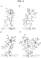
- a particle dispersion of the non-fluorescent nanoparticle 5a in which the primary antibody 1a as a monoclonal antibody is concentrated and bound in large number on a surface of the particle is applied on a sample slide, and an antigen and antibody reaction is allowed to occur. Accordingly, together with the non-fluorescent nanoparticle 5a, monoclonal antibody 1a binds to plural epitopes ep1 included in the antigen P of the sample slide.
- use amount of the non-fluorescent nanoparticle 5a in which primary antibody is bound on a surface of the particle is mainly determined based on the binding density of the primary antibody on a surface of the non-fluorescent nanoparticle 5a, binding affinity of the primary antibody for the antigen, and average particle size of the non-fluorescent nanoparticle 5a, and it is not particularly limited as long as it is a concentration allowing the binding of the primary antibody to an antigen.
- the non-fluorescent nanoparticle 5a can be used after being dispersed in a buffer like PBS such that the concentration of the primary antibody present on a surface of the non-fluorescent nanoparticle 5a is identical to the concentration of the free primary antibody which has been explained above in the first embodiment.
- mole number (or number) and molar concentration of the primary antibody on a surface of the non-fluorescent nanoparticle are preferably set at 10 times or higher, although it may vary depending on antibody titer.
- binding of a monoclonal antibody 1a to the antigen can be carried out at the same conditions as common primary antibody reaction, and it can be carried out by incubation for 4 to 12 hours at 1 to 10°C, for example.
- the second reaction step in which Fab region (part or whole) of the primary antibody 1a that is concentrated in large number on a surface of the non-fluorescent nanoparticle 5a fixed on the antigen P, or Fc region (part or whole) thereof not bound to the antigen P (that is, primary antibody part not contributing to the reaction with the antigen P) is bound with the secondary antibody 2d as plural free polyclonal antibodies is carried out.
- the fluorescent nanoparticle 5 is fixed on the secondary antibody 2d.
- 2 or more secondary antibody 2d and fluorescent nanoparticle 5 are bound and fixed to one antigen P.
- the fluorescent immunostaining method of the eighth embodiment because plural primary antibodies 1a are present in concentrated manner on a surface of the non-fluorescent nanoparticle 5a, compared to a case in which the primary antibody 1a is in a free state, binding of the primary antibody 1a to the antigen P can be obtained at higher probability. Furthermore, for one antigen P, the primary antibody 1a as a monoclonal antibody binds only at one site, and thus the specificity is high.
- plural secondary antibodies 2d bind to plural primary antibodies 1a that are present in concentrated manner on a surface of the non-fluorescent nanoparticle 5a but not reacted with the antigen P, and the fluorescent nanoparticle 5 binds to each of those the secondary antibody 2d, high sensitization can be achieved with very favorable efficiency while the specificity is suitably maintained. It is particularly advantageous in that, by adjusting the amount of the primary antibody 1a, the secondary antibody 2d, or the fluorescent nanoparticle 5 to be used, the detection sensitivity can be controlled within a broad range and a broader dynamic range of the sensitivity is obtained.
- a particle dispersion of the non-fluorescent nanoparticle 5a in which the primary antibody 1a as a monoclonal antibody is concentrated and bound in large number on a surface of the particle is applied on a sample slide, and an antigen and antibody reaction is allowed to occur. Accordingly, the primary antibody 1a binds to plural epitopes ep1 included in the antigen P of the sample slide.
- the use amount of the non-fluorescent nanoparticle 5b having the primary antibody 1a concentrated and bound in large number on a surface of the particle is the same as that of the eighth embodiment.
- Fc region (part or whole) of plural primary antibodies 1a that are not bound to the antigen P, which are present on a surface of the non-fluorescent nanoparticle 5b fixed on the antigen P, or Fab region (part or whole) thereof (that is, primary antibody part not contributing to the reaction with the antigen P) is bound with the secondary antibodies 2a and 2d as plural free polyclonal antibodies.
- the fluorescent nanoparticle 5 is fixed on the secondary antibodies 2a and 2d.
- 2 or more secondary antibody 2d and fluorescent nanoparticle 5 are bound and fixed to one antigen P.
- the fluorescent immunostaining method of the ninth embodiment because plural primary antibodies 1a are present in concentrated manner on a surface of the non-fluorescent nanoparticle 5b, compared to a case in which the primary antibody 1a is in a free state, binding of the primary antibody 1a to the antigen P can be obtained at higher probability. Furthermore, for one antigen P, the primary antibody 1a as a monoclonal antibody binds only at one site, and thus the specificity is high.
- the secondary antibodies 2a and 2d as a polyclonal antibody bind to multiple sites of plural primary antibodies 1a present on a surface of the non-fluorescent nanoparticle 5a and not reacted with the antigen P, and also the fluorescent nanoparticle 5a binds to those secondary antibodies 2a and 2d, and thus an increased binding number of the secondary antibody is obtained and high sensitization can be achieved with favorable efficiency compared to a case of using a monoclonal antibody as the secondary antibody.
- a broader dynamic range of the sensitivity is obtained, and also, by adjusting the amount of the primary antibody, secondary antibody, or fluorescent nanoparticle to be used within the broad dynamic range, the detection sensitivity can be controlled suitably in broad range.
- a particle dispersion of the non-fluorescent nanoparticle 5a in which the primary antibodies 1a and 1b as a polyclonal antibody are concentrated and bound in large number on a surface of the particle is applied on a sample slide, and an antigen and antibody reaction is allowed to occur. Accordingly, the primary antibodies 1a and 1b bind to plural epitopes ep1 and ep2 included in the antigen P of the sample slide.
- the use amount of the non-fluorescent nanoparticle 5a having the primary antibodies 1a and 1b concentrated and bound in large number on a surface of the particle is the same as that of the eighth embodiment.
- Fc region (part or whole) of plural primary antibodies 1a and 1b present on a surface of the non-fluorescent nanoparticle 5a fixed on the antigen P, or Fab region (part or whole) thereof (that is, primary antibody part not contributing to the reaction with the antigen P) is bound with plural secondary antibodies 2d as a free monoclonal antibody.
- the fluorescent nanoparticle 5 is fixed on the secondary antibody 2d.
- 2 or more secondary antibody 2d and fluorescent nanoparticle 5 are bound and fixed to one antigen P.
- the fluorescent immunostaining method of the tenth embodiment because plural primary antibodies 1a and 1b are present on a surface of the non-fluorescent nanoparticle 5a, compared to a case in which the primary antibodies 1a and 1b are in a free state, binding of the primary antibodies 1a and 1b to the antigen P can be obtained at higher probability. Furthermore, because the primary antibodies 1a and 1b are a polyclonal antibody, the non-fluorescent nanoparticle is more firmly fixed on the antigen P compared to the case of the ninth embodiment (case in which a monoclonal antibody is fixed on a surface of the non-fluorescent nanoparticle 5a). Thus, by having high specificity, high sensitization can be suitably achieved.
- a particle dispersion of the non-fluorescent nanoparticle 5a in which plural polyclonal antibodies 1a and 1b are bound on a surface of the particle is applied on a sample slide, and an antigen and antibody reaction is allowed to occur.
- the primary antibodies 1a and 1b as a polyclonal antibody bind to plural epitopes ep1 and ep2 included in the antigen P of the sample slide.
- the use amount of the non-fluorescent nanoparticle 5a having the primary antibodies 1a and 1b concentrated and bound in large number on a surface of the particle is the same as that of the eighth embodiment.
- Fc region (part or whole) of plural primary antibodies 1a which have not bound to the antigen P and are present on a surface of the non-fluorescent nanoparticle 5a fixed on the antigen P, or Fab region (part or whole) thereof (that is, primary antibody part not contributing to the reaction with the antigen P) is bound with plural secondary antibodies 2a, 2d, and 2f as a free polyclonal antibody.
- the fluorescent nanoparticle 5 is fixed on the secondary antibodies 2a, 2d, and 2f.
- 2 or more secondary antibodies 2a, 2d, and 2f and fluorescent nanoparticle 5 are bound and fixed to one antigen P.
- the fluorescent immunostaining method of the eleventh embodiment because plural primary antibodies 1a and 1b are present on a surface of the non-fluorescent nanoparticle 5a, compared to a case in which the primary antibodies 1a and 1b are in a free state, binding of the primary antibody to the antigen P can be obtained at higher probability. Furthermore, because the primary antibodies 1a and 1b are a polyclonal antibody, the non-fluorescent nanoparticle 5a is more firmly fixed on the antigen P compared to a case in which a monoclonal antibody is fixed on a surface of the non-fluorescent nanoparticle 5a. Thus, higher specificity is obtained.
- the secondary antibodies 2a, 2d, and 2f as a free polyclonal antibody bind to two or more of the part or whole of Fc region or Fab region of the plural primary antibodies 1a and 1b present on a surface of the non-fluorescent nanoparticle 5a fixed on the antigen and also the fluorescent nanoparticle 5 binds to the secondary antibody, the binding number of the secondary antibody and fluorescent nanoparticle is higher and the dynamic range of detection is broader due to higher detection sensitivity than a case in which the secondary antibody is a monoclonal antibody.
- a particle dispersion of the non-fluorescent nanoparticle 5a in which plural monoclonal antibodies 1a are bound on a surface of the particle is applied on a sample slide, and an antigen and antibody reaction is allowed to occur.
- the primary antibody 1a as a monoclonal antibody binds to epitope ep1 included in the antigen P of the sample slide.
- the use amount of the non-fluorescent nanoparticle 5a having the primary antibody 1a concentrated and bound in large number on a surface of the particle is the same as that of the eighth embodiment.
- Fc region (part or whole) of plural primary antibodies 1a which are present on a surface of the non-fluorescent nanoparticle 5a fixed to one antigen P, or Fab region (part or whole) thereof (that is, primary antibody part not contributing to the reaction with the antigen P) is bound with plural secondary antibodies 2c as a monoclonal antibody bound in large number on a surface of the fluorescent nanoparticle 5, and, by way of the binding of 2 or more secondary antibody 2c to one antigen P, one or more fluorescent nanoparticles 5 are fixed.
- the primary antibody 1a is a monoclonal antibody
- the primary antibody binding is obtained while the specificity for the antigen is maintained, in particular.
- plural primary antibodies 1a are present in concentrated manner on a surface of the non-fluorescent nanoparticle 5a, compared to a case in which the primary antibody 1a is in a free state, binding of the primary antibody to the antigen P can be obtained at higher probability.
- the secondary antibody 2c which is bound in large number on a surface of the fluorescent nanoparticle 5, binds to plural primary antibodies 1a on a surface of the non-fluorescent nanoparticle 5a which have not reacted with the antigen P, the secondary antibody 2c can be bound to the primary antibody 1a at high probability, and thus high sensitization can be suitably achieved.
- a particle dispersion of the non-fluorescent nanoparticle 5a in which the monoclonal antibody 1a is concentrated and bound in large number on a surface of the particle is applied on a sample slide, and an antigen and antibody reaction is allowed to occur. Accordingly, the monoclonal antibody 1a (primary antibody) binds to epitope ep1 included in the antigen P of the sample slide.
- the use amount of the non-fluorescent nanoparticle 5a having the primary antibody 1a concentrated and bound in large number on a surface of the particle is the same as that of the eighth embodiment.
- Fc region (part or whole) of plural primary antibodies 1a which are present on a surface of the non-fluorescent nanoparticle 5a fixed on the antigen P and not bound to the antigen P, or Fab region (part or whole) thereof (that is, primary antibody part not contributing to the reaction with the antigen P) is bound with plural secondary antibodies 2d and 2c as a polyclonal antibody bound in large number on a surface of the fluorescent nanoparticle 5. Accordingly, by way of the binding of 2 or more of the secondary antibody 1a to one antigen P, one or more fluorescent nanoparticles 5 are fixed.
- the primary antibody 1a is a monoclonal antibody
- binding of the primary antibody 1a is obtained while the specificity for the antigen P is maintained, in particular.
- plural primary antibodies 1a are present on a surface of the non-fluorescent nanoparticle 5a, compared to a case in which the primary antibody 1a is in a free state, binding of the primary antibody 1a to the antigen P can be obtained at higher probability.
- the polyclonal antibodies 2c and 2d which are concentrated and bound in large number on a surface of the fluorescent nanoparticle 5, bind, as a secondary antibody, to plural primary antibodies 1a on a surface of the non-fluorescent nanoparticle 5a which have not reacted with the antigen P, the secondary antibody can bind to multiples sites of the primary antibody 1a at high probability, and thus high sensitization can be suitably achieved.
- a particle dispersion of the non-fluorescent nanoparticle 5a in which the primary antibodies 1a and 1b as a polyclonal antibody are concentrated and bound in large number on a surface of the particle is applied on a sample slide, and an antigen and antibody reaction is allowed to occur. Accordingly, the primary antibodies 1a and 1b bind to plural epitopes ep1 and ep2 included in the antigen P of the sample slide, and thus the non-fluorescent nanoparticle 5a is fixed on the antigen P.
- the use amount of the non-fluorescent nanoparticle 5a having the primary antibodies 1a and 1b bound on a surface of the particle is the same as that of the eighth embodiment.
- Fc region (part or whole) of plural primary antibodies 1a and 1b, which are present on a surface of the non-fluorescent nanoparticle 5a fixed on the antigen P and not bound to the antigen P, or Fab region (part or whole) thereof (that is, primary antibody part not contributing to the reaction with the antigen P) is bound with plural secondary antibodies 2d as a monoclonal antibody bound in large number on a surface of the fluorescent nanoparticle 5, and, by way of the binding of 2 or more antibodies to one antigen, one or more fluorescent nanoparticles are fixed.
- mole number (or number) and molar concentration of the secondary antibody on a surface of the fluorescent nanoparticle are preferably set at 10 times or higher, although it may vary depending on antibody titer (ditto for the followings).
- the fluorescent immunostaining method of the fourteenth embodiment because plural primary antibodies 1a and 1b are present on a surface of the non-fluorescent nanoparticle 5a, compared to a case in which the primary antibody is in a free state, binding of the primary antibody 1a to the antigen P can be obtained at higher probability. Furthermore, because plural primary antibodies 1a and 1b are a polyclonal antibody, the non-fluorescent nanoparticle 5a is more firmly fixed on the antigen P than a case in which a monoclonal antibody is fixed on a surface of the non-fluorescent nanoparticle 5a, and thus higher specificity is obtained.
- the monoclonal antibody 2d bound in large number on a surface of the fluorescent nanoparticle 5 binds, and thus the binding can be achieved regardless of the type of the primary antibody 1a, and also the secondary antibodies 2c and 2d can bind to the primary antibody 1a at high probability. Accordingly, high sensitization can be suitably obtained.
- a particle dispersion of the non-fluorescent nanoparticle 5a in which the primary antibodies 1a and 1b as a polyclonal antibody are concentrated and bound in large number on a surface of the particle is applied on a sample slide, and an antigen and antibody reaction is allowed to occur. Accordingly, each of the primary antibodies 1a and 1b binds to plural epitopes ep1 and ep2 included in the antigen P of the sample slide.
- the use amount of the non-fluorescent nanoparticle 5a having the primary antibodies 1a and 1b bound on a surface of the particle is the same as that of the eighth embodiment.
- Fc region (part or whole) of the primary antibodies 1a and 1b, which are present on a surface of the non-fluorescent nanoparticle 5a fixed on the antigen P, or Fab region (part or whole) thereof (that is, primary antibody part not contributing to the reaction with the antigen P) is bound with polyclonal antibodies 2d and 2c (secondary antibody) that are bound in large number on a surface of the fluorescent nanoparticle 5, and, by way of the binding of 2 or more secondary antibodies 2c and 2d to one antigen P, one or more fluorescent nanoparticles 5 are fixed.
- the fluorescent immunostaining method of the fifteenth embodiment because plural primary antibodies 1a and 1b are present in concentrated manner on a surface of the non-fluorescent nanoparticle 5a, compared to a case of a free primary antibody, binding of the primary antibodies 1a and 1b to the antigen P can be obtained at higher probability. Furthermore, because the primary antibodies 1a and 1b on a surface of the non-fluorescent nanoparticle 5a are a polyclonal antibody, the non-fluorescent nanoparticle 5a is more firmly fixed on the antigen P than a case in which a monoclonal antibody is fixed on a surface of the non-fluorescent nanoparticle 5a, and thus higher specificity is obtained.
- the polyclonal antibody bound in large number on a surface of the fluorescent nanoparticle 5 binds, as a secondary antibody, to different binding sites of the primary antibodies 1a and 1b (e.g., whole Fc region and part of Fab region), and thus the secondary antibody can bind firmly to the primary antibody at high probability. Accordingly, high sensitization can be suitably obtained.
- Steps (3-1) to (3-3) the melamine-based fluorescent nanoparticles modified with maleimide group were prepared.
- a non-fluorescent melamine nanoparticle bound with anti PD-L1 antibody "Anti-PD-Ll antibody (ab58810)” was prepared in the same manner as Preparation Example 2 except that, with regard to Preparation Example 2, the above non-fluorescent nanoparticle is used instead of the fluorescent nanoparticles, and "Anti-PD-L1 antibody (ab58810)” (animal species: rabbit, Abcam), which is a polyclonal antibody capable of binding to PD-L1, is used instead of "Biotin labeled anti rabbit IgG antibody (animal species: goat)".
- the binding density of a primary antibody on a surface of the "Biotin labeled anti rabbit IgG antibody (animal species: goat)" was about 1000 antibodies per particle and it corresponds to about 3.54 ⁇ 10 14 (antibody/mm 2 of particle surface).
- the amount of proteins bound on a surface of the particle was measured, and based on the weight molecular weight of the antibody used, mole number or number of the antibody fixed on a surface of the particle was calculated.
- the average particle surface area was calculated based on 4 ⁇ r 2 . Furthermore, by using a particle counter, the dispersion concentration (particles/ ⁇ L) of the non-fluorescent nanoparticle was determined, and from the total volume of the dispersion ( ⁇ L), total number (particles) of the non-fluorescent nanoparticle was calculated. By multiplying the average particle surface area (mm2/particle) by the total number (particles) of the particles that are measured, total area (mm2) of the non-fluorescent nanoparticle was calculated. From the mole number of the antibody and total particle area (mm 2 ), the antibody binding density (mole number of antibody/mm 2 ) was calculated.
- the non-fluorescent melamine nanoparticle bound with "PD-L1 (E1L3N (registered trademark) XP (registered trademark) Rabbit mAb” was prepared in the same manner as Preparation Example 3 except that, with regard to Preparation Example 3, the above non-fluorescent nanoparticle is used instead of the fluorescent nanoparticles, and PD-L1 (E1L3N (registered trademark) XP (registered trademark) Rabbit mAb (Cell Signaling Technology Japan, K.K.)" is used instead of "Biotin labeled anti rabbit IgG antibody (animal species: goat)" (product number: 426011, 426012, NICHIREI BIOSCIENCES INC.).
- the binding density of a primary antibody on a surface of a non-fluorescent nanoparticle in that case, it was found that about 1000 antibodies are bound to one particle and it corresponds to 3.54 ⁇ 10 14 (antibody/mm 2 of particle surface).
- the fluorescent nanoparticles added with a maleimide group were dispersed in PBS containing 2 mM EDTA to 0.67 nM.
- This dispersion of fluorescent nanoparticles (740 ⁇ L) and the streptavidin introduced with SH group (0.04 mg) were admixed with each other, and allowed to react for 1 hour at room temperature.
- the reaction solution was added with 10 mM mercaptoethanol to terminate the reaction.
- the obtained solution was concentrated using a centrifuge filter, and by using a gel filtration column for purification, non-reacted antifluorescein antibody or the like are removed to obtain sulforhodamine-containing nanoparticle bound with streptavidin by way of linker.
- a sample slide of PD-L1 positive culture cells fixed by paraffin was prepared.
- sample slide for noise measurement, PD-L1 negative cells were cultured and fixed with paraffin to give a sample slide (hereinbelow, simply described as "sample slide (for noise measurement)).
- NCI-H460 organism: homo sapiens, human/tissue: lung: pleural effusion/disorder: cancer: large cell lung cancer
- NCI-H446 organism: homo sapiens, human/tissue: lung: pleural effusion; originating from metastasis area/disorder: cancer: small cell lung cancer
- ATCC registered trademark
- the subculture was carried out according to generally known subculture. As shown in Table 2, 0.25% trypsin was used for cell surface washing during subculture. The ratio of the volume of fresh medium to addition amount of cell suspension to be added to the medium was set at 1 : 3 to 1 : 8 (or 1 : 3 to 1 : 6).
- the sample slide obtained after a series of the above steps was illuminated with a prescribed excitation light to allow fluorescence to be emitted.
- the sample slide in this state was observed and photographed under a fluorescence microscope ("BX-53", manufactured by Olympus Corporation). Furthermore, the bright spot measurement was carried out based on ImageJ Find Maxima method.
- the excitation light was set at 545 to 565 nm according to transmission through an optical filter.
- the wavelength range (nm) of the fluorescence to be observed was set at 570 to 590 nm according to transmission through an optical filter.
- the conditions of the excitation wavelength in the microscope observation and image acquisition were set such that the irradiation energy in the vicinity of the center of the visual field is 900 W/cm 2 for excitation at 550 nm.
- a photograph was taken by arbitrarily setting the exposure time such that the image brightness is not saturated (for example, the exposure time was set at 4,000 ⁇ s).
- the photographed microscopic image taken by a fluorescence microscope or the like, when brightness is higher than predetermined threshold value, it is measured as bright spot, and number of the fluorescent nanoparticle or fluorescent signal intensity (signal (relative value) per cell was calculated.
- Example 1 except that the second reaction step of Comparative Example 1 is carried out as described below, other steps like the deparaffinization treatment step, the activation treatment step, the first reaction step, the third reaction step, the washing step, and the observation step were carried out in the same manner as Comparative Example 1.
- the signal value compared to Comparative Example 1 was found to be 2.
- the noise value indicates a value when the measured value of the sample slide (for noise measurement) of Comparative Example 1 is set at 1, and it was found to be 1.5.
- S/N ratio (relative value) was 1.33.
- the quantification property was " ⁇ ".
- Example 2 except that the first reaction step of Comparative Example 1 is carried out as described below, other steps like the deparaffinization treatment step, the activation treatment step, the second reaction step, the washing steps 1 to 3, the third reaction step, and the observation step were carried out in the same manner as Comparative Example 1.
- the primary antibody was used after it has been adjusted to have the same mole number as the mole number of the primary antibody used in Comparative Example 1.
- Example 3 except that the first reaction step and the second reaction step of Comparative Example 1 are carried out as described below, other steps including the deparaffinization treatment step, the activation treatment step, the washing steps 1 to 3, the third reaction step, and the observation step were carried out in the same manner as Comparative Example 1.
- the primary antibody was used after it has been adjusted to have the same mole number as the mole number of the primary antibody used in Comparative Example 1.
- Example 3 As a result of Example 3, the signal value compared to Comparative Example 1 was found to be 4, and the noise value (relative value) was found to be 3. S/N ratio (relative value) was 1.33. Furthermore, the quantification property was " ⁇ ". Result of Example 3 is shown in Table 3.
- Example 3 except that the first reaction step of Comparative Example 1 is carried out as described below, other steps including the deparaffinization treatment step, the activation treatment step, the washing steps 1 to 3, the second reaction step, the third reaction step, and the observation step were carried out in the same manner as Comparative Example 1.
- mole number of antibody contained in 1 ⁇ L of the dispersion liquid of non-fluorescent nanoparticles that are bound with the primary antibody is calculated by the following formula 1, and based on the obtained information, use amount of the primary antibody was adjusted to have the same mole number and the same concentration (mol/ ⁇ L) as the mole number of the primary antibody used in Comparative Example 1.
- Mole number/ ⁇ L "Concentration of dispersion liquid of non-fluorescent nanoparticles (particles/ ⁇ L)" ⁇ "Average particle surface area (mm 2 /particle)” ⁇ "Antibody binding density (antibody mole number/mm 2 )" on surface of non-fluorescent nanoparticle
- Example 4 As a result of Example 4, the signal value compared to Comparative Example 1 was found to be 4, and the noise value (relative value) was found to be 3. S/N ratio (relative value) was 1.33. Furthermore, the quantification property was " ⁇ ". Result of Example 4 is shown in Table 3.
- Example 5 except that the first reaction step and the second reaction step of Comparative Example 1 are carried out as described below, other steps including the deparaffinization treatment step, the activation treatment step, the washing steps 1 to 3, the third reaction step, and the observation step were carried out in the same manner as Comparative Example 1.
- Example 5 As a result of Example 5, the signal value compared to Comparative Example 1 was found to be 8, and the noise value (relative value) was found to be 6. S/N ratio (relative value) was 1.33. Furthermore, the quantification property was " ⁇ ". Result of Example 5 is shown in Table 3.
- Example 6 except that the first reaction step and the second reaction step of Comparative Example 1 are carried out as described below, other steps including the deparaffinization treatment step, the activation treatment step, the washing steps 1 to 3, the third reaction step, and the observation step were carried out in the same manner as Comparative Example 1.
- Example 6 As a result of Example 6, the signal value compared to Comparative Example 1 was found to be 8, and the noise value (relative value) was found to be 4. S/N ratio (relative value) was 2. Furthermore, the quantification property was " ⁇ ". Result of Example 6 is shown in Table 3.
- Example 7 except that the first reaction step and the second reaction step of Comparative Example 1 are carried out as described below, other steps including the deparaffinization treatment step, the activation treatment step, the washing steps 1 to 3, the third reaction step, and the observation step were carried out in the same manner as Comparative Example 1.
- Example 7 As a result of Example 7, the signal value compared to Comparative Example 1 was found to be 12, and the noise value (relative value) was found to be 8. S/N ratio (relative value) was 1.50. Furthermore, the quantification property was " ⁇ ". Result of Example 7 is shown in Table 3.
- Example 8 except that the second reaction step of Comparative Example 1 is carried out as described below, other steps including the deparaffinization treatment step, the activation treatment step, the washing steps 1 to 3, the first reaction step, the third reaction step, and the observation step were carried out in the same manner as Comparative Example 1.
- Example 8 As a result of Example 8, the signal value compared to Comparative Example 1 was found to be 1, and the noise value (relative value) was found to be 0.8. S/N ratio (relative value) was 1.25. Furthermore, the quantification property was " ⁇ ". Result of Example 8 is shown in Table 3.
- Example 9 except that the second reaction step of Comparative Example 1 is carried out as described below, other steps including the deparaffinization treatment step, the activation treatment step, the washing steps 1 to 3, the first reaction step, the third reaction step, and the observation step were carried out in the same manner as Comparative Example 1.
- Example 9 the signal value compared to Comparative Example 1 was found to be 2, and the noise value (relative value) was found to be 1.5. S/N ratio (relative value) was 1.33. Furthermore, the quantification property was " ⁇ ". Result of Example 9 is shown in Table 3.
- Example 10 except that the first reaction step and the second reaction step of Comparative Example 1 are carried out as described below, other steps including the deparaffinization treatment step, the activation treatment step, the washing steps 1 to 3, the third reaction step, and the observation step were carried out in the same manner as Comparative Example 1.
- the primary antibody was used after it has been adjusted to have the same mole number as the mole number of the primary antibody used in Comparative Example 1.
- Example 10 As a result of Example 10, the signal value compared to Comparative Example 1 was found to be 4, and the noise value (relative value) was found to be 2.5. S/N ratio (relative value) was 1.60. Furthermore, the quantification property was " ⁇ ". Result of Example 10 is shown in Table 3.
- Example 11 except that the first reaction step and the second reaction step of Comparative Example 1 are carried out as described below, other steps including the deparaffinization treatment step, the activation treatment step, the washing steps 1 to 3, the third reaction step, and the observation step were carried out in the same manner as Comparative Example 1.
- the primary antibody was used after it has been adjusted to have the same mole number as the mole number of the primary antibody used in Comparative Example 1.
- Example 11 As a result of Example 11, the signal value compared to Comparative Example 1 was found to be 8, and the noise value (relative value) was found to be 4.5. S/N ratio (relative value) was 1.78. Furthermore, the quantification property was " ⁇ ". Result of Example 11 is shown in Table 3.
- Example 12 except that the first reaction step and the second reaction step of Comparative Example 1 are carried out as described below, other steps including the deparaffinization treatment step, the activation treatment step, the washing steps 1 to 3, the third reaction step, and the observation step were carried out in the same manner as Comparative Example 1.
- Example 12 As a result of Example 12, the signal value compared to Comparative Example 1 was found to be 8, and the noise value (relative value) was found to be 5.5. S/N ratio (relative value) was 1.45. Furthermore, the quantification property was " ⁇ ". Result of Example 12 is shown in Table 3.
- Example 13 except that the first reaction step and the second reaction step of Comparative Example 1 are carried out as described below, other steps including the deparaffinization treatment step, the activation treatment step, the washing steps 1 to 3, the third reaction step, and the observation step were carried out in the same manner as Comparative Example 1.
- Example 13 As a result of Example 13, the signal value compared to Comparative Example 1 was found to be 12, and the noise value (relative value) was found to be 6.5. S/N ratio (relative value) was 1.85. Furthermore, the quantification property was " ⁇ ". Result of Example 13 is shown in Table 3.
- Example 14 except that the first reaction step and the second reaction step of Comparative Example 1 are carried out as described below, other steps including the deparaffinization treatment step, the activation treatment step, the washing steps 1 to 3, the third reaction step, and the observation step were carried out in the same manner as Comparative Example 1.
- Example 14 As a result of Example 14, the signal value compared to Comparative Example 1 was found to be 16, and the noise value (relative value) was found to be 10. S/N ratio (relative value) was 1.60. Furthermore, the quantification property was " ⁇ ". Result of Example 14 is shown in Table 3.
- Example 15 except that the first reaction step and the second reaction step of Comparative Example 1 are carried out as described below, other steps including the deparaffinization treatment step, the activation treatment step, the washing steps 1 to 3, the third reaction step, and the observation step were carried out in the same manner as Comparative Example 1.
- Example 15 As a result of Example 15, the signal value compared to Comparative Example 1 was found to be 24, and the noise value (relative value) was found to be 16. S/N ratio (relative value) was 1.50. Furthermore, the quantification property was " ⁇ ". Result of Example 15 is shown in Table 3.
- a free polyclonal antibody in case of using a free polyclonal antibody, it is preferable from the viewpoint of the S/N ratio to have a fluorescent immunostaining method in which a free polyclonal antibody is used as a primary antibody and, simultaneously, a free monoclonal antibody is used as a secondary antibody so that plural secondary antibodies can be fixed to one molecule of the antigen. Furthermore, from the viewpoint of signal enhancement, it is preferable to have a fluorescent immunostaining method in which a free polyclonal antibody is used for both the primary antibody and the secondary antibody so that plural secondary antibodies can be fixed to one molecule of the antigen.
- a fluorescent immunostaining method in which a polyclonal antibody bound in large number on a surface of a non-fluorescent nanoparticle is used as a primary antibody, and, simultaneously, a free polyclonal antibody is used as a secondary antibody so that plural secondary antibodies can be fixed to one molecule of the antigen.
- a fluorescent immunostaining method in which a polyclonal antibody bound in large number on a surface of a non-fluorescent nanoparticle is used as a primary antibody, and, simultaneously, a polyclonal antibody bound in large number on a surface of a fluorescent nanoparticle is used as a secondary antibody so that plural secondary antibodies can be fixed to one molecule of the antigen.
Landscapes
- Health & Medical Sciences (AREA)
- Life Sciences & Earth Sciences (AREA)
- Immunology (AREA)
- Engineering & Computer Science (AREA)
- Chemical & Material Sciences (AREA)
- Molecular Biology (AREA)
- Biomedical Technology (AREA)
- Hematology (AREA)
- Urology & Nephrology (AREA)
- Analytical Chemistry (AREA)
- Pathology (AREA)
- General Physics & Mathematics (AREA)
- General Health & Medical Sciences (AREA)
- Biochemistry (AREA)
- Physics & Mathematics (AREA)
- Medicinal Chemistry (AREA)
- Food Science & Technology (AREA)
- Biotechnology (AREA)
- Cell Biology (AREA)
- Microbiology (AREA)
- Nanotechnology (AREA)
- Investigating, Analyzing Materials By Fluorescence Or Luminescence (AREA)
- Investigating Or Analysing Materials By The Use Of Chemical Reactions (AREA)
Applications Claiming Priority (2)
| Application Number | Priority Date | Filing Date | Title |
|---|---|---|---|
| JP2016076433 | 2016-04-06 | ||
| PCT/JP2017/008064 WO2017175523A1 (fr) | 2016-04-06 | 2017-03-01 | Procédé d'immunocoloration fluorescente |
Publications (2)
| Publication Number | Publication Date |
|---|---|
| EP3441761A1 true EP3441761A1 (fr) | 2019-02-13 |
| EP3441761A4 EP3441761A4 (fr) | 2019-04-10 |
Family
ID=60001572
Family Applications (1)
| Application Number | Title | Priority Date | Filing Date |
|---|---|---|---|
| EP17778902.1A Withdrawn EP3441761A4 (fr) | 2016-04-06 | 2017-03-01 | Procédé d'immunocoloration fluorescente |
Country Status (4)
| Country | Link |
|---|---|
| US (1) | US20190079081A1 (fr) |
| EP (1) | EP3441761A4 (fr) |
| JP (1) | JPWO2017175523A1 (fr) |
| WO (1) | WO2017175523A1 (fr) |
Families Citing this family (4)
| Publication number | Priority date | Publication date | Assignee | Title |
|---|---|---|---|---|
| JP7054378B2 (ja) * | 2018-11-08 | 2022-04-13 | 富士レビオ株式会社 | Ca19-9測定方法及びca19-9測定キット、並びに、これらに用いる抗体固定化担体及びその製造方法 |
| CN111257264B (zh) * | 2020-03-18 | 2023-02-28 | 上海纽脉医疗科技有限公司 | 一种生物组织包含的醛基含量的检测方法 |
| CN113866407A (zh) * | 2021-12-01 | 2021-12-31 | 上海思路迪医学检验所有限公司 | 一种外泌体her2蛋白的磁免疫化学发光检测试剂盒 |
| JPWO2024143431A1 (fr) * | 2022-12-27 | 2024-07-04 |
Family Cites Families (16)
| Publication number | Priority date | Publication date | Assignee | Title |
|---|---|---|---|---|
| JPH0763761A (ja) * | 1993-08-31 | 1995-03-10 | Tosoh Corp | 生理活性物質固定化磁性微粒子の製造法 |
| AU7382900A (en) | 1999-09-15 | 2001-04-17 | Miraibio Inc. | Method for processing biological samples |
| US20070292903A1 (en) * | 2004-11-25 | 2007-12-20 | Takashi Kobayashi | Method of detemining tropomyosin in chitosan |
| JP4929635B2 (ja) | 2005-07-14 | 2012-05-09 | 富士ゼロックス株式会社 | マレイミド基含有多孔質架橋ポリスチレン粒子及びその製造方法 |
| US8119572B2 (en) * | 2005-10-24 | 2012-02-21 | Wisconsin Alumni Research Foundation | Methods for determining protein binding specificity using peptide libraries |
| DK2339014T3 (en) * | 2005-11-16 | 2015-07-20 | Ambrx Inc | Methods and compositions comprising non-natural amino acids |
| JP2010096677A (ja) * | 2008-10-17 | 2010-04-30 | Toray Ind Inc | 抗体/抗原結合能を有する高感度免疫学測定用ナノ粒子 |
| JP2010210444A (ja) * | 2009-03-10 | 2010-09-24 | Beacle Inc | 多抗原同時検出用ナノ粒子 |
| AU2011274369A1 (en) * | 2010-07-02 | 2012-12-06 | Ventana Medical Systems, Inc. | Hapten conjugates for target detection |
| EP2437061A1 (fr) * | 2010-09-30 | 2012-04-04 | Ikonomopoulos, John | Détection de protéine mycobactérienne |
| US8494401B2 (en) | 2011-09-07 | 2013-07-23 | Xerox Corporation | Active ozone scrubber |
| ES2636483T3 (es) * | 2012-02-21 | 2017-10-05 | Laboratory Corporation Of America Holdings | Métodos y sistemas para la amplificación de señal de bioensayos |
| JP6286966B2 (ja) | 2013-09-18 | 2018-03-07 | コニカミノルタ株式会社 | 蛍光標識用樹脂粒子およびその製造方法、並びに、これを含む免疫染色用蛍光標識剤 |
| US10509039B2 (en) * | 2014-03-06 | 2019-12-17 | Konica Minolta, Inc. | Integrated phosphor nanoparticle marking agent, and fluorescent immunostaining employing same |
| WO2015146896A1 (fr) * | 2014-03-24 | 2015-10-01 | コニカミノルタ株式会社 | Procédé d'analyse quantitative de matériau biologique sur la base d'une immunocoloration à antigènes multiples |
| US10746740B2 (en) * | 2015-01-22 | 2020-08-18 | Konica Minolta, Inc. | Biological substance quantitation method, pathological diagnosis support system, and recording medium storing computer readable program |
-
2017
- 2017-03-01 WO PCT/JP2017/008064 patent/WO2017175523A1/fr not_active Ceased
- 2017-03-01 EP EP17778902.1A patent/EP3441761A4/fr not_active Withdrawn
- 2017-03-01 JP JP2018510272A patent/JPWO2017175523A1/ja active Pending
- 2017-03-01 US US16/084,374 patent/US20190079081A1/en not_active Abandoned
Also Published As
| Publication number | Publication date |
|---|---|
| WO2017175523A1 (fr) | 2017-10-12 |
| EP3441761A4 (fr) | 2019-04-10 |
| JPWO2017175523A1 (ja) | 2019-02-14 |
| US20190079081A1 (en) | 2019-03-14 |
Similar Documents
| Publication | Publication Date | Title |
|---|---|---|
| EP3124969B1 (fr) | Procédé d'analyse quantitative de matériau biologique sur la base d'une immunocoloration à antigènes multiples | |
| US10509039B2 (en) | Integrated phosphor nanoparticle marking agent, and fluorescent immunostaining employing same | |
| JP6687018B2 (ja) | 目的生体物質の検出方法および検出システム | |
| EP3441761A1 (fr) | Procédé d'immunocoloration fluorescente | |
| US11391728B2 (en) | Phosphor-integrated nanoparticles used in fluorescence observation | |
| JP6658330B2 (ja) | 組織切片から蛍光ナノ粒子の解離を防止する方法 | |
| US20200165666A1 (en) | Diluent for fluorescent nano particles, kit for immunofluorescent staining which utilizes same, solution for immunofluorescent staining, immunofluorescent staining method, and gene staining method | |
| EP3327427A1 (fr) | Procédé d'analyse et système d'analyse de substances biologiques cibles | |
| EP3608669A1 (fr) | Particules fluorescentes pour prémélange, colorant fluorescent en contenant et procédé de marquage fluorescent les utilisant | |
| JP6583011B2 (ja) | 酸性水溶液を用いた免疫染色スライドの洗浄方法 | |
| EP3173785B1 (fr) | Réactif de marquage contenant un médicament à cible moléculaire | |
| WO2021192910A1 (fr) | Procédé et dispositif de génération d'images et programme | |
| WO2021230134A1 (fr) | Procédé de formation d'image | |
| US20210318323A1 (en) | Information Acquiring Method, Information Acquiring Device, and Program | |
| WO2022196203A1 (fr) | Procédé de formation d'image | |
| JP2023126996A (ja) | 免疫染色方法及び免疫染色組織標本の作製方法 | |
| WO2019130503A1 (fr) | Procédé d'acquisition d'informations |
Legal Events
| Date | Code | Title | Description |
|---|---|---|---|
| STAA | Information on the status of an ep patent application or granted ep patent |
Free format text: STATUS: THE INTERNATIONAL PUBLICATION HAS BEEN MADE |
|
| PUAI | Public reference made under article 153(3) epc to a published international application that has entered the european phase |
Free format text: ORIGINAL CODE: 0009012 |
|
| STAA | Information on the status of an ep patent application or granted ep patent |
Free format text: STATUS: REQUEST FOR EXAMINATION WAS MADE |
|
| 17P | Request for examination filed |
Effective date: 20181005 |
|
| AK | Designated contracting states |
Kind code of ref document: A1 Designated state(s): AL AT BE BG CH CY CZ DE DK EE ES FI FR GB GR HR HU IE IS IT LI LT LU LV MC MK MT NL NO PL PT RO RS SE SI SK SM TR |
|
| AX | Request for extension of the european patent |
Extension state: BA ME |
|
| A4 | Supplementary search report drawn up and despatched |
Effective date: 20190307 |
|
| RIC1 | Information provided on ipc code assigned before grant |
Ipc: G01N 33/53 20060101ALI20190301BHEP Ipc: G01N 33/536 20060101ALI20190301BHEP Ipc: G01N 33/48 20060101AFI20190301BHEP Ipc: G01N 33/543 20060101ALI20190301BHEP |
|
| DAV | Request for validation of the european patent (deleted) | ||
| DAX | Request for extension of the european patent (deleted) | ||
| STAA | Information on the status of an ep patent application or granted ep patent |
Free format text: STATUS: EXAMINATION IS IN PROGRESS |
|
| 17Q | First examination report despatched |
Effective date: 20200407 |
|
| STAA | Information on the status of an ep patent application or granted ep patent |
Free format text: STATUS: THE APPLICATION IS DEEMED TO BE WITHDRAWN |
|
| 18D | Application deemed to be withdrawn |
Effective date: 20201020 |