EP3449930B1 - Synapsenbildungsmittel - Google Patents
Synapsenbildungsmittel Download PDFInfo
- Publication number
- EP3449930B1 EP3449930B1 EP17789738.6A EP17789738A EP3449930B1 EP 3449930 B1 EP3449930 B1 EP 3449930B1 EP 17789738 A EP17789738 A EP 17789738A EP 3449930 B1 EP3449930 B1 EP 3449930B1
- Authority
- EP
- European Patent Office
- Prior art keywords
- administration
- medicament
- cells
- mscs
- blood
- Prior art date
- Legal status (The legal status is an assumption and is not a legal conclusion. Google has not performed a legal analysis and makes no representation as to the accuracy of the status listed.)
- Active
Links
Images









Classifications
-
- A—HUMAN NECESSITIES
- A61—MEDICAL OR VETERINARY SCIENCE; HYGIENE
- A61K—PREPARATIONS FOR MEDICAL, DENTAL OR TOILETRY PURPOSES
- A61K35/00—Medicinal preparations containing materials or reaction products thereof with undetermined constitution
- A61K35/12—Materials from mammals; Compositions comprising non-specified tissues or cells; Compositions comprising non-embryonic stem cells; Genetically modified cells
- A61K35/28—Bone marrow; Haematopoietic stem cells; Mesenchymal stem cells of any origin, e.g. adipose-derived stem cells
-
- A—HUMAN NECESSITIES
- A61—MEDICAL OR VETERINARY SCIENCE; HYGIENE
- A61K—PREPARATIONS FOR MEDICAL, DENTAL OR TOILETRY PURPOSES
- A61K9/00—Medicinal preparations characterised by special physical form
- A61K9/0012—Galenical forms characterised by the site of application
- A61K9/0019—Injectable compositions; Intramuscular, intravenous, arterial, subcutaneous administration; Compositions to be administered through the skin in an invasive manner
-
- A—HUMAN NECESSITIES
- A61—MEDICAL OR VETERINARY SCIENCE; HYGIENE
- A61P—SPECIFIC THERAPEUTIC ACTIVITY OF CHEMICAL COMPOUNDS OR MEDICINAL PREPARATIONS
- A61P25/00—Drugs for disorders of the nervous system
-
- C—CHEMISTRY; METALLURGY
- C12—BIOCHEMISTRY; BEER; SPIRITS; WINE; VINEGAR; MICROBIOLOGY; ENZYMOLOGY; MUTATION OR GENETIC ENGINEERING
- C12N—MICROORGANISMS OR ENZYMES; COMPOSITIONS THEREOF; PROPAGATING, PRESERVING, OR MAINTAINING MICROORGANISMS; MUTATION OR GENETIC ENGINEERING; CULTURE MEDIA
- C12N5/00—Undifferentiated human, animal or plant cells, e.g. cell lines; Tissues; Cultivation or maintenance thereof; Culture media therefor
- C12N5/06—Animal cells or tissues; Human cells or tissues
- C12N5/0602—Vertebrate cells
- C12N5/0652—Cells of skeletal and connective tissues; Mesenchyme
- C12N5/0662—Stem cells
- C12N5/0663—Bone marrow mesenchymal stem cells (BM-MSC)
Definitions
- MSCs Mesenchymal stem cells
- neurotrophic and protective effects of neurotrophic factors For the therapeutic mechanism of MSCs, many mechanisms of action have been supposed and these are classified into three categories: neurotrophic and protective effects of neurotrophic factors, angiogenesis effect (restoration of the cerebral blood flow), and nerve regeneration.
- the neurotrophic and protective effects are expected to be mediated by humoral factors such as the neurotrophic factors brain derived neurotrophic factor (BDNF) and glial derived neurotrophic factor (GDNF).
- BDNF brain derived neurotrophic factor
- GDNF glial derived neurotrophic factor
- the angiogenesis effect is considered to involve two mechanisms: one is the secretion of angiogenesis factors by MSCs accumulated in the lesion to induce angiogenesis and the other one is differentiation of administered MSCs themselves into vascular endothelia to form new blood vessels.
- the nerve regeneration effect is also considered to involve two mechanisms: one is the promotion of endogenous neurogenesis by MSCs accumulated in the lesion and the other one is differentiation of administered MSCs themselves into
- US 2012/0009271 A1 relates generally to stem cell therapy and describes neuroprotection through the administration of amnion derived stem cells and melatonin.
- An object of the present invention is to provide a new method for treating an intractable neurological disease that has conventionally been considered to be difficult to treat by elucidating a therapeutic mechanism of mesenchymal stem cells (MSCs) and constructing the rationale for clinical applications thereof.
- MSCs mesenchymal stem cells
- the inventors have demonstrated that intravenously administered MSCs reach the hippocampus, differentiate into nerve cells, and form synapses. They have also demonstrated that administration of MSCs in a cerebral infarction model activates not only motor and sensory areas in the infarction region but also contralesional motor and sensory areas. Furthermore, they have demonstrated that administration of MSCs in a vascular dementia model rat improves the cognitive function.
- the present invention relates to the following (1) to (10) claims.
- intravenously administered MSCs ameliorate neurological diseases such as cerebral infarction and vascular dementia by forming synapses and rebuilding neural circuits as well as promoting brain plasticity.
- administration of MSCs is effective for dementia (vascular dementia, Alzheimer-type dementia), intractable neurological diseases such as chronic-phase cerebral infarction and chronic-phase spinal cord injury, and mental diseases, which have conventionally been considered to be difficult to treat and the effect restores not only the motor function but also higher functions such as memory impairment.
- the "synapse formation agent” used in the present invention is a cellular preparation containing CD24-negative mesenchymal stem cells (MSCs) derived from human bone marrow or blood and is a medicament as defined in the appended claim having the effect of rebuilding neural circuits, since the administered MSCs reach the affected portion, differentiate into nerve cells, and form synapses.
- MSCs mesenchymal stem cells
- the synapse formation agent used in the present invention has the effect of promoting brain plasticity.
- the nerve cell has a structure in which dendrites and an axon extend from a cell body having a nucleus and the dendrites receive signals from other cells and the axon sends signals to other cells.
- the synapse is a minute gap between an axon terminal of a nerve cell and a dendrite of another nerve cell and has an important role as a signaling junction of the nerve cell.
- the "synapse formation” is a process in which an axon extending from a nerve cell appropriately elongates to the vicinity of a target cell with which the nerve cell is to establish a neural connection and reaches the target to form a synapse between the axonal terminal and the target cell and is an important process for forming a correct neural circuit.
- the present invention also provides the use of a brain plasticity promoter comprising CD24-negative mesenchymal stem cells (MSCs) derived from human bone marrow or blood as defined in the appended claims.
- MSCs CD24-negative mesenchymal stem cells
- the phenomenon in which nerve cells and/or brain circuits make up the optimal processing system according to the environment or a need is referred to as the "brain plasticity.”
- the MSCs used in the present invention as defined in the appended claims also have the function of promoting the "brain plasticity" with which sites that have not been damaged function beyond usual ranges to compensate the function of the damage sites. Accordingly, intravenously administered MSCs exhibit the therapeutic effect for neurodegenerative diseases such as dementia, cerebral infarction, spinal cord injury, and Parkinson's disease by promoting reconstruction of neural circuits by the synapse formation as well as promoting brain plasticity.
- the "mesenchymal stem cells” used in the present invention as defined in the appended claims are stem cells having multipotency and the self-renewal present in a very small amount among the stroma cells in mesenchymal tissue and known to not only differentiate into connective tissue cells such as osteocytes, chondrocytes, and adipocytes, but also have differentiation potency into nerve cells and cardiomyocytes.
- Sources of the mesenchymal stem cells include bone marrow, peripheral blood, umbilical cord blood, fetal embryos, and brain, but are preferably mesenchymal stem cells derived from human bone marrow or blood (bone marrow mesenchymal stem cells), in particular, human bone marrow mesenchymal stem cells in the present invention.
- the bone marrow mesenchymal stem cells have advantages in that 1) marked effects can be expected, 2) risk of side effects is low, 3) sufficient supply of donor cells can be expected, and 4) therapies with them are noninvasive and they can be autografted and therefore 5) risk of infection is low, 6) immunorejection does not need to be concerned about, 7) they have no ethical problems, 8) they are likely to be accepted socially, and 9) they are likely to become a therapy widely used as a general medical care.
- the bone marrow transplantation therapy is a therapy already used on the clinical site and its safety is confirmed.
- stem cells derived from bone marrow are highly migratory and not only by the transplant to the local site but also by intravenous administration, they can be delivered to lesional tissue and the therapeutic effect can be expected there.
- the cells may be cells obtained by inducing differentiation of ES cells or induced pluripotent stem cells (iPS cells or the like), an established cell line, or cells isolated from the living body and proliferated.
- the cells may be derived from allogeneic cells or autologous cells, but they are preferably mesenchymal stem cells derived from autologous cells (derived from patient's own cells).
- the mesenchymal stem cells used in the present invention as defined in the appended claims are cells that are negative for CD24, a differentiation marker, and maintained in an undifferentiated state. Therefore, the cells have properties of having high proliferation and survival rates after the introduction into the living body.
- the inventors have developed a method for obtaining such undifferentiated mesenchymal stem cells, details of which are described in WO2009/002503 .
- the mesenchymal stem cells used in the present invention are characterized by being positive for at least one or more selected from CD73, CD90, CD105, and CD200 and/or negative for at least one or more selected from CD19, CD34, CD45, CD74, CD79 ⁇ , and HLA-DR.
- the mesenchymal stem cells used in the present invention are characterized by being positive for 2 or more of CD73, CD90, CD105, and CD200 and negative for 4 or more of CD19, CD34, CD45, CD74, CD79 ⁇ , and HLA-DR.
- the mesenchymal stem cells used in the present invention are characterized by being positive for CD73, CD90, CD105, and CD200 and negative for CD19, CD34, CD45, CD74, CD79 ⁇ , and HLA-DR.
- cells separated from a bone marrow aspirate or the like under conditions in which the cells do not come in substantial contact with an anticoagulant are proliferated in a medium containing human serum (preferably autologous serum) and containing no anticoagulant (heparin or the like) or an anticoagulant at a very low concentration.
- a medium containing human serum preferably autologous serum
- no anticoagulant heparin or the like
- an anticoagulant at a very low concentration means not containing an amount of anticoagulant effective as an anticoagulant.
- the amount effective as an anticoagulant is usually about 20 to 40 U/mL, for example, for heparin or a derivative thereof
- the method developed by the inventors decreases, by minimizing the amount added beforehand into a blood collection tube for sampling, the amount in a sample harvested from a living body to less than 5 U/mL, preferably less than 2 U/mL, and further preferably less than 0.2 U/mL and the amount present in a medium in which the cells are cultured to less than 0.5 U/mL, preferably less than 0.2 U/mL, and further preferably less than 0.02 U/mL in volume of medium (see WO2009/034708 ).
- the density of the cells in the medium has an effect on properties and the direction of differentiation of the cells.
- cell densities in a medium higher than 8,500 cells/cm 2 change the properties of the cells and therefore it is preferred to passage the cells at a cell density lower than or, at most, equal to 8500/cm 2 and it is more preferable to passage the cells at a time point when the cell density become equal to or higher than 5500/cm 2 .
- a human serum-containing medium is used and therefore, in consideration of the burden on the serum donor, it is desirable that the number of the medium change is as little as possible and, for example, the medium change is conducted at least once a week and more preferably 1 to 2 times a week.
- the cells are repeatedly passaged until the total number of the cells reaches 10 8 or more.
- the number of required cells may vary depending on the purpose of use, but for example, the number of mesenchymal stem cells required for the transplant for treating cerebral infarction is considered to be equal to or higher than 10 7 . According to the method that the inventors developed, 10 7 mesenchymal stem cells can be obtained in about 12 days.
- the proliferated MSCs may be stored by techniques such as the cryopreservation (for example, in a deep freezer at -152 degrees Celsius) until use as needed.
- a medium a medium for mammalian cells such as RPMI
- serum preferably human serum, more preferably autologous serum
- DMSO dextran
- cells can be suspended in a cryopreservation medium containing 20.5 mL of RPMI sterilized by usual filtration, 20.5 mL of self-serum collected from a patient, 5 mL of dextran, and 5 mL of DMSO and cryopreserved at -150 degrees Celsius.
- DMSO and dextran include, but are not limited to, Cryoserv made by Nipro Corporation and Low Molecular Dextran L Injection made by Otsuka Pharmaceutical Co., Ltd., respectively.
- the number of mesenchymal stem cells is 10 7 or more, preferably 5 ⁇ 10 7 or more, more preferably 10 8 or more, further preferably 5 ⁇ 10 8 or more.
- the number of the dose is not limited to once and may be administered 2 or more times.
- the synapse formation agent and brain plasticity promoter used in the present invention as defined in the appended claims is preferably a formulation for parenteral administration, more preferably a formulation for parenteral systemic administration, and particularly a formulation for intravenous administration.
- the dosage form suitable for the parenteral administration include injections such as solution-type injections, suspension-type injections, emulsion-type injections, and injections prepared at time of use and grafts.
- the formulation for parenteral administration is in the form of an aqueous or nonaqueous isotonic aseptic solution or suspension and is formulated into an appropriate unit dosage form in combination with, for example, a pharmacologically acceptable carrier or vehicle, such as, specifically, sterile water or physiological saline, a medium (medium particularly used in the culture of mammalian cells, such as RPMI), a physiological buffer solution such as PBS, a vegetable oil, an emulsifier, a suspension, a surfactant, a stabilizer, an excipient, a vehicle, a preservative, a binder, or the like, as appropriate.
- a pharmacologically acceptable carrier or vehicle such as, specifically, sterile water or physiological saline, a medium (medium particularly used in the culture of mammalian cells, such as RPMI), a physiological buffer solution such as PBS, a vegetable oil, an emulsifier, a suspension, a surfactant, a stabilizer,
- an aqueous solution for injection examples include physiological saline, a medium, physiological buffer solutions such as PBS, isotonic solutions containing glucose and/or another adjuvant, for example, D-sorbitol, D-mannose, D-mannitol, sodium chloride, or the like, which may be used in combination with a suitable solubilizing agent, for example, alcohol, more specifically; ethanol, polyalcohol, propylene glycol, polyethyleneglycol, and non-ionic surfactants, for example, polysorbate 80, HCO-50, or the like.
- physiological saline a medium
- physiological buffer solutions such as PBS
- isotonic solutions containing glucose and/or another adjuvant for example, D-sorbitol, D-mannose, D-mannitol, sodium chloride, or the like
- a suitable solubilizing agent for example, alcohol, more specifically; ethanol, polyalcohol, propylene glycol, polyethyleneglycol, and non-ionic surfactants
- the synapse formation agent and brain plasticity promoter used in the present invention as defined in the appended claims are useful in the treatment a chronic phase of cerebral infarction or a chronic phase of spinal cord injury because of their synapse formation and plasticity promoting effects in lesions in the hippocampus or the like.
- the inventors have demonstrated that the cognitive function is improved by intravenous administration of MSCs and vascular dementia can be treated with MSCs in stroke-prone spontaneously hypertensive rats.
- vascular dementia the blood-brain barrier is disrupted by high blood pressure and the decline of the cognitive function (dementia) develops by the occurrence of lacunar infarction, cerebral white matter lesions, and microhemorrhage.
- the disruption of the blood-brain barrier is also observed in Alzheimer-type dementia and deposition of ⁇ amyloid is also found in vascular dementia. Meanwhile, the accumulation of ⁇ amyloid does not always result in the development of Alzheimer-type dementia. Accordingly, vascular dementia and Alzheimer-type dementia have similarities in pathology and the border between them is not clear. Therefore, the improvement of the cognitive function by MSCs can be expected also in Alzheimer-type dementia.
- Cerebral infarction refers to a pathological condition in which cerebral ischemia occurs due to cerebral artery occlusion or stenosis and brain tissue undergoes necrosis or a similar state.
- MSCs have the protective effect on the brain (parenchyma and blood vessels) and the intravenous administration of MSCs reduces the ischemic lesion volume and improves the behavioral function in the acute and subacute phases of cerebral infarction.
- the main objective of treatment in the chronic phase of cerebral infarction has been considered to be the prevention of the recurrence as well as the restoration of survived cells around the necrotized cells and/or dysfunctioned cells to alleviate the pathological conditions.
- the synapse formation agent and brain plasticity promoter used in the present invention as defined in the appended claims make it possible to restore the motor function and the brain functions by promoting the reconstruction of neural circuits and compensation by normal tissue even in the chronic phase of cerebral infarction.
- the central nervous system including the spinal cord unlike the peripheral nerves, is not restored or reproduced once injured.
- the treatment for chronic-phase spinal cord injury with advanced scarring is difficult and clinical trials using ES cells have been conducted, but with no success.
- the synapse formation agent and brain plasticity promoter used in the present invention as defined in the appended claims make it possible to restore the motor function and the neural functions by promoting the reconstruction of neural circuits and compensation by normal tissue even in the chronic phase of cerebral infarction.
- a synapse formation agent and brain plasticity promoter useful for neurodegenerative diseases, such as amyotrophic lateral sclerosis (ALS), Parkinson's disease, progressive supranuclear palsy (PSP), Huntington's disease, multiple system atrophy (MSA), striatonigral degeneration (SND), Shy-Drager syndrome, olivopontocerebellar atrophy (OPCA), and spinocerebellar degeneration (SCD).
- ALS amyotrophic lateral sclerosis
- PPP progressive supranuclear palsy
- MSA multiple system atrophy
- SND striatonigral degeneration
- Shy-Drager syndrome Shy-Drager syndrome
- OPCA olivopontocerebellar atrophy
- SCD spinocerebellar degeneration
- a synapse formation agent and brain plasticity promoter useful for mental diseases such as schizophrenia, manic depression, personality disorder, mood disorder, impaired mental development, stress-related disorder, autism, learning disability, behavior/emotional disorder, mental retardation, sleep disorder, eating disorder, identity disorder, dissociative disorder, adjustment disorder, alcoholic disorder, and dependence.
- the synapse formation agent and brain plasticity promoter used in the present invention as defined in the appended claims can improve higher functions in attentional dysfunction, memory impairment, aphasia, lapse of memory, apraxia, executive function disorder, emotional disorder, and the like, in addition to the improvement of the motor function and simple cognitive functions.
- the effect of the treatment with the synapse formation agent and brain plasticity promoter used in the present invention as defined in the appended claims is markedly increased by using it in combination with rehabilitation. It is known that rehabilitation improves the plasticity in patients with cerebral infarction or spinal cord injury. However, the combination of the treatment with the synapse formation agent and brain plasticity promoter used in the present invention as defined in the appended claims and rehabilitation synergistically improves their plasticity-promoting functions.
- the synapse formation agent and brain plasticity promoter used in the present invention as defined in the appended claims make it possible to treat chronic-phase cerebral infarction, or chronic-phase spinal cord injury which have conventionally been considered to be difficult to treat, by the promotion of reconstruction of neural circuits and the plasticity by the synapse formation along with the tissue repair of the damage sites.
- Example 1 Synapse formation and promotion of plasticity in cerebral infarction rat
- the bone marrow obtained from femoral bones of adult SD rats was diluted to 25 ml with Dulbecco's modified Eagle medium (DMEM) supplemented with 10% heat-inactivated FBS, 2 mM l-glutamine, 100 U/ml penicillin, and 0.1 mg/ml streptomycin were added, and the bone marrow was incubated for 3 days at 37 degrees Celsius in 5% CO 2 atmosphere ( Kim S. et al., Brain Res. 2006, 1123:27-33 . Ukai R. et al., J. Neurotrauma. 2007, 24:508-520 .). The bone marrow was cultured until confluent and adherent cells were detached with trypsin-EDTA and passaged at a density of 1 ⁇ 10 4 cells/ml three times to obtain mesenchymal stem cells (MSCs).
- DMEM Dulbecco's modified Eagle medium
- FBS heat-inactivated FBS
- tMCAO rat transient middle cerebral artery occlusion
- DMEM containing MSCs 1.0 ⁇ 10 6 cells each labeled with GFP was administered intravenously to rats at 8 weeks after establishing occlusion.
- Rats were anesthetized with ketamine (75 mg/kg) and xylazine (10 mg/kg) in week 6 after administration of GFP-MSCs and perfused with 200 ml of phosphate-buffered saline (PBS) and 4% PFA.
- PBS phosphate-buffered saline
- Brain tissue was dissected out. 4% PFA was infiltrated into the brain tissue for 4 hours and then PBS containing 30% sucrose was infiltrated into the tissue for 24 hours.
- the brain tissue was then immersed in an embedding agent for cryosectioning (Tissue-Tek, Torrance, CA) and then stored at -80 degrees Celsius until use. Coronal sections were cut to 50- ⁇ m thickness, stained with DAPI, then coverslipped with VECTASHIELD (Vector Laboratories, Burlingame, CA), and observed using a confocal microscope with Ex/Em (405; 561: LSM780 ELYRA S.1 system).
- DMEM containing MSCs 1.0 ⁇ 10 6 cells each
- Cyclosporine A 10 mg/kg
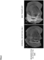
- the measurement with fMRI was conducted under anesthesia 42 days after the administration of MSCs.
- the fMRI analysis of change in signal in the right somatic sensory area of the cortex was conducted with T2-weighted images obtained by producing electrical stimulation (1 mA, pulse; 3 times/sec) using Electric Pulse Generator: Master-8 (A. M. P. I.) through an indwelling electrical stimulation needle in the left upper limb of the rats.
- the brain was fixed by perfusion 42 days after onset of cerebral infarction and the fixed brain was immersed in 4% PFA for 2 weeks or longer.
- the fixed brain was transferred into a centrifuge tube after 2 weeks and the test tube was filled with Fluorinert (a fluorine-based inert liquid) to prepare a specimen for MRI imaging.
- Fluorinert a fluorine-based inert liquid
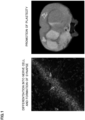
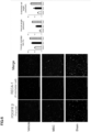
- the DAPI staining image indicated that intravenously administered GFP-MSCs reach the hippocampus and differentiate into neurons to grow neurites and form synapses ( Figure 1 , left).
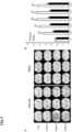
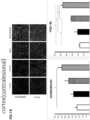
- MSCs facilitates not only the reproduction and plasticity in the lesion and surrounding tissue, but also the reproduction and plasticity of the whole central nervous system including the contralesional side of the brain. Therefore, it has become possible to induce not only the restoration of relatively simple functions including the restoration of the motor function, but also the restoration of higher and complex neural functions such as the restoration of higher brain functions (including aphasia).
- SHRSP Stroke-prone spontaneously hypertensive rats
- BBB blood-brain barrier
- NOP novel object placement test
- Vascular dementia model rat SHRSP rat
- the SHRSP rats were purchased from Hoshino Laboratory Animals, Inc. These rats are stroke-prone spontaneously hypertensive rats established by selecting the offspring having a parent who died of stroke every generation and crossbreeding the offspring and vascular dementia model rats that develop dementia by having a disruption of the blood-brain barrier due to high blood pressure and generating lacunar infarction or the like.
- rats having experienced cerebral infarction or cerebral hemorrhage at 16 to 20 weeks were selected as subjects and divided, after pre-treatment evaluation, into two groups: the MSC administration group and the vehicle (DMEM) administration group for the following tests.
- a circular swimming pool having a diameter of 1.3 m was filled to a depth of 30 cm with opaque water at a water temperature of 24 degrees Celsius and a platform to be the goal was placed just under the surface of the water. Rats were put in the swimming pool at the edge of the pool and latency to reach the platform [LRP] was measured.
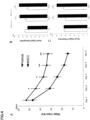
- the measurement was carried out by video tracking (Anymaze tracking software (Stoelting Co.; Wood Dale, IL, USA)) for consecutive 6 days in week 5 after transplant. 4 cycles of 1 minute device adaptation was conducted on day 1 and LRP was measured for consecutive 5 days from day 2. The measurement was conducted 4 cycles a day and the mean of the measurements was used.
- NOR was composed of (1) the sample phase, (2) the delay phase, and (3) the test phase.
- sample phase two identical objects were placed at the positions that are 10 cm from two respective walls, in an open field and the subjects were allowed to explore freely. After the sample phase, the subjects were returned to their home cages. 5 minutes later, the subjects were brought back in the open field again to start the test phase.
- object used in the sample phase (Familiar object: Object F) was placed near one wall and a new object (Novel object: Object N) was placed near the other wall.
- object exploration action was defined as an action of a subject that brings its nose within 2 cm from an object.
- the evaluation was made based on the value (N/N + F) expressed in % obtained by dividing the exploration time for Object N by the total exploration time for the two objects using the object exploration time in the test phase.
- NOP was tested on day 5. NOP was composed of three phases like NOR. In the test phase, the same objects as those used in the sample phase were used, but one object was placed at the same position as that in the sample phase (Familiar object: Object F) and the other one was placed at a different position (Novel object: Object N).
- the evaluation was made based on the value (N/N + F) expressed in % obtained by dividing the exploration time for Object N by the total exploration time for the two objects using the object exploration time in the test phase.
- Model rats 1 week after the intervention were anesthetized with ketamine (75 mg/kg) and xylazine (10 mg/kg) and FITC-lectin (1.6 mg/kg, Sigma, Taufkirchen, Germany) and Evans Blue (EvB) (4 % EvB in saline, 4 mL/kg, Sigma) were administered to the model rats from the femoral vein.
- the rats were sacrificed just after the administration and perfused with 200 ml of phosphate-buffered saline (PBS).
- PBS phosphate-buffered saline
- the brain tissue was dissected out, shock frozen in isopentane, and stored at -80 degrees until use.
- sample preparation the tissue was sliced into 30 ⁇ m coronal sections and post-fixed in 4% PFA.
- the sites at 1.60 to 6.80 mm posterior to the bregma were observed with LSM780 confocal laser microscope (Laser: Argon 488 for FITC-lectin, 561 for EvB; Objective: Plan-Apochromat 10x/0.45 M27, Zeiss, Jena, Germany).
- Rats were anesthetized with ketamine (75 mg/kg) and xylazine (10 mg/kg) in week 6 after transplant and perfused with 200 ml of phosphate-buffered saline (PBS) and 4% PFA.
- PBS phosphate-buffered saline
- the brain tissue was dissected out.
- 4% PFA was infiltrated into the brain tissue for 4 hours and then 15% and 30% sucrose were infiltrated into the tissue for 24 hours.
- the brain tissue was then immersed in an embedding agent for cryosectioning (Tissue-Tek, Torrance, CA), then flash-frozen in isopentane, and stored at -80 degrees.
- the tissue was sliced into 30 ⁇ m coronal sections. The sites at 1.60 to 6.80 mm posterior to the bregma were observed so as to include the whole hippocampus.
- the samples were blocked with 10% goat serum for 30 minutes and stored with a primary antibody dissolved in 5% goat serum in a refrigerator at 4 degrees Celsius overnight.
- the samples were washed with PBS on the next day and then allowed to react with a secondary antibody dissolved in 5% goat serum at room temperature for 2 hours.
- the antibody used for pericytes was an anti-PDGFR ⁇ antibody and the antibody used for vascular endothelium was an anti-RECA antibody.
- the RECA-positive blood vessel length was measured as the vascular endothelium length and the PDGFR ⁇ -positive blood vessel length was measured as the pericyte-positive blood vessel length.
- the measurement was performed using Image J. Each length was measured and the pericyte coverage rate was calculated by dividing the pericyte-positive blood vessel length by the vascular endothelium length and expressed in % for evaluation.
- Rats were anesthetized with ketamine (75 mg/kg) and xylazine (10 mg/kg) and the head was fixed in a coil and photographed.
- MRI measurements were performed before intervention, and in week 1, week 3, and week 4 after intervention.
- the MRI measurements were performed using a 7-Teslar, 18-cm-bore superconducting magnet (Oxford Magnet Technologies) interfaced to a UNITYINOVA console (Oxford Instruments) as described previously ( Honma T. et al., Exp. Neurol. 2006, 199:56-66 ., Komatsu K. et al., Brain Res. 2010, 1334:84-92 .).
- T2-weighted images were obtained.
- the lateral ventricle volumes were measured using image processing software (Scion Image, Version Beta 4.0.2, Scion Corporation) from serial images obtained from the T2-weighted images (Neumann-Haefelin et al., 2000).
- PBS phosphate-buffered saline
- the samples were cut into 30 ⁇ m coronal sections and subjected to Nissl staining.
- the thicknesses of cerebral cortex and corpus callosum in the M1 to S1 region were measured at three places each in a slice 3.3 mm posterior to the bregma using the polarizing microscope Olympus BX51 (4 ⁇ objective) and Stereo Investigator software (MicroBrightField) and the means were expressed as the measurements.
- PBS phosphate-buffered saline
- the samples were cut into 30 ⁇ m coronal sections and subjected to Nissl staining. Nerve cell counts in the whole hippocampus were determined using the polarizing microscope Olympus BX51 and StereoInvestigator software (MicroBrightField).
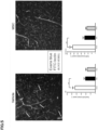
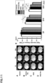
- Evans Blue staining indicated that Evans Blue (red), which should remain in the blood vessels in the normal brain, is leaked out to the outer tissue from the blood vessels and the blood-brain barrier is broken in the control (vehicle) group ( Figure 5 , left), but improvement is found in the MSC administration group ( Figure 5 , right).
- the blood-brain barrier is composed of endothelial cells, pericytes, and astrocytes.
- the improvement in coverage of endothelial cells, which are important for maintaining the function of the blood-brain barrier, by pericytes (pericyte coverage rate) was indicated.
- MSCs were intravenously administered to patients with chronic-phase cerebral infarction and the improvement of the higher function level was evaluated.
- Bone marrow aspirates were harvested from the ilium of patients with cerebral infarction under local anesthesia. Cells of interest were separated from the bone marrow aspirates in the Cell Processing Center (CPC) and cultured to obtain about 10000 times of cells in about 2 weeks. About 1 ⁇ 10 8 cells were enclosed in a bag with a capacity of about 40 ml under the GMP management to produce a cellular preparation. This cellular preparation was transplanted by intravenous administration for 30 minutes to 1 hour.
- CPC Cell Processing Center
- a placebo was administered for 150 days in the first half (Clinical trial I). MSCs were administered on day 150. Higher functions were evaluated until day 250 (Clinical trial II).
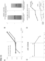
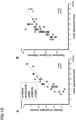
- the WAB aphasia test was performed on day 40 after onset (the time of changing hospital), day 76 after onset, day 141 after onset (before administration of cells), day 187 after onset (34 days after administration of cells), and day 250 after onset (97 days after administration of cells).
- This test include 38 test items under the eight main items: spontaneous speech, auditory comprehension, repetition, naming, reading, writing, praxis, and construction and allows the classification of aphasia as well as the calculation of aphasia quotient, which represents the severity of aphasia.
- the WAIS-III test was performed on day 40 after onset (the time of transfer to another hospital), day 141 after onset (before administration of cells), and day 250 after onset (97 days after administration of cells) and the processing speed was calculated.
- MSCs were administered after day 150 (chronic phase, Clinical trial II),
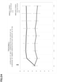
- the FUGL MEYER scores were maintained stably at low levels due to the placebo administration in the first half (Clinical trial I), but the administration of MSCs markedly improved the function in the latter half (Clinical trial II). As seen above, the motor function was also markedly improved ( Figure 10D ).
- the bone marrow obtained from femoral bones of adult SD rats was diluted to 25 ml with Dulbecco's modified Eagle medium (DMEM) and heat-inactivated 10% FBS, 2 mM l-glutamine, 100 U/ml penicillin, and 0.1 mg/ml streptomycin were added.
- DMEM Dulbecco's modified Eagle medium
- FBS fetal bovine serum
- 2 mM l-glutamine 100 U/ml penicillin
- streptomycin 0.1 mg/ml streptomycin
- tMCAO rat transient middle cerebral artery occlusion
- Cyclosporin A (10 mg/kg) was administered intraperitoneally to all rats every day. The intravenous administration was all from the left femoral vein.
- the rats were forced to run on a treadmill every day for 20 minutes.
- the exercise was started 1 day after arterial obstruction, at a speed of 3 m/min with a slope of 0 degrees during the first one week, and the speed was increased by 3 m/min every week until the subsequent histologic evaluation.
- Rats were anesthetized with ketamine (75 mg/kg) and xylazine (10 mg/kg) and MRI measurements were performed.
- the MRI measurements were performed using a 7-Teslar, 18-cm-bore superconducting magnet (Oxford Magnet Technologies) interfaced to a UNITYINOVA console (Oxford Instruments) as described previously (id.).
- T2WI-MRI measurements were performed 1, 14, and 35 days after occlusion.
- the ischemia lesion area was calculated from the MRI image using Scion Image, Version Beta 4.0.2 (Scion Corporation).
- Lesion volume (mm 3 ) was determined by analysis of high intensity areas on serial images collected through the cerebrum. For each slice, the higher intensity lesions in T2WI-MRI, where the signal intensity was 1.25 times higher than the counterpart in the contralateral brain lesion, were marked as the ischemic lesion area, and infarct volume was calculated taking slice thickness (1 mm) into account. The presence of intracerebral hemorrhage was counted when there is a low intensity area in the T2WI section. Animals with an initial stroke volume less than the standard were excluded from the experiment.
- the nerve cell count (synapse density) was measured using Nissl-stained samples according to the description in (9) in Example 2.
- the limb function was evaluated by the following six items for rats.
- a rat is slowly brought closer toward a table with the rat being about to get on the table.
- normal rats stretch and place both forelimbs on the table.
- a stroke rat may lose contact with the table with the forelimb contralateral to the injured hemisphere.
- the rat is placed on the table and gently pushed from behind the table edge. A normal rat will grip on the table edge, but an injured rat may drop the forelimb contralateral to the injured hemisphere from the table.
- the rat is placed on the table and gently pushed laterally toward the table edge toward the forelimb contralateral to the injured hemisphere.
- Rats were anesthetized with ketamine (75 mg/kg) and xylazine (10 mg/kg) in week 6 after transplant and perfused with 200 ml of phosphate-buffered saline (PBS) and 4% PFA.
- PBS phosphate-buffered saline
- the brain tissue was dissected out. 4% PFA was infiltrated into the brain tissue for 4 hours and then 15% and 30% sucrose were infiltrated into the tissue for 24 hours.
- the brain tissue was then immersed in an embedding agent for cryosectioning (Tissue-Tek, Torrance, CA), then flash-frozen in isopentane, and stored at -80 degrees. Cortex and striatum samples were cut out from the brain tissue and the expression levels of synaptophysin and PSD-95 were measured using an anti-synaptophysin antibody and an anti-PSD-95 antibody.
- a spinal cord injury model was performed according to previous reports (Matsushita et al., 2015).
- Adult male SD rats 250 to 300 g were anesthetized with ketamine (90 mg/kg) and xylazine (4 mg/kg), a laminectomy performed at the T9-10 level spinal cord, and a contusion delivered using the apparatus for generating spinal cord injury (Infinite Horizon Impactor, 60-kilodyne).
- Rats at 10 weeks after establishing spinal cord injury were received intravenous infusion of MSCs (1.0 ⁇ 10 6 cells each) labeled with GFP in 1 ml of DMEM.
- the rats were anesthetized with ketamine (75 mg/kg) and xylazine (10 mg/kg) after infusion of GFP-MSCs and perfused with 200 ml of phosphate-buffered saline (PBS) and 4% PFA.
- the spinal cord was dissected out and stained with DAPI, then coverslipped with VECTASHIELD (Vector Laboratories, Burlingame, CA), and observed using a confocal microscope with Ex/Em (405; 561: LSM780 ELYRA S.1 system).
- Rats at 10 weeks after establishing spinal cord injury were received intravenous infusion of MSCs (1.0 ⁇ 10 6 cells each) in 1 ml of DMEM. Evans Blue was administered to the rats from the thighbone blood vessel 1 week after transplant.
- the rats were anesthetized with ketamine (75 mg/kg) and xylazine (10 mg/kg) 6 hours later and perfused with 200 ml of phosphate-buffered saline (PBS) and 4% PFA.
- PBS phosphate-buffered saline
- the spinal cord was dissected out.
- the spinal cord sample was observed under a microscope and the state of the BSCB (blood spinal cord barrier) was evaluated.
- the RECA-positive blood vessel length was measured as the vascular endothelium length and the PDGFR ⁇ -positive blood vessel length was measured as the pericyte-positive blood vessel length.
- the measurement was performed using Image J. Each length was measured and the pericyte coverage rate was calculated by dividing the pericyte-positive blood vessel length by the vascular endothelium length and expressed in % for evaluation.
- the rats were perfused and fixed 20 weeks after spinal cord injury and immersed in 4% PFA for 2 weeks or longer. Two weeks later, the fixed spinal cord was transferred into a centrifuge tube and the test tube was filled with Fluorinert (a fluorine-based inert liquid) to prepare a specimen for MRI imaging.
- Fluorinert a fluorine-based inert liquid
- the MRI measurements were performed using a 7-Teslar, 18-cm-bore superconducting magnet (Oxford Magnet Technologies) interfaced to a UNITYINOVA console (Oxford Instruments) as described previously (id.).
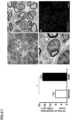
- the immunological analysis using an anti-P0 antibody and the analysis with an electron microscope indicated the presence of remyelinated axons, by the administration of MSCs, having a peripheral nerve type of myelin sheath (Schwann cells) characterized by a large nucleus and the basement membrane.
- MSCs having a peripheral nerve type of myelin sheath (Schwann cells) characterized by a large nucleus and the basement membrane.
- the result of staining the spinal cord injury lesion with toluidine blue and evaluating the number of remyelinated axons indicated a significantly larger number of remyelinated axons in the MSC group than in the Vehicle group. Therefore, it was indicated that the transplant of MSCs caused remyelination ( Figure 21 ).
- MCAO Permanent middle cerebral artery occlusion
- MSC group DMEM containing 1.0 ⁇ 10 6 MSCs P2 from an SD rat 8 weeks after MCAO in 1 ml was administered from the femoral vein.
- DMEM group 1 ml of DMEM was administered from the femoral vein.
- the present invention makes it possible to rebuild neural circuits and promote the brain plasticity by the formation of synapses and is available in treating dementia, chronic-phase cerebral infarction, chronic-phase spinal cord injury, mental diseases, and the like, which have conventionally been considered to be difficult to treat.
Landscapes
- Health & Medical Sciences (AREA)
- Life Sciences & Earth Sciences (AREA)
- Engineering & Computer Science (AREA)
- Biomedical Technology (AREA)
- Chemical & Material Sciences (AREA)
- Developmental Biology & Embryology (AREA)
- General Health & Medical Sciences (AREA)
- Cell Biology (AREA)
- Immunology (AREA)
- Veterinary Medicine (AREA)
- Public Health (AREA)
- Animal Behavior & Ethology (AREA)
- Pharmacology & Pharmacy (AREA)
- Medicinal Chemistry (AREA)
- Biotechnology (AREA)
- Organic Chemistry (AREA)
- Zoology (AREA)
- Bioinformatics & Cheminformatics (AREA)
- Hematology (AREA)
- Epidemiology (AREA)
- Wood Science & Technology (AREA)
- Genetics & Genomics (AREA)
- Neurology (AREA)
- Virology (AREA)
- Neurosurgery (AREA)
- General Chemical & Material Sciences (AREA)
- Nuclear Medicine, Radiotherapy & Molecular Imaging (AREA)
- Chemical Kinetics & Catalysis (AREA)
- Biochemistry (AREA)
- General Engineering & Computer Science (AREA)
- Rheumatology (AREA)
- Microbiology (AREA)
- Dermatology (AREA)
- Medicines Containing Material From Animals Or Micro-Organisms (AREA)
- Acyclic And Carbocyclic Compounds In Medicinal Compositions (AREA)
- Polymers With Sulfur, Phosphorus Or Metals In The Main Chain (AREA)
- Medicinal Preparation (AREA)
Claims (10)
- Medikament, umfassend CD24-negative mesenchymale Stammzellen (MSCs), die aus menschlichem Knochenmark oder Blut stammen, zur Anwendung in einem Therapieverfahren durch den Wiederaufbau neuraler Schaltkreise in einem Patienten, der an einer chronischen Phase eines Hirninfarkts oder einer chronischen Phase einer Rückenmarksverletzung leidet, wobei das Medikament die Plastizität des Gehirns fördert.
- Medikament zur Anwendung nach Anspruch 1, wobei die Zellen positiv für mindestens eines oder mehrere ausgewählt aus CD73, CD90, CD105 und CD200 sind und/oder negativ für mindestens eines oder mehrere ausgewählt aus CD19, CD34, CD45, CD74, CD79α und HLA-DR sind.
- Medikament zur Anwendung nach Anspruch 1 oder 2, wobei das menschliche Knochenmark oder Blut Knochenmark oder Blut des Patienten ist.
- Medikament zur Anwendung nach irgendeinem der Ansprüche 1 bis 3, wobei die Zellen in einem Medium, das menschliches Serum enthält, vermehrt und angereichert worden sind.
- Medikament zur Anwendung nach Anspruch 4, wobei das menschliche Serum autologes Serum des Patienten ist.
- Medikament zur Anwendung nach irgendeinem der Ansprüche 1 bis 5, wobei das Medikament eine Formulierung zur intravenösen Verabreichung, eine Formulierung zur Verabreichung durch Lumbalpunktion, eine Formulierung zur intrazerebralen Verabreichung, eine Formulierung zur intrazerebroventrikulären Verabreichung, eine Formulierung zur lokalen Verabreichung oder eine Formulierung zur intraarteriellen Verabreichung ist.
- Medikament zur Anwendung nach irgendeinem der Ansprüche 1 bis 5, wobei das Medikament eine Formulierung zur intravenösen Verabreichung ist.
- Medikament zur Anwendung nach irgendeinem der Ansprüche 1 bis 7, wobei die Zellen in einem Medium, das keinen Gerinnungshemmer oder einen Gerinnungshemmer von weniger als 0,02 U/mL enthält, vermehrt und angereichert wurden.
- Medikament zur Anwendung nach Anspruch 8, wobei das menschliche Knochenmark oder Blut so vorbereitet wurde, sodass die zum Zeitpunkt der Entnahme zugesetzte Menge des Gerinnungshemmers weniger als 0,2 U/mL, bezogen auf das Volumen des Knochenmarks oder Bluts, beträgt.
- Medikament zur Anwendung nach Anspruch 8 oder 9, wobei der Gerinnungshemmer Heparin, ein Heparinderivat oder ein Salz davon ist.
Applications Claiming Priority (3)
| Application Number | Priority Date | Filing Date | Title |
|---|---|---|---|
| JP2016091286 | 2016-04-28 | ||
| JP2016091300 | 2016-04-28 | ||
| PCT/JP2017/017325 WO2017188457A1 (ja) | 2016-04-28 | 2017-04-26 | シナプス形成剤 |
Publications (4)
| Publication Number | Publication Date |
|---|---|
| EP3449930A1 EP3449930A1 (de) | 2019-03-06 |
| EP3449930A4 EP3449930A4 (de) | 2019-11-20 |
| EP3449930B1 true EP3449930B1 (de) | 2025-04-02 |
| EP3449930C0 EP3449930C0 (de) | 2025-04-02 |
Family
ID=60160788
Family Applications (1)
| Application Number | Title | Priority Date | Filing Date |
|---|---|---|---|
| EP17789738.6A Active EP3449930B1 (de) | 2016-04-28 | 2017-04-26 | Synapsenbildungsmittel |
Country Status (10)
| Country | Link |
|---|---|
| US (2) | US11666601B2 (de) |
| EP (1) | EP3449930B1 (de) |
| JP (2) | JP7101367B2 (de) |
| KR (2) | KR20220166875A (de) |
| CN (1) | CN109069542A (de) |
| AU (2) | AU2017258058B2 (de) |
| CA (1) | CA3022275A1 (de) |
| IL (2) | IL299420B2 (de) |
| SG (1) | SG11201809512TA (de) |
| WO (1) | WO2017188457A1 (de) |
Families Citing this family (12)
| Publication number | Priority date | Publication date | Assignee | Title |
|---|---|---|---|---|
| CN109069542A (zh) * | 2016-04-28 | 2018-12-21 | 北海道公立大学法人札幌医科大学 | 突触形成剂 |
| US20190183937A1 (en) * | 2016-08-18 | 2019-06-20 | Sapporo Medical University | Life extension agent |
| EP3698376A4 (de) * | 2017-10-16 | 2021-10-20 | Performance Phenomics Inc. | Verfahren und vorrichtung zur verwendung von gehirnbildgebung zur vorhersage der leistung |
| JP7387613B2 (ja) * | 2018-02-09 | 2023-11-28 | ザ トラスティーズ オブ ダートマス カレッジ | 神経変性疾患及び障害の治療のためのキメラ抗原受容体 |
| US20220125850A1 (en) | 2019-02-07 | 2022-04-28 | Sapporo Medical University | Pharmaceutical composition for preventing in-stent restenosis |
| JP7541699B2 (ja) * | 2019-03-14 | 2024-08-29 | 北海道公立大学法人 札幌医科大学 | 筋萎縮性側索硬化症の治療用医薬組成物 |
| US20220226386A1 (en) * | 2019-05-23 | 2022-07-21 | Mesoblast International Sárl | Functional recovery from cerebral infarction |
| JP7618233B2 (ja) | 2019-08-09 | 2025-01-21 | 国立大学法人東北大学 | 脳血管性認知症の治療または予防剤 |
| JP7545714B2 (ja) | 2020-07-31 | 2024-09-05 | ニューロテックメディカル株式会社 | 神経障害の治療剤 |
| JP7671950B2 (ja) | 2020-11-30 | 2025-05-07 | ニューロテックメディカル株式会社 | 神経障害の治療剤 |
| EP4279919B1 (de) * | 2021-01-12 | 2026-04-29 | Cellatoz Therapeutics, Inc. | Verfahren zum screening von nervenregenerationsfördernden zellen mit nervenregenerationsaktivität |
| CN116802501A (zh) * | 2021-01-12 | 2023-09-22 | 雪拉托兹治疗株式会社 | 具有神经元再生活性的神经元再生促进细胞的筛选方法 |
Citations (1)
| Publication number | Priority date | Publication date | Assignee | Title |
|---|---|---|---|---|
| US20120009271A1 (en) * | 2009-03-12 | 2012-01-12 | Medical College Of Georgia | Method of disease-induced and receptor-mediated stem cell neuroprotection |
Family Cites Families (8)
| Publication number | Priority date | Publication date | Assignee | Title |
|---|---|---|---|---|
| EP2295539A1 (de) | 2000-06-26 | 2011-03-16 | Nc Medical Research Inc. | Zellfraktion mit Zellen welche in der Lage sind sich in Nervenzellen zu differenzieren |
| WO2008036374A2 (en) * | 2006-09-21 | 2008-03-27 | Medistem Laboratories, Inc. | Allogeneic stem cell transplants in non-conditioned recipients |
| US9387226B2 (en) | 2006-11-30 | 2016-07-12 | Medipost Co., Ltd | Neural cell proliferation induced through the culture of neural cells with umbilical cord blood-derived mesenchymal stem cells |
| AU2008298816B2 (en) | 2007-09-11 | 2013-09-26 | Sapporo Medical University | Cell growth method and pharmaceutical preparation for tissue repair and regeneration |
| KR20100054711A (ko) | 2008-11-14 | 2010-05-25 | 메디포스트(주) | 간엽 줄기세포 또는 이의 배양액을 포함하는 신경질환의 예방 또는 치료용 조성물 |
| DK2602310T3 (da) * | 2010-08-03 | 2020-02-24 | Univ Sapporo Medical | Autoserum-indeholdende knoglemarvcelledyrkningssystem, autoserum-indeholdende knoglemarvcelledyrkningsfremgangsmåde, og fremgangsmåde til fremstilling af en lægemiddelsammensætning omfattende (...) |
| JP5598864B2 (ja) * | 2011-12-05 | 2014-10-01 | 北海道公立大学法人 札幌医科大学 | 細胞増殖方法ならびに組織の修復および再生のための医薬 |
| CN109069542A (zh) | 2016-04-28 | 2018-12-21 | 北海道公立大学法人札幌医科大学 | 突触形成剂 |
-
2017
- 2017-04-26 CN CN201780026329.7A patent/CN109069542A/zh active Pending
- 2017-04-26 AU AU2017258058A patent/AU2017258058B2/en active Active
- 2017-04-26 KR KR1020227042142A patent/KR20220166875A/ko not_active Ceased
- 2017-04-26 SG SG11201809512TA patent/SG11201809512TA/en unknown
- 2017-04-26 WO PCT/JP2017/017325 patent/WO2017188457A1/ja not_active Ceased
- 2017-04-26 JP JP2018514748A patent/JP7101367B2/ja active Active
- 2017-04-26 EP EP17789738.6A patent/EP3449930B1/de active Active
- 2017-04-26 KR KR1020187034088A patent/KR20190004307A/ko not_active Ceased
- 2017-04-26 CA CA3022275A patent/CA3022275A1/en active Pending
- 2017-04-26 IL IL299420A patent/IL299420B2/en unknown
- 2017-04-26 US US16/096,962 patent/US11666601B2/en active Active
-
2018
- 2018-10-25 IL IL262621A patent/IL262621B2/en unknown
-
2022
- 2022-06-24 JP JP2022101637A patent/JP7450896B2/ja active Active
-
2023
- 2023-04-26 US US18/139,694 patent/US20240122988A1/en active Pending
- 2023-07-07 AU AU2023204405A patent/AU2023204405B2/en active Active
Patent Citations (1)
| Publication number | Priority date | Publication date | Assignee | Title |
|---|---|---|---|---|
| US20120009271A1 (en) * | 2009-03-12 | 2012-01-12 | Medical College Of Georgia | Method of disease-induced and receptor-mediated stem cell neuroprotection |
Non-Patent Citations (3)
| Title |
|---|
| MATCHYNSKI-FRANKS JESSICA J. ET AL: "Mesenchymal Stem Cells as Treatment for Behavioral Deficits and Neuropathology in the 5xFAD Mouse Model of Alzheimer's Disease", CELL TRANSPLANTATION, vol. 25, no. 4, 1 April 2016 (2016-04-01), US, pages 687 - 703, XP055782789, ISSN: 0963-6897, Retrieved from the Internet <URL:http://www.ingentaconnect.com/content/cog/ct/2016/00000025/00000004/art00008> DOI: 10.3727/096368916X690818 * |
| NAALDIJK Y. ET AL: "Effect of systemic transplantation of bone marrow-derived mesenchymal stem cells on neuropathology markers in APP/PS1 Alzheimer mice", NEUROPATHOLOGY AND APPLIED NEUROBIOLOGY., vol. 43, no. 4, 19 April 2016 (2016-04-19), GB, pages 299 - 314, XP055782819, ISSN: 0305-1846, Retrieved from the Internet <URL:https://api.wiley.com/onlinelibrary/tdm/v1/articles/10.1111%2Fnan.12319> DOI: 10.1111/nan.12319 * |
| NANETTE JOYCE ET AL: "Mesenchymal stem cells for the treatment of neurodegenerative disease", REGENERATIVE MEDICINE, vol. 5, no. 6, 1 November 2010 (2010-11-01), GB, pages 933 - 946, XP055412982, ISSN: 1746-0751, DOI: 10.2217/rme.10.72 * |
Also Published As
| Publication number | Publication date |
|---|---|
| KR20190004307A (ko) | 2019-01-11 |
| JP7450896B2 (ja) | 2024-03-18 |
| US20240122988A1 (en) | 2024-04-18 |
| CN109069542A (zh) | 2018-12-21 |
| AU2023204405A1 (en) | 2023-08-03 |
| US20190117700A1 (en) | 2019-04-25 |
| IL262621A (en) | 2018-12-31 |
| KR20220166875A (ko) | 2022-12-19 |
| IL262621B2 (en) | 2023-05-01 |
| IL299420B1 (en) | 2025-03-01 |
| IL262621B1 (en) | 2023-01-01 |
| US11666601B2 (en) | 2023-06-06 |
| SG11201809512TA (en) | 2018-11-29 |
| CA3022275A1 (en) | 2017-11-02 |
| IL299420B2 (en) | 2025-07-01 |
| AU2023204405B2 (en) | 2025-12-11 |
| JP7101367B2 (ja) | 2022-07-15 |
| EP3449930C0 (de) | 2025-04-02 |
| JP2022126812A (ja) | 2022-08-30 |
| AU2017258058B2 (en) | 2023-04-13 |
| JPWO2017188457A1 (ja) | 2019-03-07 |
| EP3449930A4 (de) | 2019-11-20 |
| WO2017188457A1 (ja) | 2017-11-02 |
| EP3449930A1 (de) | 2019-03-06 |
| IL299420A (en) | 2023-02-01 |
| AU2017258058A1 (en) | 2018-11-15 |
Similar Documents
| Publication | Publication Date | Title |
|---|---|---|
| AU2023204405B2 (en) | Synapse formation agent | |
| Alvarado-Velez et al. | Immuno-suppressive hydrogels enhance allogeneic MSC survival after transplantation in the injured brain | |
| Hour et al. | Magnetic targeted delivery of the SPIONs-labeled mesenchymal stem cells derived from human Wharton's jelly in Alzheimer's rat models | |
| Ding et al. | Enhancement of neuroplasticity through upregulation of β1-integrin in human umbilical cord-derived stromal cell implanted stroke model | |
| JP5841952B2 (ja) | 腫瘍性疾患の治療的処置にて用いるための成体幹細胞由来の微細小胞(mv) | |
| Yang et al. | Subarachnoid transplantation of human umbilical cord mesenchymal stem cell in rodent model with subacute incomplete spinal cord injury: preclinical safety and efficacy study | |
| US20080131405A1 (en) | Composition for treating a disease caused by neuronal insult comprising a human umbilical cord blood-derived mesenchymal stem cell as an active ingredient | |
| Ozdemir et al. | Regenerative treatment in spinal cord injury | |
| Blazquez et al. | Intrapericardial administration of mesenchymal stem cells in a large animal model: a bio-distribution analysis | |
| Long et al. | MRI tracking of bone marrow mesenchymal stem cells labeled with ultra-small superparamagnetic iron oxide nanoparticles in a rat model of temporal lobe epilepsy | |
| KR102213890B1 (ko) | 엑소좀 기반의 면역세포의 교차분화 방법 | |
| Hu et al. | Targeting and reprogramming microglial phagocytosis of neutrophils by ginsenoside Rg1 nanovesicles promotes stroke recovery | |
| Lim et al. | Evaluation of autologous bone marrow–derived mesenchymal stem cells on renal regeneration after experimentally induced acute kidney injury in dogs | |
| Lundberg et al. | Endovascular transplantation of stem cells to the injured rat CNS | |
| Penna et al. | Early homing of adult mesenchymal stem cells in normal and infarcted isolated beating hearts | |
| Yu et al. | NanoCsA improves the survival of human iPSC transplant in hemiparkinsonian rats | |
| Amin et al. | Microanatomical evidences for potential of mesenchymal stem cells in amelioration of striatal degeneration | |
| Chen et al. | Intranasal delivery of hypoxia-preconditioned extracellular vesicles derived from BMSCs alleviates neuroinflammation and brain dysfunction in TBI | |
| EP3501525A1 (de) | Lebensdauerverlängerungsmittel | |
| Ito et al. | Validity of bone marrow stromal cell expansion by animal serum-free medium for cell transplantation therapy of cerebral infarct in rats—a serial MRI study | |
| Ramli et al. | Intravenous versus intraarterial transplantation of human umbilical cord blood mononuclear cells for brain ischemia in rats | |
| Bernardi et al. | Absence of micronucleus formation in CHO-K1 cells cultivated in platelet lysate enriched medium | |
| US10130683B1 (en) | Combination therapy of stem cell mobilizing agents and stem cell transplantation | |
| Shuster-Hyman | Biodistribution and Fate of Intravenously-Delivered First Trimester Human Umbilical Cord Perivascular Cells in a Murine Model of Systemic Inflammation | |
| Bonab et al. | Mesenchymal stem cell therapy for multiple sclerosis |
Legal Events
| Date | Code | Title | Description |
|---|---|---|---|
| STAA | Information on the status of an ep patent application or granted ep patent |
Free format text: STATUS: THE INTERNATIONAL PUBLICATION HAS BEEN MADE |
|
| PUAI | Public reference made under article 153(3) epc to a published international application that has entered the european phase |
Free format text: ORIGINAL CODE: 0009012 |
|
| STAA | Information on the status of an ep patent application or granted ep patent |
Free format text: STATUS: REQUEST FOR EXAMINATION WAS MADE |
|
| 17P | Request for examination filed |
Effective date: 20181120 |
|
| AK | Designated contracting states |
Kind code of ref document: A1 Designated state(s): AL AT BE BG CH CY CZ DE DK EE ES FI FR GB GR HR HU IE IS IT LI LT LU LV MC MK MT NL NO PL PT RO RS SE SI SK SM TR |
|
| AX | Request for extension of the european patent |
Extension state: BA ME |
|
| DAV | Request for validation of the european patent (deleted) | ||
| DAX | Request for extension of the european patent (deleted) | ||
| A4 | Supplementary search report drawn up and despatched |
Effective date: 20191017 |
|
| RIC1 | Information provided on ipc code assigned before grant |
Ipc: A61P 25/28 20060101ALI20191011BHEP Ipc: A61P 25/00 20060101ALI20191011BHEP Ipc: C12N 5/0775 20100101ALI20191011BHEP Ipc: A61K 35/28 20150101AFI20191011BHEP |
|
| STAA | Information on the status of an ep patent application or granted ep patent |
Free format text: STATUS: EXAMINATION IS IN PROGRESS |
|
| 17Q | First examination report despatched |
Effective date: 20210311 |
|
| GRAP | Despatch of communication of intention to grant a patent |
Free format text: ORIGINAL CODE: EPIDOSNIGR1 |
|
| STAA | Information on the status of an ep patent application or granted ep patent |
Free format text: STATUS: GRANT OF PATENT IS INTENDED |
|
| INTG | Intention to grant announced |
Effective date: 20240702 |
|
| GRAJ | Information related to disapproval of communication of intention to grant by the applicant or resumption of examination proceedings by the epo deleted |
Free format text: ORIGINAL CODE: EPIDOSDIGR1 |
|
| STAA | Information on the status of an ep patent application or granted ep patent |
Free format text: STATUS: EXAMINATION IS IN PROGRESS |
|
| GRAP | Despatch of communication of intention to grant a patent |
Free format text: ORIGINAL CODE: EPIDOSNIGR1 |
|
| STAA | Information on the status of an ep patent application or granted ep patent |
Free format text: STATUS: GRANT OF PATENT IS INTENDED |
|
| INTC | Intention to grant announced (deleted) | ||
| INTG | Intention to grant announced |
Effective date: 20241029 |
|
| GRAS | Grant fee paid |
Free format text: ORIGINAL CODE: EPIDOSNIGR3 |
|
| GRAA | (expected) grant |
Free format text: ORIGINAL CODE: 0009210 |
|
| STAA | Information on the status of an ep patent application or granted ep patent |
Free format text: STATUS: THE PATENT HAS BEEN GRANTED |
|
| AK | Designated contracting states |
Kind code of ref document: B1 Designated state(s): AL AT BE BG CH CY CZ DE DK EE ES FI FR GB GR HR HU IE IS IT LI LT LU LV MC MK MT NL NO PL PT RO RS SE SI SK SM TR |
|
| REG | Reference to a national code |
Ref country code: GB Ref legal event code: FG4D |
|
| REG | Reference to a national code |
Ref country code: CH Ref legal event code: EP |
|
| REG | Reference to a national code |
Ref country code: IE Ref legal event code: FG4D |
|
| U01 | Request for unitary effect filed |
Effective date: 20250402 |
|
| U07 | Unitary effect registered |
Designated state(s): AT BE BG DE DK EE FI FR IT LT LU LV MT NL PT RO SE SI Effective date: 20250408 |
|
| U20 | Renewal fee for the european patent with unitary effect paid |
Year of fee payment: 9 Effective date: 20250606 |
|
| PG25 | Lapsed in a contracting state [announced via postgrant information from national office to epo] |
Ref country code: ES Free format text: LAPSE BECAUSE OF FAILURE TO SUBMIT A TRANSLATION OF THE DESCRIPTION OR TO PAY THE FEE WITHIN THE PRESCRIBED TIME-LIMIT Effective date: 20250402 |
|
| PG25 | Lapsed in a contracting state [announced via postgrant information from national office to epo] |
Ref country code: NO Free format text: LAPSE BECAUSE OF FAILURE TO SUBMIT A TRANSLATION OF THE DESCRIPTION OR TO PAY THE FEE WITHIN THE PRESCRIBED TIME-LIMIT Effective date: 20250702 Ref country code: GR Free format text: LAPSE BECAUSE OF FAILURE TO SUBMIT A TRANSLATION OF THE DESCRIPTION OR TO PAY THE FEE WITHIN THE PRESCRIBED TIME-LIMIT Effective date: 20250703 |
|
| PG25 | Lapsed in a contracting state [announced via postgrant information from national office to epo] |
Ref country code: PL Free format text: LAPSE BECAUSE OF FAILURE TO SUBMIT A TRANSLATION OF THE DESCRIPTION OR TO PAY THE FEE WITHIN THE PRESCRIBED TIME-LIMIT Effective date: 20250402 |
|
| PG25 | Lapsed in a contracting state [announced via postgrant information from national office to epo] |
Ref country code: HR Free format text: LAPSE BECAUSE OF FAILURE TO SUBMIT A TRANSLATION OF THE DESCRIPTION OR TO PAY THE FEE WITHIN THE PRESCRIBED TIME-LIMIT Effective date: 20250402 |
|
| PG25 | Lapsed in a contracting state [announced via postgrant information from national office to epo] |
Ref country code: RS Free format text: LAPSE BECAUSE OF FAILURE TO SUBMIT A TRANSLATION OF THE DESCRIPTION OR TO PAY THE FEE WITHIN THE PRESCRIBED TIME-LIMIT Effective date: 20250702 |
|
| PG25 | Lapsed in a contracting state [announced via postgrant information from national office to epo] |
Ref country code: IS Free format text: LAPSE BECAUSE OF FAILURE TO SUBMIT A TRANSLATION OF THE DESCRIPTION OR TO PAY THE FEE WITHIN THE PRESCRIBED TIME-LIMIT Effective date: 20250802 |
|
| REG | Reference to a national code |
Ref country code: CH Ref legal event code: H13 Free format text: ST27 STATUS EVENT CODE: U-0-0-H10-H13 (AS PROVIDED BY THE NATIONAL OFFICE) Effective date: 20251125 |
|
| PG25 | Lapsed in a contracting state [announced via postgrant information from national office to epo] |
Ref country code: SM Free format text: LAPSE BECAUSE OF FAILURE TO SUBMIT A TRANSLATION OF THE DESCRIPTION OR TO PAY THE FEE WITHIN THE PRESCRIBED TIME-LIMIT Effective date: 20250402 |
|
| PG25 | Lapsed in a contracting state [announced via postgrant information from national office to epo] |
Ref country code: CH Free format text: LAPSE BECAUSE OF NON-PAYMENT OF DUE FEES Effective date: 20250430 |
|
| PG25 | Lapsed in a contracting state [announced via postgrant information from national office to epo] |
Ref country code: CZ Free format text: LAPSE BECAUSE OF FAILURE TO SUBMIT A TRANSLATION OF THE DESCRIPTION OR TO PAY THE FEE WITHIN THE PRESCRIBED TIME-LIMIT Effective date: 20250402 |
|
| PG25 | Lapsed in a contracting state [announced via postgrant information from national office to epo] |
Ref country code: SK Free format text: LAPSE BECAUSE OF FAILURE TO SUBMIT A TRANSLATION OF THE DESCRIPTION OR TO PAY THE FEE WITHIN THE PRESCRIBED TIME-LIMIT Effective date: 20250402 |
|
| PG25 | Lapsed in a contracting state [announced via postgrant information from national office to epo] |
Ref country code: MC Free format text: LAPSE BECAUSE OF FAILURE TO SUBMIT A TRANSLATION OF THE DESCRIPTION OR TO PAY THE FEE WITHIN THE PRESCRIBED TIME-LIMIT Effective date: 20250402 |
|
| PLBE | No opposition filed within time limit |
Free format text: ORIGINAL CODE: 0009261 |
|
| STAA | Information on the status of an ep patent application or granted ep patent |
Free format text: STATUS: NO OPPOSITION FILED WITHIN TIME LIMIT |
|
| REG | Reference to a national code |
Ref country code: CH Ref legal event code: L10 Free format text: ST27 STATUS EVENT CODE: U-0-0-L10-L00 (AS PROVIDED BY THE NATIONAL OFFICE) Effective date: 20260211 |
|
| 26N | No opposition filed |
Effective date: 20260105 |
|
| GBPC | Gb: european patent ceased through non-payment of renewal fee |
Effective date: 20250702 |
|
| PG25 | Lapsed in a contracting state [announced via postgrant information from national office to epo] |
Ref country code: GB Free format text: LAPSE BECAUSE OF NON-PAYMENT OF DUE FEES Effective date: 20250702 |
|
| PG25 | Lapsed in a contracting state [announced via postgrant information from national office to epo] |
Ref country code: IE Free format text: LAPSE BECAUSE OF NON-PAYMENT OF DUE FEES Effective date: 20250426 |