EP3478331B1 - Biomatériau - Google Patents
Biomatériau Download PDFInfo
- Publication number
- EP3478331B1 EP3478331B1 EP17751229.0A EP17751229A EP3478331B1 EP 3478331 B1 EP3478331 B1 EP 3478331B1 EP 17751229 A EP17751229 A EP 17751229A EP 3478331 B1 EP3478331 B1 EP 3478331B1
- Authority
- EP
- European Patent Office
- Prior art keywords
- biomaterial
- material fraction
- fraction
- biomaterial according
- tissue
- Prior art date
- Legal status (The legal status is an assumption and is not a legal conclusion. Google has not performed a legal analysis and makes no representation as to the accuracy of the status listed.)
- Active
Links
Images


Classifications
-
- A—HUMAN NECESSITIES
- A61—MEDICAL OR VETERINARY SCIENCE; HYGIENE
- A61L—METHODS OR APPARATUS FOR STERILISING MATERIALS OR OBJECTS IN GENERAL; DISINFECTION, STERILISATION OR DEODORISATION OF AIR; CHEMICAL ASPECTS OF BANDAGES, DRESSINGS, ABSORBENT PADS OR SURGICAL ARTICLES; MATERIALS FOR BANDAGES, DRESSINGS, ABSORBENT PADS OR SURGICAL ARTICLES
- A61L27/00—Materials for grafts or prostheses or for coating grafts or prostheses
- A61L27/50—Materials characterised by their function or physical properties, e.g. injectable or lubricating compositions, shape-memory materials, surface modified materials
- A61L27/56—Porous materials, e.g. foams or sponges
-
- A—HUMAN NECESSITIES
- A61—MEDICAL OR VETERINARY SCIENCE; HYGIENE
- A61F—FILTERS IMPLANTABLE INTO BLOOD VESSELS; PROSTHESES; DEVICES PROVIDING PATENCY TO, OR PREVENTING COLLAPSING OF, TUBULAR STRUCTURES OF THE BODY, e.g. STENTS; ORTHOPAEDIC, NURSING OR CONTRACEPTIVE DEVICES; FOMENTATION; TREATMENT OR PROTECTION OF EYES OR EARS; BANDAGES, DRESSINGS OR ABSORBENT PADS; FIRST-AID KITS
- A61F2/00—Filters implantable into blood vessels; Prostheses, i.e. artificial substitutes or replacements for parts of the body; Appliances for connecting them with the body; Devices providing patency to, or preventing collapsing of, tubular structures of the body, e.g. stents
- A61F2/02—Prostheses implantable into the body
- A61F2/28—Bones
-
- A—HUMAN NECESSITIES
- A61—MEDICAL OR VETERINARY SCIENCE; HYGIENE
- A61L—METHODS OR APPARATUS FOR STERILISING MATERIALS OR OBJECTS IN GENERAL; DISINFECTION, STERILISATION OR DEODORISATION OF AIR; CHEMICAL ASPECTS OF BANDAGES, DRESSINGS, ABSORBENT PADS OR SURGICAL ARTICLES; MATERIALS FOR BANDAGES, DRESSINGS, ABSORBENT PADS OR SURGICAL ARTICLES
- A61L27/00—Materials for grafts or prostheses or for coating grafts or prostheses
- A61L27/14—Macromolecular materials
- A61L27/18—Macromolecular materials obtained otherwise than by reactions only involving carbon-to-carbon unsaturated bonds
-
- A—HUMAN NECESSITIES
- A61—MEDICAL OR VETERINARY SCIENCE; HYGIENE
- A61L—METHODS OR APPARATUS FOR STERILISING MATERIALS OR OBJECTS IN GENERAL; DISINFECTION, STERILISATION OR DEODORISATION OF AIR; CHEMICAL ASPECTS OF BANDAGES, DRESSINGS, ABSORBENT PADS OR SURGICAL ARTICLES; MATERIALS FOR BANDAGES, DRESSINGS, ABSORBENT PADS OR SURGICAL ARTICLES
- A61L27/00—Materials for grafts or prostheses or for coating grafts or prostheses
- A61L27/14—Macromolecular materials
- A61L27/22—Polypeptides or derivatives thereof, e.g. degradation products
- A61L27/24—Collagen
-
- A—HUMAN NECESSITIES
- A61—MEDICAL OR VETERINARY SCIENCE; HYGIENE
- A61L—METHODS OR APPARATUS FOR STERILISING MATERIALS OR OBJECTS IN GENERAL; DISINFECTION, STERILISATION OR DEODORISATION OF AIR; CHEMICAL ASPECTS OF BANDAGES, DRESSINGS, ABSORBENT PADS OR SURGICAL ARTICLES; MATERIALS FOR BANDAGES, DRESSINGS, ABSORBENT PADS OR SURGICAL ARTICLES
- A61L27/00—Materials for grafts or prostheses or for coating grafts or prostheses
- A61L27/50—Materials characterised by their function or physical properties, e.g. injectable or lubricating compositions, shape-memory materials, surface modified materials
-
- A—HUMAN NECESSITIES
- A61—MEDICAL OR VETERINARY SCIENCE; HYGIENE
- A61L—METHODS OR APPARATUS FOR STERILISING MATERIALS OR OBJECTS IN GENERAL; DISINFECTION, STERILISATION OR DEODORISATION OF AIR; CHEMICAL ASPECTS OF BANDAGES, DRESSINGS, ABSORBENT PADS OR SURGICAL ARTICLES; MATERIALS FOR BANDAGES, DRESSINGS, ABSORBENT PADS OR SURGICAL ARTICLES
- A61L27/00—Materials for grafts or prostheses or for coating grafts or prostheses
- A61L27/50—Materials characterised by their function or physical properties, e.g. injectable or lubricating compositions, shape-memory materials, surface modified materials
- A61L27/58—Materials at least partially resorbable by the body
-
- A—HUMAN NECESSITIES
- A61—MEDICAL OR VETERINARY SCIENCE; HYGIENE
- A61F—FILTERS IMPLANTABLE INTO BLOOD VESSELS; PROSTHESES; DEVICES PROVIDING PATENCY TO, OR PREVENTING COLLAPSING OF, TUBULAR STRUCTURES OF THE BODY, e.g. STENTS; ORTHOPAEDIC, NURSING OR CONTRACEPTIVE DEVICES; FOMENTATION; TREATMENT OR PROTECTION OF EYES OR EARS; BANDAGES, DRESSINGS OR ABSORBENT PADS; FIRST-AID KITS
- A61F2/00—Filters implantable into blood vessels; Prostheses, i.e. artificial substitutes or replacements for parts of the body; Appliances for connecting them with the body; Devices providing patency to, or preventing collapsing of, tubular structures of the body, e.g. stents
- A61F2/02—Prostheses implantable into the body
- A61F2/30—Joints
- A61F2002/30001—Additional features of subject-matter classified in A61F2/28, A61F2/30 and subgroups thereof
- A61F2002/30003—Material related properties of the prosthesis or of a coating on the prosthesis
- A61F2002/30004—Material related properties of the prosthesis or of a coating on the prosthesis the prosthesis being made from materials having different values of a given property at different locations within the same prosthesis
- A61F2002/30014—Material related properties of the prosthesis or of a coating on the prosthesis the prosthesis being made from materials having different values of a given property at different locations within the same prosthesis differing in elasticity, stiffness or compressibility
-
- A—HUMAN NECESSITIES
- A61—MEDICAL OR VETERINARY SCIENCE; HYGIENE
- A61F—FILTERS IMPLANTABLE INTO BLOOD VESSELS; PROSTHESES; DEVICES PROVIDING PATENCY TO, OR PREVENTING COLLAPSING OF, TUBULAR STRUCTURES OF THE BODY, e.g. STENTS; ORTHOPAEDIC, NURSING OR CONTRACEPTIVE DEVICES; FOMENTATION; TREATMENT OR PROTECTION OF EYES OR EARS; BANDAGES, DRESSINGS OR ABSORBENT PADS; FIRST-AID KITS
- A61F2/00—Filters implantable into blood vessels; Prostheses, i.e. artificial substitutes or replacements for parts of the body; Appliances for connecting them with the body; Devices providing patency to, or preventing collapsing of, tubular structures of the body, e.g. stents
- A61F2/02—Prostheses implantable into the body
- A61F2/30—Joints
- A61F2002/30001—Additional features of subject-matter classified in A61F2/28, A61F2/30 and subgroups thereof
- A61F2002/30108—Shapes
- A61F2002/3011—Cross-sections or two-dimensional shapes
- A61F2002/30138—Convex polygonal shapes
- A61F2002/30143—Convex polygonal shapes hexagonal
-
- A—HUMAN NECESSITIES
- A61—MEDICAL OR VETERINARY SCIENCE; HYGIENE
- A61L—METHODS OR APPARATUS FOR STERILISING MATERIALS OR OBJECTS IN GENERAL; DISINFECTION, STERILISATION OR DEODORISATION OF AIR; CHEMICAL ASPECTS OF BANDAGES, DRESSINGS, ABSORBENT PADS OR SURGICAL ARTICLES; MATERIALS FOR BANDAGES, DRESSINGS, ABSORBENT PADS OR SURGICAL ARTICLES
- A61L2430/00—Materials or treatment for tissue regeneration
- A61L2430/02—Materials or treatment for tissue regeneration for reconstruction of bones; weight-bearing implants
-
- A—HUMAN NECESSITIES
- A61—MEDICAL OR VETERINARY SCIENCE; HYGIENE
- A61L—METHODS OR APPARATUS FOR STERILISING MATERIALS OR OBJECTS IN GENERAL; DISINFECTION, STERILISATION OR DEODORISATION OF AIR; CHEMICAL ASPECTS OF BANDAGES, DRESSINGS, ABSORBENT PADS OR SURGICAL ARTICLES; MATERIALS FOR BANDAGES, DRESSINGS, ABSORBENT PADS OR SURGICAL ARTICLES
- A61L2430/00—Materials or treatment for tissue regeneration
- A61L2430/24—Materials or treatment for tissue regeneration for joint reconstruction
-
- A—HUMAN NECESSITIES
- A61—MEDICAL OR VETERINARY SCIENCE; HYGIENE
- A61L—METHODS OR APPARATUS FOR STERILISING MATERIALS OR OBJECTS IN GENERAL; DISINFECTION, STERILISATION OR DEODORISATION OF AIR; CHEMICAL ASPECTS OF BANDAGES, DRESSINGS, ABSORBENT PADS OR SURGICAL ARTICLES; MATERIALS FOR BANDAGES, DRESSINGS, ABSORBENT PADS OR SURGICAL ARTICLES
- A61L2430/00—Materials or treatment for tissue regeneration
- A61L2430/38—Materials or treatment for tissue regeneration for reconstruction of the spine, vertebrae or intervertebral discs
Definitions
- the invention relates to a biomaterial.
- This is a synthetic or non-living natural material used in medicine for therapeutic or diagnostic purposes, in direct contact with biological tissues of the body.
- the material enters into chemical, physical and biological interactions with the corresponding biological systems.
- This also includes short-term contact via the outer body surface, via body openings and via mucous membranes accessible from the outside.
- the invention particularly relates to materials that are placed inside the body for long-term retention.
- biomaterial refers to the material, in particular the chemical and physical, properties of the material.
- a biocompatibility resulting from its properties is characteristic of a biomaterial, which includes both the functional similarity to the body's own structures and an appropriate biological compatibility in the body.
- biomaterials are used in the form of implants inside the body. They are used, for example, as part of a treatment known as osteosynthesis to support healing after a fracture as well as the permanent replacement of bones that have been irreparably destroyed by an accident or bone cancer. It is also possible to replace joint structures that have been worn down by chronic diseases or long-term stress. Blood vessels can be replaced with vascular prostheses, and stents serve to support the walls of blood vessels. Examples of the replacement of organ parts or entire organs with biomaterials are artificial heart valves, artificial bladders or artificial hearts. Biomaterials are also used in plastic surgery, such as glass for artificial eyes, silicone for breast reconstruction or augmentation, or titanium for skull reconstruction.
- biomaterials also serve to support tissue regeneration, in which the damaged or missing tissue is regenerated and/or remodeled by the body that it is equivalent in function to the original intact tissue or at least comes close to it.
- the biomaterial has no permanent but only a temporary function and is ideally broken down in the body over time until it has completely disappeared and is replaced by the newly formed tissue. Examples include the regeneration of bones, skin, nerves, muscles, tendons and ligaments, connective tissue, cartilage, esophagus, vocal cords, heart muscle and heart valves.
- the biomaterials that are used here must therefore have additional properties, since in particular they must be at least partially degraded or remodeled by processes taking place in the body, with the degradation products also having to be biocompatible.
- bioresorbable biomaterials can consist of natural materials of autogenous, allogeneic or xenogeneic or plant origin (e.g. collagen, elastin, fibrin, hydroxyapatite, chitosan, alginate) or synthetic materials (e.g. PLA, PGA, PCL, PLGA) .
- natural materials of autogenous, allogeneic or xenogeneic or plant origin e.g. collagen, elastin, fibrin, hydroxyapatite, chitosan, alginate
- synthetic materials e.g. PLA, PGA, PCL, PLGA
- regenerative medicine In regenerative medicine, the aim is to fully restore the function of damaged, diseased or lost tissue (e.g. due to trauma).
- Regenerative medicine deals with so-called "tissue engineering", the cultivation of replacement tissue with properties that are identical or as similar as possible to the tissue to be replaced, but also with the restoration of function using the body's own, so-called “endogenous” regeneration mechanisms and - principles. Endogenous regeneration mechanisms can be supported, for example, by introducing biomaterials, cytokines (e.g. growth factors) or cells or a combination of these. Insofar as the biomaterial is able, e.g.
- tissue engineering approaches the most exact reproduction possible of the intact tissue should ensure rapid integration of the biomaterial and restoration of the tissue function.
- the complexity of native tissue has not yet been fully mapped, so that corresponding tissue engineering products have not yet been comparable with native autologous tissue.
- the object of the invention is to find a material that makes it easier to restore the original situation or to achieve a desired situation after a structural and/or functional change within the organism.
- the first material proportion is the proportion that has a lower stiffness than the average stiffness of the material biomaterials.
- the mean stiffness is determined based on the volume.
- the volume fraction of the stiffer material is understood to mean the actual volume of the second material fraction, excluding the cavities, in relation to the total volume of the biomaterial.
- the total volume is the volume of the mass plus the volume of the cavities enclosed by the mass. This means that a porous cuboid has the same total volume as a solid material of the same size.
- the mechanics of the material are to be used on the one hand to stabilize the softer material portion during the regeneration process and on the other hand to regulate cell functions and tissue differentiation.
- the spatially specific design of the components translates the forces acting on the material into the expansion required for the respective differentiation. Due to the structure, the material should also perform a stretching function, which should prevent the softer biomaterial structure from collapsing during the healing process.
- One embodiment provides a multi-component biomaterial consisting of at least two components that differ primarily in their mechanical and structural properties.
- the biomaterial referred to below as “mechanohybridscaffold”, is mechanically and structurally hierarchical.
- the component with lower rigidity and smaller structural size serves as a carrier material for the cells that are intended to bring about the regeneration (of local origin or obtained and/or cultivated) and as a cell-controlling material;
- the component with higher structural rigidity and structure size (second material portion) serves as a mechanically stabilizing element for realizing the desired macroscopic properties.
- Stiffness here means the structural rigidity.
- the first portion of material is structurally integrated in a form-fitting manner into the second portion of material.
- Mechanical forces acting on the second material portion act are thus preferably transmitted to the first material portion both via a form fit and in the case of adhesive forces between the two material components via a non-positive fit, with the rigidity at a corresponding mechanical load determining the strain acting on the second material portion in the first material portion.
- the term "scaffold” refers to "framework structures" which can be partially or completely remodeled in the course of regeneration by endogenous processes.
- the function of the structuring of the mechanohybrid scaffold is primarily to direct the regeneration process along a given direction.
- This principle has already been used successfully in the regeneration of e.g. B. Skin, soft tissue and peripheral nerves ( US6447701 (B1) - Method for producing porous structures) and used by the inventors in bone regeneration.
- B. Skin, soft tissue and peripheral nerves US6447701 (B1) - Method for producing porous structures
- a structured and directed regeneration process is also important for the regeneration of many other tissues (e.g. muscles, tendons, ligaments, vessels, tubular hollow organs).
- at least one of the material components has a structural anisotropy.
- the structurally hierarchical structure also ensures that the architecture and the local mechanical rigidity of the first material portion are influenced as little as possible by the second.
- the first part of the material in the body which is soft and mechanically and structurally optimized for the induction, is deformed by cell forces and forces from the surrounding tissue and its pore architecture is impaired and loses its guiding function. It is therefore necessary to combine the first material portion with a second mechanically stabilizing material portion in order to maintain or further increase the regeneration potential of the material. With a second mechanically stabilizing material component, the rigidity of the first material portion can be reduced even further, so that even greater expansions, which are more advantageous for tissue regeneration of certain tissues, can be achieved by the cells without the overall structure of the mechanohybrid scaffold collapsing.
- At least one part of the material and preferably both parts of the material have oriented structures.
- a structure is directional if the largest and smallest distance between structural elements (pore walls, webs, etc.) that can be found along all spatial directions differs by at least three times. The direction with the greatest distance between the structural elements corresponds to the preferred orientation of the material or the direction of the structural anisotropy.
- one or both parts of the material are biodegradable.
- the absorbability is matched to the speed of the regeneration process and the tissue remodeling processes taking place.
- Both parts of the material are connected to one another in a positive or non-positive manner to the extent that mechanical signals are transmitted from one part of the material to the other.
- the anisotropic structuring of the first and/or second material portion also allows the realization of different mechanical rigidities in different spatial directions (anisotropic rigidity). This makes it possible to design the mechanohybrid scaffold in such a way that it is easily compressible, e.g. perpendicular to the load-bearing direction, which offers great advantages when it is inserted using minimally invasive surgical methods (e.g. endoscopy). Elastic material properties ensure that both parts of the material can be stretched again after minimally invasive insertion.
- At least one material portion and preferably even both material portions are elastically deformable.
- the range of elastic deformability in one spatial direction is a minimum of 0.1 times the length for compression and a maximum of 10 times the length for stretching.
- the first portion of material can consist of several materials or components, which are arranged, for example, in the form of a bi- or multi-layer scaffold. These can differ in the material (e.g. hydrogel, collagen, chitosan, etc.) or in the material parameters (e.g. solids content, density, porosity, etc.).
- the second material portion can consist of several materials or components and can be constructed as a bi- or multi-layer scaffold.
- the properties of the material components can vary spatially, either as discrete layers or strata or with a smooth (gradual) transition or with a soft or firm core. Such a design can be used, for example, to achieve different expansions in different areas of the material.
- transitions between different tissues can be simulated (eg muscle-tendon, tendon-bone, cartilage-bone).
- the spatial differences in elongation, pore size or stiffness can be used to differentiate the regenerating tissue into different tissue types within a material.
- the first and second portions of material are structurally interwoven rather than formed as discrete layers.
- the support structure is an integral part of the biomaterial and is surrounded by the scaffold.
- an endoskeleton also called inner skeleton
- skeleton mechanical support structure
- the hierarchically structured biomaterial can also contain other components in the form of growth factors, bioactive particles or additives that modify the bioactivity of the material.
- the additional factors can be integrated in the first material portion and/or the second material portion or introduced into the porous structure.
- the hierarchically structured biomaterial Before being introduced into the body, the hierarchically structured biomaterial can also be combined with cells that support the regeneration process. These cells can either be obtained during the procedure (e.g. from tissue or blood) or taken in a previous procedure and possibly preserved and/or expanded in vitro.
- the lead structure causes a structural and/or mechanical anisotropy of at least one of the components for aligning the tissue regeneration process in tissues with specific structural properties such as bones, ligaments, muscles and/or cartilage.
- Bimodal mechanical properties imply a microscopically soft and macroscopically load-bearing function at the cellular level to stabilize scaffold integrity.
- a hierarchical structure enables optimized mechanics and geometry on different length scales with the same construction principle, which leads to special mechanical and geometric properties and also enables the seamless transmission of mechanical signals between the components.
- the construction from different material proportions enables spatially adjustable mechanical properties.
- Long-term mechanical stability i.e. dimensional stability of biodegradable scaffolds, is achieved by a clamping function of the support structure. If at least the second material portion is compressible and elastic, the result is a scaffold that is particularly suitable for use in minimally invasive surgical techniques.
- a mechanical anisotropy very strong in the mechanical functional direction, easily compressible perpendicular to it) can be used here.
- the second portion of material spans the first portion of material again after compression.
- the first material portion can be optimized for controlling cellular processes and the second material portion for stabilizing the primary structure and for transmitting biomechanical signals to the first material portion. This allows for a discrete distribution or a continuous transition in the mechanical properties of the material. By combining or integrating particles or additives/active ingredients, mechanical, chemical, biological and physical properties can be enhanced. In addition, the possibility of cell colonization opens up.
- all soft and degradable, biocompatible materials can be used for the first material component. These are, for example, collagen, fibrin, hyaluronic acid, elastin, alginate, polyethylene glycol (PEG), polyvinyl alcohol (PVA), polysaccharides, chitosan, silk and other materials.
- all degradable and non-degradable biocompatible materials can be considered for the second material component.
- PA polyamide
- PCL polycaprolactone
- PLA polylactide-co-glycolide
- PLA polylactide
- PEG polyethylene glycol
- PGA polyglycolide
- PVA polyvinyl alcohol
- PEEK polyetheretherketone
- PMMA polymethyl methacrylate
- metals e.g. nitinol, surgical steels, titanium and titanium alloys, magnesium
- ceramics and ceramic composites and many other materials e.g. nitinol, surgical steels, titanium and titanium alloys, magnesium
- a highly porous structure with a porosity > 70% and an open-pored spherical, channel-shaped or more complex geometry is proposed for the first material portion.
- a framework structure with the lowest possible material volume or high porosity (>70%) is proposed, which is constructed, for example, from polygonal elements (honeycomb structure), channels or spherical geometries.
- the pores or structure sizes can be between 10-1000 ⁇ m for the first portion of material and between 100 ⁇ m-10 mm for the second portion of material.
- the stiffnesses can be E ⁇ 1 MPa for the first material portion and E>100 kPa for the second material portion.
- a material for the first part of the material, a material is proposed that does not significantly change its shape (pore geometry) in combination with the second part of the material after cyclic compression and relaxation, and for the second part of the material an elastic, largely reversibly deformable material with little or no energy dissipation in a cyclical cycle compression and relaxation.
- the porosity of a biomaterial should be as high as possible in order to use endogenous regeneration processes for tissue healing.
- a high porosity enables a better supply of the cells by means of diffusion and convection of dissolved substances (e.g. supply of nutrients and oxygen, removal of metabolic products and gases).
- a high porosity is necessary so that the endogenous tissue processes are not spatially impeded by the biomaterial.
- the architecture of the second material portion is chosen so that its structural rigidity is as high as possible compared to the volume portion it occupies. This is only possible through a corresponding design process that takes engineering principles into account.
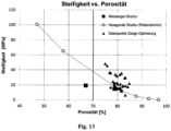
- the prior art shows a relative structural stiffness of only 6.3% with a porosity of 67 percent by volume, the value of a design-optimized structure is >15%.
- the relative structural stiffness is the structural stiffness divided by the material stiffness).
- both material components do not have any oriented structures and therefore cannot direct the regeneration process along defined directions, which is disadvantageous for the regeneration of musculoskeletal tissue and other tissues with oriented structures (eg nerves).
- the aim of the invention is to create a structurally and mechanically optimized environment for tissue regeneration. This requires porosities with values >70 percent by volume, particularly preferably >80 percent by volume. For resorbable materials, such porosities can only be achieved while maintaining the mechanical structural rigidity through a material architecture that is tailored to the mechanical function. The amount of material is distributed in such a way that the structural rigidity is as high as possible in the desired spatial direction. The identification of an optimal architecture is possible, for example, using "finite element" computer simulations.
- the stabilization structure proposed by Weisberger has very different material thicknesses/material diameters within the unit cell.
- this leads to inhomogeneous resorption. Areas of small material diameter degrade faster and lead to mechanical failure of the structure.
- areas with a large material diameter lead to a tissue reaction over a longer period of time without fulfilling a mechanical function. For this reason, it is advantageous for the structural elements within a unit cell to have a diameter that is as uniform as possible.
- the first material portion consists of collagen and is optimized in terms of solids content, pore size and degradation time.
- Such an embodiment could induce bone growth over a cartilaginous phase (enchondral ossification) in a bone defect of critical size.
- the material can be used in different areas of tissue defect regeneration.
- a particularly relevant application is in vertebral body fusion, the regeneration of larger defects in long tubular bones and in the regeneration of osteochondral defects, e.g. in the knee joint.
- the use of the material in the regeneration of tendons, tendon-muscle and tendon-bone transitions after a rupture should be mentioned.
- the biomaterial can be used, for example, to produce bone regeneration materials for healing bone defects, e.g. in the tibia or radius, bone regeneration material for the fusion of vertebral bodies, material for the regeneration of osteochondral defects, e.g.
- the structure of both material components can be built up from simple basic geometries/basic structures. Analogously to crystallography, such a basic structure can be described as a unit cell from which the overall structure arises through displacement and/or rotation in certain spatial directions or along certain axes. This creates a repetitive pattern with structural symmetries in displacement and rotation.
- the second portion of material has stiffening structures.
- the component contains at least one part of the material and preferably both parts of the material, substances, elements or structures that can be displayed using imaging methods used in the clinic, so that the alignment and positioning of the biomaterial can be determined with the help of X-rays, images in the computer tomograph or checked by magnetic resonance imaging and the healing process can be checked.
- the shape of the first or second material portion can be changed by an external stimulus (e.g. force, temperature, electric or magnetic fields) in order, for example, to induce cyclic deformations that have mechanobiological effects on the regenerating cells.
- the triggered deformation of the second material portion can simplify the minimally invasive implantation of the biomaterial.
- the external stimulation is non-invasive, i.e. it works (e.g. through the magnetic field) without touching the biomaterial.
- Cyclic deformation of the biomaterial can be advantageous for the targeted differentiation of the cells and the type and orientation of the extracellular matrix formed by the cells.
- the second material portion of an implant for the regeneration of a specific tissue is adapted in such a way that the expansions required for the regeneration of this tissue result in the first material portion in the body and a collapse of the first material portion is prevented.
- the stiffness of the intact material and the mechanical forces acting there are taken from the literature.
- values of tissue stretching are taken from the literature, which are particularly beneficial for a regeneration process.
- unit cells are designed, which match the rigidity in the different spatial directions to the forces occurring after implantation (e.g. greatest rigidity in the main load direction).
- the maximum stiffness of the material must be the same as or below the stiffness of the intact tissue in order not to block a mechanical stimulus for regeneration.
- different designs of the unit cell are tested for their mechanical properties by moving/rotating/adding/deleting individual material webs using computer simulations based on the finite element method. This is used to determine the strains occurring in the material as a function of the forces acting.
- the design is selected that meets the criteria in terms of stiffness/elongation in the different spatial directions and at the same time has the highest possible porosity that can be achieved.
- the Indian figure 1 The first portion of material shown has a directional pore structure. This serves as a lead structure and at the same time represents the actual cell substrate with optimized mechanical and structural properties (e.g. pore diameter) and thus the material component for inducing the regeneration processes.
- the mechanical properties are optimized for the cellular processes of migration, matrix formation and differentiation (intrinsic biomechanical and structural signals).
- the second material portion shown is the also structurally anisotropic, load-bearing support structure in the shown implementation. It has no direct influence on the cellular processes. However, the higher rigidity of this component ensures the macroscopic integrity of the first material portion.
- the stable structure implements a clamping function and prevents the collapse of the first material portion, which serves as the inner guiding structure, during the tissue formation and remodeling processes.
- the rigidity of the second material portion is used to set the mechanical expansions that occur in the first material portion due to the locally acting musculoskeletal forces. Elongation values are set, which support the differentiation into the desired tissue types (e.g. cartilage or bone).
- the inside figure 3 Mechanohybridscaffold shown represents a three-dimensional combination of first and second material portion and figure 4 an embodiment in which the different requirements for new tissue formation (cartilage versus bone) are realized by two areas with different properties.
- the extensibility in the lower area of the scaffold for bone regeneration is smaller than in the upper area for cartilage regeneration.
- figure 5 is a view for including the in figure 3 shown combination in a tissue region of a bone-cartilage defect, the use of the embodiment according to figure 4 is an even more advantageous variant for the regeneration of the two tissue types bone and cartilage.
- the mechanohybrid scaffold shown consists of a material portion, for example collagen, and a second material portion, for example PCL.
- the structure of the second part of the material was optimized in terms of mechanical and structural properties (anisotropy, defined rigidity, high porosity, no struts with Alignment perpendicular to the longitudinal direction/pore direction).
- the hierarchical structure was created by multiplying unit cells from the first and second material components.
- the deformation under load shown was simulated using the finite element method for the design process in order to achieve the desired beneficial strains.
- a soft collagen scaffold with vertically aligned pores was combined as the first material portion with a 3D printed support structure as the second material portion.
- the support structure was made of polyamide (PA) using SLM (Selective Laser Melting).
- the diameter of the pores in the support structure ie the diameter of a honeycomb of the second material portion
- the mechano hybrid scaffold ( Figure 11a , Scaffold 2) is created by fabricating a very soft collagen scaffold with oriented pores within the support structure ( Figure 11 b) been manufactured.
- the figure 12 thus shows the mechanical stabilization of a collagen scaffold by introducing a 3D-printed support structure made of PA to create a mechanohybrid scaffold.
- the pictures of figure 12 show the pure collagen scaffold (a) and the mechanohybrid scaffold (b) after wetting with aqueous buffer solution.
- the result of the mechanical compression test shows the more than 1000-fold increase in scaffold stiffness through the introduction of the support structure (c).
- the modulus of elasticity of the pure collagen scaffold and the collagen/PA hybrid scaffold was determined by means of monoaxial mechanical compression testing in an open reservoir with aqueous solution ("unconfined compression test").
- the modulus of elasticity E was analyzed in the linear region of the stress-strain curve.
Landscapes
- Health & Medical Sciences (AREA)
- Chemical & Material Sciences (AREA)
- Life Sciences & Earth Sciences (AREA)
- Public Health (AREA)
- Oral & Maxillofacial Surgery (AREA)
- Transplantation (AREA)
- Veterinary Medicine (AREA)
- Animal Behavior & Ethology (AREA)
- General Health & Medical Sciences (AREA)
- Epidemiology (AREA)
- Medicinal Chemistry (AREA)
- Dermatology (AREA)
- Engineering & Computer Science (AREA)
- Vascular Medicine (AREA)
- Heart & Thoracic Surgery (AREA)
- Dispersion Chemistry (AREA)
- Biomedical Technology (AREA)
- Cardiology (AREA)
- Orthopedic Medicine & Surgery (AREA)
- Chemical Kinetics & Catalysis (AREA)
- Biophysics (AREA)
- Materials For Medical Uses (AREA)
- Prostheses (AREA)
Claims (14)
- Matériau biologique, destiné notamment à la régénération tissulaire, comportant une première proportion de matériau biorésorbable à pores ouverts et une deuxième proportion de matériau qui est plus rigide que la première proportion de matériau et forme une structure de soutien supportant les charges, la proportion volumique du matériau plus rigide étant inférieure à 30 % par rapport au volume total dudit matériau biologique, le volume total étant constitué du volume dudit matériau biologique et du volume des cavités entourées par le matériau biologique, la rigidité structurelle de la deuxième proportion de matériau étant au moins 10 fois supérieure à celle de la première proportion de matériau, et la deuxième proportion de matériau faisant partie intégrante dudit matériau biologique tout en étant entourée, à la manière d'un endosquelette, par la première proportion de matériau, faisant en sorte que la première proportion de matériau soit intégrée structurellement à la deuxième proportion de matériau par complémentarité de forme.
- Matériau biologique selon la revendication 1, caractérisé en ce qu'au moins une proportion de matériau peut subir une déformation élastique.
- Matériau biologique selon l'une des revendications précédentes, caractérisé en ce que les deux proportions de matériau peuvent subir une déformation élastique.
- Matériau biologique selon l'une des revendications précédentes, caractérisé en ce que la proportion volumique du matériau plus rigide représente moins de 25 %, et préférentiellement moins de 20 %, du volume total dudit matériau biologique.
- Matériau biologique selon l'une des revendications précédentes, caractérisé en ce que la deuxième proportion de matériau présente une rigidité structurelle qui est 100 fois supérieure, et préférentiellement 1000 fois supérieure, à celle de la première proportion de matériau.
- Matériau biologique selon l'une des revendications précédentes, caractérisé en ce que la première et la deuxième proportions de matériau présentent une structuration hiérarchique.
- Matériau biologique selon l'une des revendications précédentes, caractérisé en ce que l'une des proportions de matériau, et préférentiellement les deux proportions de matériau, présentent des structures orientées.
- Matériau biologique selon l'une des revendications précédentes, caractérisé en ce que la première et la deuxième proportions de matériau présentent des zones comportant différentes structures orientées ou non-orientées.
- Matériau biologique selon l'une des revendications précédentes, caractérisé en ce que la structure d'au moins une des proportion de matériau, et préférentiellement des deux proportions de matériau, est réalisée sous forme d'un schéma répétitif présentant des symétries structurelles.
- Matériau biologique selon l'une des revendications précédentes, caractérisé en ce que ladite structure d'au moins une des proportions de matériau, et préférentiellement des deux proportions de matériau, contient des éléments ou structures pouvant être représentés au moyen de méthodes d'imagerie.
- Matériau biologique selon l'une des revendications précédentes, caractérisé en ce qu 'au moins une proportion de matériau peut être déformée par un stimulus externe afin de favoriser la régénération.
- Matériau biologique selon l'une des revendications précédentes, caractérisé en ce la deuxième proportion de matériau présente sa plus forte rigidité dans la direction selon laquelle ledit matériau subira, après l'implantation dans un tissu, les forces les plus importantes (direction de charge principale).
- Matériau biologique selon l'une des revendications précédentes, caractérisé en ce que l'architecture de la deuxième proportion de matériau est optimisée de manière à ce qu'elle permette d'atteindre une porosité la plus élevée possible afin de réaliser une certaine rigidité de matériau sur le plan macroscopique.
- Matériau biologique selon l'une des revendications précédentes, caractérisé en ce que les diamètres moyens des éléments structurels de la deuxième proportion de matériau varient, au sein d'une géométrie de base / cellule unitaire, d'un facteur inférieur à deux.
Applications Claiming Priority (2)
| Application Number | Priority Date | Filing Date | Title |
|---|---|---|---|
| DE102016007931.2A DE102016007931A1 (de) | 2016-06-30 | 2016-06-30 | Biomaterial |
| PCT/DE2017/000183 WO2018001401A1 (fr) | 2016-06-30 | 2017-06-28 | Biomatériau |
Publications (3)
| Publication Number | Publication Date |
|---|---|
| EP3478331A1 EP3478331A1 (fr) | 2019-05-08 |
| EP3478331B1 true EP3478331B1 (fr) | 2023-06-21 |
| EP3478331C0 EP3478331C0 (fr) | 2023-06-21 |
Family
ID=59581684
Family Applications (1)
| Application Number | Title | Priority Date | Filing Date |
|---|---|---|---|
| EP17751229.0A Active EP3478331B1 (fr) | 2016-06-30 | 2017-06-28 | Biomatériau |
Country Status (5)
| Country | Link |
|---|---|
| US (1) | US11554196B2 (fr) |
| EP (1) | EP3478331B1 (fr) |
| CA (1) | CA3028521C (fr) |
| DE (2) | DE102016007931A1 (fr) |
| WO (1) | WO2018001401A1 (fr) |
Families Citing this family (4)
| Publication number | Priority date | Publication date | Assignee | Title |
|---|---|---|---|---|
| US8728387B2 (en) | 2005-12-06 | 2014-05-20 | Howmedica Osteonics Corp. | Laser-produced porous surface |
| CA3139954A1 (fr) * | 2019-05-31 | 2020-12-03 | Tepha, Inc. | Implants assembles a partir de cellules unitaires de polyedre squelettique, de cellules unitaires enroulees ou de cellules unitaires maillees |
| CA3213145A1 (fr) | 2021-03-11 | 2022-09-15 | Tepha, Inc. | Implant de reconstruction mammaire |
| US20250025307A1 (en) * | 2023-07-18 | 2025-01-23 | Boneforger Holding Company | Custom Bone Scaffold Using Tessellation of Polygons |
Citations (1)
| Publication number | Priority date | Publication date | Assignee | Title |
|---|---|---|---|---|
| US7758882B2 (en) * | 2000-01-31 | 2010-07-20 | Indiana University Research And Technology Corporation | Composite biomaterial including anisometric calcium phosphate reinforcement particles and related methods |
Family Cites Families (5)
| Publication number | Priority date | Publication date | Assignee | Title |
|---|---|---|---|---|
| ES2088690T3 (es) * | 1990-12-19 | 1996-08-16 | Harle Anton | Procedimiento para la fabricacion de un implante medico e implante fabricado de acuerdo con dicho procedimiento. |
| DE19751031A1 (de) | 1997-11-19 | 1999-06-24 | Ingo Dipl Ing Heschel | Verfahren zur Herstellung poröser Strukturen |
| US8518123B2 (en) * | 2005-09-09 | 2013-08-27 | Board Of Trustees Of The University Of Arkansas | System and method for tissue generation and bone regeneration |
| WO2009049650A2 (fr) | 2007-10-19 | 2009-04-23 | Stryker Trauma Gmbh | Substitut osseux synthétique, son procédé de préparation et procédé pour combler une cavité dans un substrat |
| EP2450066A1 (fr) | 2010-10-19 | 2012-05-09 | Protip Sas | Nouvel implant hybride |
-
2016
- 2016-06-30 DE DE102016007931.2A patent/DE102016007931A1/de not_active Withdrawn
-
2017
- 2017-06-28 DE DE112017003254.7T patent/DE112017003254A5/de not_active Withdrawn
- 2017-06-28 US US16/313,937 patent/US11554196B2/en active Active
- 2017-06-28 WO PCT/DE2017/000183 patent/WO2018001401A1/fr not_active Ceased
- 2017-06-28 CA CA3028521A patent/CA3028521C/fr active Active
- 2017-06-28 EP EP17751229.0A patent/EP3478331B1/fr active Active
Patent Citations (1)
| Publication number | Priority date | Publication date | Assignee | Title |
|---|---|---|---|---|
| US7758882B2 (en) * | 2000-01-31 | 2010-07-20 | Indiana University Research And Technology Corporation | Composite biomaterial including anisometric calcium phosphate reinforcement particles and related methods |
Also Published As
| Publication number | Publication date |
|---|---|
| CA3028521C (fr) | 2024-10-01 |
| DE102016007931A1 (de) | 2018-01-04 |
| WO2018001401A1 (fr) | 2018-01-04 |
| DE112017003254A5 (de) | 2019-03-21 |
| CA3028521A1 (fr) | 2018-01-04 |
| EP3478331A1 (fr) | 2019-05-08 |
| DE102016007931A8 (de) | 2018-03-01 |
| US11554196B2 (en) | 2023-01-17 |
| EP3478331C0 (fr) | 2023-06-21 |
| US20200179570A1 (en) | 2020-06-11 |
Similar Documents
| Publication | Publication Date | Title |
|---|---|---|
| EP2536446B1 (fr) | Dispositif pour le recouvrement et/ou la reconstruction d'un défaut d'un os, et son procédé de fabrication | |
| EP0984745B1 (fr) | Structures pour le remplacement de l'os ou du cartilage | |
| US9763788B2 (en) | Bone regeneration using biodegradable polymeric nanocomposite materials and applications of the same | |
| US9308076B2 (en) | Bi-phasic compressed porous reinforcement materials suitable for implant | |
| WO2015185219A1 (fr) | Fabrication d'un composant de culture d'un implant de surface articulaire | |
| CN111920552B (zh) | 具有多个通孔的椎间融合器 | |
| DE102011107577B4 (de) | Implantat, Bauteilset, Verfahren zur Herstellung eines Implantats und/oder eines Bauteilsets und Vorrichtung zum Durchführen eines Verfahrens zur Herstellung eines Implantats und/oder eines Bauteils | |
| WO2018001401A1 (fr) | Biomatériau | |
| DE102009032218A1 (de) | Überhöhtes Implantat zur Rekonstruktion von Meniskusdefekten oder Meniskusteildefekten | |
| DE10061704A1 (de) | Verfahren und Vorrichtung zur Herstellung von biologischem Gewebe in einer Wachstumskammer | |
| EP1732618B1 (fr) | Procede pour realiser un materiau d'implant osseux | |
| DE102016211201A1 (de) | Medizinisches Produkt, vorzugsweise zur Anwendung bei der Behandlung einer Knochenkavität, Verfahren zur Herstellung des medizinischen Produkts sowie medizinisches Kit | |
| DE102015006154B4 (de) | Vorrichtung zur Abdeckung und/oder Rekonstruktion einer Knochendefektstelle; Verfahren zur Herstellung eines Aufsatzes einer Abdeckvorrichtung für eine Knochendefektstelle | |
| CN116158824A (zh) | 一种用于断骨再生的可调节式骨重建支架 | |
| DE102009032216B4 (de) | Spacer-Implantat zur Behandlung von menschlichen oder tierischen Gelenkknorpelschäden | |
| CH673579A5 (fr) | ||
| DE19945529A1 (de) | Implantat zur funktionserhaltenden Rekonstruktion von Skelettdefekten und Verfahren für die Herstellung | |
| DE102011087899B4 (de) | Gelenkscaffold | |
| DE102012109266B4 (de) | Implantat zur Erzeugung eines transplantierbaren Gewebestücks | |
| DE102017205688B4 (de) | Endoprothesenvorrichtung aus Keramik zum Einsetzen in eine Mulde eines Knochens | |
| DE102012003541A1 (de) | Bakterielle Nanocellulose-Körper mit kanalartiger Hohlraumstruktur sowie deren Herstellung und Verwendung | |
| WO2006039733A2 (fr) | Element de remplacement osseux | |
| DE102010026322A1 (de) | Chirurgische Implantate aus bioresorbierbaren und/oder biodegradierbaren Polymeren sowie Verfahren zu deren Herstellung | |
| Sidun et al. | Bone Ingrowth Processes on Porous Metalic Implants | |
| DE102006026592A1 (de) | Träger mit Colloss |
Legal Events
| Date | Code | Title | Description |
|---|---|---|---|
| STAA | Information on the status of an ep patent application or granted ep patent |
Free format text: STATUS: UNKNOWN |
|
| STAA | Information on the status of an ep patent application or granted ep patent |
Free format text: STATUS: THE INTERNATIONAL PUBLICATION HAS BEEN MADE |
|
| PUAI | Public reference made under article 153(3) epc to a published international application that has entered the european phase |
Free format text: ORIGINAL CODE: 0009012 |
|
| STAA | Information on the status of an ep patent application or granted ep patent |
Free format text: STATUS: REQUEST FOR EXAMINATION WAS MADE |
|
| 17P | Request for examination filed |
Effective date: 20190121 |
|
| AK | Designated contracting states |
Kind code of ref document: A1 Designated state(s): AL AT BE BG CH CY CZ DE DK EE ES FI FR GB GR HR HU IE IS IT LI LT LU LV MC MK MT NL NO PL PT RO RS SE SI SK SM TR |
|
| AX | Request for extension of the european patent |
Extension state: BA ME |
|
| DAV | Request for validation of the european patent (deleted) | ||
| DAX | Request for extension of the european patent (deleted) | ||
| STAA | Information on the status of an ep patent application or granted ep patent |
Free format text: STATUS: EXAMINATION IS IN PROGRESS |
|
| 17Q | First examination report despatched |
Effective date: 20200617 |
|
| REG | Reference to a national code |
Ref country code: DE Ref legal event code: R079 Free format text: PREVIOUS MAIN CLASS: A61L0027180000 Ipc: A61L0027560000 Ref document number: 502017014951 Country of ref document: DE |
|
| GRAP | Despatch of communication of intention to grant a patent |
Free format text: ORIGINAL CODE: EPIDOSNIGR1 |
|
| STAA | Information on the status of an ep patent application or granted ep patent |
Free format text: STATUS: GRANT OF PATENT IS INTENDED |
|
| RIC1 | Information provided on ipc code assigned before grant |
Ipc: A61F 2/28 20060101ALI20230117BHEP Ipc: A61L 27/58 20060101ALI20230117BHEP Ipc: A61L 27/50 20060101ALI20230117BHEP Ipc: A61L 27/24 20060101ALI20230117BHEP Ipc: A61L 27/18 20060101ALI20230117BHEP Ipc: A61L 27/56 20060101AFI20230117BHEP |
|
| INTG | Intention to grant announced |
Effective date: 20230210 |
|
| RAP3 | Party data changed (applicant data changed or rights of an application transferred) |
Owner name: CHARITE - UNIVERSITAETSMEDIZIN BERLIN Owner name: MATRICEL GMBH |
|
| GRAS | Grant fee paid |
Free format text: ORIGINAL CODE: EPIDOSNIGR3 |
|
| GRAA | (expected) grant |
Free format text: ORIGINAL CODE: 0009210 |
|
| STAA | Information on the status of an ep patent application or granted ep patent |
Free format text: STATUS: THE PATENT HAS BEEN GRANTED |
|
| AK | Designated contracting states |
Kind code of ref document: B1 Designated state(s): AL AT BE BG CH CY CZ DE DK EE ES FI FR GB GR HR HU IE IS IT LI LT LU LV MC MK MT NL NO PL PT RO RS SE SI SK SM TR |
|
| REG | Reference to a national code |
Ref country code: CH Ref legal event code: EP |
|
| REG | Reference to a national code |
Ref country code: DE Ref legal event code: R096 Ref document number: 502017014951 Country of ref document: DE |
|
| REG | Reference to a national code |
Ref country code: AT Ref legal event code: REF Ref document number: 1580432 Country of ref document: AT Kind code of ref document: T Effective date: 20230715 |
|
| REG | Reference to a national code |
Ref country code: IE Ref legal event code: FG4D Free format text: LANGUAGE OF EP DOCUMENT: GERMAN |
|
| U01 | Request for unitary effect filed |
Effective date: 20230718 |
|
| U07 | Unitary effect registered |
Designated state(s): AT BE BG DE DK EE FI FR IT LT LU LV MT NL PT SE SI Effective date: 20230823 |
|
| REG | Reference to a national code |
Ref country code: LT Ref legal event code: MG9D |
|
| U20 | Renewal fee for the european patent with unitary effect paid |
Year of fee payment: 7 Effective date: 20230915 |
|
| PG25 | Lapsed in a contracting state [announced via postgrant information from national office to epo] |
Ref country code: NO Free format text: LAPSE BECAUSE OF FAILURE TO SUBMIT A TRANSLATION OF THE DESCRIPTION OR TO PAY THE FEE WITHIN THE PRESCRIBED TIME-LIMIT Effective date: 20230921 |
|
| PG25 | Lapsed in a contracting state [announced via postgrant information from national office to epo] |
Ref country code: RS Free format text: LAPSE BECAUSE OF FAILURE TO SUBMIT A TRANSLATION OF THE DESCRIPTION OR TO PAY THE FEE WITHIN THE PRESCRIBED TIME-LIMIT Effective date: 20230621 Ref country code: HR Free format text: LAPSE BECAUSE OF FAILURE TO SUBMIT A TRANSLATION OF THE DESCRIPTION OR TO PAY THE FEE WITHIN THE PRESCRIBED TIME-LIMIT Effective date: 20230621 Ref country code: GR Free format text: LAPSE BECAUSE OF FAILURE TO SUBMIT A TRANSLATION OF THE DESCRIPTION OR TO PAY THE FEE WITHIN THE PRESCRIBED TIME-LIMIT Effective date: 20230922 |
|
| PG25 | Lapsed in a contracting state [announced via postgrant information from national office to epo] |
Ref country code: SK Free format text: LAPSE BECAUSE OF FAILURE TO SUBMIT A TRANSLATION OF THE DESCRIPTION OR TO PAY THE FEE WITHIN THE PRESCRIBED TIME-LIMIT Effective date: 20230621 |
|
| PG25 | Lapsed in a contracting state [announced via postgrant information from national office to epo] |
Ref country code: ES Free format text: LAPSE BECAUSE OF FAILURE TO SUBMIT A TRANSLATION OF THE DESCRIPTION OR TO PAY THE FEE WITHIN THE PRESCRIBED TIME-LIMIT Effective date: 20230621 |
|
| PG25 | Lapsed in a contracting state [announced via postgrant information from national office to epo] |
Ref country code: IS Free format text: LAPSE BECAUSE OF FAILURE TO SUBMIT A TRANSLATION OF THE DESCRIPTION OR TO PAY THE FEE WITHIN THE PRESCRIBED TIME-LIMIT Effective date: 20231021 |
|
| PG25 | Lapsed in a contracting state [announced via postgrant information from national office to epo] |
Ref country code: SM Free format text: LAPSE BECAUSE OF FAILURE TO SUBMIT A TRANSLATION OF THE DESCRIPTION OR TO PAY THE FEE WITHIN THE PRESCRIBED TIME-LIMIT Effective date: 20230621 Ref country code: SK Free format text: LAPSE BECAUSE OF FAILURE TO SUBMIT A TRANSLATION OF THE DESCRIPTION OR TO PAY THE FEE WITHIN THE PRESCRIBED TIME-LIMIT Effective date: 20230621 Ref country code: RO Free format text: LAPSE BECAUSE OF FAILURE TO SUBMIT A TRANSLATION OF THE DESCRIPTION OR TO PAY THE FEE WITHIN THE PRESCRIBED TIME-LIMIT Effective date: 20230621 Ref country code: IS Free format text: LAPSE BECAUSE OF FAILURE TO SUBMIT A TRANSLATION OF THE DESCRIPTION OR TO PAY THE FEE WITHIN THE PRESCRIBED TIME-LIMIT Effective date: 20231021 Ref country code: ES Free format text: LAPSE BECAUSE OF FAILURE TO SUBMIT A TRANSLATION OF THE DESCRIPTION OR TO PAY THE FEE WITHIN THE PRESCRIBED TIME-LIMIT Effective date: 20230621 Ref country code: CZ Free format text: LAPSE BECAUSE OF FAILURE TO SUBMIT A TRANSLATION OF THE DESCRIPTION OR TO PAY THE FEE WITHIN THE PRESCRIBED TIME-LIMIT Effective date: 20230621 |
|
| PG25 | Lapsed in a contracting state [announced via postgrant information from national office to epo] |
Ref country code: PL Free format text: LAPSE BECAUSE OF FAILURE TO SUBMIT A TRANSLATION OF THE DESCRIPTION OR TO PAY THE FEE WITHIN THE PRESCRIBED TIME-LIMIT Effective date: 20230621 |
|
| PG25 | Lapsed in a contracting state [announced via postgrant information from national office to epo] |
Ref country code: MC Free format text: LAPSE BECAUSE OF FAILURE TO SUBMIT A TRANSLATION OF THE DESCRIPTION OR TO PAY THE FEE WITHIN THE PRESCRIBED TIME-LIMIT Effective date: 20230621 |
|
| REG | Reference to a national code |
Ref country code: DE Ref legal event code: R097 Ref document number: 502017014951 Country of ref document: DE |
|
| REG | Reference to a national code |
Ref country code: IE Ref legal event code: MM4A |
|
| PG25 | Lapsed in a contracting state [announced via postgrant information from national office to epo] |
Ref country code: MC Free format text: LAPSE BECAUSE OF FAILURE TO SUBMIT A TRANSLATION OF THE DESCRIPTION OR TO PAY THE FEE WITHIN THE PRESCRIBED TIME-LIMIT Effective date: 20230621 |
|
| PG25 | Lapsed in a contracting state [announced via postgrant information from national office to epo] |
Ref country code: IE Free format text: LAPSE BECAUSE OF NON-PAYMENT OF DUE FEES Effective date: 20230628 |
|
| PLBE | No opposition filed within time limit |
Free format text: ORIGINAL CODE: 0009261 |
|
| STAA | Information on the status of an ep patent application or granted ep patent |
Free format text: STATUS: NO OPPOSITION FILED WITHIN TIME LIMIT |
|
| PG25 | Lapsed in a contracting state [announced via postgrant information from national office to epo] |
Ref country code: IE Free format text: LAPSE BECAUSE OF NON-PAYMENT OF DUE FEES Effective date: 20230628 |
|
| 26N | No opposition filed |
Effective date: 20240322 |
|
| U20 | Renewal fee for the european patent with unitary effect paid |
Year of fee payment: 8 Effective date: 20240509 |
|
| U20 | Renewal fee for the european patent with unitary effect paid |
Year of fee payment: 9 Effective date: 20250509 |
|
| PGFP | Annual fee paid to national office [announced via postgrant information from national office to epo] |
Ref country code: GB Payment date: 20250401 Year of fee payment: 9 |
|
| PG25 | Lapsed in a contracting state [announced via postgrant information from national office to epo] |
Ref country code: CY Free format text: LAPSE BECAUSE OF FAILURE TO SUBMIT A TRANSLATION OF THE DESCRIPTION OR TO PAY THE FEE WITHIN THE PRESCRIBED TIME-LIMIT; INVALID AB INITIO Effective date: 20170628 |
|
| PG25 | Lapsed in a contracting state [announced via postgrant information from national office to epo] |
Ref country code: HU Free format text: LAPSE BECAUSE OF FAILURE TO SUBMIT A TRANSLATION OF THE DESCRIPTION OR TO PAY THE FEE WITHIN THE PRESCRIBED TIME-LIMIT; INVALID AB INITIO Effective date: 20170628 |
|
| PGFP | Annual fee paid to national office [announced via postgrant information from national office to epo] |
Ref country code: CH Payment date: 20250701 Year of fee payment: 9 |
|
| PG25 | Lapsed in a contracting state [announced via postgrant information from national office to epo] |
Ref country code: TR Free format text: LAPSE BECAUSE OF FAILURE TO SUBMIT A TRANSLATION OF THE DESCRIPTION OR TO PAY THE FEE WITHIN THE PRESCRIBED TIME-LIMIT Effective date: 20230621 |