EP3478331B1 - Biomaterial - Google Patents
Biomaterial Download PDFInfo
- Publication number
- EP3478331B1 EP3478331B1 EP17751229.0A EP17751229A EP3478331B1 EP 3478331 B1 EP3478331 B1 EP 3478331B1 EP 17751229 A EP17751229 A EP 17751229A EP 3478331 B1 EP3478331 B1 EP 3478331B1
- Authority
- EP
- European Patent Office
- Prior art keywords
- biomaterial
- material fraction
- fraction
- biomaterial according
- tissue
- Prior art date
- Legal status (The legal status is an assumption and is not a legal conclusion. Google has not performed a legal analysis and makes no representation as to the accuracy of the status listed.)
- Active
Links
Images


Classifications
-
- A—HUMAN NECESSITIES
- A61—MEDICAL OR VETERINARY SCIENCE; HYGIENE
- A61L—METHODS OR APPARATUS FOR STERILISING MATERIALS OR OBJECTS IN GENERAL; DISINFECTION, STERILISATION OR DEODORISATION OF AIR; CHEMICAL ASPECTS OF BANDAGES, DRESSINGS, ABSORBENT PADS OR SURGICAL ARTICLES; MATERIALS FOR BANDAGES, DRESSINGS, ABSORBENT PADS OR SURGICAL ARTICLES
- A61L27/00—Materials for grafts or prostheses or for coating grafts or prostheses
- A61L27/50—Materials characterised by their function or physical properties, e.g. injectable or lubricating compositions, shape-memory materials, surface modified materials
- A61L27/56—Porous materials, e.g. foams or sponges
-
- A—HUMAN NECESSITIES
- A61—MEDICAL OR VETERINARY SCIENCE; HYGIENE
- A61F—FILTERS IMPLANTABLE INTO BLOOD VESSELS; PROSTHESES; DEVICES PROVIDING PATENCY TO, OR PREVENTING COLLAPSING OF, TUBULAR STRUCTURES OF THE BODY, e.g. STENTS; ORTHOPAEDIC, NURSING OR CONTRACEPTIVE DEVICES; FOMENTATION; TREATMENT OR PROTECTION OF EYES OR EARS; BANDAGES, DRESSINGS OR ABSORBENT PADS; FIRST-AID KITS
- A61F2/00—Filters implantable into blood vessels; Prostheses, i.e. artificial substitutes or replacements for parts of the body; Appliances for connecting them with the body; Devices providing patency to, or preventing collapsing of, tubular structures of the body, e.g. stents
- A61F2/02—Prostheses implantable into the body
- A61F2/28—Bones
-
- A—HUMAN NECESSITIES
- A61—MEDICAL OR VETERINARY SCIENCE; HYGIENE
- A61L—METHODS OR APPARATUS FOR STERILISING MATERIALS OR OBJECTS IN GENERAL; DISINFECTION, STERILISATION OR DEODORISATION OF AIR; CHEMICAL ASPECTS OF BANDAGES, DRESSINGS, ABSORBENT PADS OR SURGICAL ARTICLES; MATERIALS FOR BANDAGES, DRESSINGS, ABSORBENT PADS OR SURGICAL ARTICLES
- A61L27/00—Materials for grafts or prostheses or for coating grafts or prostheses
- A61L27/14—Macromolecular materials
- A61L27/18—Macromolecular materials obtained otherwise than by reactions only involving carbon-to-carbon unsaturated bonds
-
- A—HUMAN NECESSITIES
- A61—MEDICAL OR VETERINARY SCIENCE; HYGIENE
- A61L—METHODS OR APPARATUS FOR STERILISING MATERIALS OR OBJECTS IN GENERAL; DISINFECTION, STERILISATION OR DEODORISATION OF AIR; CHEMICAL ASPECTS OF BANDAGES, DRESSINGS, ABSORBENT PADS OR SURGICAL ARTICLES; MATERIALS FOR BANDAGES, DRESSINGS, ABSORBENT PADS OR SURGICAL ARTICLES
- A61L27/00—Materials for grafts or prostheses or for coating grafts or prostheses
- A61L27/14—Macromolecular materials
- A61L27/22—Polypeptides or derivatives thereof, e.g. degradation products
- A61L27/24—Collagen
-
- A—HUMAN NECESSITIES
- A61—MEDICAL OR VETERINARY SCIENCE; HYGIENE
- A61L—METHODS OR APPARATUS FOR STERILISING MATERIALS OR OBJECTS IN GENERAL; DISINFECTION, STERILISATION OR DEODORISATION OF AIR; CHEMICAL ASPECTS OF BANDAGES, DRESSINGS, ABSORBENT PADS OR SURGICAL ARTICLES; MATERIALS FOR BANDAGES, DRESSINGS, ABSORBENT PADS OR SURGICAL ARTICLES
- A61L27/00—Materials for grafts or prostheses or for coating grafts or prostheses
- A61L27/50—Materials characterised by their function or physical properties, e.g. injectable or lubricating compositions, shape-memory materials, surface modified materials
-
- A—HUMAN NECESSITIES
- A61—MEDICAL OR VETERINARY SCIENCE; HYGIENE
- A61L—METHODS OR APPARATUS FOR STERILISING MATERIALS OR OBJECTS IN GENERAL; DISINFECTION, STERILISATION OR DEODORISATION OF AIR; CHEMICAL ASPECTS OF BANDAGES, DRESSINGS, ABSORBENT PADS OR SURGICAL ARTICLES; MATERIALS FOR BANDAGES, DRESSINGS, ABSORBENT PADS OR SURGICAL ARTICLES
- A61L27/00—Materials for grafts or prostheses or for coating grafts or prostheses
- A61L27/50—Materials characterised by their function or physical properties, e.g. injectable or lubricating compositions, shape-memory materials, surface modified materials
- A61L27/58—Materials at least partially resorbable by the body
-
- A—HUMAN NECESSITIES
- A61—MEDICAL OR VETERINARY SCIENCE; HYGIENE
- A61F—FILTERS IMPLANTABLE INTO BLOOD VESSELS; PROSTHESES; DEVICES PROVIDING PATENCY TO, OR PREVENTING COLLAPSING OF, TUBULAR STRUCTURES OF THE BODY, e.g. STENTS; ORTHOPAEDIC, NURSING OR CONTRACEPTIVE DEVICES; FOMENTATION; TREATMENT OR PROTECTION OF EYES OR EARS; BANDAGES, DRESSINGS OR ABSORBENT PADS; FIRST-AID KITS
- A61F2/00—Filters implantable into blood vessels; Prostheses, i.e. artificial substitutes or replacements for parts of the body; Appliances for connecting them with the body; Devices providing patency to, or preventing collapsing of, tubular structures of the body, e.g. stents
- A61F2/02—Prostheses implantable into the body
- A61F2/30—Joints
- A61F2002/30001—Additional features of subject-matter classified in A61F2/28, A61F2/30 and subgroups thereof
- A61F2002/30003—Material related properties of the prosthesis or of a coating on the prosthesis
- A61F2002/30004—Material related properties of the prosthesis or of a coating on the prosthesis the prosthesis being made from materials having different values of a given property at different locations within the same prosthesis
- A61F2002/30014—Material related properties of the prosthesis or of a coating on the prosthesis the prosthesis being made from materials having different values of a given property at different locations within the same prosthesis differing in elasticity, stiffness or compressibility
-
- A—HUMAN NECESSITIES
- A61—MEDICAL OR VETERINARY SCIENCE; HYGIENE
- A61F—FILTERS IMPLANTABLE INTO BLOOD VESSELS; PROSTHESES; DEVICES PROVIDING PATENCY TO, OR PREVENTING COLLAPSING OF, TUBULAR STRUCTURES OF THE BODY, e.g. STENTS; ORTHOPAEDIC, NURSING OR CONTRACEPTIVE DEVICES; FOMENTATION; TREATMENT OR PROTECTION OF EYES OR EARS; BANDAGES, DRESSINGS OR ABSORBENT PADS; FIRST-AID KITS
- A61F2/00—Filters implantable into blood vessels; Prostheses, i.e. artificial substitutes or replacements for parts of the body; Appliances for connecting them with the body; Devices providing patency to, or preventing collapsing of, tubular structures of the body, e.g. stents
- A61F2/02—Prostheses implantable into the body
- A61F2/30—Joints
- A61F2002/30001—Additional features of subject-matter classified in A61F2/28, A61F2/30 and subgroups thereof
- A61F2002/30108—Shapes
- A61F2002/3011—Cross-sections or two-dimensional shapes
- A61F2002/30138—Convex polygonal shapes
- A61F2002/30143—Convex polygonal shapes hexagonal
-
- A—HUMAN NECESSITIES
- A61—MEDICAL OR VETERINARY SCIENCE; HYGIENE
- A61L—METHODS OR APPARATUS FOR STERILISING MATERIALS OR OBJECTS IN GENERAL; DISINFECTION, STERILISATION OR DEODORISATION OF AIR; CHEMICAL ASPECTS OF BANDAGES, DRESSINGS, ABSORBENT PADS OR SURGICAL ARTICLES; MATERIALS FOR BANDAGES, DRESSINGS, ABSORBENT PADS OR SURGICAL ARTICLES
- A61L2430/00—Materials or treatment for tissue regeneration
- A61L2430/02—Materials or treatment for tissue regeneration for reconstruction of bones; weight-bearing implants
-
- A—HUMAN NECESSITIES
- A61—MEDICAL OR VETERINARY SCIENCE; HYGIENE
- A61L—METHODS OR APPARATUS FOR STERILISING MATERIALS OR OBJECTS IN GENERAL; DISINFECTION, STERILISATION OR DEODORISATION OF AIR; CHEMICAL ASPECTS OF BANDAGES, DRESSINGS, ABSORBENT PADS OR SURGICAL ARTICLES; MATERIALS FOR BANDAGES, DRESSINGS, ABSORBENT PADS OR SURGICAL ARTICLES
- A61L2430/00—Materials or treatment for tissue regeneration
- A61L2430/24—Materials or treatment for tissue regeneration for joint reconstruction
-
- A—HUMAN NECESSITIES
- A61—MEDICAL OR VETERINARY SCIENCE; HYGIENE
- A61L—METHODS OR APPARATUS FOR STERILISING MATERIALS OR OBJECTS IN GENERAL; DISINFECTION, STERILISATION OR DEODORISATION OF AIR; CHEMICAL ASPECTS OF BANDAGES, DRESSINGS, ABSORBENT PADS OR SURGICAL ARTICLES; MATERIALS FOR BANDAGES, DRESSINGS, ABSORBENT PADS OR SURGICAL ARTICLES
- A61L2430/00—Materials or treatment for tissue regeneration
- A61L2430/38—Materials or treatment for tissue regeneration for reconstruction of the spine, vertebrae or intervertebral discs
Definitions
- the invention relates to a biomaterial.
- This is a synthetic or non-living natural material used in medicine for therapeutic or diagnostic purposes, in direct contact with biological tissues of the body.
- the material enters into chemical, physical and biological interactions with the corresponding biological systems.
- This also includes short-term contact via the outer body surface, via body openings and via mucous membranes accessible from the outside.
- the invention particularly relates to materials that are placed inside the body for long-term retention.
- biomaterial refers to the material, in particular the chemical and physical, properties of the material.
- a biocompatibility resulting from its properties is characteristic of a biomaterial, which includes both the functional similarity to the body's own structures and an appropriate biological compatibility in the body.
- biomaterials are used in the form of implants inside the body. They are used, for example, as part of a treatment known as osteosynthesis to support healing after a fracture as well as the permanent replacement of bones that have been irreparably destroyed by an accident or bone cancer. It is also possible to replace joint structures that have been worn down by chronic diseases or long-term stress. Blood vessels can be replaced with vascular prostheses, and stents serve to support the walls of blood vessels. Examples of the replacement of organ parts or entire organs with biomaterials are artificial heart valves, artificial bladders or artificial hearts. Biomaterials are also used in plastic surgery, such as glass for artificial eyes, silicone for breast reconstruction or augmentation, or titanium for skull reconstruction.
- biomaterials also serve to support tissue regeneration, in which the damaged or missing tissue is regenerated and/or remodeled by the body that it is equivalent in function to the original intact tissue or at least comes close to it.
- the biomaterial has no permanent but only a temporary function and is ideally broken down in the body over time until it has completely disappeared and is replaced by the newly formed tissue. Examples include the regeneration of bones, skin, nerves, muscles, tendons and ligaments, connective tissue, cartilage, esophagus, vocal cords, heart muscle and heart valves.
- the biomaterials that are used here must therefore have additional properties, since in particular they must be at least partially degraded or remodeled by processes taking place in the body, with the degradation products also having to be biocompatible.
- bioresorbable biomaterials can consist of natural materials of autogenous, allogeneic or xenogeneic or plant origin (e.g. collagen, elastin, fibrin, hydroxyapatite, chitosan, alginate) or synthetic materials (e.g. PLA, PGA, PCL, PLGA) .
- natural materials of autogenous, allogeneic or xenogeneic or plant origin e.g. collagen, elastin, fibrin, hydroxyapatite, chitosan, alginate
- synthetic materials e.g. PLA, PGA, PCL, PLGA
- regenerative medicine In regenerative medicine, the aim is to fully restore the function of damaged, diseased or lost tissue (e.g. due to trauma).
- Regenerative medicine deals with so-called "tissue engineering", the cultivation of replacement tissue with properties that are identical or as similar as possible to the tissue to be replaced, but also with the restoration of function using the body's own, so-called “endogenous” regeneration mechanisms and - principles. Endogenous regeneration mechanisms can be supported, for example, by introducing biomaterials, cytokines (e.g. growth factors) or cells or a combination of these. Insofar as the biomaterial is able, e.g.
- tissue engineering approaches the most exact reproduction possible of the intact tissue should ensure rapid integration of the biomaterial and restoration of the tissue function.
- the complexity of native tissue has not yet been fully mapped, so that corresponding tissue engineering products have not yet been comparable with native autologous tissue.
- the object of the invention is to find a material that makes it easier to restore the original situation or to achieve a desired situation after a structural and/or functional change within the organism.
- the first material proportion is the proportion that has a lower stiffness than the average stiffness of the material biomaterials.
- the mean stiffness is determined based on the volume.
- the volume fraction of the stiffer material is understood to mean the actual volume of the second material fraction, excluding the cavities, in relation to the total volume of the biomaterial.
- the total volume is the volume of the mass plus the volume of the cavities enclosed by the mass. This means that a porous cuboid has the same total volume as a solid material of the same size.
- the mechanics of the material are to be used on the one hand to stabilize the softer material portion during the regeneration process and on the other hand to regulate cell functions and tissue differentiation.
- the spatially specific design of the components translates the forces acting on the material into the expansion required for the respective differentiation. Due to the structure, the material should also perform a stretching function, which should prevent the softer biomaterial structure from collapsing during the healing process.
- One embodiment provides a multi-component biomaterial consisting of at least two components that differ primarily in their mechanical and structural properties.
- the biomaterial referred to below as “mechanohybridscaffold”, is mechanically and structurally hierarchical.
- the component with lower rigidity and smaller structural size serves as a carrier material for the cells that are intended to bring about the regeneration (of local origin or obtained and/or cultivated) and as a cell-controlling material;
- the component with higher structural rigidity and structure size (second material portion) serves as a mechanically stabilizing element for realizing the desired macroscopic properties.
- Stiffness here means the structural rigidity.
- the first portion of material is structurally integrated in a form-fitting manner into the second portion of material.
- Mechanical forces acting on the second material portion act are thus preferably transmitted to the first material portion both via a form fit and in the case of adhesive forces between the two material components via a non-positive fit, with the rigidity at a corresponding mechanical load determining the strain acting on the second material portion in the first material portion.
- the term "scaffold” refers to "framework structures" which can be partially or completely remodeled in the course of regeneration by endogenous processes.
- the function of the structuring of the mechanohybrid scaffold is primarily to direct the regeneration process along a given direction.
- This principle has already been used successfully in the regeneration of e.g. B. Skin, soft tissue and peripheral nerves ( US6447701 (B1) - Method for producing porous structures) and used by the inventors in bone regeneration.
- B. Skin, soft tissue and peripheral nerves US6447701 (B1) - Method for producing porous structures
- a structured and directed regeneration process is also important for the regeneration of many other tissues (e.g. muscles, tendons, ligaments, vessels, tubular hollow organs).
- at least one of the material components has a structural anisotropy.
- the structurally hierarchical structure also ensures that the architecture and the local mechanical rigidity of the first material portion are influenced as little as possible by the second.
- the first part of the material in the body which is soft and mechanically and structurally optimized for the induction, is deformed by cell forces and forces from the surrounding tissue and its pore architecture is impaired and loses its guiding function. It is therefore necessary to combine the first material portion with a second mechanically stabilizing material portion in order to maintain or further increase the regeneration potential of the material. With a second mechanically stabilizing material component, the rigidity of the first material portion can be reduced even further, so that even greater expansions, which are more advantageous for tissue regeneration of certain tissues, can be achieved by the cells without the overall structure of the mechanohybrid scaffold collapsing.
- At least one part of the material and preferably both parts of the material have oriented structures.
- a structure is directional if the largest and smallest distance between structural elements (pore walls, webs, etc.) that can be found along all spatial directions differs by at least three times. The direction with the greatest distance between the structural elements corresponds to the preferred orientation of the material or the direction of the structural anisotropy.
- one or both parts of the material are biodegradable.
- the absorbability is matched to the speed of the regeneration process and the tissue remodeling processes taking place.
- Both parts of the material are connected to one another in a positive or non-positive manner to the extent that mechanical signals are transmitted from one part of the material to the other.
- the anisotropic structuring of the first and/or second material portion also allows the realization of different mechanical rigidities in different spatial directions (anisotropic rigidity). This makes it possible to design the mechanohybrid scaffold in such a way that it is easily compressible, e.g. perpendicular to the load-bearing direction, which offers great advantages when it is inserted using minimally invasive surgical methods (e.g. endoscopy). Elastic material properties ensure that both parts of the material can be stretched again after minimally invasive insertion.
- At least one material portion and preferably even both material portions are elastically deformable.
- the range of elastic deformability in one spatial direction is a minimum of 0.1 times the length for compression and a maximum of 10 times the length for stretching.
- the first portion of material can consist of several materials or components, which are arranged, for example, in the form of a bi- or multi-layer scaffold. These can differ in the material (e.g. hydrogel, collagen, chitosan, etc.) or in the material parameters (e.g. solids content, density, porosity, etc.).
- the second material portion can consist of several materials or components and can be constructed as a bi- or multi-layer scaffold.
- the properties of the material components can vary spatially, either as discrete layers or strata or with a smooth (gradual) transition or with a soft or firm core. Such a design can be used, for example, to achieve different expansions in different areas of the material.
- transitions between different tissues can be simulated (eg muscle-tendon, tendon-bone, cartilage-bone).
- the spatial differences in elongation, pore size or stiffness can be used to differentiate the regenerating tissue into different tissue types within a material.
- the first and second portions of material are structurally interwoven rather than formed as discrete layers.
- the support structure is an integral part of the biomaterial and is surrounded by the scaffold.
- an endoskeleton also called inner skeleton
- skeleton mechanical support structure
- the hierarchically structured biomaterial can also contain other components in the form of growth factors, bioactive particles or additives that modify the bioactivity of the material.
- the additional factors can be integrated in the first material portion and/or the second material portion or introduced into the porous structure.
- the hierarchically structured biomaterial Before being introduced into the body, the hierarchically structured biomaterial can also be combined with cells that support the regeneration process. These cells can either be obtained during the procedure (e.g. from tissue or blood) or taken in a previous procedure and possibly preserved and/or expanded in vitro.
- the lead structure causes a structural and/or mechanical anisotropy of at least one of the components for aligning the tissue regeneration process in tissues with specific structural properties such as bones, ligaments, muscles and/or cartilage.
- Bimodal mechanical properties imply a microscopically soft and macroscopically load-bearing function at the cellular level to stabilize scaffold integrity.
- a hierarchical structure enables optimized mechanics and geometry on different length scales with the same construction principle, which leads to special mechanical and geometric properties and also enables the seamless transmission of mechanical signals between the components.
- the construction from different material proportions enables spatially adjustable mechanical properties.
- Long-term mechanical stability i.e. dimensional stability of biodegradable scaffolds, is achieved by a clamping function of the support structure. If at least the second material portion is compressible and elastic, the result is a scaffold that is particularly suitable for use in minimally invasive surgical techniques.
- a mechanical anisotropy very strong in the mechanical functional direction, easily compressible perpendicular to it) can be used here.
- the second portion of material spans the first portion of material again after compression.
- the first material portion can be optimized for controlling cellular processes and the second material portion for stabilizing the primary structure and for transmitting biomechanical signals to the first material portion. This allows for a discrete distribution or a continuous transition in the mechanical properties of the material. By combining or integrating particles or additives/active ingredients, mechanical, chemical, biological and physical properties can be enhanced. In addition, the possibility of cell colonization opens up.
- all soft and degradable, biocompatible materials can be used for the first material component. These are, for example, collagen, fibrin, hyaluronic acid, elastin, alginate, polyethylene glycol (PEG), polyvinyl alcohol (PVA), polysaccharides, chitosan, silk and other materials.
- all degradable and non-degradable biocompatible materials can be considered for the second material component.
- PA polyamide
- PCL polycaprolactone
- PLA polylactide-co-glycolide
- PLA polylactide
- PEG polyethylene glycol
- PGA polyglycolide
- PVA polyvinyl alcohol
- PEEK polyetheretherketone
- PMMA polymethyl methacrylate
- metals e.g. nitinol, surgical steels, titanium and titanium alloys, magnesium
- ceramics and ceramic composites and many other materials e.g. nitinol, surgical steels, titanium and titanium alloys, magnesium
- a highly porous structure with a porosity > 70% and an open-pored spherical, channel-shaped or more complex geometry is proposed for the first material portion.
- a framework structure with the lowest possible material volume or high porosity (>70%) is proposed, which is constructed, for example, from polygonal elements (honeycomb structure), channels or spherical geometries.
- the pores or structure sizes can be between 10-1000 ⁇ m for the first portion of material and between 100 ⁇ m-10 mm for the second portion of material.
- the stiffnesses can be E ⁇ 1 MPa for the first material portion and E>100 kPa for the second material portion.
- a material for the first part of the material, a material is proposed that does not significantly change its shape (pore geometry) in combination with the second part of the material after cyclic compression and relaxation, and for the second part of the material an elastic, largely reversibly deformable material with little or no energy dissipation in a cyclical cycle compression and relaxation.
- the porosity of a biomaterial should be as high as possible in order to use endogenous regeneration processes for tissue healing.
- a high porosity enables a better supply of the cells by means of diffusion and convection of dissolved substances (e.g. supply of nutrients and oxygen, removal of metabolic products and gases).
- a high porosity is necessary so that the endogenous tissue processes are not spatially impeded by the biomaterial.
- the architecture of the second material portion is chosen so that its structural rigidity is as high as possible compared to the volume portion it occupies. This is only possible through a corresponding design process that takes engineering principles into account.
- the prior art shows a relative structural stiffness of only 6.3% with a porosity of 67 percent by volume, the value of a design-optimized structure is >15%.
- the relative structural stiffness is the structural stiffness divided by the material stiffness).
- both material components do not have any oriented structures and therefore cannot direct the regeneration process along defined directions, which is disadvantageous for the regeneration of musculoskeletal tissue and other tissues with oriented structures (eg nerves).
- the aim of the invention is to create a structurally and mechanically optimized environment for tissue regeneration. This requires porosities with values >70 percent by volume, particularly preferably >80 percent by volume. For resorbable materials, such porosities can only be achieved while maintaining the mechanical structural rigidity through a material architecture that is tailored to the mechanical function. The amount of material is distributed in such a way that the structural rigidity is as high as possible in the desired spatial direction. The identification of an optimal architecture is possible, for example, using "finite element" computer simulations.
- the stabilization structure proposed by Weisberger has very different material thicknesses/material diameters within the unit cell.
- this leads to inhomogeneous resorption. Areas of small material diameter degrade faster and lead to mechanical failure of the structure.
- areas with a large material diameter lead to a tissue reaction over a longer period of time without fulfilling a mechanical function. For this reason, it is advantageous for the structural elements within a unit cell to have a diameter that is as uniform as possible.
- the first material portion consists of collagen and is optimized in terms of solids content, pore size and degradation time.
- Such an embodiment could induce bone growth over a cartilaginous phase (enchondral ossification) in a bone defect of critical size.
- the material can be used in different areas of tissue defect regeneration.
- a particularly relevant application is in vertebral body fusion, the regeneration of larger defects in long tubular bones and in the regeneration of osteochondral defects, e.g. in the knee joint.
- the use of the material in the regeneration of tendons, tendon-muscle and tendon-bone transitions after a rupture should be mentioned.
- the biomaterial can be used, for example, to produce bone regeneration materials for healing bone defects, e.g. in the tibia or radius, bone regeneration material for the fusion of vertebral bodies, material for the regeneration of osteochondral defects, e.g.
- the structure of both material components can be built up from simple basic geometries/basic structures. Analogously to crystallography, such a basic structure can be described as a unit cell from which the overall structure arises through displacement and/or rotation in certain spatial directions or along certain axes. This creates a repetitive pattern with structural symmetries in displacement and rotation.
- the second portion of material has stiffening structures.
- the component contains at least one part of the material and preferably both parts of the material, substances, elements or structures that can be displayed using imaging methods used in the clinic, so that the alignment and positioning of the biomaterial can be determined with the help of X-rays, images in the computer tomograph or checked by magnetic resonance imaging and the healing process can be checked.
- the shape of the first or second material portion can be changed by an external stimulus (e.g. force, temperature, electric or magnetic fields) in order, for example, to induce cyclic deformations that have mechanobiological effects on the regenerating cells.
- the triggered deformation of the second material portion can simplify the minimally invasive implantation of the biomaterial.
- the external stimulation is non-invasive, i.e. it works (e.g. through the magnetic field) without touching the biomaterial.
- Cyclic deformation of the biomaterial can be advantageous for the targeted differentiation of the cells and the type and orientation of the extracellular matrix formed by the cells.
- the second material portion of an implant for the regeneration of a specific tissue is adapted in such a way that the expansions required for the regeneration of this tissue result in the first material portion in the body and a collapse of the first material portion is prevented.
- the stiffness of the intact material and the mechanical forces acting there are taken from the literature.
- values of tissue stretching are taken from the literature, which are particularly beneficial for a regeneration process.
- unit cells are designed, which match the rigidity in the different spatial directions to the forces occurring after implantation (e.g. greatest rigidity in the main load direction).
- the maximum stiffness of the material must be the same as or below the stiffness of the intact tissue in order not to block a mechanical stimulus for regeneration.
- different designs of the unit cell are tested for their mechanical properties by moving/rotating/adding/deleting individual material webs using computer simulations based on the finite element method. This is used to determine the strains occurring in the material as a function of the forces acting.
- the design is selected that meets the criteria in terms of stiffness/elongation in the different spatial directions and at the same time has the highest possible porosity that can be achieved.
- the Indian figure 1 The first portion of material shown has a directional pore structure. This serves as a lead structure and at the same time represents the actual cell substrate with optimized mechanical and structural properties (e.g. pore diameter) and thus the material component for inducing the regeneration processes.
- the mechanical properties are optimized for the cellular processes of migration, matrix formation and differentiation (intrinsic biomechanical and structural signals).
- the second material portion shown is the also structurally anisotropic, load-bearing support structure in the shown implementation. It has no direct influence on the cellular processes. However, the higher rigidity of this component ensures the macroscopic integrity of the first material portion.
- the stable structure implements a clamping function and prevents the collapse of the first material portion, which serves as the inner guiding structure, during the tissue formation and remodeling processes.
- the rigidity of the second material portion is used to set the mechanical expansions that occur in the first material portion due to the locally acting musculoskeletal forces. Elongation values are set, which support the differentiation into the desired tissue types (e.g. cartilage or bone).
- the inside figure 3 Mechanohybridscaffold shown represents a three-dimensional combination of first and second material portion and figure 4 an embodiment in which the different requirements for new tissue formation (cartilage versus bone) are realized by two areas with different properties.
- the extensibility in the lower area of the scaffold for bone regeneration is smaller than in the upper area for cartilage regeneration.
- figure 5 is a view for including the in figure 3 shown combination in a tissue region of a bone-cartilage defect, the use of the embodiment according to figure 4 is an even more advantageous variant for the regeneration of the two tissue types bone and cartilage.
- the mechanohybrid scaffold shown consists of a material portion, for example collagen, and a second material portion, for example PCL.
- the structure of the second part of the material was optimized in terms of mechanical and structural properties (anisotropy, defined rigidity, high porosity, no struts with Alignment perpendicular to the longitudinal direction/pore direction).
- the hierarchical structure was created by multiplying unit cells from the first and second material components.
- the deformation under load shown was simulated using the finite element method for the design process in order to achieve the desired beneficial strains.
- a soft collagen scaffold with vertically aligned pores was combined as the first material portion with a 3D printed support structure as the second material portion.
- the support structure was made of polyamide (PA) using SLM (Selective Laser Melting).
- the diameter of the pores in the support structure ie the diameter of a honeycomb of the second material portion
- the mechano hybrid scaffold ( Figure 11a , Scaffold 2) is created by fabricating a very soft collagen scaffold with oriented pores within the support structure ( Figure 11 b) been manufactured.
- the figure 12 thus shows the mechanical stabilization of a collagen scaffold by introducing a 3D-printed support structure made of PA to create a mechanohybrid scaffold.
- the pictures of figure 12 show the pure collagen scaffold (a) and the mechanohybrid scaffold (b) after wetting with aqueous buffer solution.
- the result of the mechanical compression test shows the more than 1000-fold increase in scaffold stiffness through the introduction of the support structure (c).
- the modulus of elasticity of the pure collagen scaffold and the collagen/PA hybrid scaffold was determined by means of monoaxial mechanical compression testing in an open reservoir with aqueous solution ("unconfined compression test").
- the modulus of elasticity E was analyzed in the linear region of the stress-strain curve.
Landscapes
- Health & Medical Sciences (AREA)
- Life Sciences & Earth Sciences (AREA)
- Chemical & Material Sciences (AREA)
- Public Health (AREA)
- Oral & Maxillofacial Surgery (AREA)
- Transplantation (AREA)
- Animal Behavior & Ethology (AREA)
- General Health & Medical Sciences (AREA)
- Veterinary Medicine (AREA)
- Epidemiology (AREA)
- Medicinal Chemistry (AREA)
- Dermatology (AREA)
- Biophysics (AREA)
- Chemical Kinetics & Catalysis (AREA)
- Orthopedic Medicine & Surgery (AREA)
- Cardiology (AREA)
- Engineering & Computer Science (AREA)
- Biomedical Technology (AREA)
- Heart & Thoracic Surgery (AREA)
- Vascular Medicine (AREA)
- Dispersion Chemistry (AREA)
- Materials For Medical Uses (AREA)
- Prostheses (AREA)
Description
- Die Erfindung betrifft ein Biomaterial. Dies ist ein synthetischer oder nichtlebender natürlicher Werkstoff, der in der Medizin für therapeutische oder diagnostische Zwecke eingesetzt wird und dabei in unmittelbaren Kontakt mit biologischem Gewebe des Körpers kommt.
- Das Material tritt dabei in chemische, physikalische und biologische Wechselwirkungen mit den entsprechenden biologischen Systemen.
- Dies schließt auch den kurzzeitigen Kontakt über die äußere Körperoberfläche, über Körperöffnungen und über von außen zugängliche Schleimhäute ein. Die Erfindung betrifft insbesondere solche Materialien, die zum längerfristigen Verbleib ins Körperinnere eingebracht werden.
- Der Begriff Biomaterial bezieht sich dabei auf die stofflichen, insbesondere die chemischen und physikalischen, Eigenschaften des Materials. Kennzeichnend für ein Biomaterial ist eine aus seinen Eigenschaften resultierende Biokompatibilität, die sowohl die funktionale Ähnlichkeit zu körpereigenen Strukturen als auch eine angemessene biologische Verträglichkeit im Körper umfasst.
- Viele Biomaterialien werden in Form von Implantaten im Körperinneren eingesetzt. Sie dienen dabei beispielsweise im Rahmen einer als Osteosynthese bezeichneten Behandlung zur Unterstützung der Heilung nach einem Knochenbruch ebenso wie dem dauerhaften Ersatz von Knochen, die durch einen Unfall oder durch Knochenkrebs irreparabel zerstört wurden. Möglich ist auch der Ersatz von Gelenkstrukturen, die durch chronische Erkrankungen oder langjährige Belastung abgenutzt sind. Mit Gefäßprothesen können Blutgefäße ersetzt werden, Stents dienen der Abstützung der Wand von Blutgefäßen. Beispiele für den Ersatz von Organteilen oder ganzen Organen durch Biomaterialien sind künstliche Herzklappen, künstliche Harnblasen oder Kunstherzen. Auch in der plastischen Chirurgie werden Biomaterialien eingesetzt, wie beispielsweise Glas für Kunstaugen, Silikon zur Brustrekonstruktion oder -vergrößerung oder Titan zur Schädelrekonstruktion.
- Biomaterialien dienen jedoch auch der Unterstützung der Geweberegeneration, bei der das beschädigte oder fehlende Gewebe durch den Körper neu gebildet und/oder so umgebaut wird, dass es in seiner Funktion dem ursprünglichen intakten Gewebe gleichwertig ist oder ihm zumindest nahe kommt. In diesem Fall hat das Biomaterial keine dauerhafte sondern nur eine zeitlich beschränkte Funktion und wird idealerweise im Laufe der Zeit im Körper abgebaut bis es vollständig verschwunden und durch das neugebildete Gewebe ersetzt ist. Als Beispiele sind die Regeneration von Knochen, Haut, Nerven, Muskeln, Sehnen und Bändern, Bindegewebe, Knorpel, Speiseröhre, Stimmband, Herzmuskel, Herzklappen zu nennen. Die Biomaterialien, die hierbei zum Einsatz kommen, müssen daher weitere Eigenschaften aufweisen, da sie insbesondere zumindest zum Teil durch im Körper ablaufende Prozesse abgebaut bzw. remodelliert werden müssen, wobei auch die Abbauprodukte biokompatibel sein müssen. Solche "bioresorbierbaren" Biomaterialien können aus natürlichen Materialien autogenen, allogenen oder xenogenen oder auch pflanzlichen Ursprungs bestehen (wie z.B. aus Kollagen, Elastin, Fibrin, Hydroxylapatit, Chitosan, Alginat) oder aus synthetischen Materialien (wie z.B. PLA, PGA, PCL, PLGA).
- In der regenerativen Medizin wird die vollständige Wiederherstellung der Funktion von beschädigtem, erkranktem oder verlorengegangenem Gewebe (z.B. durch Trauma) angestrebt. Die regenerative Medizin befasst sich sowohl mit dem sogenannten "Tissue Engineering", dem Züchten von Ersatzgewebe mit Eigenschaften, welche dem zu ersetzenden Gewebe identisch oder möglichst ähnlich sind, aber auch mit der Wiederherstellung der Funktion unter Ausnutzung körpereigener, sogenannter "endogener" Regenerationsmechanismen und -prinzipien. Endogene Regenerationsmechanismen können z.B. durch das Einbringen von Biomaterialien, Zytokinen (z.B. Wachstumsfaktoren) oder Zellen oder einer Kombination aus diesen unterstützt werden. Insofern das Biomaterial, z.B. durch seine mechanischen, physikalischen oder chemischen Eigenschaften, in der Lage ist Zellen aus dem umliegenden Gewebe zu rekrutieren und im endogenen Regenerationsprozess zu unterstützen und zu leiten, ist auch ein reiner Biomaterialansatz denkbar, bei dem von außen keine Zellen oder Zytokine in den Körper eingebracht werden müssen.
- Das Prinzip endogene Regenerationsprozesse zur Geweberegeneration zu nutzen hat unter Einsatz bestehender Biomaterialansätze zu widersprüchlichen Ergebnissen bezüglich der Qualität des resultierenden Gewebes geführt. Die mangelnde Qualität des Regenerats und ein damit verbundener unklarer Langzeiterfolg führen dazu, dass sich bisher wenige bzw. keines dieser vorgefertigten und gebrauchsfertigen "off the shelf" Materialien oder Materialkombinationen zur Geweberegeneration in der Klinik durchsetzen konnte.
- In sogenannten Tissue-Engineering-Ansätzen soll der möglichst exakte Nachbau des intakten Gewebes eine schnelle Integration des Biomaterials und eine Wiederherstellung der Gewebefunktion gewährleisten. Allerdings ist die Komplexität des nativen Gewebes bisher nicht vollständig abbildbar, sodass entsprechende Tissue-Engineering Produkte bislang nicht mit dem nativen autologen Gewebe vergleichbar sind.
- In "Weisgerber DW, Erning K, Flanagan CL, Hollister SJ, Harley BAC. Evaluation of multiscale mineralized collagen-polycaprolactone composites for bone tissue engineering. J Mech Behav Biomed Mater. 2016 Aug;61:318-327" wird ein PCL-Traggerüst, das ein Kollagen-Glycosaminoglycan-Gerüst umgibt offenbart.
- Die Aufgabe der Erfindung liegt darin, ein Material zu finden, das es erleichtert nach einer Struktur- und/oder Funktionsveränderung innerhalb des Organismus die ursprüngliche Situation wieder herzustellen oder eine angestrebte Situation zu erreichen.
- Diese Aufgabe wird mit einem Biomaterial mit den Merkmalen des Patentanspruchs 1 gelöst.
- Vorteilhafte Ausgestaltungen sind Gegenstand der Unteransprüche.
- Um die Materialanteile bestimmen zu können, wenn Materialqualitäten als fließender Übergang ineinander übergehen oder wenn die Steifheit bei ähnlichem Material im Wesentlichen durch die Struktur bestimmt wird, wird festgelegt, dass der erste Materialanteil derjenige Anteil ist, der eine geringere Steifheit hat als die mittlere Steifheit des Biomaterials. Die mittlere Steifheit wird auf das Volumen bezogen ermittelt.
- Unter dem Volumenanteil des steiferen Materials wird das tatsächliche Volumen des zweiten Materialanteils unter Ausschluss der Hohlräume im Verhältnis zum Gesamtvolumen des Biomaterials verstanden. Dabei ist das Gesamtvolumen jeweils das Volumen der Masse plus das Volumen der von der Masse umschlossenen Hohlräume. Das heißt ein poröser Quader hat das gleiche Gesamtvolumen wie ein gleichgroßes Vollmaterial.
- Studien z.B. von Engler et.al, Harley et. al., Park et al. und Reilly et. al. haben gezeigt, dass die Stammzellmigration und Differenzierung entscheidend durch die Eigenschaften Steifigkeit und Struktur der Mikroumgebung der Zelle beeinflusst werden.
- Weitere Untersuchungen in 3D Hydrogelen bestätigten, dass eine mechanische Steifigkeit mit einem Elastizitätsmodul zwischen ca. 10 und 100 kPa die osteogene Differenzierung von mesenchymalen Stammzellen (MSCs) induzieren, während geringere Steifigkeiten zu einer adipogenen Differenzierung führen.
- Experimentelle Untersuchungen und Computermodelle haben gezeigt, dass neben der Substratsteifigkeit auch die lokale mechanische Dehnung bzw. Stauchung ein entscheidender Einflussfaktor für die Differenzierung von Zellen- und Gewebe bei der Knochenregeneration darstellt Nachfolgend soll der Begriff "Stauchung" auch unter dem Oberbegriff "Dehnung" subsummiert werden, so dass die bezifferten Dehnungen auch Verkürzungen um den betragsmäßigen Prozentsatz beinhalten sollen. Zu hohe Dehnungen (ε > 30 %) behindern die Regeneration, mittlere Dehnungen (ε = ca. 4 bis 12 %) unterstützen die Bildung von Knorpel, geringe Dehnungen (ε = ca. 0,04 bis 4 %) die Mineralisation von Knorpel und die Formation von Knochen.
- Bei der Erfindung soll die Mechanik des Materials einerseits zur Stabilisierung des weicheren Materialanteils während des Regenerationsprozesses und andererseits als Regulator der Zellfunktionen und der Gewebedifferenzierung genutzt werden. Über die räumlich spezifische Gestaltung der Komponenten werden die auf das Material wirkenden Kräfte in die für die jeweilige Differenzierung notwendige Dehnung übersetzt. Durch die Struktur soll das Material außerdem eine Aufspann-Funktion wahrnehmen, die einen Kollaps der weicheren Biomaterialstruktur während des Heilungsprozesses verhindern soll.
- Eine Ausführungsform sieht ein Mehrkomponenten-Biomaterial vor, das aus mindestens zwei Komponenten besteht, die sich in erster Linie in ihren mechanischen und strukturellen Eigenschaften unterscheiden. Das im Weiteren als "Mechanohybridscaffold" bezeichnete Biomaterial ist mechanisch und strukturell hierarchisch aufgebaut. Bei dem Zwei-Komponenten-Biomaterial dient die Komponente mit geringerer Steifigkeit und geringerer Strukturgröße (erster Materialanteil) als Trägermaterial für die Zellen, die die Regeneration bewirken sollen (lokalen Ursprungs bzw. gewonnen und/oder kultiviert) und als zell-steuerndes Material; die Komponente mit höherer Struktursteifigkeit und Strukturgröße (zweiter Materialanteil) dient als mechanisch stabilisierendes Element zur Realisierung der gewünschten makroskopischen Eigenschaften. Hierbei ist als Steifigkeit die Struktursteifigkeit gemeint. Diese wird von den Eigenschaften der verwendeten Materialien, deren Struktur und von der geometrischen Anordnung der Komponenten bei Mehrkomponentenmaterialien bestimmt. Der erste Materialanteil ist in den zweiten Materialanteil formschlüssig strukturell integriert. Mechanische Kräfte, die auf den zweiten Materialanteil wirken, werden somit auf den ersten Materialanteil vorzugsweise sowohl über einen Formschluss als auch bei Adhäsionskräften zwischen den beiden Materialkomponenten über Kraftschluss übertragen, wobei die Steifigkeit bei einer entsprechenden mechanischen Belastung die von dem zweiten Materialanteil in dem ersten Materialanteil wirkende Dehnung bestimmt. Mit dem Begriff "Scaffold" werden dabei "Gerüststrukturen" bezeichnet, die im Verlauf der Regeneration durch körpereigene Prozesse teilweise oder ganz remodelliert werden können.
- Die Funktion der Strukturierung des Mechanohybridscaffolds ist es, in erster Linie den Regenerationsprozess entlang einer vorgegebenen Richtung zu leiten. Dieses Prinzip wurde bereits erfolgreich bei der Regeneration von z. B. Haut, Weichgewebe und peripheren Nerven (
US6447701 (B1) - Method for producing porous structures) und von den Erfindern bei der Regeneration von Knochen eingesetzt. Ein strukturierter und gerichteter Regenerationsprozess ist erwartungsgemäß jedoch auch für die Regeneration vieler weiterer Gewebe (z.B. Muskel, Sehnen, Bänder, Gefäße, röhrenförmige Hohlorgane) von Bedeutung. Entsprechend weist zumeist mindestens einer der Materialanteile eine strukturelle Anisotropie auf. Der strukturell hierarchische Aufbau gewährleistet dabei auch, dass die Architektur und die lokale mechanische Steifigkeit des ersten Materialanteils durch den zweiten möglichst wenig beeinflusst wird. - In eigenen Tierstudien wurde belegt, dass bereits der erste Materialanteil alleine bei geeigneter Wahl der Materialparameter (Steifigkeit, Porenarchitektur, Resorbierbarkeit) den Knochenregenerationsprozess in einem Knochendefekt unterstützen kann. In den Studien wurde ein Knochenwachstum entlang der Scaffoldporen über den Prozess der enchondralen Ossifikation beobachtet, während beim gleichen Tiermodell ohne Scaffold eine direkte - sogenannte intramembranöse - oberflächliche Knochenformation und eine geringere volumetrische Knochenneubildung beobachtet wurde. Bei der enchondralen Ossifikation wird Knochen über die Mineralisation einer Knorpelphase gebildet, wie es auch in der Wachstumsfuge bei Kindern und Jugendlichen beobachtet wird. Im Gegensatz hierzu wird bei der direkten intramembranösen Knochenformation Knochengewebe direkt auf bestehendem Knochen ohne Knorpelphase abgelegt. Es wurde jedoch auch beobachtet, dass bei diesem Ansatz der weiche, für die Induktion mechanisch und strukturell optimierte, erste Materialanteil im Körper durch Zellkräfte und Kräfte von umliegendem Gewebe verformt und seine Porenarchitektur beeinträchtigt wird und seine Leitfunktion verliert. Es ist daher notwendig den ersten Materialanteil mit einem zweiten mechanisch stabilisierenden Materialanteil zu kombinieren, um das Regenerationspotential des Materials aufrecht zu erhalten bzw. weiter zu erhöhen. Bei einem zweiten mechanisch stabilisierenden Materialanteil kann zudem die Steifigkeit des ersten Materialanteils noch weiter reduziert werden, so dass noch größere - für die Geweberegeneration bestimmter Gewebe vorteilhaftere Dehnungen durch die Zellen erzielt werden können, ohne dass die Gesamtstruktur des Mechanohybridscaffolds kollabiert.
- Vorteilhaft ist es, wenn mindestens ein Materialanteil und vorzugsweise beide Materialanteile gerichtete Strukturen aufweisen. Eine Struktur ist gerichtet, wenn sich der größte und der kleinste Abstand zwischen Strukturelementen (Porenwänden, Stegen, etc.) der entlang aller Raumrichtungen zu finden ist um mindestens das Dreifache unterscheidet. Die Richtung mit dem größten Abstand der Strukturelemente entspricht der bevorzugten Ausrichtung des Materials bzw. der Richtung der strukturellen Anisotropie.
- In einer möglichen Realisierung sind einer oder beide Materialanteile bioabbaubar. In diesem Fall ist die Resorbierbarkeit auf die Geschwindigkeit des Regenerationsprozesses und die ablaufenden Gewebeumbauprozesse abgestimmt. Beide Materialanteile sind miteinander soweit form- oder kraftschlüssig verbunden, dass mechanische Signale von dem einen zu dem anderen Materialanteil übertragen werden.
- Die anisotrope Strukturierung des ersten und/oder zweiten Materialanteils erlaubt zusätzlich die Realisierung unterschiedlicher mechanischer Steifigkeiten in unterschiedliche Raumrichtungen (anisotrope Steifigkeit). Dies ermöglicht es, den Mechanohybridscaffold so zu gestalten, dass er z.B. senkrecht zur lasttragenden Richtung leicht komprimierbar ist, was große Vorteile beim Einbringen mittels minimalinvasiven Operationsmethoden (z.B. Endoskopie) bietet. Elastische Materialeigenschaften gewährleisten das Wiederaufspannen von beiden Materialanteilen nach minimal-invasivem Einbringen.
- Es wird daher vorgeschlagen, dass mindestens ein Materialanteil und vorzugsweise sogar beide Materialanteile elastisch verformbar sind. Der Bereich der elastischen Verformbarkeit in einer Raumrichtung liegt in der Praxis bei einer Kompression auf minimal die 0,1-fache Länge bzw. bei Dehnung auf maximal die 10-fache Länge.
- Der erste Materialanteil kann aus mehreren Materialien oder Komponenten bestehen, die z.B. in Form eines Bi- oder Multilayerscaffolds angeordnet sind. Diese können sich im Material (z.B. Hydrogel, Kollagen, Chitosan, usw.) oder in den Materialparametern (z.B. Feststoffgehalt, Dichte, Porosität, etc.) unterscheiden. Ebenso kann der zweite Materialanteil aus mehreren Materialien oder Komponenten bestehen und als Bi- oder Multilayerscaffold aufgebaut sein. Die mechanischen Eigenschaften der Materialanteile können räumlich unterschiedlich gestaltet sein, entweder als diskrete Lagen bzw. Schichten oder mit fließendem (graduellem) Übergang oder mit einem weichen oder festen Kern. Eine solche Gestaltung kann z.B. genutzt werden, um unterschiedliche Dehnungen in unterschiedlichen Bereichen des Materials zu realisieren. Hierdurch können z.B. Übergänge zwischen unterschiedlichen Geweben nachgebildet werden (z.B. Muskel-Sehne, Sehne-Knochen, Knorpel-Knochen). Die räumlichen Unterschiede in z.B. Dehnung, Porengröße oder Steifigkeit können zur Differenzierung des regenerierenden Gewebes in unterschiedliche Gewebetypen innerhalb eines Materials genutzt werden.
- Gemäß der beanspruchten Erfindung sind die ersten und zweiten Materialanteile strukturell miteinander verwoben und nicht als diskrete Lagen gestaltet. Die Stützstruktur ist wie bei einem Endoskelett integraler und vom Scaffold umgebener Teil des Biomaterials. (Def aus Wikipedia: Als Endoskelett (auch Innenskelett genannt) bezeichnet man in der Biologie eine mechanische Stützstruktur (Skelett), die nicht Teil der äußeren Hülle des Organismus ist.)
- Das hierarchisch strukturierte Biomaterial kann zusätzlich weitere Komponenten in Form von Wachstumsfaktoren, bioaktiven Partikeln oder Zusatzstoffen, die die Bioaktivität des Materials modifizieren, beinhalten. Die Zusatzfaktoren können im ersten Materialanteil und/oder dem zweiten Materialanteil integriert oder in die poröse Struktur eingebracht sein.
- Das hierarchisch strukturierte Biomaterial kann zusätzlich vor dem Einbringen in den Körper mit Zellen kombiniert werden, welche den Regenerationsprozess unterstützen. Diese Zellen können entweder während des Eingriffs gewonnen werden (z.B. aus Gewebe oder Blut) oder in einer vorausgehenden Prozedur entnommen und ggf. konserviert und/oder in vitro expandiert worden sein.
- Die Erfindung hat gegenüber bekannten Scaffolds mehrere Vorteile. Die Leitstruktur bedingt eine strukturelle und/oder mechanische Anisotropie mindestens eines der Anteile zur Ausrichtung des Geweberegenerationsprozesses in Geweben mit spezifischen strukturellen Eigenschaften wie beispielsweise von Knochen, Bändern, Muskeln und/oder Knorpel. Bimodale mechanische Eigenschaften bedingen eine auf zellulärer Ebene mikroskopisch weiche und makroskopisch lasttragende Funktion zur Stabilisierung der Scaffoldintegrität.
- Ein hierarchischer Aufbau ermöglicht eine optimierte Mechanik und Geometrie auf unterschiedlichen Längenskalen bei gleichem Bauprinzip, der zu speziellen mechanischen und geometrischen Eigenschaften führt und ermöglicht außerdem auch die nahtlose Übertragung von mechanischen Signalen zwischen den Komponenten. Der Aufbau aus unterschiedlichen Materialanteilen ermöglicht räumlich einstellbare mechanische Eigenschaften. Durch eine Aufspannfunktion der Stützstruktur wird eine mechanische Langzeitstabilität, das heißt eine Formstabilität von bioabbaubaren Scaffolds erreicht. Sofern mindestens der zweite Materialanteil eine Komprimierbarkeit und Elastizität aufweist, ergibt sich ein Scaffold, das besonders für den Einsatz bei minimalinvasiven Operationstechniken geeignet ist. Dabei kann eine mechanische Anisotropie (sehr fest in mechanischer Funktionsrichtung, leicht komprimierbar senkrecht dazu) genutzt werden. Der zweite Materialanteil spannt dabei den ersten Materialanteil nach Kompression wieder auf. Einzelne Komponenten oder Materialanteile können auf die jeweilige Aufgabe mechanisch, strukturell und ggf. biochemisch optimiert werden. Dabei kann der erste Materialanteil zur Steuerung von zellulären Prozessen optimiert werden und der zweite Materialanteil zur Stabilisierung der primären Struktur und zur Übertragung von biomechanischen Signalen an den ersten Materialanteil. Dies ermöglicht eine diskrete Verteilung oder einen kontinuierlichen Übergang der mechanischen Eigenschaften des Materials. Durch die Kombination oder Integration von Partikeln oder Zusatzstoffen/-wirkstoffen können mechanische, chemische, biologische und physikalische Eigenschaften erweitert werden. Außerdem erschließt sich die Möglichkeit der Zellbesiedlung.
- Folgende Materialien können beispielhaft für die Realisierung der beiden Komponenten zum Einsatz kommen: Für den ersten Materialanteil kommen prinzipiell alle weichen und degradierbaren, biokompatiblen Materialien einzeln oder in Kombination in Frage. Dies sind beispielsweise Kollagen, Fibrin, Hyaluronsäure, Elastin, Alginat, Polyethylenglykol (PEG), Polyvinylalkohol (PVA), Polysaccharide, Chitosan, Seide und andere Materialien. Für den zweiten Materialanteil kommen prinzipiell alle degradierbaren und nicht-degradierbaren biokompatiblen Materialien in Frage. Dies sind beispielsweise Polyamid (PA), Polycaprolacton (PCL), Polylactid-co-Glycolid (PLGA), Polylactid (PLA), Polyethylenglykol (PEG), Polyglycolid (PGA), Polyvinylalkohol (PVA), Polyetheretherketon (PEEK), Polymethylmethacrylat (PMMA), Metalle (z.B. Nitinol, chirurgische Stähle, Titan- und Titanlegierungen, Magnesium), Keramiken und Keramik-Komposite und viele andere Materialien.
- Verschiedene Materialparameter können beispielhaft für die Realisierung der beiden Komponenten zum Einsatz kommen. Im Hinblick auf die Struktur wird für den ersten Materialanteil eine hochporöse Struktur mit einer Porosität > 70 % und eine offenporöse sphärische, kanalförmige oder komplexere Geometrie vorgeschlagen. Für den zweiten Materialanteil wird eine Gerüststruktur mit möglichst geringem Materialvolumen bzw. hoher Porosität (> 70%) vorgeschlagen, die beispielsweise aus polygonförmigen Elementen (Wabenstruktur), Kanälen oder sphärischen Geometrien aufgebaut ist. Die Poren bzw. Strukturgrößen können für den ersten Materialanteil zwischen 10 - 1000 µm und für den zweiten Materialanteil zwischen 100 µm - 10 mm liegen. Die Steifigkeiten können für den ersten Materialanteil bei E < 1 MPa und für den zweiten Materialanteil bei E > 100 kPa liegen. Für den ersten Materialanteil wird ein Material vorgeschlagen, das nach zyklischer Kompression und Entspannung seine Form (Porengeometrie) in Kombination mit dem zweiten Materialanteil nicht wesentlich verändert und für den zweiten Materialanteil ein elastisches, weitgehend reversibel verformbares Material mit nur geringer oder keiner Energiedissipation bei einer zyklischen Kompression und Entspannung.
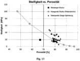
- Allgemein ist bekannt, dass die Porosität eines Biomaterials zur Nutzung endogener Regenrationsprozesse für die Gewebeheilung möglichst hoch sein sollte. Eine hohe Porosität ermöglicht eine bessere Versorgung der Zellen mittels Diffusion und Konvektion von gelösten Stoffen (z.B. Zufuhr von Nährstoffen und Sauerstoff, Abfuhr von Stoffwechselprodukten und Gasen). Außerdem ist eine hohe Porosität notwendig, um die endogenen Gewebeprozesse durch das Biomaterial räumlich nicht zu behindern. Da andererseits die äußere mechanische Steifigkeit des in der Erfindung beschriebenen Biomaterials für die Funktionalität des ersten Materialanteils essentiell ist, wird die Architektur des zweiten Materialanteils so gewählt, dass seine Struktursteifigkeit im Vergleich zu dem von ihm eingenommenen Volumenanteil möglichst hoch ist. Dies ist nur durch einen entsprechenden Designprozess unter Berücksichtigung ingenieurswissenschaftlicher Prinzipien möglich. Eigene Computersimulationen mittels Finite Elemente Methode (FEM) haben gezeigt, dass durch einen solchen Prozess die Struktursteifigkeit des zweiten Materialanteils bezogen auf die Steifigkeit des eingesetzten Materials deutlich erhöht werden kann. Erst dadurch sind unter Berücksichtigung der notwendigen Steifigkeit des Biomaterials, z.B. für den Einsatz in Knochendefekten, Porositäten von >70 Volumenprozent (70 % (v/v) erreichbar. Durch das Design der zweiten Materialkomponente können die in
Figur 13 angegebenen Steifigkeiten erreicht oder überschritten werden (Struktur entsprechend Ausführungsbeispiel inFig. 6 ). Zwischenwerte sind durch Interpolation zu berechnen. Stiffness ist Struktursteifigkeit in MPa und Porosity ist Porosität in Volumenprozent. Der Stand der Technik (Weisberger et al., 2016) zeigt bei einer Porosität von 67 Volumenprozent lediglich eine relative Struktursteifigkeit von 6.3 %, der Wert einer designoptimierten Struktur liegt bei >15 %. Die relative Struktursteifigkeit ist dabei die Struktursteifigkeit geteilt durch die Materialsteifigkeit). - In einer Publikation zeigten Weisberger et al. (Journal of the Mechanical Behavior of Biomedical Materials 2016, Volume 61, Pages 318-327, DOI: 10.1016/j.jmbbm.2016.03.032), dass die Steifigkeit eines weichen Biomaterials (Mineralisierter Kollagen/Glycosaminoglycan Scaffold) durch Kombination mit einer gedruckten Stabilisierungsstruktur (Polycaprolacton, PCL) signifikant erhöht werden kann. Allerdings ist die Porosität der Stabilisierungsstruktur gering (67 Volumenprozent), sodass mit einer deutlichen Beeinträchtigung endogener Regenerationsprozesse bei Implantation zu rechnen ist. Außerdem weisen beide Materialkomponenten keine gerichteten Strukturen auf und können damit den Regenerationsprozess nicht entlang definierter Richtungen leiten, was für die Regeneration muskuloskeletaler Gewebe und anderer Gewebe mit gerichteten Strukturen (z.B. Nerven) von Nachteil ist. Ziel der Erfindung ist es, eine strukturell und mechanisch optimierte Umgebung für die Geweberegeneration zu schaffen. Hierfür sind Porositäten mit Werten >70 Volumenprozent, besonders bevorzugt >80 Volumenprozent notwendig. Für resorbierbare Materialien lassen sich solche Porositäten unter Erhalt der mechanischen Struktursteifigkeit nur durch eine auf die mechanische Funktion abgestimmte Materialarchitektur erreichen. Hierbei wird die Materialmenge so verteilt, dass sich in der gewünschten Raumrichtung eine möglichst hohe strukturelle Steifigkeit ergibt. Die Identifikation einer optimalen Architektur ist z.B. unter Nutzung von "Finite Elemente"-Computersimulationen möglich.
- Weiterhin weist die von Weisberger vorgeschlagene Stabilisierungsstruktur innerhalb der Einheitszelle ("Unit cell") stark unterschiedliche Materialstärken/Materialdurchmesser auf. Im Zuge der Materialdegradation nach Implantation kommt es hierdurch zu einer inhomogenen Resorption. Bereiche geringen Materialdurchmessers werden schneller abgebaut und führen zum mechanischen Versagen der Struktur. Bereiche großen Materialdurchmessers führen im Zuge der Degradation über einen längeren Zeitraum zu einer Gewebereaktion ohne eine mechanische Funktion zu erfüllen. Aus diesem Grund ist ein möglichst einheitlicher Durchmesser der Strukturelemente innerhalb einer Einheitszelle von Vorteil.
- Besonders vorteilhaft ist, wenn der erste Materialanteil aus Kollagen besteht und im Hinblick auf Feststoffgehalt, Porengröße und Abbauzeit optimiert wird. Eine derartige Ausführungsform konnte in einem Knochendefekt kritischer Größe ein Knochenwachstum über eine Knorpelphase (enchondrale Ossifikation) induzieren.
- Das Material kann in unterschiedlichen Bereichen der Gewebedefektregeneration eingesetzt werden. Eine besonders relevante Anwendung liegt in der Wirbelkörperfusion, der Regeneration von größeren Defekten der langen Röhrenknochen und bei der Regeneration von osteochondralen Defekten z.B. im Kniegelenk. Über diese Anwendungen hinaus ist der Einsatz des Materials bei der Regeneration von Sehnen, Sehnen-Muskel und Sehnen-Knochen Übergängen nach Ruptur zu erwähnen. Dazu können aus den Biomaterial beispielsweise Knochenregenerationsmaterialien zur Heilung von Knochendefekten z.B. in Tibia oder Radius, Knochenregenerationsmaterial zur Fusion von Wirbelkörpern, Material zur Regeneration von osteochondralen Defekten z.B. nach Trauma oder Osteochondrosis dissecans, Material zur Regeneration von Hohlorganen (Gefäße, Speiseröhre etc....), Material zur Regeneration von Stimmbändern, Material zur Regeneration von Haut, Material zur Regeneration von Herzklappen und Herzmuskel und Material zur Regeneration von peripheren Nerven und des zentralen Nervensystems (Rückenmark) hergestellt werden.
- Die Struktur beider Materialkomponenten kann aus einfachen Grundgeometrien/ Grundstrukturen aufgebaut sein. Eine solche Grundstruktur kann, analog zur Kristallographie, als Einheitszelle beschrieben werden, aus der die Gesamtstruktur durch Verschiebung und/oder Rotation in bestimmten Raumrichtungen oder entlang bestimmter Achsen entsteht. Hierdurch entsteht ein repetitives Muster mit Struktursymmetrien in Verschiebung und Rotation.
- Besonders vorteilhaft ist, wenn der zweite Materialanteil Versteifungsstrukturen aufweist. Das Volumen oder die Masse des zweiten Materialanteils kann so verteilt sein, dass sich eine möglichst hohe mechanische Steifigkeit oder Festigkeit (oder auch Dehnbarkeit) bei möglichst geringem Materialvolumen bzw. bei möglichst hoher Porosität ergibt. Dies kann z.B. durch eine geeignete Anordnung von dünnen Materialstegen oder Wänden in bestimmten Raumrichtungen erreicht werden (=Leichtbauprinzip). Diese Strukturen können beispielsweise wie Bienenwaben aufgebaut sein.
- Besonders vorteilhaft ist es, wenn die Komponente mindestens eines Materialanteils und vorzugsweise beider Materialanteile Stoffe, Elemente oder Strukturen enthält, welche mittels in der Klinik eingesetzter Bildgebender Verfahren darstellbar sind, so dass die Ausrichtung und Positionierung des Biomaterials mit Hilfe von Röntgenaufnahmen, Aufnahmen im Computertomographen oder durch Magnetresonanztomographie überprüft und der Heilungsprozess kontrolliert werden kann.
- Besonders vorteilhaft ist es, wenn der erste oder zweite Materialanteil durch einen externen Stimulus (z.B. Kraft, Temperatur, elektrische oder magnetische Felder) in seiner Form verändert werden kann, um z.B. zyklische Verformungen zu induzieren, die mechanobiologische Auswirkungen auf die regenerierenden Zellen haben. Die getriggerte Verformung des zweiten Materialanteils kann die minimal invasive Implantation des Biomaterials vereinfachen. Im Idealfall erfolgt die externe Stimulation nicht invasiv, d.h. sie gelingt (z.B. durch das magnetische Feld) ohne Berührung des Biomaterials. Eine zyklische Verformung des Biomaterials kann für die gezielte Differenzierung der Zellen und die Art und Orientierung der von den Zellen gebildeten extrazellulären Matrix von Vorteil sein.
- Biologic-free mechanically induced muscle regeneration, Proc Natl Acad Sci USA. 2016 Feb 9;113(6):1534-9. doi: 10.1073/pnas beschreibt die mechanische Verformung eines Ferrogels mit magnetischen Partikeln durch Anlegen eines magnetischen Feldes. Im Unterscheid zur Erfindung ist es ein ein-Phasen Material ohne Ausrichtung und ohne Stützstruktur. Zur Erläuterung der Erfindung wird aber vollinhaltlich auf diese Veröffentlichung Bezug genommen.
- Bei einem Verfahren zur Herstellung eines derartigen Biomaterials wird der zweite Materialanteil eines Implantats zur Regeneration eines spezifischen Gewebes so angepasst, dass sich die für die Regeneration dieses Gewebes benötigten Dehnungen im ersten Materialanteil im Körper ergeben und ein Kollabieren des ersten Materialanteils verhindert wird.
- Hierzu werden zunächst die Steifigkeit des intakten Materials und die dort wirkenden mechanischen Kräfte aus der Literatur entnommen. Weiterhin werden aus der Literatur Werte der Gewebedehnung entnommen, die für einen Regenerationsprozess besonders förderlich sind. Mit Hilfe einer 3D Konstruktionssoftware (CAD) werden Einheitszellen entworfen, welche die Steifigkeit in den unterschiedlichen Raumrichtungen auf die nach Implantation auftretenden Kräfte abstimmt (z.B. größte Steifigkeit in Hauptbelastungsrichtung). Die maximale Steifigkeit des Materials muss dabei gleich oder unterhalb der Steifigkeit des intakten Gewebes liegen, um einen mechanischen Reiz für die Regeneration nicht zu blockieren. Im Laufe des Prozesses werden unterschiedliche Designs der Einheitszelle durch Verschieben/Rotieren/Ergänzen/Löschen einzelner Materialstege mittels Computersimulationen basierend auf Finiter Elemente Methode auf ihre mechanischen Eigenschaften getestet. Hierüber werden die im Material auftretenden Dehnungen in Abhängigkeit der wirkenden Kräfte ermittelt. Es wird das Design ausgewählt, welches die Kriterien bezüglich Steifigkeit/Dehnung in den unterschiedlichen Raumrichtungen erfüllt und gleichzeitig die höchst möglich realisierbare Porosität aufweist.
- Mehrere Referenzbeispiele sind in den Zeichnungen dargestellt und werden im Folgenden näher erläutert. In diesen Referenzbeispielen ist der zweite Materialanteil nicht wie bei einem Endoskelett integraler und vom ersten Materialanteil umgebener Teil des Biomaterials. Es zeigt
- Figur 1
- eine dreidimensionale Ansicht eines offen porösen, bioresorbierbaren ersten Materialanteils,
- Figur 2
- eine dreidimensionale Ansicht eines zweiten Materialanteils, der steifer ist als der erste Materialanteil,
- Figur 3
- eine dreidimensionale Ansicht einer Kombination aus erstem und zweitem Materialanteil,
- Figur 4
- eine dreidimensionale Ansicht einer Kombination aus erstem und zweitem Materialanteil mit unterschiedlichen Strukturen,
- Figur 5
- eine Ansicht zur Einbindung der in
Figur 3 gezeigten Kombination in einen Gewebebereich, - Figur 6
- eine dreidimensionale Ansicht eines Mechanohybridscaffolds,
- Figur 7
- einen Schnitt durch den in
Figur 6 gezeigten Mechanohybridscaffold, - Figur 8
- eine Seitenansicht des in
Figur 6 gezeigten komprimierten Mechanohybridscaffold, - Figur 9
- zwei Ansichten eines Strukturelements der zweiten Materialkomponente als Basis für Computersimulationen,
- Figur 10
- die unteren Grenzwerte der relativen Struktursteifigkeit des in
Figur 9 gezeigten Strukturelements als Tabelle, - Figur 11
- einen reinen Kollagenscaffold (a, 1) und einen Mechanohybridscaffold (a, 2) nach Zuschneiden der Prototypen für die mechanische Testung und eine mittels SLM hergestellte Stützstruktur vor Einbringen in den Mechanohybridscaffold (b),
- Figur 12
- Bilder des reinen Kollagenscaffolds (a) und des Mechanohybridscaffolds (b) nach Benetzung mit wässriger Pufferlösung sowie das Ergebnis der mechanischen Kompressionstestung (c) und
- Figur 13
- die Struktursteifigkeit über der Porosität.
- Der in der
Figur 1 gezeigte erste Materialanteil hat eine gerichtete Porenstruktur. Diese dient als Leitstruktur und stellt gleichzeitig das eigentliche Zellsubstrat mit optimierten mechanischen und strukturellen Eigenschaften (z.B. Porendurchmesser) und damit die Materialkomponente zur Induktion der Regenerationsprozesse dar. Die mechanischen Eigenschaften sind für die zellulären Prozesse der Migration, Matrixformation und Differenzierung optimiert (intrinsische biomechanische und strukturelle Signale). - Der in der
Figur 2 gezeigte zweite Materialanteil (sekundäre Struktur) ist in der gezeigten Realisierung die, ebenfalls strukturell anisotrope, lasttragende Stützstruktur. Sie hat keinen direkten Einfluss auf die zellulären Vorgänge. Allerdings wird durch die höhere Steifigkeit dieser Komponente die makroskopische Integrität des ersten Materialanteils sichergestellt. Die stabile Struktur realisiert eine Aufspann-Funktion und verhindert den Kollaps des als innere Leitstruktur dienenden ersten Materialanteils, während der Gewebeformations- und Umbauprozesse. Außerdem werden über die Steifigkeit des zweiten Materialanteils die mechanischen Dehnungen eingestellt, die aufgrund der lokal wirkenden muskuloskeletalen Kräfte im ersten Materialanteil entstehen. Es werden Dehnungswerte eingestellt, welche die Differenzierung in die gewünschten Gewebetypen (z.B. Knorpel oder Knochen) unterstützen. - Der in
Figur 3 gezeigte Mechanohybridscaffold stellt eine dreidimensionale Kombination aus erstem und zweitem Materialanteil dar undFigur 4 eine Ausführungsform, bei der die unterschiedlichen Anforderungen an die Neugewebebildung (Knorpel gegenüber Knochen) durch zwei Bereiche unterschiedlicher Eigenschaften realisiert werden. Hier ist die Dehnbarkeit entsprechend den zellulären Erfordernissen im unteren Bereich des Scaffolds für die knöcherne Regeneration kleiner als im oberen Bereich für die Knorpelregeneration. - In
Figur 5 ist eine Ansicht zur Einbindung der inFigur 3 gezeigten Kombination in einen Gewebebereich eines Knochen-Knorpeldefekts dargestellt, wobei die Verwendung der Ausführungsform gemäßFigur 4 eine noch vorteilhaftere Variante zur Regeneration der beiden Gewebstypen Knochen bzw. Knorpel ist. - Der in den
Figuren 6 bis 8 gezeigte Mechanohybridscaffold besteht aus einem Materialanteil, beispielsweise aus Kollagen und einem zweiten Materialanteil beispielsweise aus PCL. Die Struktur des zweiten Materialanteils wurde bezüglich der mechanischen und strukturellen Eigenschaften optimiert (Anisotropie, definierte Steifigkeit, hohe Porosität, keine Streben mit Ausrichtung senkrecht zur Längsrichtung/Porenrichtung). Die hierarchische Struktur ist durch Vervielfältigung von Einheitszellen aus erster und zweiter Materialkomponente entstanden. Die inFigur 8 gezeigte Verformung bei Belastung wurde mittels Finite Elemente Methode für den Designprozess simuliert, um die gewünschten vorteilhaften Dehnungen zu erzielen. - In einem Referenzbeispiel wurde als erster Materialanteil ein weicher Kollagenscaffold mit senkrecht gerichteten Poren mit einer 3D gedruckten Stützstruktur als zweitem Materialanteil kombiniert. Die Stützstruktur wurde aus Polyamid (PA) mittels SLM (Selective Laser Melting) hergestellt. Der Durchmesser der Poren in der Stützstruktur (d.h. der Durchmesser einer Wabe des zweiten Materialanteils) war hierbei ca. 50x größer als der Durchmesser der Poren im ersten Materialanteil, dem Kollagenscaffold. Das Mechanohybridscaffold (
Figur 11 a , Scaffold 2) ist durch Herstellung eines sehr weichen Kollagenscaffolds mit gerichteten Poren innerhalb der Stützstruktur (Figur 11 b) hergestellt worden. - Während das reine Kollagenscaffold nach Benetzen mit wässriger Lösung (Phosphatpuffer) kollabiert und damit seine äußere Form verändert (
Figur 12a ) zeigt das Mechanohybridscaffold als Zeichen der Aufspannfunktion der Sekundärstruktur (=Skelett!!) keine Veränderung der äußeren Geometrie (Figur 12b ). DieFigur 12 zeigt somit die mechanische Stabilisierung eines Kollagenscaffolds durch Einbringen einer 3D-gedruckten Stützstruktur aus PA zur Erzeugung eines Mechanohybridscaffolds. Die Bilder derFigur 12 zeigen den reinen Kollagenscaffold (a) und den Mechanohybridscaffold (b) nach Benetzung mit wässriger Pufferlösung. Das Ergebnis der mechanischen Kompressionstestung zeigt den mehr als 1000-fachen Anstieg der Scaffoldsteifigkeit durch Einbringen der Stützstruktur (c). Das Elastizitätsmodul des reinen Kollagenscaffolds und des Kollagen/PA Hybridscaffolds wurde mittels monoaxialer mechanischer Kompressionstestung in einem offenen Reservoir mit wässriger Lösung bestimmt ("unconfined compression test"). Das Elastizitätsmodul E wurde im linearen Bereich der Spannungs-Dehnungs-Kurve analysiert. Die mechanische Testung belegte eine Erhöhung der makroskopischen Steifigkeit von E=0.3(±0.1) kPa (reines Kollagenscaffold) auf 550(±64) kP (Mechanohybridscaffold), also um mehr als das 1000-fache (sieheFigur 12c ).
Claims (14)
- Biomaterial, insbesondere für die Geweberegeneration, mit einem offen porösen, bioresorbierbaren ersten Materialanteil und einem zweiten Materialanteil, der steifer ist als der erste Materialanteil und eine lasttragende Stützstruktur bildet, wobei der Volumenanteil des steiferen Materials weniger als 30 %, des Gesamtvolumens des Biomaterials beträgt, wobei das Gesamtvolumen aus dem Volumen des Biomaterials und dem Volumen vom Biomaterial umschlossener Hohlräume besteht, die Struktursteifigkeit des zweiten Materialanteils mindestens 10 Mal höher ist als die des ersten Materialanteils, und der zweite Materialanteil wie bei einem Endoskelett integraler und vom ersten Materialanteil umgebener Teil des Biomaterials ist, sodass der erste Materialanteil in den zweiten Materialanteil formschlüssig strukturell integriert ist.
- Biomaterial nach Anspruch 1, dadurch gekennzeichnet, dass mindestens ein Materialanteil elastisch verformbar ist.
- Biomaterial nach einem der vorhergehenden Ansprüche, dadurch gekennzeichnet, dass beide Materialanteile elastisch verformbar sind.
- Biomaterial nach einem der vorhergehenden Ansprüche, dadurch gekennzeichnet, dass der Volumenanteil des steiferen Materials weniger als 25 % und bevorzugt weniger als 20 % des Gesamtvolumens des Biomaterials beträgt.
- Biomaterial nach einem der vorhergehenden Ansprüche, dadurch gekennzeichnet, dass die Struktursteifigkeit des zweiten Materialanteils 100 Mal und bevorzugt 1000 Mal höher ist als die des ersten Materialanteils.
- Biomaterial nach einem der vorhergehenden Ansprüche, dadurch gekennzeichnet, dass die Strukturierung von erstem und zweitem Materialanteil hierarchisch ist.
- Biomaterial nach einem der vorhergehenden Ansprüche, dadurch gekennzeichnet, dass mindestens ein Materialanteil und vorzugsweise beide Materialanteile gerichtete Strukturen aufweisen.
- Biomaterial nach einem der vorhergehenden Ansprüche, dadurch gekennzeichnet, dass der erste und der zweite Materialanteil Bereiche mit unterschiedlichen gerichteten oder ungerichteten Strukturen aufweisen.
- Biomaterial nach einem der vorhergehenden Ansprüche, dadurch gekennzeichnet, dass die Struktur mindestens eines Materialanteils und vorzugsweise beider Materialanteile als repetitives Muster mit Struktursymmetrien aufgebaut ist.
- Biomaterial nach einem der vorhergehenden Ansprüche, dadurch gekennzeichnet, dass die Struktur mindestens eines Materialanteils und vorzugsweise beider Materialanteile Stoffe, Elemente oder Strukturen enthält, welche mittels bildgebenden Verfahren darstellbar sind.
- Biomaterial nach einem der vorhergehenden Ansprüche, dadurch gekennzeichnet, dass mindestens ein Materialanteil durch einen externen Stimulus zur Regenerationsförderung verformbar ist.
- Biomaterial nach einem der vorhergehenden Ansprüche, dadurch gekennzeichnet, dass die Steifigkeit des zweiten Materialanteils in der Richtung am höchsten ist, in der nach Implantation in ein Gewebe die größten Kräfte auf das Material wirken (Hauptbelastungsrichtung).
- Biomaterial nach einem der vorhergehenden Ansprüche, dadurch gekennzeichnet, dass die Architektur des zweiten Materialanteils so optimiert ist, dass zur Realisierung einer bestimmten makroskopischen Materialsteifigkeit eine möglichst hohe Porosität erreicht werden kann.
- Biomaterial nach einem der vorhergehenden Ansprüche, dadurch gekennzeichnet, dass sich die mittleren Durchmesser der Strukturelemente des zweiten Materialanteils innerhalb einer Grundgeometrie/ Einheitszelle um weniger als einen Faktor zwei unterscheiden.
Applications Claiming Priority (2)
| Application Number | Priority Date | Filing Date | Title |
|---|---|---|---|
| DE102016007931.2A DE102016007931A1 (de) | 2016-06-30 | 2016-06-30 | Biomaterial |
| PCT/DE2017/000183 WO2018001401A1 (de) | 2016-06-30 | 2017-06-28 | Biomaterial |
Publications (3)
| Publication Number | Publication Date |
|---|---|
| EP3478331A1 EP3478331A1 (de) | 2019-05-08 |
| EP3478331B1 true EP3478331B1 (de) | 2023-06-21 |
| EP3478331C0 EP3478331C0 (de) | 2023-06-21 |
Family
ID=59581684
Family Applications (1)
| Application Number | Title | Priority Date | Filing Date |
|---|---|---|---|
| EP17751229.0A Active EP3478331B1 (de) | 2016-06-30 | 2017-06-28 | Biomaterial |
Country Status (5)
| Country | Link |
|---|---|
| US (1) | US11554196B2 (de) |
| EP (1) | EP3478331B1 (de) |
| CA (1) | CA3028521C (de) |
| DE (2) | DE102016007931A1 (de) |
| WO (1) | WO2018001401A1 (de) |
Families Citing this family (4)
| Publication number | Priority date | Publication date | Assignee | Title |
|---|---|---|---|---|
| US8728387B2 (en) | 2005-12-06 | 2014-05-20 | Howmedica Osteonics Corp. | Laser-produced porous surface |
| WO2020242694A1 (en) * | 2019-05-31 | 2020-12-03 | Tepha, Inc. | Implants assembled from skeletal polyhedron unit cells, coiled unit cells or mesh unit cells |
| JP2024510182A (ja) | 2021-03-11 | 2024-03-06 | テファ, インコーポレイテッド | 乳房再建インプラント |
| US20250025307A1 (en) * | 2023-07-18 | 2025-01-23 | Boneforger Holding Company | Custom Bone Scaffold Using Tessellation of Polygons |
Citations (1)
| Publication number | Priority date | Publication date | Assignee | Title |
|---|---|---|---|---|
| US7758882B2 (en) * | 2000-01-31 | 2010-07-20 | Indiana University Research And Technology Corporation | Composite biomaterial including anisometric calcium phosphate reinforcement particles and related methods |
Family Cites Families (5)
| Publication number | Priority date | Publication date | Assignee | Title |
|---|---|---|---|---|
| ES2088690T3 (es) * | 1990-12-19 | 1996-08-16 | Harle Anton | Procedimiento para la fabricacion de un implante medico e implante fabricado de acuerdo con dicho procedimiento. |
| DE19751031A1 (de) | 1997-11-19 | 1999-06-24 | Ingo Dipl Ing Heschel | Verfahren zur Herstellung poröser Strukturen |
| EP1931401A2 (de) * | 2005-09-09 | 2008-06-18 | University of Arkansas at Little Rock | System und verfahren für die gewebegeneration und knochenregeneration |
| EP2197506B1 (de) * | 2007-10-19 | 2012-06-13 | Stryker Trauma GmbH | Synthetisches knochensubstitut, verfahren zu seiner herstellung und verfahren zum auffüllen eines hohlraums in einem substrat |
| EP2450066A1 (de) * | 2010-10-19 | 2012-05-09 | Protip Sas | Neues Hybridimplantat |
-
2016
- 2016-06-30 DE DE102016007931.2A patent/DE102016007931A1/de not_active Withdrawn
-
2017
- 2017-06-28 WO PCT/DE2017/000183 patent/WO2018001401A1/de not_active Ceased
- 2017-06-28 US US16/313,937 patent/US11554196B2/en active Active
- 2017-06-28 DE DE112017003254.7T patent/DE112017003254A5/de not_active Withdrawn
- 2017-06-28 EP EP17751229.0A patent/EP3478331B1/de active Active
- 2017-06-28 CA CA3028521A patent/CA3028521C/en active Active
Patent Citations (1)
| Publication number | Priority date | Publication date | Assignee | Title |
|---|---|---|---|---|
| US7758882B2 (en) * | 2000-01-31 | 2010-07-20 | Indiana University Research And Technology Corporation | Composite biomaterial including anisometric calcium phosphate reinforcement particles and related methods |
Also Published As
| Publication number | Publication date |
|---|---|
| EP3478331A1 (de) | 2019-05-08 |
| DE102016007931A8 (de) | 2018-03-01 |
| CA3028521C (en) | 2024-10-01 |
| CA3028521A1 (en) | 2018-01-04 |
| DE112017003254A5 (de) | 2019-03-21 |
| DE102016007931A1 (de) | 2018-01-04 |
| US20200179570A1 (en) | 2020-06-11 |
| EP3478331C0 (de) | 2023-06-21 |
| US11554196B2 (en) | 2023-01-17 |
| WO2018001401A1 (de) | 2018-01-04 |
Similar Documents
| Publication | Publication Date | Title |
|---|---|---|
| EP2536446B1 (de) | Vorrichtung zur abdeckung und/oder rekonstruktion einer knochendefektstelle und verfahren zu deren herstellung | |
| EP0984745B1 (de) | Knochen- und knorpel-ersatzstrukturen | |
| US9763788B2 (en) | Bone regeneration using biodegradable polymeric nanocomposite materials and applications of the same | |
| EP3478331B1 (de) | Biomaterial | |
| WO2015185219A1 (de) | Herstellung einer komponente zur züchtung eines gelenkflächenimplantats | |
| CN111920552B (zh) | 具有多个通孔的椎间融合器 | |
| DE102011107577B4 (de) | Implantat, Bauteilset, Verfahren zur Herstellung eines Implantats und/oder eines Bauteilsets und Vorrichtung zum Durchführen eines Verfahrens zur Herstellung eines Implantats und/oder eines Bauteils | |
| DE102009032218A1 (de) | Überhöhtes Implantat zur Rekonstruktion von Meniskusdefekten oder Meniskusteildefekten | |
| EP2836244B1 (de) | Implantat aus einem faserverbundwerkstoff | |
| DE10061704A1 (de) | Verfahren und Vorrichtung zur Herstellung von biologischem Gewebe in einer Wachstumskammer | |
| EP1732618B1 (de) | Verfahren zur herstellung eines knochen-implantatmaterials | |
| DE102016211201A1 (de) | Medizinisches Produkt, vorzugsweise zur Anwendung bei der Behandlung einer Knochenkavität, Verfahren zur Herstellung des medizinischen Produkts sowie medizinisches Kit | |
| DE102015006154B4 (de) | Vorrichtung zur Abdeckung und/oder Rekonstruktion einer Knochendefektstelle; Verfahren zur Herstellung eines Aufsatzes einer Abdeckvorrichtung für eine Knochendefektstelle | |
| CN116158824A (zh) | 一种用于断骨再生的可调节式骨重建支架 | |
| DE102009032216B4 (de) | Spacer-Implantat zur Behandlung von menschlichen oder tierischen Gelenkknorpelschäden | |
| CH673579A5 (de) | ||
| DE19945529A1 (de) | Implantat zur funktionserhaltenden Rekonstruktion von Skelettdefekten und Verfahren für die Herstellung | |
| DE102011087899B4 (de) | Gelenkscaffold | |
| DE102012109266B4 (de) | Implantat zur Erzeugung eines transplantierbaren Gewebestücks | |
| DE102017205688B4 (de) | Endoprothesenvorrichtung aus Keramik zum Einsetzen in eine Mulde eines Knochens | |
| DE102012003541A1 (de) | Bakterielle Nanocellulose-Körper mit kanalartiger Hohlraumstruktur sowie deren Herstellung und Verwendung | |
| WO2006039733A2 (de) | Knochenersatzteil aus endlosfaser-verbundwerkstoff | |
| US9289300B2 (en) | La fuente porus implant matrix | |
| DE102010026322A1 (de) | Chirurgische Implantate aus bioresorbierbaren und/oder biodegradierbaren Polymeren sowie Verfahren zu deren Herstellung | |
| Sidun et al. | Bone Ingrowth Processes on Porous Metalic Implants |
Legal Events
| Date | Code | Title | Description |
|---|---|---|---|
| STAA | Information on the status of an ep patent application or granted ep patent |
Free format text: STATUS: UNKNOWN |
|
| STAA | Information on the status of an ep patent application or granted ep patent |
Free format text: STATUS: THE INTERNATIONAL PUBLICATION HAS BEEN MADE |
|
| PUAI | Public reference made under article 153(3) epc to a published international application that has entered the european phase |
Free format text: ORIGINAL CODE: 0009012 |
|
| STAA | Information on the status of an ep patent application or granted ep patent |
Free format text: STATUS: REQUEST FOR EXAMINATION WAS MADE |
|
| 17P | Request for examination filed |
Effective date: 20190121 |
|
| AK | Designated contracting states |
Kind code of ref document: A1 Designated state(s): AL AT BE BG CH CY CZ DE DK EE ES FI FR GB GR HR HU IE IS IT LI LT LU LV MC MK MT NL NO PL PT RO RS SE SI SK SM TR |
|
| AX | Request for extension of the european patent |
Extension state: BA ME |
|
| DAV | Request for validation of the european patent (deleted) | ||
| DAX | Request for extension of the european patent (deleted) | ||
| STAA | Information on the status of an ep patent application or granted ep patent |
Free format text: STATUS: EXAMINATION IS IN PROGRESS |
|
| 17Q | First examination report despatched |
Effective date: 20200617 |
|
| REG | Reference to a national code |
Ref country code: DE Ref legal event code: R079 Free format text: PREVIOUS MAIN CLASS: A61L0027180000 Ipc: A61L0027560000 Ref document number: 502017014951 Country of ref document: DE |
|
| GRAP | Despatch of communication of intention to grant a patent |
Free format text: ORIGINAL CODE: EPIDOSNIGR1 |
|
| STAA | Information on the status of an ep patent application or granted ep patent |
Free format text: STATUS: GRANT OF PATENT IS INTENDED |
|
| RIC1 | Information provided on ipc code assigned before grant |
Ipc: A61F 2/28 20060101ALI20230117BHEP Ipc: A61L 27/58 20060101ALI20230117BHEP Ipc: A61L 27/50 20060101ALI20230117BHEP Ipc: A61L 27/24 20060101ALI20230117BHEP Ipc: A61L 27/18 20060101ALI20230117BHEP Ipc: A61L 27/56 20060101AFI20230117BHEP |
|
| INTG | Intention to grant announced |
Effective date: 20230210 |
|
| RAP3 | Party data changed (applicant data changed or rights of an application transferred) |
Owner name: CHARITE - UNIVERSITAETSMEDIZIN BERLIN Owner name: MATRICEL GMBH |
|
| GRAS | Grant fee paid |
Free format text: ORIGINAL CODE: EPIDOSNIGR3 |
|
| GRAA | (expected) grant |
Free format text: ORIGINAL CODE: 0009210 |
|
| STAA | Information on the status of an ep patent application or granted ep patent |
Free format text: STATUS: THE PATENT HAS BEEN GRANTED |
|
| AK | Designated contracting states |
Kind code of ref document: B1 Designated state(s): AL AT BE BG CH CY CZ DE DK EE ES FI FR GB GR HR HU IE IS IT LI LT LU LV MC MK MT NL NO PL PT RO RS SE SI SK SM TR |
|
| REG | Reference to a national code |
Ref country code: CH Ref legal event code: EP |
|
| REG | Reference to a national code |
Ref country code: DE Ref legal event code: R096 Ref document number: 502017014951 Country of ref document: DE |
|
| REG | Reference to a national code |
Ref country code: AT Ref legal event code: REF Ref document number: 1580432 Country of ref document: AT Kind code of ref document: T Effective date: 20230715 |
|
| REG | Reference to a national code |
Ref country code: IE Ref legal event code: FG4D Free format text: LANGUAGE OF EP DOCUMENT: GERMAN |
|
| U01 | Request for unitary effect filed |
Effective date: 20230718 |
|
| U07 | Unitary effect registered |
Designated state(s): AT BE BG DE DK EE FI FR IT LT LU LV MT NL PT SE SI Effective date: 20230823 |
|
| REG | Reference to a national code |
Ref country code: LT Ref legal event code: MG9D |
|
| U20 | Renewal fee for the european patent with unitary effect paid |
Year of fee payment: 7 Effective date: 20230915 |
|
| PG25 | Lapsed in a contracting state [announced via postgrant information from national office to epo] |
Ref country code: NO Free format text: LAPSE BECAUSE OF FAILURE TO SUBMIT A TRANSLATION OF THE DESCRIPTION OR TO PAY THE FEE WITHIN THE PRESCRIBED TIME-LIMIT Effective date: 20230921 |
|
| PG25 | Lapsed in a contracting state [announced via postgrant information from national office to epo] |
Ref country code: RS Free format text: LAPSE BECAUSE OF FAILURE TO SUBMIT A TRANSLATION OF THE DESCRIPTION OR TO PAY THE FEE WITHIN THE PRESCRIBED TIME-LIMIT Effective date: 20230621 Ref country code: HR Free format text: LAPSE BECAUSE OF FAILURE TO SUBMIT A TRANSLATION OF THE DESCRIPTION OR TO PAY THE FEE WITHIN THE PRESCRIBED TIME-LIMIT Effective date: 20230621 Ref country code: GR Free format text: LAPSE BECAUSE OF FAILURE TO SUBMIT A TRANSLATION OF THE DESCRIPTION OR TO PAY THE FEE WITHIN THE PRESCRIBED TIME-LIMIT Effective date: 20230922 |
|
| PG25 | Lapsed in a contracting state [announced via postgrant information from national office to epo] |
Ref country code: SK Free format text: LAPSE BECAUSE OF FAILURE TO SUBMIT A TRANSLATION OF THE DESCRIPTION OR TO PAY THE FEE WITHIN THE PRESCRIBED TIME-LIMIT Effective date: 20230621 |
|
| PG25 | Lapsed in a contracting state [announced via postgrant information from national office to epo] |
Ref country code: ES Free format text: LAPSE BECAUSE OF FAILURE TO SUBMIT A TRANSLATION OF THE DESCRIPTION OR TO PAY THE FEE WITHIN THE PRESCRIBED TIME-LIMIT Effective date: 20230621 |
|
| PG25 | Lapsed in a contracting state [announced via postgrant information from national office to epo] |
Ref country code: IS Free format text: LAPSE BECAUSE OF FAILURE TO SUBMIT A TRANSLATION OF THE DESCRIPTION OR TO PAY THE FEE WITHIN THE PRESCRIBED TIME-LIMIT Effective date: 20231021 |
|
| PG25 | Lapsed in a contracting state [announced via postgrant information from national office to epo] |
Ref country code: SM Free format text: LAPSE BECAUSE OF FAILURE TO SUBMIT A TRANSLATION OF THE DESCRIPTION OR TO PAY THE FEE WITHIN THE PRESCRIBED TIME-LIMIT Effective date: 20230621 Ref country code: SK Free format text: LAPSE BECAUSE OF FAILURE TO SUBMIT A TRANSLATION OF THE DESCRIPTION OR TO PAY THE FEE WITHIN THE PRESCRIBED TIME-LIMIT Effective date: 20230621 Ref country code: RO Free format text: LAPSE BECAUSE OF FAILURE TO SUBMIT A TRANSLATION OF THE DESCRIPTION OR TO PAY THE FEE WITHIN THE PRESCRIBED TIME-LIMIT Effective date: 20230621 Ref country code: IS Free format text: LAPSE BECAUSE OF FAILURE TO SUBMIT A TRANSLATION OF THE DESCRIPTION OR TO PAY THE FEE WITHIN THE PRESCRIBED TIME-LIMIT Effective date: 20231021 Ref country code: ES Free format text: LAPSE BECAUSE OF FAILURE TO SUBMIT A TRANSLATION OF THE DESCRIPTION OR TO PAY THE FEE WITHIN THE PRESCRIBED TIME-LIMIT Effective date: 20230621 Ref country code: CZ Free format text: LAPSE BECAUSE OF FAILURE TO SUBMIT A TRANSLATION OF THE DESCRIPTION OR TO PAY THE FEE WITHIN THE PRESCRIBED TIME-LIMIT Effective date: 20230621 |
|
| PG25 | Lapsed in a contracting state [announced via postgrant information from national office to epo] |
Ref country code: PL Free format text: LAPSE BECAUSE OF FAILURE TO SUBMIT A TRANSLATION OF THE DESCRIPTION OR TO PAY THE FEE WITHIN THE PRESCRIBED TIME-LIMIT Effective date: 20230621 |
|
| PG25 | Lapsed in a contracting state [announced via postgrant information from national office to epo] |
Ref country code: MC Free format text: LAPSE BECAUSE OF FAILURE TO SUBMIT A TRANSLATION OF THE DESCRIPTION OR TO PAY THE FEE WITHIN THE PRESCRIBED TIME-LIMIT Effective date: 20230621 |
|
| REG | Reference to a national code |
Ref country code: DE Ref legal event code: R097 Ref document number: 502017014951 Country of ref document: DE |
|
| REG | Reference to a national code |
Ref country code: IE Ref legal event code: MM4A |
|
| PG25 | Lapsed in a contracting state [announced via postgrant information from national office to epo] |
Ref country code: MC Free format text: LAPSE BECAUSE OF FAILURE TO SUBMIT A TRANSLATION OF THE DESCRIPTION OR TO PAY THE FEE WITHIN THE PRESCRIBED TIME-LIMIT Effective date: 20230621 |
|
| PG25 | Lapsed in a contracting state [announced via postgrant information from national office to epo] |
Ref country code: IE Free format text: LAPSE BECAUSE OF NON-PAYMENT OF DUE FEES Effective date: 20230628 |
|
| PLBE | No opposition filed within time limit |
Free format text: ORIGINAL CODE: 0009261 |
|
| STAA | Information on the status of an ep patent application or granted ep patent |
Free format text: STATUS: NO OPPOSITION FILED WITHIN TIME LIMIT |
|
| PG25 | Lapsed in a contracting state [announced via postgrant information from national office to epo] |
Ref country code: IE Free format text: LAPSE BECAUSE OF NON-PAYMENT OF DUE FEES Effective date: 20230628 |
|
| 26N | No opposition filed |
Effective date: 20240322 |
|
| U20 | Renewal fee for the european patent with unitary effect paid |
Year of fee payment: 8 Effective date: 20240509 |
|
| U20 | Renewal fee for the european patent with unitary effect paid |
Year of fee payment: 9 Effective date: 20250509 |
|
| PGFP | Annual fee paid to national office [announced via postgrant information from national office to epo] |
Ref country code: GB Payment date: 20250401 Year of fee payment: 9 |
|
| PG25 | Lapsed in a contracting state [announced via postgrant information from national office to epo] |
Ref country code: CY Free format text: LAPSE BECAUSE OF FAILURE TO SUBMIT A TRANSLATION OF THE DESCRIPTION OR TO PAY THE FEE WITHIN THE PRESCRIBED TIME-LIMIT; INVALID AB INITIO Effective date: 20170628 |
|
| PG25 | Lapsed in a contracting state [announced via postgrant information from national office to epo] |
Ref country code: HU Free format text: LAPSE BECAUSE OF FAILURE TO SUBMIT A TRANSLATION OF THE DESCRIPTION OR TO PAY THE FEE WITHIN THE PRESCRIBED TIME-LIMIT; INVALID AB INITIO Effective date: 20170628 |
|
| PGFP | Annual fee paid to national office [announced via postgrant information from national office to epo] |
Ref country code: CH Payment date: 20250701 Year of fee payment: 9 |
|
| PG25 | Lapsed in a contracting state [announced via postgrant information from national office to epo] |
Ref country code: TR Free format text: LAPSE BECAUSE OF FAILURE TO SUBMIT A TRANSLATION OF THE DESCRIPTION OR TO PAY THE FEE WITHIN THE PRESCRIBED TIME-LIMIT Effective date: 20230621 |