EP3496603B1 - Vorrichtung zur verwendung bei der blutsauerstoffsättigungsmessung - Google Patents
Vorrichtung zur verwendung bei der blutsauerstoffsättigungsmessung Download PDFInfo
- Publication number
- EP3496603B1 EP3496603B1 EP17748785.7A EP17748785A EP3496603B1 EP 3496603 B1 EP3496603 B1 EP 3496603B1 EP 17748785 A EP17748785 A EP 17748785A EP 3496603 B1 EP3496603 B1 EP 3496603B1
- Authority
- EP
- European Patent Office
- Prior art keywords
- wavelength
- flow
- ppg
- tissue region
- signals
- Prior art date
- Legal status (The legal status is an assumption and is not a legal conclusion. Google has not performed a legal analysis and makes no representation as to the accuracy of the status listed.)
- Active
Links
Images




Classifications
-
- A—HUMAN NECESSITIES
- A61—MEDICAL OR VETERINARY SCIENCE; HYGIENE
- A61B—DIAGNOSIS; SURGERY; IDENTIFICATION
- A61B5/00—Measuring for diagnostic purposes; Identification of persons
- A61B5/02—Detecting, measuring or recording for evaluating the cardiovascular system, e.g. pulse, heart rate, blood pressure or blood flow
- A61B5/026—Measuring blood flow
- A61B5/0261—Measuring blood flow using optical means, e.g. infrared light
-
- A—HUMAN NECESSITIES
- A61—MEDICAL OR VETERINARY SCIENCE; HYGIENE
- A61B—DIAGNOSIS; SURGERY; IDENTIFICATION
- A61B5/00—Measuring for diagnostic purposes; Identification of persons
- A61B5/145—Measuring characteristics of blood in vivo, e.g. gas concentration or pH-value ; Measuring characteristics of body fluids or tissues, e.g. interstitial fluid or cerebral tissue
- A61B5/1455—Measuring characteristics of blood in vivo, e.g. gas concentration or pH-value ; Measuring characteristics of body fluids or tissues, e.g. interstitial fluid or cerebral tissue using optical sensors, e.g. spectral photometrical oximeters
- A61B5/14551—Measuring characteristics of blood in vivo, e.g. gas concentration or pH-value ; Measuring characteristics of body fluids or tissues, e.g. interstitial fluid or cerebral tissue using optical sensors, e.g. spectral photometrical oximeters for measuring blood gases
- A61B5/14552—Details of sensors specially adapted therefor
-
- A—HUMAN NECESSITIES
- A61—MEDICAL OR VETERINARY SCIENCE; HYGIENE
- A61B—DIAGNOSIS; SURGERY; IDENTIFICATION
- A61B5/00—Measuring for diagnostic purposes; Identification of persons
- A61B5/68—Arrangements of detecting, measuring or recording means, e.g. sensors, in relation to patient
- A61B5/6801—Arrangements of detecting, measuring or recording means, e.g. sensors, in relation to patient specially adapted to be attached to or worn on the body surface
- A61B5/6813—Specially adapted to be attached to a specific body part
- A61B5/6825—Hand
- A61B5/6826—Finger
-
- A—HUMAN NECESSITIES
- A61—MEDICAL OR VETERINARY SCIENCE; HYGIENE
- A61B—DIAGNOSIS; SURGERY; IDENTIFICATION
- A61B5/00—Measuring for diagnostic purposes; Identification of persons
- A61B5/72—Signal processing specially adapted for physiological signals or for diagnostic purposes
- A61B5/7221—Determining signal validity, reliability or quality
-
- G—PHYSICS
- G02—OPTICS
- G02B—OPTICAL ELEMENTS, SYSTEMS OR APPARATUS
- G02B27/00—Optical systems or apparatus not provided for by any of the groups G02B1/00 - G02B26/00, G02B30/00
- G02B27/48—Laser speckle optics
-
- A—HUMAN NECESSITIES
- A61—MEDICAL OR VETERINARY SCIENCE; HYGIENE
- A61B—DIAGNOSIS; SURGERY; IDENTIFICATION
- A61B2560/00—Constructional details of operational features of apparatus; Accessories for medical measuring apparatus
- A61B2560/02—Operational features
- A61B2560/0223—Operational features of calibration, e.g. protocols for calibrating sensors
-
- A—HUMAN NECESSITIES
- A61—MEDICAL OR VETERINARY SCIENCE; HYGIENE
- A61B—DIAGNOSIS; SURGERY; IDENTIFICATION
- A61B2562/00—Details of sensors; Constructional details of sensor housings or probes; Accessories for sensors
- A61B2562/02—Details of sensors specially adapted for in-vivo measurements
- A61B2562/0233—Special features of optical sensors or probes classified in A61B5/00
- A61B2562/0238—Optical sensor arrangements for performing transmission measurements on body tissue
-
- A—HUMAN NECESSITIES
- A61—MEDICAL OR VETERINARY SCIENCE; HYGIENE
- A61B—DIAGNOSIS; SURGERY; IDENTIFICATION
- A61B2562/00—Details of sensors; Constructional details of sensor housings or probes; Accessories for sensors
- A61B2562/02—Details of sensors specially adapted for in-vivo measurements
- A61B2562/0233—Special features of optical sensors or probes classified in A61B5/00
- A61B2562/0242—Special features of optical sensors or probes classified in A61B5/00 for varying or adjusting the optical path length in the tissue
-
- A—HUMAN NECESSITIES
- A61—MEDICAL OR VETERINARY SCIENCE; HYGIENE
- A61B—DIAGNOSIS; SURGERY; IDENTIFICATION
- A61B5/00—Measuring for diagnostic purposes; Identification of persons
- A61B5/72—Signal processing specially adapted for physiological signals or for diagnostic purposes
- A61B5/7271—Specific aspects of physiological measurement analysis
- A61B5/7278—Artificial waveform generation or derivation, e.g. synthesizing signals from measured signals
Definitions
- the present invention relates to the field of medical technology, and in particular to a processing device, system and method for use in blood oxygen saturation measurement of a subject.
- the present invention further relates to a corresponding computer program for carrying out said method.
- Vital signs of a person for example the heart rate (HR), the respiration rate (RR) or the blood oxygen saturation, serve as indicators of the current state of a person and as powerful predictors of serious medical events. For this reason, vital signs are extensively monitored in inpatient and outpatient care settings, at home or in further health, leisure and fitness settings.
- HR heart rate
- RR respiration rate
- blood oxygen saturation serve as indicators of the current state of a person and as powerful predictors of serious medical events. For this reason, vital signs are extensively monitored in inpatient and outpatient care settings, at home or in further health, leisure and fitness settings.
- Plethysmography generally refers to the measurement of volume changes of an organ or a body part and in particular to the detection of volume changes due to a cardio-vascular pulse wave traveling through the body of a subject with every heartbeat.
- Photoplethysmography is an optical measurement technique that evaluates a time-variant pulsatile change of light absorption of a tissue region or volume of interest. Besides information about the heart rate, a PPG waveform can comprise information attributable to further physiological phenomena such as the respiration.
- the blood oxygen saturation can be determined. This is due to the fact that oxygenated hemoglobin (HbO 2 ) and reduced hemoglobin (Hb) show a different absorption spectrum. The blood color thus depends on the blood oxygen saturation.
- a typical pulse oximeter comprises a red LED and an infrared LED as light sources and one photodiode for detecting light that has been transmitted through patient tissue.
- Commercially available pulse oximeters quickly switch between measurements at a red and an infrared wavelength and thereby measure the transmissivity of the same area or volume of tissue at two different wavelengths. This is referred to as time-division-multiplexing.
- the pulsatile absorption at the first and at the second wavelength leads to a first PPG signal indicative of an absorption of light within the tissue region at the first wavelength, and a second PPG signal indicative of an absorption of light within the tissue region at the second wavelength.
- the arterial oxygen saturation (SpO2) can estimated from a relative pulsatile amplitude at the different wavelengths.
- a problem involved with conventional pulse oximetry (POX) is the limited accuracy, typically ⁇ 3 saturation percent. While such an accuracy is generally acceptable for adults, a higher accuracy would be desirable for neonate patients in a neonate intensive care unit (NICU) because a target range for the blood oxygen saturation may be 91 %-95%. Too low oxygen saturation can lead to undersupply, whereas a too high oxygen saturation can lead to blindness of neonates.
- the target range can be controlled by a measurement of the fraction of inspired oxygen (FiO 2 ) or based on blood samples.
- a further problem in the context of conventional pulse oximetry is alarm fatigue.
- Alarm fatigue occurs when one is exposed to a large number of frequent alarms and consequently becomes desensitized to them. Desensitization can lead to longer response time or to missing important alarms.
- alarm thresholds may be set at 89 % and 95 %.
- ⁇ 3 saturation percent there is a high risk of false alarms. It would thus be advantageous to improve the accuracy to reduce false alarms and alarm fatigue.
- WO 2013/030744 A1 discloses a wearable pulse oximetry device.
- the pulse oximetry device is mounted on a wrist strap and fixates an area above a distal end of the ulna with a domed-shaped structure. This area is used as the measuring area.
- the measurement is carried out by a detector positioned above the fixated area that detects light emitted by light sources having different wavelengths that are located at a periphery of the fixated area. Hence, the reflections are measured at neither a reflection mode nor a transmission mode, but are at an angle between 20° and 160° from the emitted light.
- US 2011/0082355 A1 discloses a photoplethysmography device and method wherein a PPG measurement is correlated with a dynamic light scattering (DLS) measurement.
- LDS dynamic light scattering
- Light-absorption related blood analyte concentration parameters are measured in accordance with a temporal correlation between the PPG and the DLS measurements.
- a system for use in blood oxygen saturation measurement of a subject comprising
- the herein presented solutions provide a possibility to enable blood oxygen saturation measurements with improved reliability.
- aspects of the proposed solutions provide a possibility to improve the accuracy of oxygen saturation measurements.
- Conventional blood oxygen saturation measurements essentially evaluate a color change of blood due to oxygenated hemoglobin (HbO 2 ) and reduced hemoglobin (Hb).
- the present invention is based on the idea to further use flow information at the same wavelengths used for blood oxygen saturation measurement as a 'color-blind' method to calibrate or correct the blood oxygen saturation measurement. It has been found that from a flow signal indicative of a flow of light scattering particles within the tissue region, in particular based on interferometric techniques such as laser speckle imaging or laser Doppler, information on the potentially different pulsatile tissue layers probed at the first wavelength and at the second wavelength can be obtained. Hence, the impact of probing a different vasculature at the first wavelength and at the second wavelength can be evaluated. Based on this common concept, according to a first aspect, the PPG signals can be corrected based on said flow signals. According to a second aspect, a feedback signal indicative of a quality or reliability of the PPG signals can be provided.
- perfusion refers to how much blood flows through a given tissue region, i.e., units of blood volume per time per tissue volume. Perfusion can thus be high even in absence of pulsatile blood volume or pulsatile blood flow, i.e., even be high for a completely non-pulsatile flow. Vice versa, a strong PPG signal can be found in cases of low perfusion, for example, if there is a blockage of flow but high pulsatile input. Hence, as used herein, a (pulsatile) PPG signal originates from the pulsatile blood volume, i.e., is an absorption dominated signal indicative of absorption of light within the tissue region.
- the (pulsatile) flow signal originates from the (pulsatile) movement of particles within the tissue region.
- a speckle pattern can be disturbed or a frequency shift can occur due to moving particles.
- the flow signal can be determined using the principles of Laser Doppler or laser speckle imaging (LSI).
- PPG signals and flow signals may thus be described as color-based probing of blood absorption and color-blind probing of the blood motion, respectively.
- the first flow signal should correspond to the second flow signal, because - despite a potentially different absorption - the same blood flow is measured.
- different pulsatile tissue layers are probed at the first wavelength and at the second wavelength, there can be a mismatch between the first flow signal and the second flow signal. The mismatch is thus indicative of differences in the probed pulsatile tissue layers, which can then be used (a) to correct the PPG signals and/or (b) to provide a feedback signal.
- the PPG signals at the first and the second wavelength can be corrected based on the flow signals at these wavelengths to provide a corrected pulsatile absorption signal.
- a difference in the probed pulsatile layers at the first wavelength and at the second wavelength can be corrected for, thereby improving an accuracy of a subsequent determination of a blood oxygen saturation based on the corrected PPG signal.
- a feedback signal indicative of a quality of the PPG signals can be provided based on a comparison of the first and second flow signals at the first wavelength and at the second wavelength, respectively.
- a mismatch between the probed pulsatile layers at the first wavelength and the second wavelength can be identified.
- a good correspondence of the two signals can be indicative of a high quality of the PPG signals.
- a mismatch between the PPG signals can be indicative of probing different pulsatile layers. In that case only a reduced reliability of an obtained blood oxygen saturation value can be expected.
- Such a feedback signal can be particularly advantageous to assess the likelihood of a false alarm.
- a feedback signal indicative of a low PPG signal quality can be issued if a modulation depth (AC/DC) of the first flow signal differs from a modulation of the second flow signal by more than a predetermined threshold.
- the first wavelength and the second wavelength refer to wavelengths suitable for determining a blood oxygen saturation based thereon, such as for example red and infrared in the wavelength range of 660 nm and 840 or 900 nm. However, also different combinations such as green and red light can be used. More than two wavelengths can be used.
- first detection data of the tissue region refers to data indicative of detected radiation at a first wavelength ( ⁇ 1) and at a second wavelength ( ⁇ 2) received from said tissue region; and second detection data of the tissue region refers to data indicative of detected radiation at the first wavelength and at the second wavelength received from said tissue region in response to coherent light at the first wavelength and coherent light at the second wavelength being emitted towards the tissue region.
- a PPG signal as used herein can be seen as conventional PPG signal indicative of an absorption of light within the tissue region at a given wavelength.
- a flow signal as used herein can be obtained by techniques such as laser speckle imaging (LSI) or laser Doppler.
- Coherent (laser) light scattered from moving objects or particles produces intensity fluctuations that can be used to measure the velocity of the scatterers.
- LSI laser speckle imaging
- laser Doppler Coherent
- speckle An overview of laser Doppler and speckle is given by Briers, "Laser Doppler and time-varying speckle: a reconciliation” in the Journal of the Optical Society of America, vol. 13, no. 2, 1996 as well as in the topical review paper by Briers, "Laser Doppler, speckle and related techniques for blood perfusion mapping and imaging", physiol. Meas. 22, R35-R66, 2001 .
- the flow signal can indicate the degree at which light received from the tissue region of the subject is perturbed by moving particles such as blood cells.
- laser Doppler velocimetry uses the frequency shift produced by the Doppler effect to measure velocity. It can be used to monitor blood flow in the body.
- Laser speckle refers to a random interference effect that gives a grainy appearance to objects illuminated by laser light. If the object comprises individual moving scatterers (such as blood cells), the speckle pattern fluctuates.
- a pulsatile signal indicates a fluctuation due to a cardio-vascular pulse wave travelling through the body of the subject with every heartbeat.
- the pulse wave will lead to a pulsatile PPG signal indicative of an absorption of light within the tissue region at the first wavelength and at the second wavelength.
- a pulsatile flow signal indicative of a flow or movement of light scattering within the tissue region at the first wavelength and at the second wavelength.
- the system for use in blood oxygen saturation measurement is arranged as a reflectance based system.
- the solution disclosed herein is particularly advantageous for a reflectance based system because a larger mismatch between light at the first wavelength and at the second wavelength with respect to the probed pulsatile tissue layers can be expected.
- a conventional transmission based system such as a conventional pulse oximetry finger clip
- the optical path at the first and the second wavelength can be rather similar.
- the proposed system can be arranged as a remote PPG system, in particular comprising a camera as the detector.
- a remote PPG system for the general concept of remote PPG and remote blood oxygen saturation measurement, reference is made to Kong, et al. "Non-contact detection of oxygen saturation based on visible light imaging device using ambient light", Optics Express 21:17464, 2013 and Wieringa, et al., "Contactless multiple wavelength photoplethysmographic imaging: a first step toward "SpO2 camera” technology, "Ann. Biomed. Eng. 33, 1034 - 1041, 2005 , which discloses a remote PPG system for contactless imaging of arterial blood oxygen saturation in tissue based upon the measurement of PPG signals at different wavelengths.
- the system comprises a monochrome CMOS-camera and a light source comprising (incoherent) LEDs of three different wavelengths.
- a conventional CCD or CMOS camera can be used as the detector unit.
- Conventional calibration of a remote PPG system based on a calibration curve is described by the inventors in Verkruysse et al., "Calibration of Contactless Pulse Oximetry", Anesthesia & Analgesia, 2016 .
- Camera-based, contactless pulse oximetry is an implementation of RPOX which may particularly benefit from the solution proposed herein. Interrogated skin depths are typically much shallower than in conventional pulse oximeter geometries. It has been found that in a remote PPG system, much of the light contributing to the detected PPG signals only travels through shallow, typically ⁇ 0.3 mm deep, skin layers.
- the processing unit can be configured to correct said PPG signals based on said flow signals and to provide an output indicative of a blood oxygen saturation of the subject based on said corrected PPG signals.
- An advantage of this embodiment is that a more accurate blood oxygen saturation value can be determined, since the corrected PPG signals are used for determining an output. Hence, a difference in the probed pulsatile tissue layers can be corrected for.
- a ratio of ratios of the corrected PPG signals may be provided as the output indicative of the blood oxygen saturation.
- the blood oxygen saturation may be calculated using a calibration curve or determined using a lookup table in a next step.
- the processing unit may directly provide the determined oxygen saturation at its output. The oxygen saturation can be determined for example based on the ratio of ratios of the corrected PPG signals in conjunction with a lookup table or calibration curve which provides a correspondence between such a ratio and the corresponding blood oxygen saturation.
- the processing unit can be configured to provide said feedback signal based on said flow signals, said feedback signal being indicative of a quality of the PPG signals and derived from a mismatch between the first flow signal at the first wavelength and the second flow signal at the second wavelength.
- the feedback signal can thus serve as an indicator regarding the reliability of the PPG signal. This can help to distinguish between true and false alarms. Further, if a large mismatch is detected, an operator may reposition the detector and/or light source(s) for acquisition of better signals. The mismatch can be indicative of a different modulation and/or modulation depth (AC/DC) of the flow signals at the first and at the second wavelength.
- AC/DC modulation and/or modulation depth
- the PPG unit can be configured to determine the first and the second PPG signal based on an average of the detected radiation at the first wavelength and at the second wavelength.
- Spatial and/or temporal averaging can be applied to the detection data at each wavelength. For example, an average light intensity as a spatial average of a region of interest can be determined based of a sequence of image frames of a video signal obtained from a camera as the detection data.
- a temporal average can be determined.
- the averaging time can be selected such that at least two sample values are provided per cardiac cycle.
- Each PPG signal can indicate a signal trace of the average of the detected data over time. Based thereon, a temporal modulation of this signal trace can be evaluated.
- normalization can be applied.
- the flow unit can be configured to derive the first and/or second flow signal based on laser Doppler and/or laser speckle technique.
- Doppler light hitting moving particles such as blood cells undergo a change in wavelength/frequency (also referred to as Doppler shift), while light particles which encounter static structures return unchanged. Some of the light can be registered by a detector such as a photodiode.
- the flow signals can be calculated based on the detection data since the magnitude and frequency distribution of the Doppler shifted light are directly related to the number and velocity of the blood cells.
- the output signal can thus provide information about the microcirculatory blood flow at the first wavelength and at the second wavelength.
- the flow unit can be configured to determine the first and the second flow signal based on a standard deviation of the detected radiation at the first wavelength and at the second wavelength.
- a spatial and/or temporal standard deviation of the detection data at each wavelength can be determined.
- a spatial standard deviation can be determined based on the detected intensities from a spatial region of interest, for example based of a sequence of image frames of a video signal obtained from a camera as the detection data.
- a temporal standard deviation of a temporal sequence of measurement values can be determined.
- Each PPG signal can indicate a signal trace of the standard deviation of the detection data over time. Based thereon, a temporal modulation of the signal traces can be evaluated.
- normalization can be applied.
- the standard deviation can be evaluated to determine the flow signals at the first wavelength and at the second wavelength respectively.
- the flow unit can be configured to determine the first and the second flow signals based on a speckle contrast at the first wavelength and the second wavelength.
- the speckle contrast can be a temporal or spatial speckle contrast.
- a diffuse medium is illuminated with coherent radiation, interference creates a speckle pattern.
- the speckle pattern is static. If any of the scatterers moves, the speckle pattern becomes dynamic. Or, with a non-zero acquisition time, the speckle contrast decreases (blurs).
- the contrast, or standard deviation goes down which can be interpreted as the flow going up.
- the flow unit and/or processing unit can thus be configured to evaluate motion blur by detecting speckle contrast variations. For example, a spatial standard deviation over pixels can be divided by an average pixel value.
- SC t stdev pixel intensity_i avg pixel intensity_i , with i being the index of the pixels for the image.
- This approach can use just one single frame to determine a speckle contrast.
- the detector can comprise a plurality of pixels and the signal processor can be configured to determine the pulsatile scattering signal based on a spatial speckle contrast at the first wavelength and at the second wavelength.
- a spatial speckle contrast can be obtained from an image taken with a camera as the detector units such as a CMOS or CCD camera.
- An advantage of this embodiment is that a commercially available camera operating at video frame rates can be used such that no high speed detection means are necessary.
- Laser speckle imaging using a consumer-grade color camera has been demonstrated by Yang et al. in "Laser speckle imaging using a consumer-grade color camera", Optics Letters, Vol. 37, Issue 19, pp. 3957-3959, 2012 .
- the processing unit can be configured to correct the first and the second PPG signal based on a temporal modulation of the first and the second flow signal at the first wavelength and the second wavelength.
- the blood oxygen saturation is determined based on the so-called ratio of ratios of the PPG signals, i.e., a ratio of an AC component divided by a DC component of the first PPG signal divided by a ratio of an AC component divided by a DC component of the second PPG signal.
- This ratio of ratios (RR) can be corrected based on a modulation depth (AC/DC) of the first flow signal at the first wavelength and a modulation depth (AC/DC) of the second flow signal at the second wavelength.
- the processing unit can be configured to correct the first PPG signal based on the first flow signal at the first wavelength and to correct the second PPG signal based on the second flow signal at the second wavelength.
- the processing unit can be configured to correct the PPG signals by scaling amplitudes of the first and the second PPG signal at the first wavelength and the second wavelength based on amplitudes of the first and the second flow signal at the first wavelength and the second wavelength.
- absolute amplitudes or normalized amplitudes can be used.
- the processing unit can be configured to determine a ratio of ratios of the PPG signals and to correct said ratio of ratios of the PPG signals based on a ratio of ratios of the flow signals.
- said second detection data can be used as said first detection data.
- a PPG measurement may use coherent light
- a flow measurement such as laser speckle imaging or laser Doppler as used herein, must use coherent light.
- the first and the second detection data can be acquired separately.
- the second detection data may also be used as the first detection data.
- a signal processor or processor configured to perform the steps of the processing method described herein can, for example, refer to a microcontroller, digital signal processor (DSP), FPGA, general purpose CPU, or the like. It should further be noted, that the processing device and/or processor described herein can also be implemented as a distributed processing device or a cloud base service, where different processing steps may be executed by different physical entities at the same or different geographic locations.
- DSP digital signal processor
- FPGA field-programmable gate array
- Fig. 1 shows a schematic diagram of a first embodiment of a system 1 and a processing device 10 for use in blood oxygen saturation measurement of a subject 100 according to an aspect of the present invention.
- the processing device 10 may be briefly referred to as device 10.
- the subject 100 in this example a patient lies in a bed 20, e.g., in a hospital or other healthcare facility, but may also be a neonate or premature infant, e.g. lying in an incubator, or a person at home or in a different environment such as a sports or leisure activity setting.
- the system 1 comprises a coherent light source 16, also referred to as illumination source or illumination unit, arranged to emit coherent light at a first wavelength ⁇ 1 and at a second wavelength ⁇ 2 towards a tissue region of the subject 100.
- the coherent light source 16 comprises a first laser source 16A, in particular comprising a laser diode for emitting coherent light at the first wavelength, and a second laser source 16B, advantageously also comprising a laser diode for emitting coherent light at the second wavelength.
- the coherent light source 16 thus is arranged to emit coherent light at a first wavelength ⁇ 1 and at a second wavelength ⁇ 2 towards a tissue region 101 of the subject 100, for example to the forehead of the subject or other bare skin region such as a hand or arm region 102.
- the system 1 further comprises a detector 18 for acquiring first and second detection data of the tissue region of the subject said first detection data being acquired over time by detecting radiation at a first wavelength and at a second wavelength received from said tissue region, said second detection data being acquired of a time by detecting radiation at the first wavelength and at the second wavelength received from said tissue region in response to coherent light at the first wavelength and coherent light at the second wavelength being emitted towards the tissue region by the coherent light source 16.
- the device 10 can derive PPG signals and flow signals as will be explained in more detail further below.
- a detector also referred to as acquisition device or a signal acquisition unit
- the detector 18 can be a camera such as a CCD or CMOS camera.
- CMOS camera For the general concept of remote PPG and remote blood oxygen saturation using a camera, reference is made to the aforementioned paper by Verkruysse et al.
- the detector 18 For acquiring detection data of the tissue region 101 of the subject 100, the detector 18 comprises a camera including a suitable photo sensor for (remotely and unobtrusively) capturing image frames of the subject 100 in particular for acquiring a sequence of image frames of the subject 100 as detection data over time.
- the image frames captured by the camera may in particular correspond to a video sequence captured by means of an analog or digital photo sensor, e.g. in a (digital) camera.
- a camera can comprise a CMOS or CCD sensor, which may also operate in a specific spectral range (visible, IR) or provide information for different spectral ranges such as R, G, B channels.
- the image frames can include a plurality of image pixels having associated pixel values.
- the detector 18 can comprise filters adapted for transmission at the first and filters adapted for transmission at the second wavelength to distinguish between the contributions at the first wavelength and at the second wavelength in the spectral domain.
- time division multiplexing can be applied.
- a light control unit 31 can be provided which controls the emission of light at the respective first and second wavelength by the coherent light source 16. The contributions at different wavelengths can thus be separated in time domain from the output signal of the detection unit 18 comprising the detection data.
- the device 10 is further preferably connected to an interface for displaying the determined signals and/or information and/or for providing medical personnel with an interface to change settings of the device 10, the coherent light source 16, the detector 18 and/or any other parameters of the system 1.
- an interface 32 may comprise different displays, buttons, touchscreens, keyboards, communication interfaces or other human machine interface (HMI) means.
- a common control unit 30 can comprise the device 10, the light control unit 31 and optionally also the interface 32.
- the control unit 30 can be a patient monitor comprising additional functionalities.
- the interface 32 can be configured to provide information indicative of a quality of the PPG signals to a user, for example, in case the processing unit is configured to provide a feedback signal based on a comparison of the first and second flow signals.
- the feedback signal can be indicative of a mismatch between the first flow signal and the second flow signal. The feedback signal can thus assist a user in determining whether and/or when a reliable pulse oxygen saturation can be measured.
- a system 1 as illustrated in Fig. 1 may, e.g., be located in a hospital, healthcare facility, elderly care facility, neonate intensive care unit (NICU) or the like. Apart from the monitoring of patients, aspects of the present invention may also be applied in other fields such as neonate monitoring, general surveillance applications, security monitoring or so-called lifestyle environments such as fitness equipment, wearable devices, or handheld-devices like a smartphone or the like.
- the uni- or bidirectional communication between the device 10, the detector 18, the coherent light source 16 and the interface 32 may work via a wireless or wired communication interface.
- Other embodiments of the present invention may include a device 10, which is not provided stand-alone but integrated into another system component such as the detector 18, the interface 32 or a patient monitor.
- the device 10 can be implemented as a distributed device by a plurality of components, including a cloud-based solution wherein at least some of the processing steps are performed at a remote location.
- Fig. 2 shows a schematic diagram of a first embodiment of a device 10 according to an aspect of the present invention, which may be used as device 10 in the system 1 shown in Fig. 1 .
- the device 10 comprises an input or input interface 11 for obtaining, i.e. receiving or retrieving, first and second detection 42, 43 data of the tissue region of the subject, said first detection data 42 being data acquired over time by detecting ration at a first wavelength and at a second wavelength received from said tissue region; said second detection data 43 being data acquired over time by detecting radiation at the first wavelength and at the second wavelength received from said tissue region in response to coherent light the first wavelength and coherent light at the second wavelength being emitted towards the tissue region.
- the device 10 further comprises a PPG unit 12 for deriving, from said first detection data 42, a first PPG signal PPG ⁇ 1 indicative of an absorption of light within the tissue region at the first wavelength and a second PPG signal PPG ⁇ 2 indicative of an absorption of light within a tissue region at the second wavelength.
- the device 10 further comprises a flow unit 13 for deriving, from said second detection data 43, a first flow signal FL ⁇ 1 indicative of a flow of light scattering particles within the tissue region of the subject probed at the first wavelength, and a second flow signal FL ⁇ 2 of a flow of light scattering particles within the tissue region of the subject probed at the second wavelength.
- a processing unit 14 is provided for correcting said PPG signals PPG ⁇ 1 , PPG ⁇ 2 based on said flow signals FL ⁇ 1 , FL ⁇ 2 and/or for providing a feedback signal based on a comparison of the first and the second flow signals FL ⁇ 1 , FL ⁇ 2 which can be provided as an output 44 of the device 10.
- the PPG unit 12, the flow unit 13 and the processing unit 14 can be implemented in hard- and/or software, for example, by one or more programmed processors or computers.
- Fig. 1 shows an embodiment, wherein the system 1 is arranged for non-contact, i.e., remote measurement.
- Fig. 3 and Fig. 4 show a second and a third embodiment of a system 1 for use in blood oxygen saturation measurement of a subject arranged for contact-based measurement.
- the system 1 for use in blood oxygen saturation measurements of the subject comprises a finger clip 34 and a control unit 30.
- the control unit 30 in turn comprises the device 10 and a light control unit 31 for operating a coherent light source 16.
- a finger clip for use in pulse oximetry can take various forms as known in the art.
- a conventional finger clip for pulse oximetry typically comprises a red LED as the first light emitter for emitting light at a first, red wavelength, for example at 660 nm, and a second, infrared LED, for example at 840 or 900 nm, for emitting infrared light towards a tissue region of the subject, here as a transmission-based measurement through the finger 103 of the subject.
- the finger 103 comprises blood vessels 104 which are probed thereby.
- a coherent light source 16 comprising a first laser source 16a in form of a red laser diode 16A for emitting coherent light at a first, red wavelength towards the finger 103 comprising the blood vessels 104 to be probed, and a second lasers source 16b in form of an infrared laser diode, for emitting coherent radiation at a second, infrared wavelength towards the same tissue region of the subject.
- the detector 18 in the shown embodiment comprises a first photodiode 18a and a second photodiode 18b.
- a narrow band red filter centered at the first wavelength of the first laser source 16a can be provided in front of the first photodiode 18a and a second filter centered at the second wavelength of the second laser source 16b can be provided in front of the second photodiode 18b.
- contributions at the first wavelength and at the second wavelength can be distinguished in the spectra domain.
- a single detector element such as a single photo diode can be provided and the signals may be separated for example by time division multiplexing and controlling the light control unit 31 for the first laser source 16a and a second laser source 16b accordingly.
- the detection data from the detector 18 is provided to the device 10, where it can be processed as described with reference to Fig. 2
- Fig. 4 shows a third embodiment of a contact-based system 1 for use in blood oxygen saturation measurement according to an aspect of the present invention.
- the probe is applied to a finger 103 of a subject comprising blood vessels 104 to be probed.
- a single photodiode is provided as a detector 18 which provides detection data of the tissue region of the subject to the device 10.
- the coherent light source 16 comprising the first laser source16a and the second laser source 16b can be integrated into the probe 35.
- the probe 35 is arranged for attachment to the skin of the subject.
- a probe as shown in Fig. 4 can also be applied to other body parts such as the forehead of the subject and a transmission-based solution as shown in Fig. 3 can also be applied in different forms, for example arranged for being wrapped around a foot of a newborn in a neonatology department.
- the first detection data is not mandatory to use coherent light for obtaining the first detection data from which the PPG signal can be derived.
- a non-coherent light source can be provided or even ambient light can be used for obtaining the first detection data at the first wavelength and at the second wavelength.
- the second detection data i.e., data being acquired over time by detecting radiation at a first wavelength and at a second wavelength received from said tissue region in response to coherent light at the first wavelength and coherent light at the second wavelength being emitted towards the tissue region, is also used as the first detection data.
- Pulse oximetry generally measures blood oxygen saturation, also referred to SpO2, by comparing relative PPG amplitudes at different wavelengths (amplitudes defined as AC/DC), for example at a first wavelength and at a second wavelength such as red and infrared, because these relative amplitudes depend strongly on the blood oxygenation.
- amplitudes defined as AC/DC amplitudes defined as AC/DC
- a second wavelength such as red and infrared
- Fig. 5A shows the blood absorption coefficients ⁇ a for oxygenated hemoglobin HbO2 and reduced hemoglobin Hb.
- the blood absorption coefficients are strongly wavelength dependent.
- a ratio of the concentrations of oxygenated hemoglobin HbO2 and reduced hemoglobin Hb can be derived.
- Fig. 5B shows diagrams of a first PPG signal PPG ⁇ 1 and a second PPG signal PPG ⁇ 2 .
- the signals can also be referred to as pulsatile PPG signals.
- the vertical axis denotes the detected radiation, i.e. the received light intensity I, whereas the horizontal axis denotes the time t.
- Contact pulse oximeters typically transmit red (R) and infrared (IR) (or, more precisely, in some cases near infrared) light through a vascular tissue of the subject of interest.
- the respective light portions (R/IR) can be transmitted and detected in an alternating (fast-switching) manner.
- R/IR red and infrared
- HbO 2 oxygenated hemoglobin
- Hb reduced hemoglobin
- An oxygen saturation (SO 2 ) estimation algorithm can make use of a ratio of the signals related to the red and the infrared portion.
- the algorithm can consider a non-pulsatile signal component.
- the PPG signal comprises a DC component and a relatively small pulsatile AC component.
- SO2 estimation generally involves an empirically derived calibration factor applied to the processed values.
- the calibration factor (or, calibration curve) is determined upon reference measurements involving invasive blood oxygen saturation measurements.
- a calibration factor is required since a PPG device basically detects a ratio of (spectral) signal portions which has to be transferred into a blood oxygen saturation value which typically involves a ratio of HbO 2 and Hb.
- a measured PPG signal PPG ⁇ 1 , PPG ⁇ 2 as a characteristic signal is considered to contain a considerably constant (DC) portion and an alternating (AC) portion superimposing the DC portion.
- the AC portion can be extracted and, furthermore, compensated for disturbances.
- the AC portion of the characteristic signal can comprise a dominant frequency which can be highly indicative of the subject's 100 vascular activity, in particular the heartbeat.
- the characteristic signal, in particular the AC portion can be indicative of further vital parameters.
- the detection of arterial blood oxygen saturation is an important field of application. Basically, arterial blood oxygen saturation-representative values can be computed taking into account the behavior of the AC portion of the PPG signals at distinct spectral portions.
- a degree of arterial blood oxygen saturation can be reflected in different radiation absorbance at blood vessels.
- the difference in absorbance due to the grade of oxygenation also varies significantly across different spectral portions.
- the DC portion of the signal can be utilized for blood oxygen saturation detection.
- the DC component represents the overall light absorption of the tissue, venous blood, and non-pulsatile arterial blood.
- C may stand for a large variety of calibration parameters applicable to the AC/DC relationship and should therefore not be interpreted in the strict algebraic sense of equation (2).
- C may, for example, represent a fixed constant value, a set of fixed constants or an adjustable calibration parameter.
- the signal calibration parameter determination can be directed to adjust or adapt the parameter C 1 .
- SaO 2 derivation may also be based on value tables deposited in (or accessible by) the system 1 and/or device 10.
- the value tables lookup-tables or data bases) may provide for a discrete representation of the relationship between detected PPG signals und the desired calibration parameter. Also in that case an adaptable calibration parameter may be applied to improve the accuracy of the vital parameter determination.
- equations (2) and (3) are primarily presented for illustrative purposes. They should not be construed as limiting the scope of the present disclosure. In practice, the skilled person may determine and establish further appropriate SaO 2 derivation models. Alternative wavelength combinations, for example green and red, can be used depending on the substance to be detected.
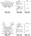
- Figs. 6A and Fig. 7A illustrate differences in light paths in two pulse oximetry light-detection geometries.
- Fig. 6A illustrates a light interrogation path for transmission-based pulse oximeter probe such as a finger-clip pulse oximeter configuration.
- Fig. 7A illustrates a light interrogation path in a remote setup such as a camera-based setup.
- the light source is denoted by Tx and the receiver or detector is denoted by Rx.
- Fig. 6A Conventional transmission-based probes, as shown in Fig. 6A can probe relatively deep vascular layers, typically > 1 mm, while in contactless, in particular camera-based PPG, the interrogation depth is much shallower, typically ⁇ 0.3 mm.
- Pulsatility dissipates as the cardiovascular pulse travels from the arteries to arterioles. It is typically assumed that volume pulsatility does not exist in capillaries any more. In the skin, arterioles rise from the deeper layer towards the surface where they branch in two capillaries. Hence, probing different depths potentially means probing a different vasculature.
- a transmission-based pulse oximeter having a light source -detector (Tx-Rx) geometry as shown in Fig. 6A has a much deeper interrogation depth as shown in the relative contribution to detected intensity I(z) shown in Fig. 6B .
- a remote or camera-based pulse oximeter having a source-detector (Tx-Rx) geometry as shown in Fig. 7A has a much shallower interrogation depth as indicated by the exponential decay shaped curve of the relative contribution to the detected intensity I(z) shown in Fig. 7B .
- the actual pulsatility profile of the probed tissue volume is typically not known.
- Fig. 6C and Fig. 7C show an assumed pulsatility P(z) versus skin penetration depth z.
- radiation emitted towards the tissue region may have different penetration depths at the first wavelength and as at the second wavelength, for example, at a red and an infrared wavelength.
- the penetration depth can also vary in time when optical and/or physiological properties of the tissue vary, for example due to venous pooling or vasoconstriction or -dilation. If pulsatility varies between vascular layers, the difference in penetration depth can disturb the SpO2 calibration and consequently the accuracy.
- the proposed solution can be particularly advantageous for remote SpO2 measurement using a camera, because due to its illumination-detection geometry, it may suffer from the invalid assumption that red and infrared wavelength, or any other combination of wavelengths used for deriving SpO2, would interrogate the same skin depths.
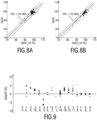
- the different penetration depth of the different wavelengths in the interrogated tissue might be a cause for the relatively larger spread in the camera-based method for SpO2 measurement shown in the diagram in Fig. 8A , as compared to the spread in the transmission-based finger probe measurement shown in Fig. 8B .
- the vertical axis denotes the SpO2 measured with a PPG camera in Fig. 8A and a finger probe in Fig. 8B .
- the horizontal axis denotes a reference SpO2 measurement.
- each dot indicates a median value over an entire SpO2(t) trace of several minutes.
- a remote measurement by for example an SpO2 camera may suffer more from the effects of shunt light if the different wavelengths probe different tissue depths.
- the DC levels and thus the AC/DC ratios may be differently affected by the optical shunting independently of the SpO2, which may lead to miscalibration and inaccuracy.
- Fig. 9 shows a graph of repeated blood oxygen saturation measurements for a number of volunteers repeated on different days which show a reproducibility of relative SpO2 deviation with respect to a group calibration.
- the vertical axis in Fig. 9 denotes a deviation ⁇ between a measured and a reference SpO2 value, ⁇ SpO2(%).
- the horizontal axis gives the different volunteers V labeled from 1 to H.
- the number of repeated measurements n is given below.
- some individuals repeatedly show an overestimation while others repeatedly show an underestimation. Such an individual bias suggests that pulsatile layers may indeed be interrogated to a larger extent by one wavelength than by the other.
- the results shown in Fig. 9 shows a graph of repeated blood oxygen saturation measurements for a number of volunteers repeated on different days which show a reproducibility of relative SpO2 deviation with respect to a group calibration.
- the vertical axis in Fig. 9 denotes a deviation ⁇ between a measured and a reference SpO2 value,
- specular reflected light from the tissue surface contributes to DC only, not to AC. Specularly reflected light is thus pure shunt light, i.e. light that did not travel into the skin before it was emitted towards the detector to be detected, which can distort the ratio of ratios on which the determination of the blood oxygen saturation is based.
- the solution according to an aspect of the present invention suggests to perform a blood oxygen saturation measurement using coherent - rather than conventionally used incoherent - light sources such as cheap diffuse laser diodes, at a wavelength appropriate for SpO2 monitoring and/or imaging, to allow (near-) a simultaneous acquisition of PPG signals and flow signals indicative of a flow of lights scattering particles within the tissue region of the subject.
- a flow signal can be obtained using, for example, laser Doppler and/or laser speckle techniques such as laser speckle imaging.
- the flow signals, for example laser speckle images, and the modulations therein can be used to correct the PPG signals at the first and at the second wavelength for different potential penetration depths at the different wavelength and the associated miscalibrations to allow for assessment with improved accuracy.
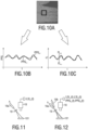
- Fig. 10A shows an illustration of data acquisition with a camera-based setup.
- a 'raw', laser speckle image can be created using the same first and second for example red and infrared, wavelength, as used in a conventional PPG measurement.
- a tissue region that is used for the measurement is indicated by the rectangle in Fig. 10A .
- a corresponding image is obtained at the first wavelength and at the second wavelength. It should be noted that further wavelengths can be used.
- detection data can be acquired over time.
- Fig. 10B shows exemplary PPG signals at the first wavelength and at the second wavelength, wherein each PPG signal is indicative of an absorption of light within the tissue region at the respective wavelength.
- the absorption-based PPG signal can be seen as a 'color-based', probing of pulsatility.
- the PPG signal PPG ⁇ 1 here obtained using red light, has a smaller PPG amplitude indicating a smaller absorption coefficient for HbO2 at ⁇ 1 compared to a higher absorption at a second wavelength, here using infrared light.
- the approach disclosed herein further suggests combining such a conventional PPG measurement with a flow measurement to obtain a first and a second flow signal indicative of a flow of light scattering particles within the tissue region of the subject at the first wavelength and at the second wavelength.
- a measurement can be performed as a 'color blind', measurement based on interferometric techniques such as laser speckle imaging or Doppler.
- laser speckle imaging is used to determine the flow.
- the flow signal is pulsatile due to pulsatile flow. For equal penetration depths, a modulation of the flow signal (ratio of AC/DC) would be the same both wavelengths.
- the proposed solution is thus based on a combination of a conventional 'color-based' PPG measurement and a 'color blind' flow measurement based on interference effects.
- an interferometric or a speckle-based technique such as LSI requires coherent (laser) light because it is an interferometric technique.
- the speckle pattern can be seen as an interferogram which blurs upon motion.
- the absorption-based PPG signal may use coherent light but can also work with incoherent light sources.
- the proposed device 10 and/or system 1 can be implemented by two or more separate physical entities. These entities can be wired or wirelessly connected and exchange data and/or information, for example using a data numeric interface, or Bluetooth, Wi-Fi and the like.
- the proposed device 10 and/or system 1 can be implemented by a flow measurement device and a pulse oximeter device. They can be provided as separate physical entities.
- the pulse oximeter device can comprise a first input for receiving the first detection data, said first detection data being data acquired over time by detecting radiation at a first wavelength ( ⁇ 1) and at a second wavelength ( ⁇ 2) received from said tissue region.
- the flow measurement device can comprise a second input for receiving the second detection data, said second detection data being data acquired over time by detecting radiation at the first wavelength and at the second wavelength received from said tissue region in response to coherent light at the first wavelength and coherent light at the second wavelength being emitted towards the tissue region.
- the input 11 can thus be implemented comprising said first input and said second input.
- the pulse oximeter device can comprise the PPG unit 12 for deriving, from said first detection data, a first PPG signal indicative of an absorption of light within the tissue region at the first wavelength, and a second PPG signal indicative of an absorption of light within the tissue region at the second wavelength.
- the flow measurement device can comprise the flow unit 13 for deriving, from said second detection data, a first flow signal indicative of a flow of light scattering particles within the tissue region at the first wavelength, and a second flow signal indicative of a flow of light scattering particles within the tissue region at the second wavelength.
- the flow measurement device may comprise a first coherent light source arranged to emit coherent light at a first wavelength and at a second wavelength towards a tissue region of the subject.
- the flow measurement device may comprise a detector for acquiring the second detection data.
- the pulse oximeter device may comprise a second light source arranged to emit coherent or non-coherent light at the first wavelength and at the second wavelength towards a tissue region of the subject.
- the pulse oximeter device may comprise a second detector for acquiring the first detection data. It shall be understood that advantageously a detector and/or a coherent light source can be shared by the flow measurement device and the pulse oximeter device.
- the processing unit 14 for correcting said PPG signals based on said flow signals and/or for providing a feedback signal based on a comparison of the first and second flow signals can at least in part be comprised in the flow measurement device, the pulse oximeter device and/or a further physical entity.
- the processing unit can also be distributed between, for example, the pulse oximeter device and the flow measurement device. For example, a ratio of ratios can be determined based on the flow signals in the flow measurement device as correction data for correcting said PPG signals based thereon.
- the pulse oximeter device can comprise an input for receiving said correction data and can further be configured to correct the PPG signals based thereon.
- the pulse oximeter device may not constantly have to be provided with correction data. It can be sufficient to provide such correction data for correcting said PPG signals based on said flow signals, for example, only at the beginning of a measurement, in time intervals or when it has been determined that the measurement conditions have changed. In such a case, it can be sufficient to perform the flow measurement only when needed for calibration, whereas the PPG measurement may be performed over an extended period of time.
- the pulse oximeter device can constantly monitor the subject, while the flow measurement device for measuring the flow signals at the first and at the second wavelength is only provided for calibration.
- such a flow measurement device may even be shared between multiple subjects.
- a ratio between modulation depths of the first and second flow signal at the first and at the second wavelength can be used to correct the ratio of ratios (RR) of the PPG signals at the first and at the second wavelength.
- the SpO2 measurement can be corrected for differences in penetration depth of the two wavelengths.
- Fig. 11 and Fig. 12 illustrate the differences between a conventional laser speckle imaging setup and an embodiment of the system for use in blood oxygen saturation as disclosed herein.
- the laser speckle imaging setup shown in Fig. 11 comprises a single laser source 16a for emitting coherent light at only one wavelength ⁇ 1 towards a tissue region 101 of a subject.
- An optional expander 19 can be provided for expanding the beam of the coherent light source 16a.
- the illuminated tissue region 101 is imaged by a camera, here a CCD camera, as a detector 18. Based on the output signal of the camera, a laser speckle imaging signal LSI ⁇ 1 (t) can be obtained.
- the system shown in Fig. 12 comprises a coherent light source 16 comprising a first laser source 16a and a second laser source 16b that illuminate the tissue region of the subject with coherent light at the first and at the second wavelength ⁇ 1, ⁇ 2.
- the detector 18 can again be a camera such as a CCD camera or a CMOS camera.
- a first laser speckle imaging signal LSI ⁇ 1 (t) as the first flow signal indicative of a flow of lights scattering particles within the tissue region of the subject at the first wavelength
- a second laser speckle imaging signal LSI ⁇ 2 (t) as the second flow signal indicative of a flow of light scattering particles within the tissue region of the subject at the second wavelength
- a second PPG signal PPG ⁇ 2 (t) indicative of an absorption of light within the tissue region of the subject at the second wavelength ⁇ 2 can be obtained.
- such a dual wavelength setup can provide PPG and LSI signals from which PPG and flow signal modulation depths can be derived for blood oxygen saturation measurement with improved accuracy.
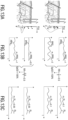
- Figs. 13A to 14B illustrate the scenario of blood oxygen saturation measurement with equal penetration depths.
- a PPG signal amplitude is proportional to PPG ⁇ ⁇ ⁇ a ⁇ ⁇ I ⁇ z P z dz , wherein ⁇ a ( ⁇ ) is the wavelength dependent absorption coefficient, I(z) denotes a relative contribution to detected intensity and P (z) denotes pulsatility versus skin depth z.
- ⁇ a ( ⁇ ) is the wavelength dependent absorption coefficient
- I(z) denotes a relative contribution to detected intensity
- P (z) denotes pulsatility versus skin depth z.
- an actual profile of the pulsatility as a function of depth (z), P (z) is generally unknown.
- the radiation at the first wavelength ⁇ 1 and at the second wavelength ⁇ 2 penetrate the tissue to the same extent. Hence, the assumption that the first and the second wavelength probe the same tissue depth is not violated.
- the different vascular pulsatility profile does not substantially affect the ratio of ratios of the PPG amplitudes m_PPG ⁇ 1 and m_PPG ⁇ 2 .
- the unknown pulsatility profile may not affect the SpO2 determination based on the PPG signals PPG ⁇ 1 and PPG ⁇ 2 as shown in Fig. 13B .
- the flow signals at the first and at the second wavelength FL ⁇ 1 and FL ⁇ 2 also show a same modulation depth (AC/DC) given by m_FL ⁇ 1 and m_FL ⁇ 2 , respectively.
- Fig. 14A shows a graph of the relative contribution to detected intensity I ⁇ (z) versus skin depth z.
- the curves I ⁇ 1 (z) and I ⁇ 2 (z) are approximately similar at the first wavelength and at the second wavelength.
- Fig. 14B the impact of three different pulsatility profiles P1, P2 and P3 as indicated by the dashed curve is shown. As can be seen, the ratio of ratios is not substantially affected and leads to the correct blood oxygen saturation value, here 92%.
- Fig. 15A to 16B the variation of pulsatility profile with depth can have significantly more impact for a reflectance-based or remote blood oxygen saturation measurement. Substantially different penetration depths for the first and the second wavelength may occur.
- the PPG amplitude at the first and at the second wavelength as given by the formula above is now not only affected by the blood absorption coefficient ⁇ a ( ⁇ ) but also depends on the wavelength-dependent relative penetration depths I ⁇ (z) and the actual pulsatility profile P(z).
- Penetration depths can depend on scattering and absorption. While infrared light scatters slightly less than red, it is not safe to conclude that infrared does typically penetrate slightly deeper than red. This is because HbO2 absorbs the light significantly better at infrared than at red, which has the opposite effect on penetration depth. And, for Hb this is slightly less true, compared to HbO2. Thus, a priori it is not clear which wavelength penetrates deeper. In fact, this can also change dynamically depending on venous pooling, and/or a changed saturation of the venous blood and/or opening and closing of shunts, e.g., due to centralization of a patient. Such physiological effects would impact both P(z) and I ⁇ (z).
- the skin when the skin regulates more blood towards the upper layer, it reduces the interrogation depth of both the red and infrared light, but extensively more for the infrared light, so disproportionally more than for the red light, due to its higher absorption coefficient.
- the infrared PPG signal amplitude may be reduced to a greater extent than the red PPG signal amplitude, which would then lead to an underestimation of SpO2.
- Fig. 15A illustrates the case where light at a first wavelength ⁇ 1 and at a second wavelength ⁇ 2 shows a different penetration depth as indicated by the vertical arrows. Furthermore, the upper and the lower graphs in Fig. 15A show a different vascular pulsatility profile P(z) in the insets. In consequence, as shown in Fig. 15B , the PPG signals at the first wavelength PPG ⁇ 1 and at the second wavelength PPG ⁇ 2 will experience a different modulation m_PPG ⁇ 1 and m_PPG ⁇ 2 . For example, the infrared light may penetrate less deep than red light.
- this error can be detected by looking at the differences in the flow signals acquired at the same wavelengths used for the PPG signals. For example speckle contrast modulation depths at the first and at the second wavelengths can be evaluated. If penetration depths of, for example, the red and infrared were equal, the infrared/red ratio of the speckle signal would be 1. However, if the probed pulsatile tissue layers differ, the flow signals at the first wavelength FL ⁇ 1 and at the second wavelength FL ⁇ 2 will experience a different modulation m_FL ⁇ 1 and m_FL ⁇ 2 , as shown in Fig. 15C . In the given example, the LSI modulation depths indicate that red indeed sees more pulsatile layers than infrared. Hence, a modulation of the first flow signal at the first, red wavelength m_FL ⁇ 1 is higher than a modulation of the second flow signal at the second, infrared wavelength m_FL ⁇ 2 .
- Fig. 16A shows a relative contribution to the detected intensity I ⁇ (z) versus skin depth z at the first wavelength ⁇ 1 and at the second wavelength ⁇ 2.
- the contribution can be approximated by a wavelength-dependent exponential decay function.
- red and infrared light may experience different penetration depths in a remote, camera-based setup, which can have non-linear consequences for the measured PPG signal at both wavelengths and their ratio, depending on the pulsatility profile.
- Fig. 16B this is exemplarily indicated for three different pulsatility profiles P1, P2, and P3 leading to an erroneous ratio of ratios of the first PPG signal and the second PPG signal on which the SpO2 estimation is based. In the shown example, this leads to an overestimation of SpO2.
- An important factor can be attributed to the gradually decreasing pulsatility as one comes closer to the surface or as arterioles come from beneath and branch into capillaries and thereby show reduced pulsatility.
- the relative PPG amplitudes can be described as vector (5, 3, 2), or, as a normalized vector (0.81, 0.49, 0.32).
- the scaling of a PPG signal with the flow signal at the respective wavelengths can take place before or after normalization.
- an absolute modulation depth or a relative, i.e., normalized, modulation depth of the flow signal can be used.
- an absolute modulation depth may increase due to an increased pulsatile component or a reduced DC component, for example due to a steady, non-pulsatile flow.
- the modulation depths would change for both the wavelengths to the same degree and the correction of the PPG amplitudes by these different flow signals would still be correct since the same ratio applies. Hence, such a difference would not corrupt the proposed correction.
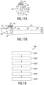
- FIG. 17A shows a cross-sectional view through a finger 103 of a subject similar to the embodiment shown in Fig. 3 .
- the finger 103 further comprises a finger nail 105 and bones 106.
- the coherent light source 16 comprises a first laser diode 16a arranged to emit coherent light at a first wavelength through the finger 103 of the subject and a second laser diode 16b arranged to emit coherent light at a second wavelength through the finger 103 of the subject.
- a detector 18 acquires detection data over time by detecting radiation at the first and at the second wavelength received from said tissue region in response to the coherent light at the first wavelength and coherent light at the second wavelength being emitted towards the tissue region.
- a different volume comprising different pulsatile tissue layers may be probed at the different wavelengths.
- a first volume 108 may be probed at the first wavelength ⁇ 1 and a second volume 109 may be probed at the second wavelength ⁇ 2. With the solution disclosed herein, this can be advantageously corrected.
- red may be absorbed less than infrared, e.g. because most of the blood is well saturated.
- the probing volume 108 for red is much wider than the probing volume 109 for infrared. Only those infrared photons that travel in an almost straight line from source to detector have a chance of reaching the detector. Red photons have more freedom to scatter around in the pulsatile volume before arriving at the source since the attenuation is much weaker.
- another example could also be construed in which the difference between wavelengths is turned around, i.e., infrared may be absorbed less than red.
- a bone 106 causes an obstruction in the light paths such that light has to go around.
- infrared will be perturbed more by this obstruction than red.
- the different probing volumes cause slightly different modulation depths that are unrelated to the oxygen saturation of the blood in the pulsatile vessels.
- a correction based on flow signals can therefore also help to improve the reliability in transmission-based measurements.
- a single photodiode can be as the detector 18.
- the first and second flow signals can be determined based on a laser Doppler technique.
- both the flow and PPG signals can result in signals averaged over a region of interest.
- the signals can essentially be condensed down to point measurements without spatial x or y information, only having temporal variation, similar to a signal derived using a single (non-image) detector.
- the principle of scaling PPG signals with flow signals at the respective wavelengths can thus also be applied for non-image signals.
- a flow chart of a method 200 according to an aspect of the present invention is illustrated in Fig. 18 .
- first detection data of a tissue region of a subject are obtained (i.e., received or retrieved).
- second detection data of the tissue region of the subject is obtained (i.e., received or retrieved).
- the second detection data can be obtained and used as the first detection data.
- the same detection data can be used.
- a first PPG signal and a second PPG signal are derived from said first detection data.
- a fourth step S204 a first flow signal and a second flow signal are derived from said second detection data.
- step S204 may also be carried out before or simultaneously to the third step S203.
- said PPG signals can be corrected based on said flow signals and/or a feedback signal can be provided based on a comparison of the first and the second flow signals.
- a blood oxygen saturation value of the subject can be determined based on the corrected PPG signals.
- a computer program may be stored/distributed on a suitable medium, such as an optical storage medium or a solid-state medium supplied together with or as part of other hardware, but may also be distributed in other forms, such as via the Internet or other wired or wireless telecommunication systems.
- a suitable medium such as an optical storage medium or a solid-state medium supplied together with or as part of other hardware, but may also be distributed in other forms, such as via the Internet or other wired or wireless telecommunication systems.
Landscapes
- Health & Medical Sciences (AREA)
- Life Sciences & Earth Sciences (AREA)
- Physics & Mathematics (AREA)
- Engineering & Computer Science (AREA)
- Public Health (AREA)
- Surgery (AREA)
- Veterinary Medicine (AREA)
- General Health & Medical Sciences (AREA)
- Animal Behavior & Ethology (AREA)
- Biophysics (AREA)
- Pathology (AREA)
- Biomedical Technology (AREA)
- Heart & Thoracic Surgery (AREA)
- Medical Informatics (AREA)
- Molecular Biology (AREA)
- Physiology (AREA)
- Optics & Photonics (AREA)
- Psychiatry (AREA)
- Computer Vision & Pattern Recognition (AREA)
- Artificial Intelligence (AREA)
- Signal Processing (AREA)
- Hematology (AREA)
- Cardiology (AREA)
- Spectroscopy & Molecular Physics (AREA)
- General Physics & Mathematics (AREA)
- Measurement Of The Respiration, Hearing Ability, Form, And Blood Characteristics Of Living Organisms (AREA)
- Measuring Pulse, Heart Rate, Blood Pressure Or Blood Flow (AREA)
Claims (13)
- System (1) zur Verwendung bei der Messung der Blutsauerstoffsättigung eines Subjekts (100), wobei das System umfasst:- eine kohärente Lichtquelle (16), welche dazu angeordnet ist, kohärentes Licht bei einer ersten Wellenlänge und einer zweiten Wellenlänge in Richtung eines Gewebebereichs (101) des Subjekts zu emittieren;- einen Detektor (18) zum Erfassen erster und zweiter Detektionsdaten des Gewebebereichs des Subjekts,
wobei die ersten Detektionsdaten im Laufe der Zeit durch Erfassen von Strahlung bei der ersten Wellenlänge (λ1) und bei der zweiten Wellenlänge (λ2) erfasst werden, welche von dem Gewebebereich empfangen werden; wobei die zweiten Detektionsdaten im Laufe der Zeit durch Erfassen von Strahlung bei der ersten Wellenlänge und bei der zweiten Wellenlänge, welche von dem Gewebebereich empfangen werden, als Reaktion darauf erfasst werden, dass das kohärente Licht bei der ersten Wellenlänge und das kohärente Licht bei der zweiten Wellenlänge in Richtung des Gewebebereichs emittiert werden; und- eine Verarbeitungsvorrichtung zum Verarbeiten der ersten und zweiten Detektionsdaten des Gewebebereichs des Subjekts, wobei die Verarbeitungsvorrichtung umfasst:- einen Eingang (11) zum Empfangen der ersten und zweiten Detektionsdaten;- eine PPG-Einheit (12) zum Ableiten eines ersten PPG-Signals, welches eine Absorption von Licht innerhalb des Gewebebereichs bei der ersten Wellenlänge angibt, und eines zweiten PPG-Signals, welches eine Absorption von Licht innerhalb des Gewebebereichs bei der zweiten Wellenlänge angibt, aus den ersten Detektionsdaten;- eine Strömungseinheit (13) zum Ableiten eines ersten Strömungssignals, welches eine Strömung von lichtstreuenden Partikeln angibt, welche bei der ersten Wellenlänge innerhalb des Gewebebereichs untersucht wurden, aus den zweiten Detektionsdaten, und eines zweiten Strömungssignals, welches eine Strömung von lichtstreuenden Partikeln angibt, welche bei der zweiten Wellenlänge innerhalb des Gewebebereichs untersucht wurden; und- eine Verarbeitungseinheit (14) zum Korrigieren der PPG-Signale basierend auf den Strömungssignalen und/oder zum Bereitstellen eines Rückmeldungssignals basierend auf einem Vergleich des ersten und zweiten Strömungssignals. - System nach Anspruch 1, wobei die Verarbeitungseinheit (14) dazu konfiguriert ist, die PPG-Signale basierend auf den Strömungssignalen zu korrigieren und eine Ausgabe bereitzustellen, welche basierend auf den korrigierten PPG-Signalen eine Blutsauerstoffsättigung des Subjekts angibt.
- System nach Anspruch 1 oder 2, wobei die Verarbeitungseinheit (14) dazu konfiguriert ist, das Rückmeldungssignal basierend auf den Strömungssignalen bereitzustellen, wobei das Rückmeldungssignal eine Qualität der PPG-Signale angibt und aus einer Nichtübereinstimmung zwischen dem ersten Strömungssignal bei der ersten Wellenlänge und dem zweiten Strömungssignal bei der zweiten Wellenlänge abgeleitet wird.
- System nach einem der vorstehenden Ansprüche, wobei die PPG-Einheit (12) dazu konfiguriert ist, das erste und das zweite PPG-Signal basierend auf einem Durchschnitt der erfassten Strahlung bei der ersten Wellenlänge und bei der zweiten Wellenlänge zu bestimmen.
- System nach einem der vorstehenden Ansprüche, wobei die Strömungseinheit (13) dazu konfiguriert ist, das erste und zweite Strömungssignal basierend auf einer Laser-Doppler- und/oder Laser-Speckle-Technik abzuleiten.
- System nach einem der vorstehenden Ansprüche, wobei die Strömungseinheit (13) dazu konfiguriert ist, das erste und das zweite Strömungssignal basierend auf einer Standardabweichung der Detektionsdaten der erfassten Strahlung bei der ersten Wellenlänge und bei der zweiten Wellenlänge zu bestimmen.
- System nach einem der vorstehenden Ansprüche, wobei die Strömungseinheit (13) dazu konfiguriert ist, das erste und das zweite Strömungssignal basierend auf einem Speckle-Kontrast bei der ersten Wellenlänge und der zweiten Wellenlänge zu bestimmen.
- System nach einem der vorstehenden Ansprüche, wobei die Verarbeitungseinheit (14) dazu konfiguriert ist, das erste und das zweite PPG-Signal basierend auf einer zeitlichen Modulation des ersten und des zweiten Strömungssignals bei der ersten Wellenlänge und der zweiten Wellenlänge zu korrigieren.
- System nach einem der vorstehenden Ansprüche, wobei die Verarbeitungseinheit (14) dazu konfiguriert ist, die PPG-Signale durch Skalieren von Amplituden des ersten und des zweiten PPG-Signals bei der ersten Wellenlänge und der zweiten Wellenlänge basierend auf Amplituden des ersten und des zweiten Strömungssignals bei der ersten Wellenlänge und der zweiten Wellenlänge zu korrigieren.
- System nach einem der vorstehenden Ansprüche, wobei die Verarbeitungseinheit (14) dazu konfiguriert ist, ein Verhältnis von Verhältnissen der PPG-Signale zu bestimmen und das Verhältnis von Verhältnissen der PPG-Signale basierend auf einem Verhältnis von Verhältnissen der Strömungssignale zu korrigieren.
- System nach einem der vorstehenden Ansprüche, angeordnet als ein Remote-PPG-System, insbesondere eine Kamera als Detektor (18) umfassend.
- Verfahren (200) zur Verwendung bei der Messung der Blutsauerstoffsättigung eines Subjekts (100) mit einem System nach einem der vorstehenden Ansprüche, wobei das Verfahren folgende Schritte umfasst:- Empfangen erster und zweiter Detektionsdaten eines Gewebebereichs (101) des Subjekts, wobei die ersten Detektionsdaten Daten sind, welche im Laufe der Zeit durch Erfassen von Strahlung bei einer ersten Wellenlänge (λ1) und bei einer zweiten Wellenlänge (λ2) erfasst werden, welche von dem Gewebebereich empfangen werden; wobei die zweiten Detektionsdaten Daten sind, welche im Laufe der Zeit durch Erfassen von Strahlung bei der ersten Wellenlänge und bei der zweiten Wellenlänge, welche von dem Gewebebereich empfangen werden, als Reaktion darauf erfasst werden, dass kohärentes Licht bei der ersten Wellenlänge und kohärentes Licht bei der zweiten Wellenlänge in Richtung des Gewebebereichs emittiert werden (S201, S202);- Ableiten eines ersten PPG-Signals, welches eine Absorption von Licht innerhalb des Gewebebereichs bei der ersten Wellenlänge angibt, und eines zweiten PPG-Signals, welches eine Absorption von Licht innerhalb des Gewebebereichs bei der zweiten Wellenlänge angibt, aus den ersten Detektionsdaten (S203);- Ableiten eines ersten Strömungssignals, welches eine Strömung von lichtstreuenden Partikeln angibt, welche bei der ersten Wellenlänge innerhalb des Gewebebereichs untersucht wurden, aus den zweiten Detektionsdaten, und eines zweiten Strömungssignals, welches eine Strömung von lichtstreuenden Partikeln angibt, welche bei der zweiten Wellenlänge innerhalb des Gewebebereichs untersucht wurden (S204); und- Korrigieren der PPG-Signale basierend auf den Strömungssignalen und/oder Bereitstellen eines Rückmeldungssignals basierend auf einem Vergleich des ersten und zweiten Strömungssignals (S205).
- Computerprogramm, umfassend Programmcodemittel zum Veranlassen eines Systems nach einem der Ansprüche 1-11, die Schritte des Verfahrens (200) nach Anspruch 12 auszuführen, wenn das Computerprogramm auf einem System ausgeführt wird.
Applications Claiming Priority (3)
| Application Number | Priority Date | Filing Date | Title |
|---|---|---|---|
| US201662372376P | 2016-08-09 | 2016-08-09 | |
| EP16183404 | 2016-08-09 | ||
| PCT/EP2017/069883 WO2018029123A1 (en) | 2016-08-09 | 2017-08-07 | Device for use in blood oxygen saturation measurement |
Publications (2)
| Publication Number | Publication Date |
|---|---|
| EP3496603A1 EP3496603A1 (de) | 2019-06-19 |
| EP3496603B1 true EP3496603B1 (de) | 2025-04-02 |
Family
ID=59523151
Family Applications (1)
| Application Number | Title | Priority Date | Filing Date |
|---|---|---|---|
| EP17748785.7A Active EP3496603B1 (de) | 2016-08-09 | 2017-08-07 | Vorrichtung zur verwendung bei der blutsauerstoffsättigungsmessung |
Country Status (4)
| Country | Link |
|---|---|
| US (1) | US11202582B2 (de) |
| EP (1) | EP3496603B1 (de) |
| JP (1) | JP7061996B6 (de) |
| CN (1) | CN109561842B (de) |
Families Citing this family (75)
| Publication number | Priority date | Publication date | Assignee | Title |
|---|---|---|---|---|
| CN108135591B (zh) | 2016-02-24 | 2021-05-28 | 禾木(中国)生物工程有限公司 | 柔性增强的神经血管导管 |
| CN109561843B (zh) * | 2016-08-09 | 2022-07-15 | 皇家飞利浦有限公司 | 用于监测对象的外围动脉灌注的设备、系统和方法 |
| JP7264581B2 (ja) | 2017-01-06 | 2023-04-25 | インセプト、リミテッド、ライアビリティ、カンパニー | 動脈瘤治療装置向けの抗血栓性コーティング |
| US10813597B2 (en) * | 2017-04-14 | 2020-10-27 | The Regents Of The University Of California | Non-invasive hemodynamic assessment via interrogation of biological tissue using a coherent light source |
| GB2572626B (en) * | 2018-04-05 | 2021-04-07 | Life Meter Srl | Pulse oximetry device, system and method |
| US11395665B2 (en) | 2018-05-01 | 2022-07-26 | Incept, Llc | Devices and methods for removing obstructive material, from an intravascular site |
| CN119405388A (zh) | 2018-05-01 | 2025-02-11 | 因赛普特有限责任公司 | 用于从血管内部位去除闭塞性物质的装置和方法 |
| US12059239B2 (en) | 2018-05-08 | 2024-08-13 | Know Labs, Inc. | Electromagnetic shielding in non-invasive analyte sensors |
| US11903689B2 (en) | 2019-12-20 | 2024-02-20 | Know Labs, Inc. | Non-invasive analyte sensor device |
| JP7229676B2 (ja) * | 2018-05-21 | 2023-02-28 | 株式会社日立製作所 | 生体情報検出装置および生体情報検出方法 |
| US11471582B2 (en) | 2018-07-06 | 2022-10-18 | Incept, Llc | Vacuum transfer tool for extendable catheter |
| US11517335B2 (en) | 2018-07-06 | 2022-12-06 | Incept, Llc | Sealed neurovascular extendable catheter |
| US11766539B2 (en) | 2019-03-29 | 2023-09-26 | Incept, Llc | Enhanced flexibility neurovascular catheter |
| US11857298B1 (en) | 2019-09-06 | 2024-01-02 | Apple Inc. | Devices having matter differentiation detectors |
| US11717197B2 (en) | 2019-09-27 | 2023-08-08 | Apple Inc. | Physiological monitoring system for measuring oxygen saturation |
| EP4044906B1 (de) | 2019-10-15 | 2025-03-12 | Kandu Health, Inc. | Systeme und verfahren zur mehrgrössenhubdetektion |
| CN111093487B (zh) * | 2019-11-29 | 2023-02-17 | 深圳市汇顶科技股份有限公司 | 自适应的生物特征检测方法及装置、电子设备 |
| US11457936B2 (en) | 2019-12-18 | 2022-10-04 | Imperative Care, Inc. | Catheter system for treating thromboembolic disease |
| JP2023507553A (ja) | 2019-12-18 | 2023-02-24 | インパラティブ、ケア、インク. | 静脈血栓塞栓症を治療するための方法及びシステム |
| US11633272B2 (en) | 2019-12-18 | 2023-04-25 | Imperative Care, Inc. | Manually rotatable thrombus engagement tool |
| EP3838144A1 (de) * | 2019-12-19 | 2021-06-23 | Koninklijke Philips N.V. | Vorrichtung, system und verfahren zur bestimmung der sauerstoffsättigung einer person |
| US12629045B2 (en) | 2019-12-20 | 2026-05-19 | Know Labs, Inc. | Non-invasive analyte sensor device |
| IL294553A (en) | 2020-01-08 | 2022-09-01 | Activ Surgical Inc | Assessment of power feedback of laser marking |
| IL295510B2 (en) * | 2020-02-14 | 2025-08-01 | Activ Surgical Inc | Systems and methods for processing laser speckle signals |
| US12089927B2 (en) | 2020-02-20 | 2024-09-17 | Know Labs, Inc. | Non-invasive analyte sensing and notification system with decoupled and inefficient transmit and receive antennas |
| US12023151B2 (en) | 2020-02-20 | 2024-07-02 | Know Labs, Inc. | Non-invasive analyte sensing and notification system with decoupled transmit and receive antennas |
| US11832926B2 (en) | 2020-02-20 | 2023-12-05 | Know Labs, Inc. | Non-invasive detection of an analyte and notification of results |
| US11573351B2 (en) | 2020-03-06 | 2023-02-07 | Apple, Inc. | Optical sensor having a magnetic optical barrier |
| WO2021183444A1 (en) | 2020-03-10 | 2021-09-16 | Imperative Care, Inc. | Enhanced flexibility neurovascular catheter |
| US12092589B1 (en) | 2020-04-03 | 2024-09-17 | Know Labs, Inc. | In vitro analyte sensor using one or more detector arrays that operate in radio/microwave frequency bands |
| US11819318B2 (en) * | 2020-04-27 | 2023-11-21 | Open Water Internet Inc. | Optical imaging from light coherence |
| US11207497B1 (en) | 2020-08-11 | 2021-12-28 | Imperative Care, Inc. | Catheter with enhanced tensile strength |
| US11764488B2 (en) | 2020-09-09 | 2023-09-19 | Know Labs, Inc. | Methods for determining variability of a state of a medium |
| US12019034B2 (en) | 2020-09-09 | 2024-06-25 | Know Labs, Inc. | In vitro sensing methods for analyzing in vitro flowing fluids |
| US11689274B2 (en) | 2020-09-09 | 2023-06-27 | Know Labs, Inc. | Systems for determining variability in a state of a medium |
| US11510597B2 (en) | 2020-09-09 | 2022-11-29 | Know Labs, Inc. | Non-invasive analyte sensor and automated response system |
| US12007338B2 (en) | 2020-09-09 | 2024-06-11 | Know Labs Inc. | In vitro sensor for analyzing in vitro flowing fluids |
| US12204289B1 (en) | 2020-09-11 | 2025-01-21 | Apple Inc. | Device removal indication using different object proximity thresholds |
| US11723563B1 (en) | 2020-09-11 | 2023-08-15 | Apple Inc. | Correcting for emitted light wavelength variation in blood-oxygen saturation measurements at wearable electronic device |
| US12074244B2 (en) | 2020-09-14 | 2024-08-27 | Apple Inc. | Optical sensor package with magnetic component for device attachment |
| US12164027B1 (en) | 2020-09-14 | 2024-12-10 | Apple Inc. | Multi-pathway distance measurements for optical sensors |
| EP4014853A1 (de) * | 2020-12-17 | 2022-06-22 | Koninklijke Philips N.V. | Vorrichtung, verfahren und systeme zur abbildung eines oder mehrerer aspekte der blutperfusion |
| CN112932475B (zh) * | 2021-02-01 | 2023-02-21 | 武汉泰利美信医疗科技有限公司 | 血氧饱和度的计算方法、装置、电子设备及存储介质 |
| US11033208B1 (en) | 2021-02-05 | 2021-06-15 | Know Labs, Inc. | Fixed operation time frequency sweeps for an analyte sensor |
| US12076141B2 (en) | 2021-02-26 | 2024-09-03 | Covidien Lp | System and method for digitally calibrating a medical sensor |
| CN113598761B (zh) * | 2021-05-10 | 2024-10-08 | 浙江大学台州研究院 | 一种基于ccd的双波长红外血氧检测系统 |
| US12629018B2 (en) | 2021-08-08 | 2026-05-19 | The General Hospital Corporation | Using the dynamic forward scattering signal for optical coherence tomography based flow quantification |
| US11864921B2 (en) | 2021-08-09 | 2024-01-09 | JMad Creations, LLC | Melanin-bias reducing pulse oximeter and patient monitoring systems and devices |
| US12558175B2 (en) | 2021-08-12 | 2026-02-24 | Imperative Care, Inc. | Multi catheter method of performing a robotic neurovascular procedure |
| US12201396B2 (en) | 2021-09-10 | 2025-01-21 | Rockley Photonics Limited | Optical speckle receiver |
| US12458295B2 (en) | 2021-10-01 | 2025-11-04 | Covidien Lp | System and method for correcting for errors dependent upon spectral characteristics of tissue |
| US20230110673A1 (en) | 2021-10-12 | 2023-04-13 | Covidien Lp | Light emitting diode temperature estimation |
| CN118302109A (zh) * | 2021-10-13 | 2024-07-05 | 欧拉健康公司 | 用于测量血氧水平的技术 |
| USD1077996S1 (en) | 2021-10-18 | 2025-06-03 | Imperative Care, Inc. | Inline fluid filter |
| US12259281B2 (en) | 2021-11-10 | 2025-03-25 | Know Labs, Inc. | Non-invasive analyte sensor with superheterodyne circuit |
| US12146841B2 (en) | 2021-11-10 | 2024-11-19 | Know Labs, Inc. | Non-invasive analyte sensor with temperature compensation |
| US12390117B2 (en) | 2021-11-16 | 2025-08-19 | Rockley Photonics Limited | Optical sensor module for speckleplethysmography (SPG) and photoplethysmography (PPG) |
| US12603427B2 (en) | 2022-01-26 | 2026-04-14 | Know Labs, Inc. | Shape changing antenna and method for use thereof |
| US20230355140A1 (en) | 2022-05-05 | 2023-11-09 | Know Labs, Inc. | High performance glucose sensor |
| CN119300753A (zh) * | 2022-06-10 | 2025-01-10 | 洛克利光子有限公司 | 用于校准基于散斑的传感器的系统和方法 |
| US11802843B1 (en) | 2022-07-15 | 2023-10-31 | Know Labs, Inc. | Systems and methods for analyte sensing with reduced signal inaccuracy |
| US12318203B2 (en) | 2022-08-15 | 2025-06-03 | Know Labs, Inc. | Vehicle interface systems and methods for analyte-based access control |
| US12033451B2 (en) | 2022-08-15 | 2024-07-09 | Know Labs, Inc. | Systems and methods for analyte-based access controls |
| US20240358292A1 (en) * | 2022-08-23 | 2024-10-31 | Samsung Electronics Co., Ltd. | Polarized photoplethysmography (ppg) with improved perfusion signal for advanced biosensing |
| US20240090788A1 (en) * | 2022-09-16 | 2024-03-21 | Guy P. Curtis | System and method for using blood flow measurements to determine ventricular contractility |
| US12318182B2 (en) | 2022-10-03 | 2025-06-03 | Know Labs, Inc. | Analyte sensors with antenna array |
| US11696698B1 (en) | 2022-10-03 | 2023-07-11 | Know Labs, Inc. | Analyte sensors with position adjustable transmit and/or receive components |
| US12193810B2 (en) | 2023-03-21 | 2025-01-14 | Know Labs, Inc. | System and method for performing surgery with real-time health parameter monitoring |
| US11903701B1 (en) | 2023-03-22 | 2024-02-20 | Know Labs, Inc. | Enhanced SPO2 measuring device |
| US12170145B2 (en) | 2023-03-22 | 2024-12-17 | Know Labs, Inc. | System and method for software and hardware activation based on real-time health parameters |
| USD1099332S1 (en) | 2023-06-06 | 2025-10-21 | Know Labs, Inc. | Non-invasive analyte sensor |
| US12685449B2 (en) | 2023-07-21 | 2026-07-21 | Rockley Photonics Limited | System and method for measuring blood pressure |
| EP4783967A1 (de) * | 2023-09-28 | 2026-08-05 | Sonion Nederland B.V. | System und verfahren zur bestimmung eines physiologischen parameters eines körpers mit blutperfundiertem gewebe |
| US12484796B1 (en) | 2024-11-27 | 2025-12-02 | Rockley Photonics Limited | System and method for measuring pulse wave velocity |
| US12396648B1 (en) | 2024-11-27 | 2025-08-26 | Rockley Photonics Limited | Wearable device with light source and optical sensor |
Family Cites Families (12)
| Publication number | Priority date | Publication date | Assignee | Title |
|---|---|---|---|---|
| US20040127800A1 (en) | 1995-07-06 | 2004-07-01 | Kimball Victor E. | Device for assessing perfusion failure in a patient by measurement of blood flow |
| AUPO676397A0 (en) | 1997-05-13 | 1997-06-05 | Dunlop, Colin | Method and apparatus for monitoring haemodynamic function |
| US6763256B2 (en) * | 2002-08-16 | 2004-07-13 | Optical Sensors, Inc. | Pulse oximeter |
| US7254429B2 (en) | 2004-08-11 | 2007-08-07 | Glucolight Corporation | Method and apparatus for monitoring glucose levels in a biological tissue |
| EP2079363B1 (de) * | 2006-10-30 | 2020-06-10 | Elfi-Tech Ltd | Verfahren zur in-vivo-messung von biologischen parametern |
| CN101808570B (zh) | 2007-09-27 | 2013-08-28 | 皇家飞利浦电子股份有限公司 | 血氧计 |
| US20110082355A1 (en) * | 2009-07-30 | 2011-04-07 | Oxitone Medical Ltd. | Photoplethysmography device and method |
| US8868149B2 (en) | 2009-07-30 | 2014-10-21 | Oxitone Medical Ltd. | Photoplethysmography device and method |
| US20120130215A1 (en) * | 2010-05-05 | 2012-05-24 | Ilya Fine | Optical measurement of parameters related to motion of light-scattering particles within a fluid by manipulating analog electrical signals |
| GB2494622A (en) * | 2011-08-30 | 2013-03-20 | Oxitone Medical Ltd | Wearable pulse oximetry device |
| EP2914163A1 (de) | 2012-11-02 | 2015-09-09 | Koninklijke Philips N.V. | Vorrichtung und verfahren zum extrahieren physiologischer daten |
| WO2016015999A1 (en) | 2014-07-30 | 2016-02-04 | Koninklijke Philips N.V. | Hemoglobin detection and photoplethysmography using spectral modulation |
-
2017
- 2017-08-07 CN CN201780048716.0A patent/CN109561842B/zh active Active
- 2017-08-07 JP JP2019507092A patent/JP7061996B6/ja active Active
- 2017-08-07 EP EP17748785.7A patent/EP3496603B1/de active Active
- 2017-08-07 US US16/323,560 patent/US11202582B2/en active Active
Also Published As
| Publication number | Publication date |
|---|---|
| CN109561842B (zh) | 2022-07-12 |
| JP2019527598A (ja) | 2019-10-03 |
| JP7061996B2 (ja) | 2022-05-02 |
| US11202582B2 (en) | 2021-12-21 |
| CN109561842A (zh) | 2019-04-02 |
| EP3496603A1 (de) | 2019-06-19 |
| JP7061996B6 (ja) | 2022-06-06 |
| US20190167124A1 (en) | 2019-06-06 |
Similar Documents
| Publication | Publication Date | Title |
|---|---|---|
| US11202582B2 (en) | Device for use in blood oxygen saturation measurement | |
| WO2018029123A1 (en) | Device for use in blood oxygen saturation measurement | |
| EP3496604B1 (de) | Vorrichtung und system zur überwachung der peripheren arteriellen perfusion einer person | |
| EP3383258B1 (de) | Vorrichtung, system und verfahren zur bestimmung der vitalzeicheninformationen eines patienten | |
| JP6336141B2 (ja) | 対象の血液内の物質の濃度を決定する装置、システム及び方法 | |
| US8123695B2 (en) | Method and apparatus for detection of venous pulsation | |
| EP3019078B1 (de) | System zum prüfen des zustands der oxygenierung eines patienten | |
| JP6615176B2 (ja) | 非干渉的な皮膚組織ハイドレーション測定デバイス及び関連方法 | |
| US8109882B2 (en) | System and method for venous pulsation detection using near infrared wavelengths | |
| EP3479754A1 (de) | Vorrichtung, system und verfahren zur bestimmung von mindestens einem vitalzeichen einer person | |
| US20190200871A1 (en) | System and method for vital signs detection | |
| US20130131475A1 (en) | Photoplethysmography device and method | |
| US20220265150A1 (en) | Device, system and method for determining physiological information | |
| US11712185B2 (en) | Device, system and method for extracting physiological information indicative of at least one vital sign of a subject | |
| EP4037567B1 (de) | Verfahren und system zur bewertung der qualität des verhältnisses von verhältniswerten | |
| Segman et al. | Oximeter behavior while using a tourniquet |
Legal Events
| Date | Code | Title | Description |
|---|---|---|---|
| STAA | Information on the status of an ep patent application or granted ep patent |
Free format text: STATUS: UNKNOWN |
|
| STAA | Information on the status of an ep patent application or granted ep patent |
Free format text: STATUS: THE INTERNATIONAL PUBLICATION HAS BEEN MADE |
|
| PUAI | Public reference made under article 153(3) epc to a published international application that has entered the european phase |
Free format text: ORIGINAL CODE: 0009012 |
|
| STAA | Information on the status of an ep patent application or granted ep patent |
Free format text: STATUS: REQUEST FOR EXAMINATION WAS MADE |
|
| 17P | Request for examination filed |
Effective date: 20190311 |
|
| AK | Designated contracting states |
Kind code of ref document: A1 Designated state(s): AL AT BE BG CH CY CZ DE DK EE ES FI FR GB GR HR HU IE IS IT LI LT LU LV MC MK MT NL NO PL PT RO RS SE SI SK SM TR |
|
| AX | Request for extension of the european patent |
Extension state: BA ME |
|
| DAV | Request for validation of the european patent (deleted) | ||
| DAX | Request for extension of the european patent (deleted) | ||
| RAP1 | Party data changed (applicant data changed or rights of an application transferred) |
Owner name: KONINKLIJKE PHILIPS N.V. |
|
| STAA | Information on the status of an ep patent application or granted ep patent |
Free format text: STATUS: EXAMINATION IS IN PROGRESS |
|
| 17Q | First examination report despatched |
Effective date: 20220811 |
|
| GRAP | Despatch of communication of intention to grant a patent |
Free format text: ORIGINAL CODE: EPIDOSNIGR1 |
|
| STAA | Information on the status of an ep patent application or granted ep patent |
Free format text: STATUS: GRANT OF PATENT IS INTENDED |
|
| INTG | Intention to grant announced |
Effective date: 20241031 |
|
| RIN1 | Information on inventor provided before grant (corrected) |
Inventor name: CIUHU, CALINA Inventor name: BEZEMER, RICK Inventor name: PRESURA, CRISTIAN NICOLAE Inventor name: ORDELMAN, SIMONE CORNELIA MARIA ANNA Inventor name: VERKRUIJSSE, WILLEM |
|
| GRAS | Grant fee paid |
Free format text: ORIGINAL CODE: EPIDOSNIGR3 |
|
| GRAA | (expected) grant |
Free format text: ORIGINAL CODE: 0009210 |
|
| STAA | Information on the status of an ep patent application or granted ep patent |
Free format text: STATUS: THE PATENT HAS BEEN GRANTED |
|
| AK | Designated contracting states |
Kind code of ref document: B1 Designated state(s): AL AT BE BG CH CY CZ DE DK EE ES FI FR GB GR HR HU IE IS IT LI LT LU LV MC MK MT NL NO PL PT RO RS SE SI SK SM TR |
|
| REG | Reference to a national code |
Ref country code: GB Ref legal event code: FG4D |
|
| REG | Reference to a national code |
Ref country code: CH Ref legal event code: EP |
|
| REG | Reference to a national code |
Ref country code: DE Ref legal event code: R096 Ref document number: 602017088683 Country of ref document: DE |
|
| REG | Reference to a national code |
Ref country code: IE Ref legal event code: FG4D |
|
| REG | Reference to a national code |
Ref country code: NL Ref legal event code: MP Effective date: 20250402 |
|
| PG25 | Lapsed in a contracting state [announced via postgrant information from national office to epo] |
Ref country code: NL Free format text: LAPSE BECAUSE OF FAILURE TO SUBMIT A TRANSLATION OF THE DESCRIPTION OR TO PAY THE FEE WITHIN THE PRESCRIBED TIME-LIMIT Effective date: 20250402 |
|
| REG | Reference to a national code |
Ref country code: AT Ref legal event code: MK05 Ref document number: 1780461 Country of ref document: AT Kind code of ref document: T Effective date: 20250402 |
|
| PG25 | Lapsed in a contracting state [announced via postgrant information from national office to epo] |
Ref country code: PT Free format text: LAPSE BECAUSE OF FAILURE TO SUBMIT A TRANSLATION OF THE DESCRIPTION OR TO PAY THE FEE WITHIN THE PRESCRIBED TIME-LIMIT Effective date: 20250804 Ref country code: ES Free format text: LAPSE BECAUSE OF FAILURE TO SUBMIT A TRANSLATION OF THE DESCRIPTION OR TO PAY THE FEE WITHIN THE PRESCRIBED TIME-LIMIT Effective date: 20250402 Ref country code: FI Free format text: LAPSE BECAUSE OF FAILURE TO SUBMIT A TRANSLATION OF THE DESCRIPTION OR TO PAY THE FEE WITHIN THE PRESCRIBED TIME-LIMIT Effective date: 20250402 |
|
| PGFP | Annual fee paid to national office [announced via postgrant information from national office to epo] |
Ref country code: DE Payment date: 20250827 Year of fee payment: 9 |
|
| REG | Reference to a national code |
Ref country code: LT Ref legal event code: MG9D |
|
| PG25 | Lapsed in a contracting state [announced via postgrant information from national office to epo] |
Ref country code: NO Free format text: LAPSE BECAUSE OF FAILURE TO SUBMIT A TRANSLATION OF THE DESCRIPTION OR TO PAY THE FEE WITHIN THE PRESCRIBED TIME-LIMIT Effective date: 20250702 Ref country code: GR Free format text: LAPSE BECAUSE OF FAILURE TO SUBMIT A TRANSLATION OF THE DESCRIPTION OR TO PAY THE FEE WITHIN THE PRESCRIBED TIME-LIMIT Effective date: 20250703 |
|
| PG25 | Lapsed in a contracting state [announced via postgrant information from national office to epo] |
Ref country code: PL Free format text: LAPSE BECAUSE OF FAILURE TO SUBMIT A TRANSLATION OF THE DESCRIPTION OR TO PAY THE FEE WITHIN THE PRESCRIBED TIME-LIMIT Effective date: 20250402 |
|
| PG25 | Lapsed in a contracting state [announced via postgrant information from national office to epo] |
Ref country code: BG Free format text: LAPSE BECAUSE OF FAILURE TO SUBMIT A TRANSLATION OF THE DESCRIPTION OR TO PAY THE FEE WITHIN THE PRESCRIBED TIME-LIMIT Effective date: 20250402 |
|
| PGFP | Annual fee paid to national office [announced via postgrant information from national office to epo] |
Ref country code: GB Payment date: 20250826 Year of fee payment: 9 |
|
| PG25 | Lapsed in a contracting state [announced via postgrant information from national office to epo] |
Ref country code: HR Free format text: LAPSE BECAUSE OF FAILURE TO SUBMIT A TRANSLATION OF THE DESCRIPTION OR TO PAY THE FEE WITHIN THE PRESCRIBED TIME-LIMIT Effective date: 20250402 |
|
| PG25 | Lapsed in a contracting state [announced via postgrant information from national office to epo] |
Ref country code: AT Free format text: LAPSE BECAUSE OF FAILURE TO SUBMIT A TRANSLATION OF THE DESCRIPTION OR TO PAY THE FEE WITHIN THE PRESCRIBED TIME-LIMIT Effective date: 20250402 |
|
| PGFP | Annual fee paid to national office [announced via postgrant information from national office to epo] |
Ref country code: FR Payment date: 20250825 Year of fee payment: 9 |
|
| PG25 | Lapsed in a contracting state [announced via postgrant information from national office to epo] |
Ref country code: RS Free format text: LAPSE BECAUSE OF FAILURE TO SUBMIT A TRANSLATION OF THE DESCRIPTION OR TO PAY THE FEE WITHIN THE PRESCRIBED TIME-LIMIT Effective date: 20250702 |
|
| PG25 | Lapsed in a contracting state [announced via postgrant information from national office to epo] |
Ref country code: IS Free format text: LAPSE BECAUSE OF FAILURE TO SUBMIT A TRANSLATION OF THE DESCRIPTION OR TO PAY THE FEE WITHIN THE PRESCRIBED TIME-LIMIT Effective date: 20250802 |
|
| PG25 | Lapsed in a contracting state [announced via postgrant information from national office to epo] |
Ref country code: LV Free format text: LAPSE BECAUSE OF FAILURE TO SUBMIT A TRANSLATION OF THE DESCRIPTION OR TO PAY THE FEE WITHIN THE PRESCRIBED TIME-LIMIT Effective date: 20250402 |
|
| REG | Reference to a national code |
Ref country code: DE Ref legal event code: R097 Ref document number: 602017088683 Country of ref document: DE |
|
| PG25 | Lapsed in a contracting state [announced via postgrant information from national office to epo] |
Ref country code: SM Free format text: LAPSE BECAUSE OF FAILURE TO SUBMIT A TRANSLATION OF THE DESCRIPTION OR TO PAY THE FEE WITHIN THE PRESCRIBED TIME-LIMIT Effective date: 20250402 Ref country code: DK Free format text: LAPSE BECAUSE OF FAILURE TO SUBMIT A TRANSLATION OF THE DESCRIPTION OR TO PAY THE FEE WITHIN THE PRESCRIBED TIME-LIMIT Effective date: 20250402 |
|
| PG25 | Lapsed in a contracting state [announced via postgrant information from national office to epo] |
Ref country code: CZ Free format text: LAPSE BECAUSE OF FAILURE TO SUBMIT A TRANSLATION OF THE DESCRIPTION OR TO PAY THE FEE WITHIN THE PRESCRIBED TIME-LIMIT Effective date: 20250402 |
|
| PG25 | Lapsed in a contracting state [announced via postgrant information from national office to epo] |
Ref country code: EE Free format text: LAPSE BECAUSE OF FAILURE TO SUBMIT A TRANSLATION OF THE DESCRIPTION OR TO PAY THE FEE WITHIN THE PRESCRIBED TIME-LIMIT Effective date: 20250402 |
|
| PG25 | Lapsed in a contracting state [announced via postgrant information from national office to epo] |
Ref country code: SK Free format text: LAPSE BECAUSE OF FAILURE TO SUBMIT A TRANSLATION OF THE DESCRIPTION OR TO PAY THE FEE WITHIN THE PRESCRIBED TIME-LIMIT Effective date: 20250402 Ref country code: RO Free format text: LAPSE BECAUSE OF FAILURE TO SUBMIT A TRANSLATION OF THE DESCRIPTION OR TO PAY THE FEE WITHIN THE PRESCRIBED TIME-LIMIT Effective date: 20250402 |
|
| PG25 | Lapsed in a contracting state [announced via postgrant information from national office to epo] |
Ref country code: IT Free format text: LAPSE BECAUSE OF FAILURE TO SUBMIT A TRANSLATION OF THE DESCRIPTION OR TO PAY THE FEE WITHIN THE PRESCRIBED TIME-LIMIT Effective date: 20250402 |
|
| PLBE | No opposition filed within time limit |
Free format text: ORIGINAL CODE: 0009261 |
|
| STAA | Information on the status of an ep patent application or granted ep patent |
Free format text: STATUS: NO OPPOSITION FILED WITHIN TIME LIMIT |
|
| REG | Reference to a national code |
Ref country code: CH Ref legal event code: L10 Free format text: ST27 STATUS EVENT CODE: U-0-0-L10-L00 (AS PROVIDED BY THE NATIONAL OFFICE) Effective date: 20260211 |
|
| 26N | No opposition filed |
Effective date: 20260105 |
|
| REG | Reference to a national code |
Ref country code: CH Ref legal event code: H13 Free format text: ST27 STATUS EVENT CODE: U-0-0-H10-H13 (AS PROVIDED BY THE NATIONAL OFFICE) Effective date: 20260324 |
|
| PG25 | Lapsed in a contracting state [announced via postgrant information from national office to epo] |
Ref country code: MC Free format text: LAPSE BECAUSE OF FAILURE TO SUBMIT A TRANSLATION OF THE DESCRIPTION OR TO PAY THE FEE WITHIN THE PRESCRIBED TIME-LIMIT Effective date: 20250402 |
|
| PG25 | Lapsed in a contracting state [announced via postgrant information from national office to epo] |
Ref country code: LU Free format text: LAPSE BECAUSE OF NON-PAYMENT OF DUE FEES Effective date: 20250807 |
|
| PG25 | Lapsed in a contracting state [announced via postgrant information from national office to epo] |
Ref country code: CH Free format text: LAPSE BECAUSE OF NON-PAYMENT OF DUE FEES Effective date: 20250831 |
|
| REG | Reference to a national code |
Ref country code: BE Ref legal event code: MM Effective date: 20250831 |
|
| PG25 | Lapsed in a contracting state [announced via postgrant information from national office to epo] |
Ref country code: LT Free format text: LAPSE BECAUSE OF FAILURE TO SUBMIT A TRANSLATION OF THE DESCRIPTION OR TO PAY THE FEE WITHIN THE PRESCRIBED TIME-LIMIT Effective date: 20250402 |
|
| PG25 | Lapsed in a contracting state [announced via postgrant information from national office to epo] |
Ref country code: IE Free format text: LAPSE BECAUSE OF NON-PAYMENT OF DUE FEES Effective date: 20250807 Ref country code: BE Free format text: LAPSE BECAUSE OF NON-PAYMENT OF DUE FEES Effective date: 20250831 |
|
| PG25 | Lapsed in a contracting state [announced via postgrant information from national office to epo] |
Ref country code: SE Free format text: LAPSE BECAUSE OF FAILURE TO SUBMIT A TRANSLATION OF THE DESCRIPTION OR TO PAY THE FEE WITHIN THE PRESCRIBED TIME-LIMIT Effective date: 20250402 |