EP3528823B1 - Déplacement latéral déterministe dans la préparation de cellules et de compositions à usages thérapeutiques - Google Patents
Déplacement latéral déterministe dans la préparation de cellules et de compositions à usages thérapeutiques Download PDFInfo
- Publication number
- EP3528823B1 EP3528823B1 EP17865812.6A EP17865812A EP3528823B1 EP 3528823 B1 EP3528823 B1 EP 3528823B1 EP 17865812 A EP17865812 A EP 17865812A EP 3528823 B1 EP3528823 B1 EP 3528823B1
- Authority
- EP
- European Patent Office
- Prior art keywords
- cell
- cells
- activation
- dld
- carriers
- Prior art date
- Legal status (The legal status is an assumption and is not a legal conclusion. Google has not performed a legal analysis and makes no representation as to the accuracy of the status listed.)
- Active
Links
Images





Classifications
-
- A—HUMAN NECESSITIES
- A61—MEDICAL OR VETERINARY SCIENCE; HYGIENE
- A61P—SPECIFIC THERAPEUTIC ACTIVITY OF CHEMICAL COMPOUNDS OR MEDICINAL PREPARATIONS
- A61P35/00—Antineoplastic agents
-
- B—PERFORMING OPERATIONS; TRANSPORTING
- B01—PHYSICAL OR CHEMICAL PROCESSES OR APPARATUS IN GENERAL
- B01L—CHEMICAL OR PHYSICAL LABORATORY APPARATUS FOR GENERAL USE
- B01L3/00—Containers or dishes for laboratory use, e.g. laboratory glassware; Droppers
- B01L3/50—Containers for the purpose of retaining a material to be analysed, e.g. test tubes
- B01L3/502—Containers for the purpose of retaining a material to be analysed, e.g. test tubes with fluid transport, e.g. in multi-compartment structures
- B01L3/5027—Containers for the purpose of retaining a material to be analysed, e.g. test tubes with fluid transport, e.g. in multi-compartment structures by integrated microfluidic structures, i.e. dimensions of channels and chambers are such that surface tension forces are important, e.g. lab-on-a-chip
- B01L3/502761—Containers for the purpose of retaining a material to be analysed, e.g. test tubes with fluid transport, e.g. in multi-compartment structures by integrated microfluidic structures, i.e. dimensions of channels and chambers are such that surface tension forces are important, e.g. lab-on-a-chip specially adapted for handling suspended solids or molecules independently from the bulk fluid flow, e.g. for trapping or sorting beads or physically stretching molecules
-
- C—CHEMISTRY; METALLURGY
- C07—ORGANIC CHEMISTRY
- C07K—PEPTIDES
- C07K14/00—Peptides having more than 20 amino acids; Gastrins; Somatostatins; Melanotropins; Derivatives thereof
- C07K14/435—Peptides having more than 20 amino acids; Gastrins; Somatostatins; Melanotropins; Derivatives thereof from animals; from humans
- C07K14/705—Receptors; Cell surface antigens; Cell surface determinants
- C07K14/70503—Immunoglobulin superfamily
- C07K14/7051—T-cell receptor (TcR)-CD3 complex
-
- C—CHEMISTRY; METALLURGY
- C07—ORGANIC CHEMISTRY
- C07K—PEPTIDES
- C07K14/00—Peptides having more than 20 amino acids; Gastrins; Somatostatins; Melanotropins; Derivatives thereof
- C07K14/435—Peptides having more than 20 amino acids; Gastrins; Somatostatins; Melanotropins; Derivatives thereof from animals; from humans
- C07K14/705—Receptors; Cell surface antigens; Cell surface determinants
- C07K14/70503—Immunoglobulin superfamily
- C07K14/70517—CD8
-
- C—CHEMISTRY; METALLURGY
- C07—ORGANIC CHEMISTRY
- C07K—PEPTIDES
- C07K14/00—Peptides having more than 20 amino acids; Gastrins; Somatostatins; Melanotropins; Derivatives thereof
- C07K14/435—Peptides having more than 20 amino acids; Gastrins; Somatostatins; Melanotropins; Derivatives thereof from animals; from humans
- C07K14/705—Receptors; Cell surface antigens; Cell surface determinants
- C07K14/70503—Immunoglobulin superfamily
- C07K14/70521—CD28, CD152
-
- C—CHEMISTRY; METALLURGY
- C07—ORGANIC CHEMISTRY
- C07K—PEPTIDES
- C07K14/00—Peptides having more than 20 amino acids; Gastrins; Somatostatins; Melanotropins; Derivatives thereof
- C07K14/435—Peptides having more than 20 amino acids; Gastrins; Somatostatins; Melanotropins; Derivatives thereof from animals; from humans
- C07K14/705—Receptors; Cell surface antigens; Cell surface determinants
- C07K14/70578—NGF-receptor/TNF-receptor superfamily, e.g. CD27, CD30, CD40, CD95
-
- C—CHEMISTRY; METALLURGY
- C12—BIOCHEMISTRY; BEER; SPIRITS; WINE; VINEGAR; MICROBIOLOGY; ENZYMOLOGY; MUTATION OR GENETIC ENGINEERING
- C12N—MICROORGANISMS OR ENZYMES; COMPOSITIONS THEREOF; PROPAGATING, PRESERVING, OR MAINTAINING MICROORGANISMS; MUTATION OR GENETIC ENGINEERING; CULTURE MEDIA
- C12N5/00—Undifferentiated human, animal or plant cells, e.g. cell lines; Tissues; Cultivation or maintenance thereof; Culture media therefor
- C12N5/06—Animal cells or tissues; Human cells or tissues
- C12N5/0602—Vertebrate cells
- C12N5/0634—Cells from the blood or the immune system
- C12N5/0636—T lymphocytes
-
- A—HUMAN NECESSITIES
- A61—MEDICAL OR VETERINARY SCIENCE; HYGIENE
- A61K—PREPARATIONS FOR MEDICAL, DENTAL OR TOILETRY PURPOSES
- A61K35/00—Medicinal preparations containing materials or reaction products thereof with undetermined constitution
- A61K35/12—Materials from mammals; Compositions comprising non-specified tissues or cells; Compositions comprising non-embryonic stem cells; Genetically modified cells
- A61K35/14—Blood; Artificial blood
- A61K35/17—Lymphocytes; B-cells; T-cells; Natural killer cells; Interferon-activated or cytokine-activated lymphocytes
-
- B—PERFORMING OPERATIONS; TRANSPORTING
- B01—PHYSICAL OR CHEMICAL PROCESSES OR APPARATUS IN GENERAL
- B01L—CHEMICAL OR PHYSICAL LABORATORY APPARATUS FOR GENERAL USE
- B01L2200/00—Solutions for specific problems relating to chemical or physical laboratory apparatus
- B01L2200/06—Fluid handling related problems
- B01L2200/0647—Handling flowable solids, e.g. microscopic beads, cells, particles
- B01L2200/0652—Sorting or classification of particles or molecules
-
- B—PERFORMING OPERATIONS; TRANSPORTING
- B01—PHYSICAL OR CHEMICAL PROCESSES OR APPARATUS IN GENERAL
- B01L—CHEMICAL OR PHYSICAL LABORATORY APPARATUS FOR GENERAL USE
- B01L2300/00—Additional constructional details
- B01L2300/08—Geometry, shape and general structure
- B01L2300/0861—Configuration of multiple channels and/or chambers in a single devices
- B01L2300/0864—Configuration of multiple channels and/or chambers in a single devices comprising only one inlet and multiple receiving wells, e.g. for separation, splitting
-
- C—CHEMISTRY; METALLURGY
- C07—ORGANIC CHEMISTRY
- C07K—PEPTIDES
- C07K2319/00—Fusion polypeptide
- C07K2319/01—Fusion polypeptide containing a localisation/targetting motif
- C07K2319/03—Fusion polypeptide containing a localisation/targetting motif containing a transmembrane segment
-
- C—CHEMISTRY; METALLURGY
- C07—ORGANIC CHEMISTRY
- C07K—PEPTIDES
- C07K2319/00—Fusion polypeptide
- C07K2319/33—Fusion polypeptide fusions for targeting to specific cell types, e.g. tissue specific targeting, targeting of a bacterial subspecies
Definitions
- the present invention is directed primarily to in vitro methods of preparing cells and compositions for therapeutic uses.
- the methods employ microfluidic devices that use Deterministic Lateral Displacement to separate cells based on size.
- B-ALL B-acute lymphoid leukemia
- B-Cell Lymphomas B-acute lymphoid leukemia
- glioblastomas Vonderheide, et al., Immunol. Rev. 257:7-13 (2014 ); Fousek, et al., Clin. Cancer Res. 21:3384-3392 (2015 ); Wang, et al., Mol. Ther.
- the present invention is directed, inter alia, to in vitro methods of collecting and rapidly processing cells, particularly cells that have therapeutic uses.
- the methods rely on Deterministic Lateral Displacement (DLD), a process that involves flowing a sample through a microfluidic device containing a specifically designed array of microposts that are tilted at a small angle from the direction of fluid flow ( Davis, et al., Proc. Natl. Acad. Sci. USA 103:14779-14784 (2006 ); Inglis, et al., Lab Chip 6:655-658 (2006 ); Chen, et al., Biomicrofluidics. 9(5):054105 (2015 )).
- DLD Deterministic Lateral Displacement
- Cells larger than the target size of the micropost array may be gently deflected ("bumped") by the microposts into a stream of clean buffer, effectively separating them from smaller, nondeflected cells and particles, while simultaneously washing the cells in a process that is non-injurious.
- Advantageous characteristics of DLD with respect to cell processing are described in Table 1:
- the invention includes a method of producing CAR T cells by obtaining a crude fluid composition comprising T cells (especially natural killer T cells and memory T cells) and performing DLD on the composition using a microfluidic device.
- the crude fluid composition comprising T cells will be an apheresis or leukapheresis product derived from the blood of a patient and containing leukocytes.
- the microfluidic device must have at least one channel extending from a sample inlet to one or more fluid outlets, wherein the channel is bounded by a first wall and a second wall opposite from the first wall.
- An array of obstacles is arranged in rows in the channel, each subsequent row of obstacles being shifted laterally with respect to a previous row. These obstacles are disposed in a manner such that, when the crude fluid composition comprising T cells is applied to an inlet of the device and fluidically passed through the channel, the T cells flow to one or more collection outlets where an enriched product is collected and other cells (e.g., red blood cells, and platelets) or other particles of a different (generally smaller) size than the T cells flow to one or more waste outlets that are separate from the collection outlets.
- cells e.g., red blood cells, and platelets
- the T cells are genetically engineered to produce chimeric antigen receptors (CARs) on their surface using procedures well established in the art.
- CARs chimeric antigen receptors
- These receptors should generally bind antigens that are on the surface of a cell associated with a disease or abnormal condition.
- the receptors may bind antigens that are unique to, or overexpressed on, the surface of cancer cells.
- CD19 may sometimes be such an antigen.
- the genetic engineering of CAR-expressing T cells will generally comprise transfecting or transducing T cells with nucleic acids and, once produced, the CAR T cells may be expanded in number by growing the cells in vitro. Activators or other factors may be added during this process to promote growth, with IL-2 and IL-15 being among the agents that may be used.
- the yield of T cells expressing chimeric receptors on their surface after DLD, recombinant engineering and expansion should, in some embodiments be at least 10% greater than T cells prepared in the same manner but not subjected to DLD and preferably at least 20, 30, 40 or 50% greater. Similarly, in some embodiments, the yield of T cells expressing the chimeric receptors on their surface should be at least 10% greater than T cells isolated by Ficoll centrifugation and not subjected to DLD and preferably at least 20, 30, 40 or 50% greater.
- Chimeric receptors will typically have a) an extracellular region with an antigen binding domain; b) a transmembrane region and c) an intracellular region.
- the cells may also be recombinantly engineered with sequences that provide the cells with a molecular switch that, when triggered, reduce CAR T cell number or activity.
- the antigen binding domain is a single chain variable fragment (scFv) from the antigen binding regions of both heavy and light chains of a monoclonal antibody.
- scFv single chain variable fragment
- the transmembrane region may have CD3 zeta, CD4, CD8, or CD28 protein sequences and the intracellular region should have a signaling domain, typically derived from CD3-zeta, CD137 or a CD28. Other signaling sequences may also be included that serve to regulate or stimulate activity.
- the T cells may, for the reasons discussed above, be bound to one or more carriers in a way that promotes DLD separation. This will preferably take place before performing DLD. However, it may also occur after performing DLD and either before or after cells are transfected or transduced for the first time.
- the carriers should comprise on their surface an affinity agent (e.g., an antibody, activator, hapten or aptamer) that binds with specificity to T cells, preferably natural killer T cells.
- affinity agent e.g., an antibody, activator, hapten or aptamer
- specificity as used in this context means that the carriers bind preferentially to the desired T cells as compared to any other cells in the composition.
- the carriers may bind to 100 or 1000 CD8+ T cells for each instance in which it binds a different type of cell.
- Carriers may, in some embodiments, have a spherical shape and be made of either biological or synthetic material, including collagen, polysaccharides including polystyrene, acrylamide, alginate and magnetic material. In addition, carriers may act in a way that complements DLD separation.
- the diameter of the complex formed between T cells and carriers should preferably be at least 20% larger than the uncomplexed T cells and preferably at least 50% larger, at least twice as large or at least ten times as large. This increase in size may be either due to the binding of a single large carrier to the cells or due to the binding of several smaller carriers.
- Binding may involve using: a) only carriers with a diameter at least as large (or in other embodiments, at least twice as large or at least ten times as large) as that of the T cells; b) only carriers with a diameter no more than 50% (or in other embodiments, no more than 25% or 15%) as large as that of the T cells; or c) mixtures of large and small carriers with these size characteristics (e.g. , there may be one group of carriers with a diameter at least as large (or at least twice or ten times as large) as the T cells and a second group of carriers with a diameter no more than 50% (or no more than 25% or 15%) as large as that of the T cells.
- a carrier will have a diameter of 1-1000 ⁇ m (and often in the range of 5-600 or 5-400 ⁇ m).
- the complexes will be separated from uncomplexed cells or contaminants by DLD on a microfluidic device having an array of obstacles with a critical size lower than the size of the complexes but higher than the size of uncomplexed non-target cells or contaminants.
- the purification of T cells may involve a two step process.
- DLD may be performed on T cells that are not bound to carriers using an array of obstacles with a critical size smaller than the T cells.
- a composition containing the separated T cells together with other cells or particles may then be recovered and bound to one or more carriers in a way that promotes DLD separation and in which T cells are bound with specificity.
- the complexes thereby formed may then be separated on an array of obstacles with a critical size smaller than the complexes but larger than uncomplexed cells.
- the DLD steps could be performed in either order, i.e., it might be performed on the complexes first or on the uncomplexed T cells first.
- no more than four hours should elapse from the time that the obtaining of the crude fluid composition comprising T cells is completed (e.g., from the time that apheresis or leukapheresis is completed) until the T cells are bound to a carrier.
- no more than five hours should elapse from the time that the obtaining of T cells is completed until the first time that T cells are transfected or transduced.
- all steps in producing the CAR T cells are performed at the same facility where the crude fluid composition comprising T cells is obtained and all steps are completed in no more than four (and preferably no more than three) hours and without the cells being frozen.
- protocols for collecting and processing cells from a patient which are designed to process cells quickly, and which can generally be performed at sites where the cells are collected.
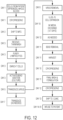
- the protocols may be used as a part of the methods for preparing target cells and CAR T cells described above. Aspects of some of these protocols are illustrated in figures 13 and 14 and may be contrasted with the protocol shown in figure 12 .
- a composition obtained by apheresis of whole blood is obtained and T cells in the composition are then selected.
- the term "selected" in this context means that the T cells are bound by agents that recognize the T cells with specificity (as defined above). DLD is then used to isolate the selected T cells and transfer these cells into a chosen fluid medium.
- Also described is a method of collecting target cells by: a) obtaining a crude fluid composition comprising the target cells from a patient; and b) performing Deterministic Lateral Displacement (DLD) on the crude fluid composition to obtain a composition enriched in target cells wherein either before, or after DLD, the target cells are bound to a carrier in a way that promotes DLD separation.
- a carrier may be used that has on its surface an affinity agent (e.g., an antibody, activator, hapten or aptamer) that binds with specificity (as defined above) to the target cells.
- Carrier may, if desired, be bound to target cells during the time that the cells are being collected from the patient and no more than five hours (and preferably no more than four, three, two or one hour(s)) should elapse from the time that the obtaining of the crude fluid composition comprising target cells is completed until the target cells are bound to the carrier.
- the diameter of the complex formed between target cells and one or more carriers should preferably be at least 20% larger than the uncomplexed cells and preferably at least 50% larger, at least twice as large or at least ten times as large. This increase in size may be either due to the binding of a single large carrier to the target cells or due to the binding of several smaller carriers.
- Binding may involve using: i) only carriers with a diameter at least as large (or in other embodiments, at least twice as large or at least ten times as large) as that of the target cells; ii) only carriers with a diameter no more than 50% (or in other embodiments, no more than 25% or 15%) as large as that of the target cells; or iii) mixtures of large and small carriers with these size characteristics (e.g ., there may be one group of carriers with a diameter at least as large (or at least twice or ten times as large) as the target cells and a second group of carriers with a diameter no more than 50% (or no more than 25% or 15%) as large as that of the target cells.
- a carrier will have a diameter of 1-1000 ⁇ m (and often in the range of 5-600 or 5-400 ⁇ m).
- the complexes would be separated from other cells or contaminants by DLD on a microfluidic device having an array of obstacles with a critical size lower than the size of the complexes but higher than the size of uncomplexed cells or contaminants.
- the crude fluid composition comprising target cells may be obtained by performing apheresis or leukapheresis on blood from the patient.
- This composition may include one or more additives that act as anticoagulants or that prevent the activation of platelets.
- additives include ticlopidine, inosine, protocatechuic acid, acetylsalicylic acid, and tirofiban alone or in combination.
- the microfluidic devices must have at least one channel extending from a sample inlet to one or more fluid outlets, wherein the channel is bounded by a first wall and a second wall opposite from the first wall.
- target cells are T cells selected from the group consisting of: Natural Killer T cells; Central Memory T cells; Helper T cells and Regulatory T cells, with Natural Killer T cells being the most preferred.
- the target cells are stem cells, B cells, macrophages, monocytes, dendritic cells, or progenitor cells.
- the method of the invention may include: c) genetically engineering cells by transducing them using a viral vector.
- the cells may be transfected electrically, chemically or by means of nanoparticles and/or expanded cells in number.
- the collected cells may be cultured and/or cryopreserved.
- culturing should generally be carried out in the presence of an activator, preferably an activator that is bound to a carrier.
- an activator preferably an activator that is bound to a carrier.
- factors that may be included in T cell cultures are IL-2 and IL-15.
- the invention includes the target cells produced by the methods.
- DLD deposition-dependent deposition
- the invention is directed to a system for purifying cells from large volume leukapheresis processes in which at least one microfluidic device is used that separates materials by DLD.
- the objective is to obtain leukocytes that may be used therapeutically or that secrete agents that may be used therapeutically.
- the invention includes binding specific types of leukocytes to one or more carriers in a way that promotes and, optionally, also complements DLD separation and then performing DLD on the complex. In this way, specific types of leukocytes may be separated from cells that are about the same size and that, in the absence of complex formation, could not be resolved by DLD.
- a two step procedure as discussed above may sometimes be advantageous in which a one DLD procedure separates unbound leukocytes from smaller material and a another DLD procedure separates a carrier-leukocyte complex from uncomplexed cells.
- a one DLD procedure separates unbound leukocytes from smaller material and a another DLD procedure separates a carrier-leukocyte complex from uncomplexed cells.
- the same technique can be used in other contexts as well, e.g., on cultured cells, provided that cell specific carriers are available.
- the cells may be recombinantly genetically engineered to alter the expression of one or more of their genes.
- the microfluidic devices must have at least one channel extending from a sample inlet to both a "collection outlet” for recovering white blood cells (WBCs) or specific leukocyte-carrier complexes and a "waste outlet” through which material of a different size (generally smaller) than WBCs or uncomplexed leukocytes flow.
- the channel is bounded by a first wall and a second wall opposite from the first wall and includes an array of obstacles arranged in rows, with each successive row being shifted laterally with respect to a previous row.
- the obstacles are disposed in a manner such that, when leukapheresis material is applied to an inlet of the device and fluidically passed through the channel, cells or cell complexes are deflected to the collection outlet (or outlets) where an enriched product is collected and material of a different (generally smaller) size flows to one or more separate waste outlets.
- the obstacles in microfluidic devices may be designed in the shape of diamonds or triangles and each device may have 6-40 channels.
- the microfluidic devices may be part of a system comprising 2-20 microfluidic devices (see Fig. 7 ). Individual devices may be operated at flow rates of 14 ml/hr but flow rates of at least 25 ml/hr (preferably at least 40, 60, 80 or 100 ml per hour) are preferable and allow large sample volumes (at least 200 ml and preferably 400-600 ml) to be processed within an hour.
- adherent target cells preferably cells of therapeutic value, e.g., adherent stem cells
- DLD Deterministic Lateral Displacement
- the adherent target cells may be bound to one or more carriers in a way that promotes or complements DLD separation.
- carriers may have on their surface an affinity agent (e.g ., an antibody, activator, hapten or aptamer) that binds with specificity (as defined above) to the adherent target cells and may be transfected or transduced with nucleic acids designed to impart on the cells a desired phenotype, e.g., to express a chimeric molecule (preferably a protein that makes the cells of greater therapeutic value).
- an affinity agent e.g ., an antibody, activator, hapten or aptamer
- nucleic acids designed to impart on the cells a desired phenotype, e.g., to express a chimeric molecule (preferably a protein that makes the cells of greater therapeutic value).
- Carriers may be added at the time that the crude fluid composition is being collected or, alternatively after collection is completed but before DLD is performed for the first time.
- DLD may be performed for a first time before carrier is added. For example, if the adherent cell has a size less than the critical size, the crude fluid composition may be applied to the device before the carrier is added, the adherent cell may be recovered, the cells may then be attached to one or more carriers to form a complex that is larger than the critical size of a device, a second DLD step may then be preformed and the carrier adherent cell complexes may be collected.
- no more than three hours (and more preferably no more than two hours, or one hour) elapse from the time that the obtaining of the crude fluid composition from the patient is completed until the adherent cell is bound to a carrier for the first time.
- no more than four hours (and preferably no more than three or two hours) elapse from the time that the obtaining of the crude fluid composition from the patient is complete until the first time that the adherent cell or a carrier adherent cell complex is collected from the device for the first time.
- the methodology described above may be used to separate adherent target cells, e.g ., adherent stem cells, from a plurality of other cells.
- the method involves: a) contacting a crude fluid composition comprising the adherent target cells and the plurality of other cells, wherein the adherent target cells are at least partially associated with one or more carriers in a way that promotes DLD separation and form carrier associated adherent target cell complexes, wherein the complexes comprise an increased size relative to the plurality of other cells, and wherein the size of the carrier associated adherent cell complexes is preferably at least 50% greater than a critical size, and other, uncomplexed cells comprise a size less than the critical size; b) applying the crude fluid composition containing the carrier associated adherent cell complexes to a device, wherein the device comprises an array of obstacles arranged in rows, wherein the rows are shifted laterally with respect to one another, wherein the rows are configured to deflect cells or complexes greater than or equal to the critical size in
- the diameter of the complex formed between adherent target cells and one or more carriers should preferably be at least 20% larger than the uncomplexed cells and preferably at least 50% larger, at least twice as large or at least ten times as large. This increase in size may be either due to the binding of a single large carrier to the adherent target cells or due to the binding of several smaller carriers.
- Binding may involve using: a) only carriers with a diameter at least as large (or in other embodiments, at least twice as large or at least ten times as large) as that of the adherent target cells; b) only carriers with a diameter no more than 50% (or in other embodiments, no more than 25% or 15%) as large as that of the adherent target cells; or c) mixtures of large and small carriers with these size characteristics (e.g ., there may be one group of carriers with a diameter at least as large (or at least twice or ten times as large) as the adherent target cells and a second group of carriers with a diameter no more than 50% (or no more than 25% or 15%) as large as that of the adherent target cells.
- a carrier will have a diameter of 1-1000 ⁇ m (and often in the range of 5-600 or 5-400 ⁇ m).
- the carriers may be made of any of the materials that are known in the art for the culturing of adherent cells including polypropylene, polystyrene, glass, gelatin, collagen, polysaccharides, plastic, acrylamide and alginate. They may be uncoated or coated with materials that promote adhesion and growth (e.g., serum, collagen, proteins or polymers) and may have agents (e.g ., antibodies, antibody fragments, substrates, activators or other materials) attached to their surfaces.
- the diluent can be growth media, the steps can be performed sequentially and, after step (d), buffer exchange can be performed.
- Examples of specific adherent cells that may be isolated in the methods described above include: an MRC-5 cell; a HeLa cell; a Vero cell; an NIH 3T3 cell; an L929 cell; a Sf21 cell; a Sf9 cell; an A549 cell; an A9 cell; an AtT-20 cell; a BALB/3T3 cell; a BHK-21 cell; a BHL-100 cell; a BT cell; a Caco-2 cell; a Chang cell; a Clone 9 cell; a Clone M-3 cell; a COS-1 cell; a COS-3 cell; a COS-7 cell; a CRFK cell; a CV-1 cell; a D-17 cell; a Daudi cell; a GH1 cell; a GH3 cell; an HaK cell; an HCT-15 cell; an HL-60 cell; an HT-1080 cell; a HEK cell, HT-29 cell; an HUVEC cell; an I-10 cell; an IM-9 cell;
- the invention also includes methods of purifying cells capable of activation using the procedures described above.
- the invention is directed to a method of separating an activated cell from a plurality of other cells by: a) contacting a crude fluid composition comprising a cell capable of activation and the plurality of other cells with one or more carriers, in a way that promotes DLD separation, wherein one or more of the carriers comprise a cell activator, wherein one or more carriers are at least partially associated with the cell capable of activation by the cell activator upon or after contact to generate a carrier associated cell complex, wherein the association of the cell activator with the cell capable of activation at least partially activates the cell capable of activation, wherein the carrier associated cell complex comprises an increased size relative to other cells, and wherein a size of the carrier associated cell complex is greater than or equal to a critical size, and the cells in the plurality of other cells comprise a size less than the critical size; b) applying the crude fluid composition to a device, wherein the device comprises an array of obstacles arranged
- the fluid composition comprising the separated carrier associated cell complex may then be collected.
- the cells may optionally be transfected or transduced with nucleic acids designed to impart on the cells a desired phenotype, e.g ., to express a chimeric molecule (preferably a protein that makes the cells of greater therapeutic value).
- the cell capable of activation may be selected from the group consisting of: a T cell, a B cell, a macrophage, a dendritic cell, a granulocyte, an innate lymphoid cell, a megakaryocyte, a natural killer cell, a thrombocyte, a synoviocyte, a beta cell, a liver cell, a pancreatic cell; a DE3 lysogenized cell, a yeast cell, a plant cell, and a stem cell.
- the cell activator may be selected from the group consisting of: an antibody or antibody fragment, CD3, CD28, an antigen, a helper T cell, a receptor, a cytokine, a glycoprotein, and any combination thereof.
- the activator may be a small compound and may be selected from the group consisting of insulin, IPTG, lactose, allolactose, a lipid, a glycoside, a terpene, a steroid, an alkaloid, and any combination thereof.
- the cell capable of activation has been collected from a patient as part of a crude fluid composition comprising the cell capable of activation and a plurality of other cells, wherein no more than four hours (and preferably no more than three hours, two hours or one hour) elapse from the time that the obtaining of the crude fluid composition from the patient is completed until the cell capable of activation is bound to the carrier. It is also preferable that no more than four hours elapse from the time that the obtaining of the crude fluid composition from the patient is completed until step c) is completed.
- the method may be altered by binding activator before collection of cells begins.
- the diameter of the complex formed between a cell capable of activation and one or more carriers should be at least 20% larger than the uncomplexed cells and more preferably at least 50% larger, at least twice as large or at least ten times as large. This increase in size may be either due to the binding of a single large carrier to the cell capable of activation or due to the binding of several smaller carriers.
- Binding may involve using: a) only carriers with a diameter at least as large (or in other embodiments, at least twice as large or at least ten times as large) as that of the cell capable of activation; b) only carriers with a diameter no more than 50% (or in other embodiments, no more than 25% or 15%) as large as that of the cell capable of activation; or c) mixtures of large and small carriers with these size characteristics (e.g ., there may be one group of carriers with a diameter at least as large (or at least twice or ten times as large) as the cell capable of activation and a second group of carriers with a diameter no more than 50% (or no more than 25% or 15%) as large as that of the cell capable of activation.
- a carrier will have a diameter of 1-1000 ⁇ m (and often in the range of 5-600 or 5-400 ⁇ m).
- Apheresis As used herein this term refers to a procedure in which blood from a patient or donor is separated into its components, e.g., plasma, white blood cells and red blood cells. More specific terms are “plateletpheresis” (referring to the separation of platelets) and “leukapheresis” (referring to the separation of leukocytes). In this context, the term “separation” refers to the obtaining of a product that is enriched in a particular component compared to whole blood and does not mean that absolute purity has been attained.
- CAR T cells The term "CAR” is an acronym for "chimeric antigen receptor.”
- a “CAR T cell” is therefore a T cell that has been genetically engineered to express a chimeric receptor.
- CAR T cell therapy This term refers to any procedure in which a disease is treated with CAR T cells.
- Diseases that may be treated include hematological and solid tumor cancers, autoimmune diseases and infectious diseases.
- Carrier refers an agent, e.g., a bead, or particle, made of either biological or synthetic material that is added to a preparation for the purpose of binding directly or indirectly (i.e ., through one or more intermediate cells, particles or compounds) to some or all of the compounds or cells present.
- Carriers may be made from a variety of different materials, including DEAE-dextran, glass, polystyrene plastic, acrylamide, collagen, and alginate and will typically have a size of 1-1000 ⁇ m.
- the carriers may be coated or uncoated and have surfaces that are modified to include affinity agents (e.g ., antibodies, activators, haptens, aptamers, particles or other compounds) that recognize antigens or other molecules on the surface of cells.
- the carriers may also be magnetized and this may provide an additional means of purification to complement DLD and they may comprise particles (e.g., Janus or Strawberry-like particles) that confer upon cells or cell complexes non-size related secondary properties.
- the particles may result in chemical, electrochemical, or magnetic properties that can be used in downstream processes, such as magnetic separation, electroporation, gene transfer, and/or specific analytical chemistry processes.
- Particles may also cause metabolic changes in cells, activate cells or promote cell division.
- Carriers that bind "in a way that promotes DLD separation” refers to carriers and methods of binding carriers that affect the way that, depending on context, a cell, protein or particle behaves during DLD.
- binding in a way that promotes DLD separation means that: a) the binding must exhibit specificity for a particular target cell type, protein or particle; and b) must result in a complex that provides for an increase in size of the complex relative to the unbound cell, protein or particle. In the case of binding to a target cell, there must be an increase of at least 2 ⁇ m (and alternatively at least 20, 50, 100, 200, 500 or 1000% when expressed as a percentage).
- the term "in a way that promotes DLD separation” also requires that the complexes permit such release, for example by chemical or enzymatic cleavage, chemical dissolution, digestion, due to competition with other binders, or by physical shearing ( e.g ., using a pipette to create shear stress) and the freed target cells, proteins or other particles must maintain activity; e.g ., therapeutic cells after release from a complex must still maintain the biological activities that make them therapeutically useful.
- Carriers may also bind "in a way that complements DLD separation”: This term refers to carriers and methods of binding carriers that change the chemical, electrochemical, or magnetic properties of cells or cell complexes or that change one or more biological activities of cells, regardless of whether they increase size sufficiently to promote DLD separation. Carriers that complement DLD separation also do not necessarily bind with specificity to target cells, i.e. , they may have to be combined with some other agent that makes them specific or they may simply be added to a cell preparation and be allowed to bind non-specifically.
- the terms "in a way that complements DLD separation” and "in a way that promotes DLD separation” are not exclusive of one another. Binding may both complement DLD separation and also promote DLD separation. For example a polysaccharide carrier may have an activator on its surface that increases the rate of cell growth and the binding of one or more of these carriers may also promote DLD separation. Alternatively binding may just promote DLD separation or just complement DLD separation.
- Target cells are the cells that various procedures described herein require or are designed to purify, collect, engineer etc. What the specific cells are will depend on the context in which the term is used. For example, if the objective of a procedure is to isolate a particular kind of stem cell, that cell would be the target cell of the procedure.
- Isolate, purify Unless otherwise indicated, these terms, as used herein, are synonymous and refer to the enrichment of a desired product relative to unwanted material. The terms do not necessarily mean that the product is completely isolated or completely pure. For example, if a starting sample had a target cell that constituted 2% of the cells in a sample, and a procedure was performed that resulted in a composition in which the target cell was 60% of the cells present, the procedure would have succeeded in isolating or purifying the target cell.
- Bump array and "obstacle array” are used synonymously herein and describe an ordered array of obstacles that are disposed in a flow channel through which a cell or particle-bearing fluid can be passed.
- Deterministic Lateral Displacement refers to a process in which particles are deflected on a path through an array, deterministically, based on their size in relation to some of the array parameters. This process can be used to separate cells, which is generally the context in which it is discussed herein. However, it is important to recognize that DLD can also be used to concentrate cells and for buffer exchange. Processes are generally described herein in terms of continuous flow (DC conditions; i.e. , bulk fluid flow in only a single direction). However, DLD can also work under oscillatory flow (AC conditions; i.e ., bulk fluid flow alternating between two directions).
- DC conditions continuous flow
- AC conditions oscillatory flow
- Critical size The "critical size" or "predetermined size” of particles passing through an obstacle array describes the size limit of particles that are able to follow the laminar flow of fluid. Particles larger than the critical size can be 'bumped' from the flow path of the fluid while particles having sizes lower than the critical size (or predetermined size) will not necessarily be so displaced.
- the critical size can be identical for both sides of the gap; however when the profile is asymmetrical, the critical sizes of the two sides of the gap can differ.
- Fluid flow refers to the macroscopic movement of fluid in a general direction across an obstacle array. These terms do not take into account the temporary displacements of fluid streams for fluid to move around an obstacle in order for the fluid to continue to move in the general direction.
- Tilt angle ⁇ In a bump array device, the tilt angle is the angle between the direction of bulk fluid flow and the direction defined by alignment of rows of sequential (in the direction of bulk fluid flow) obstacles in the array.
- the "array direction” is a direction defined by the alignment of rows of sequential obstacles in the array.
- a particle is “bumped” in a bump array if, upon passing through a gap and encountering a downstream obstacle, the particle's overall trajectory follows the array direction of the bump array (i.e., travels at the tilt angle ⁇ relative to bulk fluid flow).
- a particle is not bumped if its overall trajectory follows the direction of bulk fluid flow under those circumstances.
- the present invention is primarily concerned with the use of DLD in preparing cells that are of therapeutic value.
- the text below provides guidance regarding methods disclosed herein and information that may aid in the making and use of devices involved in carrying out those methods.
- Cells may be isolated by performing DLD using microfluidic devices that contain a channel through which fluid flows from an inlet at one end of the device to outlets at the opposite end.
- DLD liquid crystal deposition
- microfluidic devices that contain a channel through which fluid flows from an inlet at one end of the device to outlets at the opposite end.
- Basic principles of size based microfluidic separations and the design of obstacle arrays for separating cells have been provided elsewhere (see, US 2014/0342375 ; US 2016/0139012 ; 7,318,902 and US 7,150,812 , which are hereby incorporated herein in their entirety) and are also summarized in the sections below.
- a fluid sample containing cells is introduced into a device at an inlet and is carried along with fluid flowing through the device to outlets.
- cells in the sample traverse the device, they encounter posts or other obstacles that have been positioned in rows and that form gaps or pores through which the cells must pass.
- Each successive row of obstacles is displaced relative to the preceding row so as to form an array direction that differs from the direction of fluid flow in the flow channel.
- the "tilt angle" defined by these two directions, together with the width of gaps between obstacles, the shape of obstacles, and the orientation of obstacles forming gaps are primary factors in determining a "critical size" for an array.
- Cells having a size greater than the critical size travel in the array direction, rather than in the direction of bulk fluid flow and particles having a size less than the critical size travel in the direction of bulk fluid flow.
- array characteristics may be chosen that result in white blood cells being diverted in the array direction whereas red blood cells and platelets continue in the direction of bulk fluid flow.
- a carrier may then be used that binds to that cell with in a way that promotes DLD separation and which thereby results in a complex that is larger than uncomplexed leukocytes. It may then be possible to carry out a separation on a device having a critical size smaller than the complexes but bigger than the uncomplexed cells.
- the obstacles used in devices may take the shape of columns or be triangular, square, rectangular, diamond shaped, trapezoidal, hexagonal or teardrop shaped.
- adjacent obstacles may have a geometry such that the portions of the obstacles defining the gap are either symmetrical or asymmetrical about the axis of the gap that extends in the direction of bulk fluid flow.
- microfluidic devices that are capable of separating cells on the basis of size are well known in the art. Such devices include those described in US 5,837,115 ; US 7,150,812 ; US 6,685,841 ; US 7,318,902 ; 7,472,794 ; and US 7,735,652 ; all of which are hereby incorporated by reference in their entirety.
- a device can be made using any of the materials from which micro- and nano-scale fluid handling devices are typically fabricated, including silicon, glasses, plastics, and hybrid materials.
- materials from which micro- and nano-scale fluid handling devices are typically fabricated including silicon, glasses, plastics, and hybrid materials.
- thermoplastic materials suitable for microfluidic fabrication is available, offering a wide selection of mechanical and chemical properties that can be leveraged and further tailored for specific applications.
- High-throughput embossing methods such as reel-to-reel processing of thermoplastics is an attractive method for industrial microfluidic chip production.
- the use of single chip hot embossing can be a cost-effective technique for realizing high-quality microfluidic devices during the prototyping stage.
- Methods for the replication of microscale features in two thermoplastics, polymethylmethacrylate (PMMA) and/or polycarbonate (PC) are described in " Microfluidic device fabrication by thermoplastic hot-embossing” by Yang, etal. (MethodsMol. Biol. 949: 115-23 (2013 )).
- the flow channel can be constructed using two or more pieces which, when assembled, form a closed cavity (preferably one having orifices for adding or withdrawing fluids) having the obstacles disposed within it.
- the obstacles can be fabricated on one or more pieces that are assembled to form the flow channel, or they can be fabricated in the form of an insert that is sandwiched between two or more pieces that define the boundaries of the flow channel.
- the obstacles may be solid bodies that extend across the flow channel, in some cases from one face of the flow channel to an opposite face of the flow channel. Where an obstacle is integral with (or an extension of) one of the faces of the flow channel at one end of the obstacle, the other end of the obstacle can be sealed to or pressed against the opposite face of the flow channel.
- a small space (preferably too small to accommodate any particles of interest for an intended use) is tolerable between one end of an obstacle and a face of the flow channel, provided the space does not adversely affect the structural stability of the obstacle or the relevant flow properties of the device.
- the number of obstacles present should be sufficient to realize the particle-separating properties of the arrays.
- the obstacles can generally be organized into rows and columns (Note: Use of the term "rows and columns" does not mean or imply that the rows and columns are perpendicular to one another). Obstacles that are generally aligned in a direction transverse to fluid flow in the flow channel can be referred to as obstacles in a column. Obstacles adjacent to one another in a column may define a gap through which fluid flows.
- Obstacles in adjacent columns can be offset from one another by a degree characterized by a tilt angle, designated ⁇ (epsilon).
- ⁇ tilt angle
- corresponding obstacles in the columns can be offset from one another such that the corresponding obstacles form a row of obstacles that extends at the angle ⁇ relative to the direction of fluid flow past the columns.
- the tilt angle can be selected and the columns can be spaced apart from each other such that 1/ ⁇ (when expressed in radians) is an integer, and the columns of obstacles repeat periodically.
- the obstacles in a single column can also be offset from one another by the same or a different tilt angle.
- the rows and columns can be arranged at an angle of 90 degrees with respect to one another, with both the rows and the columns tilted, relative to the direction of bulk fluid flow through the flow channel, at the same angle of ⁇ .
- Surfaces can be coated to modify their properties and polymeric materials employed to fabricate devices, can be modified in many ways.
- functional groups such as amines or carboxylic acids that are either in the native polymer or added by means of wet chemistry or plasma treatment are used to crosslink proteins or other molecules.
- DNA can be attached to COC and PMMA substrates using surface amine groups.
- Surfactants such as Pluronic ® can be used to make surfaces hydrophilic and protein repellant by adding Pluronic ® to PDMS formulations.
- a layer of PMMA is spin coated on a device, e.g., microfluidic chip and PMMA is "doped" with hydroxypropyl cellulose to vary its contact angle.
- one or more walls may be chemically modified to be non-adherent or repulsive.
- the walls may be coated with a thin film coating (e.g., a monolayer) of commercial non-stick reagents, such as those used to form hydrogels.
- Charged polymers may also be employed to repel oppositely charged species.
- the type of chemical species used for repulsion and the method of attachment to the channel walls can depend on the nature of the species being repelled and the nature of the walls and the species being attached. Such surface modification techniques are well known in the art.
- the walls may be functionalized before or after the device is assembled.
- the DLD devices described herein can be used to purify cells, cellular fragments, cell adducts, or nucleic acids. As discussed herein, these devices can also be used to separate a cell population of interest from a plurality of other cells. Separation and purification of blood components using devices can be found, for example, in US Publication No. US2016/0139012 , the teaching of which is incorporated by reference herein in its entirety. A brief discussion of a few illustrative separations is provided below.
- devices are used in procedures designed to separate a viable cell from a nonviable cell.
- the term "viable cell” refers to a cell that is capable of growth, is actively dividing, is capable of reproduction, or the like.
- DLD devices can be designed to comprise a critical size that is greater than a predetermined size of the nonviable cell and less than a predetermined size of the viable cell.
- the critical size may be as little as 1.1 fold greater than (or less than) the predetermined size of the nonviable cell but generally, larger degrees (or smaller) are preferred, e.g., about 1.2 fold - 2 fold, and preferably 3-10 fold.
- DLD devices can be used to in procedures to separate adherent cells.
- adherent cell refers to a cell capable of adhering to a surface.
- Adherent cells include immortalized cells used in cell culturing and can be derived from mammalian hosts. In some instances, the adherent cell may be trypsinized prior to purification.
- adherent cells include MRC-5 cells; HeLa cells; Vero cells; NIH 3T3 cells; L929 cells; Sf21 cells; Sf9 cells; A549 cells; A9 cells; AtT-20 cells; BALB/3T3 cells; BHK-21 cells; BHL-100 cells; BT cells; Caco-2 cells; Chang cells; Clone 9 cells; Clone M-3 cells; COS-1 cells; COS-3 cells; COS-7 cells; CRFK cells; CV-1 cells; D-17 cells; Daudi cells; GH1 cells; GH3 cells; HaK cells; HCT-15 cells; HL-60 cells; HT-1080 cells; HT-29 cells; HUVEC cells; I-10 cells; IM-9 cells; JEG-2 cells; Jensen cells; Jurkat cells; K-562 cells; KB cells; KG-1 cells; L2 cells; LLC-WRC 256 cells; McCoy cells; MCF7 cells; WI-38 cells; WISH cells; XC cells; Y-1 cells; CHO cells; Raw 26
- procedures may involve separating cells from a diluent such as growth media, which may provide for the efficient maintenance of a culture of the adherent cells.
- a culture of adherent cells in a growth medium can be exchanged into a transfection media comprising transfection reagents, into a second growth medium designed to elicit change within the adherent cell such as differentiation of a stem cell, or into sequential wash buffers designed to remove compounds from the culture.
- adherent cells are purified through association with one or more carriers that bind in a way that promotes DLD separation.
- the carriers may be of the type described herein and binding may stabilize and/or activate the cells.
- a carrier will typically be in the rage of 1-1000 ⁇ m but may sometimes also be outside of this range.
- a complex may be anywhere from a few percent larger than the uncomplexed cell to many times the size of the uncomplexed cell. In order to facilitate separations, an increase of at least 20% is desirable with higher percentages (50; 100; 1000 or more) being preferred.
- the DLD devices can also be used in procedures for separating an activated cell or a cell capable of activation, from a plurality of other cells.
- the cells undergoing activation may be grown on a large scale but, in a preferred embodiment, the cells are derived from a single patient and DLD is performed within at least few hours after collection.
- activated cell or "cell capable of activation” refers to a cell that has been, or can be activated, respectively, through association, incubation, or contact with a cell activator.
- Examples of cells capable of activation can include cells that play a role in the immune or inflammatory response such as: T cells, B cells; regulatory T cells, macrophages, dendritic cells, granulocytes, innate lymphoid cells, megakaryocytes, natural killer cells, thrombocytes, synoviocytes, and the like; cells that play a role in metabolism, such as beta cells, liver cells, and pancreatic cells; and recombinant cells capable of inducible protein expression such as DE3 lysogenized E. coli cells, yeast cells, plant cells, etc.
- one or more carriers will have the activator on their surface.
- cell activators include proteins, antibodies, cytokines, CD3, CD28, antigens against a specific protein, helper T cells, receptors, and glycoproteins; hormones such as insulin, glucagon and the like; IPTG, lactose, allolactose, lipids, glycosides, terpenes, steroids, and alkaloids.
- the activatable cell should be at least partially associated with carriers through interaction between the activatable cell and cell activator on the surface of the carriers.
- the complexes formed may be just few percent larger than the uncomplexed cell or many times the size of the uncomplexed cell. In order to facilitate separations, an increase of at least 20% is desirable with higher percentages (40, 50, 100, 1000 or more) being preferred.
- DLD can also be used in purifications designed to remove compounds that may be toxic to a cell or to keep the cells free from contamination by a toxic compound. Examples include an antibiotic, a cryopreservative, an antifungal, a toxic metabolite, sodium azide, a metal ion, a metal ion chelator, an endotoxin, a plasticizer, a pesticide, and any combination thereof.
- the device can be used to remove toxic compounds from cells to ensure consistent production of material from the cells.
- the cell can be a log phase cell.
- the term "log phase cell" refers to an actively dividing cell at a stage of growth characterized by exponential logarithmic growth. In log phase, a cell population can double at a constant rate such that plotting the natural logarithm of cell number against time produces a straight line.
- the ability to separate toxic material may be important for a wide variety of cells including: bacterial strains such as BL21, Tuner, Origami, Origami B, Rosetta, C41, C43, DH5 ⁇ , DH10 ⁇ , or XL1Blue; yeast strains such as those of genera Saccharomyces, Pichia, Kluyveromyces, Hansenula and Yarrowia; algae; and mammalian cell cultures, including cultures of MRC-5 cells; HeLa cells; Vero cells; NIH 3T3 cells; L929 cells; Sf21 cells; Sf9 cells; A549 cells; A9 cells; AtT-20 cells; BALB/3T3 cells; BHK-21 cells; BHL-100 cells; BT cells; Caco-2 cells; Chang cells; Clone 9 cells; Clone M-3 cells; COS-1 cells; COS-3 cells; COS-7 cells; CRFK cells; CV-1 cells; D-17 cells; Daudi cells; GH1 cells; GH3 cells; HaK cells; HCT
- the DLD devices may also be used in the purification of material secreted from a cell.
- secreted materials includes proteins, peptides, enzymes, antibodies, fuel, biofuels such as those derived from algae, polymers, small molecules such as simple organic molecules, complex organic molecules, drugs and pro-drugs, carbohydrates and any combination thereof.
- Secreted products can include therapeutically useful proteins such as insulin, Imatinib, T cells, T cell receptors, Fc fusion proteins, anticoagulants, blood factors, bone morphogenetic proteins, engineered protein scaffolds, enzymes, growth factors, hormones, interferons, interleukins, and thrombolytics.
- FIG. 15 is a schematic depicting the use of DLD in the purification of secreted products.
- the cells may be in an aqueous suspension of buffer, growth medium, or the like, such that the cell secretes product into the suspension.
- secreted products include proteins, peptides, enzymes, antibodies, fuel, biofuels such as those derived from algae, polymers, small molecules such as simple organic molecules, complex organic molecules, drugs and pro-drugs, carbohydrates and any combination thereof.
- Secreted products can include therapeutically useful proteins such as insulin, Imatinib, T cells, T cell receptors, Fc fusion proteins, anticoagulants, blood factors, bone morphogenetic proteins, engineered protein scaffolds, enzymes, growth factors, hormones, interferons, interleukins, and thrombolytics.
- therapeutically useful proteins such as insulin, Imatinib, T cells, T cell receptors, Fc fusion proteins, anticoagulants, blood factors, bone morphogenetic proteins, engineered protein scaffolds, enzymes, growth factors, hormones, interferons, interleukins, and thrombolytics.
- Purification might be carried out, for example, in situations where cells have a predetermined size that is greater than a predetermined size of the secreted compound, where the predetermined size of the cell is greater than or equal to a critical size, and the predetermined size of the secreted compound is less than the critical size.
- the cells when applied to a DLD device, the cells can be deflected in a first direction while the secreted compound can be deflected in a second direction, thereby separating the secreted compound from the cell.
- a secreted protein may be captured by a large carrier that binds in a way that promotes DLD separation. DLD may then be performed and the carrier-protein complex may then be treated to further purify, or release, the protein.
- FIGS. 16 and 17 are schematics of an iterative process in which separated cells are looped back into the DLD device after separation.
- the cells may be looped from a first device into a second, different device with obstacles comprising different critical sizes.
- Such a system can allow systematic separation of a plurality of size ranges by manipulating the range of critical sizes.
- cells may be looped back to the same device used previously to separate the isolated particles. This system can be advantageous for continuous purification of actively dividing cells or compounds being actively expressed.
- such a method could be combined with the method of purifying the secreted product to both collect the secreted product from one flow stream and the cell producing the secreted product from another flow stream. Because the cells can continuously produce the secreted product, the purified cells can be reapplied to the device to continuously collect the secreted product from the cells.
- the purity, yields and viability of cells produced by the DLD methods discussed herein will vary based on a number of factors including the nature of the starting material, the exact procedure employed and the characteristics of the DLD device.
- purifications, yields and viabilities of at least 60% should be obtained with, higher percentages, at least 70, 80 or 90% being more preferred.
- methods may be used to isolate leukocytes from whole blood, apheresis products or leukapheresis products with at least 70% purity, yield and viability with higher percentages (at least 80%, 85%, or 90%) being preferred.
- microfabricated sieving matrices have been disclosed for separating particles ( Chou, et. al., Proc. Natl. Acad. Sci. 96:13762 (1999 ); Han, et al., Science 288:1026 (2000 ); Huang, et al., Nat. Biotechnol. 20:1048 (2002 ); Turner et al., Phys. Rev. Lett. 88(12):128103 (2002 ); Huang, et al., Phys. Rev. Lett. 89:178301 (2002 ); U.S. Pat. No.
- Bump array also known as "obstacle array” devices have been described, and their basic operation is explained, for example in U.S. Pat. No. 7,150,812 .
- a bump array operates essentially by segregating particles passing through an array (generally, a periodically-ordered array) of obstacles, with segregation occurring between particles that follow an "array direction" that is offset from the direction of bulk fluid flow or from the direction of an applied field ( U.S. 7,150,812 ).
- the geometry of adjacent obstacles is such that the portions of the obstacles defining the gap are symmetrical about the axis of the gap that extends in the direction of bulk fluid flow.
- the velocity or volumetric profile of fluid flow through such gaps is approximately parabolic across the gap, with fluid velocity and flux being zero at the surface of each obstacle defining the gap (assuming no-slip flow conditions) and reaching a maximum value at the center point of the gap.
- the profile being parabolic a fluid layer of a given width adjacent to one of the obstacles defining the gap contains an equal proportion of fluid flux as a fluid layer of the same width adjacent to the other obstacle that defines the gap, meaning that the critical size of particles that are 'bumped' during passage through the gap is equal regardless of which obstacle the particle travels near.
- particle size-segregating performance of an obstacle array can be improved by shaping and disposing the obstacles such that the portions of adjacent obstacles that deflect fluid flow into a gap between obstacles are not symmetrical about the axis of the gap that extends in the direction of bulk fluid flow.
- Such lack of flow symmetry into the gap can lead to a non-symmetrical fluid flow profile within the gap. Concentration of fluid flow toward one side of a gap (i.e., a consequence of the non-symmetrical fluid flow profile through the gap) can reduce the critical size of particles that are induced to travel in the array direction, rather than in the direction of bulk fluid flow.
- the non-symmetry of the flow profile causes differences between the width of the flow layer adjacent to one obstacle that contains a selected proportion of fluid flux through the gap and the width of the flow layer that contains the same proportion of fluid flux and that is adjacent to the other obstacle that defines the gap.
- the different widths of the fluid layers adjacent to obstacles define a gap that exhibits two different critical particle sizes. A particle traversing the gap can be bumped (i.e., travel in the array direction, rather than the bulk fluid flow direction) if it exceeds the critical size of the fluid layer in which it is carried.
- a particle traversing a gap having a non-symmetrical flow profile it is possible for a particle traversing a gap having a non-symmetrical flow profile to be bumped if the particle travels in the fluid layer adjacent to one obstacle, but to be not-bumped if it travels in the fluid layer adjacent to the other obstacle defining the gap.
- decreasing the roundness of edges of obstacles that define gaps can improve the particle size-segregating performance of an obstacle array.
- arrays of obstacles having a triangular cross-section with sharp vertices can exhibit a lower critical particle size than do arrays of identically-sized and -spaced triangular obstacles having rounded vertices.
- the critical size of particles deflected in the array direction under the influence of bulk fluid flow can be decreased without necessarily reducing the size of the obstacles.
- obstacles having sharper edges can be spaced farther apart than, but still yield particle segregation properties equivalent to, identically-sized obstacles having less sharp edges.
- Objects separated by size on microfluidic include cells, biomolecules, inorganic beads, and other objects. Typical sizes fractionated range from 100 nanometers to 50 micrometers. However, larger and smaller particles may also sometimes be fractionated.
- a device or combination of devices might be used to process between about 10 ⁇ l to at least 500 ⁇ l of sample, between about 500 ⁇ l and about 40 mL of sample, between about 500 ⁇ l and about 20 mL of sample, between about 20 mL of sample and about 200 mL of sample, between about 40 mL of sample and about 200 mL of sample, or at least 200 mL of sample.
- a device can comprise one or multiple channels with one or more inlets and one or more outlets.
- Inlets may be used for sample or crude (i.e. , unpurified) fluid compositions, for buffers or to introduce reagents.
- Outlets may be used for collecting product or may be used as an outlet for waste.
- Channels may be about 0.5 to 100 mm in width and about 2-200 mm long but different widths and lengths are also possible.
- Depth may be 1- 1000 ⁇ m and there may be anywhere from 1 to 100 channels or more present. Volumes may vary over a very wide range from a few ⁇ l to many ml and devices may have a plurality of zones (stages, or sections) with different configurations of obstacles.
- Gap size in an array of obstacles can vary from about a few ( e.g ., 1-500) micrometers or be more than a millimeter. Obstacles may, in some embodiments have a diameter of 1-3000 micrometers and may have a variety of shapes (round, triangular, teardrop shaped, diamond shaped, square, rectangular etc.).
- a first row of posts can be located close to (e.g. within 5 ⁇ m) the inlet or be more than 1 mm away.
- a device can include a plurality of stackable chips.
- a device can comprise about 1 - 50 chips.
- a device may have a plurality of chips placed in series or in parallel or both.
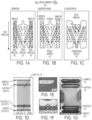
- the DLD array used in this study consisted of a single-zone, mirrored, diamond post design (see D'Silva, J., "Throughout Microfluidic Capture of Rare Cells from Large Volumes of Blood;” A Dissertation Presented to the Faculty of Princeton University in Candidacy for the Degree of Doctor of Philosophy (2016 )). There were 14 parallel arrays per chip resulting in a 14-lane DLD device ( Fig. 1D ). The device was designed with a 16 ⁇ m gap between posts and a 1/42 tilt, resulting in a critical diameter of ⁇ 4 ⁇ m. The plastic DLD device was generated using a process called soft-embossing.
- a silicon (Si) master for the plastic DLD microchip was made using standard photolithographic and deep reactive ion etching techniques (Princeton University, PRISM).
- the features on the silicon master were then transferred to a soft elastomeric mold (Edge Embossing, Medford, MA) by casting and curing the elastomer over the Si features.
- the elastomer was peeled off to create a reusable, negative imprint of the silicon master.
- a plastic blank sheet was placed between the elastomer molds, and then using a combination of pressure and temperature, the plastic was extruded into the features (wells) of the soft-elastomer negative mold, replicating the positive features and depth of the original silicon master.
- the soft tool was then peeled off from the plastic device, producing a flat piece of plastic surface-embossed to a depth ⁇ 100 ⁇ m with a pattern of flow channels and trenches around an array of microposts ( Fig. 1D , inset). Ports were created for fluidic access to the Input and Output ends of the microchip.
- the device was lidded with a heat-sensitive, hydrophilic adhesive (ARFlow Adhesives Research, Glen Rock, PA).
- the overall chip was 40 ⁇ 75 mm, and 1 mm thick - smaller than the size of a credit card.
- the microfluidic device was assembled inside an optically transparent and pressure resistant manifold with fluidic connections. Fluids were driven through the DLD microchip using a constant pneumatic pressure controller (MFCS-EZ, Fluigent, Lowell, MA). Two separate pressure controls were used, one for buffer and one for sample.
- the flow path for the buffer line included tubing connecting a buffer reservoir (60 mL syringe), an in-line degasser (Biotech DEGASi, Minneapolis, MN) and the buffer inlet port of the manifold.
- the flow path for the sample included tubing connecting a sample reservoir (20 mL syringe), a 20 ⁇ m PureFlow nylon filter of 25mm diameter (Clear Solutions, Inc. San Clemente, CA) to retain aggregates larger than the microchips nominal gap size (16 ⁇ m), and the sample inlet port on the manifold.
- the outlet ports of the manifold were connected by tubing to collection reservoirs for the waste and product fractions.
- the microchips, filter and tubing were primed and blocked for 15 min with running buffer before the sample was loaded.
- the DLD setup was primed by loading running buffer into the buffer reservoir (60 mL syringe) and then pressurizing; fluid then passed through the tubing and into the manifold "Buffer in” port ( Fig. 1 ). Air in the manifold port was vented via another port on that inlet, and then that port was sealed. The buffer was then driven through the microchip and out both the product and waste outlets, evacuating all air in the micropost array. At the same time, buffer was back flushed up through the "Sample IN" port on the manifold and through the in-line filter, flushing any air.
- This priming step took ⁇ 5 min of hands-on time, and removed all air from the microchip, manifold and tubing. Following the prime step, buffer continued to flush the setup for an additional 15 minutes to block all the interior surfaces; this step was automated and did not require hands-on time.
- the system was depressurized, and sample was loaded into the sample container (20 mL syringe).
- the sample (see below) was diluted 1-part sample to 4 parts running buffer (0.2x) prior to loading on the DLD.
- the buffer source was re-pressurized first, then the sample source, resulting in both buffer and sample entering their respective ports on the manifold and microchip and flowing through the microchip in parallel (see separation mode, Fig. 1 Ai).
- the system automatically processed the entire sample volume. Both product and waste fractions were collected in pre-weighed sterile conical 50mL tubes and weighed after the collection to determine the volumes collected.
- EDTA free buffer formulations Three different EDTA free buffer formulations were tested on the DLD: 0.5% F127 (Pluronic F-127, Sigma Aldrich, St. Louis, MO) in phosphate-buffered saline [Ca ++ /Mg ++ free) (Quality biological, Gaithersburg, MD), 1% Bovine Serum Albumin (BSA) (Affymetrix, Santa Clara, CA) in phosphate-buffered saline [Ca ++ /Mg ++ free], and an isotonic Elutriation Buffer (EB) composed of 50% Plasmalyte A (Baxter, Deerfield, IL) and 50% of a mixture containing 1.0% BSA (Affymetrix, Santa Clara, CA) 1.0mM N-Acetyl-Cysteine, 2% Dextrose and 0.45% NaCl (all from Sigma-Aldrich, St.
- F127 Puronic F-127, Sigma Aldrich, St. Louis, MO
- the buffers were prepared fresh each day, and were sterile-filtered through a 0.2 ⁇ m filter flask prior to use on the DLD. All samples in the expansion group were processed using the isotonic elutriation buffer to best align with current CAR-T-cell manufacturing approaches, even though better DLD performance has been established with the addition of poloxamer ( Johnson, et al., Cancer Cell Res. 27:38-58 (2017 )).
- PBMCs Peripheral blood mononuclear cells
- RPMI Sigma-Aldrich. St Louis, MO
- Ficoll-Hypaque GE, Pittsburgh, PA
- the top layer was discarded and the interface PBMC fraction transferred to a new 50mL tube and brought up to 20mL of RPMI.
- PBMCs were washed by centrifugation for 10 min at 400xg, the supernatant discarded and the pellet resuspended with 20 mL of RPMI and washed again at 200xg for 10min. The supernatant was removed and the pellet resuspended in full media containing RPMI-1640 + 10% Fetal Bovine Serum (FBS) (Sigma-Aldrich, St. Louis, MO) plus penicillin 100 units/mL and streptomycin 100 ⁇ g/mL antibiotics (Thermo-Fisher, Waltham, MA).
- FBS Fetal Bovine Serum
- penicillin 100 units/mL and streptomycin 100 ⁇ g/mL antibiotics Thermo-Fisher, Waltham, MA.
- Antibodies (mAb) conjugated to fluorochromes were obtained from BioLegend (San Diego, CA): CD25-PE, CD25-APC, CD95-FITC, CD45RA-BV605, CD45RO-PECy7, CD197/CCR7 PE, CD279-PE, CD28 PE-Cy5, CD45-PerCP, CD3-FITC, CD3-BV421, CD4-AF700, CD8-APC-AF780, CD61-FITC, CD41-FITC, CD45-Alexa647. Viability of the WBCs obtained by DLD and PBMCs purified by Ficoll-Hypaque was determined by Trypan blue exclusion.
- T-cell stimulations in expansion group DLD, Ficoll and LRS product were diluted to 1 ⁇ 10 7 T cells/mL then activated with CD3/CD28 washed and equilibrated anti-CD3/CD28 conjugated magnetic beads (5.0 ⁇ m) (Thermo-Fisher, Waltham, MA) at a ratio of 3.2:1 beads per cell for 60 min, and then the activated T cells were separated by a magnetic depletion for 5 min. Unbound cells were removed, and the bead-bound cells were cultured further in full media (below).
- LRS sample (same donor as was processed via DLD or Ficoll) was incubated with immunomagnetic CD3/CD28 beads for one hour. The mixture was then placed against a magnet for 5 minutes to capture the T cells. The magnetic bead-bound cells (activated cells) were removed and then diluted to 0.5 ⁇ 10 6 /mL as above for culture in full media.
- recombinant human IL-2 (BioLegend, San Diego, CA) was added at 200 IU/mL to wells. Following cell culture for up to 15 days, beads were removed from cells and cells counted at each time point. To remove beads, the cells in the well were resuspended by passing the cells through a 5-mL pipette for 10 times. Next, the cell suspension was passed throughout a 1 mL pipette 40 times followed by vigorous pipetting using a 200 ⁇ L tip for 1 min. Then the cell suspension was placed on the side of a magnet for 5 min and the nonmagnetic fraction was transferred to a fresh tube and counted. The number of cells in the culture wells was determined using a Scepter hand-held cell counter and by flow cytometry.
- the cells were then diluted to 250 ⁇ L of PBS with a final DRAQ5 TM DNA dye (Thermo-Fisher, Waltham, MA) concentration of 1.0mM.
- the stained cells were fixed with an additional 250 ⁇ L 1.2% p-formaldehyde in PBS overnight prior to acquisition.
- a minimum of 25,000 events or 2500 bead events were acquired on a BD FACSCalibur (BD Biosciences, San Jose, CA) using a fluorescence threshold (CD45 PerCP). Phenotypic analysis was also performed at all time points, using a 7-color activation/anergy panel consisting of CD3, CD45RA, CD95, CD279, CD25, CD4, and CD8.
- the panel was modified to create a 9-color panel focused on T central memory cells which added CD45RO PE-Cy7, CD28 PE-Cy5 and substituted CD197/CCR7 PE for CD279/PD1 PE.
- For multicolor staining 100 ⁇ l of a cell suspension was stained as above, and resuspended in 750 ⁇ L PBS and washed by centrifugation at 400xg and then resuspending in 250 ⁇ L 1.2% p-formaldehyde and fixed overnight prior to acquiring 20,000 events using forward scatter threshold on a four laser BD FACS Aria II. (BD Biosciences, San Jose, CA). All data analysis was performed using Flowlogic Software (Inivai, Melbourne, Australia).
- the DLD and Ficoll separation methods were used to process 12 LRS samples obtained from 12 separate normal donors. Of those 12 samples received and processed, 11 samples clustered around a mean of 148.7 ⁇ 10 3 / ⁇ L WBC and 2.52 ⁇ 10 6 / ⁇ L platelet counts respectively ( Fig. 2A, 2B ). The 12 th sample, with 313.3 ⁇ 10 3 / ⁇ L WBC and 4.87 ⁇ 10 6 / ⁇ L platelet counts can be seen in the scatter plot as a triangle, ( Fig. 2A ). This sample was sufficiently aggregated at the time of processing that it rapidly clogged the 20 ⁇ m prefilter and thus did not fully enter the DLD.
- FIG. 2A A representative image of the input material (LRS product diluted to 0.2x) is shown in ( Fig. 2A ).
- the respective cell products as collected in tubes ( Fig. 2 G, H ).
- DLD processing automated the process of removing the WBCs from the RBCs and platelets, generating one tube for product and one for waste, while the Ficoll sample still requires further manual processing to pipet the PMBC layer at the operationally-defined interface of the plasma layer above and Ficoll layer below ( Fig. 2H ); plus, an additional minimum of two centrifugal washes are required to remove most of the contaminating platelets.
- Flow cytometric characterization of the cultures was performed at each time point to assess consistency of cell activation. Changes in CD25 expression of CD3+ cells, as measured on Day 8, for Ficoll, DLD and direct magnet ( Fig. 3 ). IL-2 Receptor positive (CD25) CD3 cells were shown in Blue (CD4+ plots) and Red (CD8+ plots). DLD prepared cells show more consistent phenotypic expression across the 4 donors for CD25, an indicator of response to CD3/CD28 stimulation, as compared to both Ficoll and direct magnet preparations.
- DLD prepared CD3+ cells had an average 73% response to co-stimulation compared to Ficoll at 51% (both stimulated at 2.3 beads/cell), while the direct magnet fraction, stimulated at a higher 5:1 ratio, had only a 54% response.
- the fold expansion of the individual cultures was determined at day 3, day 8 and day 15; that data is shown in Figure 5A .
- the plot shows the expansion of each donor sample, across each method. While the direct magnet approach appears to show higher expansion, the counts are likely significantly affected by the different bead:cell ratios (and corresponding differences in plating density). Regardless, the 4 donors show significant variability in the fold expansion.
- the day 15 culture for the direct magnet arm donor #21 became contaminated and had to be discarded, despite having antibiotics present. It is not possible to know if the day 8 expansion data for donor #21 were influenced by the contaminant.
- Figure 6 shows the phenotypic approach to identifying memory cells used in this study, which is designed to eliminate any issues with shed antigens such as CD62L ( Mahnke, et al., Eur. J. of Immunol. 43:2797-2809 (2013 )).
- Central memory cells are sequentially gated and then backgated to show the CD3+ T cells are positive for CD45R0+, CD95+, CD28+ and CD197/CCR7+ against all other CD3+ cells in the culture.
- the DLD arm showed 100% (4/4) donors achieving central memory conversion with an average of 74% of cells being of memory phenotype, with coefficient of variation across donors of 13%.
- the Ficoll arm showed 50% (2/4) converting with an average of 47% memory cells, and a 29% variation.
- the direct magnet arm achieved 33% (1/3) conversion with an average of 48% memory cells and an associated 79% variation.
Landscapes
- Health & Medical Sciences (AREA)
- Chemical & Material Sciences (AREA)
- Life Sciences & Earth Sciences (AREA)
- Organic Chemistry (AREA)
- Immunology (AREA)
- General Health & Medical Sciences (AREA)
- Zoology (AREA)
- Genetics & Genomics (AREA)
- Biochemistry (AREA)
- Cell Biology (AREA)
- Medicinal Chemistry (AREA)
- Engineering & Computer Science (AREA)
- Gastroenterology & Hepatology (AREA)
- Biophysics (AREA)
- Molecular Biology (AREA)
- Proteomics, Peptides & Aminoacids (AREA)
- Toxicology (AREA)
- Biomedical Technology (AREA)
- Wood Science & Technology (AREA)
- Bioinformatics & Cheminformatics (AREA)
- Biotechnology (AREA)
- Hematology (AREA)
- Chemical Kinetics & Catalysis (AREA)
- General Engineering & Computer Science (AREA)
- Microbiology (AREA)
- Veterinary Medicine (AREA)
- Public Health (AREA)
- Animal Behavior & Ethology (AREA)
- Pharmacology & Pharmacy (AREA)
- Nuclear Medicine, Radiotherapy & Molecular Imaging (AREA)
- General Chemical & Material Sciences (AREA)
- Fluid Mechanics (AREA)
- Physics & Mathematics (AREA)
- Dispersion Chemistry (AREA)
- Analytical Chemistry (AREA)
- Clinical Laboratory Science (AREA)
- Medicines Containing Material From Animals Or Micro-Organisms (AREA)
- Micro-Organisms Or Cultivation Processes Thereof (AREA)
- Apparatus Associated With Microorganisms And Enzymes (AREA)
- Preparation Of Compounds By Using Micro-Organisms (AREA)
Claims (15)
- Procédé in vitro de séparation d'une cellule activée à partir d'une pluralité d'autres cellules comprenant :a) la mise en contact d'une composition fluidique brute comprenant une cellule capable d'activation et la pluralité d'autres cellules avec un ou plusieurs supports, dans lequel le au moins un support comprend un activateur cellulaire,dans lequel l'activateur cellulaire est au moins partiellement associé à la cellule capable d'activation par l'activateur cellulaire lors de ou après un contact afin de générer un complexe cellulaire associé à un support,dans lequel l'association de l'activateur cellulaire avec la cellule capable d'activation par l'activateur cellulaire active au moins partiellement la cellule capable d'activation,dans lequel le complexe cellulaire associé à un support comprend une taille augmentée par rapport aux cellules dans la pluralité d'autres cellules, etdans lequel une taille du complexe cellulaire associé à un support est supérieure ou égale à une taille critique, et les cellules dans la pluralité d'autres cellules comprennent une taille inférieure à la taille critique ;b) l'application de l'échantillon à un dispositif,dans lequel le dispositif comprend un réseau d'obstacles disposés en rangées ; dans lequel les rangées sont décalées latéralement les unes par rapport aux autres,dans lequel les rangées sont configurées pour dévier une particule ayant une taille supérieure ou égale à la taille critique dans une première direction et une particule ayant une taille inférieure à la taille critique dans une deuxième direction ; etc) la circulation de l'échantillon dans le dispositif,dans lequel l'association du support comprenant l'activateur cellulaire avec la cellule capable d'activation par l'activateur cellulaire entraîne une augmentation de la taille du complexe par rapport à la cellule non liée de sorte que le complexe cellulaire associé à un support soit dévié par les obstacles dans la première direction, et que les cellules dans la pluralité d'autres cellules soient déviées dans la deuxième direction,en séparant ainsi la cellule activée des autres cellules de la pluralité ;d) la collecte d'une composition fluidique comprenant le complexe cellulaire associé à un support séparé.
- Procédé selon la revendication 1, dans lequel la cellule capable d'activation est sélectionnée dans le groupe consistant en : une cellule T, une cellule B, une cellule T régulatrice, un macrophage, une cellule dendritique, un granulocyte, une cellule lymphoïde innée, un mégacaryocyte, une cellule tueuse naturelle, un thrombocyte, un synoviocyte, une cellule bêta, une cellule hépatique, une cellule pancréatique ; une cellule lysogénisée DE3, une cellule de levure, une cellule végétale, une cellule souche, et une cellule recombinante capable d'expression d'une protéine inductible.
- Procédé selon la revendication 1 ou 2, dans lequel l'activateur cellulaire est une protéine.
- Procédé selon la revendication 3, dans lequel la protéine est un anticorps.
- Procédé selon la revendication 1 ou 2, dans lequel la protéine est sélectionnée dans le groupe consistant en : CD3, CD28, un antigène, une cellule T auxiliaire, un récepteur, une cytokine, une glycoprotéine, et une quelconque combinaison de ceux-ci.
- Procédé selon la revendication 1, dans lequel l'activateur cellulaire est sélectionné dans le groupe consistant en l'insuline, l'IPTG, le lactose, l'allolactose, un lipide, un glycoside, un terpène, un stéroïde, un alcaloïde, et une quelconque combinaison de ceux-ci.
- Procédé selon l'une quelconque des revendications 1-6, dans lequel ladite cellule capable d'activation a été collectée chez un patient en tant qu'élément d'une composition fluidique brute comprenant ladite cellule capable d'activation et une pluralité d'autres cellules, et dans lequel il ne s'écoule pas plus de quatre heures entre le moment où la composition fluidique brute est obtenue à partir du patient et la liaison de la cellule capable d'activation au support.
- Procédé selon la revendication 1-6, dans lequel il ne s'écoule pas plus de deux heures entre le moment où la composition fluidique brute est obtenue chez le patient et la liaison de la cellule capable d'activation au support.
- Procédé selon l'une quelconque des revendications 1-8, dans lequel les diamètres de l'ensemble desdits supports sont au moins aussi grands que celui de la cellule capable d'activation.
- Procédé selon l'une quelconque des revendications 1-9, dans lequel les diamètres de l'ensemble desdits supports sont au moins dix fois plus grands que celui de la cellule capable d'activation.
- Procédé selon l'une quelconque des revendications 1-9, dans lequel les diamètres de l'ensemble desdits supports sont au maximum 25 % plus grands que celui de la cellule capable d'activation.
- Procédé selon l'une quelconque des revendications 1-11, comprenant en outre la transfection ou la transduction de la cellule capable d'activation ou de la cellule au moins partiellement activée avec des acides nucléiques, dans lequel les acides nucléiques sont conçus pour conférer un phénotype souhaité aux cellules.
- Procédé selon l'une quelconque des revendications 1-11, comprenant en outre la transfection ou la transduction de la cellule capable d'activation ou de la cellule au moins partiellement activée avec des acides nucléiques, dans lequel la transfection ou la transduction avec les acides nucléiques génère des cellules exprimant une molécule chimérique.
- Procédé selon la revendication 12 ou 13, dans lequel la cellule capable d'activation est une cellule T.
- Procédé selon l'une quelconque des revendications 1-14, dans lequel le diamètre du complexe formé entre la cellule capable d'activation et un ou plusieurs supports est au moins 20 % plus grand que celui des cellules non complexées.
Applications Claiming Priority (4)
| Application Number | Priority Date | Filing Date | Title |
|---|---|---|---|
| US201662412180P | 2016-10-24 | 2016-10-24 | |
| US201762553723P | 2017-09-01 | 2017-09-01 | |
| US201762567553P | 2017-10-03 | 2017-10-03 | |
| PCT/US2017/057876 WO2018080997A1 (fr) | 2016-10-24 | 2017-10-23 | Déplacement latéral déterministe dans la préparation de cellules et de compositions à usages thérapeutiques |
Publications (3)
| Publication Number | Publication Date |
|---|---|
| EP3528823A1 EP3528823A1 (fr) | 2019-08-28 |
| EP3528823A4 EP3528823A4 (fr) | 2020-04-15 |
| EP3528823B1 true EP3528823B1 (fr) | 2024-09-18 |
Family
ID=62025395
Family Applications (1)
| Application Number | Title | Priority Date | Filing Date |
|---|---|---|---|
| EP17865812.6A Active EP3528823B1 (fr) | 2016-10-24 | 2017-10-23 | Déplacement latéral déterministe dans la préparation de cellules et de compositions à usages thérapeutiques |
Country Status (7)
| Country | Link |
|---|---|
| US (1) | US20190366342A1 (fr) |
| EP (1) | EP3528823B1 (fr) |
| JP (2) | JP2020503887A (fr) |
| CN (1) | CN110381961A (fr) |
| AU (1) | AU2017350739B2 (fr) |
| CA (1) | CA3041522A1 (fr) |
| WO (1) | WO2018080997A1 (fr) |
Families Citing this family (21)
| Publication number | Priority date | Publication date | Assignee | Title |
|---|---|---|---|---|
| US8008032B2 (en) | 2008-02-25 | 2011-08-30 | Cellective Dx Corporation | Tagged ligands for enrichment of rare analytes from a mixed sample |
| EP3608022A1 (fr) | 2013-03-15 | 2020-02-12 | The Trustees of Princeton University | Procédés et dispositifs de purification à haut rendement |
| US12590955B2 (en) | 2013-03-15 | 2026-03-31 | Zeon Corporation | Methods and systems for processing particles |
| EP2971287B1 (fr) | 2013-03-15 | 2019-08-14 | GPB Scientific, LLC | Traitement microfluidique intégré de particules |
| US20150064153A1 (en) | 2013-03-15 | 2015-03-05 | The Trustees Of Princeton University | High efficiency microfluidic purification of stem cells to improve transplants |
| US10976232B2 (en) | 2015-08-24 | 2021-04-13 | Gpb Scientific, Inc. | Methods and devices for multi-step cell purification and concentration |
| EP3675876A4 (fr) | 2017-09-01 | 2021-06-02 | GPB Scientific, Inc. | Procédés de préparation de cellules thérapeutiquement actives au moyen de la microfluidique |
| CN107723207B (zh) * | 2017-11-01 | 2019-01-01 | 深圳市瑞格生物科技有限公司 | 一种分离捕获细胞的芯片及其在肿瘤细胞分选中的应用 |
| CN112188930B (zh) * | 2018-05-13 | 2023-08-15 | Gpb科学有限公司 | 通过确定性侧向位移与产物再循环的组合的纯化和浓缩 |
| CN108841727B (zh) * | 2018-05-31 | 2022-03-04 | 东南大学 | 基于微流控芯片的car-t细胞自动化制备系统 |
| CN110643577A (zh) * | 2018-06-26 | 2020-01-03 | 深圳市北科生物科技有限公司 | 基于机械臂的全自动细胞培养方法及其系统 |
| WO2020014538A1 (fr) * | 2018-07-12 | 2020-01-16 | Gpb Scientific, Llc | Procédé microfluidique pour la préparation de cellules |
| JP2022518934A (ja) * | 2019-01-29 | 2022-03-17 | ジーピービー・サイエンティフィック・インコーポレイテッド | 向上した生産性および医薬的特徴を持つ細胞集団 |
| US20230028754A1 (en) * | 2019-12-28 | 2023-01-26 | Gpb Scientific, Inc. | Microfluidic cartridges for processing particles and cells |
| WO2021150488A1 (fr) * | 2020-01-20 | 2021-07-29 | Gpb Scientific, Inc. | Prévention de l'activation plaquettaire et de la différenciation cellulaire pendant le traitement du sang et de compositions associées au sang |
| CA3197019A1 (fr) * | 2020-10-30 | 2022-05-05 | Onecyte Biotechnologies, Inc. | Systemes et procedes de developpement a haut debit de lignees cellulaires |
| CN116783283A (zh) * | 2020-11-13 | 2023-09-19 | Gpb科学有限公司 | 产生具有增加的核酸摄取的细胞群体的方法 |
| JP2023550050A (ja) * | 2020-11-13 | 2023-11-30 | ジーピービー・サイエンティフィック・インコーポレイテッド | 核酸取込み量が増加した細胞集団を生成する方法 |
| CN113462522B (zh) * | 2021-08-09 | 2023-04-25 | 江西中医药大学 | 一种从体外血液分离磁珠的确定性侧向位移微流控芯片 |
| US20230139871A1 (en) * | 2021-11-02 | 2023-05-04 | Terumo Bct, Inc. | Systems and Methods For Using Microfluidic Devices With Apheresis Systems |
| US20250059506A1 (en) * | 2021-12-15 | 2025-02-20 | Zeon Corporation | Label-free cell isolation |
Family Cites Families (12)
| Publication number | Priority date | Publication date | Assignee | Title |
|---|---|---|---|---|
| BR0108545A (pt) * | 2000-02-24 | 2004-06-29 | Xcyte Therapies Inc | Estimulação e concentração simultâneas das células |
| JP4259825B2 (ja) * | 2002-08-20 | 2009-04-30 | 株式会社日立メディコ | 細胞分離キットおよびそれを用いた細胞分離方法 |
| DE60334250D1 (de) * | 2003-05-08 | 2010-10-28 | Life Technologies Corp | Erzeugung und isolierung antigenspezifischer t-zellen |
| TW200523534A (en) * | 2003-09-04 | 2005-07-16 | Arryx Inc | Multiple laminar flow-based particle and cellular separation with laser steering |
| EP1874920A4 (fr) * | 2005-04-05 | 2009-11-04 | Cellpoint Diagnostics | Dispositifs et procédés permettant d'enrichir et de modifier des cellules tumorales circulantes et d'autres particules |
| JP5140780B2 (ja) * | 2010-12-28 | 2013-02-13 | エスシーワールド株式会社 | 血液中の標的細胞の検査方法、標的細胞検索装置、及びバイオチップ |
| EP2971287B1 (fr) * | 2013-03-15 | 2019-08-14 | GPB Scientific, LLC | Traitement microfluidique intégré de particules |
| EP3608022A1 (fr) * | 2013-03-15 | 2020-02-12 | The Trustees of Princeton University | Procédés et dispositifs de purification à haut rendement |
| SG2013090790A (en) * | 2013-12-04 | 2015-07-30 | Clearbridge Mfluidics Pte Ltd | A microfluidic device |
| WO2015162211A1 (fr) * | 2014-04-24 | 2015-10-29 | Miltenyi Biotec Gmbh | Procédé de production automatisée de cellules t génétiquement modifiées |
| EP3738597A1 (fr) * | 2014-04-25 | 2020-11-18 | Bluebird Bio, Inc. | Procédés améliorés de production de thérapies reposant sur des cellules adoptives |
| EP3263693A4 (fr) * | 2015-02-27 | 2018-02-21 | Toppan Printing Co., Ltd. | Procédé de séparation de cellules et dispositif associé |
-
2017
- 2017-10-23 CN CN201780081343.7A patent/CN110381961A/zh active Pending
- 2017-10-23 US US16/343,754 patent/US20190366342A1/en not_active Abandoned
- 2017-10-23 CA CA3041522A patent/CA3041522A1/fr active Pending
- 2017-10-23 JP JP2019543181A patent/JP2020503887A/ja active Pending
- 2017-10-23 WO PCT/US2017/057876 patent/WO2018080997A1/fr not_active Ceased
- 2017-10-23 AU AU2017350739A patent/AU2017350739B2/en not_active Ceased
- 2017-10-23 EP EP17865812.6A patent/EP3528823B1/fr active Active
-
2022
- 2022-12-01 JP JP2022192856A patent/JP2023022241A/ja active Pending
Also Published As
| Publication number | Publication date |
|---|---|
| US20190366342A1 (en) | 2019-12-05 |
| EP3528823A4 (fr) | 2020-04-15 |
| CN110381961A (zh) | 2019-10-25 |
| EP3528823A1 (fr) | 2019-08-28 |
| AU2017350739B2 (en) | 2023-03-16 |
| JP2023022241A (ja) | 2023-02-14 |
| AU2017350739A1 (en) | 2019-06-13 |
| WO2018080997A1 (fr) | 2018-05-03 |
| JP2020503887A (ja) | 2020-02-06 |
| CA3041522A1 (fr) | 2018-05-03 |
Similar Documents
| Publication | Publication Date | Title |
|---|---|---|
| AU2017350739B2 (en) | Deterministic lateral displacement in the preparation of cells and compositions for therapeutic uses | |
| US12612597B2 (en) | Methods for preparing therapeutically active cells using microfluidics | |
| JP7827107B2 (ja) | 生成物の再循環を用いた決定論的横方向変位(Deterministic Lateral Displacement)による結合精製および濃縮 | |
| US20220251507A1 (en) | Methods for producing cell populations with increased nucleic acid uptake | |
| HK40011954A (en) | Deterministic lateral displacement in the preparation of cells and compositions for therapeutic uses | |
| WO2021202892A1 (fr) | Groupement à déplacement latéral déterministe doté de colonne unique d'obstacles de choc | |
| US20250059506A1 (en) | Label-free cell isolation | |
| HK40022390A (en) | Methods for preparing therapeutically active cells using microfluidics | |
| HK40022390B (zh) | 使用微流体制备治疗活性细胞的方法 | |
| CN116783283A (zh) | 产生具有增加的核酸摄取的细胞群体的方法 |
Legal Events
| Date | Code | Title | Description |
|---|---|---|---|
| STAA | Information on the status of an ep patent application or granted ep patent |
Free format text: STATUS: THE INTERNATIONAL PUBLICATION HAS BEEN MADE |
|
| PUAI | Public reference made under article 153(3) epc to a published international application that has entered the european phase |
Free format text: ORIGINAL CODE: 0009012 |
|
| STAA | Information on the status of an ep patent application or granted ep patent |
Free format text: STATUS: REQUEST FOR EXAMINATION WAS MADE |
|
| 17P | Request for examination filed |
Effective date: 20190503 |
|
| AK | Designated contracting states |
Kind code of ref document: A1 Designated state(s): AL AT BE BG CH CY CZ DE DK EE ES FI FR GB GR HR HU IE IS IT LI LT LU LV MC MK MT NL NO PL PT RO RS SE SI SK SM TR |
|
| AX | Request for extension of the european patent |
Extension state: BA ME |
|
| DAV | Request for validation of the european patent (deleted) | ||
| DAX | Request for extension of the european patent (deleted) | ||
| A4 | Supplementary search report drawn up and despatched |
Effective date: 20200312 |
|
| RIC1 | Information provided on ipc code assigned before grant |
Ipc: C12N 5/00 20060101ALI20200306BHEP Ipc: C12N 5/0783 20100101ALI20200306BHEP Ipc: C07K 14/705 20060101ALI20200306BHEP Ipc: C07K 14/725 20060101ALI20200306BHEP Ipc: A61K 35/12 20150101AFI20200306BHEP Ipc: A61K 35/17 20150101ALI20200306BHEP Ipc: A61P 35/00 20060101ALI20200306BHEP |
|
| RAP1 | Party data changed (applicant data changed or rights of an application transferred) |
Owner name: UNIVERSITY OF MARYLAND, BALTIMORE Owner name: GPB SCIENTIFIC, INC. Owner name: THE TRUSTEES OF PRINCETON UNIVERSITY |
|
| STAA | Information on the status of an ep patent application or granted ep patent |
Free format text: STATUS: EXAMINATION IS IN PROGRESS |
|
| 17Q | First examination report despatched |
Effective date: 20211115 |
|
| GRAP | Despatch of communication of intention to grant a patent |
Free format text: ORIGINAL CODE: EPIDOSNIGR1 |
|
| STAA | Information on the status of an ep patent application or granted ep patent |
Free format text: STATUS: GRANT OF PATENT IS INTENDED |
|
| INTG | Intention to grant announced |
Effective date: 20230310 |
|
| GRAJ | Information related to disapproval of communication of intention to grant by the applicant or resumption of examination proceedings by the epo deleted |
Free format text: ORIGINAL CODE: EPIDOSDIGR1 |
|
| STAA | Information on the status of an ep patent application or granted ep patent |
Free format text: STATUS: EXAMINATION IS IN PROGRESS |
|
| GRAP | Despatch of communication of intention to grant a patent |
Free format text: ORIGINAL CODE: EPIDOSNIGR1 |
|
| STAA | Information on the status of an ep patent application or granted ep patent |
Free format text: STATUS: GRANT OF PATENT IS INTENDED |
|
| P01 | Opt-out of the competence of the unified patent court (upc) registered |
Effective date: 20230622 |
|
| INTC | Intention to grant announced (deleted) | ||
| INTG | Intention to grant announced |
Effective date: 20230727 |
|
| GRAJ | Information related to disapproval of communication of intention to grant by the applicant or resumption of examination proceedings by the epo deleted |
Free format text: ORIGINAL CODE: EPIDOSDIGR1 |
|
| STAA | Information on the status of an ep patent application or granted ep patent |
Free format text: STATUS: EXAMINATION IS IN PROGRESS |
|
| INTC | Intention to grant announced (deleted) | ||
| GRAP | Despatch of communication of intention to grant a patent |
Free format text: ORIGINAL CODE: EPIDOSNIGR1 |
|
| STAA | Information on the status of an ep patent application or granted ep patent |
Free format text: STATUS: GRANT OF PATENT IS INTENDED |
|
| INTG | Intention to grant announced |
Effective date: 20240116 |
|
| GRAS | Grant fee paid |
Free format text: ORIGINAL CODE: EPIDOSNIGR3 |
|
| GRAA | (expected) grant |
Free format text: ORIGINAL CODE: 0009210 |
|
| STAA | Information on the status of an ep patent application or granted ep patent |
Free format text: STATUS: THE PATENT HAS BEEN GRANTED |
|
| AK | Designated contracting states |
Kind code of ref document: B1 Designated state(s): AL AT BE BG CH CY CZ DE DK EE ES FI FR GB GR HR HU IE IS IT LI LT LU LV MC MK MT NL NO PL PT RO RS SE SI SK SM TR |
|
| REG | Reference to a national code |
Ref country code: GB Ref legal event code: FG4D |
|
| REG | Reference to a national code |
Ref country code: CH Ref legal event code: EP |
|
| REG | Reference to a national code |
Ref country code: DE Ref legal event code: R096 Ref document number: 602017084966 Country of ref document: DE |
|
| REG | Reference to a national code |
Ref country code: IE Ref legal event code: FG4D |
|
| REG | Reference to a national code |
Ref country code: LT Ref legal event code: MG9D |
|
| PG25 | Lapsed in a contracting state [announced via postgrant information from national office to epo] |
Ref country code: NO Free format text: LAPSE BECAUSE OF FAILURE TO SUBMIT A TRANSLATION OF THE DESCRIPTION OR TO PAY THE FEE WITHIN THE PRESCRIBED TIME-LIMIT Effective date: 20241218 |
|
| PG25 | Lapsed in a contracting state [announced via postgrant information from national office to epo] |
Ref country code: GR Free format text: LAPSE BECAUSE OF FAILURE TO SUBMIT A TRANSLATION OF THE DESCRIPTION OR TO PAY THE FEE WITHIN THE PRESCRIBED TIME-LIMIT Effective date: 20241219 Ref country code: FI Free format text: LAPSE BECAUSE OF FAILURE TO SUBMIT A TRANSLATION OF THE DESCRIPTION OR TO PAY THE FEE WITHIN THE PRESCRIBED TIME-LIMIT Effective date: 20240918 |
|
| PG25 | Lapsed in a contracting state [announced via postgrant information from national office to epo] |
Ref country code: BG Free format text: LAPSE BECAUSE OF FAILURE TO SUBMIT A TRANSLATION OF THE DESCRIPTION OR TO PAY THE FEE WITHIN THE PRESCRIBED TIME-LIMIT Effective date: 20240918 |
|
| PG25 | Lapsed in a contracting state [announced via postgrant information from national office to epo] |
Ref country code: LV Free format text: LAPSE BECAUSE OF FAILURE TO SUBMIT A TRANSLATION OF THE DESCRIPTION OR TO PAY THE FEE WITHIN THE PRESCRIBED TIME-LIMIT Effective date: 20240918 |
|
| PG25 | Lapsed in a contracting state [announced via postgrant information from national office to epo] |
Ref country code: HR Free format text: LAPSE BECAUSE OF FAILURE TO SUBMIT A TRANSLATION OF THE DESCRIPTION OR TO PAY THE FEE WITHIN THE PRESCRIBED TIME-LIMIT Effective date: 20240918 |
|
| REG | Reference to a national code |
Ref country code: NL Ref legal event code: MP Effective date: 20240918 |
|
| PG25 | Lapsed in a contracting state [announced via postgrant information from national office to epo] |
Ref country code: RS Free format text: LAPSE BECAUSE OF FAILURE TO SUBMIT A TRANSLATION OF THE DESCRIPTION OR TO PAY THE FEE WITHIN THE PRESCRIBED TIME-LIMIT Effective date: 20241218 |
|
| PG25 | Lapsed in a contracting state [announced via postgrant information from national office to epo] |
Ref country code: RS Free format text: LAPSE BECAUSE OF FAILURE TO SUBMIT A TRANSLATION OF THE DESCRIPTION OR TO PAY THE FEE WITHIN THE PRESCRIBED TIME-LIMIT Effective date: 20241218 Ref country code: NO Free format text: LAPSE BECAUSE OF FAILURE TO SUBMIT A TRANSLATION OF THE DESCRIPTION OR TO PAY THE FEE WITHIN THE PRESCRIBED TIME-LIMIT Effective date: 20241218 Ref country code: LV Free format text: LAPSE BECAUSE OF FAILURE TO SUBMIT A TRANSLATION OF THE DESCRIPTION OR TO PAY THE FEE WITHIN THE PRESCRIBED TIME-LIMIT Effective date: 20240918 Ref country code: HR Free format text: LAPSE BECAUSE OF FAILURE TO SUBMIT A TRANSLATION OF THE DESCRIPTION OR TO PAY THE FEE WITHIN THE PRESCRIBED TIME-LIMIT Effective date: 20240918 Ref country code: GR Free format text: LAPSE BECAUSE OF FAILURE TO SUBMIT A TRANSLATION OF THE DESCRIPTION OR TO PAY THE FEE WITHIN THE PRESCRIBED TIME-LIMIT Effective date: 20241219 Ref country code: FI Free format text: LAPSE BECAUSE OF FAILURE TO SUBMIT A TRANSLATION OF THE DESCRIPTION OR TO PAY THE FEE WITHIN THE PRESCRIBED TIME-LIMIT Effective date: 20240918 Ref country code: BG Free format text: LAPSE BECAUSE OF FAILURE TO SUBMIT A TRANSLATION OF THE DESCRIPTION OR TO PAY THE FEE WITHIN THE PRESCRIBED TIME-LIMIT Effective date: 20240918 |
|
| REG | Reference to a national code |
Ref country code: AT Ref legal event code: MK05 Ref document number: 1724127 Country of ref document: AT Kind code of ref document: T Effective date: 20240918 |
|
| PG25 | Lapsed in a contracting state [announced via postgrant information from national office to epo] |
Ref country code: NL Free format text: LAPSE BECAUSE OF FAILURE TO SUBMIT A TRANSLATION OF THE DESCRIPTION OR TO PAY THE FEE WITHIN THE PRESCRIBED TIME-LIMIT Effective date: 20240918 |
|
| RAP2 | Party data changed (patent owner data changed or rights of a patent transferred) |
Owner name: UNIVERSITY OF MARYLAND, BALTIMORE Owner name: THE TRUSTEES OF PRINCETON UNIVERSITY Owner name: ZEON CORPORATION |
|
| PG25 | Lapsed in a contracting state [announced via postgrant information from national office to epo] |
Ref country code: IS Free format text: LAPSE BECAUSE OF FAILURE TO SUBMIT A TRANSLATION OF THE DESCRIPTION OR TO PAY THE FEE WITHIN THE PRESCRIBED TIME-LIMIT Effective date: 20250118 Ref country code: PT Free format text: LAPSE BECAUSE OF FAILURE TO SUBMIT A TRANSLATION OF THE DESCRIPTION OR TO PAY THE FEE WITHIN THE PRESCRIBED TIME-LIMIT Effective date: 20250120 |
|
| PG25 | Lapsed in a contracting state [announced via postgrant information from national office to epo] |
Ref country code: RO Free format text: LAPSE BECAUSE OF FAILURE TO SUBMIT A TRANSLATION OF THE DESCRIPTION OR TO PAY THE FEE WITHIN THE PRESCRIBED TIME-LIMIT Effective date: 20240918 Ref country code: SM Free format text: LAPSE BECAUSE OF FAILURE TO SUBMIT A TRANSLATION OF THE DESCRIPTION OR TO PAY THE FEE WITHIN THE PRESCRIBED TIME-LIMIT Effective date: 20240918 |
|
| PG25 | Lapsed in a contracting state [announced via postgrant information from national office to epo] |
Ref country code: ES Free format text: LAPSE BECAUSE OF FAILURE TO SUBMIT A TRANSLATION OF THE DESCRIPTION OR TO PAY THE FEE WITHIN THE PRESCRIBED TIME-LIMIT Effective date: 20240918 |
|
| PG25 | Lapsed in a contracting state [announced via postgrant information from national office to epo] |
Ref country code: AT Free format text: LAPSE BECAUSE OF FAILURE TO SUBMIT A TRANSLATION OF THE DESCRIPTION OR TO PAY THE FEE WITHIN THE PRESCRIBED TIME-LIMIT Effective date: 20240918 Ref country code: EE Free format text: LAPSE BECAUSE OF FAILURE TO SUBMIT A TRANSLATION OF THE DESCRIPTION OR TO PAY THE FEE WITHIN THE PRESCRIBED TIME-LIMIT Effective date: 20240918 |
|
| PG25 | Lapsed in a contracting state [announced via postgrant information from national office to epo] |
Ref country code: CZ Free format text: LAPSE BECAUSE OF FAILURE TO SUBMIT A TRANSLATION OF THE DESCRIPTION OR TO PAY THE FEE WITHIN THE PRESCRIBED TIME-LIMIT Effective date: 20240918 Ref country code: PL Free format text: LAPSE BECAUSE OF FAILURE TO SUBMIT A TRANSLATION OF THE DESCRIPTION OR TO PAY THE FEE WITHIN THE PRESCRIBED TIME-LIMIT Effective date: 20240918 |
|
| PG25 | Lapsed in a contracting state [announced via postgrant information from national office to epo] |
Ref country code: IT Free format text: LAPSE BECAUSE OF FAILURE TO SUBMIT A TRANSLATION OF THE DESCRIPTION OR TO PAY THE FEE WITHIN THE PRESCRIBED TIME-LIMIT Effective date: 20240918 Ref country code: SK Free format text: LAPSE BECAUSE OF FAILURE TO SUBMIT A TRANSLATION OF THE DESCRIPTION OR TO PAY THE FEE WITHIN THE PRESCRIBED TIME-LIMIT Effective date: 20240918 |
|
| RAP4 | Party data changed (patent owner data changed or rights of a patent transferred) |
Owner name: UNIVERSITY OF MARYLAND, BALTIMORE Owner name: THE TRUSTEES OF PRINCETON UNIVERSITY Owner name: ZEON CORPORATION |
|
| REG | Reference to a national code |
Ref country code: CH Ref legal event code: PL |
|
| REG | Reference to a national code |
Ref country code: DE Ref legal event code: R097 Ref document number: 602017084966 Country of ref document: DE |
|
| PG25 | Lapsed in a contracting state [announced via postgrant information from national office to epo] |
Ref country code: MC Free format text: LAPSE BECAUSE OF FAILURE TO SUBMIT A TRANSLATION OF THE DESCRIPTION OR TO PAY THE FEE WITHIN THE PRESCRIBED TIME-LIMIT Effective date: 20240918 |
|
| PG25 | Lapsed in a contracting state [announced via postgrant information from national office to epo] |
Ref country code: DK Free format text: LAPSE BECAUSE OF FAILURE TO SUBMIT A TRANSLATION OF THE DESCRIPTION OR TO PAY THE FEE WITHIN THE PRESCRIBED TIME-LIMIT Effective date: 20240918 |
|
| PG25 | Lapsed in a contracting state [announced via postgrant information from national office to epo] |
Ref country code: LU Free format text: LAPSE BECAUSE OF NON-PAYMENT OF DUE FEES Effective date: 20241023 Ref country code: BE Free format text: LAPSE BECAUSE OF NON-PAYMENT OF DUE FEES Effective date: 20241031 |
|
| PG25 | Lapsed in a contracting state [announced via postgrant information from national office to epo] |
Ref country code: CH Free format text: LAPSE BECAUSE OF NON-PAYMENT OF DUE FEES Effective date: 20241031 |
|
| PLBE | No opposition filed within time limit |
Free format text: ORIGINAL CODE: 0009261 |
|
| STAA | Information on the status of an ep patent application or granted ep patent |
Free format text: STATUS: NO OPPOSITION FILED WITHIN TIME LIMIT |
|
| REG | Reference to a national code |
Ref country code: BE Ref legal event code: MM Effective date: 20241031 |
|
| REG | Reference to a national code |
Ref country code: DE Ref legal event code: R081 Ref document number: 602017084966 Country of ref document: DE Owner name: THE TRUSTEES OF PRINCETON UNIVERSITY, PRINCETO, US Free format text: FORMER OWNERS: GPB SCIENTIFIC, INC., RICHMOND, VA, US; THE TRUSTEES OF PRINCETON UNIVERSITY, PRINCETON, NJ, US; UNIVERSITY OF MARYLAND, BALTIMORE, BALTIMORE, MD, US Ref country code: DE Ref legal event code: R081 Ref document number: 602017084966 Country of ref document: DE Owner name: UNIVERSITY OF MARYLAND, BALTIMORE, BALTIMORE, US Free format text: FORMER OWNERS: GPB SCIENTIFIC, INC., RICHMOND, VA, US; THE TRUSTEES OF PRINCETON UNIVERSITY, PRINCETON, NJ, US; UNIVERSITY OF MARYLAND, BALTIMORE, BALTIMORE, MD, US Ref country code: DE Ref legal event code: R081 Ref document number: 602017084966 Country of ref document: DE Owner name: ZEON CORPORATION, JP Free format text: FORMER OWNERS: GPB SCIENTIFIC, INC., RICHMOND, VA, US; THE TRUSTEES OF PRINCETON UNIVERSITY, PRINCETON, NJ, US; UNIVERSITY OF MARYLAND, BALTIMORE, BALTIMORE, MD, US |
|
| 26N | No opposition filed |
Effective date: 20250619 |
|
| PG25 | Lapsed in a contracting state [announced via postgrant information from national office to epo] |
Ref country code: SE Free format text: LAPSE BECAUSE OF FAILURE TO SUBMIT A TRANSLATION OF THE DESCRIPTION OR TO PAY THE FEE WITHIN THE PRESCRIBED TIME-LIMIT Effective date: 20240918 |
|
| PG25 | Lapsed in a contracting state [announced via postgrant information from national office to epo] |
Ref country code: IE Free format text: LAPSE BECAUSE OF NON-PAYMENT OF DUE FEES Effective date: 20241023 |
|
| PGFP | Annual fee paid to national office [announced via postgrant information from national office to epo] |
Ref country code: DE Payment date: 20250929 Year of fee payment: 9 |
|
| PGFP | Annual fee paid to national office [announced via postgrant information from national office to epo] |
Ref country code: GB Payment date: 20251024 Year of fee payment: 9 |
|
| PGFP | Annual fee paid to national office [announced via postgrant information from national office to epo] |
Ref country code: FR Payment date: 20251024 Year of fee payment: 9 |
|
| PG25 | Lapsed in a contracting state [announced via postgrant information from national office to epo] |
Ref country code: CY Free format text: LAPSE BECAUSE OF FAILURE TO SUBMIT A TRANSLATION OF THE DESCRIPTION OR TO PAY THE FEE WITHIN THE PRESCRIBED TIME-LIMIT; INVALID AB INITIO Effective date: 20171023 |
|
| PG25 | Lapsed in a contracting state [announced via postgrant information from national office to epo] |
Ref country code: HU Free format text: LAPSE BECAUSE OF FAILURE TO SUBMIT A TRANSLATION OF THE DESCRIPTION OR TO PAY THE FEE WITHIN THE PRESCRIBED TIME-LIMIT; INVALID AB INITIO Effective date: 20171023 |