EP3629335A1 - Procédé de sélection automatique des paramètres d'imagerie pour un procédé d'imagerie - Google Patents
Procédé de sélection automatique des paramètres d'imagerie pour un procédé d'imagerie Download PDFInfo
- Publication number
- EP3629335A1 EP3629335A1 EP18196783.7A EP18196783A EP3629335A1 EP 3629335 A1 EP3629335 A1 EP 3629335A1 EP 18196783 A EP18196783 A EP 18196783A EP 3629335 A1 EP3629335 A1 EP 3629335A1
- Authority
- EP
- European Patent Office
- Prior art keywords
- imaging
- image data
- ranking list
- selection
- parameter
- Prior art date
- Legal status (The legal status is an assumption and is not a legal conclusion. Google has not performed a legal analysis and makes no representation as to the accuracy of the status listed.)
- Withdrawn
Links
Images


Classifications
-
- G—PHYSICS
- G16—INFORMATION AND COMMUNICATION TECHNOLOGY [ICT] SPECIALLY ADAPTED FOR SPECIFIC APPLICATION FIELDS
- G16H—HEALTHCARE INFORMATICS, i.e. INFORMATION AND COMMUNICATION TECHNOLOGY [ICT] SPECIALLY ADAPTED FOR THE HANDLING OR PROCESSING OF MEDICAL OR HEALTHCARE DATA
- G16H30/00—ICT specially adapted for the handling or processing of medical images
- G16H30/20—ICT specially adapted for the handling or processing of medical images for handling medical images, e.g. DICOM, HL7 or PACS
-
- G—PHYSICS
- G01—MEASURING; TESTING
- G01R—MEASURING ELECTRIC VARIABLES; MEASURING MAGNETIC VARIABLES
- G01R33/00—Arrangements or instruments for measuring magnetic variables
- G01R33/20—Arrangements or instruments for measuring magnetic variables involving magnetic resonance
- G01R33/44—Arrangements or instruments for measuring magnetic variables involving magnetic resonance using nuclear magnetic resonance [NMR]
- G01R33/48—NMR imaging systems
- G01R33/54—Signal processing systems, e.g. using pulse sequences ; Generation or control of pulse sequences; Operator console
- G01R33/543—Control of the operation of the MR system, e.g. setting of acquisition parameters prior to or during MR data acquisition, dynamic shimming, use of one or more scout images for scan plane prescription
-
- A—HUMAN NECESSITIES
- A61—MEDICAL OR VETERINARY SCIENCE; HYGIENE
- A61B—DIAGNOSIS; SURGERY; IDENTIFICATION
- A61B5/00—Measuring for diagnostic purposes; Identification of persons
- A61B5/05—Detecting, measuring or recording for diagnosis by means of electric currents or magnetic fields; Measuring using microwaves or radio waves
- A61B5/055—Detecting, measuring or recording for diagnosis by means of electric currents or magnetic fields; Measuring using microwaves or radio waves involving electronic [EMR] or nuclear [NMR] magnetic resonance, e.g. magnetic resonance imaging
-
- A—HUMAN NECESSITIES
- A61—MEDICAL OR VETERINARY SCIENCE; HYGIENE
- A61B—DIAGNOSIS; SURGERY; IDENTIFICATION
- A61B5/00—Measuring for diagnostic purposes; Identification of persons
- A61B5/0033—Features or image-related aspects of imaging apparatus, e.g. for MRI, optical tomography or impedance tomography apparatus; Arrangements of imaging apparatus in a room
-
- A—HUMAN NECESSITIES
- A61—MEDICAL OR VETERINARY SCIENCE; HYGIENE
- A61B—DIAGNOSIS; SURGERY; IDENTIFICATION
- A61B6/00—Apparatus or devices for radiation diagnosis; Apparatus or devices for radiation diagnosis combined with radiation therapy equipment
- A61B6/02—Arrangements for diagnosis sequentially in different planes; Stereoscopic radiation diagnosis
- A61B6/03—Computed tomography [CT]
- A61B6/032—Transmission computed tomography [CT]
-
- A—HUMAN NECESSITIES
- A61—MEDICAL OR VETERINARY SCIENCE; HYGIENE
- A61B—DIAGNOSIS; SURGERY; IDENTIFICATION
- A61B6/00—Apparatus or devices for radiation diagnosis; Apparatus or devices for radiation diagnosis combined with radiation therapy equipment
- A61B6/52—Devices using data or image processing specially adapted for radiation diagnosis
-
- G—PHYSICS
- G06—COMPUTING OR CALCULATING; COUNTING
- G06F—ELECTRIC DIGITAL DATA PROCESSING
- G06F17/00—Digital computing or data processing equipment or methods, specially adapted for specific functions
- G06F17/10—Complex mathematical operations
- G06F17/18—Complex mathematical operations for evaluating statistical data, e.g. average values, frequency distributions, probability functions, regression analysis
-
- G—PHYSICS
- G06—COMPUTING OR CALCULATING; COUNTING
- G06T—IMAGE DATA PROCESSING OR GENERATION, IN GENERAL
- G06T12/00—Tomographic reconstruction from projections
- G06T12/10—Image preprocessing, e.g. calibration, positioning of sources or scatter correction
-
- G—PHYSICS
- G06—COMPUTING OR CALCULATING; COUNTING
- G06T—IMAGE DATA PROCESSING OR GENERATION, IN GENERAL
- G06T7/00—Image analysis
- G06T7/0002—Inspection of images, e.g. flaw detection
- G06T7/0012—Biomedical image inspection
-
- G—PHYSICS
- G16—INFORMATION AND COMMUNICATION TECHNOLOGY [ICT] SPECIALLY ADAPTED FOR SPECIFIC APPLICATION FIELDS
- G16H—HEALTHCARE INFORMATICS, i.e. INFORMATION AND COMMUNICATION TECHNOLOGY [ICT] SPECIALLY ADAPTED FOR THE HANDLING OR PROCESSING OF MEDICAL OR HEALTHCARE DATA
- G16H40/00—ICT specially adapted for the management or administration of healthcare resources or facilities; ICT specially adapted for the management or operation of medical equipment or devices
- G16H40/60—ICT specially adapted for the management or administration of healthcare resources or facilities; ICT specially adapted for the management or operation of medical equipment or devices for the operation of medical equipment or devices
- G16H40/63—ICT specially adapted for the management or administration of healthcare resources or facilities; ICT specially adapted for the management or operation of medical equipment or devices for the operation of medical equipment or devices for local operation
-
- G—PHYSICS
- G06—COMPUTING OR CALCULATING; COUNTING
- G06T—IMAGE DATA PROCESSING OR GENERATION, IN GENERAL
- G06T2207/00—Indexing scheme for image analysis or image enhancement
- G06T2207/10—Image acquisition modality
- G06T2207/10072—Tomographic images
- G06T2207/10088—Magnetic resonance imaging [MRI]
-
- G—PHYSICS
- G06—COMPUTING OR CALCULATING; COUNTING
- G06T—IMAGE DATA PROCESSING OR GENERATION, IN GENERAL
- G06T2207/00—Indexing scheme for image analysis or image enhancement
- G06T2207/20—Special algorithmic details
- G06T2207/20081—Training; Learning
-
- G—PHYSICS
- G06—COMPUTING OR CALCULATING; COUNTING
- G06T—IMAGE DATA PROCESSING OR GENERATION, IN GENERAL
- G06T2207/00—Indexing scheme for image analysis or image enhancement
- G06T2207/20—Special algorithmic details
- G06T2207/20084—Artificial neural networks [ANN]
Definitions
- the present invention relates to an automatic method for selecting imaging parameters for imaging methods, in particular for selecting magnetic resonance imaging (MRI) sequences, a corresponding imaging method, in particular an MRI method, and a corresponding imaging device, in particular an MRI device. comprising a detection algorithm and a selection algorithm.
- MRI magnetic resonance imaging
- Imaging devices e.g. MRI devices
- CT computed tomography
- Imaging devices can typically be configured to store a combination of different imaging parameters as a protocol. These protocols can then be used to scan patients with specific clinical questions in order to obtain the best possible image data for diagnosis.
- a clear referral or clinical question for example "follow-up for multiple sclerosis", a "multiple sclerosis protocol” is usually selected, and optimal image data for diagnosing the clinical question is thus generated.
- the diagnosis of the images can be time-consuming, especially when radiologists have to be called in first. This time can lead to delays in the process. This is uncomfortable for the patient (e.g. waiting time in a narrow MR scanner) and can even lead to serious damage to the patient in the event of a time-critical diagnosis (e.g. stroke).
- the MTRA has to be very well trained in order to be able to select suitable imaging parameters based on the images with the standard protocol. If the MTRA is poorly designed, it may be the case that the standard protocol is run through and it only becomes apparent afterwards when the radiologist reports that additional images or image data with adapted imaging parameters are required.
- the object of the present invention is therefore to provide a method with which imaging parameters which are as optimal as possible for establishing image data or images for diagnosis are ascertained in a time-saving manner.
- the present invention provides a method for the automatic selection of imaging parameters according to claim 1 as well as a corresponding imaging method and an imaging device according to the further independent claims.
- Advantageous refinements and developments of the present invention are the subject of the respective dependent claims. Individual features and combinations of features of the refinements and developments can be combined with one another in any technologically meaningful manner, unless this is explicitly excluded below.
- the further embodiments and further developments thus formed are also encompassed by the present invention for the person skilled in the art and therefore belong within their scope.
- an imaging device comprises a control device with an imaging module and a selection module.
- the control device is set up to carry out the imaging method described above.
- the imaging module is set up to carry out steps A) to C) and D) to E).
- the selection module is set up to carry out steps a) to d).
- the essence of the present invention is that a combination of the detection algorithm (DA) and the selection algorithm (SA) analyzes recorded image data and the result of the Image analysis has a direct influence on the subsequent behavior of the imaging method or the imaging device (MR scanner), in that imaging parameters (eg contrasts or sequences) are automatically selected based on the said result of the image analysis.
- DA detection algorithm
- SA selection algorithm
- At least one primary imaging parameter is first loaded by the imaging module.
- This at least one primary imaging parameter can define a standard protocol or a primary protocol or can be comprised by this.
- the standard protocol can be used, for example, to generate an overview image of the part of the body in question or even of the entire patient (spiral CT image).
- the at least one loaded primary imaging parameter (primary) image data are generated which serve as the basis for the subsequent selection of imaging parameters.
- the generated (primary) image data are transmitted from the imaging module to the selection module.
- a ranking list with probable clinical indications or diagnoses is created based on the image data by the recognition algorithm (DA).
- the ranking is based on at least one qualifying value for the clinical indications (e.g. their probability and / or a confidence / significance of this probability).
- the recognition algorithm (DA) carries out an image analysis of the image data.
- the selection algorithm can be based on artificial intelligence (AI) and can include, for example, a neural network (NN) or a deep NN (deep NN, DNN) or a "convolutional NN” (CNN) or alternatively a "support vector machine”"(SVM) or a" Random Forest Classifier "(RFC).
- AI artificial intelligence
- NN neural network
- DNN deep NN
- CNN convolutional NN
- SVM support vector machine
- RRC Random Forest Classifier
- a detection algorithm based on an NN can initially be based on training data for the detection of clinical indications / diagnoses or only a specific one clinical indication / diagnosis can be trained in image data.
- Training image data sets of individual imaging parameters e.g. sequences / contrasts
- technical configuration parameters which can be stored, for example, in metadata such as a DICOM header
- metadata such as a DICOM header
- the trained NN can be used as a detection algorithm (DA).
- DA detection algorithm
- the NN can be applied to the image data (for example in DICOM format) and output at least one corresponding qualifying value for each clinical indication / diagnosis on which the NN was trained.
- the selection algorithm is then carried out based on the ranking with the qualifying values for the individual clinical indications / diagnoses.
- the aim of the selection algorithm is to select the at least one imaging parameter for the next imaging or scanning sequence.
- the at least one imaging parameter is selected or defined in such a way that the at least one qualifying value for the respective clinical indications in the ranking list will change maximally in the image data subsequently recorded.
- This maximum change means, for example, that a probability for a clinical indication or diagnosis (e.g. stroke) changes in such a way that the clinical indication / diagnosis is (reliably) excluded (e.g. probability of a clinical indication / diagnosis ⁇ 5% [Percent]) or (sure) can be confirmed (e.g. P Indication ⁇ 95%).
- the termination criterion can be, for example, a lower and / or an upper limit for the probability of a clinical indication / diagnosis (e.g. 0.05 ⁇
- the at least one imaging parameter selected by the selection algorithm is transmitted from the selection module to the imaging module. After the at least one imaging parameter has been received by the imaging module, it is loaded by the imaging module for renewed imaging. The renewed imaging is then carried out with the corresponding at least one imaging parameter and the image data obtained is fed back to the selection module.
- the last image data created can be output to a user (MTRA and / or doctor or radiologist) after the termination criterion has been met.
- the last updated ranking list and / or the previous image data can also be output.
- the user can then make a diagnosis for the patient on the basis of the last bill data created and optionally also on the basis of the ranking list and / or the previous image data.
- information from the referring doctor eg clinical question that is to be clarified (suspicion of a specific clinical indication / diagnosis)
- the selection algorithm (SA) is used to select the at least one imaging parameter in step c) using a data table and / or using a model.
- the aim of the selection algorithm is to optimally design the next imaging by selecting at least one imaging parameter (e.g. sequence or contrast) that is as optimal as possible for the imaging method or the imaging device (e.g. MR scanner).
- the basis for the selection is the result of the previous detection algorithm (DA) (in particular the probability of the presence of one or more specific clinical indication (s) / diagnosis (s) and / or the confidence (s) of this probability ( en)).
- DA previous detection algorithm
- the data table and the model can also be combined with each other.
- SA Selection algorithm
- the selection of the imaging parameters by the selection algorithm (SA) on the basis of a data table can be implemented particularly easily and is therefore inexpensive. If the imaging parameters are selected instead or in addition on the basis of a model (e.g. NN), a safe selection means is also available for a large number of selection options that are very similar to one another.
- SA selection algorithm
- the termination criterion is met if a qualifying value of at least one of the clinical indications in the ranking list fulfills a predetermined limit criterion (e.g. threshold value for confidence (threshold confidence)) or all possible imaging parameters have been run through.
- a predetermined limit criterion e.g. threshold value for confidence (threshold confidence)
- the further selection of imaging parameters and renewed imaging can be carried out be canceled.
- the further selection of imaging parameters and renewed imaging can be terminated. The method for selecting or the imaging method can be terminated if the at least one qualifying value (e.g.
- Different termination criteria (T indication , T confidence ) can be specified for different qualifying values.
- the automatic selection process or the imaging process can be terminated when all possible imaging parameters that are suitable for the clinical indications in the ranking list have been run through.
- a restrictive or less restrictive termination criterion can be preset. This helps optimize the number of iterations of automatic selection of imaging parameters. The time required for the method for automatic selection or for the imaging method can thus be reduced.
- the user e.g. radiologist or MTRA
- the termination criteria e.g. threshold confidence
- the maximum change in the qualifying values in the ranking list is based on a maximum change in one of the qualifying values of an individual clinical indication in the ranking list or on a cumulative maximum change in qualifying values of several clinical indications in the ranking list.
- the at least one imaging parameter can be selected by the selection algorithm (SA) in such a way that the at least one qualifying value is only for the clinical indication / diagnosis to be clarified changes at most, so that after renewed imaging with the appropriately selected at least one imaging parameter, the clinical indication / diagnosis to be clarified can be confirmed or excluded as much better as possible on the basis of the new image data.
- the at least one qualifying value for several or as many clinical indications / diagnoses from the ranking can be changed as much as possible. For this purpose, the changes in the at least one qualifying value are added to the cumulative change (in terms of amount) and the cumulative maximum change is then determined accordingly.
- the user By selecting suitable criteria for the selection algorithm (SA), the user (radiologist / MTRA) can control the imaging method in a specific direction if this is desired or necessary.
- SA selection algorithm
- the at least one qualifying value is a probability of the clinical indications or a confidence of a probability of the clinical indications.
- the selection algorithm (SA) selects the at least one imaging parameter (eg sequence or contrast) based on the probabilities (P indication ) and / or the confidence (C indication ) for the clinical indications in the ranking. For example, the probability that a brain tumor is present and the confidence of this probability, which is based on the analysis of the previous image data by the recognition algorithm (DA), can be taken into account in the targeted selection of MRI sequences by the selection algorithm (SA) or the selection can be based on this.
- the likelihood of clinical indications enables the selection of the imaging parameters for the renewed imaging to be focused in a targeted manner in order to continue to follow only the most plausible clinical indications / diagnoses.
- the confidence of this probability enables the further imaging to be controlled by the selection of the imaging parameters in such a way that only diagnoses with a high degree of certainty can be made by the user (eg radiologist or MTRA) with the resulting image data.
- step b) the creation of the ranking list is based on the image data from all previous iterations.
- the recognition algorithm (DA) takes into account not only the image data that were currently transferred to the selection module, but also previous image data from previous iterations of the imaging method.
- the detection algorithm (DA) can also take into account image data that were created at an earlier point in time, for example in the case of a previous tumor staging.
- a recognition algorithm comprising an NN can make more precise statements (e.g. higher confidence in the probabilities of the potential clinical indications) with additional image data of the same patient and thus create a more reliable ranking list.
- the at least one imaging parameter is a magnetic resonance tomography (MRT) sequence.
- MRT magnetic resonance tomography
- the imaging method is an MRI method.
- the imaging device is an MRI device.
- the many different MRT sequences which comprise many different contrasts, slice thicknesses (“slice thickness”), pulse patterns, etc., can be used to generate many different image data, each for others clinical indications or diagnoses are particularly suitable.
- the automatic selection of the MRI sequences can thus achieve a particularly high time saving and particularly large increase in the security with which a diagnosis is made by the user in MRI devices / methods.
- the primary imaging parameter was selected on the basis of a primary question (eg information from the referring doctor).
- a primary question for example "suspected stroke" or a planned tumor staging
- this information is used to select the at least one primary imaging parameter (e.g. sequence or contrast) so that the resulting (primary) Image data is already as good as possible for diagnosing the clinical question / diagnosis, which is based on the primary question.
- a primary imaging parameter e.g. sequence or contrast
- the duration and quality of the imaging can be further increased.
- the predefined termination criterion if the predefined termination criterion is met, at least the most recently generated image data or the most recently created ranking list are output.
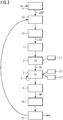
- a flowchart of a method for automatically selecting imaging parameters according to the present invention is shown schematically.
- reception 1 of image data BD takes place.
- the image data may have been created by an image processing device such as, for example, a magnetic resonance tomography (MRT) device, a computed tomography (CT) device, ultrasound device or the like.
- the image data BD can originate from an imaging module of the image processing device and can be received, for example, by a selection module of the image processing device.
- a ranking list RL of at least one qualifying value for potential clinical indications is created 2 in a second step b).
- the creation 2 can additionally be based on image data BD from previous iterations or also on image data from other imaging.
- the ranking list RL is created 2 by means of a detection algorithm (DA). The detection algorithm can be carried out, for example, by the selection module.
- DA detection algorithm
- the detection algorithm can be carried out, for example, by the selection module.
- at least one imaging parameter BP is selected 3 in a third step c).
- the at least one imaging parameter BP is suitable for generating image data BD in a subsequent (renewed) imaging (for example MRT scan), which enable a maximum change in the qualifying values in the ranking list RL.
- the at least one imaging parameter BP is selected 3 by means of a selection algorithm (selection algorithm) SA.
- the selection algorithm can be carried out, for example, by the selection module.
- the selected at least one imaging parameter BP is then transmitted in a fourth step d) by sending 4 the selected imaging parameter BP, for example from the selection module to the imaging module.
- the first to fourth steps are repeated iteratively until an abort criterion is met.
- Imaging parameters are automatically selected using a dynamic diagnostic exclusion protocol, which essentially comprises the detection algorithm DA and the selection algorithm SA.
- this information is returned to the selection algorithm SA by the detection algorithm DA.
- the returned information is thus used, for example, to improve the information in the data table 3.1 or to adapt the weights in the model (NN) 3.2.
- the iteration through the first to fourth step is terminated as soon as the termination criterion is met.
- the termination criterion specifies, for example, a minimum probability (T high indication ) with which a clinical indication must exist so that it can be confirmed. Conversely, a highest probability can also be specified as a termination criterion (T low indication ) up to which a clinical indication can be confirmed as not being present. Additionally or alternatively, a minimum confidence can be specified as a termination criterion (T confidence ), from which a statement about the probability of a clinical indication can be regarded as certain.
- a possible output of a ranking list RL of the detection algorithm DA based on image data BD from a patient's head is given below in an exemplary manner in a first table.
- clinical indication probability Confidence tumor 5% 3% multiple sclerosis 2% 5% Alzheimer 1 % 2% stroke 1 % 6%
- the qualifying values are given as an example in a ranking list RL after a further iteration.
- the selection algorithm SA has chosen, for example, an MRI sequence with which the clinical indication "stroke” can be excluded with certainty (high confidence) (low probability). This can be done using criterion b.1. can be achieved.
- clinical indication probability Confidence tumor 5% 3% multiple sclerosis 1 % 40% Alzheimer 1 % 60% stroke 0.1% 98%
- the ranking list RL from the second table has been updated in a subsequent iteration based on image data that originate from imaging with imaging parameters (MRI sequence) that also include all other clinical indications, if possible make it ascertainable with high security. This is due to the criterion a. possible. clinical indication probability Confidence tumor 10% 75% multiple sclerosis 1 % 80% Alzheimer 1 % 60% stroke 0.1% 99%
- FIG. 2 Figure 3 is a schematic flow diagram of an imaging method including the method for automatically selecting imaging parameters from Fig. 1 shown. Only the additional steps of the imaging method are described below.
- a first imaging step A at least one primary imaging parameter pBP is loaded 11, for example by an imaging module.
- a second Imaging step performing 12 imaging based on the loaded imaging parameter pBP / BP and generating image data BD (for example with an imaging device such as an MR scanner by its imaging module).
- the generated image data BD is then sent 13 in a third imaging step (for example, to a selection module of the MR scanner through its imaging module). This is followed by the four steps of the procedure for automatically selecting imaging parameters BP as previously described.
- a reception 14 of the selected imaging parameter BP which can originate from the selection module of the MR scanner, is carried out by its imaging module in a fourth imaging step.
- the received imaging parameter BP is loaded 15, for example by the imaging module of the MR scanner.
- the second to third imaging step, the first to fourth step of the method for automatically selecting imaging parameters and the fourth to fifth imaging step are carried out iteratively until the termination criterion as described above is met.
- FIG. 3 is a schematic of an imaging device comprising a control device that is set up to perform the imaging method Fig. 2 to perform.
- the imaging device 100 here an MRT device (MR scanner), comprises a control device 101 with an imaging module 102 and a selection module 103.
- the imaging module is designed and set up in such a way that it can carry out the first to fifth imaging steps.
- the selection module is designed and set up such that it can carry out the first to fourth step of the method for automatically selecting imaging parameters.
- the imaging module 102 sends generated image data BD to the selection module 103 and the selection module 103 sends selected imaging parameters BP and optionally determined ones Ranking lists RL to the imaging module 102.
- control device 101 can be made up of one or more data processing devices, such as, for. B. microcontrollers (pC), integrated circuits, application-specific integrated circuits (ASIC), application-specific standard products (ASSP), digital signal processors (DSP), programmable (logic) gates in the field Arrangement (Field Programmable Gate Arrays, FPGA) and the like can be constructed.
- the imaging module 102 and the selection module can be implemented on the data processing device (s) of the control device 101 or each can comprise its own data processing devices which are communicatively connected to the control unit 101.
- connection "or” is to be understood as a recording (“and / or") and not exclusive (“either ... or”).
Landscapes
- Engineering & Computer Science (AREA)
- Health & Medical Sciences (AREA)
- Physics & Mathematics (AREA)
- Life Sciences & Earth Sciences (AREA)
- Medical Informatics (AREA)
- General Physics & Mathematics (AREA)
- Nuclear Medicine, Radiotherapy & Molecular Imaging (AREA)
- General Health & Medical Sciences (AREA)
- Radiology & Medical Imaging (AREA)
- Theoretical Computer Science (AREA)
- Public Health (AREA)
- Biomedical Technology (AREA)
- High Energy & Nuclear Physics (AREA)
- Data Mining & Analysis (AREA)
- Veterinary Medicine (AREA)
- Surgery (AREA)
- Molecular Biology (AREA)
- Animal Behavior & Ethology (AREA)
- Heart & Thoracic Surgery (AREA)
- Biophysics (AREA)
- Pathology (AREA)
- Computer Vision & Pattern Recognition (AREA)
- Computational Mathematics (AREA)
- Mathematical Optimization (AREA)
- Pure & Applied Mathematics (AREA)
- Mathematical Analysis (AREA)
- Mathematical Physics (AREA)
- Quality & Reliability (AREA)
- Primary Health Care (AREA)
- Optics & Photonics (AREA)
- Epidemiology (AREA)
- Evolutionary Biology (AREA)
- Bioinformatics & Cheminformatics (AREA)
- Algebra (AREA)
- Probability & Statistics with Applications (AREA)
- Operations Research (AREA)
- Software Systems (AREA)
- Bioinformatics & Computational Biology (AREA)
- Databases & Information Systems (AREA)
- Condensed Matter Physics & Semiconductors (AREA)
Priority Applications (3)
| Application Number | Priority Date | Filing Date | Title |
|---|---|---|---|
| EP18196783.7A EP3629335A1 (fr) | 2018-09-26 | 2018-09-26 | Procédé de sélection automatique des paramètres d'imagerie pour un procédé d'imagerie |
| US16/583,568 US11099251B2 (en) | 2018-09-26 | 2019-09-26 | Method for automatic selection of imaging parameters for imaging methods |
| CN201910916605.8A CN110946578B (zh) | 2018-09-26 | 2019-09-26 | 自动选择成像参数的方法、相应的成像方法和成像设备 |
Applications Claiming Priority (1)
| Application Number | Priority Date | Filing Date | Title |
|---|---|---|---|
| EP18196783.7A EP3629335A1 (fr) | 2018-09-26 | 2018-09-26 | Procédé de sélection automatique des paramètres d'imagerie pour un procédé d'imagerie |
Publications (1)
| Publication Number | Publication Date |
|---|---|
| EP3629335A1 true EP3629335A1 (fr) | 2020-04-01 |
Family
ID=63685654
Family Applications (1)
| Application Number | Title | Priority Date | Filing Date |
|---|---|---|---|
| EP18196783.7A Withdrawn EP3629335A1 (fr) | 2018-09-26 | 2018-09-26 | Procédé de sélection automatique des paramètres d'imagerie pour un procédé d'imagerie |
Country Status (3)
| Country | Link |
|---|---|
| US (1) | US11099251B2 (fr) |
| EP (1) | EP3629335A1 (fr) |
| CN (1) | CN110946578B (fr) |
Families Citing this family (1)
| Publication number | Priority date | Publication date | Assignee | Title |
|---|---|---|---|---|
| KR102673155B1 (ko) * | 2021-11-12 | 2024-06-10 | 한국과학기술원 | 스코어 기반의 확산 모델을 이용한 자기공명영상 복원 방법 및 그 장치 |
Citations (3)
| Publication number | Priority date | Publication date | Assignee | Title |
|---|---|---|---|---|
| US20040254439A1 (en) * | 2003-06-11 | 2004-12-16 | Siemens Medical Solutions Usa, Inc. | System and method for adapting the behavior of a diagnostic medical ultrasound system based on anatomic features present in ultrasound images |
| US20090118614A1 (en) * | 2006-12-27 | 2009-05-07 | Fujifilm Corporation | Medical imaging system and method |
| US20150080733A1 (en) * | 2012-05-25 | 2015-03-19 | Fujifilm Corporation | Ultrasound diagnostic apparatus and ultrasound diagnostic image data processing method |
Family Cites Families (22)
| Publication number | Priority date | Publication date | Assignee | Title |
|---|---|---|---|---|
| DE69321653T2 (de) * | 1992-05-27 | 1999-05-20 | Koninklijke Philips Electronics N.V., Eindhoven | Verfahren und Gerät zur Bilderzeugung mittels magnetischer Resonanz |
| US20020097902A1 (en) * | 1993-09-29 | 2002-07-25 | Roehrig Jimmy R. | Method and system for the display of regions of interest in medical images |
| US6986019B1 (en) * | 2003-04-21 | 2006-01-10 | Maxtor Corporation | Method and apparatus for detection and management of data streams |
| US20050209882A1 (en) * | 2004-03-22 | 2005-09-22 | Jacobsen Jeffry B | Clinical data processing system |
| EP1736907A3 (fr) * | 2005-06-10 | 2016-07-06 | Siemens Healthcare GmbH | Amélioration de l'acquisition de données de mesure et de la reconstruction d'image pour les images MR |
| US20080253509A1 (en) * | 2005-10-06 | 2008-10-16 | Koninklijke Philips Electronics, N.V. | Acquisition Parameter Optimization For Csct |
| CN102113897B (zh) * | 2009-12-31 | 2014-10-15 | 深圳迈瑞生物医疗电子股份有限公司 | 一种在图像中提取及测量感兴趣目标的方法及其装置 |
| CN102654568A (zh) * | 2011-03-01 | 2012-09-05 | 西门子公司 | 用来确定对于磁共振成像的激励参数的方法和装置 |
| EP2791838B1 (fr) * | 2011-12-15 | 2019-10-16 | Koninklijke Philips N.V. | Reconstruction d'imagerie médicale optimisée pour destinataire |
| US8989465B2 (en) * | 2012-01-17 | 2015-03-24 | Mayo Foundation For Medical Education And Research | System and method for medical image reconstruction and image series denoising using local low rank promotion |
| DE102012205711B4 (de) * | 2012-04-05 | 2023-08-31 | Siemens Healthcare Gmbh | Verfahren zum Betreiben eines bildgebenden Diagnosegerätes sowie medizinisches bildgebendes System |
| US8965094B2 (en) * | 2012-04-14 | 2015-02-24 | Nocimed, Llc | Magnetic resonance spectroscopy pulse sequence, acquisition, and processing system and method |
| HUE063524T2 (hu) * | 2013-09-18 | 2024-01-28 | Siemens Medical Solutions Usa Inc | Eljárás és rendszer adatok statisztikai modellezésére kvadratikus valószínûségfüggvény használatával |
| CN103705239B (zh) * | 2013-12-05 | 2016-01-20 | 深圳先进技术研究院 | 磁共振参数成像方法和系统 |
| CN103654789B (zh) * | 2013-12-10 | 2015-12-30 | 深圳先进技术研究院 | 磁共振快速参数成像方法和系统 |
| JP6027065B2 (ja) * | 2014-08-21 | 2016-11-16 | 富士フイルム株式会社 | 類似画像検索装置、類似画像検索装置の作動方法、および類似画像検索プログラム |
| CN106510744B (zh) * | 2016-04-27 | 2021-01-08 | 上海联影医疗科技股份有限公司 | Pet扫描中多示踪剂动态参数的估计方法 |
| WO2018107371A1 (fr) * | 2016-12-13 | 2018-06-21 | 上海联影医疗科技有限公司 | Système et procédé de recherche d'image |
| CN106691486A (zh) * | 2016-12-30 | 2017-05-24 | 上海联影医疗科技有限公司 | 医学成像系统及方法 |
| US10478134B2 (en) * | 2017-09-26 | 2019-11-19 | General Electric Company | Systems and methods for improved diagnostics for nuclear medicine imaging |
| EP3467770B1 (fr) * | 2017-10-05 | 2022-11-23 | Siemens Healthcare GmbH | Procédé d'analyse d'un ensemble de données d'imagerie médicale, système d'analyse d'un ensemble de données d'imagerie médicale, produit-programme d'ordinateur et support lisible par ordinateur |
| JP7303677B2 (ja) * | 2019-07-03 | 2023-07-05 | キヤノンメディカルシステムズ株式会社 | 医用データ処理装置、医用データ処理方法、医用データ処理プログラム及び磁気共鳴イメージング装置 |
-
2018
- 2018-09-26 EP EP18196783.7A patent/EP3629335A1/fr not_active Withdrawn
-
2019
- 2019-09-26 CN CN201910916605.8A patent/CN110946578B/zh active Active
- 2019-09-26 US US16/583,568 patent/US11099251B2/en not_active Expired - Fee Related
Patent Citations (3)
| Publication number | Priority date | Publication date | Assignee | Title |
|---|---|---|---|---|
| US20040254439A1 (en) * | 2003-06-11 | 2004-12-16 | Siemens Medical Solutions Usa, Inc. | System and method for adapting the behavior of a diagnostic medical ultrasound system based on anatomic features present in ultrasound images |
| US20090118614A1 (en) * | 2006-12-27 | 2009-05-07 | Fujifilm Corporation | Medical imaging system and method |
| US20150080733A1 (en) * | 2012-05-25 | 2015-03-19 | Fujifilm Corporation | Ultrasound diagnostic apparatus and ultrasound diagnostic image data processing method |
Non-Patent Citations (1)
| Title |
|---|
| UNKNOWN: "Patient-centered CT imaging: New methods for patient-specific optimization", 17 December 2015 (2015-12-17), XP055528950, Retrieved from the Internet <URL:http://incenter.medical.philips.com/doclib/enc/fetch/2000/4504/577242/577249/586938/587315/iPatient_WP_HR.pdf?nodeid=13207522&vernum=-2> [retrieved on 20181130] * |
Also Published As
| Publication number | Publication date |
|---|---|
| US11099251B2 (en) | 2021-08-24 |
| CN110946578B (zh) | 2023-10-31 |
| US20200096586A1 (en) | 2020-03-26 |
| CN110946578A (zh) | 2020-04-03 |
Similar Documents
| Publication | Publication Date | Title |
|---|---|---|
| EP3301642B1 (fr) | Vérification d'image automatisée dans l'imagerie par rayons x | |
| DE69629732T2 (de) | Vorrichtung zur rechnerunterstützten Diagnose | |
| DE102005004383B4 (de) | Verfahren und Vorrichtung zur Steuerung einer bildgebenden Modalität | |
| EP3143592B1 (fr) | Procédé et dispositif de réduction d'artéfacts dans des images obtenues par tomodensitométrie | |
| EP2648122B1 (fr) | Procédé de chargement de données d'images médicales ainsi que le dispositif de réalisation du procédé | |
| DE102019113493A1 (de) | Abgestimmte medizinische ultraschallbildgebung | |
| DE102011002928A1 (de) | Verfahren zur rechnergestützten Konfiguration einer medizinischen Bildgebungsvorrichtung | |
| DE102007028226B4 (de) | Auswertungsverfahren für eine zeitliche Sequenz von Röntgenbildern und hiermit korrespondierende Gegenstände | |
| EP3540632B1 (fr) | Procédé pour la classification des échantillons tissulaires | |
| DE102022201347A1 (de) | Verfahren und System zur automatisierten Bestimmung von Untersuchungsergebnissen in einer Bildsequenz | |
| DE69819269T2 (de) | Bildverarbeitungssystem | |
| DE10349661B4 (de) | Einrichtung und Verfahren zur Überwachung der Parameterwahl beim Betrieb eines technischen Gerätes | |
| DE102020206059A1 (de) | Computerimplementiertes Verfahren und System zum Trainieren eines Auswertungsalgorithmus, Computerprogramm und elektronisch lesbarer Datenträger | |
| EP3843011A1 (fr) | Système et procédé d'assurance qualité des modèles à base de données | |
| EP1519301A2 (fr) | Dispositif de classification d'événements physiologiques | |
| EP3547254A1 (fr) | Procédé d'analyse et unité d'analyse permettant de déterminer des données de résultats radiologiques | |
| EP3629335A1 (fr) | Procédé de sélection automatique des paramètres d'imagerie pour un procédé d'imagerie | |
| DE102004056589A1 (de) | Verfahren und Vorrichtung zur Durchführung segmentierungsbasierter Bildoperationen | |
| EP1981582A1 (fr) | Procédé, dispositif et programme informatique utilisés pour produire un signal de commande pour un implant cochléaire sur la base d'un signal audio | |
| DE102005034160A1 (de) | Verfahren zur Optimierung der Durchführung von Messungen | |
| EP4332982A1 (fr) | Dispositif de commande et procédé de commande d'un système d'imagerie médicotechnique | |
| DE112018003757T5 (de) | Nervenimpulsabtastung für hochdichte implantierbare neuraleaufzeichnungssysteme | |
| DE102005055657A1 (de) | Verfahren zum Betreiben einer medizinischen Diagnoseeinrichtung sowie medizinische Diagnoseeinrichtung | |
| DE202021102338U1 (de) | Steuergerät zum Erzeugen von Trainingsdaten zum Trainieren eines Algorithmus des maschinellen Lernens | |
| DE102006025422B4 (de) | Bildauswertungsverfahren für zweidimensionale Projektionsbilder und hiermit korrespondierende Gegenstände |
Legal Events
| Date | Code | Title | Description |
|---|---|---|---|
| PUAI | Public reference made under article 153(3) epc to a published international application that has entered the european phase |
Free format text: ORIGINAL CODE: 0009012 |
|
| STAA | Information on the status of an ep patent application or granted ep patent |
Free format text: STATUS: THE APPLICATION HAS BEEN PUBLISHED |
|
| AK | Designated contracting states |
Kind code of ref document: A1 Designated state(s): AL AT BE BG CH CY CZ DE DK EE ES FI FR GB GR HR HU IE IS IT LI LT LU LV MC MK MT NL NO PL PT RO RS SE SI SK SM TR |
|
| AX | Request for extension of the european patent |
Extension state: BA ME |
|
| STAA | Information on the status of an ep patent application or granted ep patent |
Free format text: STATUS: REQUEST FOR EXAMINATION WAS MADE |
|
| 17P | Request for examination filed |
Effective date: 20200928 |
|
| RBV | Designated contracting states (corrected) |
Designated state(s): AL AT BE BG CH CY CZ DE DK EE ES FI FR GB GR HR HU IE IS IT LI LT LU LV MC MK MT NL NO PL PT RO RS SE SI SK SM TR |
|
| STAA | Information on the status of an ep patent application or granted ep patent |
Free format text: STATUS: EXAMINATION IS IN PROGRESS |
|
| 17Q | First examination report despatched |
Effective date: 20230503 |
|
| STAA | Information on the status of an ep patent application or granted ep patent |
Free format text: STATUS: THE APPLICATION HAS BEEN WITHDRAWN |
|
| 18W | Application withdrawn |
Effective date: 20230901 |