EP3633029A2 - Method for culturing natural killer cell, using transformed t cell - Google Patents
Method for culturing natural killer cell, using transformed t cell Download PDFInfo
- Publication number
- EP3633029A2 EP3633029A2 EP18805231.0A EP18805231A EP3633029A2 EP 3633029 A2 EP3633029 A2 EP 3633029A2 EP 18805231 A EP18805231 A EP 18805231A EP 3633029 A2 EP3633029 A2 EP 3633029A2
- Authority
- EP
- European Patent Office
- Prior art keywords
- cells
- gene
- cell
- natural killer
- killer cells
- Prior art date
- Legal status (The legal status is an assumption and is not a legal conclusion. Google has not performed a legal analysis and makes no representation as to the accuracy of the status listed.)
- Pending
Links
- 210000004027 cell Anatomy 0.000 title claims abstract description 275
- 210000000822 natural killer cell Anatomy 0.000 title claims abstract description 219
- 238000012258 culturing Methods 0.000 title claims abstract description 105
- 238000000034 method Methods 0.000 title abstract description 27
- 210000001744 T-lymphocyte Anatomy 0.000 claims abstract description 176
- 108090000623 proteins and genes Proteins 0.000 claims description 349
- 210000003819 peripheral blood mononuclear cell Anatomy 0.000 claims description 116
- 108010082808 4-1BB Ligand Proteins 0.000 claims description 68
- 108010042215 OX40 Ligand Proteins 0.000 claims description 41
- 238000012136 culture method Methods 0.000 claims description 29
- 239000002773 nucleotide Substances 0.000 claims description 24
- 125000003729 nucleotide group Chemical group 0.000 claims description 24
- 125000003275 alpha amino acid group Chemical group 0.000 claims description 18
- 108010002350 Interleukin-2 Proteins 0.000 claims description 11
- 239000000203 mixture Substances 0.000 claims description 11
- 239000004480 active ingredient Substances 0.000 claims description 8
- 241000713666 Lentivirus Species 0.000 claims description 7
- 102000015696 Interleukins Human genes 0.000 claims description 6
- 108010063738 Interleukins Proteins 0.000 claims description 6
- 210000005259 peripheral blood Anatomy 0.000 claims description 4
- 239000011886 peripheral blood Substances 0.000 claims description 4
- FWMNVWWHGCHHJJ-SKKKGAJSSA-N 4-amino-1-[(2r)-6-amino-2-[[(2r)-2-[[(2r)-2-[[(2r)-2-amino-3-phenylpropanoyl]amino]-3-phenylpropanoyl]amino]-4-methylpentanoyl]amino]hexanoyl]piperidine-4-carboxylic acid Chemical compound C([C@H](C(=O)N[C@H](CC(C)C)C(=O)N[C@H](CCCCN)C(=O)N1CCC(N)(CC1)C(O)=O)NC(=O)[C@H](N)CC=1C=CC=CC=1)C1=CC=CC=C1 FWMNVWWHGCHHJJ-SKKKGAJSSA-N 0.000 claims description 3
- 229940127174 UCHT1 Drugs 0.000 claims description 2
- 210000000265 leukocyte Anatomy 0.000 claims description 2
- 230000035755 proliferation Effects 0.000 abstract description 36
- 238000004519 manufacturing process Methods 0.000 abstract description 2
- 238000002659 cell therapy Methods 0.000 abstract 2
- 230000001461 cytolytic effect Effects 0.000 abstract 1
- 238000010361 transduction Methods 0.000 description 116
- 230000026683 transduction Effects 0.000 description 116
- 230000014509 gene expression Effects 0.000 description 90
- 102100032101 Tumor necrosis factor ligand superfamily member 9 Human genes 0.000 description 52
- 239000003550 marker Substances 0.000 description 46
- 238000001943 fluorescence-activated cell sorting Methods 0.000 description 40
- 206010028980 Neoplasm Diseases 0.000 description 33
- 102100026890 Tumor necrosis factor ligand superfamily member 4 Human genes 0.000 description 32
- 239000002609 medium Substances 0.000 description 28
- 239000000872 buffer Substances 0.000 description 25
- 238000005119 centrifugation Methods 0.000 description 21
- 201000011510 cancer Diseases 0.000 description 19
- 230000000694 effects Effects 0.000 description 17
- 230000022534 cell killing Effects 0.000 description 16
- 239000006228 supernatant Substances 0.000 description 16
- 239000012980 RPMI-1640 medium Substances 0.000 description 14
- 108060008682 Tumor Necrosis Factor Proteins 0.000 description 12
- 102000000852 Tumor Necrosis Factor-alpha Human genes 0.000 description 12
- 238000004458 analytical method Methods 0.000 description 11
- 239000011259 mixed solution Substances 0.000 description 11
- MZOFCQQQCNRIBI-VMXHOPILSA-N (3s)-4-[[(2s)-1-[[(2s)-1-[[(1s)-1-carboxy-2-hydroxyethyl]amino]-4-methyl-1-oxopentan-2-yl]amino]-5-(diaminomethylideneamino)-1-oxopentan-2-yl]amino]-3-[[2-[[(2s)-2,6-diaminohexanoyl]amino]acetyl]amino]-4-oxobutanoic acid Chemical compound OC[C@@H](C(O)=O)NC(=O)[C@H](CC(C)C)NC(=O)[C@H](CCCN=C(N)N)NC(=O)[C@H](CC(O)=O)NC(=O)CNC(=O)[C@@H](N)CCCCN MZOFCQQQCNRIBI-VMXHOPILSA-N 0.000 description 10
- 108020004414 DNA Proteins 0.000 description 10
- 101000581981 Homo sapiens Neural cell adhesion molecule 1 Proteins 0.000 description 10
- 102100027347 Neural cell adhesion molecule 1 Human genes 0.000 description 10
- RXWNCPJZOCPEPQ-NVWDDTSBSA-N puromycin Chemical compound C1=CC(OC)=CC=C1C[C@H](N)C(=O)N[C@H]1[C@@H](O)[C@H](N2C3=NC=NC(=C3N=C2)N(C)C)O[C@@H]1CO RXWNCPJZOCPEPQ-NVWDDTSBSA-N 0.000 description 10
- 239000013598 vector Substances 0.000 description 10
- 102000004127 Cytokines Human genes 0.000 description 9
- 108090000695 Cytokines Proteins 0.000 description 9
- 230000004913 activation Effects 0.000 description 9
- 238000010790 dilution Methods 0.000 description 9
- 239000012895 dilution Substances 0.000 description 9
- 239000008188 pellet Substances 0.000 description 9
- 108010004217 Natural Cytotoxicity Triggering Receptor 1 Proteins 0.000 description 8
- 102100032870 Natural cytotoxicity triggering receptor 1 Human genes 0.000 description 8
- 230000000638 stimulation Effects 0.000 description 8
- 102100038077 CD226 antigen Human genes 0.000 description 7
- 101000884298 Homo sapiens CD226 antigen Proteins 0.000 description 7
- 101000917858 Homo sapiens Low affinity immunoglobulin gamma Fc region receptor III-A Proteins 0.000 description 7
- 101000917839 Homo sapiens Low affinity immunoglobulin gamma Fc region receptor III-B Proteins 0.000 description 7
- 101001109501 Homo sapiens NKG2-D type II integral membrane protein Proteins 0.000 description 7
- 101000589305 Homo sapiens Natural cytotoxicity triggering receptor 2 Proteins 0.000 description 7
- 102100029185 Low affinity immunoglobulin gamma Fc region receptor III-B Human genes 0.000 description 7
- 102100022680 NKG2-D type II integral membrane protein Human genes 0.000 description 7
- 108010004222 Natural Cytotoxicity Triggering Receptor 3 Proteins 0.000 description 7
- 102100032851 Natural cytotoxicity triggering receptor 2 Human genes 0.000 description 7
- 102100032852 Natural cytotoxicity triggering receptor 3 Human genes 0.000 description 7
- 239000003814 drug Substances 0.000 description 7
- 230000002062 proliferating effect Effects 0.000 description 7
- 238000005057 refrigeration Methods 0.000 description 7
- 239000000243 solution Substances 0.000 description 7
- ORILYTVJVMAKLC-UHFFFAOYSA-N Adamantane Natural products C1C(C2)CC3CC1CC2C3 ORILYTVJVMAKLC-UHFFFAOYSA-N 0.000 description 6
- 102100028990 C-X-C chemokine receptor type 3 Human genes 0.000 description 6
- 101000916050 Homo sapiens C-X-C chemokine receptor type 3 Proteins 0.000 description 6
- 238000006243 chemical reaction Methods 0.000 description 6
- 208000015181 infectious disease Diseases 0.000 description 6
- 239000012528 membrane Substances 0.000 description 6
- 230000002265 prevention Effects 0.000 description 6
- 210000002966 serum Anatomy 0.000 description 6
- 238000010186 staining Methods 0.000 description 6
- 229940124597 therapeutic agent Drugs 0.000 description 6
- 239000011534 wash buffer Substances 0.000 description 6
- 102000043279 ADAM17 Human genes 0.000 description 5
- 108091007505 ADAM17 Proteins 0.000 description 5
- 208000035473 Communicable disease Diseases 0.000 description 5
- 230000003833 cell viability Effects 0.000 description 5
- 238000002826 magnetic-activated cell sorting Methods 0.000 description 5
- 239000002953 phosphate buffered saline Substances 0.000 description 5
- 229950010131 puromycin Drugs 0.000 description 5
- 108020003175 receptors Proteins 0.000 description 5
- 102000005962 receptors Human genes 0.000 description 5
- 239000012146 running buffer Substances 0.000 description 5
- 230000003248 secreting effect Effects 0.000 description 5
- 230000035899 viability Effects 0.000 description 5
- YXHLJMWYDTXDHS-IRFLANFNSA-N 7-aminoactinomycin D Chemical compound C[C@H]1OC(=O)[C@H](C(C)C)N(C)C(=O)CN(C)C(=O)[C@@H]2CCCN2C(=O)[C@@H](C(C)C)NC(=O)[C@H]1NC(=O)C1=C(N)C(=O)C(C)=C2OC(C(C)=C(N)C=C3C(=O)N[C@@H]4C(=O)N[C@@H](C(N5CCC[C@H]5C(=O)N(C)CC(=O)N(C)[C@@H](C(C)C)C(=O)O[C@@H]4C)=O)C(C)C)=C3N=C21 YXHLJMWYDTXDHS-IRFLANFNSA-N 0.000 description 4
- 108700012813 7-aminoactinomycin D Proteins 0.000 description 4
- 101001057504 Homo sapiens Interferon-stimulated gene 20 kDa protein Proteins 0.000 description 4
- 101001055144 Homo sapiens Interleukin-2 receptor subunit alpha Proteins 0.000 description 4
- 101001023379 Homo sapiens Lysosome-associated membrane glycoprotein 1 Proteins 0.000 description 4
- 102100037850 Interferon gamma Human genes 0.000 description 4
- 108010074328 Interferon-gamma Proteins 0.000 description 4
- 102100026878 Interleukin-2 receptor subunit alpha Human genes 0.000 description 4
- 102100035133 Lysosome-associated membrane glycoprotein 1 Human genes 0.000 description 4
- 101710187830 Tumor necrosis factor receptor superfamily member 1B Proteins 0.000 description 4
- 102100033733 Tumor necrosis factor receptor superfamily member 1B Human genes 0.000 description 4
- 239000000427 antigen Substances 0.000 description 4
- 108091007433 antigens Proteins 0.000 description 4
- 102000036639 antigens Human genes 0.000 description 4
- 238000003501 co-culture Methods 0.000 description 4
- 238000010276 construction Methods 0.000 description 4
- 239000013604 expression vector Substances 0.000 description 4
- MHMNJMPURVTYEJ-UHFFFAOYSA-N fluorescein-5-isothiocyanate Chemical compound O1C(=O)C2=CC(N=C=S)=CC=C2C21C1=CC=C(O)C=C1OC1=CC(O)=CC=C21 MHMNJMPURVTYEJ-UHFFFAOYSA-N 0.000 description 4
- UYTPUPDQBNUYGX-UHFFFAOYSA-N guanine Chemical compound O=C1NC(N)=NC2=C1N=CN2 UYTPUPDQBNUYGX-UHFFFAOYSA-N 0.000 description 4
- 238000011534 incubation Methods 0.000 description 4
- 230000002147 killing effect Effects 0.000 description 4
- 230000035772 mutation Effects 0.000 description 4
- 230000001105 regulatory effect Effects 0.000 description 4
- 230000002441 reversible effect Effects 0.000 description 4
- 230000003068 static effect Effects 0.000 description 4
- 210000004881 tumor cell Anatomy 0.000 description 4
- 239000004474 valine Substances 0.000 description 4
- 108091026890 Coding region Proteins 0.000 description 3
- 101000638251 Homo sapiens Tumor necrosis factor ligand superfamily member 9 Proteins 0.000 description 3
- 239000012097 Lipofectamine 2000 Substances 0.000 description 3
- 206010025323 Lymphomas Diseases 0.000 description 3
- KWYUFKZDYYNOTN-UHFFFAOYSA-M Potassium hydroxide Chemical compound [OH-].[K+] KWYUFKZDYYNOTN-UHFFFAOYSA-M 0.000 description 3
- HEMHJVSKTPXQMS-UHFFFAOYSA-M Sodium hydroxide Chemical compound [OH-].[Na+] HEMHJVSKTPXQMS-UHFFFAOYSA-M 0.000 description 3
- 108091008874 T cell receptors Proteins 0.000 description 3
- 102000016266 T-Cell Antigen Receptors Human genes 0.000 description 3
- 241000700605 Viruses Species 0.000 description 3
- 230000002159 abnormal effect Effects 0.000 description 3
- 108700025316 aldesleukin Proteins 0.000 description 3
- 229930189065 blasticidin Natural products 0.000 description 3
- 210000000170 cell membrane Anatomy 0.000 description 3
- 230000003247 decreasing effect Effects 0.000 description 3
- 238000005516 engineering process Methods 0.000 description 3
- 238000002474 experimental method Methods 0.000 description 3
- 239000004615 ingredient Substances 0.000 description 3
- 210000004698 lymphocyte Anatomy 0.000 description 3
- 229920002113 octoxynol Polymers 0.000 description 3
- 239000008194 pharmaceutical composition Substances 0.000 description 3
- 239000013612 plasmid Substances 0.000 description 3
- 229940087463 proleukin Drugs 0.000 description 3
- 230000002195 synergetic effect Effects 0.000 description 3
- 102000035160 transmembrane proteins Human genes 0.000 description 3
- 108091005703 transmembrane proteins Proteins 0.000 description 3
- UZOVYGYOLBIAJR-UHFFFAOYSA-N 4-isocyanato-4'-methyldiphenylmethane Chemical compound C1=CC(C)=CC=C1CC1=CC=C(N=C=O)C=C1 UZOVYGYOLBIAJR-UHFFFAOYSA-N 0.000 description 2
- QGZKDVFQNNGYKY-UHFFFAOYSA-N Ammonia Chemical compound N QGZKDVFQNNGYKY-UHFFFAOYSA-N 0.000 description 2
- IJGRMHOSHXDMSA-UHFFFAOYSA-N Atomic nitrogen Chemical compound N#N IJGRMHOSHXDMSA-UHFFFAOYSA-N 0.000 description 2
- 239000006144 Dulbecco’s modified Eagle's medium Substances 0.000 description 2
- KCXVZYZYPLLWCC-UHFFFAOYSA-N EDTA Chemical compound OC(=O)CN(CC(O)=O)CCN(CC(O)=O)CC(O)=O KCXVZYZYPLLWCC-UHFFFAOYSA-N 0.000 description 2
- 102100025137 Early activation antigen CD69 Human genes 0.000 description 2
- 102100021260 Galactosylgalactosylxylosylprotein 3-beta-glucuronosyltransferase 1 Human genes 0.000 description 2
- 101000934374 Homo sapiens Early activation antigen CD69 Proteins 0.000 description 2
- 101000894906 Homo sapiens Galactosylgalactosylxylosylprotein 3-beta-glucuronosyltransferase 1 Proteins 0.000 description 2
- 101001018097 Homo sapiens L-selectin Proteins 0.000 description 2
- 101001109503 Homo sapiens NKG2-C type II integral membrane protein Proteins 0.000 description 2
- 101000764263 Homo sapiens Tumor necrosis factor ligand superfamily member 4 Proteins 0.000 description 2
- 102100034343 Integrase Human genes 0.000 description 2
- 101150069255 KLRC1 gene Proteins 0.000 description 2
- 102100033467 L-selectin Human genes 0.000 description 2
- 101100404845 Macaca mulatta NKG2A gene Proteins 0.000 description 2
- 102100022682 NKG2-A/NKG2-B type II integral membrane protein Human genes 0.000 description 2
- 102100022683 NKG2-C type II integral membrane protein Human genes 0.000 description 2
- NBIIXXVUZAFLBC-UHFFFAOYSA-N Phosphoric acid Chemical compound OP(O)(O)=O NBIIXXVUZAFLBC-UHFFFAOYSA-N 0.000 description 2
- 108010092799 RNA-directed DNA polymerase Proteins 0.000 description 2
- QAOWNCQODCNURD-UHFFFAOYSA-N Sulfuric acid Chemical compound OS(O)(=O)=O QAOWNCQODCNURD-UHFFFAOYSA-N 0.000 description 2
- 108700019146 Transgenes Proteins 0.000 description 2
- 102100040247 Tumor necrosis factor Human genes 0.000 description 2
- 239000003242 anti bacterial agent Substances 0.000 description 2
- 229940088710 antibiotic agent Drugs 0.000 description 2
- 238000010923 batch production Methods 0.000 description 2
- 238000002306 biochemical method Methods 0.000 description 2
- BQRGNLJZBFXNCZ-UHFFFAOYSA-N calcein am Chemical compound O1C(=O)C2=CC=CC=C2C21C1=CC(CN(CC(=O)OCOC(C)=O)CC(=O)OCOC(C)=O)=C(OC(C)=O)C=C1OC1=C2C=C(CN(CC(=O)OCOC(C)=O)CC(=O)OCOC(=O)C)C(OC(C)=O)=C1 BQRGNLJZBFXNCZ-UHFFFAOYSA-N 0.000 description 2
- OPTASPLRGRRNAP-UHFFFAOYSA-N cytosine Chemical compound NC=1C=CNC(=O)N=1 OPTASPLRGRRNAP-UHFFFAOYSA-N 0.000 description 2
- 210000001151 cytotoxic T lymphocyte Anatomy 0.000 description 2
- 201000010099 disease Diseases 0.000 description 2
- 208000037265 diseases, disorders, signs and symptoms Diseases 0.000 description 2
- 230000008014 freezing Effects 0.000 description 2
- 238000007710 freezing Methods 0.000 description 2
- 230000005251 gamma ray Effects 0.000 description 2
- 239000003102 growth factor Substances 0.000 description 2
- 239000001963 growth medium Substances 0.000 description 2
- 238000009169 immunotherapy Methods 0.000 description 2
- 230000003834 intracellular effect Effects 0.000 description 2
- 239000003446 ligand Substances 0.000 description 2
- 230000001404 mediated effect Effects 0.000 description 2
- 230000008823 permeabilization Effects 0.000 description 2
- 229920003023 plastic Polymers 0.000 description 2
- 230000008569 process Effects 0.000 description 2
- 235000018102 proteins Nutrition 0.000 description 2
- 102000004169 proteins and genes Human genes 0.000 description 2
- 230000028327 secretion Effects 0.000 description 2
- 238000004114 suspension culture Methods 0.000 description 2
- 238000005406 washing Methods 0.000 description 2
- IJRKANNOPXMZSG-SSPAHAAFSA-N 2-hydroxypropane-1,2,3-tricarboxylic acid;(2r,3s,4r,5r)-2,3,4,5,6-pentahydroxyhexanal Chemical compound OC[C@@H](O)[C@@H](O)[C@H](O)[C@@H](O)C=O.OC(=O)CC(O)(C(O)=O)CC(O)=O IJRKANNOPXMZSG-SSPAHAAFSA-N 0.000 description 1
- 229930024421 Adenine Natural products 0.000 description 1
- GFFGJBXGBJISGV-UHFFFAOYSA-N Adenine Chemical compound NC1=NC=NC2=C1N=CN2 GFFGJBXGBJISGV-UHFFFAOYSA-N 0.000 description 1
- 241000283690 Bos taurus Species 0.000 description 1
- 101100161935 Caenorhabditis elegans act-4 gene Proteins 0.000 description 1
- 201000009030 Carcinoma Diseases 0.000 description 1
- 229920000209 Hexadimethrine bromide Polymers 0.000 description 1
- QNAYBMKLOCPYGJ-REOHCLBHSA-N L-alanine Chemical compound C[C@H](N)C(O)=O QNAYBMKLOCPYGJ-REOHCLBHSA-N 0.000 description 1
- 241001465754 Metazoa Species 0.000 description 1
- 108091028043 Nucleic acid sequence Proteins 0.000 description 1
- 102000004473 OX40 Ligand Human genes 0.000 description 1
- 206010035226 Plasma cell myeloma Diseases 0.000 description 1
- ONIBWKKTOPOVIA-UHFFFAOYSA-N Proline Natural products OC(=O)C1CCCN1 ONIBWKKTOPOVIA-UHFFFAOYSA-N 0.000 description 1
- 206010039491 Sarcoma Diseases 0.000 description 1
- BQCADISMDOOEFD-UHFFFAOYSA-N Silver Chemical compound [Ag] BQCADISMDOOEFD-UHFFFAOYSA-N 0.000 description 1
- 102100022153 Tumor necrosis factor receptor superfamily member 4 Human genes 0.000 description 1
- 101710165473 Tumor necrosis factor receptor superfamily member 4 Proteins 0.000 description 1
- 150000007513 acids Chemical class 0.000 description 1
- 230000003213 activating effect Effects 0.000 description 1
- 229960000643 adenine Drugs 0.000 description 1
- 235000004279 alanine Nutrition 0.000 description 1
- 229910000147 aluminium phosphate Inorganic materials 0.000 description 1
- 229910021529 ammonia Inorganic materials 0.000 description 1
- 210000004102 animal cell Anatomy 0.000 description 1
- 230000001093 anti-cancer Effects 0.000 description 1
- 150000007514 bases Chemical class 0.000 description 1
- 239000011324 bead Substances 0.000 description 1
- 230000003115 biocidal effect Effects 0.000 description 1
- 229940088623 biologically active substance Drugs 0.000 description 1
- 230000015572 biosynthetic process Effects 0.000 description 1
- 210000004369 blood Anatomy 0.000 description 1
- 239000008280 blood Substances 0.000 description 1
- 210000001124 body fluid Anatomy 0.000 description 1
- 239000010839 body fluid Substances 0.000 description 1
- 239000006143 cell culture medium Substances 0.000 description 1
- 230000003915 cell function Effects 0.000 description 1
- 238000010367 cloning Methods 0.000 description 1
- 238000007796 conventional method Methods 0.000 description 1
- 229940104302 cytosine Drugs 0.000 description 1
- 210000004443 dendritic cell Anatomy 0.000 description 1
- 230000037213 diet Effects 0.000 description 1
- 235000005911 diet Nutrition 0.000 description 1
- LOKCTEFSRHRXRJ-UHFFFAOYSA-I dipotassium trisodium dihydrogen phosphate hydrogen phosphate dichloride Chemical compound P(=O)(O)(O)[O-].[K+].P(=O)(O)([O-])[O-].[Na+].[Na+].[Cl-].[K+].[Cl-].[Na+] LOKCTEFSRHRXRJ-UHFFFAOYSA-I 0.000 description 1
- 239000012153 distilled water Substances 0.000 description 1
- 229940079593 drug Drugs 0.000 description 1
- 239000003937 drug carrier Substances 0.000 description 1
- 230000007613 environmental effect Effects 0.000 description 1
- 230000017188 evasion or tolerance of host immune response Effects 0.000 description 1
- 230000001605 fetal effect Effects 0.000 description 1
- 238000009472 formulation Methods 0.000 description 1
- 239000012595 freezing medium Substances 0.000 description 1
- 230000006870 function Effects 0.000 description 1
- 230000007849 functional defect Effects 0.000 description 1
- 108020001507 fusion proteins Proteins 0.000 description 1
- 102000037865 fusion proteins Human genes 0.000 description 1
- 238000001476 gene delivery Methods 0.000 description 1
- 230000002068 genetic effect Effects 0.000 description 1
- 230000036541 health Effects 0.000 description 1
- 201000005787 hematologic cancer Diseases 0.000 description 1
- 208000024200 hematopoietic and lymphoid system neoplasm Diseases 0.000 description 1
- 210000004408 hybridoma Anatomy 0.000 description 1
- 230000036737 immune function Effects 0.000 description 1
- 230000001771 impaired effect Effects 0.000 description 1
- 238000000338 in vitro Methods 0.000 description 1
- 230000002779 inactivation Effects 0.000 description 1
- 239000003112 inhibitor Substances 0.000 description 1
- 230000015788 innate immune response Effects 0.000 description 1
- 208000032839 leukemia Diseases 0.000 description 1
- 150000002632 lipids Chemical class 0.000 description 1
- 239000007788 liquid Substances 0.000 description 1
- 210000002540 macrophage Anatomy 0.000 description 1
- -1 mbIL-21 Proteins 0.000 description 1
- 238000005259 measurement Methods 0.000 description 1
- 230000007246 mechanism Effects 0.000 description 1
- 230000002503 metabolic effect Effects 0.000 description 1
- 239000002207 metabolite Substances 0.000 description 1
- 239000010445 mica Substances 0.000 description 1
- 229910052618 mica group Inorganic materials 0.000 description 1
- 238000002156 mixing Methods 0.000 description 1
- 239000003068 molecular probe Substances 0.000 description 1
- 210000001616 monocyte Anatomy 0.000 description 1
- 201000000050 myeloid neoplasm Diseases 0.000 description 1
- 229910052757 nitrogen Inorganic materials 0.000 description 1
- 210000005105 peripheral blood lymphocyte Anatomy 0.000 description 1
- 238000000053 physical method Methods 0.000 description 1
- 239000002243 precursor Substances 0.000 description 1
- 238000002360 preparation method Methods 0.000 description 1
- 230000002035 prolonged effect Effects 0.000 description 1
- 239000002994 raw material Substances 0.000 description 1
- 108091008146 restriction endonucleases Proteins 0.000 description 1
- 239000000523 sample Substances 0.000 description 1
- 238000012163 sequencing technique Methods 0.000 description 1
- 230000002269 spontaneous effect Effects 0.000 description 1
- 239000012192 staining solution Substances 0.000 description 1
- 230000002459 sustained effect Effects 0.000 description 1
- 238000003786 synthesis reaction Methods 0.000 description 1
- 230000008685 targeting Effects 0.000 description 1
- 238000002560 therapeutic procedure Methods 0.000 description 1
- 238000001890 transfection Methods 0.000 description 1
- 238000003151 transfection method Methods 0.000 description 1
- 230000009261 transgenic effect Effects 0.000 description 1
- 230000032258 transport Effects 0.000 description 1
- 239000013638 trimer Substances 0.000 description 1
- 241001430294 unidentified retrovirus Species 0.000 description 1
- XLYOFNOQVPJJNP-UHFFFAOYSA-N water Chemical compound O XLYOFNOQVPJJNP-UHFFFAOYSA-N 0.000 description 1
Images


Classifications
-
- C—CHEMISTRY; METALLURGY
- C12—BIOCHEMISTRY; BEER; SPIRITS; WINE; VINEGAR; MICROBIOLOGY; ENZYMOLOGY; MUTATION OR GENETIC ENGINEERING
- C12N—MICROORGANISMS OR ENZYMES; COMPOSITIONS THEREOF; PROPAGATING, PRESERVING, OR MAINTAINING MICROORGANISMS; MUTATION OR GENETIC ENGINEERING; CULTURE MEDIA
- C12N5/00—Undifferentiated human, animal or plant cells, e.g. cell lines; Tissues; Cultivation or maintenance thereof; Culture media therefor
- C12N5/06—Animal cells or tissues; Human cells or tissues
- C12N5/0602—Vertebrate cells
- C12N5/0634—Cells from the blood or the immune system
- C12N5/0646—Natural killers cells [NK], NKT cells
-
- A—HUMAN NECESSITIES
- A61—MEDICAL OR VETERINARY SCIENCE; HYGIENE
- A61K—PREPARATIONS FOR MEDICAL, DENTAL OR TOILETRY PURPOSES
- A61K40/00—Cellular immunotherapy
- A61K40/10—Cellular immunotherapy characterised by the cell type used
- A61K40/15—Natural-killer [NK] cells; Natural-killer T [NKT] cells
-
- A—HUMAN NECESSITIES
- A61—MEDICAL OR VETERINARY SCIENCE; HYGIENE
- A61K—PREPARATIONS FOR MEDICAL, DENTAL OR TOILETRY PURPOSES
- A61K40/00—Cellular immunotherapy
- A61K40/40—Cellular immunotherapy characterised by antigens that are targeted or presented by cells of the immune system
- A61K40/41—Vertebrate antigens
- A61K40/42—Cancer antigens
-
- C—CHEMISTRY; METALLURGY
- C12—BIOCHEMISTRY; BEER; SPIRITS; WINE; VINEGAR; MICROBIOLOGY; ENZYMOLOGY; MUTATION OR GENETIC ENGINEERING
- C12N—MICROORGANISMS OR ENZYMES; COMPOSITIONS THEREOF; PROPAGATING, PRESERVING, OR MAINTAINING MICROORGANISMS; MUTATION OR GENETIC ENGINEERING; CULTURE MEDIA
- C12N5/00—Undifferentiated human, animal or plant cells, e.g. cell lines; Tissues; Cultivation or maintenance thereof; Culture media therefor
- C12N5/06—Animal cells or tissues; Human cells or tissues
- C12N5/0602—Vertebrate cells
- C12N5/0634—Cells from the blood or the immune system
- C12N5/0636—T lymphocytes
- C12N5/0638—Cytotoxic T lymphocytes [CTL] or lymphokine activated killer cells [LAK]
-
- C—CHEMISTRY; METALLURGY
- C12—BIOCHEMISTRY; BEER; SPIRITS; WINE; VINEGAR; MICROBIOLOGY; ENZYMOLOGY; MUTATION OR GENETIC ENGINEERING
- C12N—MICROORGANISMS OR ENZYMES; COMPOSITIONS THEREOF; PROPAGATING, PRESERVING, OR MAINTAINING MICROORGANISMS; MUTATION OR GENETIC ENGINEERING; CULTURE MEDIA
- C12N2501/00—Active agents used in cell culture processes, e.g. differentation
- C12N2501/20—Cytokines; Chemokines
- C12N2501/23—Interleukins [IL]
- C12N2501/2302—Interleukin-2 (IL-2)
-
- C—CHEMISTRY; METALLURGY
- C12—BIOCHEMISTRY; BEER; SPIRITS; WINE; VINEGAR; MICROBIOLOGY; ENZYMOLOGY; MUTATION OR GENETIC ENGINEERING
- C12N—MICROORGANISMS OR ENZYMES; COMPOSITIONS THEREOF; PROPAGATING, PRESERVING, OR MAINTAINING MICROORGANISMS; MUTATION OR GENETIC ENGINEERING; CULTURE MEDIA
- C12N2501/00—Active agents used in cell culture processes, e.g. differentation
- C12N2501/20—Cytokines; Chemokines
- C12N2501/23—Interleukins [IL]
- C12N2501/2321—Interleukin-21 (IL-21)
-
- C—CHEMISTRY; METALLURGY
- C12—BIOCHEMISTRY; BEER; SPIRITS; WINE; VINEGAR; MICROBIOLOGY; ENZYMOLOGY; MUTATION OR GENETIC ENGINEERING
- C12N—MICROORGANISMS OR ENZYMES; COMPOSITIONS THEREOF; PROPAGATING, PRESERVING, OR MAINTAINING MICROORGANISMS; MUTATION OR GENETIC ENGINEERING; CULTURE MEDIA
- C12N2501/00—Active agents used in cell culture processes, e.g. differentation
- C12N2501/20—Cytokines; Chemokines
- C12N2501/25—Tumour necrosing factors [TNF]
-
- C—CHEMISTRY; METALLURGY
- C12—BIOCHEMISTRY; BEER; SPIRITS; WINE; VINEGAR; MICROBIOLOGY; ENZYMOLOGY; MUTATION OR GENETIC ENGINEERING
- C12N—MICROORGANISMS OR ENZYMES; COMPOSITIONS THEREOF; PROPAGATING, PRESERVING, OR MAINTAINING MICROORGANISMS; MUTATION OR GENETIC ENGINEERING; CULTURE MEDIA
- C12N2501/00—Active agents used in cell culture processes, e.g. differentation
- C12N2501/50—Cell markers; Cell surface determinants
- C12N2501/515—CD3, T-cell receptor complex
-
- C—CHEMISTRY; METALLURGY
- C12—BIOCHEMISTRY; BEER; SPIRITS; WINE; VINEGAR; MICROBIOLOGY; ENZYMOLOGY; MUTATION OR GENETIC ENGINEERING
- C12N—MICROORGANISMS OR ENZYMES; COMPOSITIONS THEREOF; PROPAGATING, PRESERVING, OR MAINTAINING MICROORGANISMS; MUTATION OR GENETIC ENGINEERING; CULTURE MEDIA
- C12N2502/00—Coculture with; Conditioned medium produced by
- C12N2502/11—Coculture with; Conditioned medium produced by blood or immune system cells
- C12N2502/1114—T cells
-
- C—CHEMISTRY; METALLURGY
- C12—BIOCHEMISTRY; BEER; SPIRITS; WINE; VINEGAR; MICROBIOLOGY; ENZYMOLOGY; MUTATION OR GENETIC ENGINEERING
- C12N—MICROORGANISMS OR ENZYMES; COMPOSITIONS THEREOF; PROPAGATING, PRESERVING, OR MAINTAINING MICROORGANISMS; MUTATION OR GENETIC ENGINEERING; CULTURE MEDIA
- C12N2502/00—Coculture with; Conditioned medium produced by
- C12N2502/11—Coculture with; Conditioned medium produced by blood or immune system cells
- C12N2502/1157—Monocytes, macrophages
-
- C—CHEMISTRY; METALLURGY
- C12—BIOCHEMISTRY; BEER; SPIRITS; WINE; VINEGAR; MICROBIOLOGY; ENZYMOLOGY; MUTATION OR GENETIC ENGINEERING
- C12N—MICROORGANISMS OR ENZYMES; COMPOSITIONS THEREOF; PROPAGATING, PRESERVING, OR MAINTAINING MICROORGANISMS; MUTATION OR GENETIC ENGINEERING; CULTURE MEDIA
- C12N2502/00—Coculture with; Conditioned medium produced by
- C12N2502/99—Coculture with; Conditioned medium produced by genetically modified cells
-
- C—CHEMISTRY; METALLURGY
- C12—BIOCHEMISTRY; BEER; SPIRITS; WINE; VINEGAR; MICROBIOLOGY; ENZYMOLOGY; MUTATION OR GENETIC ENGINEERING
- C12N—MICROORGANISMS OR ENZYMES; COMPOSITIONS THEREOF; PROPAGATING, PRESERVING, OR MAINTAINING MICROORGANISMS; MUTATION OR GENETIC ENGINEERING; CULTURE MEDIA
- C12N2506/00—Differentiation of animal cells from one lineage to another; Differentiation of pluripotent cells
- C12N2506/11—Differentiation of animal cells from one lineage to another; Differentiation of pluripotent cells from blood or immune system cells
-
- C—CHEMISTRY; METALLURGY
- C12—BIOCHEMISTRY; BEER; SPIRITS; WINE; VINEGAR; MICROBIOLOGY; ENZYMOLOGY; MUTATION OR GENETIC ENGINEERING
- C12N—MICROORGANISMS OR ENZYMES; COMPOSITIONS THEREOF; PROPAGATING, PRESERVING, OR MAINTAINING MICROORGANISMS; MUTATION OR GENETIC ENGINEERING; CULTURE MEDIA
- C12N2510/00—Genetically modified cells
Definitions
- the present invention relates to a culture method for natural killer cells using transformed T cells.
- natural killer cells activate dendritic cells and induce cytotoxic T lymphocytes (CTLs) to specifically respond to tumors, thereby eliminating tumor cells.
- CTLs cytotoxic T lymphocytes
- Natural killer cells directly kill malignant tumors such as sarcoma, myeloma, carcinoma, lymphoma, and leukemia.
- malignant tumors such as sarcoma, myeloma, carcinoma, lymphoma, and leukemia.
- most natural killer cells present in the bodies of normal subjects exist in an inactive state, and activated natural killer cells are required to eliminate tumors.
- the natural killer cells have functional defects due to the immune evasion mechanism of cancer cells.
- natural killer cells in order to use natural killer cells as a therapeutic agent, it is very important to activate the natural killer cells.
- the number of natural killer cells present in the body is limited, it is essential to develop a technique for massively proliferating and freezing natural killer cells in the blood from normal subjects or patients.
- an ex vivo expansion culture method is used.
- Peripheral blood mononuclear cells (PBMCs), CD3- cells, CD3-CD56+ cells, CD56+ cells, and the like are used as seed cells, and cytokines such as IL-2, IL-12, IL-15, and IL-21, LPS ( Goodier et al., J. Immunol. 165(1):139-147, 2000 ), OKT-3 antibodies that stimulate CD3 ( Condiotti et al., Experimental Hematol. 29(1):104-113, 2001 ) are used as proliferation factors for natural killer cells.
- cytokines such as IL-2, IL-12, IL-15, and IL-21
- LPS Goodier et al., J. Immunol. 165(1):139-147, 2000
- OKT-3 antibodies that stimulate CD3 Condiotti et al., Experimental Hematol. 29(1):104-113, 2001
- proliferation factors Using the above-mentioned proliferation factors only
- K562 cell line is a blood cancer-derived cell line that lacks HLA and is a representative target cell line that natural killer cells can easily attack.
- the culture method using K562-based feeder cells for NK cells are as follows; a method in which cultures NK cells using 4-1BBL and membrane-bound IL-15 expressed K562 cell line, ( Fujisaki et al., Cancer Res.
- the present inventors have developed a method for effectively proliferating natural killer cells ex vivo , comprising expressing co-stimulatory molecules and growth factors, which can enhance proliferation of the natural killer cells, in CD4+ T cells.
- the present inventors have established transformed or genetically modified CD4(+) T cells.
- the present inventors have identified that in a case where the genetically modified CD4(+) T cells are co-cultured with peripheral blood mononuclear cells, such co-culture increases proliferation and abnormal cell-killing activity of natural killer cells, and thus have completed the present invention. Therefore, there is provided a culture method for proliferating natural killer cells in an efficient and stable manner using the genetically modified CD4(+) T cells.
- a genetically modified or transformed CD4+ T cell that expresses at least one gene selected from the group consisting of 4-1BBL gene, mbIL-21 gene, OX40L gene, and mTNF-a gene.
- a culture method for natural killer cells comprising a step of co-culturing the genetically modified CD4+ T cells and seed cells.
- composition for culturing natural killer cells comprising the genetically modified CD4+ T cell as an active ingredient.
- the culture method for natural killer cells using the genetically modified T cell of the present invention allows natural killer cells to be effectively proliferated and produced from a small number of seed cells.
- the culture method also improves abnormal cell-killing activity of natural killer cells. Therefore, the culture method for natural killer cells using the genetically modified T cell of the present invention can be usefully used for commercialization into a cell therapeutic agent.
- a natural killer cell produced by the culture method of the present invention can be usefully used as a cell therapeutic agent.
- a transformed CD4+ T cell that expresses at least one gene selected from the group consisting of 4-1BBL gene, mbIL-21 gene, OX40L gene, and mTNF-a gene.
- the gene when one gene is transduced, the gene may be 4-1BBL, mbIL-21, OX40L, or mTNF-a.
- a combination of the genes when two genes are transduced, a combination of the genes may be mbIL-21/4-1BBL, 4-1BBL/OX40L, mTNF- ⁇ /4-1BBL, mbIL-21/OX40L, mbIL-21/mTNF- ⁇ , or mTNF- ⁇ /OX40L.
- combinations of genes, mbIL-21/4-1BBL, mTNF- ⁇ /OX40L, mTNF- ⁇ /4-1BBL, and mbIL-21/OX40L were transduced into T cells.
- a combination of the genes may be 4-1BBL/mbIL-21/OX40L, mbIL-21/OX40L/mTNF- ⁇ , mTNF- ⁇ /mbIL-21/4-1BBL, or 4-1BBL/OX40L/mTNF- ⁇ .
- a combination of genes, mTNF- ⁇ /mbIL-21/4-1BBL was transduced into T cells.
- a combination of the genes may be mTNF- ⁇ /mbIL-21/OX40L/4-1BBL.
- a combination of genes, mTNF- ⁇ /mbIL-21/OX40L/4-1BBL was transduced into T cells.
- 4-1BBL refers to one of TNF superfamily (TNFSF) called CD137L, and means a ligand that forms a trimer and binds to 4-1BB as a receptor.
- TNFSF TNF superfamily
- the 4-1BBL gene may be derived from human.
- the 4-1BBL gene may be NCBI Reference Sequence: NM_003811, but is not limited thereto.
- the 4-1BBL gene may have a nucleotide sequence encoding the amino acid sequence represented by SEQ ID NO: 1.
- the nucleotide sequence encoding the amino acid sequence represented by SEQ ID NO: 1 may be the nucleotide sequence represented by SEQ ID NO: 2.
- mbIL-21 may be IL-21 designed to be bound to a cell membrane.
- mbIL-21 may be a fusion protein formed by binding between IL-21 and a transmembrane protein.
- the transmembrane protein may be CD8 ⁇ .
- the transmembrane protein may be a transmembrane domain of CD8 ⁇ .
- the IL-21 gene may be NCBI Reference Sequence: NM_021803.3, but is not limited thereto.
- the CD8 ⁇ gene may be NCBI Reference Sequence: NM_001768, but is not limited thereto.
- the mbIL-21 is expressed in the form of IL-21 bound to a cell membrane.
- the mbIL-21 gene may have a nucleotide sequence encoding the amino acid sequence represented by SEQ ID NO: 3.
- the nucleotide sequence encoding the amino acid sequence represented by SEQ ID NO: 3 may be the nucleotide sequence represented by SEQ ID NO: 4.
- OX40L as used herein is also referred to as ACT-4 receptor, TNF4-human, GP34, or CD134L, and means a ligand that binds to OX40.
- the OX40L gene may be NCBI Reference Sequence: NM_003326, but is not limited thereto.
- the OX40L gene may have a nucleotide sequence encoding the amino acid sequence represented by SEQ ID NO: 5.
- the nucleotide sequence encoding the amino acid sequence represented by SEQ ID NO: 5 may be the nucleotide sequence represented by SEQ ID NO: 6.
- mTNF- ⁇ as used herein means a gene obtained by causing a point mutation to occur on DNA of tumor necrosis factor-alpha so that alanine-valine, a site recognized by tumor necrosis factor-alpha-converting enzyme (TACE) in the amino acid sequence of tumor necrosis factor-alpha, is changed to proline-valine. Mutation of alanine to proline is randomly selected.
- TACE tumor necrosis factor-alpha-converting enzyme
- the mTNF-a gene may have a nucleotide sequence encoding the amino acid sequence represented by SEQ ID NO: 8.
- the nucleotide sequence encoding the amino acid sequence represented by SEQ ID NO: 8 may be the nucleotide sequence represented by SEQ ID NO: 9.
- the 4-1BBL gene, mbIL-21 gene, OX40L gene, or mTNF-a gene may be transduced through a recombinant lentivirus.
- transduction is not limited thereto.
- a biochemical method As a method of transfecting the gene into cells, a biochemical method, a physical method, or a virus-mediated transfection method may be used.
- a biochemical method FuGene6 (Roche, USA), lipofectamine (LipofectamineTM 2000, Invitrogen, USA), or ExGen 500 (MBI Fermentas International Inc., CANADA) may be used.
- a lipid-mediated method using lipofectamine may be used.
- vector is an expression vector capable of expressing a gene of interest in a cell into which the vector is transduced, and refers to a gene construct containing an essential regulatory element operably linked so that a gene insert transduced into the vector is expressed.
- any expression vector capable of expressing the gene in CD4+ T cell line may be used, and pCDH-CMV-MCS-EF1-Puro (SBI, CD510B-1) or pCDH-CMV-MCS-EF1-Neo (SBI, CD514B-1) lentiviral vector was used in a specific embodiment of the present invention.
- the lentivirus means a virus which belongs to a retrovirus family and is characterized by a long latency period.
- the lentivirus can deliver genetic information into DNA of a host cell.
- Use of the lentivirus is one of the most effective methods of using gene delivery vectors that can replicate in non-dividing cells.
- the CD4+ T cells may be ex vivo isolated CD4+ T cells, ex vivo expansion-cultured CD4+ T cells, or CD4+ T cell line (T lymphoma cell line).
- the CD4+ T cells may be auxiliary T cells, and may be a hybridoma obtained by fusing CD4+ T cells with cancer cells.
- the CD4+ T cells may be any one selected from the group consisting of Hut78, H9, Jurkat, Loucy, Molt-3, Molt-13, PEER, RPMI8402, and TALL-01 cells. Hut78, H9, or Jurkat T cells may be preferred.
- a culture method for natural killer cells comprising a step of co-culturing the transformed CD4+ T cells and seed cells.
- feeder cell as used herein is also referred to as an auxiliary cell for culture, and means a cell which does not proliferate but has metabolic activity so that the cell produces various metabolites and thus helps proliferation of a target cell.
- the feeder cell may be a transformed CD4+ T cell that expresses at least one gene selected from the group consisting of 4-1BBL gene, mbIL-21 gene, OX40L gene, and mTNF-a gene.
- T cells used as the feeder cells may be inactivated cells whose division/proliferation is inhibited or cells which have not been inactivated.
- the T cells may be inactivated to ensure safety.
- an inactivation method a conventional method known in the art may be used. For example, a gamma ray-irradiation method may be used. In a case where T cells which have not been inactivated are used, most of them are tumor cells, and thus may be killed by activated natural killer cells during culture.
- seed cell means a cell capable of proliferating to natural killer cells through appropriate culture.
- the seed cells may be any one selected from the group consisting of peripheral blood, peripheral blood leukocytes, peripheral blood mononuclear cells (PBMCs), enriched natural killer cells, and isolated natural killer cells.
- the seed cells may preferably be, but are not limited to, CD3(-) cells from which CD3(+) cells have been eliminated.
- culture may be performed by mixing the feeder cells and the seed cells at a ratio of 0.1 or higher.
- the feeder cells and the seed cells may be at a ratio of 0.1:1 to 50:1. More specifically, the ratio may be 0.5:1 to 40:1.
- the ratio may be 1:1 to 30:1. Most specifically, the ratio may be 2:1 to 20:1.
- the feeder cells and the seed cells may be at a ratio of 5:1, but is not particularly limited thereto.
- the "ratio" means a ratio based on the number of cells.
- the seed cells may be mixed once with the feeder cells and culture may be performed for 5 days to 60 days, or may be mixed two or more times with the feeder cells and culture may be performed for 60 days or longer.
- the seed cells may be mixed once with the feeder cells and culture was performed for 14 days to 21 days.
- the culture method is not limited thereto.
- natural killer cells and T lymphoma cell line are co-cultured in conventional animal cell culture medium such as AIM-V medium, RPMI1640, CellGro SCGM, X-VIVO20, IMDM, and DMEM.
- AIM-V medium RPMI1640, CellGro SCGM, X-VIVO20, IMDM, and DMEM.
- culture may be performed with addition of an antibody that has low affinity for T cells and stimulates T cells, and an interleukin.
- the culture method is not limited thereto.
- antibody which has low affinity for T cells and stimulates T cells means a protein that specifically responds to CD3 antigens, a group of molecules each of which associates with T cell receptor (TCR) to form an antigen recognition complex.
- TCR T cell receptor
- the CD3 molecule has a longer intracellular region and plays a role in delivering an antigen recognition signal into a cell.
- the antibody that has low affinity for T cells and stimulates T cells may preferably be an anti-CD3 antibody.
- the anti-CD3 antibody may be OKT-3, UCHT1, or HIT3a.
- interleukin refers to a group belonging to cytokines, and means a proteinaceous biologically-active substance produced by immunocompetent cells such as lymphocytes, monocytes, and macrophages.
- the interleukin may be IL-2, IL-15, IL-12, IL-18, or IL-21.
- culture was performed with addition of OKT-3 antibody and IL-2.
- a concentration of the OKT-3 antibody to be added may be 0.1 ng/ml to 100 ng/ml.
- the concentration of the OKT-3 antibody may be 10 ng/ ⁇ l.
- a concentration of IL-2 may be 10 U/ml to 2,000 U/ml.
- the concentration of IL-2 may be 500 U/ml.
- serum or plasma and an additional growth factor that supports growth of lymphocytes may be added and culture may be performed.
- the type of serum or plasma to be added to the medium is not particularly limited, and a variety of commercially available animal-derived serum or plasma may be used.
- human-derived serum or plasma in particular, self-derived serum or plasma may be used.
- culture means a method of growing cells under an environmental condition that is appropriately artificially regulated.
- the method of culturing the genetically modified CD4+ T cells may be carried out using methods well known in the art. Specifically, the culture may be performed in a batch or fed-batch process, or may be continuously performed in a repeated fed-batch process.
- precursors suitable for the culture medium may be used.
- the above-mentioned raw materials may be added to a culture, during a culture process, in a batch, fed-batch, or continuous manner by an appropriate method.
- the present invention is not particularly limited thereto.
- a pH of the culture can be regulated by using, in an appropriate manner, basic compounds such as sodium hydroxide, potassium hydroxide, and ammonia, or acidic compounds such as phosphoric acid or sulfuric acid.
- the culture method using the T cells as feeder cells allows natural killer cells to be selectively cultured from seed cells such as PBMCs, and enables a stable culture due to the fact that there is no difference in proliferation of natural killer cells as compared with a case where PBMC feeder cells are used depending on donors. Therefore, a large amount of natural killer cell therapeutic agents can be efficiently and stably obtained.
- composition for culturing natural killer cells comprising the genetically modified CD4+ T cell as an active ingredient.
- a natural killer cell produced by the culture method for natural killer cells.
- the natural killer cells cultured according to the culture method for natural killer cells can be frozen and do not exhibit impaired cell function even in a case of being thawed again.
- an activating receptor such as NKp46
- the natural killer cells exhibit increased killing activity against a tumor cell line and increased cytokine secretion, and thus an excellent anticancer effect can be expected. Therefore, it is possible to prepare a cell therapeutic agent effective for tumor treatment, using a large number of activated natural killer cells which can be clinically applied.
- composition for the prevention or treatment of an infectious disease comprising, as an active ingredient, the natural killer cell produced by the culture method for natural killer cells
- the natural killer cell may be contained in an amount of 10% to 95% by weight with respect to the total weight of the composition.
- the composition for the prevention or treatment of an infectious disease of the present invention may further comprise, in addition to the active ingredient, one or more active ingredients that exhibit the same or similar functions.
- composition for the prevention or treatment of an infectious disease may be formulated into a pharmaceutical composition for administration by further including one or more pharmaceutically acceptable carriers in addition to the above-mentioned active ingredients.
- a dose of the pharmaceutical composition for the prevention or treatment of an infectious disease may be regulated depending on various factors including type of disease, severity of disease, types and amounts of active ingredients and other ingredients included in the composition, type of formulation and the patient's age, weight, general health condition, sex, and diet, time of administration, route of administration and secretion rate of composition, duration of treatment, and simultaneously used drugs.
- a dose of the natural killer cells according to the present invention may be 0.01 ⁇ 10 7 cells/kg to 1.0 ⁇ 10 9 cells/kg, and may be 0.5 ⁇ 10 7 cells/kg to 1.0 ⁇ 10 8 cells/kg.
- the dose may be administered once a day or may be divided into several times a day.
- compositions for the prevention or treatment of an infectious disease may be administered to an individual by various methods known in the art.
- the route of administration may be appropriately selected by those skilled in the art in consideration of administration method, body fluid volume, viscosity, and the like.
- Example 1.1 Construction of recombinant lentiviral vector
- pCDH-CMV-MCS-EF1-Puro SBI, CD510B-1
- pCDH-CMV-MCS-EF1-Neo SBI, CD514B-1
- genes for transduction 4-1BBL (TNF superfamily member 9, TNFSF9), mbIL-21 (membrane-bound IL-21), OX40L (TNF superfamily member 4 (TNFSF4) transcript variant 1) and mTNF-a (membrane-bound TNF alpha) genes were used.
- 4-1BBL gene expression vector (OriGene, RC211160) was used.
- mbIL-21 gene (SEQ ID NO: 4)
- pcDNA3.1 vector (GenScript, US) into which a codon-optimized mbIL-21 gene sequence is inserted was used.
- OX40L gene (SEQ ID NO: 6), a request for synthesis thereof was made to BIONEER CORPORATION.
- RNA was extracted from peripheral blood mononuclear cells (PBMCs), and then CDS was obtained therefrom by reverse transcriptase (RT)-PCR.
- PBMCs peripheral blood mononuclear cells
- RT reverse transcriptase
- TNF- ⁇ TNF- ⁇ was cleaved by tumor necrosis factor-alpha-converting enzyme (TACE); however, a point mutation was caused to occur on DNA of TNF- ⁇ so that alanine-valine (A-V), a site recognized by TACE in the amino acid sequence of TNF- ⁇ , became proline-valine (P-V), thereby allowing TNF- ⁇ to remain attached to a cell membrane.
- TACE tumor necrosis factor-alpha-converting enzyme
- the point mutation was performed, in human mTNF-a gene represented by SEQ ID NO: 7, by replacing guanine, which is the 226 th base, with cytosine, and replacing adenine, which is the 228 th base, with guanine.
- Table 1 shows the primers used in the experiment.
- the transgene and the lentiviral vector were treated with EcoRI and BamHI restriction enzymes. Thereafter, ligation was performed using an In-Fusion HD cloning kit (Clontech, 639649). The ligated lentiviral vector was transformed into DH5 ⁇ competent cells and culture was performed. Plasmid DNA was obtained from the transformed DH5 ⁇ competent cells using a plasmid mini-prep kit (MACHEREY-NAGEL/740422.50). For all plasmid DNA, a request for sequencing thereof was made to an outside company, and it was confirmed that DNA sequences thereof were accurate.
- Example 1.2 Construction of enriched lentiviruses
- 293T cell line was inoculated into a 75T flask (Nunc, 156499) at 1.5 ⁇ 10 6 to 2 ⁇ 10 6 cells two days prior to transfection, and incubated in an incubator at a condition of 5% CO 2 and 37°C.
- the medium was replaced with 6 ml of OPTI-MEM (Gibco, 31985-088) and incubated for 30 minutes under a 5% CO 2 condition at a temperature of 37°C.
- OPTI-MEM Gibco, 31985-088
- Table 2 shows the DNA mixed solution and the lipofectamine (lipofectamine 2000, Life technologies, 11668500) mixed solution.
- 0.5 ⁇ 10 6 cells of T cell line in culture 1 ml of OPTI-MEM, 50 ⁇ l of lentiviral thaw, 10 ⁇ g/ml of polybrene (Santa Cruz, C2013) were mixed.
- the mixture was placed in a 6-well plate (Nunc, 140675), and spinoculation was performed at 1800 g and a temperature of 32°C for 90 minutes. Then, incubation was performed for 2 hours in an incubator at a condition of 5% CO 2 and 37°C. Thereafter, the medium was replaced with the same culture medium and incubation was performed for 48 hours.
- Hut78 cell line (ATCC, TIB-161TM) was cultured in IMDM (ATCC, 30-2005) medium containing 20% (v/v) FBS. In subculture, the cell concentration thereof was maintained at 1.5 ⁇ 10 5 cells/ml to 2.0 ⁇ 10 5 cells/ml.
- H9 cell line (ATCC, HTB-176TM) and Jurkat T cell line (ATCC, TIB-152TM) were cultured in RPMI1640 (ATCC, 30-2001) medium containing 10% (v/v) FBS. In subculture, the cell concentrations thereof were maintained at 1.0 ⁇ 10 5 cells/ml to 1.5 ⁇ 10 5 cells/ml and 0.5 ⁇ 10 5 cells/ml to 1.0 ⁇ 10 5 cells/ml, respectively. Subculture of all cell lines was performed at 2-day to 3-day intervals. As a culture vessel, a 75T flask was used, and the medium volume was maintained at 15 ml to 20 ml.
- the recombinant lentivirus-infected cell line was selected using antibiotics (Table 3). [TABLE 3] Combination of transduced genes Cell line Antibiotic concentration used mTNF- ⁇ Hut78 1 ⁇ g/ml of puromycin mbIL-21 H9 (Life technologies, A1113802) Jurkat or 6 ⁇ g/ml of blasticidin (Gibco, R210-01) OX40L Hut78 1 ⁇ g/ml of puromycin 4-1BBL H9 or 50 ⁇ g/ml of G418 (Sigma Aldrich, G8168) Jurkat 1 mg/ml of G418 mTNF- ⁇ /OX40L Hut78 1 ⁇ g/ml of puromycin or 6 ⁇ g/ml of blasticidin mbIL-21/OX40L H9 50 ⁇ g/ml of G418 mbIL-21/4-1BBL Jurkat 0.5 ⁇ g/ml of puromycin 1 mg/ml of G418 m
- Table 3 shows antibiotics used for the cell lines having transduced genes.
- the T cell line subcultured in Example 2.1. was collected and centrifuged at 1,200 rpm for 5 minutes. Then, the culture was removed by suction.
- FACS buffer was made by adding 2% (v/v) FBS to PBS. Dilution with 1 ml of the FACS buffer was performed and the cell number was measured. Dilution with the FACS buffer was performed so as to give a concentration of 5 ⁇ 10 6 cells/ml. 100 ⁇ l of the diluted cell solution was added to each 5 ml FACS tube (Falcon, 352052).
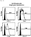
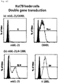
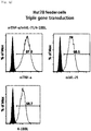
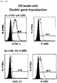
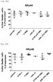
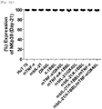
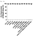
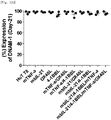
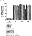
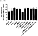
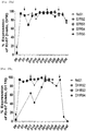
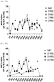
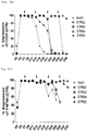
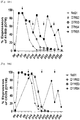
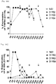
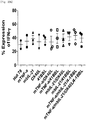
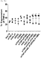
- Staining was performed with anti-human TNF- ⁇ (membrane)-PE (R&D Systems, FAB210P), anti-human OX40L-PE (BD, 558184), anti-human 4-1BBL-PE (BD, 559446), anti-human IL-21-PE (eBioscience, 12-7219-42), 7-AAD (Beckman Coulter, Inc., IM3630c), PE mouse IgG1 k isotype control (BD Pharmingen, 555749), PerCP-Cy5.5 mouse IgG1 k isotype control (BD, 550795) antibodies, and then an expression rate of each gene was analyzed using FACS equipment ( Figs. 1a to 3b ).
- Phosphate buffered saline PBS, LONZA, 17-516Q
- PBS Phosphate buffered saline
- 2% (v/v) FBS and 2 mM EDTA were added to PBS, to make MACS running buffer.
- the PBMC pellets were suspended in 10 ml of the MACS running buffer and the cell number was measured using an Adam cell counter system.
- 5 ⁇ 10 7 PBMCs were transferred to a new 50-ml tube, and then centrifuged at 1,200 rpm and a temperature of 4°C for 10 minutes.
- 400 ⁇ l of the MACS running buffer and 100 ⁇ l of CD3 magnetic beads (Miltenyi Biotech, 130050101) were added to 5 ⁇ 10 7 PBMC cell pellets and reaction was allowed to occur at a temperature of 4°C for 20 minutes.
- 10 ml to 20 ml of the MACS running buffer was added thereto and washing was performed. Then, centrifugation was performed at 1,200 rpm and a temperature of 4°C for 10 minutes, and the resultant was suspended again in 2 ml of the MACS running buffer.
- the cells were isolated using VarioMACS (Miltenyi Biotech) equipped with a CS column (Miltenyi Biotech, 130-041-305). The cells were recovered by washing the column until it reached a final volume of 20 ml. The cell number was measured using the Adam cell counter system. 1 ⁇ 10 7 cells were placed in a new 50-ml tube, and centrifuged at 1,200 rpm and a temperature of 4°C for 10 minutes. The cell pellets were suspended in freezing medium, and freezing was performed in liquid nitrogen to achieve 1 ⁇ 10 7 cells per vial.
- CD3(-) PBMC vial was thawed and transferred to a 50-ml tube.
- CD3(-) PBMCs were suspended in PBS containing 0.6% (v/v) ACD (citrate-dextrose solution, Sigma-Aldrich, C3821), 0.2% (v/v) fetal serum bovine (FBS), and 2 mM EDTA, and centrifuged at 1,200 rpm and a temperature of 4°C for 10 minutes.
- CD3(-) PBMC pellets were suspended in 1% (v/v) CellGro medium (Cellgenix, 20802-0500), and the cell number was measured using the Adam cell counter system.
- CD3(-) PBMC seed cells were suspended in 1% (v/v) CellGro medium at a concentration of 1 ⁇ 10 6 cells/ml.
- Example 3.2 Co-culture of CD3(-) PBMC seed cells and T feeder cells having transduced genes
- T cells having transduced gene were recovered from the culture flask and centrifuged at 1,200 rpm and a temperature of 4°C for 10 minutes. Thereafter, the cells were suspended in 1% (v/v) CellGro medium, and the cell number was measured using the Adam cell counter system.
- T feeder cells having transduced gene(s) were prepared by being suspended in 1% (v/v) CellGro medium at a concentration of 5 ⁇ 10 6 cells/ml and then being irradiated with 20,000 cGy in a gamma-ray irradiator and inactivated.
- IL-2 Proleukin Inj., Novartis Korea
- OKT-3 eBioscience, 16-0037-85
- the CD3(-) PBMC seed cells and the T feeder cells having transduced gene(s) were added in a volume of 0.5 ml to 1 ml each at a ratio of 1:5 or 1:2.5, and CellGro medium containing 1% (v/v) human plasma was added thereto in a volume of 0.25 ml to 0.5 ml.
- the resultant was subjected to static culture in an incubator at a temperature condition of 37°C for 4 to 7 days while adding the medium.
- the cell number was measured so that the cells had a concentration of about 0.5 ⁇ 10 6 to 1 ⁇ 10 6 cells/ml. Thereafter, the cells were diluted with CellGro medium containing 500 IU of IL-2 and 1% (v/v) autologous plasma, and placed in an appropriate culture vessel. The cells were again subjected to static culture.
- the cell number was measured at 2-day to 3-day intervals so that the cells had a concentration of 0.5 ⁇ 10 5 to 1 ⁇ 10 6 cells/ml. Thereafter, the cells were subjected to suspension culture until day 21 while performing dilution with CellGro medium containing 500 IU of IL-2 and 1% (v/v) autologous plasma. On day 21 of suspension culture, natural killer cells were obtained.
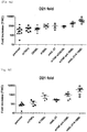
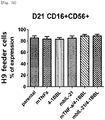
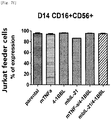
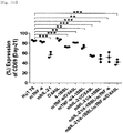
- Table 4 shows proliferation rates of the natural killer cells cultured with T cell line having undergone single or double gene transduction.
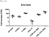
- a proliferative effect of the natural killer cells cultured with Hut78 T cell line having undergone triple gene (mTNF- ⁇ /mbIL-21/4-1BBL) or quadruple gene (mTNF- ⁇ /mbIL-21/OX40L/4-1BBL) transduction was compared together with combinations of genes for which experiments were previously made.
- the cell line that induced the highest proliferation of natural killer cells was the cell line (998-fold) into which the mbIL-21/4-1BBL gene had been transduced.
- Table 5 shows proliferation rates of the natural killer cells cultured with T cell lines having undergone multiple gene transduction.
- Hut78 cells having undergone triple gene transduction were recovered from the culture flask and centrifuged at 1,200 rpm and a temperature of 4°C for 10 minutes. Thereafter, the cells were suspended in 1% (v/v) CellGro medium, and the cell number was measured using the Adam cell counter system.
- Hut78 feeder cells having undergone triple gene transduction were prepared by being suspended in 1% (v/v) CellGro medium at a concentration of 2.5 ⁇ 10 6 cells/ml and then being irradiated with 20,000 cGy in a gamma-ray irradiator and inactivated.
- IL-2 Proleukin Inj., Novartis Korea
- OKT-3 eBioscience, 16-0037-85
- the CD3(-) PBMC seed cells and the Hut78 feeder cells having undergone triple gene transduction were added in a volume of 0.5 ml to 1 ml each at a ratio of 1:2.5, and CellGro medium containing 1% (v/v) human plasma was added thereto in a volume of 0.5 ml to 1 ml.
- the cells were subjected to static culture in an incubator at a temperature condition of 37°C for 4 to 7 days.
- half of the medium was removed and CellGro medium containing 1% (v/v) human plasma was added thereto as much as the removed volume.
- the cell number was measured so that the cells had a concentration of about 0.5 ⁇ 10 6 cells/ml to 1 ⁇ 10 6 cells/ml.
- the cell number was measured and the cells were suspended in 1% (v/v) CellGro medium at a concentration of 1 ⁇ 10 6 cells/ml.
- the Hut78 feeder cells having undergone triple gene transduction were added thereto in a volume of 0.5 ml to 1 ml each at a ratio of 1:2.5, and CellGro medium containing 500 IU of IL-2 (Proleukin Inj., Novartis Korea), 10 ng/ml of OKT-3 (eBioscience, 16-0037-85), and 1% (v/v) human plasma was added thereto in a volume of 0.5 ml to 1 ml.
- the cells were subjected to static culture in an incubator at a temperature condition of 37°C for 3 to 4 days. After that, the cell number was measured at 2-day or 3-day intervals so that the cells had a concentration of 0.5 ⁇ 10 5 cells/ml to 1 ⁇ 10 6 cells/ml.
- the re-stimulation process was repeated at 7-day or 11-day intervals, so that a total of 4 re-stimulations at 7-day intervals (D7RS5) and a total of 3 re-stimulations at 11-day intervals (D11RS4) were given.
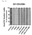
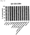
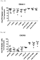
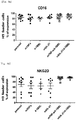
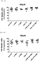
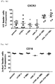
- the natural killer cells cultured for 21 days or the natural killer cells cultured with repeated re-stimulation were collected and centrifuged at 1,200 rpm for 5 minutes. The culture was removed by suction. Dilution with 1 ml of FACS buffer was performed and the cell number was measured. Dilution with the FACS buffer was performed so as to give a concentration of 5 ⁇ 10 6 cells/ml. 100 ⁇ l of the diluted cell solution was added to each 5-ml FACS tube (Falcon, 352052), and phenotypic analysis was performed with the following antibodies.
- Tube 1 Staining with anti-human CD56 was performed by selecting one of three fluorescences, and thus the same fluorescence was selected for CD3 of Tube 2 and CD56 of Tubes 3 to 9 and 11 to 15, and isotype control of Tube 10.
- the cells in the above tubes were stained at refrigeration temperature for 30 minutes. Thereafter, 2 ml of FACS buffer was added to the stained cells, and centrifugation was performed at 1,500 rpm for 5 minutes. The supernatant was removed, and 2 ml of FACS buffer was added again. Centrifugation was performed at 1,500 rpm for 5 minutes. The supernatant was removed again, and placed and suspended in 200 ⁇ l of fixation buffer (BD, 554655). Then, identification of the cells and examination for purity and phenotype thereof were performed using FACS LSRII Fortessa (BD).
- BD fixation buffer
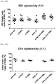
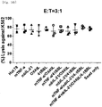
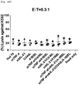
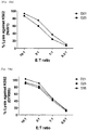
- the Calcein-AM-stained tumor cell line was washed by adding thereto 10 ml to 15 ml of RPMI1640 medium supplemented with 10% (v/v) FBS, and centrifugation was performed. Then, the pellets were suspended in 10 ml of RPMI1640 medium supplemented with 10% (v/v) FBS so that the cells had a concentration of 1 ⁇ 10 5 cells/ml. Natural killer cells were placed in a 15-ml tube at 1 ⁇ 10 6 cells, and centrifugation was performed. The pellets were suspended in RPMI1640 medium supplemented with 10% (v/v) FBS at a desired ratio (1:1) with respect to K562 cancer cell line. The prepared K562 cancer cell line and natural killer cell line were mixed using 100 ⁇ l each and dispensed into a 96-well U-bottom plate (Nunc, 163320). Each well was prepared in triplicate and a mean value thereof was obtained.
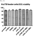
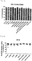
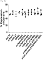
- the natural killer cells cultured with various feeder cells were reacted with K562 cancer cell line to measure their direct cell-killing activity. As compared with other conditions, the killing activity against the K562 cancer cell line was increased on average under a condition of feeder cells having undergone double gene transduction ( Figs. 13a to 14d ).
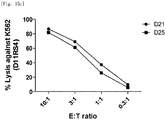
- the natural killer cells cultured with re-stimulation by T cell line due to gene transduction were reacted with K562 cancer cell line to measure their direct cell-killing activity. It was identified that under both the condition where no re-stimulation was performed and the condition where repeated re-stimulation was performed, the killing activity against K562 was well maintained even when a culture period was prolonged and fold expansion was increased ( Figs. 15a to 15c ).
- the natural killer cells cultured for 21 days or the natural killer cells cultured with repeated re-stimulation were collected and centrifuged at 1,200 rpm for 5 minutes. The culture was removed by suction. Dilution with 1 ml of FACS buffer was performed and the cell number was measured. Dilution with FACS buffer was performed so as to give a concentration of 5 ⁇ 10 6 cells/ml. 100 ⁇ l of the diluted cell solution was added to each 5-ml FACS tube (Falcon, 352052), and proliferation marker analysis was performed with the following antibodies.
- the cells in the above tubes were stained at refrigeration temperature for 30 minutes. Thereafter, 2 ml of FACS buffer was added to the stained cells, and centrifugation was performed at 1,500 rpm for 5 minutes. The supernatant was removed, and 2 ml of FACS buffer was added again. Centrifugation was performed at 1,500 rpm for 5 minutes. The supernatant was removed again, and placed and suspended in 200 ⁇ l of fixation/permeabilization buffer (eBioscience, 00-5521-00). Then, reaction was allowed to occur at refrigeration temperature for 30 minutes or longer. 2 ml of 1 ⁇ Perm/wash buffer was added and centrifugation was performed at 1,500 rpm for 5 minutes.
- fixation/permeabilization buffer eBioscience, 00-5521-00
- the natural killer cells cultured for 21 days or the natural killer cells cultured with repeated re-stimulation were collected and centrifuged at 1,200 rpm for 5 minutes. The culture was removed by suction. Dilution with 1 ml of FACS buffer was performed and the cell number was measured. Dilution with FACS buffer was performed so as to give a concentration of 5 ⁇ 10 6 cells/ml. 100 ⁇ l of the diluted cell solution was added to each 5-ml FACS tube (Falcon, 352052), and phenotypic analysis was performed with the following antibodies.
- Tube 1 Staining with anti-human CD56 was performed by selecting one of three fluorescences, and thus the same fluorescence was selected for CD56 of Tubes 2 to 4, and isotype control of Tube 5.
- the cells in the above tubes were stained at refrigeration temperature for 30 minutes. Thereafter, 2 ml of FACS buffer was added to the stained cells, and centrifugation was performed at 1,500 rpm for 5 minutes. The supernatant was removed, and 2 ml of FACS buffer was added again. Centrifugation was performed at 1,500 rpm for 5 minutes. The supernatant was removed again, and placed and suspended in 200 ⁇ l of fixation buffer (BD, 554655). Then, FACS LSRII Fortessa (BD) was used to perform identification of the cells and examination for co-stimulatory molecules.
- fixation buffer BD, 554655
- Natural killer cells were placed at 2.5 ⁇ 10 6 cells in a 15-ml tube and centrifuged. The cell pellets were suspended in RPMI1640 medium supplemented with 1 ml of 10% (v/v) FBS. In order to identify, within the cells, cytokines to be secreted out of the cells, the protein transport inhibitors Golgiplug (BD, 555029) and Golgistop (BD, 554724) were added thereto.
- Golgiplug BD, 555029
- Golgistop BD, 554724
- the prepared cells were dispensed into a 96-well U-bottom plate (Nunc, 163320), in which the natural killer cells and the medium were added in 100 ⁇ l each to well 1, and the K562 cancer cell line and the natural killer cell line were mixed using 100 ⁇ l each and added to wells 2 and 3.
- the light was blocked and reaction was allowed to occur for 4 hours in an incubator at a temperature condition of 37°C, and then the plate was centrifuged at 2,000 rpm for 3 minutes. The supernatant was removed and cytokine-secreting ability analysis was performed with the following antibodies.
- Wells 1, 2, 3 Anti-human CD3-PerCP-Cy5.5 (eBioscience, 45-0036-42), anti-human CD56-APC-Cy7 (eBioscience, 47-0567-42), 7-AAD (Beckman Coulter, Inc., A07704)
- the plate was stained at refrigeration temperature for 30 minutes. Thereafter, 100 ⁇ l of FACS buffer was added to the stained cells, and centrifugation was performed at 2,000 rpm for 3 minutes. The supernatant was removed and 200 ⁇ l FACS buffer was added again thereto. Centrifugation was performed at 2,000 rpm for 3 minutes. The supernatant was removed again, and placed and suspended in 200 ⁇ l of fixation/permeabilization buffer (BD Pharmingen, 554714). Then, reaction was allowed to occur at refrigeration temperature for 30 minutes or longer.
- fixation/permeabilization buffer BD Pharmingen, 554714
- the cytokinesecretion of the natural killer cells cultured after one stimulation by Hut78-mTNF- ⁇ /4-1BBL/mbIL-21 cell line on day 0 of culture or the natural killer cells cultured through repeated re-stimulation by Hut78-mTNF- ⁇ /4-1BBL/mbIL-21 cell line at 7-day or 11-day intervals was measured by reaction with K562 cancer cell line. It was identified that as compared with the natural killer cells cultured with one stimulation, the cytokine-secreting ability against K562 was maintained, without large difference, in the natural killer cells cultured with repeated re-stimulation, despite a long culture period and high expansion fold ( Figs. 21a to 21c ).
Landscapes
- Health & Medical Sciences (AREA)
- Engineering & Computer Science (AREA)
- Life Sciences & Earth Sciences (AREA)
- Biomedical Technology (AREA)
- Immunology (AREA)
- Chemical & Material Sciences (AREA)
- Zoology (AREA)
- Biotechnology (AREA)
- Genetics & Genomics (AREA)
- Wood Science & Technology (AREA)
- Organic Chemistry (AREA)
- Bioinformatics & Cheminformatics (AREA)
- General Health & Medical Sciences (AREA)
- Cell Biology (AREA)
- Hematology (AREA)
- Biochemistry (AREA)
- General Engineering & Computer Science (AREA)
- Microbiology (AREA)
- Animal Behavior & Ethology (AREA)
- Epidemiology (AREA)
- Public Health (AREA)
- Veterinary Medicine (AREA)
- Micro-Organisms Or Cultivation Processes Thereof (AREA)
- Medicines Containing Material From Animals Or Micro-Organisms (AREA)
- Oncology (AREA)
- Virology (AREA)
- Medicinal Chemistry (AREA)
- Pharmacology & Pharmacy (AREA)
- Developmental Biology & Embryology (AREA)
Abstract
Description
- The present invention relates to a culture method for natural killer cells using transformed T cells.
- As a therapeutic method for the treatment of cancer patients and the prevention of recurrence of cancer, immunotherapy using the patient's immune function has been developed. In particular, immunotherapy using natural killer cells that can be mass-produced and frozen has been studied. Natural killer cells are lymphoid cells that account for about 15% of peripheral blood lymphocytes, and play an important role in the innate immune response.
- Specifically, natural killer cells activate dendritic cells and induce cytotoxic T lymphocytes (CTLs) to specifically respond to tumors, thereby eliminating tumor cells.
- Natural killer cells directly kill malignant tumors such as sarcoma, myeloma, carcinoma, lymphoma, and leukemia. However, most natural killer cells present in the bodies of normal subjects exist in an inactive state, and activated natural killer cells are required to eliminate tumors. In addition, in a case of natural killer cells present in the bodies of cancer patients, the natural killer cells have functional defects due to the immune evasion mechanism of cancer cells.
- Therefore, in order to use natural killer cells as a therapeutic agent, it is very important to activate the natural killer cells. In addition, since the number of natural killer cells present in the body is limited, it is essential to develop a technique for massively proliferating and freezing natural killer cells in the blood from normal subjects or patients.
- As a method for massively proliferating natural killer cells, an ex vivo expansion culture method is used. For ex vivo expansion culture of natural killer cells, Peripheral blood mononuclear cells (PBMCs), CD3- cells, CD3-CD56+ cells, CD56+ cells, and the like are used as seed cells, and cytokines such as IL-2, IL-12, IL-15, and IL-21, LPS (Goodier et al., J. Immunol. 165(1):139-147, 2000), OKT-3 antibodies that stimulate CD3 (Condiotti et al., Experimental Hematol. 29(1):104-113, 2001) are used as proliferation factors for natural killer cells. Using the above-mentioned proliferation factors only, it is possible to proliferate natural killer cells about 3 to 10 times. However, with the above proliferation rate, it is difficult to commercialize natural killer cells into a therapeutic agent.
- Recently, methods for proliferating natural killer cells using various types of feeder cells have been studied. Representative cell lines used as feeder cells include PBMC, EBV-LCL, and K562 cell line. K562 cell line is a blood cancer-derived cell line that lacks HLA and is a representative target cell line that natural killer cells can easily attack. The culture method using K562-based feeder cells for NK cells are as follows; a method in which cultures NK cells using 4-1BBL and membrane-bound IL-15 expressed K562 cell line, (Fujisaki et al., Cancer Res. 69(9):4010-4017, 2009), a method in which cultures NK cells using MICA, 4-1BBL, and IL-15 expressed K562 cell line (Gong et al., Tissue Antigens, 76(6):467-475, 2010), and a method in which cultures NK cells using 4-1BBL and membrane-bound IL-21 expressed K562 cell (Cecele JD et al, PloSONE, 7(1):e30264, 2012)
- Accordingly, in order to activate and proliferate natural killer cells, the present inventors have developed a method for effectively proliferating natural killer cells ex vivo, comprising expressing co-stimulatory molecules and growth factors, which can enhance proliferation of the natural killer cells, in CD4+ T cells.
- Specifically, in order to increase efficiency of a culturing method for natural killer cells using the CD4(+) T cells as feeder cells, the present inventors have established transformed or genetically modified CD4(+) T cells. The present inventors have identified that in a case where the genetically modified CD4(+) T cells are co-cultured with peripheral blood mononuclear cells, such co-culture increases proliferation and abnormal cell-killing activity of natural killer cells, and thus have completed the present invention. Therefore, there is provided a culture method for proliferating natural killer cells in an efficient and stable manner using the genetically modified CD4(+) T cells.
- In an aspect of the present invention, there is provided a genetically modified or transformed CD4+ T cell that expresses at least one gene selected from the group consisting of 4-1BBL gene, mbIL-21 gene, OX40L gene, and mTNF-a gene.
- In addition, in another aspect of the present invention, there is provided a culture method for natural killer cells, comprising a step of co-culturing the genetically modified CD4+ T cells and seed cells.
- Furthermore, in yet another aspect of the present invention, there is provided a composition for culturing natural killer cells, comprising the genetically modified CD4+ T cell as an active ingredient.
- In addition, in still yet another aspect of the present invention, there is provided a natural killer cell produced by the culture method.
- The culture method for natural killer cells using the genetically modified T cell of the present invention allows natural killer cells to be effectively proliferated and produced from a small number of seed cells. In addition, the culture method also improves abnormal cell-killing activity of natural killer cells. Therefore, the culture method for natural killer cells using the genetically modified T cell of the present invention can be usefully used for commercialization into a cell therapeutic agent. Furthermore, a natural killer cell produced by the culture method of the present invention can be usefully used as a cell therapeutic agent.
-
-
Fig. 1a shows expression of single genes transduced into Hut78 cell line based on FACS. -
Fig. 1b shows expression of double genes transduced into Hut78 cell line based on FACS. -
Fig. 1c shows gene expression in Hut78 cell line based on FACS. -
Fig. 1d shows expression of single genes transduced into Hut78 cell line based on FACS. -
Fig. 1e shows expression of mTNF-α/OX40L and mTNF-α/4-1BBL double genes transduced into Hut78 cell line based on FACS. - Fig. If shows expression of mbIL-21/OX40L and mbIL-21/4-1BBL double genes transduced into Hut78 cell line based on FACS.
-
Fig. 1g shows expression of triple genes transduced into Hut78 cell line based on FACS. -
Fig. 1h shows expression of quadruple genes transduced into Hut78 cell line based on FACS. -
Fig. 2a shows expression of single genes transduced into H9 cell line based on FACS. -
Fig. 2b shows expression of double genes transduced into H9 cell line based on FACS. -
Fig. 3a shows expression of single genes transduced into Jurkat T cell line based on FACS. -
Fig. 3b shows expression of double genes transduced into Jurkat T cell line based on FACS. -
Fig. 4a shows fold increase, for each transduced gene, of natural killer cells produced by co-culturing Hut78 T cells having undergone single gene or double gene transduction, and peripheral blood mononuclear cells. -
Fig. 4b shows fold increase, for each transduced gene, of natural killer cells produced by co-culturing H9 T cells having undergone single gene or double gene transduction, and peripheral blood mononuclear cells. -
Fig. 4c shows fold increase, for each transduced gene, of natural killer cells produced by co-culturing Jurkat T cells having undergone single gene or double gene transduction, and peripheral blood mononuclear cells. -
Fig. 4d shows fold increase, for each transduced gene, of natural killer cells produced by co-culturing Hut78 T cell line having undergone single gene to quadruple gene transduction, and peripheral blood mononuclear cells. -
Fig. 5a shows fold increase of natural killer cells (D7R) produced by co-culturing Hut78 T cell line having undergone triple gene transduction, and peripheral blood mononuclear cells. -
Fig. 5b shows fold increase, calculated as log values, of natural killer cells (D7R) produced by co-culturing Hut78 T cell line having undergone triple gene transduction, and peripheral blood mononuclear cells. -
Fig. 5c shows fold increases of natural killer cells (D11R) produced by co-culturing Hut78 T cell line having undergone triple gene transduction, and peripheral blood mononuclear cells. -
Fig. 5d shows fold increase, calculated as log values, of natural killer cells (D11R) produced by co-culturing Hut78 T cell line having undergone triple gene transduction, and peripheral blood mononuclear cells. -
Fig. 6a shows viability, for each transduced gene, of natural killer cells produced by co-culturing Hut78 T cell line having undergone single gene or double gene transduction, and peripheral blood mononuclear cells. -
Fig. 6b shows viability, for each transduced gene, of natural killer cells produced by co-culturing H9 T cell line having undergone single gene or double gene transduction, and peripheral blood mononuclear cells. -
Fig. 6c shows viability, for each transduced gene, of natural killer cells produced by co-culturing Jurkat T cell line having undergone single gene or double gene transduction, and peripheral blood mononuclear cells. -
Fig. 6d shows viability, for each transduced gene, of natural killer cells produced by co-culturing Hut78 T cell line having undergone single gene to quadruple gene transduction, and peripheral blood mononuclear cells. -
Fig. 7a shows purity (CD3-CD56+), for each transduced gene, of natural killer cells produced by co-culturing Hut78 T cell line having undergone single gene or double gene transduction, and peripheral blood mononuclear cells. -
Fig. 7b shows activation (CD16+CD56+), for each transduced gene, of natural killer cells produced by co-culturing Hut78 T cell line having undergone single gene or double gene transduction, and peripheral blood mononuclear cells. -
Fig. 7c shows purity (CD3-CD56+), for each transduced gene, of natural killer cells produced by co-culturing H9 T cell line having undergone single gene or double gene transduction, and peripheral blood mononuclear cells. -
Fig. 7d shows activation (CD16+CD56+), for each transduced gene, of natural killer cells produced by co-culturing H9 T cell line having undergone single gene or double gene transduction, and peripheral blood mononuclear cells. -
Fig. 7e shows purity (CD3-CD56+), for each transduced gene, of natural killer cells produced by co-culturing Jurkat T cell line having undergone single gene or double gene transduction, and peripheral blood mononuclear cells. -
Fig. 7f shows activation (CD16+CD56+), for each transduced gene, of natural killer cells produced by co-culturing Jurkat cell line having undergone single gene or double gene transduction, and peripheral blood mononuclear cells. -
Fig. 7g shows purity (CD3-CD56+), for each transduced gene, of natural killer cells produced by co-culturing Hut78 T cell line having undergone single gene to quadruple gene transduction, and peripheral blood mononuclear cells. -
Fig. 7h shows activation (CD16+CD56+), for each transduced gene, of natural killer cells produced by co-culturing Hut78 T cell line having undergone single gene to quadruple gene transduction, and peripheral blood mononuclear cells. -
Fig. 8a shows expression levels of thephenotypic marker CD 16, for each transduced gene, in natural killer cells produced by co-culturing Hut78 T cell line having undergone single gene or double gene transduction, and peripheral blood mononuclear cells. -
Fig. 8b shows expression levels of the phenotypic marker NKG2D, for each transduced gene, in natural killer cells produced by co-culturing Hut78 T cell line having undergone single gene or double gene transduction, and peripheral blood mononuclear cells. -
Fig. 8c shows expression levels of the phenotypic marker NKp30, for each transduced gene, in natural killer cells produced by co-culturing Hut78 T cell line having undergone single gene or double gene transduction, and peripheral blood mononuclear cells. -
Fig. 8d shows expression levels of the phenotypic marker NKp44, for each transduced gene, in natural killer cells produced by co-culturing Hut78 T cell line having undergone single gene or double gene transduction, and peripheral blood mononuclear cells. -
Fig. 8e shows expression levels of the phenotypic marker NKp46, for each transduced gene, in natural killer cells produced by co-culturing Hut78 T cell line having undergone single gene or double gene transduction, and peripheral blood mononuclear cells. -
Fig. 8f shows expression levels of the phenotypic marker DNAM-1, for each transduced gene, in natural killer cells produced by co-culturing Hut78 T cell line having undergone single gene or double gene transduction, and peripheral blood mononuclear cells. -
Fig. 8g shows expression levels of the phenotypic marker CXCR3, for each transduced gene, in natural killer cells produced by co-culturing Hut78 T cell line having undergone single gene or double gene transduction, and peripheral blood mononuclear cells. -
Fig. 9a shows expression levels of thephenotypic marker CD 16, for each transduced gene, in natural killer cells produced by co-culturing H9 T cell line having undergone single gene or double gene transduction, and peripheral blood mononuclear cells. -
Fig. 9b shows expression levels of the phenotypic marker NKG2D, for each transduced gene, in natural killer cells produced by co-culturing H9 T cell line having undergone single gene or double gene transduction, and peripheral blood mononuclear cells. -
Fig. 9c shows expression levels of the phenotypic marker NKp30, for each transduced gene, in natural killer cells produced by co-culturing H9 T cell line having undergone single gene or double gene transduction, and peripheral blood mononuclear cells. -
Fig. 9d shows expression levels of the phenotypic marker NKp44, for each transduced gene, in natural killer cells produced by co-culturing H9 T cell line having undergone single gene or double gene transduction, and peripheral blood mononuclear cells. -
Fig. 9e shows expression levels of the phenotypic marker NKp46, for each transduced gene, in natural killer cells produced by co-culturing H9 T cell line having undergone single gene or double gene transduction, and peripheral blood mononuclear cells. -
Fig. 9f shows expression levels of the phenotypic marker DNAM-1, for each transduced gene, in natural killer cells produced by co-culturing H9 T cell line having undergone single gene or double gene transduction, and peripheral blood mononuclear cells. -
Fig. 9g shows expression levels of the phenotypic marker CXCR3, for each transduced gene, in natural killer cells produced by co-culturing H9 T cell line having undergone single gene or double gene transduction, and peripheral blood mononuclear cells. -
Fig. 10a shows expression levels of thephenotypic marker CD 16, for each transduced gene, in natural killer cells produced by co-culturing Jurkat T cell line having undergone single gene or double gene transduction, and peripheral blood mononuclear cells. -
Fig. 10b shows expression levels of the phenotypic marker NKG2D, for each transduced gene, in natural killer cells produced by co-culturing Jurkat T cell line having undergone single gene or double gene transduction, and peripheral blood mononuclear cells. -
Fig. 10c shows expression levels of the phenotypic marker NKp30, for each transduced gene, in natural killer cells produced by co-culturing Jurkat T cell line having undergone single gene or double gene transduction, and peripheral blood mononuclear cells. -
Fig. 10d shows expression levels of the phenotypic marker NKp44, for each transduced gene, in natural killer cells produced by co-culturing Jurkat T cell line having undergone single gene or double gene transduction, and peripheral blood mononuclear cells. -
Fig. 10e shows expression levels of the phenotypic marker NKp46, for each transduced gene, in natural killer cells produced by co-culturing Jurkat T cell line having undergone single gene or double gene transduction, and peripheral blood mononuclear cells. -
Fig. 10f shows expression levels of the phenotypic marker DNAM-1, for each transduced gene, in natural killer cells produced by co-culturing Jurkat T cell line having undergone single gene or double gene transduction, and peripheral blood mononuclear cells. -
Fig. 10g shows expression levels of the phenotypic marker CXCR3, for each transduced gene, in natural killer cells produced by co-culturing Jurkat T cell line having undergone single gene or double gene transduction, and peripheral blood mononuclear cells. -
Fig. 11a shows expression levels of thephenotypic marker CD 16, for each transduced gene, in natural killer cells produced by co-culturing Hut78 T cell line having undergone single gene to quadruple gene transduction, and peripheral blood mononuclear cells. -
Fig. 11b shows expression levels of the phenotypic marker NKG2A, for each transduced gene, in natural killer cells produced by co-culturing Hut78 T cell line having undergone single gene to quadruple gene transduction, and peripheral blood mononuclear cells. -
Fig. 11c shows expression levels of the phenotypic marker NKG2C, for each transduced gene, in natural killer cells produced by co-culturing Hut78 T cell line having undergone single gene to quadruple gene transduction, and peripheral blood mononuclear cells. -
Fig. 11d shows expression levels of the phenotypic marker NKG2D, for each transduced gene, in natural killer cells produced by co-culturing Hut78 T cell line having undergone single gene to quadruple gene transduction, and peripheral blood mononuclear cells. -
Fig. 11e shows expression levels of the phenotypic marker NKp30, for each transduced gene, in natural killer cells produced by co-culturing Hut78 T cell line having undergone single gene to quadruple gene transduction, and peripheral blood mononuclear cells. -
Fig. 11f shows expression levels of the phenotypic marker NKp44, for each transduced gene, in natural killer cells produced by co-culturing Hut78 T cell line having undergone single gene to quadruple gene transduction, and peripheral blood mononuclear cells. -
Fig. 11g shows expression levels of the phenotypic marker NKp46, for each transduced gene, in natural killer cells produced by co-culturing Hut78 T cell line having undergone single gene to quadruple gene transduction, and peripheral blood mononuclear cells. -
Fig. 11h shows expression levels of the phenotypic marker DNAM-1, for each transduced gene, in natural killer cells produced by co-culturing Hut78 T cell line having undergone single gene to quadruple gene transduction, and peripheral blood mononuclear cells. -
Fig. 11i shows expression levels of the phenotypic marker CXCR3, for each transduced gene, in natural killer cells produced by co-culturing Hut78 T cell line having undergone single gene to quadruple gene transduction, and peripheral blood mononuclear cells. -
Fig. 11j shows expression levels of the phenotypic marker CD62L, for each transduced gene, in natural killer cells produced by co-culturing Hut78 T cell line having undergone single gene to quadruple gene transduction, and peripheral blood mononuclear cells. -
Fig. 11k shows expression levels of the phenotypic marker CD57, for each transduced gene, in natural killer cells produced by co-culturing Hut78 T cell line having undergone single gene to quadruple gene transduction, and peripheral blood mononuclear cells. -
Fig. 11l shows expression levels of the phenotypic marker CD69, for each transduced gene, in natural killer cells produced by co-culturing Hut78 T cell line having undergone single gene to quadruple gene transduction, and peripheral blood mononuclear cells. -
Fig. 12a shows expression levels of the phenotypic markers CD16, NKG2A, NKG2C, and NKG2D in natural killer cells produced by co-culturing Hut78 T cell line having undergone triple gene transduction, and peripheral blood mononuclear cells. -
Fig. 12b shows expression levels of the phenotypic markers NKp30, NKp44, NKp46, and DNAM-1 in natural killer cells produced by co-culturing Hut78 T cell line having undergone triple gene transduction, and peripheral blood mononuclear cells. -
Fig. 12c shows expression levels of the phenotypic markers CD62L, CD69, CXCR3, and CD57 in natural killer cells produced by co-culturing Hut78 T cell line having undergone triple gene transduction, and peripheral blood mononuclear cells. -
Fig. 13a shows tumor cell-killing activity, for each transduced gene, of natural killer cells produced by co-culturing Hut78 T cell line having undergone single gene or double gene transduction, and peripheral blood mononuclear cells. -
Fig. 13b shows tumor cell-killing activity, for each transduced gene, of natural killer cells produced by co-culturing H9 T cell line having undergone single gene or double gene transduction, and peripheral blood mononuclear cells. -
Fig. 13c shows tumor cell-killing activity, for each transduced gene, of natural killer cells produced by co-culturing Jurkat T cell line having undergone single gene or double gene transduction, and peripheral blood mononuclear cells. -
Fig. 14a shows tumor cell-killing activity (E:T = 10:1), for each transduced gene, of natural killer cells produced by co-culturing Hut78 T cell line having undergone single gene to quadruple gene transduction, and peripheral blood mononuclear cells. -
Fig. 14b shows tumor cell-killing activity (E:T = 3:1), for each transduced gene, of natural killer cells produced by co-culturing Hut78 T cell line having undergone single gene to quadruple gene transduction, and peripheral blood mononuclear cells. -
Fig. 14c shows tumor cell-killing activity (E:T = 1:1), for each transduced gene, of natural killer cells produced by co-culturing Hut78 T cell line having undergone single gene to quadruple gene transduction, and peripheral blood mononuclear cells. -
Fig. 14d shows tumor cell-killing activity (E:T = 0.3:1), for each transduced gene, of natural killer cells produced by co-culturing Hut78 T cell line having undergone single gene to quadruple gene transduction, and peripheral blood mononuclear cells. -
Fig. 15a shows tumor cell-killing activity, for respective cell ratios, of natural killer cells (NoS1) produced by co-culturing Hut78 T cell line having undergone triple gene transduction, and peripheral blood mononuclear cells. -
Fig. 15b shows tumor cell-killing activity, for respective cell ratios, of natural killer cells (D7RS5) produced by co-culturing Hut78 T cell line having undergone triple gene transduction, and peripheral blood mononuclear cells. -
Fig. 15c shows tumor cell-killing activity, for respective cell ratios, of natural killer cells (D11RS4) produced by co-culturing Hut78 T cell line having undergone triple gene transduction, and peripheral blood mononuclear cells. -
Fig. 16a shows, , expression levels of the proliferation marker Ki-67, for each transduced gene, in natural killer cells produced by co-culturing Hut78 T cell line having undergone single gene to quadruple gene transduction, and peripheral blood mononuclear cells. -
Fig. 16b shows expression levels of the proliferation marker Ki-67 onday 16, for each transduced gene, in natural killer cells produced by co-culturing Hut78 T cell line having undergone single gene to quadruple gene transduction, and peripheral blood mononuclear cells. -
Fig. 16c shows expression levels of the proliferation marker Ki-67 for each transduced gene, in terms of mean fluorescence index (MFI) ratio values, in natural killer cells produced by co-culturing Hut78 T cell line having undergone single gene to quadruple gene transduction, and peripheral blood mononuclear cells. -
Fig. 16d shows expression levels of the proliferation marker Ki-67 for each transduced gene, in terms of mean fluorescence index ratio values, in natural killer cells produced by co-culturing Hut78 T cell line having undergone single gene to quadruple gene transduction, and peripheral blood mononuclear cells. -
Fig. 17a shows expression levels of the proliferation marker Ki-67 in natural killer cells (D7R) produced by performing re-stimulation at 7-day intervals when co-culturing Hut78 T cell line having undergone triple gene transduction, and peripheral blood mononuclear cells. -
Fig. 17b shows expression levels of the proliferation marker Ki-67 in natural killer cells (D11R) produced by performing re-stimulation at 11-day intervals when co-culturing Hut78 T cell line having undergone triple gene transduction, and peripheral blood mononuclear cells. -
Fig. 17c shows expression levels of the proliferation marker Ki-67, in terms of mean fluorescence index ratio values, in natural killer cells (D7R) produced by performing re-stimulation at 7-day intervals when co-culturing Hut78 T cell line having undergone triple gene transduction, and peripheral blood mononuclear cells. -
Fig. 17d shows expression levels of the proliferation marker Ki-67, in terms of mean fluorescence index ratio values, in natural killer cells (D11R) produced by performing re-stimulation at 11-day intervals when co-culturing Hut78 T cell line having undergone triple gene transduction, and peripheral blood mononuclear cells. -
Fig. 18a shows expression levels of the co-stimulatory molecule CD25, for each transduced gene, in natural killer cells produced by co-culturing Hut78 T cell line having undergone single gene to quadruple gene transduction, and peripheral blood mononuclear cells. -
Fig. 18b shows expression levels of the co-stimulatory molecule CD25 onday 11, for each transduced gene, in natural killer cells produced by co-culturing Hut78 T cell line having undergone single gene to quadruple gene transduction, and peripheral blood mononuclear cells. -
Fig. 18c shows expression levels of the co-stimulatory molecule TNFRII, for each transduced gene, in natural killer cells produced by co-culturing Hut78 T cell line having undergone single gene to quadruple gene transduction, and peripheral blood mononuclear cells. -
Fig. 18d shows expression levels of the co-stimulatory molecule TNFRII onday 16, for each transduced gene, in natural killer cells produced by co-culturing Hut78 T cell line having undergone single gene to quadruple gene transduction, and peripheral blood mononuclear cells. -
Fig. 18e shows expression levels of the co-stimulatory molecule 41BB, for each transduced gene, in natural killer cells produced by co-culturing Hut78 T cell line having undergone single gene to quadruple gene transduction, and peripheral blood mononuclear cells. -
Fig. 18f shows expression levels of the co-stimulatory molecule 41BB onday 7, for each transduced gene, in natural killer cells produced by co-culturing Hut78 T cell line having undergone single gene to quadruple gene transduction, and peripheral blood mononuclear cells. -
Fig. 19a shows expression levels of the co-stimulatory molecule CD25 in natural killer cells (D7R) produced by performing re-stimulation at 7-day intervals when co-culturing Hut78 T cell line having undergone triple gene transduction, and peripheral blood mononuclear cells. -
Fig. 19b shows expression levels of the co-stimulatory molecule TNFRII in natural killer cells (D7R) produced by performing re-stimulation at 7-day intervals when co-culturing Hut78 T cell line having undergone triple gene transduction, and peripheral blood mononuclear cells. -
Fig. 19c shows expression levels of the co-stimulatory molecule 41BB in natural killer cells (D7R) produced by performing re-stimulation at 7-day intervals when co-culturing Hut78 T cell line having undergone triple gene transduction, and peripheral blood mononuclear cells. -
Fig. 19d shows expression levels of the co-stimulatory molecule CD25 in natural killer cells (D11R) produced by performing re-stimulation at 11-day intervals when co-culturing Hut78 T cell line having undergone triple gene transduction, and peripheral blood mononuclear cells. -
Fig. 19e shows expression levels of the co-stimulatory molecule TNFRII in natural killer cells (D11R) produced by performing re-stimulation at 11-day intervals when co-culturing Hut78 T cell line having undergone triple gene transduction, and peripheral blood mononuclear cells. -
Fig. 19f shows expression levels of the co-stimulatory molecule 41BB in natural killer cells (D11R) produced by performing re-stimulation at 11-day intervals when co-culturing Hut78 T cell line having undergone triple gene transduction, and peripheral blood mononuclear cells. -
Fig. 20a shows expression levels of CD107a, for each transduced gene, in natural killer cells produced by co-culturing Hut78 T cell line having undergone single gene to quadruple gene transduction, and peripheral blood mononuclear cells. -
Fig. 20b shows expression levels of IFN-γ, for each transduced gene, in natural killer cells produced by co-culturing Hut78 T cell line having undergone single gene to quadruple gene transduction, and peripheral blood mononuclear cells. -
Fig. 20c shows expression levels of TNF-α, for each transduced gene, in natural killer cells produced by co-culturing Hut78 T cell line having undergone single gene to quadruple gene transduction, and peripheral blood mononuclear cells. -
Fig. 21a shows expression levels of CD107a, IFN-γ, and TNF-α in natural killer cells (NoS1) produced by co-culturing Hut78 T cell line having undergone triple gene transduction, and peripheral blood mononuclear cells. -
Fig. 21b shows expression levels of CD107a, IFN-γ, and TNF-α in natural killer cells (D7RS5) produced by co-culturing Hut78 T cell line having undergone triple gene transduction, and peripheral blood mononuclear cells. -
Fig. 21c shows expression levels of CD107a, IFN-γ, and TNF-α in natural killer cells (D11RS4) produced by co-culturing Hut78 T cell line having undergone triple gene transduction, and peripheral blood mononuclear cells. - Hereinafter, the present invention will be described in detail.
- In an aspect of the present invention, there is provided a transformed CD4+ T cell that expresses at least one gene selected from the group consisting of 4-1BBL gene, mbIL-21 gene, OX40L gene, and mTNF-a gene.
- Specifically, when one gene is transduced, the gene may be 4-1BBL, mbIL-21, OX40L, or mTNF-a. In addition, when two genes are transduced, a combination of the genes may be mbIL-21/4-1BBL, 4-1BBL/OX40L, mTNF-α/4-1BBL, mbIL-21/OX40L, mbIL-21/mTNF-α, or mTNF-α/OX40L. In an embodiment of the present invention, combinations of genes, mbIL-21/4-1BBL, mTNF-α/OX40L, mTNF-α/4-1BBL, and mbIL-21/OX40L were transduced into T cells.
- In addition, when three genes are transduced, a combination of the genes may be 4-1BBL/mbIL-21/OX40L, mbIL-21/OX40L/mTNF-α, mTNF-α/mbIL-21/4-1BBL, or 4-1BBL/OX40L/mTNF-α. In an embodiment of the present invention, a combination of genes, mTNF-α/mbIL-21/4-1BBL was transduced into T cells.
- In addition, when four genes are transduced, a combination of the genes may be mTNF-α/mbIL-21/OX40L/4-1BBL. In an embodiment of the present invention, a combination of genes, mTNF-α/mbIL-21/OX40L/4-1BBL was transduced into T cells.
- The term "4-1BBL" as used herein refers to one of TNF superfamily (TNFSF) called CD137L, and means a ligand that forms a trimer and binds to 4-1BB as a receptor. The 4-1BBL gene may be derived from human.
- Specifically, the 4-1BBL gene may be NCBI Reference Sequence: NM_003811, but is not limited thereto. The 4-1BBL gene may have a nucleotide sequence encoding the amino acid sequence represented by SEQ ID NO: 1. The nucleotide sequence encoding the amino acid sequence represented by SEQ ID NO: 1 may be the nucleotide sequence represented by SEQ ID NO: 2.
- The term "mbIL-21" as used herein may be IL-21 designed to be bound to a cell membrane. Here, mbIL-21 may be a fusion protein formed by binding between IL-21 and a transmembrane protein. The transmembrane protein may be CD8α. Specifically, the transmembrane protein may be a transmembrane domain of CD8α.
- Specifically, the IL-21 gene may be NCBI Reference Sequence: NM_021803.3, but is not limited thereto. In addition, the CD8α gene may be NCBI Reference Sequence: NM_001768, but is not limited thereto. The mbIL-21 is expressed in the form of IL-21 bound to a cell membrane. In addition, the mbIL-21 gene may have a nucleotide sequence encoding the amino acid sequence represented by SEQ ID NO: 3. The nucleotide sequence encoding the amino acid sequence represented by SEQ ID NO: 3 may be the nucleotide sequence represented by SEQ ID NO: 4.
- The term "OX40L" as used herein is also referred to as ACT-4 receptor, TNF4-human, GP34, or CD134L, and means a ligand that binds to OX40. Specifically, the OX40L gene may be NCBI Reference Sequence: NM_003326, but is not limited thereto. The OX40L gene may have a nucleotide sequence encoding the amino acid sequence represented by SEQ ID NO: 5. The nucleotide sequence encoding the amino acid sequence represented by SEQ ID NO: 5 may be the nucleotide sequence represented by SEQ ID NO: 6.
- The term "mTNF-α" as used herein means a gene obtained by causing a point mutation to occur on DNA of tumor necrosis factor-alpha so that alanine-valine, a site recognized by tumor necrosis factor-alpha-converting enzyme (TACE) in the amino acid sequence of tumor necrosis factor-alpha, is changed to proline-valine. Mutation of alanine to proline is randomly selected.
- Specifically, the mTNF-a gene may have a nucleotide sequence encoding the amino acid sequence represented by SEQ ID NO: 8. The nucleotide sequence encoding the amino acid sequence represented by SEQ ID NO: 8 may be the nucleotide sequence represented by SEQ ID NO: 9.
- The 4-1BBL gene, mbIL-21 gene, OX40L gene, or mTNF-a gene may be transduced through a recombinant lentivirus. However, transduction is not limited thereto.
- As a method of transfecting the gene into cells, a biochemical method, a physical method, or a virus-mediated transfection method may be used. In addition, as the biochemical method, FuGene6 (Roche, USA), lipofectamine (
LipofectamineTM 2000, Invitrogen, USA), or ExGen 500 (MBI Fermentas International Inc., CANADA) may be used. In addition, a lipid-mediated method using lipofectamine may be used. - The term "vector" as used herein is an expression vector capable of expressing a gene of interest in a cell into which the vector is transduced, and refers to a gene construct containing an essential regulatory element operably linked so that a gene insert transduced into the vector is expressed.
- In addition, as the expression vector containing the gene, any expression vector capable of expressing the gene in CD4+ T cell line may be used, and pCDH-CMV-MCS-EF1-Puro (SBI, CD510B-1) or pCDH-CMV-MCS-EF1-Neo (SBI, CD514B-1) lentiviral vector was used in a specific embodiment of the present invention.
- The lentivirus means a virus which belongs to a retrovirus family and is characterized by a long latency period. The lentivirus can deliver genetic information into DNA of a host cell. Use of the lentivirus is one of the most effective methods of using gene delivery vectors that can replicate in non-dividing cells.
- The CD4+ T cells may be ex vivo isolated CD4+ T cells, ex vivo expansion-cultured CD4+ T cells, or CD4+ T cell line (T lymphoma cell line). In addition, the CD4+ T cells may be auxiliary T cells, and may be a hybridoma obtained by fusing CD4+ T cells with cancer cells. Specifically, the CD4+ T cells may be any one selected from the group consisting of Hut78, H9, Jurkat, Loucy, Molt-3, Molt-13, PEER, RPMI8402, and TALL-01 cells. Hut78, H9, or Jurkat T cells may be preferred.
- In another aspect of the present invention, there is provided a culture method for natural killer cells, comprising a step of co-culturing the transformed CD4+ T cells and seed cells.
- The term "feeder cell" as used herein is also referred to as an auxiliary cell for culture, and means a cell which does not proliferate but has metabolic activity so that the cell produces various metabolites and thus helps proliferation of a target cell. The feeder cell may be a transformed CD4+ T cell that expresses at least one gene selected from the group consisting of 4-1BBL gene, mbIL-21 gene, OX40L gene, and mTNF-a gene.
- T cells used as the feeder cells may be inactivated cells whose division/proliferation is inhibited or cells which have not been inactivated. Preferably, the T cells may be inactivated to ensure safety. As an inactivation method, a conventional method known in the art may be used. For example, a gamma ray-irradiation method may be used. In a case where T cells which have not been inactivated are used, most of them are tumor cells, and thus may be killed by activated natural killer cells during culture.
- The term "seed cell" as used herein means a cell capable of proliferating to natural killer cells through appropriate culture. Specifically, the seed cells may be any one selected from the group consisting of peripheral blood, peripheral blood leukocytes, peripheral blood mononuclear cells (PBMCs), enriched natural killer cells, and isolated natural killer cells. The seed cells may preferably be, but are not limited to, CD3(-) cells from which CD3(+) cells have been eliminated.
- In a culture method for natural killer cells, culture may be performed by mixing the feeder cells and the seed cells at a ratio of 0.1 or higher. Specifically, the feeder cells and the seed cells may be at a ratio of 0.1:1 to 50:1. More specifically, the ratio may be 0.5:1 to 40:1.
- Even more specifically, the ratio may be 1:1 to 30:1. Most specifically, the ratio may be 2:1 to 20:1. In an embodiment, the feeder cells and the seed cells may be at a ratio of 5:1, but is not particularly limited thereto. The "ratio" means a ratio based on the number of cells.
- In the culture method for natural killer cells, the seed cells may be mixed once with the feeder cells and culture may be performed for 5 days to 60 days, or may be mixed two or more times with the feeder cells and culture may be performed for 60 days or longer. Preferably, the seed cells may be mixed once with the feeder cells and culture was performed for 14 days to 21 days. However, the culture method is not limited thereto.
- In the culture method for natural killer cells, natural killer cells and T lymphoma cell line are co-cultured in conventional animal cell culture medium such as AIM-V medium, RPMI1640, CellGro SCGM, X-VIVO20, IMDM, and DMEM. In co-culture, culture may be performed with addition of an antibody that has low affinity for T cells and stimulates T cells, and an interleukin. However, the culture method is not limited thereto.
- The term "antibody which has low affinity for T cells and stimulates T cells" as used herein means a protein that specifically responds to CD3 antigens, a group of molecules each of which associates with T cell receptor (TCR) to form an antigen recognition complex. As compared with TCR, the CD3 molecule has a longer intracellular region and plays a role in delivering an antigen recognition signal into a cell.
- The antibody that has low affinity for T cells and stimulates T cells, which can be used in the present invention, may preferably be an anti-CD3 antibody. Specifically, the anti-CD3 antibody may be OKT-3, UCHT1, or HIT3a.
- The term "interleukin (IL)" as used herein refers to a group belonging to cytokines, and means a proteinaceous biologically-active substance produced by immunocompetent cells such as lymphocytes, monocytes, and macrophages. The interleukin may be IL-2, IL-15, IL-12, IL-18, or IL-21.
- In an embodiment of the present invention, culture was performed with addition of OKT-3 antibody and IL-2. A concentration of the OKT-3 antibody to be added may be 0.1 ng/ml to 100 ng/ml. Preferably, the concentration of the OKT-3 antibody may be 10 ng/µl. A concentration of IL-2 may be 10 U/ml to 2,000 U/ml. Preferably, the concentration of IL-2 may be 500 U/ml. In addition, serum or plasma and an additional growth factor that supports growth of lymphocytes may be added and culture may be performed. The type of serum or plasma to be added to the medium is not particularly limited, and a variety of commercially available animal-derived serum or plasma may be used. Preferably, human-derived serum or plasma, in particular, self-derived serum or plasma may be used.
- The term "culture" as used herein means a method of growing cells under an environmental condition that is appropriately artificially regulated. The method of culturing the genetically modified CD4+ T cells may be carried out using methods well known in the art. Specifically, the culture may be performed in a batch or fed-batch process, or may be continuously performed in a repeated fed-batch process.
- In addition, precursors suitable for the culture medium may be used. The above-mentioned raw materials may be added to a culture, during a culture process, in a batch, fed-batch, or continuous manner by an appropriate method. However, the present invention is not particularly limited thereto. A pH of the culture can be regulated by using, in an appropriate manner, basic compounds such as sodium hydroxide, potassium hydroxide, and ammonia, or acidic compounds such as phosphoric acid or sulfuric acid.
- The culture method using the T cells as feeder cells allows natural killer cells to be selectively cultured from seed cells such as PBMCs, and enables a stable culture due to the fact that there is no difference in proliferation of natural killer cells as compared with a case where PBMC feeder cells are used depending on donors. Therefore, a large amount of natural killer cell therapeutic agents can be efficiently and stably obtained.
- In addition, in yet another aspect of the present invention, there is provided a composition for culturing natural killer cells, comprising the genetically modified CD4+ T cell as an active ingredient.
- Furthermore, in still yet another aspect of the present invention, there is provided a natural killer cell produced by the culture method for natural killer cells.
- The natural killer cells cultured according to the culture method for natural killer cells can be frozen and do not exhibit impaired cell function even in a case of being thawed again. In addition, due to high expression of an activating receptor such as NKp46, the natural killer cells exhibit increased killing activity against a tumor cell line and increased cytokine secretion, and thus an excellent anticancer effect can be expected. Therefore, it is possible to prepare a cell therapeutic agent effective for tumor treatment, using a large number of activated natural killer cells which can be clinically applied.
- In addition, in a composition for the prevention or treatment of an infectious disease, comprising, as an active ingredient, the natural killer cell produced by the culture method for natural killer cells, the natural killer cell may be contained in an amount of 10% to 95% by weight with respect to the total weight of the composition. In addition, the composition for the prevention or treatment of an infectious disease of the present invention may further comprise, in addition to the active ingredient, one or more active ingredients that exhibit the same or similar functions.
- The composition for the prevention or treatment of an infectious disease may be formulated into a pharmaceutical composition for administration by further including one or more pharmaceutically acceptable carriers in addition to the above-mentioned active ingredients.
- A dose of the pharmaceutical composition for the prevention or treatment of an infectious disease may be regulated depending on various factors including type of disease, severity of disease, types and amounts of active ingredients and other ingredients included in the composition, type of formulation and the patient's age, weight, general health condition, sex, and diet, time of administration, route of administration and secretion rate of composition, duration of treatment, and simultaneously used drugs. However, for desired effects, a dose of the natural killer cells according to the present invention may be 0.01 × 10⁷ cells/kg to 1.0 × 10⁹ cells/kg, and may be 0.5 × 10⁷ cells/kg to 1.0 × 10⁸ cells/kg. Here, the dose may be administered once a day or may be divided into several times a day.
- In addition, the pharmaceutical composition for the prevention or treatment of an infectious disease may be administered to an individual by various methods known in the art. The route of administration may be appropriately selected by those skilled in the art in consideration of administration method, body fluid volume, viscosity, and the like.
- Hereinafter, the present invention will be described in detail by way of examples. However, the following examples are only for illustrating the present invention, and the present invention is not limited to the following examples.
- As a lentiviral vector, pCDH-CMV-MCS-EF1-Puro (SBI, CD510B-1) or pCDH-CMV-MCS-EF1-Neo (SBI, CD514B-1) was used. As genes for transduction, 4-1BBL (
TNF superfamily member 9, TNFSF9), mbIL-21 (membrane-bound IL-21), OX40L (TNF superfamily member 4 (TNFSF4) transcript variant 1) and mTNF-a (membrane-bound TNF alpha) genes were used. - Specifically, for the 4-1BBL gene (SEQ ID NO: 2), 4-1BBL gene expression vector (OriGene, RC211160) was used. For the mbIL-21 gene (SEQ ID NO: 4), pcDNA3.1 vector (GenScript, US) into which a codon-optimized mbIL-21 gene sequence is inserted was used. For the OX40L gene (SEQ ID NO: 6), a request for synthesis thereof was made to BIONEER CORPORATION.
- For the mTNF-a gene (SEQ ID NO: 9), RNA was extracted from peripheral blood mononuclear cells (PBMCs), and then CDS was obtained therefrom by reverse transcriptase (RT)-PCR. In order for TNF-α to be secreted, TNF-α was cleaved by tumor necrosis factor-alpha-converting enzyme (TACE); however, a point mutation was caused to occur on DNA of TNF-α so that alanine-valine (A-V), a site recognized by TACE in the amino acid sequence of TNF-α, became proline-valine (P-V), thereby allowing TNF-α to remain attached to a cell membrane. The point mutation was performed, in human mTNF-a gene represented by SEQ ID NO: 7, by replacing guanine, which is the 226th base, with cytosine, and replacing adenine, which is the 228th base, with guanine.
- Primers suitable for each gene for transducrion were used to amplify, through PCR, the coding sequence (CDS) of the transgene (Table 1).
[TABLE 1] Primer Sequence information (5'->3') SEQ ID NO 4-1BBL 4-1BBL Forward 
SEQ ID NO: 10 4-1BBL Reverse TTCGCGGCCGCGGATCCTTATTCCGACCTCGGTGAAGG SEQ ID NO: 11 mbIL-21 mbIL-21 Forward TAGAGCTAGCGAATTCGCCACCGCCACCATGGCTCTGCCC SEQ ID NO: 12 mbIL-21 Reverse TCGCGGCCGCGGATCCTCAATACAGGGTGATGACC SEQ ID NO: 13 OX40L OX40L Forward TAGAGCTAGCGAATTCGCCACCATGGAACGGGTGCAAC SEQ ID NO: 14 OX40L Reverse TCGCGGCCGCGGATCCTCACAAGACACAGAACTCCCC SEQ ID NO: 15 mTNF-α mTNF-α Forward TAGAGCTAGCGAATTCGCCACCGCCACCATGGCTCTGCCC SEQ ID NO: 16 mTNF-α Reverse TCGCGGCCGCGGATCCTCACAGGGCAATGATCCC SEQIDNO: 17 - Table 1 shows the primers used in the experiment.
- The transgene and the lentiviral vector were treated with EcoRI and BamHI restriction enzymes. Thereafter, ligation was performed using an In-Fusion HD cloning kit (Clontech, 639649). The ligated lentiviral vector was transformed into DH5α competent cells and culture was performed. Plasmid DNA was obtained from the transformed DH5α competent cells using a plasmid mini-prep kit (MACHEREY-NAGEL/740422.50). For all plasmid DNA, a request for sequencing thereof was made to an outside company, and it was confirmed that DNA sequences thereof were accurate.
- For recombinant lentiviral production, 293T cell line was inoculated into a 75T flask (Nunc, 156499) at 1.5 × 10⁶ to 2 × 10⁶ cells two days prior to transfection, and incubated in an incubator at a condition of 5% CO2 and 37°C. When cell confluency of the 293T cells reached about 80% to 90%, the medium was replaced with 6 ml of OPTI-MEM (Gibco, 31985-088) and incubated for 30 minutes under a 5% CO2 condition at a temperature of 37°C. A DNA mixed solution and a lipofectamine (lipofectamine 2000, Life technologies, 11668500) mixed solution were prepared (Table 2).
[TABLE 2] Category Ingredient DNA mixed solution 6 µg of target DNA, 6 µg of Gag, 6 µg of REV, 3 µg of VSVG, 1 ml of OPTI-MEM Lipofectamine mixed solution 36 µl of lipofectamine - Table 2 shows the DNA mixed solution and the lipofectamine (lipofectamine 2000, Life technologies, 11668500) mixed solution.
- Ingredients of the respective mixed solutions were mixed well using a vortexer and allowed to stand at room temperature for 3 minutes. Then, the two mixed solutions were mixed and allowed to stand at room temperature for 20 minutes or longer. The 293T cells for which medium replacement had been made were subjected to treatment with 2 ml of the mixed solution of DNA and lipofectamine. After 4 hours, the medium was replaced with DMEM (Gibco, 11995073) medium supplemented with 10% (v/v) FBS, and incubation was performed for 48 hours under a condition of 5% CO2 at a temperature of 37°C.
- 8 ml of culture of the 293T cells obtained by performing incubation for 48 hours was collected and filtered with a 0.45 µm filter (Millipore, SLHP033RS). The filtered culture was enriched to 250 µl or less using the Amicon Ultra-15 Centrifugal Filter Unit with Ultracel-100 membrane (Merckmillipore, UFC910096). The enriched viruses were aliquoted in appropriate amounts and stored at a temperature of -80°C.
- 0.5 × 10⁶ cells of T cell line in culture, 1 ml of OPTI-MEM, 50 µl of lentiviral thaw, 10 µg/ml of polybrene (Santa Cruz, C2013) were mixed. The mixture was placed in a 6-well plate (Nunc, 140675), and spinoculation was performed at 1800 g and a temperature of 32°C for 90 minutes. Then, incubation was performed for 2 hours in an incubator at a condition of 5% CO2 and 37°C. Thereafter, the medium was replaced with the same culture medium and incubation was performed for 48 hours.
- Hut78 cell line (ATCC, TIB-161™) was cultured in IMDM (ATCC, 30-2005) medium containing 20% (v/v) FBS. In subculture, the cell concentration thereof was maintained at 1.5 × 10⁵ cells/ml to 2.0 × 10⁵ cells/ml. H9 cell line (ATCC, HTB-176™) and Jurkat T cell line (ATCC, TIB-152™) were cultured in RPMI1640 (ATCC, 30-2001) medium containing 10% (v/v) FBS. In subculture, the cell concentrations thereof were maintained at 1.0 × 10⁵ cells/ml to 1.5 × 10⁵ cells/ml and 0.5 × 10⁵ cells/ml to 1.0 × 10⁵ cells/ml, respectively. Subculture of all cell lines was performed at 2-day to 3-day intervals. As a culture vessel, a 75T flask was used, and the medium volume was maintained at 15 ml to 20 ml.
- The recombinant lentivirus-infected cell line was selected using antibiotics (Table 3).
[TABLE 3] Combination of transduced genes Cell line Antibiotic concentration used mTNF- α Hut78 1 µg/ml of puromycin mbIL-21 H9 (Life technologies, A1113802) Jurkat or 6 µg/ml of blasticidin (Gibco, R210-01) OX40L Hut78 1 µg/ml of puromycin 4-1BBL H9 or 50 µg/ml of G418 (Sigma Aldrich, G8168) Jurkat 1 mg/ml of G418 mTNF-α /OX40L Hut78 1 µg/ml of puromycin or 6 µg/ml of blasticidin mbIL-21/ OX40L H9 50 µg/ml of G418 mbIL-21/4-1BBL Jurkat 0.5 µg/ml of puromycin 1 mg/ml of G418 mTNF-α/mbIL-21/4- 1BBL Hut78 1 µg/ml of puromycin mTNF-α/mbIL-21/OX40L/4- 1BBL 6 µg/ml of blasticidin 50 µg/ml of G418 - Table 3 shows antibiotics used for the cell lines having transduced genes.
- The T cell line subcultured in Example 2.1. was collected and centrifuged at 1,200 rpm for 5 minutes. Then, the culture was removed by suction. FACS buffer was made by adding 2% (v/v) FBS to PBS. Dilution with 1 ml of the FACS buffer was performed and the cell number was measured. Dilution with the FACS buffer was performed so as to give a concentration of 5 × 10⁶ cells/ml. 100 µl of the diluted cell solution was added to each 5 ml FACS tube (Falcon, 352052). Staining was performed with anti-human TNF-α (membrane)-PE (R&D Systems, FAB210P), anti-human OX40L-PE (BD, 558184), anti-human 4-1BBL-PE (BD, 559446), anti-human IL-21-PE (eBioscience, 12-7219-42), 7-AAD (Beckman Coulter, Inc., IM3630c), PE mouse IgG1 k isotype control (BD Pharmingen, 555749), PerCP-Cy5.5 mouse IgG1 k isotype control (BD, 550795) antibodies, and then an expression rate of each gene was analyzed using FACS equipment (
Figs. 1a to 3b ). - Phosphate buffered saline (PBS, LONZA, 17-516Q) was added, at a 1:1 ratio, to PBMCs collected from healthy donors and centrifugation was performed at 1,500 rpm and a temperature of 4°C for 10 minutes. 2% (v/v) FBS and 2 mM EDTA were added to PBS, to make MACS running buffer. The PBMC pellets were suspended in 10 ml of the MACS running buffer and the cell number was measured using an Adam cell counter system.
- In order to obtain the seed cells from which CD3(+) cells have been eliminated, 5 × 10⁷ PBMCs were transferred to a new 50-ml tube, and then centrifuged at 1,200 rpm and a temperature of 4°C for 10 minutes. 400 µl of the MACS running buffer and 100 µl of CD3 magnetic beads (Miltenyi Biotech, 130050101) were added to 5 × 10⁷ PBMC cell pellets and reaction was allowed to occur at a temperature of 4°C for 20 minutes. 10 ml to 20 ml of the MACS running buffer was added thereto and washing was performed. Then, centrifugation was performed at 1,200 rpm and a temperature of 4°C for 10 minutes, and the resultant was suspended again in 2 ml of the MACS running buffer.
- The cells were isolated using VarioMACS (Miltenyi Biotech) equipped with a CS column (Miltenyi Biotech, 130-041-305). The cells were recovered by washing the column until it reached a final volume of 20 ml. The cell number was measured using the Adam cell counter system. 1 × 10⁷ cells were placed in a new 50-ml tube, and centrifuged at 1,200 rpm and a temperature of 4°C for 10 minutes. The cell pellets were suspended in freezing medium, and freezing was performed in liquid nitrogen to achieve 1 × 10⁷ cells per vial.
- One frozen CD3(-) PBMC vial was thawed and transferred to a 50-ml tube. CD3(-) PBMCs were suspended in PBS containing 0.6% (v/v) ACD (citrate-dextrose solution, Sigma-Aldrich, C3821), 0.2% (v/v) fetal serum bovine (FBS), and 2 mM EDTA, and centrifuged at 1,200 rpm and a temperature of 4°C for 10 minutes. CD3(-) PBMC pellets were suspended in 1% (v/v) CellGro medium (Cellgenix, 20802-0500), and the cell number was measured using the Adam cell counter system. CD3(-) PBMC seed cells were suspended in 1% (v/v) CellGro medium at a concentration of 1 × 10⁶ cells/ml.
- T cells having transduced gene were recovered from the culture flask and centrifuged at 1,200 rpm and a temperature of 4°C for 10 minutes. Thereafter, the cells were suspended in 1% (v/v) CellGro medium, and the cell number was measured using the Adam cell counter system. T feeder cells having transduced gene(s) were prepared by being suspended in 1% (v/v) CellGro medium at a concentration of 5 × 10⁶ cells/ml and then being irradiated with 20,000 cGy in a gamma-ray irradiator and inactivated.
- In culture of natural killer cells, 500 IU of IL-2 (Proleukin Inj., Novartis Korea) and 10 ng/ml of OKT-3 (eBioscience, 16-0037-85) were added to a culture plastic plate. On
day 0 of culture, the CD3(-) PBMC seed cells and the T feeder cells having transduced gene(s) were added in a volume of 0.5 ml to 1 ml each at a ratio of 1:5 or 1:2.5, and CellGro medium containing 1% (v/v) human plasma was added thereto in a volume of 0.25 ml to 0.5 ml. The resultant was subjected to static culture in an incubator at a temperature condition of 37°C for 4 to 7 days while adding the medium. - On
days 4 to 5 of culture, the cell number was measured so that the cells had a concentration of about 0.5 × 10⁶ to 1 × 10⁶ cells/ml. Thereafter, the cells were diluted with CellGro medium containing 500 IU of IL-2 and 1% (v/v) autologous plasma, and placed in an appropriate culture vessel. The cells were again subjected to static culture. - After that, the cell number was measured at 2-day to 3-day intervals so that the cells had a concentration of 0.5 × 10⁵ to 1 × 10⁶ cells/ml. Thereafter, the cells were subjected to suspension culture until
day 21 while performing dilution with CellGro medium containing 500 IU of IL-2 and 1% (v/v) autologous plasma. Onday 21 of suspension culture, natural killer cells were obtained. - As a result of comparing proliferation rates of the cultured natural killer cells, it was possible to identify that culture with the cell line into which one or more genes (mTNF-α, OX40L, 4-1BBL, or mbIL-21) have been transduced results in increased proliferation of natural killer cells, in terms of proliferation rate based on total nucleated cells (TNC), as compared with the cell line having no transduced gene (76-fold in H9, 382-fold in Hut78, 26-fold in Jurkat). In particular, as compared with single transduction of the 4-1BBL gene or the mbIL-21 gene, in case of the cell line into which the mbIL-21/4-1BBL gene is transduced, remarkably high proliferation rates for natural killer cells, which are 4,169-fold in Hut78-mbIL-21/4-1BBL, 3,758-fold in H9-mbIL-21/4-1BBL, 258-fold in Jurkat-mbIL-21/4-1BBL, were induced regardless of types of T cells (Table 4,
Figs. 4a to 4c ). That is, it was identified that a synergistic effect was observed when the mbIL-21 gene and the 4-1BBL gene were transduced.[TABLE 4] Transduced gene(s) Hut78 feeder cell Fold increase (D21) H9 feeder cell Fold increase (D21) Jurkat feeder cell Fold increase (D14) Parental 382.0±379.78 79.3±38.60 26.1±15.45 mTNF-α 532.5±315.48 147.7±101.04 19.1±8.84 OX40L 541.4±353.22 - - 4-1BBL 992.3±423.75 689.8±541.60 121.7±30.46 mbIL-21 979.2±745.36 277.8±71.00 1.4±1.05 mTNF-α/OX40L 916.0±204.52 - - mTNF-α/4-1BBL 1871.7±701.68 985.3±705.35 60.7±20.09 mbIL-21/4-1BBL 4169.5±2795.10 3758.5±3232.39 258.5±121.36 - Table 4 shows proliferation rates of the natural killer cells cultured with T cell line having undergone single or double gene transduction.
- In order to identify a synergistic effect obtained when a combination of two or more genes is transduced, a proliferative effect of the natural killer cells cultured with Hut78 T cell line having undergone triple gene (mTNF-α/mbIL-21/4-1BBL) or quadruple gene (mTNF-α/mbIL-21/OX40L/4-1BBL) transduction was compared together with combinations of genes for which experiments were previously made. Among the cell lines having undergone double gene transduction, the cell line that induced the highest proliferation of natural killer cells was the cell line (998-fold) into which the mbIL-21/4-1BBL gene had been transduced. However, when cultured with a cell line having undergone triple gene transduction (1,607-fold) or a cell line having undergone quadruple gene transduction (1,640-fold), it was identified that a higher synergistic effect was observed than the cell line (998-fold) into which the mbIL-21/4-1BBL gene was transduced (Table 5 and
Fig. 4d ).[TABLE 5] Transduced gene(s) Hut78 feeder cell Fold increase(D21) Parental 72.1 ±71.5 mTNF-α 121.9 ±103.0 OX40L 212.4 ±297.2 4-1BBL 235.1 ±195.1 mbIL-21 737.2 ±393.6 mTNF-α/OX40L 77.8 ±67.3 mTNF-α/4-1BBL 385.4 ±293.3 mbIL-21/4-1BBL 998.3 ±911.2 mbIL-21/OX40L 743.0 ±411.7 mTNF-α/mbIL-21/4-1BBL 1607.3 ±845.0 mTNF-α/mbIL-21/OX40L/4-1BBL 1640.6 ±1191.6 Single culture of seed cells 0.8 - Table 5 shows proliferation rates of the natural killer cells cultured with T cell lines having undergone multiple gene transduction.
- Experiments were conducted to identify whether culture of natural killer cells can be continuously maintained with repeated re-stimulation by T cell line due to gene transduction.
- Hut78 cells having undergone triple gene transduction were recovered from the culture flask and centrifuged at 1,200 rpm and a temperature of 4°C for 10 minutes. Thereafter, the cells were suspended in 1% (v/v) CellGro medium, and the cell number was measured using the Adam cell counter system. Hut78 feeder cells having undergone triple gene transduction were prepared by being suspended in 1% (v/v) CellGro medium at a concentration of 2.5 × 10⁶ cells/ml and then being irradiated with 20,000 cGy in a gamma-ray irradiator and inactivated.
- In culture of natural killer cells, 500 IU of IL-2 (Proleukin Inj., Novartis Korea) and 10 ng/ml of OKT-3 (eBioscience, 16-0037-85) were added to a culture plastic plate.
- On
day 0 of culture, the CD3(-) PBMC seed cells and the Hut78 feeder cells having undergone triple gene transduction were added in a volume of 0.5 ml to 1 ml each at a ratio of 1:2.5, and CellGro medium containing 1% (v/v) human plasma was added thereto in a volume of 0.5 ml to 1 ml. The cells were subjected to static culture in an incubator at a temperature condition of 37°C for 4 to 7 days. Onday day - On
day - The re-stimulation process was repeated at 7-day or 11-day intervals, so that a total of 4 re-stimulations at 7-day intervals (D7RS5) and a total of 3 re-stimulations at 11-day intervals (D11RS4) were given.
- As a result of comparing proliferation rates of cultured natural killer cells, it was possible to identify that culture with one or more re-stimulations resulted in increased proliferation of natural killer cells, in terms of proliferation based on total nucleated cells (TNC), as compared with a proliferation obtained in a case where no re-stimulation was given (
Figs. 5a to 5d ). When being stimulated once with Hut78-mTNF-α/4-1BBL/mbIL-21 cell line onday 0 of culture, culture was continued up to day 25 and 2,124-fold proliferation was achieved. When stimulated 5 times with Hut78-mTNF-α/4-1BBL/mbIL-21 cell line at 7-day intervals, culture was continued up to day 35 day and 436,032-fold proliferation was achieved. When stimulated 4 times with Hut78-mTNF-α/4-1BBL/mbIL-21 cell line at 11-day intervals, culture was continued up today 42 and 2,628,153-fold proliferation was achieved. Therefore, it was possible to identify that repeated stimulation with Hut78-mTNF-α/4-1BBL/mbIL-21 T cell line at 7-day or 11-day intervals resulted in a continuous increase in proliferation of natural killer cells. - In order to compare and evaluate in-vitro cell viability, the Adam cell counter system, which is one of cell counters using PI staining solution capable of binding to intracellular nuclei, was used. After the number of viable cells was calculated by subtracting the number of dead cells from the measured total number of cells, cell viability was calculated using the following Expression I.

- As a result of measuring viability of the natural killer cells after 21 days of culture, high cell viability of about 80% or more was observed under all conditions (
Figs. 6a to 6d ). - The natural killer cells cultured for 21 days or the natural killer cells cultured with repeated re-stimulation were collected and centrifuged at 1,200 rpm for 5 minutes. The culture was removed by suction. Dilution with 1 ml of FACS buffer was performed and the cell number was measured. Dilution with the FACS buffer was performed so as to give a concentration of 5 × 10⁶ cells/ml. 100 µl of the diluted cell solution was added to each 5-ml FACS tube (Falcon, 352052), and phenotypic analysis was performed with the following antibodies.
- Tube 1: Anti-human CD3-FITC (BD Pharmingen, 555332), anti-human CD16-PE (BD Pharmingen, 555407), anti-human CD56-PE-Cy5 (BD Pharmingen, 555517) or anti-human CD56-PE Cy7 (BD Pharmingen, 557747), anti-human CD56-BV421 (BD Pharmingen, 562751)
- Tube 2: Anti-human CD14-FITC (BD Pharmingen, 555397), anti-human CD19-PE (BD Pharmingen, 555413), anti-human CD3-PE-Cy5 (BD Pharmingen, 555341) or anti-human CD3-PE Cy7 (eBioscience, 25-0038-42), anti-human CD3-BV421 (BD Pharmingen, 562438)
- Tube 3: Anti-human CD3-FITC, anti-human NKG2D-PE (R&D Systems, FAB139P), anti-human CD56-PE-Cy5 or anti-human CD56-PE-Cy7, anti-human CD56-BV421
- Tube 4: Anti-human CD3-FITC, anti-human NKp30-PE (BD Pharmingen, 558407), anti-human CD56-PE-Cy5 or anti-human CD56-PE-Cy7, anti-human CD56-BV421
- Tube 5: Anti-human CD3-FITC, anti-human NKp44-PE (BD Pharmingen, 558563), anti-human CD56-PE-Cy5 or anti-human CD56-PE-Cy7, anti-human CD56-BV421
- Tube 6: Anti-human CD3-FITC, anti-human NKp46-PE (BD Pharmingen, 557991), anti-human CD56-PE-Cy5 or anti-human CD56-PE-Cy7, anti-human CD56-BV421
- Tube 7: Anti-human CD3-FITC, anti-human DNAM-1-PE (BD Pharmingen, 559789), anti-human CD56-PE-Cy5 or anti-human CD56-PE-Cy7, anti-human CD56-BV421
- Tube 8: Anti-human CD3-FITC, anti-human CXCR3-PE (BD Pharmingen, 557185), anti-human CD56-PE-Cy5 or anti-human CD56-PE-Cy7, anti-human CD56-BV421
- Tube 9: Anti-human CD3-FITC, PE mouse IgG1 k isotype control (BD Pharmingen, 555749), anti-human CD56-PE-Cy5 or anti-human CD56-PE-Cy7, anti-human CD56-BV421
- Tube 10: FITC mouse IgG1 k isotype control (BD Pharmingen, 555748), PE mouse IgG1 k isotype control, PE-Cy5 mouse IgG1 k isotype control (BD Pharmingen, 555750) or PE-Cy7 mouse IgG1 k isotype control (BD Pharmingen, 557872), BV421 mouse IgG1 k isotype control (BD Pharmingen, 562438)
- Tube 11: Anti-human CD3-FITC, anti-human NKG2A-PE (R&D Systems, FAB1059P), anti-human CD56-PE-Cy5 or anti-human CD56-PE-Cy7, anti-human CD56-BV421
- Tube 12: Anti-human CD3-FITC, anti-human NKG2C-PE (R&D Systems, FAB138P), anti-human CD56-PE-Cy5 or anti-human CD56-PE-Cy7, anti-human CD56-BV421
- Tube 13: Anti-human CD3-FITC, anti-human CD62L-PE (eBioscience, 12-0629-42), anti-human CD56-PE-Cy5 or anti-human CD56-PE-Cy7, anti-human CD56-BV421
- Tube 14: Anti-human CD3-FITC, anti-human CD69-PE (R&D Systems, FAB23591P), anti-human CD56-PE-Cy5 or anti-human CD56-PE-Cy7, anti-human CD56-BV421
- Tube 15: Anti-human CD3-FITC, anti-human CD57-PE (BD Pharmingen, 560844), anti-human CD56-PE-Cy5 or anti-human CD56-PE-Cy7, anti-human CD56-BV421
- In
Tube 1, Staining with anti-human CD56 was performed by selecting one of three fluorescences, and thus the same fluorescence was selected for CD3 ofTube 2 and CD56 ofTubes 3 to 9 and 11 to 15, and isotype control ofTube 10. - The cells in the above tubes were stained at refrigeration temperature for 30 minutes. Thereafter, 2 ml of FACS buffer was added to the stained cells, and centrifugation was performed at 1,500 rpm for 5 minutes. The supernatant was removed, and 2 ml of FACS buffer was added again. Centrifugation was performed at 1,500 rpm for 5 minutes. The supernatant was removed again, and placed and suspended in 200 µl of fixation buffer (BD, 554655). Then, identification of the cells and examination for purity and phenotype thereof were performed using FACS LSRII Fortessa (BD).
- Identification and purity analysis were performed on the natural killer cells cultured for 21 days. As a result, it was identified that the NK cell (CD3-CD56+) content was 95% or more under all conditions, and that an expression level of CD56+CD16+ marker for evaluating activation of the natural killer cells was highly maintained using the T feeder cells having undergone double gene transduction (
Figs. 7a to 7h ). - In addition, expression of representative receptors in natural killer cells was analyzed. It was identified that all natural killer cells expressed
CD 16 at a high level, and expressed the activation markers NKG2D, NKp30, NKp44, NKp46, and DNAM-1 at high levels, without variation among donors, under a condition of feeder cells having undergone double gene transduction, as compared with a condition where no gene has been transduced or a condition of feeder cells having undergone single gene transduction. In particular, it was possible to identify that the amount of CXCR3 was greatly increased under a condition of feeder cells having undergone double gene transduction. Therefore, it was identified that the feeder cells having undergone double gene transduction were useful feeder cells capable of increasing activation and tumor targeting of NK cells (Figs. 8a to 11l ). - As a result of identifying receptors and activation markers of the natural killer cells cultured with repeated re-stimulation by T cell line due to gene transduction, it was identified that under both the condition where no re-stimulation was performed and the condition where repeated re-stimulation was performed, the activation markers (CD16, NKG2D, NKp30, NKp44, NKp46, DNAM-1) were expressed with no large difference even when a re-stimulation interval or a culture period increased (
Figs. 12a to 12c ). - 1 × 10⁶ cells of K562 cancer cell line were placed in a 15-ml tube and centrifugation was performed. The cell pellets were suspended in RPMI1640 medium supplemented with 1 ml of 10% (v/v) FBS. Thereafter, 30 µl of 1 mM Calcein-AM (Molecular probe, C34852) was added thereto. Then, silver foil was used to block the light, and staining was performed for 1 hour in an incubator at a temperature condition of 37°C.
- The Calcein-AM-stained tumor cell line was washed by adding thereto 10 ml to 15 ml of RPMI1640 medium supplemented with 10% (v/v) FBS, and centrifugation was performed. Then, the pellets were suspended in 10 ml of RPMI1640 medium supplemented with 10% (v/v) FBS so that the cells had a concentration of 1 × 10⁵ cells/ml. Natural killer cells were placed in a 15-ml tube at 1 × 10⁶ cells, and centrifugation was performed. The pellets were suspended in RPMI1640 medium supplemented with 10% (v/v) FBS at a desired ratio (1:1) with respect to K562 cancer cell line. The prepared K562 cancer cell line and natural killer cell line were mixed using 100 µl each and dispensed into a 96-well U-bottom plate (Nunc, 163320). Each well was prepared in triplicate and a mean value thereof was obtained.
- To spontaneous release wells were added 100 µl of the stained K562 cancer cell line and 100 µl of RPMI1640 medium supplemented with 10% (v/v) FBS. To maximum release wells were added 100 µl of the stained K562 cancer cell line and 100 µl of triple distilled water supplemented with 2% (v/v) Triton-
X 100. - In order to correct auto-fluorescence present in RPMI1640 medium supplemented with 10% (v/v) FBS and RPMI1640 medium supplemented with 2% (v/v) Triton-
X 100, 200 µl of RPMI1640 medium supplemented with 10% (v/v) FBS was added to prepare a medium value; and 100 µl of RPMI1640 medium supplemented with 2% (v/v) Triton-X 100 was added to 100 µl of RPMI1640 medium supplemented with 10% (v/v) FBS, to prepare a mixed solution value of the two solutions. The auto-fluorescence was corrected by adding, to the maximum release value, the difference (A) obtained by subtracting the mixed solution value from the medium value. - The light was blocked and reaction was allowed to occur for 4 hours in an incubator at a temperature condition of 37°C, and then the plate was centrifuged at 2,000 rpm for 3 minutes. The supernatant was dispensed at 100 µl each into 96-well black plate (Nunc, 237108). Fluorescence values (OD480/535 nm) were measured using a fluorescence plate reader (Perkin Elmer, VICTOR X3), and tumor cell-killing ability of natural killer cells was calculated using the following Expression II.

- The natural killer cells cultured with various feeder cells were reacted with K562 cancer cell line to measure their direct cell-killing activity. As compared with other conditions, the killing activity against the K562 cancer cell line was increased on average under a condition of feeder cells having undergone double gene transduction (
Figs. 13a to 14d ). - The natural killer cells cultured with re-stimulation by T cell line due to gene transduction were reacted with K562 cancer cell line to measure their direct cell-killing activity. It was identified that under both the condition where no re-stimulation was performed and the condition where repeated re-stimulation was performed, the killing activity against K562 was well maintained even when a culture period was prolonged and fold expansion was increased (
Figs. 15a to 15c ). - The natural killer cells cultured for 21 days or the natural killer cells cultured with repeated re-stimulation were collected and centrifuged at 1,200 rpm for 5 minutes. The culture was removed by suction. Dilution with 1 ml of FACS buffer was performed and the cell number was measured. Dilution with FACS buffer was performed so as to give a concentration of 5 × 10⁶ cells/ml. 100 µl of the diluted cell solution was added to each 5-ml FACS tube (Falcon, 352052), and proliferation marker analysis was performed with the following antibodies.
- Tube 1: Anti-human CD3-FITC (BD Pharmingen, 555332), anti-human CD56-PE-Cy7 (BD Pharmingen, 557747), 7-AAD (Beckman Coulter, Inc., A07704)
- Tube 2: Anti-human CD3-FITC (BD Pharmingen, 555332), anti-human CD56-PE-Cy7 (BD Pharmingen, 557747), 7-AAD (Beckman Coulter, Inc., A07704)
- Tube 3: FITC Mouse IgG1 k isotype control (BD Pharmingen, 555748), PE-Cy7 Mouse IgG1 k isotype control (BD Pharmingen, 557872)
- The cells in the above tubes were stained at refrigeration temperature for 30 minutes. Thereafter, 2 ml of FACS buffer was added to the stained cells, and centrifugation was performed at 1,500 rpm for 5 minutes. The supernatant was removed, and 2 ml of FACS buffer was added again. Centrifugation was performed at 1,500 rpm for 5 minutes. The supernatant was removed again, and placed and suspended in 200 µl of fixation/permeabilization buffer (eBioscience, 00-5521-00). Then, reaction was allowed to occur at refrigeration temperature for 30 minutes or longer. 2 ml of 1 × Perm/wash buffer was added and centrifugation was performed at 1,500 rpm for 5 minutes. The supernatant was removed, and 2 ml of FACS buffer was added again. Centrifugation was performed at 1,500 rpm for 5 minutes. Anti-human Ki-67-V450 (BD Pharmingen, 561281) was added to the
tube 1, and V450 mouse IgG1 k isotype control (BD Pharmingen, 561504) was added to thetubes - Thereafter, 2 ml of 1 × Perm/wash buffer was added to the stained cells, and centrifugation was performed at 1,500 rpm for 5 minutes. The supernatant was removed, and 2
ml 1 × Perm/wash buffer was added again. Centrifugation was performed at 1,500 rpm for 5 minutes. The supernatant was removed again, and placed and suspended in 200 µl of fixation buffer (BD, 554655). Then, FACS LSRII Fortessa (BD) was used to examine the proliferation marker in the cells. - It was identified that expression of Ki-67 in the natural killer cells cultured with
HuT 78 cell lines into which one or more genes have been transduced was maintained at a relatively high level, except for Hut78-mTNF-α/OX40L, as compared with theHuT 78 cell line having no gene transduction (Figs. 16a to 16d ). - Analysis was performed on the proliferation marker in the natural killer cells cultured through re-stimulation by Hut78-mTNF-α/4-1BBL/mbIL-21 feeder cells. As a result, it was identified that expression of Ki-67 which had been decreasing increased with stimulation by feeder cells at 7-day or 11-day intervals. In addition, it was identified that expression of Ki-67 could be sustained through repeated re-stimulation (
Figs. 17a to 17d ). - The natural killer cells cultured for 21 days or the natural killer cells cultured with repeated re-stimulation were collected and centrifuged at 1,200 rpm for 5 minutes. The culture was removed by suction. Dilution with 1 ml of FACS buffer was performed and the cell number was measured. Dilution with FACS buffer was performed so as to give a concentration of 5 × 10⁶ cells/ml. 100 µl of the diluted cell solution was added to each 5-ml FACS tube (Falcon, 352052), and phenotypic analysis was performed with the following antibodies.
- Tube 1: Anti-human CD3-FITC (BD Pharmingen, 555332), anti-human CD25-PE (BD Pharmingen, 555432), anti-human CD56-PE-Cy5 (BD Pharmingen, 555517) or anti-human CD56-PE Cy7 (BD Pharmingen, 557747), anti-human CD56-BV421 (BD Pharmingen, 562751)
- Tube 2: Anti-human CD3-FITC, anti-human TNFRII-PE (R&D Systems, FAB226P), anti-human CD56-PE-Cy5 or anti-human CD56-PE-Cy7, anti-human CD56-BV421
- Tube 3: Anti-human CD3-FITC, anti-human 4-1BB-PE (BD Pharmingen, 559446), anti-human CD56-PE-Cy5 or anti-human CD56-PE-Cy7, anti-human CD56-BV421
- Tube 4: Anti-human CD3-FITC, PE mouse IgG1 k isotype control (BD Pharmingen, 555749), anti-human CD56-PE-Cy5 or anti-human CD56-PE-Cy7, anti-human CD56-BV421
- Tube 5: FITC mouse IgG1 k isotype control (BD Pharmingen, 555748), PE mouse IgG1 k isotype control, PE-Cy5 mouse IgG1 k isotype control (BD Pharmingen, 555750) or PE-Cy7 mouse IgG1 k isotype control (BD Pharmingen, 557872), BV421 mouse IgG1 k isotype control (BD Pharmingen, 562438)
- In
Tube 1, Staining with anti-human CD56 was performed by selecting one of three fluorescences, and thus the same fluorescence was selected for CD56 ofTubes 2 to 4, and isotype control ofTube 5. - The cells in the above tubes were stained at refrigeration temperature for 30 minutes. Thereafter, 2 ml of FACS buffer was added to the stained cells, and centrifugation was performed at 1,500 rpm for 5 minutes. The supernatant was removed, and 2 ml of FACS buffer was added again. Centrifugation was performed at 1,500 rpm for 5 minutes. The supernatant was removed again, and placed and suspended in 200 µl of fixation buffer (BD, 554655). Then, FACS LSRII Fortessa (BD) was used to perform identification of the cells and examination for co-stimulatory molecules.
- Analysis was performed on co-stimulatory molecules in the natural killer cells cultured for 21 days using Hut78 cell lines having various gene combinations. As a result, it was identified that expression of the co-stimulatory molecules in the natural killer cells cultured with the Hut78 cell lines into which one or more genes have been transduced is maintained at a relatively high level as compared with the non-transgenic Hut78 cell line. In particular, it was identified that the cell lines into which a single gene or combinations of multiple genes including the mbIL-21 gene have been transduced exhibited higher expression of co-stimulatory molecules than other cell lines (
Figs. 18a to 18f ). - Analysis was performed on co-stimulatory molecules in the natural killer cells cultured after one stimulation by Hut78-mTNF-α/4-1BBL/mbIL-21 cell line on
day 0 of culture or the natural killer cells cultured through repeated re-stimulation by Hut78-mTNF-α/4-1BBL/mbIL-21 cell line at 7-day or 11-day intervals. As a result, expression of the co-stimulatory molecules increased and then remained in a decreased state, unless additional re-stimulation is performed after the first stimulation. On the other hand, expression of the co-stimulatory molecules was continued during the culture period while repeatedly showing that upon repeated stimulation by feeder cells, expression of the co-stimulatory molecules increased at a time point of stimulation and then decreased after a certain time elapsed (Figs. 19a to 19f ). - 2.5 × 10⁶ cells of K562 cancer cell line were placed in a 15-ml tube and centrifuged. The cell pellets were suspended in 1 ml of RPMI1640 medium supplemented with 10% (v/v) FBS.
- Natural killer cells were placed at 2.5 × 10⁶ cells in a 15-ml tube and centrifuged. The cell pellets were suspended in RPMI1640 medium supplemented with 1 ml of 10% (v/v) FBS. In order to identify, within the cells, cytokines to be secreted out of the cells, the protein transport inhibitors Golgiplug (BD, 555029) and Golgistop (BD, 554724) were added thereto.
- The prepared cells were dispensed into a 96-well U-bottom plate (Nunc, 163320), in which the natural killer cells and the medium were added in 100 µl each to well 1, and the K562 cancer cell line and the natural killer cell line were mixed using 100 µl each and added to
wells -
Wells 1, 2: Anti-human CD107a-APC (BD Pharmingen, 560664) - Well 3: APC mouse IgG1 k isotype control (BD Pharmingen, 555751)
- The light was blocked and reaction was allowed to occur for 4 hours in an incubator at a temperature condition of 37°C, and then the plate was centrifuged at 2,000 rpm for 3 minutes. The supernatant was removed and cytokine-secreting ability analysis was performed with the following antibodies.
-
Wells - The plate was stained at refrigeration temperature for 30 minutes. Thereafter, 100 µl of FACS buffer was added to the stained cells, and centrifugation was performed at 2,000 rpm for 3 minutes. The supernatant was removed and 200 µl FACS buffer was added again thereto. Centrifugation was performed at 2,000 rpm for 3 minutes. The supernatant was removed again, and placed and suspended in 200 µl of fixation/permeabilization buffer (BD Pharmingen, 554714). Then, reaction was allowed to occur at refrigeration temperature for 30 minutes or longer.
- 200 µl of 1 × Perm/wash buffer was added thereto, and centrifugation was performed at 1,500 rpm for 5 minutes. The supernatant was removed, and 2 ml of FACS buffer was added again. Centrifugation was performed at 2,000 rpm for 3 minutes. Anti-human IFN-γ-FITC (BD Pharmingen, 554700), anti-human TNFα-PE-Cy7 (eBioscience, 25-7349-82) were added to
wells well 3. Staining was performed at refrigeration temperature for 30 minutes. - Thereafter, 100 µl of 1 × Perm/wash buffer was added to the stained cells, and centrifugation was performed at 2,000 rpm for 3 minutes. The supernatant was removed and 100 µl of 1 × Perm/wash buffer was added again thereto. Centrifugation was performed at 2,000 rpm for 3 minutes. The supernatant was removed again, and placed and suspended in 200 µl of fixation buffer (BD, 554655). Then, FACS LSRII Fortessa (BD) was used to examine cytokine-secreting ability of the cells.
- Measurements were performed on the cytokine-secreting ability, against K562 cancer cell line, of the natural killer cells cultured for 21 days using Hut78 cell lines having various gene combinations. As a result, it was identified that when being cultured with cell lines into which one or more genes have been transduced, a similar level of cytokine-secreting ability was observed despite large difference in expansion fold, as compared with the cell line having no gene transduction (
Figs. 20a to 20c ). - The cytokinesecretion of the natural killer cells cultured after one stimulation by Hut78-mTNF-α/4-1BBL/mbIL-21 cell line on
day 0 of culture or the natural killer cells cultured through repeated re-stimulation by Hut78-mTNF-α/4-1BBL/mbIL-21 cell line at 7-day or 11-day intervals was measured by reaction with K562 cancer cell line. It was identified that as compared with the natural killer cells cultured with one stimulation, the cytokine-secreting ability against K562 was maintained, without large difference, in the natural killer cells cultured with repeated re-stimulation, despite a long culture period and high expansion fold (Figs. 21a to 21c ).


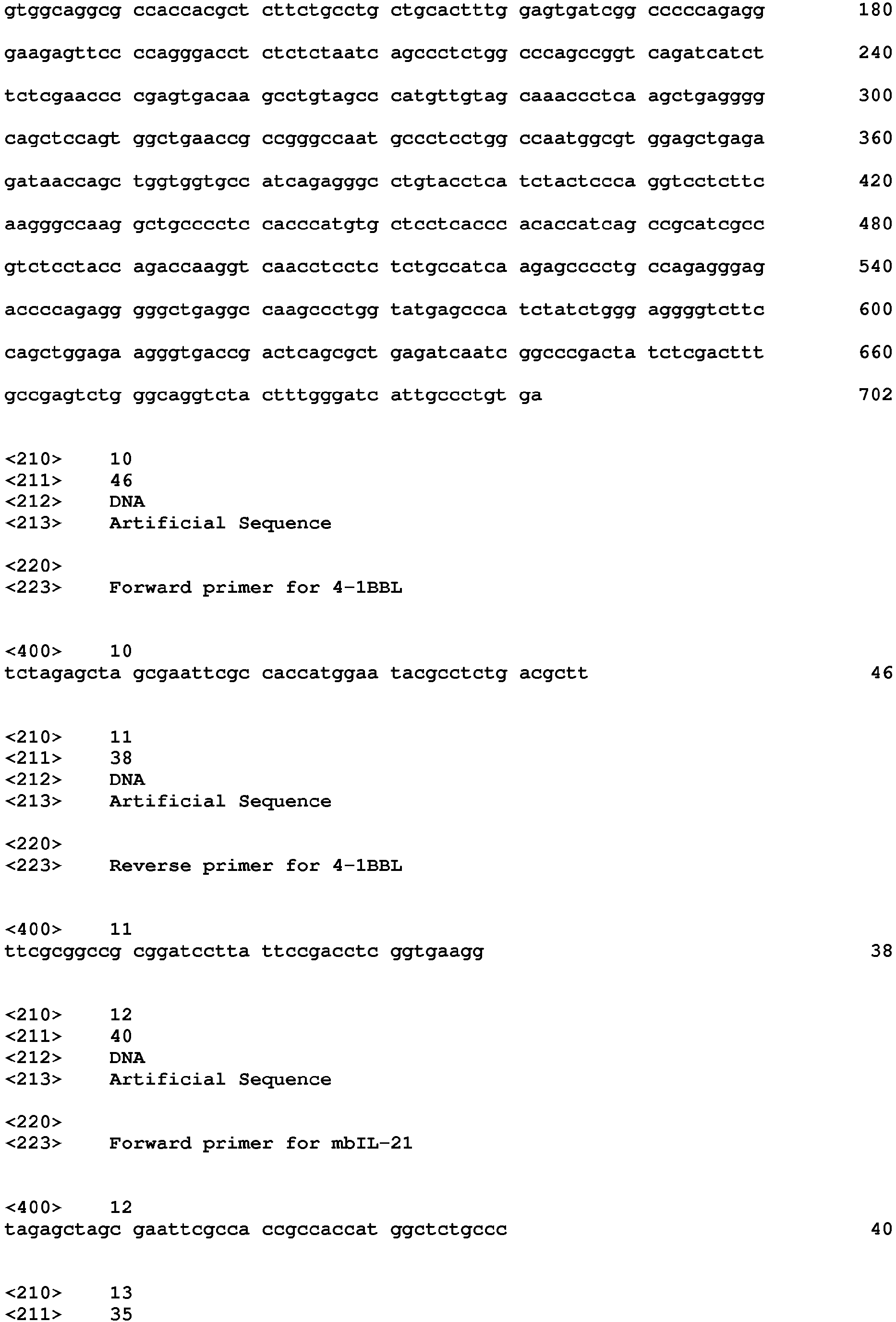

Claims (25)
- A genetically modified CD4+ T cell that expresses at least one gene selected from the group consisting of 4-1BBL gene, mbIL-21 gene, OX40L gene, and mTNF-a gene.
- The genetically modified CD4+ T cell of claim 1, wherein the 4-1BBL gene has a nucleotide sequence encoding the amino acid sequence represented by SEQ ID NO: 1.
- The genetically modified CD4+ T cell of claim 2, wherein the nucleotide sequence encoding the amino acid sequence represented by SEQ ID NO: 1 is the nucleotide sequence represented by SEQ ID NO: 2.
- The genetically modified CD4+ T cell of claim 1, wherein the mbIL-21 gene has a nucleotide sequence encoding the amino acid sequence represented by SEQ ID NO: 3.
- The genetically modified CD4+ T cell of claim 4, wherein the nucleotide sequence encoding the amino acid sequence represented by SEQ ID NO: 3 is the nucleotide sequence represented by SEQ ID NO: 4.
- The genetically modified CD4+ T cell of claim 1, wherein the OX40L gene has a nucleotide sequence encoding the amino acid sequence represented by SEQ ID NO: 5.
- The genetically modified CD4+ T cell of claim 6, wherein the nucleotide sequence encoding the amino acid sequence represented by SEQ ID NO: 5 is the nucleotide sequence represented by SEQ ID NO: 6.
- The genetically modified CD4+ T cell of claim 1, wherein the mTNF-a gene has a nucleotide sequence encoding the amino acid sequence represented by SEQ ID NO: 8.
- The genetically modified CD4+ T cell of claim 8, wherein the nucleotide sequence encoding the amino acid sequence represented by SEQ ID NO: 8 is the nucleotide sequence represented by SEQ ID NO: 9.
- The genetically modified CD4+ T cell of claim 1, wherein the gene is transduced via a recombinant lentivirus.
- The genetically modified CD4+ T cell of claim 1, wherein the CD4+ T cell expresses the 4-1BBL gene or the mbIL-21 gene.
- The genetically modified CD4+ T cell of claim 11, wherein the CD4+ T cell expresses the 4-1BBL gene and the mbIL-21 gene.
- The genetically modified CD4+ T cell of claim 1, wherein the CD4+ T cell expresses the 4-1BBL gene, the mbIL-21 gene, and the mTNF-a gene.
- The genetically modified CD4+ T cell of claim 1, wherein the CD4+ T cell expresses the 4-1BBL gene, the mbIL-21 gene, the OX40L gene, and the mTNF-a gene.
- The genetically modified CD4+ T cell of claim 1, wherein the CD4+ T cell is any one selected from the group consisting of Hut78, H9, Jurkat, Loucy, Molt-3, Molt-13, PEER, RPMI8402, and TALL-01 cells.
- A culture method for natural killer cells, comprising:
co-culturing the genetically modified CD4+ T cells of claim 1 and seed cells. - The culture method of claim 16, wherein the seed cells are any one selected from the group consisting of peripheral blood, peripheral blood mononuclear cells, peripheral blood leukocytes, enriched natural killer cells, and isolated natural killer cells.
- The culture method of claim 16, wherein the seed cells are cells from which CD3(+) cells have been removed.
- The culture method of claim 16, wherein the genetically modified CD4+ T cells and the seed cells are mixed at a ratio of 0.1:1 to 50:1 and the culture is performed.
- The culture method of claim 16, wherein the seed cells are mixed once with feeder cells and the culture is performed for 5 days to 60 days, or the seed cells are mixed two or more times with feeder cells and the culture is performed for 60 days or longer.
- The culture method of claim 16, wherein the culture is performed in a medium containing an anti-CD3 antibody and an interleukin protein.
- The culture method of claim 21, wherein the anti-CD3 antibody includes any one selected from the group consisting of OKT3, UCHT1, and HIT3a.
- The culture method of claim 21, wherein the interleukin protein includes any one selected from the group consisting of IL-2, IL-12, IL-15, IL-18, and IL-21.
- A composition for culturing natural killer cells, comprising as an active ingredient the genetically modified CD4+ T cell of any one of claims 1 to 15.
- A natural killer cell prepared by the culture method for natural killer cells of any one of claims 16 to 23.
Applications Claiming Priority (2)
| Application Number | Priority Date | Filing Date | Title |
|---|---|---|---|
| KR20170065180 | 2017-05-26 | ||
| PCT/KR2018/005983 WO2018217064A2 (en) | 2017-05-26 | 2018-05-25 | Method for culturing natural killer cell, using transformed t cell |
Publications (2)
| Publication Number | Publication Date |
|---|---|
| EP3633029A2 true EP3633029A2 (en) | 2020-04-08 |
| EP3633029A4 EP3633029A4 (en) | 2021-06-09 |
Family
ID=64396946
Family Applications (1)
| Application Number | Title | Priority Date | Filing Date |
|---|---|---|---|
| EP18805231.0A Pending EP3633029A4 (en) | 2017-05-26 | 2018-05-25 | Method for culturing natural killer cell, using transformed t cell |
Country Status (8)
| Country | Link |
|---|---|
| US (2) | US12203065B2 (en) |
| EP (1) | EP3633029A4 (en) |
| JP (1) | JP7039623B2 (en) |
| KR (1) | KR102460173B1 (en) |
| CN (1) | CN110662834B (en) |
| AU (1) | AU2018271755B2 (en) |
| CA (1) | CA3062130A1 (en) |
| WO (1) | WO2018217064A2 (en) |
Cited By (3)
| Publication number | Priority date | Publication date | Assignee | Title |
|---|---|---|---|---|
| EP3892721A4 (en) * | 2018-11-14 | 2022-08-31 | GC Cell Corporation | Method for culturing cord blood-derived natural killer cells using transformed t-cells |
| US11766456B2 (en) | 2014-11-26 | 2023-09-26 | GC Cell Corporation | Method for culturing natural killer cells using T cells |
| US12203065B2 (en) | 2017-05-26 | 2025-01-21 | GC Cell Corporation | Method for culturing natural killer cell, using transformed T cell |
Families Citing this family (14)
| Publication number | Priority date | Publication date | Assignee | Title |
|---|---|---|---|---|
| WO2018124766A2 (en) | 2016-12-28 | 2018-07-05 | 주식회사 녹십자랩셀 | Chimeric antigen receptor and natural killer cells expressing same |
| US12116417B2 (en) | 2017-11-14 | 2024-10-15 | GC Cell Corporation | Anti-HER2 antibody or antigen-binding fragment thereof, and chimeric antigen receptor comprising same |
| US11649294B2 (en) | 2017-11-14 | 2023-05-16 | GC Cell Corporation | Anti-HER2 antibody or antigen-binding fragment thereof, and chimeric antigen receptor comprising same |
| EP3746095A4 (en) | 2018-02-01 | 2021-04-21 | Nkmax Co., Ltd. | NATURAL KILLER CELL PRODUCTION PROCESS AND COMPOSITION FOR CANCER TREATMENT |
| CN111304255B (en) * | 2020-02-21 | 2023-06-09 | 中邦干细胞科技有限公司 | Nourishing cell, preparation method thereof and application of nourishing cell in efficient amplification of NK cells |
| EP4163366A4 (en) * | 2020-06-09 | 2024-08-21 | Samsung Life Public Welfare Foundation | GENETICALLY MODIFIED CELL LINE FOR NK CELL ACTIVATION AND AMPLIFICATION AND USE THEREOF |
| CN112048482A (en) * | 2020-09-09 | 2020-12-08 | 广东昭泰体内生物医药科技有限公司 | Culture method of NK (natural killer) like cells |
| CA3205631A1 (en) * | 2020-12-17 | 2022-06-23 | Artiva Biotherapeutics, Inc. | Expanded and stimulated natural killer cells |
| KR102829749B1 (en) * | 2021-06-01 | 2025-07-07 | 주식회사 박셀바이오 | Composition for expanding natural killer cells comprising feeder cells |
| CN113599395B (en) * | 2021-09-22 | 2022-08-12 | 郑州源创吉因实业有限公司 | Pharmaceutical composition for treating cancer containing NK cells |
| WO2023080895A1 (en) * | 2021-11-04 | 2023-05-11 | Artiva Biotherapeutics, Inc. | Treatment of cancer with nk cells and multispecific engagers |
| CN119013770B (en) * | 2022-10-07 | 2025-11-14 | 东京毅力科创株式会社 | Substrate processing system and edge ring mounting method |
| WO2024153124A1 (en) * | 2023-01-18 | 2024-07-25 | 南京传奇生物科技有限公司 | Modified primary t cell and use thereof |
| CN120209100B (en) * | 2025-03-28 | 2025-09-05 | 河北金要生物科技有限公司 | A virus-like particle expressing mIL21 and 4-1BBL and its application in in vitro expansion of NK cells |
Family Cites Families (75)
| Publication number | Priority date | Publication date | Assignee | Title |
|---|---|---|---|---|
| JPS5840837U (en) | 1981-09-12 | 1983-03-17 | 三菱電機株式会社 | Wafer cleaning and drying equipment |
| US5766601A (en) | 1990-08-08 | 1998-06-16 | University Of Massachusetts Medical Center | Cross-reactive influenza a immunization |
| CA2227327A1 (en) | 1995-07-25 | 1997-02-13 | Celltherapy, Inc. | Autologous immune cell therapy: cell compositions, methods and applications to treatment of human disease |
| IL125884A0 (en) | 1996-03-04 | 1999-04-11 | Targeted Genetics Corp | Modified rapid expansion methods ("modified-rem") for in vitro propagation of t lymphocytes |
| EP0947581A4 (en) | 1996-08-08 | 2004-07-28 | Mitsubishi Pharma Corp | CULTURE AND ITS APPLICATIONS |
| AU2001274931B2 (en) | 2000-05-23 | 2006-03-30 | Nexell Therapeutics, Inc. | Reagents for cell selection and methods of use |
| US20030068306A1 (en) | 2001-09-14 | 2003-04-10 | Dilber Mehmet Sirac | Medium |
| WO2005007116A2 (en) | 2003-07-18 | 2005-01-27 | Valeocyte Therapies Llc | Artificial antigen presenting cell devices |
| GB0318913D0 (en) * | 2003-08-12 | 2003-09-17 | Ist Superiore Sanita | Apoptosis inhibitors |
| ES2368152T3 (en) | 2005-07-01 | 2011-11-14 | Txcell S.A. | OBTAINING TR1 CELLS SPECIFIC TO FOOD ANTIGENS OR SELF-ORIGINS FROM A POPULATION OF LEUKOCYTES OR PBMC. |
| US9121008B2 (en) | 2005-08-31 | 2015-09-01 | University Of Utah Research Foundation | Development of natural killer cells and functional natural killer cell lines |
| US20080261299A1 (en) | 2007-04-23 | 2008-10-23 | Zeikus J Gregory | Pneumatic Bioreactor |
| US7628528B2 (en) | 2005-10-26 | 2009-12-08 | PRS Biotech, Inc. | Pneumatic bioreactor |
| KR20080008060A (en) | 2006-07-19 | 2008-01-23 | 전남대학교산학협력단 | Method of producing natural killer cells |
| RU2009110156A (en) | 2006-08-23 | 2010-09-27 | Байнекс Ко., Лтд. (Kr) | METHOD FOR PRODUCING ACTIVATED LYMPHOCYTES FOR IMMUNOTHERAPY |
| WO2008070647A1 (en) | 2006-12-07 | 2008-06-12 | Wyeth | Methods and compositions for assessing il-12 or the neutralization of il-12 in a sample |
| CA2682527C (en) * | 2007-03-30 | 2017-07-11 | Memorial Sloan-Kettering Cancer Center | Constitutive expression of costimulatory ligands on adoptively transferred t lymphocytes |
| CN101302491B (en) | 2007-05-09 | 2011-09-14 | 王歈 | Highly effective method for amplifying activated lymphocyte and cultivation system |
| JP4953403B2 (en) | 2007-11-05 | 2012-06-13 | 恒雄 倉持 | Method for producing cells for cancer immunotherapy |
| WO2009132283A2 (en) * | 2008-04-24 | 2009-10-29 | University Of Miami | Method for treating autoimmune disorders |
| US20090269849A1 (en) | 2008-04-25 | 2009-10-29 | Pbs Biotech, Inc. | Bioreactor Apparatus |
| KR101035556B1 (en) | 2008-05-22 | 2011-05-23 | 재단법인 목암생명공학연구소 | Method for producing natural killer cells, natural killer cells prepared by such a method, and a composition for treating solid tumors comprising the same as an active ingredient |
| KR20090127974A (en) | 2008-06-10 | 2009-12-15 | 주식회사 엔케이바이오 | Medium composition for self-activating lymphocyte culture |
| KR101043588B1 (en) | 2008-07-25 | 2011-06-22 | 재단법인 철원플라즈마 산업기술연구원 | Plasma ceramic coating film formation method |
| KR101133185B1 (en) | 2008-07-29 | 2012-04-06 | 서울대학교병원 | Method for Proliferating Natural Killer cell |
| US8877182B2 (en) | 2009-03-26 | 2014-11-04 | Cellprotect Nordic Pharmaceuticals Ab | Expansion of NK cells |
| US9062287B2 (en) | 2009-09-11 | 2015-06-23 | Takara Bio Inc. | Process for production of natural killer cells |
| US20130011376A1 (en) | 2009-12-29 | 2013-01-10 | Gamida Cell Ltd. | Methods for Enhancing Natural Killer Cell Proliferation and Activity |
| CN103068973A (en) | 2010-08-20 | 2013-04-24 | 免疫创新治疗有限公司 | Cells expressing Th1 properties and cytolytic properties |
| KR101298012B1 (en) | 2011-02-08 | 2013-08-26 | (주)차바이오앤디오스텍 | Process for preparing lymphocytes comprising activated natural killer cells for targeting cancer cells and pharmaceutical composition comprising the same |
| ES2856825T3 (en) | 2011-03-18 | 2021-09-28 | Glycostem Therapeutics B V | Generation of NK cells and NK cell progenitors |
| WO2012146702A1 (en) | 2011-04-28 | 2012-11-01 | INSERM (Institut National de la Santé et de la Recherche Médicale) | Methods for preparing accessory cells and uses thereof for preparing activated nk cells |
| JP5840876B2 (en) | 2011-06-24 | 2016-01-06 | テラ株式会社 | Compositions and methods for amplifying NK cells |
| JP5572863B2 (en) | 2011-06-24 | 2014-08-20 | 国立大学法人九州大学 | Method for amplifying NK cells |
| JP5847518B2 (en) | 2011-09-28 | 2016-01-20 | 株式会社日本バイオセラピー研究所 | Method for producing NK cell-enhanced blood product |
| ES2652666T3 (en) | 2011-12-22 | 2018-02-05 | Mogam Biotechnology Institute | Production process of natural cytolytic lymphocytes, natural cytolytic lymphocytes produced in this way and composition for the treatment of cancers and infectious diseases containing them |
| CN102559600A (en) * | 2011-12-29 | 2012-07-11 | 上海交通大学医学院 | Artificial antigen presenting cell and application thereof in NK (natural killer) cell amplification |
| WO2013131045A1 (en) | 2012-03-02 | 2013-09-06 | The Regents Of The University Of California | Expansion of alloantigen-reactive regulatory t cells |
| CN104321425B (en) | 2012-05-07 | 2018-08-10 | 株式会社Nkmax | Method for inducing and expanding natural killer cells derived from peripheral blood mononuclear cells |
| JP5511039B1 (en) | 2013-05-22 | 2014-06-04 | 国立大学法人九州大学 | Method for preparing NK cells |
| CA2945320A1 (en) * | 2014-04-10 | 2015-10-15 | Seattle Children's Hospital (dba Seattle Children's Research Institute) | Drug regulated transgene expression |
| WO2016029043A1 (en) | 2014-08-21 | 2016-02-25 | The General Hospital Corporation | Tumor necrosis factor superfamily and tnf-like ligand muteins and methods of preparing and using the same |
| EP3223856B1 (en) * | 2014-10-27 | 2025-09-24 | University of Central Florida Research Foundation, Inc. | Methods and compositions for natural killer cells |
| CN107106609A (en) * | 2014-10-31 | 2017-08-29 | 宾夕法尼亚大学董事会 | Stimulate and extend the composition and method of T cell |
| CN105602899B (en) | 2014-11-25 | 2019-03-08 | 天津天锐生物科技有限公司 | A kind of method of NK cell Proliferation in interleukin 21 and 4-1BBL induction peripheral blood mononuclear cells |
| KR101697473B1 (en) * | 2014-11-26 | 2017-01-18 | 주식회사 녹십자랩셀 | Method for Preparation of Natural Killer Cells Using T Cells |
| KR101706524B1 (en) | 2014-12-03 | 2017-02-14 | 주식회사 녹십자랩셀 | Efficient Method for Preparing of Stable Natural Killer Cells |
| GB201503500D0 (en) * | 2015-03-02 | 2015-04-15 | Ucl Business Plc | Cell |
| US10744157B2 (en) | 2015-03-26 | 2020-08-18 | The Trustees Of Dartmouth College | Anti-MICA antigen binding fragments, fusion molecules, cells which express and methods of using |
| CA2984794A1 (en) | 2015-05-07 | 2016-11-10 | Agenus Inc. | Anti-ox40 antibodies and methods of use thereof |
| AU2017215955B2 (en) | 2016-02-01 | 2020-01-23 | GC Cell Corporation | Medium composition for cryopreservation of cell and use thereof |
| CN106222141B (en) * | 2016-10-17 | 2018-10-19 | 湖南丰晖生物科技有限公司 | NK cell culture fluids and cell culture processes |
| WO2018124766A2 (en) | 2016-12-28 | 2018-07-05 | 주식회사 녹십자랩셀 | Chimeric antigen receptor and natural killer cells expressing same |
| CN108300697A (en) | 2017-01-13 | 2018-07-20 | 上海恒润达生生物科技有限公司 | A kind of method and purposes that trophocyte stimulation NK cells expand |
| GB201706451D0 (en) | 2017-04-24 | 2017-06-07 | Imp Innovations Ltd | Cancer treatment |
| EP3633029A4 (en) | 2017-05-26 | 2021-06-09 | Green Cross Lab Cell Corporation | Method for culturing natural killer cell, using transformed t cell |
| US12116417B2 (en) | 2017-11-14 | 2024-10-15 | GC Cell Corporation | Anti-HER2 antibody or antigen-binding fragment thereof, and chimeric antigen receptor comprising same |
| CN108300693A (en) | 2018-01-25 | 2018-07-20 | 河北省健海生物芯片技术有限责任公司 | A kind of natural killer cells amplification in vitro method |
| KR102232321B1 (en) | 2018-03-23 | 2021-03-26 | 주식회사 녹십자랩셀 | Method for Preparing NK Cell |
| WO2019182392A1 (en) | 2018-03-23 | 2019-09-26 | 주식회사 녹십자랩셀 | Method for producing natural killer cells |
| KR20200030337A (en) | 2018-09-12 | 2020-03-20 | 주식회사 녹십자랩셀 | Pharmaceutical combinations for treating tumor comprising anti-cd19 antibody and natural killer cell |
| WO2020101361A1 (en) | 2018-11-14 | 2020-05-22 | 주식회사 녹십자랩셀 | Method for culturing cord blood-derived natural killer cells using transformed t-cells |
| IL298326A (en) | 2020-05-22 | 2023-01-01 | Gc Cell Corp | An antibody against her2 or an antigen-binding fragment thereof, and a chimeric antigen receptor comprising it |
| WO2022133061A1 (en) | 2020-12-17 | 2022-06-23 | Artiva Biotherapeutics, Inc. | Infusion ready cryopreservation compositions |
| CA3205631A1 (en) | 2020-12-17 | 2022-06-23 | Artiva Biotherapeutics, Inc. | Expanded and stimulated natural killer cells |
| CA3205637A1 (en) | 2020-12-17 | 2022-06-23 | Artiva Biotherapeutics, Inc. | Treatment of cancer with nk cells and a cd20 targeted antibody |
| CA3216024A1 (en) | 2021-04-08 | 2022-10-13 | Artiva Biotherapeutics, Inc. | Chimeric antigen receptor comprising an anti-her2 antibody or antigen-binding fragment thereof and natural killer cells comprising the same |
| JP2024513958A (en) | 2021-04-08 | 2024-03-27 | アルティヴァ バイオセラピューティクス インコーポレイテッド | Cancer treatment using NK cells and EGFR targeting antibodies |
| AU2022253256A1 (en) | 2021-04-08 | 2023-11-16 | Artiva Biotherapeutics, Inc. | Treatment of cancer with nk cells and a cd38-targeted antibody |
| JP2024515169A (en) | 2021-04-08 | 2024-04-05 | アルティヴァ バイオセラピューティクス インコーポレイテッド | Cancer treatment using NK cells and CD20-targeted antibodies |
| WO2022216144A1 (en) | 2021-04-08 | 2022-10-13 | GC Cell Corporation | Fusion proteins comprising chimeric antigen receptors and il-15 |
| AU2022254689A1 (en) | 2021-04-08 | 2023-11-16 | Artiva Biotherapeutics, Inc. | Treatment of cancer with nk cells and a her2 targeted antibody |
| US20240366663A1 (en) | 2021-04-08 | 2024-11-07 | Artiva Biotherapeutics, Inc. | Chimeric antigen receptor comprising an anti-cd19 antibody or antigen-binding fragment thereof and natural killer cells comprising the same |
| WO2023080895A1 (en) | 2021-11-04 | 2023-05-11 | Artiva Biotherapeutics, Inc. | Treatment of cancer with nk cells and multispecific engagers |
| JP2024542159A (en) | 2021-11-04 | 2024-11-13 | アーティバ バイオセラピューティクス インコーポレイテッド | Treating Cancer with NK Cells and Multispecific Engagers |
-
2018
- 2018-05-25 EP EP18805231.0A patent/EP3633029A4/en active Pending
- 2018-05-25 AU AU2018271755A patent/AU2018271755B2/en active Active
- 2018-05-25 WO PCT/KR2018/005983 patent/WO2018217064A2/en not_active Ceased
- 2018-05-25 CN CN201880034893.8A patent/CN110662834B/en active Active
- 2018-05-25 CA CA3062130A patent/CA3062130A1/en active Pending
- 2018-05-25 JP JP2019565241A patent/JP7039623B2/en active Active
- 2018-05-25 US US16/613,601 patent/US12203065B2/en active Active
- 2018-05-25 KR KR1020197030133A patent/KR102460173B1/en active Active
-
2024
- 2024-12-11 US US18/977,119 patent/US20250109380A1/en active Pending
Cited By (4)
| Publication number | Priority date | Publication date | Assignee | Title |
|---|---|---|---|---|
| US11766456B2 (en) | 2014-11-26 | 2023-09-26 | GC Cell Corporation | Method for culturing natural killer cells using T cells |
| US12203065B2 (en) | 2017-05-26 | 2025-01-21 | GC Cell Corporation | Method for culturing natural killer cell, using transformed T cell |
| EP3892721A4 (en) * | 2018-11-14 | 2022-08-31 | GC Cell Corporation | Method for culturing cord blood-derived natural killer cells using transformed t-cells |
| US12398372B2 (en) | 2018-11-14 | 2025-08-26 | GC Cell Corporation | Method for culturing cord blood-derived natural killer cells using transformed T-cells |
Also Published As
| Publication number | Publication date |
|---|---|
| US12203065B2 (en) | 2025-01-21 |
| JP7039623B2 (en) | 2022-03-22 |
| AU2018271755B2 (en) | 2021-12-23 |
| CA3062130A1 (en) | 2019-11-22 |
| CN110662834A (en) | 2020-01-07 |
| EP3633029A4 (en) | 2021-06-09 |
| WO2018217064A3 (en) | 2019-03-28 |
| JP2020521462A (en) | 2020-07-27 |
| KR102460173B1 (en) | 2022-10-31 |
| WO2018217064A2 (en) | 2018-11-29 |
| US20250109380A1 (en) | 2025-04-03 |
| KR20200001595A (en) | 2020-01-06 |
| US20200108096A1 (en) | 2020-04-09 |
| CN110662834B (en) | 2023-09-12 |
| AU2018271755A1 (en) | 2019-11-21 |
Similar Documents
| Publication | Publication Date | Title |
|---|---|---|
| US20250109380A1 (en) | Method for culturing natural killer cell, using transformed t cell | |
| US20240141041A1 (en) | CHIMERIC ANTIGEN RECEPTORS (CARs), COMPOSITIONS AND METHODS THEREOF | |
| KR102437015B1 (en) | Transgenic T Cells and Chimeric Antigen Receptor T Cell Compositions and Related Methods | |
| JP6879910B2 (en) | Modification of gene expression in CART cells and their use | |
| JP6720176B2 (en) | Methods for controlled activation or elimination of therapeutic cells | |
| JP7233720B2 (en) | Immune Competent Cells Expressing Cell Surface Molecules That Specifically Recognize Human Mesothelin, IL-7, and CCL19 | |
| JP2020096640A (en) | Controlled elimination of therapeutic cells | |
| CN111247242A (en) | Chimeric Antigen Receptors (CARs), compositions and methods of use thereof | |
| EP3907280A1 (en) | Immune effector cell targeting gpc3 and application thereof | |
| US20250101081A1 (en) | Novel gene armoring | |
| JP7179986B2 (en) | Method for culturing cord blood-derived natural killer cells using transformed T cells | |
| US20230381312A1 (en) | Redirecting glucose metabolism to limit stress and improve adoptive cell therapy | |
| HK40058243A (en) | Method for culturing cord blood-derived natural killer cells using transformed t-cells | |
| HK40058243B (en) | Method for culturing cord blood-derived natural killer cells using transformed t-cells | |
| WO2024206936A2 (en) | Interleukin-18 pretreatment as a cryoprotectant for lymphocytes | |
| HK40091903B (en) | Transgenic t cell and chimeric antigen receptor t cell compositions and related methods | |
| HK40091903A (en) | Transgenic t cell and chimeric antigen receptor t cell compositions and related methods | |
| KR20250167171A (en) | A Composition for Culturing Natural Killer Cells Comprising Extracellular Vesicles Isolated from Engineered T Cells as Active Ingredients | |
| JP2023504196A (en) | Placenta-derived allogeneic CAR-T cells and uses thereof |
Legal Events
| Date | Code | Title | Description |
|---|---|---|---|
| STAA | Information on the status of an ep patent application or granted ep patent |
Free format text: STATUS: THE INTERNATIONAL PUBLICATION HAS BEEN MADE |
|
| PUAI | Public reference made under article 153(3) epc to a published international application that has entered the european phase |
Free format text: ORIGINAL CODE: 0009012 |
|
| STAA | Information on the status of an ep patent application or granted ep patent |
Free format text: STATUS: REQUEST FOR EXAMINATION WAS MADE |
|
| 17P | Request for examination filed |
Effective date: 20191120 |
|
| AK | Designated contracting states |
Kind code of ref document: A2 Designated state(s): AL AT BE BG CH CY CZ DE DK EE ES FI FR GB GR HR HU IE IS IT LI LT LU LV MC MK MT NL NO PL PT RO RS SE SI SK SM TR |
|
| AX | Request for extension of the european patent |
Extension state: BA ME |
|
| DAV | Request for validation of the european patent (deleted) | ||
| DAX | Request for extension of the european patent (deleted) | ||
| RIC1 | Information provided on ipc code assigned before grant |
Ipc: C12N 5/0783 20100101AFI20210201BHEP Ipc: A61K 35/12 20150101ALI20210201BHEP |
|
| A4 | Supplementary search report drawn up and despatched |
Effective date: 20210507 |
|
| RIC1 | Information provided on ipc code assigned before grant |
Ipc: C12N 5/0783 20100101AFI20210430BHEP Ipc: A61K 35/12 20150101ALI20210430BHEP |
|
| RAP1 | Party data changed (applicant data changed or rights of an application transferred) |
Owner name: GC CELL CORPORATION |
|
| STAA | Information on the status of an ep patent application or granted ep patent |
Free format text: STATUS: EXAMINATION IS IN PROGRESS |
|
| 17Q | First examination report despatched |
Effective date: 20241009 |