EP3638021B1 - Procédés de conservation de cellules - Google Patents
Procédés de conservation de cellules Download PDFInfo
- Publication number
- EP3638021B1 EP3638021B1 EP18817025.2A EP18817025A EP3638021B1 EP 3638021 B1 EP3638021 B1 EP 3638021B1 EP 18817025 A EP18817025 A EP 18817025A EP 3638021 B1 EP3638021 B1 EP 3638021B1
- Authority
- EP
- European Patent Office
- Prior art keywords
- cells
- channel
- microbubbles
- solution
- group
- Prior art date
- Legal status (The legal status is an assumption and is not a legal conclusion. Google has not performed a legal analysis and makes no representation as to the accuracy of the status listed.)
- Active
Links
Images


Classifications
-
- A—HUMAN NECESSITIES
- A61—MEDICAL OR VETERINARY SCIENCE; HYGIENE
- A61M—DEVICES FOR INTRODUCING MEDIA INTO, OR ONTO, THE BODY; DEVICES FOR TRANSDUCING BODY MEDIA OR FOR TAKING MEDIA FROM THE BODY; DEVICES FOR PRODUCING OR ENDING SLEEP OR STUPOR
- A61M1/00—Suction or pumping devices for medical purposes; Devices for carrying-off, for treatment of, or for carrying-over, body-liquids; Drainage systems
- A61M1/02—Blood transfusion apparatus
- A61M1/0272—Apparatus for treatment of blood or blood constituents prior to or for conservation, e.g. freezing, drying or centrifuging
-
- A—HUMAN NECESSITIES
- A01—AGRICULTURE; FORESTRY; ANIMAL HUSBANDRY; HUNTING; TRAPPING; FISHING
- A01N—PRESERVATION OF BODIES OF HUMANS OR ANIMALS OR PLANTS OR PARTS THEREOF; BIOCIDES, e.g. AS DISINFECTANTS, AS PESTICIDES OR AS HERBICIDES; PEST REPELLANTS OR ATTRACTANTS; PLANT GROWTH REGULATORS
- A01N1/00—Preservation of bodies of humans or animals, or parts thereof
- A01N1/10—Preservation of living parts
-
- C—CHEMISTRY; METALLURGY
- C12—BIOCHEMISTRY; BEER; SPIRITS; WINE; VINEGAR; MICROBIOLOGY; ENZYMOLOGY; MUTATION OR GENETIC ENGINEERING
- C12N—MICROORGANISMS OR ENZYMES; COMPOSITIONS THEREOF; PROPAGATING, PRESERVING, OR MAINTAINING MICROORGANISMS; MUTATION OR GENETIC ENGINEERING; CULTURE MEDIA
- C12N5/00—Undifferentiated human, animal or plant cells, e.g. cell lines; Tissues; Cultivation or maintenance thereof; Culture media therefor
- C12N5/0068—General culture methods using substrates
-
- C—CHEMISTRY; METALLURGY
- C12—BIOCHEMISTRY; BEER; SPIRITS; WINE; VINEGAR; MICROBIOLOGY; ENZYMOLOGY; MUTATION OR GENETIC ENGINEERING
- C12N—MICROORGANISMS OR ENZYMES; COMPOSITIONS THEREOF; PROPAGATING, PRESERVING, OR MAINTAINING MICROORGANISMS; MUTATION OR GENETIC ENGINEERING; CULTURE MEDIA
- C12N5/00—Undifferentiated human, animal or plant cells, e.g. cell lines; Tissues; Cultivation or maintenance thereof; Culture media therefor
- C12N5/54—Mechanical aspects of preservation of animal cells or human cells; Apparatus or containers therefor
- C12N5/542—Apparatus
-
- C—CHEMISTRY; METALLURGY
- C12—BIOCHEMISTRY; BEER; SPIRITS; WINE; VINEGAR; MICROBIOLOGY; ENZYMOLOGY; MUTATION OR GENETIC ENGINEERING
- C12N—MICROORGANISMS OR ENZYMES; COMPOSITIONS THEREOF; PROPAGATING, PRESERVING, OR MAINTAINING MICROORGANISMS; MUTATION OR GENETIC ENGINEERING; CULTURE MEDIA
- C12N5/00—Undifferentiated human, animal or plant cells, e.g. cell lines; Tissues; Cultivation or maintenance thereof; Culture media therefor
- C12N5/56—Physical preservation processes for animal cells or human cells
- C12N5/562—Temperature processes, e.g. following predefined temperature changes over time
-
- C—CHEMISTRY; METALLURGY
- C12—BIOCHEMISTRY; BEER; SPIRITS; WINE; VINEGAR; MICROBIOLOGY; ENZYMOLOGY; MUTATION OR GENETIC ENGINEERING
- C12N—MICROORGANISMS OR ENZYMES; COMPOSITIONS THEREOF; PROPAGATING, PRESERVING, OR MAINTAINING MICROORGANISMS; MUTATION OR GENETIC ENGINEERING; CULTURE MEDIA
- C12N5/00—Undifferentiated human, animal or plant cells, e.g. cell lines; Tissues; Cultivation or maintenance thereof; Culture media therefor
- C12N5/56—Physical preservation processes for animal cells or human cells
- C12N5/565—Pressure processes, e.g. following predefined pressure changes over time
-
- C—CHEMISTRY; METALLURGY
- C12—BIOCHEMISTRY; BEER; SPIRITS; WINE; VINEGAR; MICROBIOLOGY; ENZYMOLOGY; MUTATION OR GENETIC ENGINEERING
- C12N—MICROORGANISMS OR ENZYMES; COMPOSITIONS THEREOF; PROPAGATING, PRESERVING, OR MAINTAINING MICROORGANISMS; MUTATION OR GENETIC ENGINEERING; CULTURE MEDIA
- C12N5/00—Undifferentiated human, animal or plant cells, e.g. cell lines; Tissues; Cultivation or maintenance thereof; Culture media therefor
- C12N5/56—Physical preservation processes for animal cells or human cells
- C12N5/568—Physical preservation processes for animal cells or human cells using electromagnetic fields or radiation; using acoustic waves or corpuscular radiation
-
- A—HUMAN NECESSITIES
- A01—AGRICULTURE; FORESTRY; ANIMAL HUSBANDRY; HUNTING; TRAPPING; FISHING
- A01N—PRESERVATION OF BODIES OF HUMANS OR ANIMALS OR PLANTS OR PARTS THEREOF; BIOCIDES, e.g. AS DISINFECTANTS, AS PESTICIDES OR AS HERBICIDES; PEST REPELLANTS OR ATTRACTANTS; PLANT GROWTH REGULATORS
- A01N1/00—Preservation of bodies of humans or animals, or parts thereof
- A01N1/10—Preservation of living parts
- A01N1/14—Mechanical aspects of preservation; Apparatus or containers therefor
- A01N1/142—Apparatus
-
- A—HUMAN NECESSITIES
- A01—AGRICULTURE; FORESTRY; ANIMAL HUSBANDRY; HUNTING; TRAPPING; FISHING
- A01N—PRESERVATION OF BODIES OF HUMANS OR ANIMALS OR PLANTS OR PARTS THEREOF; BIOCIDES, e.g. AS DISINFECTANTS, AS PESTICIDES OR AS HERBICIDES; PEST REPELLANTS OR ATTRACTANTS; PLANT GROWTH REGULATORS
- A01N1/00—Preservation of bodies of humans or animals, or parts thereof
- A01N1/10—Preservation of living parts
- A01N1/16—Physical preservation processes
- A01N1/162—Temperature processes, e.g. following predefined temperature changes over time
-
- A—HUMAN NECESSITIES
- A01—AGRICULTURE; FORESTRY; ANIMAL HUSBANDRY; HUNTING; TRAPPING; FISHING
- A01N—PRESERVATION OF BODIES OF HUMANS OR ANIMALS OR PLANTS OR PARTS THEREOF; BIOCIDES, e.g. AS DISINFECTANTS, AS PESTICIDES OR AS HERBICIDES; PEST REPELLANTS OR ATTRACTANTS; PLANT GROWTH REGULATORS
- A01N1/00—Preservation of bodies of humans or animals, or parts thereof
- A01N1/10—Preservation of living parts
- A01N1/16—Physical preservation processes
- A01N1/165—Pressure processes, e.g. following predefined pressure changes over time
-
- A—HUMAN NECESSITIES
- A01—AGRICULTURE; FORESTRY; ANIMAL HUSBANDRY; HUNTING; TRAPPING; FISHING
- A01N—PRESERVATION OF BODIES OF HUMANS OR ANIMALS OR PLANTS OR PARTS THEREOF; BIOCIDES, e.g. AS DISINFECTANTS, AS PESTICIDES OR AS HERBICIDES; PEST REPELLANTS OR ATTRACTANTS; PLANT GROWTH REGULATORS
- A01N1/00—Preservation of bodies of humans or animals, or parts thereof
- A01N1/10—Preservation of living parts
- A01N1/16—Physical preservation processes
- A01N1/168—Physical preservation processes using electromagnetic fields or radiation; using acoustic waves or corpuscular radiation
-
- A—HUMAN NECESSITIES
- A61—MEDICAL OR VETERINARY SCIENCE; HYGIENE
- A61M—DEVICES FOR INTRODUCING MEDIA INTO, OR ONTO, THE BODY; DEVICES FOR TRANSDUCING BODY MEDIA OR FOR TAKING MEDIA FROM THE BODY; DEVICES FOR PRODUCING OR ENDING SLEEP OR STUPOR
- A61M2202/00—Special media to be introduced, removed or treated
- A61M2202/04—Liquids
- A61M2202/0413—Blood
- A61M2202/0429—Red blood cells; Erythrocytes
-
- A—HUMAN NECESSITIES
- A61—MEDICAL OR VETERINARY SCIENCE; HYGIENE
- A61M—DEVICES FOR INTRODUCING MEDIA INTO, OR ONTO, THE BODY; DEVICES FOR TRANSDUCING BODY MEDIA OR FOR TAKING MEDIA FROM THE BODY; DEVICES FOR PRODUCING OR ENDING SLEEP OR STUPOR
- A61M2205/00—General characteristics of the apparatus
- A61M2205/02—General characteristics of the apparatus characterised by a particular materials
- A61M2205/0244—Micromachined materials, e.g. made from silicon wafers, microelectromechanical systems [MEMS] or comprising nanotechnology
-
- A—HUMAN NECESSITIES
- A61—MEDICAL OR VETERINARY SCIENCE; HYGIENE
- A61M—DEVICES FOR INTRODUCING MEDIA INTO, OR ONTO, THE BODY; DEVICES FOR TRANSDUCING BODY MEDIA OR FOR TAKING MEDIA FROM THE BODY; DEVICES FOR PRODUCING OR ENDING SLEEP OR STUPOR
- A61M2205/00—General characteristics of the apparatus
- A61M2205/05—General characteristics of the apparatus combined with other kinds of therapy
- A61M2205/058—General characteristics of the apparatus combined with other kinds of therapy with ultrasound therapy
-
- C—CHEMISTRY; METALLURGY
- C12—BIOCHEMISTRY; BEER; SPIRITS; WINE; VINEGAR; MICROBIOLOGY; ENZYMOLOGY; MUTATION OR GENETIC ENGINEERING
- C12N—MICROORGANISMS OR ENZYMES; COMPOSITIONS THEREOF; PROPAGATING, PRESERVING, OR MAINTAINING MICROORGANISMS; MUTATION OR GENETIC ENGINEERING; CULTURE MEDIA
- C12N2500/00—Specific components of cell culture medium
- C12N2500/30—Organic components
- C12N2500/34—Sugars
-
- C—CHEMISTRY; METALLURGY
- C12—BIOCHEMISTRY; BEER; SPIRITS; WINE; VINEGAR; MICROBIOLOGY; ENZYMOLOGY; MUTATION OR GENETIC ENGINEERING
- C12N—MICROORGANISMS OR ENZYMES; COMPOSITIONS THEREOF; PROPAGATING, PRESERVING, OR MAINTAINING MICROORGANISMS; MUTATION OR GENETIC ENGINEERING; CULTURE MEDIA
- C12N2502/00—Coculture with; Conditioned medium produced by
- C12N2502/11—Coculture with; Conditioned medium produced by blood or immune system cells
- C12N2502/115—Platelets, megakaryocytes
-
- C—CHEMISTRY; METALLURGY
- C12—BIOCHEMISTRY; BEER; SPIRITS; WINE; VINEGAR; MICROBIOLOGY; ENZYMOLOGY; MUTATION OR GENETIC ENGINEERING
- C12N—MICROORGANISMS OR ENZYMES; COMPOSITIONS THEREOF; PROPAGATING, PRESERVING, OR MAINTAINING MICROORGANISMS; MUTATION OR GENETIC ENGINEERING; CULTURE MEDIA
- C12N2521/00—Culture process characterised by the use of hydrostatic pressure, flow or shear forces
- C12N2521/10—Sound, e.g. ultrasounds
Definitions
- the presently-disclosed subject matter generally relates to methods for the delivery of molecules into cells.
- certain embodiments of the presently-disclosed subject matter relate to methods for the preservation, for example cryopreservation and lyopreservation, of cells that make use of ultrasound and sonoporation.
- Red blood cells perform a vital role by delivering molecular oxygen to tissues throughout the body in order to meet the metabolic needs of the body.
- Conditions or procedures that cause significant loss of functional RBCs such as trauma, blood disorders, cancer treatment, or surgical procedures, can lead to oxygen deprivation, life-threatening organ failure, and can eventually lead to death. Therefore, many patients require blood transfusions in order to replace lost RBCs and maintain adequate oxygenation of tissues.
- blood transfusions are the most common medical procedure performed in U.S. hospitals, with over 2.9 million patients receiving a combined total of over 13.7 million units of RBCs in 2011. Approximately 85 million units of RBCs are transfused annually worldwide, and the global market size for blood preparation was $44 billion in 2015.
- RBCs are typically stored at 4 °C and must be used within 42 days. Thus, frequent blood donations are needed in order to maintain an adequate supply of RBCs for transfusions. Over 370,000 units of expired RBCs were discarded in the United States in 2011 and over 10% of surveyed U.S. hospitals reported at least one day of blood shortage per year, with an average annual RBC shortage of 19 days in these hospitals. In some special cases, RBCs can be frozen in the presence of glycerol for up to 10 years, but the glycerol must be thoroughly removed via multiple cumbersome washing steps prior to transfusion, and most hospitals do not have the capability to perform this process.
- microfluidic electro-sonoporation a multi-modal cell poration methodology through simultaneous application of electric field and ultrasonic wave
- Whitney Longsine-Parker et al., Lab Chip, 2013,13, pages 2144-2152 discloses a microfluidic electro-sonoporation device, wherein cells flowing through a microfluidic channel made of a microelectrode pair are exposed simultaneously to an electric field and to acoustic pressure generated by an ultrasound transducer placed on top of the channel.
- the channel has three inlets, a center cell inlet and two side buffer inlets.
- One aspect of the disclosure provides a system for molecule delivery into cells, comprising a microfluidic device comprising a first channel for housing one or more microbubbles, a second channel for housing soluble molecules, the second channel in fluid communication with and intersected by the first channel, and a third channel for housing a cell solution, the third channel in fluid communication with and intersected by the second channel; and an ultrasound transducer positioned below the third channel or an interdigital transducer positioned on opposite sides of the third channel.
- at least a portion of the third channel is in a spiral configuration.
- the first channel, the second channel, and/or the third channel is about 20 ⁇ m to about 700 ⁇ m wide and about 20 ⁇ m to about 400 ⁇ m deep.
- Another aspect of the disclosure provides a method for molecule delivery into cells, comprising forming a solution comprising an amount of microbubbles, an amount of soluble molecules, and one or more cells; flowing the solution through a microfluidic device 4; and sonicating the solution for a period of time sufficient to form pores in the one or more cells.
- the soluble molecules are selected from the group consisting of trehalose, sucrose, sorbitol, xylitol, myo-iositol, diglycerol phosphate, proline, ectoine, taurine, hypotaurine, and transgenic expressed group 1, group 2, or group 3 Late Embryogenesis Abundant (LEA) proteins from the brine Artemia franciscana.
- the method further comprises a step of drying and/or freezing the one or more cells.
- the one or more cells are red blood cells.
- Another aspect of the disclosure provides a method for the cryopreservation of cells, comprising forming a solution comprising an amount of microbubbles, an amount of preservation agent, and one or more cells; and sonicating the solution for a period of time sufficient to form pores in the one or more cells.
- Another aspect of the disclosure provides a method for molecule delivery into cells, comprising passing a fluid comprising an amount of microbubbles, an amount of soluble molecules, and one or more cells through a microchannel one cell at a time and sonicating the cells as the cells pass through the microchannel such that the microbubbles form pores in the one or more cells through which one or more of the soluble molecules enter the one or more cells.
- an optionally variant portion means that the portion is variant or non-variant.
- Embodiments of the disclosure provide a system and process for delivering molecules (i.e. a "target agent"), such as the cell protectant trehalose, into cells, such as RBCs or platelets, by utilizing a combination of ultrasound technology and microbubbles.
- molecules i.e. a "target agent”
- a target agent such as the cell protectant trehalose
- Any type of cells e.g. nucleated cells, are compatible with the systems and processes described herein.
- Exemplary cell types include, but are not limited to, stem cells which may be useful for stem cell therapy, insect cells e.g. for transgenic protein production, and cell lines for drug testing such as HepG2, CHO, HEK, HELA, etc.
- Microbubbles which are generally ⁇ 1-3 ⁇ m in diameter, are FDA approved for use as ultrasound contrast agents in clinical echocardiography, and are also in development for therapeutic use. Microbubbles oscillate in response to ultrasound pressure waves that cause the microbubbles to expand and compress with each positive and negative pressure cycle. This microbubble oscillation ("cavitation") exerts forces on the surrounding fluid. At sufficient ultrasound amplitudes the oscillation becomes non-linear and can induce strong microstreaming or microjetting effects in the surrounding fluid. When this occurs near a cell membrane the microjets and shock waves produced by microbubble cavitation can form transient pores in the membrane, which are generally rapidly repaired by the cell in less than a minute.
- Embodiments of the disclosure utilize this pore formation process ("sonoporation") to directly deliver target agents into the cell.
- sonoporation may be used to deliver the sugar trehalose, a cell protectant, into RBCs to promote cell survival during freezing and drying.
- microbubbles comprise about 1-20% of a solution containing the target agent and/or cells.
- the microbubbles are lipid-coated.
- Exemplary lipids that may be utilized include, but are not limited to 1,2-distearoyl-sn-glycero-3-phosphocholine (DSPC), 1,2-dipalmitoyl-sn-glycero-3-phosphocholine (DPPC), 1,2-distearoyl-sn-glycero-3-ethylphosphocholine (DSEPC), 1,2-distearoyl-sn-glycero-3-phosphoglycerol (DSPG), 1,2-distearoyl-sn-glycero-3-phosphoethanolamine-N-[methoxy(polyethylene glycol)-2000] (DSPE-PEG2000), and/or polyethylene glycol-40 stearate (PEG-40).
- DSPC 1,2-distearoyl-sn-glycero-3-phosphocholine
- DPPC 1,2-dipalmitoyl-sn-glycero-3-phosphocholine
- a formulation comprising DSPC:DSEPC:DSPG:PEG-40 in a 100:43:1:4.5 molar ratio is used.
- a lipid solution composed of 94:4 molar ratio (DPPC:DSPE-PEG2000) is used.
- Trehalose has been granted Generally Recognized As Safe (GRAS) status by the U.S. Food and Drug Administration and is used for food preservation in products such as sweets, meats, and frozen foods. It is a naturally-occurring non-reducing sugar found in many plants and lower animals that helps them survive water-limited states such as dehydration or freezing. During freezing, trehalose prevents damage from ice crystals. During drying, the sugar forms a glassy barrier around cellular membranes replacing water. However, cells such as RBCs are generally impermeable to trehalose and must be manipulated to enable intracellular delivery of this compound.
- GRAS Generally Recognized As Safe
- a system for delivery of a target agent into cells e.g. for cell preservation
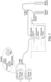
- a microfluidic device and an ultrasound transducer FIG. 1
- embodiments of the system are useful for cryopreservation and/or lyopreservation.
- Cryopreservation preserves cells by cooling to very low temperatures (e.g., lower than -80°C) whereas lyopreservation is a biomimetic strategy based on anhydrobiosis to preserve cells at ambient temperatures in a desiccated state.
- This device can be used for sonoporation and loading of any soluble molecule, e.g.
- the microfluidic device is positioned at or near the surface of a filled water tank.
- Tubing such as Tygon PVC clear tubing (1/16 inch inner diameter, 1/8 inch outer diameter) (McMaster-Carr), may be inserted into the inlet and outlet ports of the device.
- the inlet tubing may lead to a 3-way valve with one side of the valve leading to a syringe filled with the sample solution (cells, trehalose, and microbubbles) clipped into a syringe pump (e.g.
- the ultrasound transducer emits ultrasonic frequencies which induces microbubble oscillation and causes pore formation in the cell membrane which allows for the transport of trehalose through the transient pores into cells ( FIG. 2A ).
- microbubbles may be synthesized and then added to the samples prior to entering the microfluidic device.
- the microbubbles may be synthesized directly within the microfluidic device.
- microbubbles are generated inside the device using the compound octafluoropentane (OFP), which is FDA approved for clinical use in ultrasound contrast agents.
- OFP and trehalose solutions are stored inside the device itself.
- liquid perfluorocarbon droplets are utilized to form microbubbles.
- Perfluorocarbon droplets are used in a liquid phase instead of a gas phase, typically with perfluoropentane (C5F12), which has a low boiling point (29 °C in bulk).
- the liquid droplets are typically 0.1 - 5 ⁇ m in diameter and can be vaporized into microbubbles in situ using ultrasound pulses as short as 1 - 5 ⁇ s.
- the microbubbles produced by vaporized droplets can be insonified to induce sonoporation similar to the microbubbles previously described.
- the solution comprising cells, microbubbles and the target agent/molecule comprises a PBS buffer.
- a lactobionate-containing buffer is used.
- An exemplary lactobionate-containing buffer is shown in Table 1.
- Table 1. MiR05-Kit [1 vial] Formula weight [g/mol] Amount for 250 mL final volume [g] Final concentration [mM] EGTA 380.4 0.05 0.5 MgCl 2 6H 2 O 203.3 0.155 3 Lactobionic acid 358.3 free acid 5.375 60 Taurine 125.1 0.625 20 KH 2 PO 4 136.1 0.34 10 HEPES 238.3 1.19 20 D-Sucrose 342.3 9.41 110
- Example concentration ranges that are compatible with the buffer described herein are shown in Table 2. Table 2. Component Final concentration [mM] EGTA 0.1-0.9 MgCl 2 6H 2 O 0.6-5.4 Lactobionic acid 12-108 Taurine 4-36 KH 2 PO 4 2-18 HEPES 4-36 D-Sucrose 0-400 Trehalose 0-400
- a system comprising a collection of microfluidic devices with integrated ultrasound transducers is provided ( FIG. 3 ).
- Cells flow through the devices where the cells are mixed with microbubbles and a target agent solution prior to sonication with ultrasound pulses to deliver the target agent into cells.
- the microfluidic device is comprised of a first channel 310 for housing one or more microbubbles, a second channel 320 for housing a target agent, e.g. a preservation agent, and a third channel 330 for housing a cell solution.
- the channels of the microfluidic device are arranged such that the second channel 320 is in fluid communication with and intersected by the first channel 310, and the third channel 330 is intersected by and in fluid communication with the second channel 320 (and the first channel 310).
- an ultrasound transducer is positioned below the third channel, and, in some embodiments, at least a portion of the third channel is in a spiral configuration 340.
- the third channel 330 is a snake-like shape.
- interdigitated transducers IDTs
- IDTs interdigitated transducers
- the first channel 310, the second channel 320, and/or the third channel 330 is about 20 ⁇ m to about 700 ⁇ m wide and about 20 ⁇ m to about 400 ⁇ m deep.
- the first channel 310, the second channel 320, and/or the third channel 330 is about 200 ⁇ m to about 500 ⁇ m wide.
- the submillimeter channels of the microfluidic device allow for a homogeneous mix of cells, the soluble molecule to be delivered to the cells, and microbubbles to flow through while being treated with ultrasound.
- the thin layer of cells required by the channel size also helps avoid attenuation problems caused by high concentrations of microbubbles and cells while being treated with ultrasound. This also increases consistency of molecule uptake, as all the cells in line with the ultrasound transducer can be treated simultaneously and not be blocked by others.
- Various flow rates can be used in the microfluidic device, such as about 5-40 ml/hr.
- Various known methods may be used to fabricate the microfluidic device.
- the device is fabricated using a photoresist as described in Example 2.
- An alternate method involves using deep reactive ion etching ("DRIE") with the Bosch process to etch channels directly into silicon instead of using photoresist. Glass can be bonded onto the silicon using anodic bonding and holes are drilled into the devices for inlet/outlet ports. This approach requires more effort/cost per device but each device may be more durable.
- DRIE deep reactive ion etching
- ultrasound pulses may be generated by piezoelectric crystals, which convert electrical energy into acoustic pressure waves. These materials are used in audio speakers and also in ultrasound transducers for medical imaging (e.g. a Philips P4-1 ultrasound probe on a Verasonics Vantage 64LE ultrasound system).
- the thickness of the crystal determines the fundamental frequency, which is below 20 kHz for audio equipment and generally above 1 MHz for clinical ultrasound imaging applications.
- the optimal ultrasound frequency for sonoporation depends on the microbubble properties but is typically in the range of 1-5 MHz. In some embodiments, a frequency of about 2.5 MHz is used. In some embodiments, frequencies up to 50 MHz may be used to induce sonoporation within the microfluidic device.
- Microbubbles have a resonance frequency based on their size and shell properties, and in some embodiments, the ultrasound frequency for sonoporation is generally the same as the microbubble resonance frequency. In most cases, microbubble resonance frequencies are in the range of 1-3 MHz.
- the ultrasound pressure amplitude is another variable in sonoporation. Generally, pressures between 0.1 - 5 MPa are used, e.g. between 0.5 - 2 MPa.
- the ultrasound pulse duration is an additional parameter relevant to sonoporation.
- Pulse durations of 1 - 1000 ⁇ s can be used without generating significant heating. In some embodiments, a pulse duration of about 2 ⁇ s is used. In some embodiments, piezoelectric crystals may be integrated within the microfluidic device in order to generate the ultrasound waves for sonoporation. In some embodiments, the total time of sonication (including ultrasound pulses) is at least about 1-100 seconds, for example, at least about 8-10 seconds. In some embodiments, continuous ultrasound is employed to generate standing acoustic waves. Acoustic radiation force causes cells to move toward specific nodes in standing acoustic waves within each channel which enables consistent sonoporation of each cell as it passes through the device.
- the microfluidic devices of the presently-disclosed subject matter can be utilized as a stand-alone automated system that is used by hospitals and blood banks to process RBCs for long-term preservation in a frozen or dry-preserved state.
- blood is collected into special collection bags and centrifuged to separate components (i.e. plasma, platelets, RBCs) from each other.
- components i.e. plasma, platelets, RBCs
- packed RBCs are stored at 4 °C for up to 42 days, or in rare cases RBCs are frozen in the presence of glycerol.
- a method for the cryopreservation of cells comprises the steps of forming a solution including an amount of microbubbles, an amount of a preservation agent, and one or more cells; and sonicating the solution for a period of time sufficient to form pores in the one or more cells.
- the preservation agent is selected from: carbohydrates, such as trehalose and sucrose; polyols, such as sorbitol, xylitol; cylitols, such as myo-iositol; anionic polyols, such as diglycerol phosphate; amino acids and derivatives, such as proline, ectoine, taurine, and hypotaurine; and anhydrobiosis related proteins, such as transgenic expressed Late Embryogenesis Abundant (LEA) proteins from the brine Artemia franciscana (groups 1, 3, and 6).
- carbohydrates such as trehalose and sucrose
- polyols such as sorbitol, xylitol
- cylitols such as myo-iositol
- anionic polyols such as diglycerol phosphate
- amino acids and derivatives such as proline, ectoine, taurine, and hypotaurine
- the preservation agent is selected from the group consisting of trehalose, sucrose, sorbitol, xylitol, myo-iositol, diglycerol phosphate, proline, ectoine, taurine, hypotaurine, and transgenic expressed Late Embryogenesis Abundant (LEA) proteins from the brine Artemia franciscana (groups 1, 3, and 6).
- the cryopreservation method further comprises the step of drying the one or more cells, such as by freeze drying.
- the cells are frozen without drying.
- the one or more cells are red blood cells.
- microbubble products are commercially available.
- Table 3 Commercially available microbubble products.
- Product Name Vendor Approval Status Shell Composition Gas Composition DEFINITY ® Lantheus Medical Imaging Approved in US, Europe, and parts of Asia Lipid Octafluoropropane SONOVUE ® Bracco Approved in North America, Europe Lipid Sulfur hexafluoride SONAZOID ® GE Approved in Japan and Korea Lipid Perfluorobutane OPTISON ® GE Approved in US and Europe Denatured albumin Octafluoropropane
- lipid-coated microbubbles were synthesized using a straightforward process.
- the lipid shell is prepared in order to stabilize the microbubble and an inert gas with low solubility in water (such as perfluorocarbon) is used to also promote microbubble stability.
- an inert gas with low solubility in water such as perfluorocarbon
- To synthesize microbubbles a lipid solution was first prepared.
- Many different possible formulations would be feasible for this application, but in this example, a formulation comprising DSPC:DSEPC:DSPG:PEG-40 in a 100:43:1:4.5 molar ratio was primarily used.
- the lipids are mixed in an organic solvent (i.e. chloroform) which is then evaporated to form a dry lipid film.
- the lipid film is rehydrated with water or a buffer such as phosphate-buffered saline (PBS) and sonicated with a probe sonicator to disperse the lipids.
- PBS phosphate-buffered saline
- the exact sonication parameters can be varied as long as the lipid solution is completely dispersed, as indicated by the appearance of a clear solution.
- the lipid solution is placed in a 1.5 ml glass vial, capped and sealed with a septum cap and crimping tool.
- the head space of the vial is filled with the gas via 2 needles (1 for input and 1 for venting air) that are inserted through the septum. After filling the head space, the vial is amalgamated using a standard amalgamator for 15-45 seconds.
- microbubbles are ready for use. They can be stored at 4 °C for up to a day.
- the microbubbles may be synthesized as described and then added to the samples prior to entering the microfluidic device. However, as described below, the microbubbles were synthesized directly within the microfluidic device.
- a microfluidic sonoporation device is developed to improve consistency and precision of trehalose loading in red blood cells. This device can be used for sonoporation and loading of any soluble molecules into any cells.
- a solution containing trehalose (200-400 mM), microbubbles, and red blood cells was added into a 15 ml conical tube (total sample volume of 0.5 ml), placed in a water tank, and ultrasound pulses were applied for 70 seconds to induce sonoporation and trehalose delivery.
- the microbubbles and cells are distributed throughout the sample and do not consistently interact.
- the microfluidic sonoporation device enables more consistent interaction of microbubbles and cells at precise locations.
- FIG. 3 An example of the device design is shown in FIG. 3 .
- a channel 310 containing perfluorocarbon gas intersects with a channel 320 containing trehalose solution.
- the gas passes through a 10- ⁇ m orifice before entering the trehalose solution, where the gas will break apart into small bubbles at a consistent rate.
- no surfactant lipid
- the microbubble/trehalose solution intersects with the channel 330 containing the cell solution, which enters a spiral patterned channel 340.
- FIG. 3 shows inward-flowing channels 342 and outward-flowing channels 344.
- Other designs are possible, such as a snake-like shape.
- the ultrasound transducer, or piezoelectric crystal is placed directly below the spiral pattern.
- each channel is 100 ⁇ m wide and 50 ⁇ m deep, although these dimensions can be adjusted down to a minimum size of 20-30 ⁇ m (cells may become trapped if the dimensions are smaller than 20 ⁇ m).
- fabrication begins by creating a photomask of the device design.
- Photoresist (SU8-2050) is coated on a silicon wafer at a thickness of 50 ⁇ m, the photomask is aligned over the wafer with a mask aligner, and the wafer is exposed to ultraviolet light to crosslink the photoresist in the exposed sections.
- the wafer is ready for use as a mold.
- Polydimethylsiloxane (PDMS) is poured over the wafer mold and baked at 60 °C for 2 hours to cure. The PDMS is removed off the wafer mold and each device is cut away from the rest of the cured PDMS.
- PDMS Polydimethylsiloxane
- Holes are punched with a 2.5 mm hole-puncher to create the inlet/outlet ports.
- the devices are placed in a plasma asher for 1 minute and immediately bonded onto glass slides.
- Plastic tubing is placed in the holes and connected to syringes, which are automatically depressed in a syringe pump at the desired flow rate.
- FIG. 6A-B Upon thawing or rehydration, the percentage of structurally intact RBCs was counted ( FIG. 6A-B ). Cell recovery rates of up to 85% were achieved after freezing/thawing, compared to less than 40% without sonoporation. Cell recovery rates of up to 12% were achieved after freeze-drying/rehydration, compared to 0% without sonoporation. Although the recovery is still low, this was believed to be a significant result given that not a single RBC was recovered from freeze-dried samples that had not been treated with sonoporation.
- microfluidic sonoporation device can further improve the recovery rates by enabling consistent and precise trehalose delivery to each cell. This device is also effective in processing RBCs for long-term preservation in a frozen or freeze-dried state.
- the Red Blood Cells were acquired from Innovation Research (Omaha, NE, USA) and stored in CPD-AS 1 solution at 4°C for up to 42 days.
- Microbubble synthesis methods Microbubbles were synthesized for ultrasound usage. The microbubbles were composed of a gas perfluorocarbon core surrounded by a lipid shell. Phospholipids were obtained from Avanti Polar Lipids (Alabaster, AL, USA). Microbubble lipid solution was composed of 96:4 molar ratio (DPPC:DSPE-PEG2000). Lipids were dissolved in chloroform and the solvent was evaporated under argon. The dry lipid film was rehydrated in PBS to a concentration of 5 mg/mL.
- the prepared lipid solution was diluted 4x in PBS in a clear 11mm glass crimp vial and sealed with a 10mm target septa and 11mm tapones crimp, all from VWR (Radnor, PA, USA).
- the remaining air in the vial was replaced with decafluorobutane (Fluoromed, Round Rock, TX, USA) via a hypodermic needle (Becton Dickinson, Franklin Lakes, NJ, USA) and a second needle to vent.
- the vial was then mixed by a high-speed amalgamator for 30 s at 4350 CPM (Pelican)
- a 10:1 mixture of base to curing agent was combined and mixed well (SYLGARD ® 184 Silicone Elastomer Kit, Down Corning Corporation, Midland, MI). The mixture was desiccated for 1 hour to remove air bubbles and poured over the silicon wafer in a petri dish. The covered wafer was then baked for 2 hours at 60°C. Once cured, the device was cut to size using a straight razor and inlet/outlet holes were created using a 2.5 mm sampling tool (WellTech Rapid-Core). The PDMS piece was then plasma bonded to a glass side (100V for 60s, 500 mbar O 2 ) using an Axic HF-8 plasma asher.
- the device was held with clamps and ring stands at the surface of a filled 20-gallon water tank ( FIG. 1 ).
- a segment of Tygon PVC clear tubing (1/16 inch inner diameter, 1/8 inch outer diameter) (McMaster-Carr) was inserted into the inlet and outlet ports of the device.
- the inlet tubing lead to 3-way valve.
- One side of the valve lead to a syringe filled with the testing solution, clipped into a NE-300 syringe pump (Farmingdale, NY, USA).
- the other side led to a wash PBS solution, also clipped into a syringe pump.
- the outlet tubing lead to another 3-way valve which split to a waste and collection conical vial.
- Fluorescein Assay The stock blood solution was diluted 40x in fluorescein (VWR) solution and PBS. The mixture was next pipetted into 1mL aliquots and aspirated into a syringe (VWR) with the use of a hypodermic needle. The appropriate amounts of microbubbles were added shortly before running the samples on the device. The 1mL aliquot was flowed through the device at the appropriate ultrasound settings and flow rate (10-40 mL/h) and collected into a conical vial at the end. The collection was washed 3x with 1mL of PBS and a final volume of 200 ⁇ L of PBS was added after washing. The samples were kept on ice or in a 4°C refrigerator until flow cytometry analysis could take place.
- Trehalose Assay The stock blood solution was diluted 40x in PBS with dissolved trehalose at a concentration of 171 mg/mL. It was pipetted into 1mL aliquots and aspirated into a syringe with the use of a hypodermic needle. The appropriate amount of microbubbles were added shortly before running the sample on the device. The 1mL aliquot was flowed through the device at the appropriate ultrasound settings and flow rate and collected into a conical vial at the end. The collection was washed 3x with 1mL of PBS and a final volume of 100 ⁇ L of 0.1% Triton X-100 was added after washing. The samples were incubated for a minimum of 30 minutes, vortexed, and a trehalose assay kit was used to analyze results. (Megazyme, Bray, Co., Wicklow, Ireland)
- the stock blood solution was diluted 40x in PBS with dissolved trehalose at a concentration of 171 mg/mL. It was pipetted into 1mL aliquots and aspirated into a syringe with the use of a hypodermic needle. The appropriate amount of microbubbles were added shortly before running the sample on the device. The 1mL aliquot was flowed through the device at the appropriate ultrasound settings and flow rate and collected into a conical vial at the end. The collection was not washed, but rather mixed by pipetting and 0.5mL of the sample was placed in a cryovial and frozen down to - 80°C with the help of a COOLCELL ® box by Biocision.
- Flow cytometry methods Flow cytometry was run on a MACSQUANT ® Analyzer 10 (a flow cytometer by Miltenyi Biotec) for 10,000 events. Data was analyzed using FLOWJO ® software. The gate was created by forward and side scattering of a control sample on the log scale and copied to the remaining samples. The mean FITC fluorescence was calculated from the gated cells and also copied to the remaining samples. This data was then transferred to MICROSOFT ® Excel, averaged and graphed.
- Trehalose assay Completed as described in Trehalose Assay Kit by Megazyme (Bray, Co., Wicklow, Ireland).
- BCA assay Completed as described in BCA Protein Assay Kit II by BioVision Inc. (Milpitas, CA, USA).
- Calcein-AM assay Calcein-AM (1 mg/mL in DMSO) was diluted 1:10 in 1X PBS. The diluted solution was added to 200 ⁇ L of processed red blood cells. The final calcein-AM concentration was 5 ⁇ M. The samples were incubated for 30 minutes in a 37°C water bath and placed on ice until ready for flow cytometry. Flow cytometry was run on a MACSQUANT ® Analyzer 10 (a flow cytometer by Miltenyi Biotec) for 10,000 events. Data was analyzed using FLOWJO ® software. Control samples (RBCs diluted 20X with no calcein-AM) were run to analyze the samples against. The control cells were gated based on forward and side scattering. The calcein fluorescence histogram was then gated so that any cell with a higher fluorescence level indicated viability. These percentages were then graphed.
- Hemoglobin assay In a 96-well plate, blank and calibration wells were created by adding water in one well and a 4:1 ratio of water: calibrator in another well. The samples were mixed in a 4:1 ratio of reagent:blood sample in separate wells. The samples were read at 400 nm wavelength on a plate reader at least 5 minutes after mixing.
- FIG. 7A-B shows SEM images of RBCs that were rehydrated after 6 weeks of dry storage at ambient temperature.
Landscapes
- Health & Medical Sciences (AREA)
- Life Sciences & Earth Sciences (AREA)
- Engineering & Computer Science (AREA)
- Wood Science & Technology (AREA)
- Zoology (AREA)
- Biomedical Technology (AREA)
- Chemical & Material Sciences (AREA)
- Biotechnology (AREA)
- Organic Chemistry (AREA)
- Bioinformatics & Cheminformatics (AREA)
- Genetics & Genomics (AREA)
- General Health & Medical Sciences (AREA)
- Biochemistry (AREA)
- General Engineering & Computer Science (AREA)
- Microbiology (AREA)
- Cell Biology (AREA)
- Heart & Thoracic Surgery (AREA)
- Environmental Sciences (AREA)
- Dentistry (AREA)
- Anesthesiology (AREA)
- Hematology (AREA)
- Animal Behavior & Ethology (AREA)
- Public Health (AREA)
- Veterinary Medicine (AREA)
- Vascular Medicine (AREA)
- Micro-Organisms Or Cultivation Processes Thereof (AREA)
- Medicinal Preparation (AREA)
- Medicines Containing Material From Animals Or Micro-Organisms (AREA)
Claims (12)
- Procédé d'administration de molécules dans des cellules, comprenant :la formation d'une solution comprenant une quantité de microbulles, une quantité de molécules solubles et une ou plusieurs cellules ;l'écoulement de la solution à travers un dispositif microfluidique comprenant un premier canal (310) pour loger une ou plusieurs microbulles, un deuxième canal (320) pour loger des molécules solubles, le deuxième canal étant en communication fluidique avec et croisé par le premier canal ; et un troisième canal (330) pour loger une solution de cellules, le troisième canal étant en communication fluidique avec et croisé par le deuxième canal, etla sonication de la solution pendant une période de temps suffisante pour former des pores dans les une ou plusieurs cellules.
- Procédé selon la revendication 1, dans lequel les molécules solubles sont choisies dans le groupe consistant en tréhalose, saccharose, sorbitol, xylitol, myo-iositol, phosphate de diglycérol, proline, ectoïne, taurine, hypotaurine et protéines transgéniques exprimées de groupe 1, de groupe 2 ou de groupe 3 abondantes d'embryogenèse tardive (LEA, Late Embryogenesis Abundant) provenant d'Artemia franciscana d'eau salée.
- Procédé de conservation de cellules, comprenant :la formation d'une solution comprenant une quantité de microbulles, une quantité d'agent de conservation et une ou plusieurs cellules ;l'écoulement de la solution à travers un dispositif microfluidique comprenant un premier canal pour loger une ou plusieurs microbulles, un deuxième canal pour loger un agent de conservation, le deuxième canal étant en communication fluidique avec et croisé par le premier canal ; et un troisième canal pour loger une solution de cellules, le troisième canal étant en communication fluidique avec et croisé par le deuxième canal, etla sonication de la solution pendant une période de temps suffisante pour former des pores dans les une ou plusieurs cellules qui permettent l'entrée de l'agent de conservation dans les une ou plusieurs cellules.
- Procédé selon la revendication 3, dans lequel l'agent de conservation est choisi dans le groupe consistant en tréhalose, saccharose, sorbitol, xylitol, myo-iositol, phosphate de diglycérol, proline, ectoïne, taurine, hypotaurine et protéines transgéniques exprimées de groupe 1, de groupe 2 ou de groupe 3 abondantes d'embryogenèse tardive (LEA, Late Embryogenesis Abundant) provenant d'Artemia franciscana d'eau salée.
- Procédé selon la revendication 1, dans lequel la solution comprenant une quantité de microbulles, une quantité de molécules solubles et une ou plusieurs cellules passe à travers le troisième microcanal, une cellule à la fois ; et
dans lequel la sonication sonique les cellules lorsque les cellules passent à travers le microcanal de telle sorte que les microbulles forment des pores dans les une ou plusieurs cellules à travers lesquels une ou plusieurs des molécules solubles pénètrent dans les une ou plusieurs cellules. - Procédé selon la revendication 5, dans lequel les molécules solubles sont choisies dans le groupe consistant en tréhalose, saccharose, sorbitol, xylitol, myo-iositol, phosphate de diglycérol, proline, ectoïne, taurine, hypotaurine et protéines transgéniques exprimées de groupe 1, de groupe 2 ou de groupe 3 abondantes d'embryogenèse tardive (LEA, Late Embryogenesis Abundant) provenant d'Artemia franciscana d'eau salée.
- Procédé selon l'une quelconque des revendications précédentes, comprenant en outre une étape de séchage et/ou de congélation des une ou plusieurs cellules.
- Procédé selon l'une quelconque des revendications précédentes, dans lequel les une ou plusieurs cellules sont des globules rouges.
- Procédé selon l'une quelconque des revendications précédentes, dans lequel lesdites microbulles font environ 1-3 µm de diamètre.
- Procédé selon l'une quelconque des revendications précédentes, dans lequel lesdites microbulles sont enrobées de lipides.
- Procédé selon la revendication 10, dans lequel les lipides desdites microbulles sont choisis parmi la 1,2-distéaroyl-sn-glycéro-3-phosphocholine (DSPC), la 1,2-dipalmitoyl-sn-glycéro-3-phosphocholine (DPPC), la 1,2-distéaroyl-sn-glycéro-3-éthylphosphocholine (DSEPC), la 1,2-distéaroyl-sn-glycéro-3-phosphoglycérol (DSPG), la 1,2-distéaroyl-sn-glycéro-3-phosphoéthanolamine-N-[méthoxy(polyéthylèneglycol)-2000] (DSPE-PEG2000) et/ou le stéarate de polyéthylène glycol-40 (PEG-40).
- Procédé selon la revendication 11, dans lequel lesdits lipides comprennent une formulation DSPC:DSEPC:DSPG:PEG-40 dans un rapport molaire de 100:43:1:4,5.
Priority Applications (1)
| Application Number | Priority Date | Filing Date | Title |
|---|---|---|---|
| EP25153054.9A EP4548942A3 (fr) | 2017-06-14 | 2018-06-14 | Procédés de conservation de cellules |
Applications Claiming Priority (3)
| Application Number | Priority Date | Filing Date | Title |
|---|---|---|---|
| US201762519638P | 2017-06-14 | 2017-06-14 | |
| US201862667826P | 2018-05-07 | 2018-05-07 | |
| PCT/US2018/037444 WO2018232058A2 (fr) | 2017-06-14 | 2018-06-14 | Systèmes et procédés de conservation de cellules |
Related Child Applications (1)
| Application Number | Title | Priority Date | Filing Date |
|---|---|---|---|
| EP25153054.9A Division EP4548942A3 (fr) | 2017-06-14 | 2018-06-14 | Procédés de conservation de cellules |
Publications (3)
| Publication Number | Publication Date |
|---|---|
| EP3638021A2 EP3638021A2 (fr) | 2020-04-22 |
| EP3638021A4 EP3638021A4 (fr) | 2021-03-31 |
| EP3638021B1 true EP3638021B1 (fr) | 2025-01-22 |
Family
ID=64660536
Family Applications (2)
| Application Number | Title | Priority Date | Filing Date |
|---|---|---|---|
| EP25153054.9A Pending EP4548942A3 (fr) | 2017-06-14 | 2018-06-14 | Procédés de conservation de cellules |
| EP18817025.2A Active EP3638021B1 (fr) | 2017-06-14 | 2018-06-14 | Procédés de conservation de cellules |
Family Applications Before (1)
| Application Number | Title | Priority Date | Filing Date |
|---|---|---|---|
| EP25153054.9A Pending EP4548942A3 (fr) | 2017-06-14 | 2018-06-14 | Procédés de conservation de cellules |
Country Status (10)
| Country | Link |
|---|---|
| US (1) | US20200196596A1 (fr) |
| EP (2) | EP4548942A3 (fr) |
| AU (2) | AU2018284963A1 (fr) |
| CA (1) | CA3067039A1 (fr) |
| DK (1) | DK3638021T3 (fr) |
| ES (1) | ES3024110T3 (fr) |
| FI (1) | FI3638021T3 (fr) |
| PL (1) | PL3638021T3 (fr) |
| PT (1) | PT3638021T (fr) |
| WO (1) | WO2018232058A2 (fr) |
Families Citing this family (2)
| Publication number | Priority date | Publication date | Assignee | Title |
|---|---|---|---|---|
| TWI810633B (zh) * | 2020-08-24 | 2023-08-01 | 國立清華大學 | 超音波搭配微氣泡輔助之氣球導管系統及其血管擴張方法 |
| WO2022187313A1 (fr) * | 2021-03-02 | 2022-09-09 | University Of Louisville Research Foundation, Inc. | Analyse de propriétés de cellule ou de particule par acoustofluidique et apprentissage machine |
Family Cites Families (19)
| Publication number | Priority date | Publication date | Assignee | Title |
|---|---|---|---|---|
| US5242792A (en) | 1991-02-25 | 1993-09-07 | The United States Of America As Represented By The Secretary Of The Navy | Method for the preservation of red blood cells by lyophilization using glycerol or inositol with disaccharides |
| CN1188171C (zh) | 1994-06-02 | 2005-02-09 | 廓德伦特控股剑桥有限公司 | 防止各种特质在再水化或熔化时聚集的方法以及由此获得的组合物 |
| JP2002516254A (ja) | 1998-05-26 | 2002-06-04 | ライフセル コーポレイション | ヒト赤血球の低温保存 |
| GB0002856D0 (en) | 2000-02-08 | 2000-03-29 | Gendel Limited | Ultrasound sensitisation |
| AU2003268033A1 (en) * | 2002-07-26 | 2004-02-16 | The General Hospital Corporation | Systems and methods for cell preservation |
| EP1841856A1 (fr) | 2005-01-28 | 2007-10-10 | Canadian Blood Services | Methode de cryoconservation de cellules et de tissus par apport liposomal de sucres afin d'ameliorer la viabilite post-decongelation |
| GB0518273D0 (en) | 2005-09-08 | 2005-10-19 | Univ Dundee | Apparatus and method for sonoporation |
| US20090081785A1 (en) | 2007-09-24 | 2009-03-26 | Hememics Biotechnologies, Inc. | Desiccated Biologics And Methods Of Preparing The Same |
| CN101381705B (zh) * | 2008-10-21 | 2011-05-04 | 上海理工大学 | 一种海藻糖载入红细胞内的方法 |
| WO2011017524A1 (fr) * | 2009-08-05 | 2011-02-10 | The Trustees Of The University Of Pennsylvania | Formation microfluidique de microbulles fonctionnalisées à bulles encapsulées pour limagerie ultrasonore et des traitements |
| EP2470287A4 (fr) * | 2009-08-28 | 2015-01-21 | Univ Columbia | Systèmes, procédés, et dispositifs de production de microbulles remplies de gaz |
| US20130129635A1 (en) * | 2010-05-24 | 2013-05-23 | Wilson Sonsini Goodrch & Rosati | Polymerized shell lipid microbubbles and uses thereof |
| GB201110454D0 (en) * | 2011-06-21 | 2011-08-03 | College The | Microfluidic photoporation |
| JP2015520194A (ja) * | 2012-06-07 | 2015-07-16 | プレジデント・アンド・フェロウズ・オブ・ハーバード・カレッジ | 薬物標的指向化のためのナノ療法 |
| WO2015047502A2 (fr) | 2013-06-27 | 2015-04-02 | Duke University | Systèmes, appareils et procédés pour la formation de pores dans des cellules d'un appareil microfluidique à gouttelettes |
| EP3169626A4 (fr) * | 2014-07-14 | 2018-04-04 | President and Fellows of Harvard College | Systèmes et procédés pour une performance améliorée de systèmes fluidiques et microfluidiques |
| SG10201912910PA (en) * | 2015-07-09 | 2020-02-27 | Massachusetts Inst Technology | Delivery of materials to anucleate cells |
| EP3184624A1 (fr) * | 2015-12-22 | 2017-06-28 | Diagenode S.A. | Dispositifs optimisés en fréquence et procédés de sonication microfluidique |
| GB201601053D0 (en) * | 2016-01-20 | 2016-03-02 | Isis Innovation | Method and apparatus for generating bubbles |
-
2018
- 2018-06-14 EP EP25153054.9A patent/EP4548942A3/fr active Pending
- 2018-06-14 FI FIEP18817025.2T patent/FI3638021T3/fi active
- 2018-06-14 ES ES18817025T patent/ES3024110T3/es active Active
- 2018-06-14 AU AU2018284963A patent/AU2018284963A1/en not_active Abandoned
- 2018-06-14 WO PCT/US2018/037444 patent/WO2018232058A2/fr not_active Ceased
- 2018-06-14 US US16/622,361 patent/US20200196596A1/en active Pending
- 2018-06-14 EP EP18817025.2A patent/EP3638021B1/fr active Active
- 2018-06-14 PT PT188170252T patent/PT3638021T/pt unknown
- 2018-06-14 CA CA3067039A patent/CA3067039A1/fr active Pending
- 2018-06-14 PL PL18817025.2T patent/PL3638021T3/pl unknown
- 2018-06-14 DK DK18817025.2T patent/DK3638021T3/da active
-
2024
- 2024-03-28 AU AU2024202011A patent/AU2024202011B2/en active Active
Also Published As
| Publication number | Publication date |
|---|---|
| EP3638021A4 (fr) | 2021-03-31 |
| FI3638021T3 (fi) | 2025-04-24 |
| ES3024110T3 (en) | 2025-06-03 |
| AU2018284963A1 (en) | 2020-02-06 |
| CA3067039A1 (fr) | 2018-12-20 |
| DK3638021T3 (da) | 2025-04-28 |
| US20200196596A1 (en) | 2020-06-25 |
| PL3638021T3 (pl) | 2025-06-09 |
| WO2018232058A2 (fr) | 2018-12-20 |
| EP4548942A2 (fr) | 2025-05-07 |
| AU2024202011A1 (en) | 2024-05-02 |
| PT3638021T (pt) | 2025-04-28 |
| WO2018232058A3 (fr) | 2019-02-28 |
| AU2024202011B2 (en) | 2026-03-26 |
| EP4548942A3 (fr) | 2025-07-30 |
| EP3638021A2 (fr) | 2020-04-22 |
Similar Documents
| Publication | Publication Date | Title |
|---|---|---|
| AU2024202011B2 (en) | Systems and methods for preservation of cells | |
| Centner et al. | Ultrasound-induced molecular delivery to erythrocytes using a microfluidic system | |
| Xu et al. | Sperm micromotors for cargo delivery through flowing blood | |
| Tam et al. | Hydration Index A Better Parameter for Explaining Small Molecule Hydration in Inhibition of Ice Recrystallization | |
| Murray et al. | Red blood cell cryopreservation with minimal post-thaw lysis enabled by a synergistic combination of a cryoprotecting polyampholyte with DMSO/trehalose | |
| Wu et al. | Clinical-grade cryopreserved doxorubicin-loaded platelets: role of cancer cells and platelet extracellular vesicles activation loop | |
| Zou et al. | Mesenchymal stem cell-derived extracellular vesicles/exosome: a promising therapeutic strategy for intracerebral hemorrhage | |
| Janis et al. | Sonoporation enables high-throughput loading of trehalose into red blood cells | |
| EP2296620B1 (fr) | Microbulles magnétiques, leurs procédés de préparation et leurs utilisations | |
| Huang et al. | Advanced nanomaterials-assisted cell cryopreservation: a mini review | |
| Wang et al. | Advanced cryopreservation engineering strategies: the critical step to utilize stem cell products | |
| CN108040460A (zh) | 冷冻保存肿瘤浸润淋巴细胞的方法 | |
| He et al. | Optimization of deep eutectic solvents enables green and efficient cryopreservation | |
| CN109718380A (zh) | 肿瘤抗原呈递系统及其在制备抗肿瘤药物中的应用、免疫系统激活剂和抗肿瘤组合物 | |
| Wang et al. | Integration of trehalose lipids with dissociative trehalose enables cryopreservation of human RBCs | |
| Wu et al. | Engineering temperature-sensitive plateletsomes as a tailored chemotherapy platform in combination with HIFU ablation for cancer treatment | |
| He et al. | Thermally induced introduction of trehalose into primary rat hepatocytes | |
| Zhao et al. | Mechanistic study of ultrasound and microbubble enhanced cancer therapy in a 3D vascularized microfluidic cancer model | |
| Pan et al. | Dual‐Frequency Ultrasound Assisted Thrombolysis in Interventional Therapy of Deep Vein Thrombosis | |
| CN107410288B (zh) | 一种人脐带间充质干细胞的储存液 | |
| Tang et al. | Rbc-derived optical nanoparticles remain stable after a freeze–thaw cycle | |
| CN115337281A (zh) | 一种靶向的工程化载药杂化细胞膜囊泡的制备方法及其应用 | |
| US20240408367A1 (en) | Microneedle, microneedle patch, method of manufacture and method of use thereof | |
| Chen et al. | Augmentation of transgenic expression by ultrasound‑mediated liposome microbubble destruction | |
| Solis et al. | Preserving enhancement in freeze-dried contrast agent ST68: Examination of excipients |
Legal Events
| Date | Code | Title | Description |
|---|---|---|---|
| STAA | Information on the status of an ep patent application or granted ep patent |
Free format text: STATUS: THE INTERNATIONAL PUBLICATION HAS BEEN MADE |
|
| PUAI | Public reference made under article 153(3) epc to a published international application that has entered the european phase |
Free format text: ORIGINAL CODE: 0009012 |
|
| STAA | Information on the status of an ep patent application or granted ep patent |
Free format text: STATUS: REQUEST FOR EXAMINATION WAS MADE |
|
| 17P | Request for examination filed |
Effective date: 20200113 |
|
| AK | Designated contracting states |
Kind code of ref document: A2 Designated state(s): AL AT BE BG CH CY CZ DE DK EE ES FI FR GB GR HR HU IE IS IT LI LT LU LV MC MK MT NL NO PL PT RO RS SE SI SK SM TR |
|
| AX | Request for extension of the european patent |
Extension state: BA ME |
|
| DAV | Request for validation of the european patent (deleted) | ||
| DAX | Request for extension of the european patent (deleted) | ||
| A4 | Supplementary search report drawn up and despatched |
Effective date: 20210301 |
|
| RIC1 | Information provided on ipc code assigned before grant |
Ipc: A01N 1/02 20060101AFI20210224BHEP Ipc: C12N 5/00 20060101ALI20210224BHEP Ipc: A61M 1/02 20060101ALI20210224BHEP |
|
| GRAP | Despatch of communication of intention to grant a patent |
Free format text: ORIGINAL CODE: EPIDOSNIGR1 |
|
| STAA | Information on the status of an ep patent application or granted ep patent |
Free format text: STATUS: GRANT OF PATENT IS INTENDED |
|
| INTG | Intention to grant announced |
Effective date: 20240820 |
|
| RAP3 | Party data changed (applicant data changed or rights of an application transferred) |
Owner name: UNIVERSITY OF LOUISVILLE RESEARCH FOUNDATION |
|
| GRAS | Grant fee paid |
Free format text: ORIGINAL CODE: EPIDOSNIGR3 |
|
| GRAA | (expected) grant |
Free format text: ORIGINAL CODE: 0009210 |
|
| STAA | Information on the status of an ep patent application or granted ep patent |
Free format text: STATUS: THE PATENT HAS BEEN GRANTED |
|
| AK | Designated contracting states |
Kind code of ref document: B1 Designated state(s): AL AT BE BG CH CY CZ DE DK EE ES FI FR GB GR HR HU IE IS IT LI LT LU LV MC MK MT NL NO PL PT RO RS SE SI SK SM TR |
|
| REG | Reference to a national code |
Ref country code: GB Ref legal event code: FG4D |
|
| REG | Reference to a national code |
Ref country code: CH Ref legal event code: EP |
|
| REG | Reference to a national code |
Ref country code: IE Ref legal event code: FG4D |
|
| REG | Reference to a national code |
Ref country code: DE Ref legal event code: R096 Ref document number: 602018078710 Country of ref document: DE |
|
| P01 | Opt-out of the competence of the unified patent court (upc) registered |
Free format text: CASE NUMBER: APP_6823/2025 Effective date: 20250210 |
|
| REG | Reference to a national code |
Ref country code: NL Ref legal event code: FP |
|
| REG | Reference to a national code |
Ref country code: FI Ref legal event code: FGE |
|
| REG | Reference to a national code |
Ref country code: PT Ref legal event code: SC4A Ref document number: 3638021 Country of ref document: PT Date of ref document: 20250428 Kind code of ref document: T Free format text: AVAILABILITY OF NATIONAL TRANSLATION Effective date: 20250417 Ref country code: DK Ref legal event code: T3 Effective date: 20250423 |
|
| REG | Reference to a national code |
Ref country code: SE Ref legal event code: TRGR |
|
| REG | Reference to a national code |
Ref country code: ES Ref legal event code: FG2A Ref document number: 3024110 Country of ref document: ES Kind code of ref document: T3 Effective date: 20250603 |
|
| PG25 | Lapsed in a contracting state [announced via postgrant information from national office to epo] |
Ref country code: RS Free format text: LAPSE BECAUSE OF FAILURE TO SUBMIT A TRANSLATION OF THE DESCRIPTION OR TO PAY THE FEE WITHIN THE PRESCRIBED TIME-LIMIT Effective date: 20250422 |
|
| REG | Reference to a national code |
Ref country code: LT Ref legal event code: MG9D |
|
| PG25 | Lapsed in a contracting state [announced via postgrant information from national office to epo] |
Ref country code: IS Free format text: LAPSE BECAUSE OF FAILURE TO SUBMIT A TRANSLATION OF THE DESCRIPTION OR TO PAY THE FEE WITHIN THE PRESCRIBED TIME-LIMIT Effective date: 20250522 |
|
| REG | Reference to a national code |
Ref country code: AT Ref legal event code: MK05 Ref document number: 1760709 Country of ref document: AT Kind code of ref document: T Effective date: 20250122 |
|
| PG25 | Lapsed in a contracting state [announced via postgrant information from national office to epo] |
Ref country code: HR Free format text: LAPSE BECAUSE OF FAILURE TO SUBMIT A TRANSLATION OF THE DESCRIPTION OR TO PAY THE FEE WITHIN THE PRESCRIBED TIME-LIMIT Effective date: 20250122 |
|
| PG25 | Lapsed in a contracting state [announced via postgrant information from national office to epo] |
Ref country code: LV Free format text: LAPSE BECAUSE OF FAILURE TO SUBMIT A TRANSLATION OF THE DESCRIPTION OR TO PAY THE FEE WITHIN THE PRESCRIBED TIME-LIMIT Effective date: 20250122 |
|
| PG25 | Lapsed in a contracting state [announced via postgrant information from national office to epo] |
Ref country code: BG Free format text: LAPSE BECAUSE OF FAILURE TO SUBMIT A TRANSLATION OF THE DESCRIPTION OR TO PAY THE FEE WITHIN THE PRESCRIBED TIME-LIMIT Effective date: 20250122 Ref country code: GR Free format text: LAPSE BECAUSE OF FAILURE TO SUBMIT A TRANSLATION OF THE DESCRIPTION OR TO PAY THE FEE WITHIN THE PRESCRIBED TIME-LIMIT Effective date: 20250423 |
|
| PG25 | Lapsed in a contracting state [announced via postgrant information from national office to epo] |
Ref country code: AT Free format text: LAPSE BECAUSE OF FAILURE TO SUBMIT A TRANSLATION OF THE DESCRIPTION OR TO PAY THE FEE WITHIN THE PRESCRIBED TIME-LIMIT Effective date: 20250122 |
|
| PG25 | Lapsed in a contracting state [announced via postgrant information from national office to epo] |
Ref country code: SM Free format text: LAPSE BECAUSE OF FAILURE TO SUBMIT A TRANSLATION OF THE DESCRIPTION OR TO PAY THE FEE WITHIN THE PRESCRIBED TIME-LIMIT Effective date: 20250122 |
|
| PGFP | Annual fee paid to national office [announced via postgrant information from national office to epo] |
Ref country code: FI Payment date: 20250929 Year of fee payment: 8 |
|
| PGFP | Annual fee paid to national office [announced via postgrant information from national office to epo] |
Ref country code: DK Payment date: 20250929 Year of fee payment: 8 |
|
| PGFP | Annual fee paid to national office [announced via postgrant information from national office to epo] |
Ref country code: NL Payment date: 20250929 Year of fee payment: 8 |
|
| PGFP | Annual fee paid to national office [announced via postgrant information from national office to epo] |
Ref country code: BE Payment date: 20250929 Year of fee payment: 8 Ref country code: GB Payment date: 20250929 Year of fee payment: 8 |
|
| PGFP | Annual fee paid to national office [announced via postgrant information from national office to epo] |
Ref country code: FR Payment date: 20250929 Year of fee payment: 8 |
|
| PGFP | Annual fee paid to national office [announced via postgrant information from national office to epo] |
Ref country code: SE Payment date: 20250929 Year of fee payment: 8 Ref country code: CH Payment date: 20250929 Year of fee payment: 8 |
|
| PG25 | Lapsed in a contracting state [announced via postgrant information from national office to epo] |
Ref country code: EE Free format text: LAPSE BECAUSE OF FAILURE TO SUBMIT A TRANSLATION OF THE DESCRIPTION OR TO PAY THE FEE WITHIN THE PRESCRIBED TIME-LIMIT Effective date: 20250122 Ref country code: CZ Free format text: LAPSE BECAUSE OF FAILURE TO SUBMIT A TRANSLATION OF THE DESCRIPTION OR TO PAY THE FEE WITHIN THE PRESCRIBED TIME-LIMIT Effective date: 20250122 |
|
| PGFP | Annual fee paid to national office [announced via postgrant information from national office to epo] |
Ref country code: IE Payment date: 20250929 Year of fee payment: 8 |
|
| REG | Reference to a national code |
Ref country code: DE Ref legal event code: R097 Ref document number: 602018078710 Country of ref document: DE |
|
| PG25 | Lapsed in a contracting state [announced via postgrant information from national office to epo] |
Ref country code: RO Free format text: LAPSE BECAUSE OF FAILURE TO SUBMIT A TRANSLATION OF THE DESCRIPTION OR TO PAY THE FEE WITHIN THE PRESCRIBED TIME-LIMIT Effective date: 20250122 |
|
| PG25 | Lapsed in a contracting state [announced via postgrant information from national office to epo] |
Ref country code: SK Free format text: LAPSE BECAUSE OF FAILURE TO SUBMIT A TRANSLATION OF THE DESCRIPTION OR TO PAY THE FEE WITHIN THE PRESCRIBED TIME-LIMIT Effective date: 20250122 |
|
| PLBE | No opposition filed within time limit |
Free format text: ORIGINAL CODE: 0009261 |
|
| STAA | Information on the status of an ep patent application or granted ep patent |
Free format text: STATUS: NO OPPOSITION FILED WITHIN TIME LIMIT |
|
| REG | Reference to a national code |
Ref country code: CH Ref legal event code: L10 Free format text: ST27 STATUS EVENT CODE: U-0-0-L10-L00 (AS PROVIDED BY THE NATIONAL OFFICE) Effective date: 20251203 |
|
| PGFP | Annual fee paid to national office [announced via postgrant information from national office to epo] |
Ref country code: PT Payment date: 20251003 Year of fee payment: 8 |
|
| 26N | No opposition filed |
Effective date: 20251023 |
|
| PGFP | Annual fee paid to national office [announced via postgrant information from national office to epo] |
Ref country code: DE Payment date: 20250929 Year of fee payment: 8 |
|
| PGFP | Annual fee paid to national office [announced via postgrant information from national office to epo] |
Ref country code: NO Payment date: 20251022 Year of fee payment: 8 |
|
| PGFP | Annual fee paid to national office [announced via postgrant information from national office to epo] |
Ref country code: IT Payment date: 20250930 Year of fee payment: 8 |
|
| PGFP | Annual fee paid to national office [announced via postgrant information from national office to epo] |
Ref country code: TR Payment date: 20251008 Year of fee payment: 8 |
|
| PGFP | Annual fee paid to national office [announced via postgrant information from national office to epo] |
Ref country code: PL Payment date: 20251007 Year of fee payment: 8 |
|
| PG25 | Lapsed in a contracting state [announced via postgrant information from national office to epo] |
Ref country code: MC Free format text: LAPSE BECAUSE OF FAILURE TO SUBMIT A TRANSLATION OF THE DESCRIPTION OR TO PAY THE FEE WITHIN THE PRESCRIBED TIME-LIMIT Effective date: 20250122 |
|
| PGFP | Annual fee paid to national office [announced via postgrant information from national office to epo] |
Ref country code: ES Payment date: 20250929 Year of fee payment: 8 |
|
| PG25 | Lapsed in a contracting state [announced via postgrant information from national office to epo] |
Ref country code: LU Free format text: LAPSE BECAUSE OF NON-PAYMENT OF DUE FEES Effective date: 20250614 |