EP3643228A1 - Echtzeit-korrekturverfahren mindestens eines elektro-physiologischen signals - Google Patents
Echtzeit-korrekturverfahren mindestens eines elektro-physiologischen signals Download PDFInfo
- Publication number
- EP3643228A1 EP3643228A1 EP19204484.0A EP19204484A EP3643228A1 EP 3643228 A1 EP3643228 A1 EP 3643228A1 EP 19204484 A EP19204484 A EP 19204484A EP 3643228 A1 EP3643228 A1 EP 3643228A1
- Authority
- EP
- European Patent Office
- Prior art keywords
- signal
- electro
- real
- frequency
- physiological signal
- Prior art date
- Legal status (The legal status is an assumption and is not a legal conclusion. Google has not performed a legal analysis and makes no representation as to the accuracy of the status listed.)
- Ceased
Links
Images


Classifications
-
- A—HUMAN NECESSITIES
- A61—MEDICAL OR VETERINARY SCIENCE; HYGIENE
- A61B—DIAGNOSIS; SURGERY; IDENTIFICATION
- A61B5/00—Measuring for diagnostic purposes; Identification of persons
- A61B5/05—Detecting, measuring or recording for diagnosis by means of electric currents or magnetic fields; Measuring using microwaves or radio waves
- A61B5/055—Detecting, measuring or recording for diagnosis by means of electric currents or magnetic fields; Measuring using microwaves or radio waves involving electronic [EMR] or nuclear [NMR] magnetic resonance, e.g. magnetic resonance imaging
-
- A—HUMAN NECESSITIES
- A61—MEDICAL OR VETERINARY SCIENCE; HYGIENE
- A61B—DIAGNOSIS; SURGERY; IDENTIFICATION
- A61B5/00—Measuring for diagnostic purposes; Identification of persons
- A61B5/24—Detecting, measuring or recording bioelectric or biomagnetic signals of the body or parts thereof
- A61B5/30—Input circuits therefor
- A61B5/307—Input circuits therefor specially adapted for particular uses
- A61B5/308—Input circuits therefor specially adapted for particular uses for electrocardiography [ECG]
-
- A—HUMAN NECESSITIES
- A61—MEDICAL OR VETERINARY SCIENCE; HYGIENE
- A61B—DIAGNOSIS; SURGERY; IDENTIFICATION
- A61B5/00—Measuring for diagnostic purposes; Identification of persons
- A61B5/72—Signal processing specially adapted for physiological signals or for diagnostic purposes
- A61B5/7203—Signal processing specially adapted for physiological signals or for diagnostic purposes for noise prevention, reduction or removal
-
- A—HUMAN NECESSITIES
- A61—MEDICAL OR VETERINARY SCIENCE; HYGIENE
- A61B—DIAGNOSIS; SURGERY; IDENTIFICATION
- A61B5/00—Measuring for diagnostic purposes; Identification of persons
- A61B5/72—Signal processing specially adapted for physiological signals or for diagnostic purposes
- A61B5/7225—Details of analogue processing, e.g. isolation amplifier, gain or sensitivity adjustment, filtering, baseline or drift compensation
-
- A—HUMAN NECESSITIES
- A61—MEDICAL OR VETERINARY SCIENCE; HYGIENE
- A61B—DIAGNOSIS; SURGERY; IDENTIFICATION
- A61B2562/00—Details of sensors; Constructional details of sensor housings or probes; Accessories for sensors
- A61B2562/18—Shielding or protection of sensors from environmental influences, e.g. protection from mechanical damage
- A61B2562/182—Electrical shielding, e.g. using a Faraday cage
Definitions
- the present invention is in the field of measuring physiological data of patients by acquisition of electro-physiological signals in real time.
- the context may be that of the acquisition of signals recording the activity of human organs such as the heart, the brain etc.
- the acquisition of the signals is also carried out in a particular environment capable of generating disturbances on the signal, because it takes place in an environment highly exposed to magnetic fields.
- These magnetic fields result for example from a medical imaging device of MRI type (magnetic resonance imaging) to which the patient is also subjected.
- the measurement of the temporal variations of the magnetic field by one or more sensors of this type is necessary in order both to simulate and then to remove the artefacts generated in the signal of the ECG, moreover measured by the sensor or sensors.
- a Hall effect sensor is - according to this solution - placed as close as possible to the ECG sensor, in order to measure the variations in magnetic field which cause the artifacts measured by this sensor. Knowing these variations and their consequences allows processing to remove them in real time from the ECG signal as originally collected.
- the invention is also situated in a perspective of electronic and algorithmic processing of signals measured in real time on the patient's body using physiological signal sensors, and proposes a simplified and effective approach for suppressing noise in electro signals. -physiological resulting therefrom.
- the detection of the belonging to the artefacts of the measured signal points is carried out in practice, according to a possible configuration of the invention, taking into account the fact that the shapes of said artefacts are very different from the usual appearance of the rest of the normal ECG signal: these are peaks of positive and negative amplitudes much higher than the other variations of the signal, which can reach up to 400 times the amplitude of the variations specific to the ECG, or even more. It is for this reason that it is necessary that the amplification has a very large dynamic input, to prevent any saturation of the signal at output. These extremely sharp peaks are characterized by slopes, on both sides of the extreme points of reversion, very important. The slope can therefore be seen as a marker which very reliably characterizes the artifacts.
- the detection of the belonging of the signal portions to the artefacts can be carried out by measuring the slope of the signal at any point and comparison with a threshold slope value. Even more precisely, preferably, the detection of the slope at a point is carried out by measuring the derivative at this point and calculating its absolute value.
- the system of the invention in the present case materialized by a signal processing device of the microprocessor, microcontroller, DSP, FPGA or computer type, which will be considered in more detail below, stores in memory at least one threshold value of curve slope compared to which the comparison is made after each slope calculation. When the comparison returns a positive result, i.e. when the measured slope is greater than the threshold value, the system considers that the point belongs to an artifact. The interpolation operations are then implemented for the substitution of points or values in the curve.
- the continuous analysis of the values measured by the analog sensors then converted into digital coding makes it possible to validate or not their belonging to an artefact.
- an interpolation algorithm makes it possible to replace the values of the artefacts with values closer to the natural curve, for example an ECG.
- the process of the invention implements a double sampling / filtering operation, first at the start of the process from analog electro-physiological signals obtained via the sensor means, in order to be able to perform electronic processing on digitized signals, then at the end of the process for a final filtering of the redacted signal which makes the final curve close to that obtained by a recording in a context devoid of too intense electromagnetic disturbances.
- the objective is to be able to acquire the ECG signal and at the same time the parasitic signal coming from the magnetic field gradients (the artefacts in the physiological signal).
- the frequency of the artefacts is in practice of the order of 500Hz to 10kHz, causing a disturbance on the physiological ECG signal of the same frequency, the first low-pass filtering at the first cutoff frequency fc 1 must cut beyond this frequency artifacts due to the gradients to minimize the attenuation of this parasitic signal, which is not sought.
- the cutoff frequency fc 1 is then chosen between 20 kHz and 256 kHz, preferably between 20 kHz and 150 kHz to make an acquisition with greater precision with a view to processing the signal, while keeping all the information of said signal.
- the first sampling frequency fe 1 should ideally, as mentioned before, be greater than twice the frequency of the artefacts of the measured signals. It is thus chosen to be between 10 kHz and 40 kHz, preferably of the order of 16 kHz. This preferential value of 16 kHz makes it possible to precisely keep the start and the end of an artifact.
- a second low-pass filtering with a second cut-off frequency fc 2 is applied, to then allow a sub -sampling of the denoised signal at a second sampling frequency fe 2 .
- Said second sampling frequency fe 2 can be between 250Hz and 2kHz, and preferably be of the order of 1 kHz.
- the second cutoff frequency fc 2 is between 20 Hz and 150 Hz, preferably of the order of 120 Hz.
- the amplification dynamics before the analog / digital conversion must be very large both to keep a sufficient resolution for the ECG signal and also not to saturate the artifacts of magnetic field gradients having an amplitude which can go up to 400 times that of the elements of the physiological signal of ECG.
- This method of the invention can also be associated with other existing methods giving rise to signal recordings comprising steep artefacts, which it can then contribute to improve.
- the invention also relates to a corresponding device, making it possible to rid their artefacts of the electro-physiological signals measured in a patient - always in the context identified previously.
- the initial measurement of the data is carried out analogically, by the sensor means which are, for example, fixed on the patient's chest in the event of an ECG.
- the electronic stage carries out an amplification, the digital conversion and a digital filtering of the signals.
- These digitized signals are then sent to the signal processing stage, for example a microprocessor, or a microcontroller, or a processor specialized in signal processing of the DSP type, or reprogrammable integrated circuits of the FPGA type and / or a computer located remotely and on which a standard signal processing program can if necessary be implemented.
- the amplification and conversion module which includes amplification means of the operational amplifier type, is designed for high input dynamics, based on a voltage of the order of 80% of the voltage d module supply.
- the sensor means can comprise at least two surface electrodes and a reference electrode with neutral conductor which can advantageously improve the signal.
- the conductors from said electrodes are connected to the electronic amplification and conversion stage.
- 4 electrodes are used, including 3 measurement electrodes and a common mode interference reduction electrode.
- the device of the invention therefore provides 3 independent ECG signals from the same patient which are analyzed in parallel by the subsequent stages of said device.
- connection module can consist of a fiber optic connection module, which have precisely such insensitivity.
- the device as a whole is composed of sensor means, for example an ECG “patch” (electrode support) allowing the collection of physiological ECG signals, by which said electro-physiological signals are measured analogically.
- a module 2 specific to the processing of ECG signals is provided downstream of the measurement electrodes 10, 11, 12, 13. It is in fact a conventional device collecting the ECG signals and capable of transforming them into view of further processing.
- the signals ECG1, ECG2, ECG3 transmitted by the electrodes 10, 11 and 12 are analog and amplified then converted into digital signals for example by the successive electronic stages as they appear in figure 3 , before being sent to a microcontroller 3.
- the conductors at the input of the conversion module 2 therefore convey analog signals while the links in dotted lines between said interface 2 and the microcontroller 3 on the one hand, then at the output thereof to a fiber optic connection module 4 on the other hand are traversed by a digital signal sampled for example at 16 kHz.
- the fiber optic portion downstream of the module 4 insensitive to electromagnetic disturbances present in the environment of an MRI imager, conveys the digitized signal to a serial input of a conventional computer 5.
- Elements 2, 3 and 4 are placed in a Faraday cage.
- a passage filter 21 (see in figure 3 ) is therefore provided between the electrode signals and the input of the amplifier / converter 2.
- the figure 2 recalls that in the event of the recording of an ECG signal, the electrodes 10 to 13 forming the sensor means of the cardiac signals are obviously attached to the patient's chest when looking / in the vicinity of the heart.
- the figure 3 shows in more detail the electronic processing of amplification and filtering applied to the analog signals collected at the level of the electrodes 11 to 13, with a view to their conversion into digital signals before subsequent denoising.
- This first treatment is carried out in module 2 visible in figure 1 .
- the amplification being done in a Faraday cage, it is necessary to integrate into the circuit an analog low-pass filter 21 essential to keep the protection against electromagnetic radiofrequency waves (64 or 128 MHz) from the Faraday cage.
- This filter 21 must filter the radio frequency but keep the ECG and magnetic field gradient information. It is provided with a cut-off frequency of the order of 123 kHz, so that the useful frequencies of the ECG signal are preserved but not the radio frequencies.
- This filtering also constitutes the anti-aliasing filter before an amplifier 22 and the analog / digital conversion part 24 proper, separated by an anti-aliasing filter 23 which also allows the passage of disturbing signals of the magnetic field gradients.
- the two aforementioned filtering stages 21, 23 are provided such that the filtering performed achieves the first cutoff frequency fc 1 within the meaning of the invention, here of the order of 20 kHz.
- a first sampling of the electrocardiogram signals can be done for example at 400 kHz, before a final subsampling at about 16 kHz, which represents the first sampling frequency fe 1 within the meaning of the invention.
- This sampling frequency must be, as we have seen, equal to at least twice the frequencies of the artefacts that pass through the low-pass filtering carried out upstream by the filters 21,23.
- the signal processing can be done directly in the microcontroller 3 (see in figure 1 ) or in the remote computer 5. In the case of local signal processing, it is then possible to sub-sample the signal at a frequency lower than 16Khz to facilitate the transfer of information to the computer. In the case of signal processing in the computer, the complete signal must be transmitted. Despite the plurality of filtering operations that have already taken place at this stage, the artefacts are not removed at the output of the interface 2, to enable them to be treated effectively.
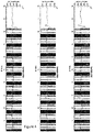
- the digitized signals ECG1, ECG2, ECG3 obtained at the output of this interface 2 have the form which appears in figure 4 , in which each diagram contains two distinct superimposed signals: signals represented in black of maximum amplitude correspond to those which come out "raw” from the interface 2 of the figure 1 , and central signals represented in gray which only present very attenuated leftovers of the artefacts, after the denoising algorithm has been implemented.
- the figure 5 details the denoising process by focusing on a time window 51 of short duration of the non-denoised digitized signal obtained at the output of the interface 2, as appears in the upper diagram 5a.
- This window is enlarged in the second diagram 5b, placed just below, which shows the same superimposition of signals, raw and noiseless, as in the diagram of the figure 4 , over a much shorter period.
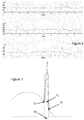
- a new time window 52 with a much more limited time scale is extracted therefrom for the purposes of explaining the operation of the denoising process.
- the raw signal from this window 52 is shown in diagram 5c, in this case comprising two artefacts.
- This signal is then derived, and the absolute value of the derivative is kept to obtain the signal of diagram 5d.
- the values of this signal derived from 5d are then subjected to an artefact suppression treatment which will be given a example below so that only the redacted signal appearing in diagram 5f remains.
- FIG. 7 An example of a simplified treatment for removing artifacts by interpolating a replacement curve is illustrated by the figure 7 .
- the slope detection algorithm comprising for example the mentioned derivation step and a step of comparison with a threshold slope, that is ie from point 71 at the start of the value replacement window 70.
- Point 71 after analysis, is detected as being part of an artifact due to its high slope: the n previous values (for example the four previous values) are then used to calculate a replacement value, for example by processing of averaging.
Landscapes
- Health & Medical Sciences (AREA)
- Life Sciences & Earth Sciences (AREA)
- Engineering & Computer Science (AREA)
- Physics & Mathematics (AREA)
- Animal Behavior & Ethology (AREA)
- Veterinary Medicine (AREA)
- Biophysics (AREA)
- Biomedical Technology (AREA)
- Heart & Thoracic Surgery (AREA)
- Medical Informatics (AREA)
- Molecular Biology (AREA)
- Surgery (AREA)
- Pathology (AREA)
- General Health & Medical Sciences (AREA)
- Public Health (AREA)
- Nuclear Medicine, Radiotherapy & Molecular Imaging (AREA)
- Signal Processing (AREA)
- Cardiology (AREA)
- High Energy & Nuclear Physics (AREA)
- Radiology & Medical Imaging (AREA)
- Artificial Intelligence (AREA)
- Computer Vision & Pattern Recognition (AREA)
- Physiology (AREA)
- Psychiatry (AREA)
- Power Engineering (AREA)
- Measurement And Recording Of Electrical Phenomena And Electrical Characteristics Of The Living Body (AREA)
Applications Claiming Priority (1)
| Application Number | Priority Date | Filing Date | Title |
|---|---|---|---|
| FR1859800A FR3087332B1 (fr) | 2018-10-23 | 2018-10-23 | Procede de correction en temps reel d'au moins un signal electro-physiologique. |
Publications (1)
| Publication Number | Publication Date |
|---|---|
| EP3643228A1 true EP3643228A1 (de) | 2020-04-29 |
Family
ID=65685604
Family Applications (1)
| Application Number | Title | Priority Date | Filing Date |
|---|---|---|---|
| EP19204484.0A Ceased EP3643228A1 (de) | 2018-10-23 | 2019-10-22 | Echtzeit-korrekturverfahren mindestens eines elektro-physiologischen signals |
Country Status (2)
| Country | Link |
|---|---|
| EP (1) | EP3643228A1 (de) |
| FR (1) | FR3087332B1 (de) |
Cited By (5)
| Publication number | Priority date | Publication date | Assignee | Title |
|---|---|---|---|---|
| US20220061771A1 (en) * | 2020-08-27 | 2022-03-03 | Ricoh Company, Ltd. | Method and apparatus for processing physiological signals and non-transitory computer-readable medium |
| CN114469132A (zh) * | 2022-02-11 | 2022-05-13 | 挂号网(杭州)科技有限公司 | 模型训练方法、装置、电子设备及存储介质 |
| CN115770048A (zh) * | 2021-09-08 | 2023-03-10 | 上海联影医疗科技股份有限公司 | 心电信号处理方法、装置、计算机设备及存储介质 |
| US11707233B1 (en) | 2022-12-16 | 2023-07-25 | Wisear | Simultaneous sub-Nyquist acquisition of a plurality of bioelectric signals |
| US20240108271A1 (en) * | 2022-09-30 | 2024-04-04 | Welch Allyn, Inc. | Methods and systems for detecting pace pulses |
Citations (4)
| Publication number | Priority date | Publication date | Assignee | Title |
|---|---|---|---|---|
| US5038785A (en) * | 1985-08-09 | 1991-08-13 | Picker International, Inc. | Cardiac and respiratory monitor with magnetic gradient noise elimination |
| US20040097802A1 (en) * | 2000-08-15 | 2004-05-20 | Cohen Mark S | Method and apparatus for reducing contamination of an electrical signal |
| US7039455B1 (en) * | 2001-10-09 | 2006-05-02 | Medrad, Inc. | Apparatus and method for removing magnetic resonance imaging-induced noise from ECG signals |
| WO2008155488A2 (fr) | 2007-04-27 | 2008-12-24 | SCHILLER MEDICAL (Société par Actions Simplifiée) | Procede, dispositif et systeme pour reduire les artefacts affectant les signaux electrophysiologiques et dus aux champs electromagnetiques |
-
2018
- 2018-10-23 FR FR1859800A patent/FR3087332B1/fr active Active
-
2019
- 2019-10-22 EP EP19204484.0A patent/EP3643228A1/de not_active Ceased
Patent Citations (4)
| Publication number | Priority date | Publication date | Assignee | Title |
|---|---|---|---|---|
| US5038785A (en) * | 1985-08-09 | 1991-08-13 | Picker International, Inc. | Cardiac and respiratory monitor with magnetic gradient noise elimination |
| US20040097802A1 (en) * | 2000-08-15 | 2004-05-20 | Cohen Mark S | Method and apparatus for reducing contamination of an electrical signal |
| US7039455B1 (en) * | 2001-10-09 | 2006-05-02 | Medrad, Inc. | Apparatus and method for removing magnetic resonance imaging-induced noise from ECG signals |
| WO2008155488A2 (fr) | 2007-04-27 | 2008-12-24 | SCHILLER MEDICAL (Société par Actions Simplifiée) | Procede, dispositif et systeme pour reduire les artefacts affectant les signaux electrophysiologiques et dus aux champs electromagnetiques |
Non-Patent Citations (1)
| Title |
|---|
| INTERNATIONAL SOCIETY FOR MAGNETIC RESONANCE IN MEDICINE, ISMRM, 2030 ADDISON STREET, 7TH FLOOR, BERKELEY, CA 94704 USA, no. 3785, 22 April 2016 (2016-04-22), XP040684825 * |
Cited By (6)
| Publication number | Priority date | Publication date | Assignee | Title |
|---|---|---|---|---|
| US20220061771A1 (en) * | 2020-08-27 | 2022-03-03 | Ricoh Company, Ltd. | Method and apparatus for processing physiological signals and non-transitory computer-readable medium |
| US12115004B2 (en) * | 2020-08-27 | 2024-10-15 | Ricoh Company, Ltd. | Method and apparatus for processing physiological signals and non-transitory computer-readable medium |
| CN115770048A (zh) * | 2021-09-08 | 2023-03-10 | 上海联影医疗科技股份有限公司 | 心电信号处理方法、装置、计算机设备及存储介质 |
| CN114469132A (zh) * | 2022-02-11 | 2022-05-13 | 挂号网(杭州)科技有限公司 | 模型训练方法、装置、电子设备及存储介质 |
| US20240108271A1 (en) * | 2022-09-30 | 2024-04-04 | Welch Allyn, Inc. | Methods and systems for detecting pace pulses |
| US11707233B1 (en) | 2022-12-16 | 2023-07-25 | Wisear | Simultaneous sub-Nyquist acquisition of a plurality of bioelectric signals |
Also Published As
| Publication number | Publication date |
|---|---|
| FR3087332A1 (fr) | 2020-04-24 |
| FR3087332B1 (fr) | 2020-11-06 |
Similar Documents
| Publication | Publication Date | Title |
|---|---|---|
| EP3643228A1 (de) | Echtzeit-korrekturverfahren mindestens eines elektro-physiologischen signals | |
| Bono et al. | Hybrid wavelet and EMD/ICA approach for artifact suppression in pervasive EEG | |
| EP2152157B1 (de) | Verfahren, vorrichtung und system zur reduzierung von artefakten, die elektrophysiologische signale beeinträchtigen und von elektromagnetischen feldern verursacht werden | |
| FR2943236A1 (fr) | Procede de surveillance d'un parametre biologique d'une personne au moyen de capteurs | |
| FR2943234A1 (fr) | Procede de surveillance d'un parametre biologique d'un occupant d'un siege avec reduction de bruit | |
| CA2491751A1 (fr) | Procede de traitement d'image acquise au moyen d'un guide constitue par une pluralite de fibres optiques | |
| FR2984720A1 (fr) | Procede et dispositif de surveillance de la mesure de la pression arterielle par catheterisme arteriel d'un patient | |
| EP2413779A1 (de) | Verfahren und system zum nachweis von okulomotorischen auffälligkeiten | |
| FR2962322A1 (fr) | Systeme de detection electroencephalographique d'une inadequation entre l'etat d'un patient place sous assistance ventilatoire et le reglage de la machine utilisee pour cette assistance, et utilisation de cette detection pour l'adaptation du reglage | |
| CN113081002A (zh) | 脑电信号的伪差去除方法、装置及电子设备 | |
| EP3552545B1 (de) | Verfahren und vorrichtung zur magnetfeldkorrektur in echtzeit | |
| WO2024003509A1 (fr) | Dispositif de détermination d'une longueur de cycle d'activation cardiaque | |
| FR2671183A1 (fr) | Procede et appareil pour analyser l'etat de protection contre la corrosion d'un ouvrage sous protection cathodique. | |
| FR2834881A1 (fr) | Enregistreur de signaux physiologiques, notamment enregistreur holter de signaux ecg, comprenant des moyens de detection du debranchement ou de la coupure des cables de liaison | |
| EP3422935B1 (de) | Verfahren zur erkennung von bestimmten elementen in elektrophysiologischen signalen und detektor | |
| Srinivasulu et al. | Basis pursuit sparse decomposition using tunable-Q wavelet transform (BPSD-TQWT) for denoising of electrocardiograms | |
| KR100689987B1 (ko) | 뇌전도 및 자기공명영상 검출 시스템에서 뇌전도 신호에포함된 잡음을 제거하기 위한 장치 및 방법 | |
| CN115755373A (zh) | 一种抑制红外干扰的太赫兹相干衍射图像处理系统及方法 | |
| Krupiński et al. | Estimation of eye blinking using biopotentials measurements for computer animation applications | |
| FR2983055A1 (fr) | Detection et estimation du complexe qrs pour le suivi d'une activite cardiaque et pulmonaire | |
| EP3073900B1 (de) | Verfahren zur konstruktion eines aktivitätsindex, vorrichtung und computerprogramm dafür | |
| Böttcher et al. | Standardizing EEG preprocessing for cross-site integration-the CLEAN pipeline | |
| WO2022148938A1 (fr) | Procédé pour une surveillance et une analyse de l'état cardiaque d'un individu | |
| EP4419007A1 (de) | Verfahren zur auswahl eines teils eines enzephalografischen signals, vorrichtungen und entsprechendes programm | |
| FR2657770A1 (fr) | Procede de traitement d'artefacts affectant des signaux respiratoires et dispositif de mise en óoeuvre. |
Legal Events
| Date | Code | Title | Description |
|---|---|---|---|
| PUAI | Public reference made under article 153(3) epc to a published international application that has entered the european phase |
Free format text: ORIGINAL CODE: 0009012 |
|
| STAA | Information on the status of an ep patent application or granted ep patent |
Free format text: STATUS: THE APPLICATION HAS BEEN PUBLISHED |
|
| AK | Designated contracting states |
Kind code of ref document: A1 Designated state(s): AL AT BE BG CH CY CZ DE DK EE ES FI FR GB GR HR HU IE IS IT LI LT LU LV MC MK MT NL NO PL PT RO RS SE SI SK SM TR |
|
| AX | Request for extension of the european patent |
Extension state: BA ME |
|
| STAA | Information on the status of an ep patent application or granted ep patent |
Free format text: STATUS: REQUEST FOR EXAMINATION WAS MADE |
|
| 17P | Request for examination filed |
Effective date: 20201027 |
|
| RBV | Designated contracting states (corrected) |
Designated state(s): AL AT BE BG CH CY CZ DE DK EE ES FI FR GB GR HR HU IE IS IT LI LT LU LV MC MK MT NL NO PL PT RO RS SE SI SK SM TR |
|
| STAA | Information on the status of an ep patent application or granted ep patent |
Free format text: STATUS: EXAMINATION IS IN PROGRESS |
|
| 17Q | First examination report despatched |
Effective date: 20221125 |
|
| STAA | Information on the status of an ep patent application or granted ep patent |
Free format text: STATUS: THE APPLICATION HAS BEEN REFUSED |
|
| 18R | Application refused |
Effective date: 20250311 |