EP3664746B1 - Constructions d'échafaudage tissulaire multiphasique - Google Patents
Constructions d'échafaudage tissulaire multiphasique Download PDFInfo
- Publication number
- EP3664746B1 EP3664746B1 EP18845151.2A EP18845151A EP3664746B1 EP 3664746 B1 EP3664746 B1 EP 3664746B1 EP 18845151 A EP18845151 A EP 18845151A EP 3664746 B1 EP3664746 B1 EP 3664746B1
- Authority
- EP
- European Patent Office
- Prior art keywords
- scaffold
- bone
- ligament
- multiphasic
- tissue scaffold
- Prior art date
- Legal status (The legal status is an assumption and is not a legal conclusion. Google has not performed a legal analysis and makes no representation as to the accuracy of the status listed.)
- Active
Links
Images












Classifications
-
- A—HUMAN NECESSITIES
- A61—MEDICAL OR VETERINARY SCIENCE; HYGIENE
- A61L—METHODS OR APPARATUS FOR STERILISING MATERIALS OR OBJECTS IN GENERAL; DISINFECTION, STERILISATION OR DEODORISATION OF AIR; CHEMICAL ASPECTS OF BANDAGES, DRESSINGS, ABSORBENT PADS OR SURGICAL ARTICLES; MATERIALS FOR BANDAGES, DRESSINGS, ABSORBENT PADS OR SURGICAL ARTICLES
- A61L27/00—Materials for grafts or prostheses or for coating grafts or prostheses
- A61L27/50—Materials characterised by their function or physical properties, e.g. injectable or lubricating compositions, shape-memory materials, surface modified materials
-
- A—HUMAN NECESSITIES
- A61—MEDICAL OR VETERINARY SCIENCE; HYGIENE
- A61F—FILTERS IMPLANTABLE INTO BLOOD VESSELS; PROSTHESES; DEVICES PROVIDING PATENCY TO, OR PREVENTING COLLAPSING OF, TUBULAR STRUCTURES OF THE BODY, e.g. STENTS; ORTHOPAEDIC, NURSING OR CONTRACEPTIVE DEVICES; FOMENTATION; TREATMENT OR PROTECTION OF EYES OR EARS; BANDAGES, DRESSINGS OR ABSORBENT PADS; FIRST-AID KITS
- A61F2/00—Filters implantable into blood vessels; Prostheses, i.e. artificial substitutes or replacements for parts of the body; Appliances for connecting them with the body; Devices providing patency to, or preventing collapsing of, tubular structures of the body, e.g. stents
- A61F2/02—Prostheses implantable into the body
- A61F2/08—Muscles; Tendons; Ligaments
-
- A—HUMAN NECESSITIES
- A61—MEDICAL OR VETERINARY SCIENCE; HYGIENE
- A61F—FILTERS IMPLANTABLE INTO BLOOD VESSELS; PROSTHESES; DEVICES PROVIDING PATENCY TO, OR PREVENTING COLLAPSING OF, TUBULAR STRUCTURES OF THE BODY, e.g. STENTS; ORTHOPAEDIC, NURSING OR CONTRACEPTIVE DEVICES; FOMENTATION; TREATMENT OR PROTECTION OF EYES OR EARS; BANDAGES, DRESSINGS OR ABSORBENT PADS; FIRST-AID KITS
- A61F2/00—Filters implantable into blood vessels; Prostheses, i.e. artificial substitutes or replacements for parts of the body; Appliances for connecting them with the body; Devices providing patency to, or preventing collapsing of, tubular structures of the body, e.g. stents
- A61F2/02—Prostheses implantable into the body
- A61F2/30—Joints
-
- A—HUMAN NECESSITIES
- A61—MEDICAL OR VETERINARY SCIENCE; HYGIENE
- A61F—FILTERS IMPLANTABLE INTO BLOOD VESSELS; PROSTHESES; DEVICES PROVIDING PATENCY TO, OR PREVENTING COLLAPSING OF, TUBULAR STRUCTURES OF THE BODY, e.g. STENTS; ORTHOPAEDIC, NURSING OR CONTRACEPTIVE DEVICES; FOMENTATION; TREATMENT OR PROTECTION OF EYES OR EARS; BANDAGES, DRESSINGS OR ABSORBENT PADS; FIRST-AID KITS
- A61F2/00—Filters implantable into blood vessels; Prostheses, i.e. artificial substitutes or replacements for parts of the body; Appliances for connecting them with the body; Devices providing patency to, or preventing collapsing of, tubular structures of the body, e.g. stents
- A61F2/02—Prostheses implantable into the body
- A61F2/30—Joints
- A61F2/42—Joints for wrists or ankles; for hands, e.g. fingers; for feet, e.g. toes
- A61F2/4261—Joints for wrists or ankles; for hands, e.g. fingers; for feet, e.g. toes for wrists
-
- A—HUMAN NECESSITIES
- A61—MEDICAL OR VETERINARY SCIENCE; HYGIENE
- A61L—METHODS OR APPARATUS FOR STERILISING MATERIALS OR OBJECTS IN GENERAL; DISINFECTION, STERILISATION OR DEODORISATION OF AIR; CHEMICAL ASPECTS OF BANDAGES, DRESSINGS, ABSORBENT PADS OR SURGICAL ARTICLES; MATERIALS FOR BANDAGES, DRESSINGS, ABSORBENT PADS OR SURGICAL ARTICLES
- A61L27/00—Materials for grafts or prostheses or for coating grafts or prostheses
- A61L27/14—Macromolecular materials
- A61L27/18—Macromolecular materials obtained otherwise than by reactions only involving carbon-to-carbon unsaturated bonds
-
- A—HUMAN NECESSITIES
- A61—MEDICAL OR VETERINARY SCIENCE; HYGIENE
- A61L—METHODS OR APPARATUS FOR STERILISING MATERIALS OR OBJECTS IN GENERAL; DISINFECTION, STERILISATION OR DEODORISATION OF AIR; CHEMICAL ASPECTS OF BANDAGES, DRESSINGS, ABSORBENT PADS OR SURGICAL ARTICLES; MATERIALS FOR BANDAGES, DRESSINGS, ABSORBENT PADS OR SURGICAL ARTICLES
- A61L27/00—Materials for grafts or prostheses or for coating grafts or prostheses
- A61L27/14—Macromolecular materials
- A61L27/22—Polypeptides or derivatives thereof, e.g. degradation products
- A61L27/222—Gelatin
-
- A—HUMAN NECESSITIES
- A61—MEDICAL OR VETERINARY SCIENCE; HYGIENE
- A61L—METHODS OR APPARATUS FOR STERILISING MATERIALS OR OBJECTS IN GENERAL; DISINFECTION, STERILISATION OR DEODORISATION OF AIR; CHEMICAL ASPECTS OF BANDAGES, DRESSINGS, ABSORBENT PADS OR SURGICAL ARTICLES; MATERIALS FOR BANDAGES, DRESSINGS, ABSORBENT PADS OR SURGICAL ARTICLES
- A61L27/00—Materials for grafts or prostheses or for coating grafts or prostheses
- A61L27/14—Macromolecular materials
- A61L27/22—Polypeptides or derivatives thereof, e.g. degradation products
- A61L27/225—Fibrin; Fibrinogen
-
- A—HUMAN NECESSITIES
- A61—MEDICAL OR VETERINARY SCIENCE; HYGIENE
- A61L—METHODS OR APPARATUS FOR STERILISING MATERIALS OR OBJECTS IN GENERAL; DISINFECTION, STERILISATION OR DEODORISATION OF AIR; CHEMICAL ASPECTS OF BANDAGES, DRESSINGS, ABSORBENT PADS OR SURGICAL ARTICLES; MATERIALS FOR BANDAGES, DRESSINGS, ABSORBENT PADS OR SURGICAL ARTICLES
- A61L27/00—Materials for grafts or prostheses or for coating grafts or prostheses
- A61L27/14—Macromolecular materials
- A61L27/22—Polypeptides or derivatives thereof, e.g. degradation products
- A61L27/227—Other specific proteins or polypeptides not covered by A61L27/222, A61L27/225 or A61L27/24
-
- A—HUMAN NECESSITIES
- A61—MEDICAL OR VETERINARY SCIENCE; HYGIENE
- A61L—METHODS OR APPARATUS FOR STERILISING MATERIALS OR OBJECTS IN GENERAL; DISINFECTION, STERILISATION OR DEODORISATION OF AIR; CHEMICAL ASPECTS OF BANDAGES, DRESSINGS, ABSORBENT PADS OR SURGICAL ARTICLES; MATERIALS FOR BANDAGES, DRESSINGS, ABSORBENT PADS OR SURGICAL ARTICLES
- A61L27/00—Materials for grafts or prostheses or for coating grafts or prostheses
- A61L27/14—Macromolecular materials
- A61L27/22—Polypeptides or derivatives thereof, e.g. degradation products
- A61L27/24—Collagen
-
- A—HUMAN NECESSITIES
- A61—MEDICAL OR VETERINARY SCIENCE; HYGIENE
- A61L—METHODS OR APPARATUS FOR STERILISING MATERIALS OR OBJECTS IN GENERAL; DISINFECTION, STERILISATION OR DEODORISATION OF AIR; CHEMICAL ASPECTS OF BANDAGES, DRESSINGS, ABSORBENT PADS OR SURGICAL ARTICLES; MATERIALS FOR BANDAGES, DRESSINGS, ABSORBENT PADS OR SURGICAL ARTICLES
- A61L27/00—Materials for grafts or prostheses or for coating grafts or prostheses
- A61L27/36—Materials for grafts or prostheses or for coating grafts or prostheses containing ingredients of undetermined constitution or reaction products thereof, e.g. transplant tissue, natural bone, extracellular matrix
- A61L27/38—Materials for grafts or prostheses or for coating grafts or prostheses containing ingredients of undetermined constitution or reaction products thereof, e.g. transplant tissue, natural bone, extracellular matrix containing added animal cells
- A61L27/3804—Materials for grafts or prostheses or for coating grafts or prostheses containing ingredients of undetermined constitution or reaction products thereof, e.g. transplant tissue, natural bone, extracellular matrix containing added animal cells characterised by specific cells or progenitors thereof, e.g. fibroblasts, connective tissue cells, kidney cells
- A61L27/3817—Cartilage-forming cells, e.g. pre-chondrocytes
-
- A—HUMAN NECESSITIES
- A61—MEDICAL OR VETERINARY SCIENCE; HYGIENE
- A61L—METHODS OR APPARATUS FOR STERILISING MATERIALS OR OBJECTS IN GENERAL; DISINFECTION, STERILISATION OR DEODORISATION OF AIR; CHEMICAL ASPECTS OF BANDAGES, DRESSINGS, ABSORBENT PADS OR SURGICAL ARTICLES; MATERIALS FOR BANDAGES, DRESSINGS, ABSORBENT PADS OR SURGICAL ARTICLES
- A61L27/00—Materials for grafts or prostheses or for coating grafts or prostheses
- A61L27/36—Materials for grafts or prostheses or for coating grafts or prostheses containing ingredients of undetermined constitution or reaction products thereof, e.g. transplant tissue, natural bone, extracellular matrix
- A61L27/38—Materials for grafts or prostheses or for coating grafts or prostheses containing ingredients of undetermined constitution or reaction products thereof, e.g. transplant tissue, natural bone, extracellular matrix containing added animal cells
- A61L27/3804—Materials for grafts or prostheses or for coating grafts or prostheses containing ingredients of undetermined constitution or reaction products thereof, e.g. transplant tissue, natural bone, extracellular matrix containing added animal cells characterised by specific cells or progenitors thereof, e.g. fibroblasts, connective tissue cells, kidney cells
- A61L27/3821—Bone-forming cells, e.g. osteoblasts, osteocytes, osteoprogenitor cells
-
- A—HUMAN NECESSITIES
- A61—MEDICAL OR VETERINARY SCIENCE; HYGIENE
- A61L—METHODS OR APPARATUS FOR STERILISING MATERIALS OR OBJECTS IN GENERAL; DISINFECTION, STERILISATION OR DEODORISATION OF AIR; CHEMICAL ASPECTS OF BANDAGES, DRESSINGS, ABSORBENT PADS OR SURGICAL ARTICLES; MATERIALS FOR BANDAGES, DRESSINGS, ABSORBENT PADS OR SURGICAL ARTICLES
- A61L27/00—Materials for grafts or prostheses or for coating grafts or prostheses
- A61L27/36—Materials for grafts or prostheses or for coating grafts or prostheses containing ingredients of undetermined constitution or reaction products thereof, e.g. transplant tissue, natural bone, extracellular matrix
- A61L27/38—Materials for grafts or prostheses or for coating grafts or prostheses containing ingredients of undetermined constitution or reaction products thereof, e.g. transplant tissue, natural bone, extracellular matrix containing added animal cells
- A61L27/3804—Materials for grafts or prostheses or for coating grafts or prostheses containing ingredients of undetermined constitution or reaction products thereof, e.g. transplant tissue, natural bone, extracellular matrix containing added animal cells characterised by specific cells or progenitors thereof, e.g. fibroblasts, connective tissue cells, kidney cells
- A61L27/3834—Cells able to produce different cell types, e.g. hematopoietic stem cells, mesenchymal stem cells, marrow stromal cells, embryonic stem cells
-
- A—HUMAN NECESSITIES
- A61—MEDICAL OR VETERINARY SCIENCE; HYGIENE
- A61L—METHODS OR APPARATUS FOR STERILISING MATERIALS OR OBJECTS IN GENERAL; DISINFECTION, STERILISATION OR DEODORISATION OF AIR; CHEMICAL ASPECTS OF BANDAGES, DRESSINGS, ABSORBENT PADS OR SURGICAL ARTICLES; MATERIALS FOR BANDAGES, DRESSINGS, ABSORBENT PADS OR SURGICAL ARTICLES
- A61L27/00—Materials for grafts or prostheses or for coating grafts or prostheses
- A61L27/36—Materials for grafts or prostheses or for coating grafts or prostheses containing ingredients of undetermined constitution or reaction products thereof, e.g. transplant tissue, natural bone, extracellular matrix
- A61L27/38—Materials for grafts or prostheses or for coating grafts or prostheses containing ingredients of undetermined constitution or reaction products thereof, e.g. transplant tissue, natural bone, extracellular matrix containing added animal cells
- A61L27/3839—Materials for grafts or prostheses or for coating grafts or prostheses containing ingredients of undetermined constitution or reaction products thereof, e.g. transplant tissue, natural bone, extracellular matrix containing added animal cells characterised by the site of application in the body
- A61L27/3843—Connective tissue
- A61L27/3847—Bones
-
- A—HUMAN NECESSITIES
- A61—MEDICAL OR VETERINARY SCIENCE; HYGIENE
- A61L—METHODS OR APPARATUS FOR STERILISING MATERIALS OR OBJECTS IN GENERAL; DISINFECTION, STERILISATION OR DEODORISATION OF AIR; CHEMICAL ASPECTS OF BANDAGES, DRESSINGS, ABSORBENT PADS OR SURGICAL ARTICLES; MATERIALS FOR BANDAGES, DRESSINGS, ABSORBENT PADS OR SURGICAL ARTICLES
- A61L27/00—Materials for grafts or prostheses or for coating grafts or prostheses
- A61L27/36—Materials for grafts or prostheses or for coating grafts or prostheses containing ingredients of undetermined constitution or reaction products thereof, e.g. transplant tissue, natural bone, extracellular matrix
- A61L27/38—Materials for grafts or prostheses or for coating grafts or prostheses containing ingredients of undetermined constitution or reaction products thereof, e.g. transplant tissue, natural bone, extracellular matrix containing added animal cells
- A61L27/3839—Materials for grafts or prostheses or for coating grafts or prostheses containing ingredients of undetermined constitution or reaction products thereof, e.g. transplant tissue, natural bone, extracellular matrix containing added animal cells characterised by the site of application in the body
- A61L27/3843—Connective tissue
- A61L27/386—Ligaments, tendons
-
- A—HUMAN NECESSITIES
- A61—MEDICAL OR VETERINARY SCIENCE; HYGIENE
- A61L—METHODS OR APPARATUS FOR STERILISING MATERIALS OR OBJECTS IN GENERAL; DISINFECTION, STERILISATION OR DEODORISATION OF AIR; CHEMICAL ASPECTS OF BANDAGES, DRESSINGS, ABSORBENT PADS OR SURGICAL ARTICLES; MATERIALS FOR BANDAGES, DRESSINGS, ABSORBENT PADS OR SURGICAL ARTICLES
- A61L27/00—Materials for grafts or prostheses or for coating grafts or prostheses
- A61L27/36—Materials for grafts or prostheses or for coating grafts or prostheses containing ingredients of undetermined constitution or reaction products thereof, e.g. transplant tissue, natural bone, extracellular matrix
- A61L27/38—Materials for grafts or prostheses or for coating grafts or prostheses containing ingredients of undetermined constitution or reaction products thereof, e.g. transplant tissue, natural bone, extracellular matrix containing added animal cells
- A61L27/3886—Materials for grafts or prostheses or for coating grafts or prostheses containing ingredients of undetermined constitution or reaction products thereof, e.g. transplant tissue, natural bone, extracellular matrix containing added animal cells comprising two or more cell types
-
- A—HUMAN NECESSITIES
- A61—MEDICAL OR VETERINARY SCIENCE; HYGIENE
- A61L—METHODS OR APPARATUS FOR STERILISING MATERIALS OR OBJECTS IN GENERAL; DISINFECTION, STERILISATION OR DEODORISATION OF AIR; CHEMICAL ASPECTS OF BANDAGES, DRESSINGS, ABSORBENT PADS OR SURGICAL ARTICLES; MATERIALS FOR BANDAGES, DRESSINGS, ABSORBENT PADS OR SURGICAL ARTICLES
- A61L27/00—Materials for grafts or prostheses or for coating grafts or prostheses
- A61L27/50—Materials characterised by their function or physical properties, e.g. injectable or lubricating compositions, shape-memory materials, surface modified materials
- A61L27/56—Porous materials, e.g. foams or sponges
-
- B—PERFORMING OPERATIONS; TRANSPORTING
- B33—ADDITIVE MANUFACTURING TECHNOLOGY
- B33Y—ADDITIVE MANUFACTURING, i.e. MANUFACTURING OF THREE-DIMENSIONAL [3D] OBJECTS BY ADDITIVE DEPOSITION, ADDITIVE AGGLOMERATION OR ADDITIVE LAYERING, e.g. BY 3D PRINTING, STEREOLITHOGRAPHY OR SELECTIVE LASER SINTERING
- B33Y80/00—Products made by additive manufacturing
-
- A—HUMAN NECESSITIES
- A61—MEDICAL OR VETERINARY SCIENCE; HYGIENE
- A61F—FILTERS IMPLANTABLE INTO BLOOD VESSELS; PROSTHESES; DEVICES PROVIDING PATENCY TO, OR PREVENTING COLLAPSING OF, TUBULAR STRUCTURES OF THE BODY, e.g. STENTS; ORTHOPAEDIC, NURSING OR CONTRACEPTIVE DEVICES; FOMENTATION; TREATMENT OR PROTECTION OF EYES OR EARS; BANDAGES, DRESSINGS OR ABSORBENT PADS; FIRST-AID KITS
- A61F2/00—Filters implantable into blood vessels; Prostheses, i.e. artificial substitutes or replacements for parts of the body; Appliances for connecting them with the body; Devices providing patency to, or preventing collapsing of, tubular structures of the body, e.g. stents
- A61F2/02—Prostheses implantable into the body
- A61F2/30—Joints
- A61F2002/30001—Additional features of subject-matter classified in A61F2/28, A61F2/30 and subgroups thereof
- A61F2002/30003—Material related properties of the prosthesis or of a coating on the prosthesis
- A61F2002/30004—Material related properties of the prosthesis or of a coating on the prosthesis the prosthesis being made from materials having different values of a given property at different locations within the same prosthesis
- A61F2002/30009—Material related properties of the prosthesis or of a coating on the prosthesis the prosthesis being made from materials having different values of a given property at different locations within the same prosthesis differing in fibre orientations
-
- A—HUMAN NECESSITIES
- A61—MEDICAL OR VETERINARY SCIENCE; HYGIENE
- A61F—FILTERS IMPLANTABLE INTO BLOOD VESSELS; PROSTHESES; DEVICES PROVIDING PATENCY TO, OR PREVENTING COLLAPSING OF, TUBULAR STRUCTURES OF THE BODY, e.g. STENTS; ORTHOPAEDIC, NURSING OR CONTRACEPTIVE DEVICES; FOMENTATION; TREATMENT OR PROTECTION OF EYES OR EARS; BANDAGES, DRESSINGS OR ABSORBENT PADS; FIRST-AID KITS
- A61F2/00—Filters implantable into blood vessels; Prostheses, i.e. artificial substitutes or replacements for parts of the body; Appliances for connecting them with the body; Devices providing patency to, or preventing collapsing of, tubular structures of the body, e.g. stents
- A61F2/02—Prostheses implantable into the body
- A61F2/30—Joints
- A61F2002/30001—Additional features of subject-matter classified in A61F2/28, A61F2/30 and subgroups thereof
- A61F2002/30003—Material related properties of the prosthesis or of a coating on the prosthesis
- A61F2002/30004—Material related properties of the prosthesis or of a coating on the prosthesis the prosthesis being made from materials having different values of a given property at different locations within the same prosthesis
- A61F2002/30011—Material related properties of the prosthesis or of a coating on the prosthesis the prosthesis being made from materials having different values of a given property at different locations within the same prosthesis differing in porosity
-
- A—HUMAN NECESSITIES
- A61—MEDICAL OR VETERINARY SCIENCE; HYGIENE
- A61F—FILTERS IMPLANTABLE INTO BLOOD VESSELS; PROSTHESES; DEVICES PROVIDING PATENCY TO, OR PREVENTING COLLAPSING OF, TUBULAR STRUCTURES OF THE BODY, e.g. STENTS; ORTHOPAEDIC, NURSING OR CONTRACEPTIVE DEVICES; FOMENTATION; TREATMENT OR PROTECTION OF EYES OR EARS; BANDAGES, DRESSINGS OR ABSORBENT PADS; FIRST-AID KITS
- A61F2/00—Filters implantable into blood vessels; Prostheses, i.e. artificial substitutes or replacements for parts of the body; Appliances for connecting them with the body; Devices providing patency to, or preventing collapsing of, tubular structures of the body, e.g. stents
- A61F2/02—Prostheses implantable into the body
- A61F2/30—Joints
- A61F2002/30001—Additional features of subject-matter classified in A61F2/28, A61F2/30 and subgroups thereof
- A61F2002/30003—Material related properties of the prosthesis or of a coating on the prosthesis
- A61F2002/3006—Properties of materials and coating materials
- A61F2002/30062—(bio)absorbable, biodegradable, bioerodable, (bio)resorbable, resorptive
-
- A—HUMAN NECESSITIES
- A61—MEDICAL OR VETERINARY SCIENCE; HYGIENE
- A61F—FILTERS IMPLANTABLE INTO BLOOD VESSELS; PROSTHESES; DEVICES PROVIDING PATENCY TO, OR PREVENTING COLLAPSING OF, TUBULAR STRUCTURES OF THE BODY, e.g. STENTS; ORTHOPAEDIC, NURSING OR CONTRACEPTIVE DEVICES; FOMENTATION; TREATMENT OR PROTECTION OF EYES OR EARS; BANDAGES, DRESSINGS OR ABSORBENT PADS; FIRST-AID KITS
- A61F2/00—Filters implantable into blood vessels; Prostheses, i.e. artificial substitutes or replacements for parts of the body; Appliances for connecting them with the body; Devices providing patency to, or preventing collapsing of, tubular structures of the body, e.g. stents
- A61F2/02—Prostheses implantable into the body
- A61F2/30—Joints
- A61F2002/30001—Additional features of subject-matter classified in A61F2/28, A61F2/30 and subgroups thereof
- A61F2002/30316—The prosthesis having different structural features at different locations within the same prosthesis; Connections between prosthetic parts; Special structural features of bone or joint prostheses not otherwise provided for
-
- A—HUMAN NECESSITIES
- A61—MEDICAL OR VETERINARY SCIENCE; HYGIENE
- A61F—FILTERS IMPLANTABLE INTO BLOOD VESSELS; PROSTHESES; DEVICES PROVIDING PATENCY TO, OR PREVENTING COLLAPSING OF, TUBULAR STRUCTURES OF THE BODY, e.g. STENTS; ORTHOPAEDIC, NURSING OR CONTRACEPTIVE DEVICES; FOMENTATION; TREATMENT OR PROTECTION OF EYES OR EARS; BANDAGES, DRESSINGS OR ABSORBENT PADS; FIRST-AID KITS
- A61F2/00—Filters implantable into blood vessels; Prostheses, i.e. artificial substitutes or replacements for parts of the body; Appliances for connecting them with the body; Devices providing patency to, or preventing collapsing of, tubular structures of the body, e.g. stents
- A61F2/02—Prostheses implantable into the body
- A61F2/30—Joints
- A61F2/3094—Designing or manufacturing processes
- A61F2/30942—Designing or manufacturing processes for designing or making customized prostheses, e.g. using templates, CT or NMR scans, finite-element analysis or CAD-CAM techniques
- A61F2002/30962—Designing or manufacturing processes for designing or making customized prostheses, e.g. using templates, CT or NMR scans, finite-element analysis or CAD-CAM techniques using stereolithography
-
- A—HUMAN NECESSITIES
- A61—MEDICAL OR VETERINARY SCIENCE; HYGIENE
- A61F—FILTERS IMPLANTABLE INTO BLOOD VESSELS; PROSTHESES; DEVICES PROVIDING PATENCY TO, OR PREVENTING COLLAPSING OF, TUBULAR STRUCTURES OF THE BODY, e.g. STENTS; ORTHOPAEDIC, NURSING OR CONTRACEPTIVE DEVICES; FOMENTATION; TREATMENT OR PROTECTION OF EYES OR EARS; BANDAGES, DRESSINGS OR ABSORBENT PADS; FIRST-AID KITS
- A61F2/00—Filters implantable into blood vessels; Prostheses, i.e. artificial substitutes or replacements for parts of the body; Appliances for connecting them with the body; Devices providing patency to, or preventing collapsing of, tubular structures of the body, e.g. stents
- A61F2/02—Prostheses implantable into the body
- A61F2/30—Joints
- A61F2/3094—Designing or manufacturing processes
- A61F2002/30985—Designing or manufacturing processes using three dimensional printing [3DP]
-
- A—HUMAN NECESSITIES
- A61—MEDICAL OR VETERINARY SCIENCE; HYGIENE
- A61F—FILTERS IMPLANTABLE INTO BLOOD VESSELS; PROSTHESES; DEVICES PROVIDING PATENCY TO, OR PREVENTING COLLAPSING OF, TUBULAR STRUCTURES OF THE BODY, e.g. STENTS; ORTHOPAEDIC, NURSING OR CONTRACEPTIVE DEVICES; FOMENTATION; TREATMENT OR PROTECTION OF EYES OR EARS; BANDAGES, DRESSINGS OR ABSORBENT PADS; FIRST-AID KITS
- A61F2/00—Filters implantable into blood vessels; Prostheses, i.e. artificial substitutes or replacements for parts of the body; Appliances for connecting them with the body; Devices providing patency to, or preventing collapsing of, tubular structures of the body, e.g. stents
- A61F2/02—Prostheses implantable into the body
- A61F2/30—Joints
- A61F2/42—Joints for wrists or ankles; for hands, e.g. fingers; for feet, e.g. toes
- A61F2/4261—Joints for wrists or ankles; for hands, e.g. fingers; for feet, e.g. toes for wrists
- A61F2002/4271—Carpal bones
- A61F2002/4287—Proximal carpal row, i.e. bones adjacent the radius and the ulna
- A61F2002/4289—Scaphoid or navicular bone
-
- A—HUMAN NECESSITIES
- A61—MEDICAL OR VETERINARY SCIENCE; HYGIENE
- A61F—FILTERS IMPLANTABLE INTO BLOOD VESSELS; PROSTHESES; DEVICES PROVIDING PATENCY TO, OR PREVENTING COLLAPSING OF, TUBULAR STRUCTURES OF THE BODY, e.g. STENTS; ORTHOPAEDIC, NURSING OR CONTRACEPTIVE DEVICES; FOMENTATION; TREATMENT OR PROTECTION OF EYES OR EARS; BANDAGES, DRESSINGS OR ABSORBENT PADS; FIRST-AID KITS
- A61F2/00—Filters implantable into blood vessels; Prostheses, i.e. artificial substitutes or replacements for parts of the body; Appliances for connecting them with the body; Devices providing patency to, or preventing collapsing of, tubular structures of the body, e.g. stents
- A61F2/02—Prostheses implantable into the body
- A61F2/30—Joints
- A61F2/42—Joints for wrists or ankles; for hands, e.g. fingers; for feet, e.g. toes
- A61F2/4261—Joints for wrists or ankles; for hands, e.g. fingers; for feet, e.g. toes for wrists
- A61F2002/4271—Carpal bones
- A61F2002/4287—Proximal carpal row, i.e. bones adjacent the radius and the ulna
- A61F2002/4292—Lunate
-
- A—HUMAN NECESSITIES
- A61—MEDICAL OR VETERINARY SCIENCE; HYGIENE
- A61L—METHODS OR APPARATUS FOR STERILISING MATERIALS OR OBJECTS IN GENERAL; DISINFECTION, STERILISATION OR DEODORISATION OF AIR; CHEMICAL ASPECTS OF BANDAGES, DRESSINGS, ABSORBENT PADS OR SURGICAL ARTICLES; MATERIALS FOR BANDAGES, DRESSINGS, ABSORBENT PADS OR SURGICAL ARTICLES
- A61L2430/00—Materials or treatment for tissue regeneration
- A61L2430/10—Materials or treatment for tissue regeneration for reconstruction of tendons or ligaments
-
- A—HUMAN NECESSITIES
- A61—MEDICAL OR VETERINARY SCIENCE; HYGIENE
- A61L—METHODS OR APPARATUS FOR STERILISING MATERIALS OR OBJECTS IN GENERAL; DISINFECTION, STERILISATION OR DEODORISATION OF AIR; CHEMICAL ASPECTS OF BANDAGES, DRESSINGS, ABSORBENT PADS OR SURGICAL ARTICLES; MATERIALS FOR BANDAGES, DRESSINGS, ABSORBENT PADS OR SURGICAL ARTICLES
- A61L2430/00—Materials or treatment for tissue regeneration
- A61L2430/24—Materials or treatment for tissue regeneration for joint reconstruction
-
- B—PERFORMING OPERATIONS; TRANSPORTING
- B29—WORKING OF PLASTICS; WORKING OF SUBSTANCES IN A PLASTIC STATE IN GENERAL
- B29C—SHAPING OR JOINING OF PLASTICS; SHAPING OF MATERIAL IN A PLASTIC STATE, NOT OTHERWISE PROVIDED FOR; AFTER-TREATMENT OF THE SHAPED PRODUCTS, e.g. REPAIRING
- B29C64/00—Additive manufacturing, i.e. manufacturing of three-dimensional [3D] objects by additive deposition, additive agglomeration or additive layering, e.g. by 3D printing, stereolithography or selective laser sintering
- B29C64/10—Processes of additive manufacturing
- B29C64/106—Processes of additive manufacturing using only liquids or viscous materials, e.g. depositing a continuous bead of viscous material
-
- B—PERFORMING OPERATIONS; TRANSPORTING
- B29—WORKING OF PLASTICS; WORKING OF SUBSTANCES IN A PLASTIC STATE IN GENERAL
- B29L—INDEXING SCHEME ASSOCIATED WITH SUBCLASS B29C, RELATING TO PARTICULAR ARTICLES
- B29L2031/00—Other particular articles
- B29L2031/753—Medical equipment; Accessories therefor
- B29L2031/7532—Artificial members, protheses
-
- B—PERFORMING OPERATIONS; TRANSPORTING
- B33—ADDITIVE MANUFACTURING TECHNOLOGY
- B33Y—ADDITIVE MANUFACTURING, i.e. MANUFACTURING OF THREE-DIMENSIONAL [3D] OBJECTS BY ADDITIVE DEPOSITION, ADDITIVE AGGLOMERATION OR ADDITIVE LAYERING, e.g. BY 3D PRINTING, STEREOLITHOGRAPHY OR SELECTIVE LASER SINTERING
- B33Y10/00—Processes of additive manufacturing
-
- B—PERFORMING OPERATIONS; TRANSPORTING
- B33—ADDITIVE MANUFACTURING TECHNOLOGY
- B33Y—ADDITIVE MANUFACTURING, i.e. MANUFACTURING OF THREE-DIMENSIONAL [3D] OBJECTS BY ADDITIVE DEPOSITION, ADDITIVE AGGLOMERATION OR ADDITIVE LAYERING, e.g. BY 3D PRINTING, STEREOLITHOGRAPHY OR SELECTIVE LASER SINTERING
- B33Y70/00—Materials specially adapted for additive manufacturing
Definitions
- the present invention relates generally to the field of tissue engineering, and more specifically to tissue engineering scaffolds capable of assisting tissue regeneration. Methods for the production of the scaffolds and their use in regenerating tissue are also provided herein.
- Tendons and ligaments can fail through trauma or chronic deterioration, and both mechanisms lead to a substantial reduction in musculoskeletal function.
- Treatments that enhance tendon and ligament repair will have a significant impact on orthopaedic surgery outcomes, reducing repair failures and repeat surgeries, facilitating a more rapid return to function, and lowering health costs.
- Many current treatments rely on using the patient's own tissue as regenerative scaffolds. This approach is often associated with donor site morbidity, as well as difficulties with size matching, anatomical shape and fixation to bone.
- Synthetic scaffolds have been manufactured using a variety of fabrication methods and polymers as an alternative for tendon and ligament repair. However, producing synthetic scaffolds with sufficient strength to withstand normal physiological forces remains challenging, as does replicating existing joint architecture. While synthetic tissue constructs have been used with varying success in larger joints such as the knee (e.g. anterior cruciate ligament repair), very little if any attempts have been made to use synthetic scaffolds in the more difficult environment of small joints.
- SLIL scapholunate interosseous ligament
- C-shaped ligament situated between the scaphoid and lunate carpal bones is a commonly torn wrist ligament.
- SLAC scapholunate advanced collapse
- DISI dorsal intercalated segment instability
- WO 2017/050837 describes a scaffold that can include three or more separate and distinct compartments having distinct structural features.
- the scaffold is porous and suitably shaped to mimic a series of fibres; it also includes a polymeric material and may be produced using a 3D printer.
- the scaffold can be used for the generation of soft tissue, in particular in the treatment of ligaments.
- the synthetic constructs produced can be used effectively in small joint repair including, for example, SLIL repair.
- the present invention alleviates at least one of the shortcomings of existing tissue engineering scaffolds and methods for their production.
- the bioscaffolds described herein are multiphasic and produced in a continuous manner to accurately imitate the morphological characteristics of the tissue to be regenerated.
- the synthetic scaffold constructs of the present invention may (i) be capable of withstanding normal physiological forces for the integrative and functional repair of soft tissue injuries, and/or (ii) closely mimic existing joint architecture, and/or (iii) allow manipulation to promote vascularization and tissue regeneration, and/or (iv) reduce or eliminate donor morbidity when harvesting autografts.
- the present invention provides: A three-dimensional multiphasic synthetic tissue scaffold comprising first, second and third compartments, wherein:
- the second axis is rotated by an angle of:
- the multiple fibre layers comprise a third layer aligned along a third axis, and the third axis is:
- the multiphasic synthetic tissue scaffold defined above further comprises mammalian cells, preferably the mammalian cells are selected from the group consisting of: ligament-derived stem cells, cartilage stem cells, mesenchymal stem cells (bone marrow stromal cells), adipose-derived stromal cells (e.g. bone-marrow derived mesenchymal stem cells, adipose-derived mesenchymal stem cells), osteoblasts, osteoblast-like cells, stem cells, progenitor cells, or any combination thereof; or preferably the mammalian cells are human mesenchymal stem cells, or human bone marrow mesenchymal stem cells.
- the mammalian cells are selected from the group consisting of: ligament-derived stem cells, cartilage stem cells, mesenchymal stem cells (bone marrow stromal cells), adipose-derived stromal cells (e.g. bone-marrow derived mesenchymal stem cells, adipose-derived mesen
- the mammalian cells are within a cell sheet wrapped around and/or inserted into one or more of the compartments of three-dimensional multiphasic tissue scaffold defined above.
- the three-dimensional multiphasic synthetic tissue scaffold defined above is produced using a three-dimensional (3D) bioprinter to continuously deposit the polymeric material onto a platform until the tissue scaffold is produced in its entirety.
- the present invention further provides a method for producing a three dimensional multiphasic synthetic tissue scaffold defined above, the method comprising using a three-dimensional (3D) bioprinter to continuously deposit polymeric material onto a platform until the tissue scaffold is produced in its entirety.
- polymer also includes a plurality of polymers.
- tissue scaffold construct “comprising” polymer type A may consist exclusively of polymer type A or may include one or more additional components (e.g. polymer type B and/or non-polymer material/s).
- a "multiphasic" tissue scaffold refers to a tissue scaffold comprising at least two (e.g. two, three, four, five or more) compartments each comprising distinct microstructural, and/or chemical, and/or mechanical properties, as compared to adjacent compartment/s of the scaffold. It will be understood that the compartments of a "multiphasic" tissue scaffold as described herein are produced in a continuous manner (e.g. in the same session of three-dimensional bioprinting), rather than being produced separately then fused and/or joined together.
- tissue scaffold is intended to exclude naturally-formed tissue scaffolds and requires that the scaffold is manufactured artificially.
- tissue scaffold or tissue scaffold compartment having an "external morphology" that "mimics” a biological component e.g. a joint, bone, ligament, cartilage
- tissue scaffold or tissue scaffold compartment having an "external morphology” that "mimics” a biological component will be understood to mean that the external/outward-facing surface of the tissue scaffold or tissue scaffold compartment is shaped and/or sized to imitate that of the biological component, such that a skilled person in the field would acknowledge the tissue scaffold or tissue scaffold compartment to be representative of the biological component.
- scapholunate joint means a joint formed by the scaphoid bone, the scapholunate ligament and the lunate bone.
- a “subject” includes any animal of economic, social or research importance including bovine, equine, ovine, primate, avian and rodent species.
- a “subject” may be a mammal such as, for example, a human or a non-human mammal.
- the term "about" when used in reference to a recited numerical value includes the recited numerical value and numerical values within plus or minus ten percent of the recited value.
- a polypeptide of between 10 residues and 20 residues in length is inclusive of a polypeptide of 10 residues in length and a polypeptide of 20 residues in length.
- the present invention provides synthetic and biocompatible scaffolds for surgical application in joint and/or ligament repair.
- multiphasic bioscaffolds with strategic biomimicry can allow for integrative and functional repair of soft tissue injuries in a manner that is superior to equivalent autografts due to factors including: (i) the capacity to more closely replicate the external morphology of the tissue under treatment allowing for identical/near identical matching of the scaffolds to existing joint architecture; and/or (ii) the maintenance of equivalent biomechanical properties as the native joint and/or ligament; and/or (iii) the capacity for manipulation to promote vascularisation and tissue regeneration thereby minimising overall healing time; and/or (iv) elimination of donor morbidity when harvesting autografts.
- the present invention relates to these scaffolds, methods for their production, and their use in tissue repair (e.g. joint and/or ligament repair).
- tissue repair e.g. joint and/or ligament repair.
- the present invention provides scaffolds constructed from polymers and methods for their production.
- the polymers may be biocompatible (i.e. non-toxic), non-immunogenic, have a capacity to act as adhesive substrates for cells, promote cell growth, and/or allow the retention of differentiated cell function.
- the polymers may comprise one or more physical characteristics allowing for mechanical strength, large surface to volume ratios, and/or straightforward processing into desired shape configurations.
- a scaffold constructed from a polymer may be rigid enough to maintain the desired shape under in vivo conditions.
- a polymer used in a method or construct described herein may be biodegradable or substantially biodegradable.
- the degraded products of the polymer are biocompatible.
- a polymer used in a method or construct described herein may be a non-degradable polymer.
- suitable non-degradable polymer materials include poly(ethylene terephthalate) (PET), Polyether ether ketone (PEEK), and combinations thereof.
- the polymer may be a homopolymer or a copolymer.
- the polymer may be synthetic or natural.
- Non-limiting examples of potentially suitable synthetic polymers include polyesters (e.g. Poly(glycolic acid), Poly(l-lactic acid), Poly(d,l-lactic acid), Poly(d,l-lactic-co-glycolic acid), Poly(caprolactone), Poly(propylene fumarate), poly (p-dioxanone), poly (trimethylene carbonate), and their copolymers, polyanhydrides (e.g. Poly [1,6 - bis(carboxyphenoxy) hexane]), Poly(phosphoesters) (e.g. poly(bis(hydroxyethyl), terephthalate-ethyl, ortho-phosphate/terephthaloyl chloride), poly(ortho esters) (e.g.
- polyesters e.g. Poly(glycolic acid), Poly(l-lactic acid), Poly(d,l-lactic acid), Poly(d,l-lactic-co-glycolic acid), Poly(caprolactone), Poly(propylene fumarate),
- polycarbonates e.g. Tyrosine-derived polycarbonate
- polyurethanes e.g. Polyurethane based on LDI and poly(glycolide-co- ⁇ -caprolactone)
- polyphosphazenes e.g. ethylglycinate polyphosphazene
- Non-limiting examples of potentially suitable natural polymers include those derived from proteins such as collagen, fibrin, gelatin, albumin and polysaccharides such as cellulose, hyaluronate, chitin, glycosaminoglycans (e.g. hyaluronic acid), proteoglycans (e.g. chondroitin sulphate, heparin), fibronectin, laminin, and alginate.
- proteins such as collagen, fibrin, gelatin, albumin and polysaccharides such as cellulose, hyaluronate, chitin, glycosaminoglycans (e.g. hyaluronic acid), proteoglycans (e.g. chondroitin sulphate, heparin), fibronectin, laminin, and alginate.
- the polymer may comprise proteins.
- the proteins may be fibrillar proteins.
- suitable fibrillar proteins include collagen, elastin, fibrinogen, fibrin, albumin and gelatin.
- a polymer used in a method or scaffold described herein may exist as a polymer in its natural state. Such polymers may be further polymerised and/or cross-linked with other polymers.
- a polymer used in a method or scaffold described herein may be prepared from monomer units using any suitable technique known in the art.
- Polymer chains may also be further polymerised by the addition of further monomer unit(s) and/or by linking with other polymer chains.
- Monomer units and/or separate polymer chains may be linked together using a suitable polymerising agent.
- Polymerisation agents and methods for their use are well known to those of skill in the art.
- Non-limiting examples of potentially suitable polymerisation agents include diisocyanates, peroxides, diimides, diols, triols, epoxides, cyanoacrylates, enzymes (e.g. polymerases) and the like.
- a polymer used in a method or scaffold described herein may be cross-linked to form a polymer network.
- the polymer networks may be two-dimensional or three-dimensional.
- Potentially suitable cross-linking agents include, but are not limited to, genipin, glutaraldehyde, carbodiimides (e.g. EDC), imidoesters (e.g. dimethyl suberimidate), N-Hydroxysuccinimide-esters (e.g. BS3), divinyl sulfone, epoxides, imidazole, sugars (e.g. pentoses or hexoses).
- EDC carbodiimides
- imidoesters e.g. dimethyl suberimidate
- N-Hydroxysuccinimide-esters e.g. BS3
- divinyl sulfone epoxides
- imidazole e.g. pentoses or hexoses
- a fibrin polymer may be formed from fibrinogen monomer precursors in the presence of a serine protease (e.g. thrombin) to initiate the spontaneous aggregation of fibrin monomers into a nanofibrous network.
- a serine protease e.g. thrombin
- Calcium ions and factor XIII a transglutaminase may then be used to covalently crosslink the fibrin polymers.
- a polymer used in a method or scaffold described herein may be a "photopolymer".
- the term "photopolymer” encompasses a polymer, and monomer units capable of assembling into a polymer, that can be made to polymerise and/or cross-link, upon exposure to a form of electromagnetic radiation (e.g. infrared light, visible light, ultraviolet light, X-rays, gamma rays).
- electromagnetic radiation e.g. infrared light, visible light, ultraviolet light, X-rays, gamma rays.
- the polymerizing and/or cross-linking may occur spontaneously upon exposure to electromagnetic radiation, or may require (or be enhanced by) the presence of one or more additional compounds (e.g. a catalyst, or a photoinitiator).
- photopolymer may be used in a method or scaffold described herein.
- Suitable photopolymers may include, but are not limited to, resins (e.g. epoxy resins, acrylate resins, Accura ® SI 10), dimethacrylate polymers, poly(propylene fumarate) (PPF), blends of PPF and diethyl fumarate (DEF), photopolymerized poly(ethylene glycol) (PEG), 2-hydroxyethyl methacrylate (HEMA), poly(ethylene glycol) diacrylate (PEGDA), and the like.
- resins e.g. epoxy resins, acrylate resins, Accura ® SI 10
- dimethacrylate polymers dimethacrylate polymers
- PPF poly(propylene fumarate)
- DEF diethyl fumarate
- PEG poly(ethylene glycol)
- HEMA 2-hydroxyethyl methacrylate
- PEGDA poly(ethylene glycol) diacrylate
- a photopolymer used in a method or scaffold described herein may be induced to polymerize, cross-link and/or cure in the presence of a photoinitiator.
- a photoinitiator is a molecule that upon absorption of light at a specific wavelength produces one or more reactive species capable of catalysing polymerization, cross-linking and/or curing reactions.
- the photoinitiator may be water-compatible and act on molecules containing an acrylate or styrene group (e.g. Irgacure 2959, 184, and 651; VA-086; or V-50).
- the photoinitiator may be a chromophore.
- Suitable photoinitiators include ruthenium II trisbipyridyl chloride [RuII(bpy) 3 ] 2+ , 2,2-dimethoxy-2-phenly acetophenone (Irgacure 651) and 2-photon sensitive chromophore (AF240).
- Scaffolds described herein are synthetic and may be designed to mimic properties of biological tissue (e.g. mammalian tissue, human tissue). As noted above, they may be produced using polymers.
- the scaffolds may be multiphasic meaning that they may comprise multiple compartments having distinct microstructural, and/or chemical, and/or mechanical properties, as compared to adjacent compartment/s in the scaffold. Accordingly, the scaffolds may comprise two, three, four, five or more than five individual compartments. Individual compartments of the scaffolds may be manufactured in a continuous fashion manner such that the entire scaffold including all of its compartments is generated as a single unit rather than producing series of individual compartments and subsequently joining, fusing or connecting them together to form the complete scaffold.
- the multiphasic scaffolds may comprise compartment/s that mimic the external morphology of a joint or a component of a joint (e.g. ligament, bone, cartilage, and combinations thereof).
- the joint may be a mammalian joint (e.g. a human joint).
- joints include fibrous joints (e.g. sutures, gomphosis, syndesmosis), cartiliginous joints (e.g. synchrondosis, symphysis), and synovial joints (e.g. gliding, hinge, pivot, condyloid, saddle, ball and socket).
- the joint may be an invertebral joint (e.g.
- an intervertebral cartilaginous joint an intervertebral synovial facet joint, a zygapophysial joint
- an atlantoaxial joint an elbow joint, a finger or thumb joint (e.g. interphalangeal joint, metacarpophalangeal joint, proximal interphalangeal joint, distal interphalangeal joint), a forearm joint (e.g. a proximal radioulnar joint, a distal radioulnar joint), a hip joint, a shoulder joint (e.g. a glenohumeral joint, a sternoclavicular joint, an acromioclavicular j oint) or a wrist joint (e.g.
- the multiphasic scaffolds may comprise compartment/s that mimic the external morphology of component/s (e.g. ligament/s, bone/s, cartilage/s) of the joints.
- component/s e.g. ligament/s, bone/s, cartilage/s
- Non limiting examples of the multiphasic scaffolds include those which comprise, in sequence, bone-ligament compartments, ligament-bone compartments, bone-ligament-bone compartments, bone-cartilage compartments, cartilage-bone compartments, bone-cartilage-bone compartments, muscle-tendon compartments, tendon-bone compartments, and muscle-tendon-bone compartments.
- the multiphasic scaffold may mimic the external morphology of scapholunate interosseous ligaments, scapholunate interosseous ligaments in connection with a scaphoid, scapholunate interosseous ligaments in connection with a lunate, or scapholunate interosseous ligaments with first ends in connection with a scaphoid and a second ends in connection with a lunate.
- the scapholonate interosseous ligaments may include dorsal (back), and/or proximal (intermediate, interosseous), and/or palmar (front, volar) ligaments.
- the scaffolds described herein may be porous.
- the porosity of the scaffold is preferably of a size that allows the migration of components (e.g. cells, proteins, growth factors, nutrients, and/or cellular wastes) within and/or through the scaffold.
- the constructs may comprise pores of between about 100 ⁇ m and about 1000 ⁇ m in width or diameter, between about 100 ⁇ m and about 900 ⁇ m in width or diameter, between about 100 ⁇ m and about 800 ⁇ m in width or diameter, between about 100 ⁇ m and about 700 ⁇ m in width or diameter, between about 100 ⁇ m and about 600 ⁇ m in width or diameter, less than about 800 ⁇ m in width or diameter, or less than about 700 ⁇ m, 600 ⁇ m, 500 ⁇ m, 400 ⁇ m, 300 ⁇ m, 200 ⁇ m, 100 ⁇ m, or 50 ⁇ m in width or diameter.
- the constructs may comprise substantially circular pores of less than about 800 ⁇ m in diameter, or less than about 700 ⁇ m, 600 ⁇ m, 500 ⁇ m, 400 ⁇ m, 300 ⁇ m , 200 ⁇ m, 100 ⁇ m, or 50 ⁇ m in diameter.
- Scaffolds described herein may be produced using a suitable bioprinting technique.
- the bioprinting technique is a three-dimensional bioprinting technique.
- suitable bioprinting techniques include inkjet-based bioprinting (e.g. thermal inkjet bioprinting, piezoelectric inkjet bioprinting) in which droplets bioink are generated at a nozzle by a thermal or piezoelectric actuator, pressure-assisted bioprinting (e.g. microextrusion) in which biomaterials are extruded under pressure (e.g. by screw- or plunger-based pressure or pneumatic pressure) thorough a microscale nozzle or a microneedle onto a substrate and may be used to fabricate three-dimensional scaffolds layer by layer, laser-assisted bioprinting (e.g.
- laser scanning photolithography two-photon laser scanning photolithography
- a laser is used as an energy source to deposit biomaterials onto a substrate and fabricate the scaffold
- stereolithography in which patterned light is used to polymerize a liquid photopolymer selectively, layer by layer.
- CAD computer-aided design
- CAD computer-aided design
- the term "CAD” includes all manner of computer aided design systems, including pure CAD systems, CAD/CAM systems, and the like, provided that such systems are used at least in part to develop or process a model of a three-dimensional scaffold construct described herein.
- Non-limiting examples include Solidworks (Solidworks Corp.) and LSM software (Zeiss).
- the scaffolds described herein may be seeded with cells. While no particular limitation exists as to the particular type/s of cells that may be used to seed the scaffolds, it is envisaged that the inclusion of stem cells and/or precursor cells capable of differentiating into cells type/s within the tissue of interest (i.e. the tissue that the scaffold seeks to mimic).
- the stem cells and/or precursor cells may be capable of differentiating into any one or more of mesenchymal osteoprogenitor cells, osteoblasts, osteocytes, osteoclasts, chondrocytes, and chondrocyte progenitor cells.
- scaffolds comprising one or more compartment/s mimicking a bone may be seeded with any cell that is capable of differentiating or expanding into an osteoblast cell.
- mesenchymal stem cells bone marrow stromal cells
- adipose-derived stromal cells e.g. bone-marrow derived mesenchymal stem cells, adipose-derived mesenchymal stem cells
- Scaffolds comprising one or more compartment/s mimicking cartilage may be seeded with any cell that is capable of differentiating or expanding into a cartilage cell.
- cartilage stem cells mesenchymal stem cells, (bone marrow stromal cells) and/or adipose-derived stromal cells (e.g. bone-marrow derived mesenchymal stem cells, adipose-derived mesenchymal stem cells) may be used for this purpose.
- Scaffolds comprising one or more compartment/s mimicking ligament/s may be seeded with any cell that is capable of differentiating or expanding into a ligament cell.
- ligament-derived stem cells mesenchymal stem cells, (bone marrow stromal cells) and/or adipose-derived stromal cells (e.g. bone-marrow derived mesenchymal stem cells, adipose-derived mesenchymal stem cells) may be used for this purpose.
- Non-limiting cell types that may be used to seed the scaffolds described herein include chord blood stem cells, embryonic stem cells, induced pluripotent stem cells, adult stem cells, blast cells, and/or placental cells.
- cell types that may be used to seed the scaffolds described herein include any cell as mesenchymal osteoprogenitor cells, osteoblasts, osteocytes, osteoclasts, chondrocytes, and chondrocyte progenitor cells.
- the cells are from a compatible human donor. More preferably, the cells are from the patient (i.e., autologous cells).
- the cells may be seeded during the construction of the scaffold itself.
- the cells can be included in or mixed with the material used to fabricate the scaffold (e.g. in a liquid mixture of polymeric material and cells).
- the resulting construct may be cellularized with, for example, cells distributed throughout, cells distributed in specific compartment/s, and/or different cell types placed and/or orientated to mimic the desired natural tissue.
- the scaffold may be produced independently of the cells which can be seeded subsequently.
- the cells may be placed in an appropriate culture medium and the scaffold placed in the medium to facilitate seeding of the scaffold.
- Various techniques of enhancing cell seeding in this context are known in to those skilled in the art, and may involve manual shaking, techniques to concentrate the cells, the addition of cell growth, differentiation and/or adhesion factors, and the like.
- Preformed multiphasic scaffolds may be seeded using cell sheet technology.
- Cell sheet technology is known to those of skill in the art. By way of non-limiting example only, reference is made to Costa et al. Journal of clinical periodontology. 2014; 41(3):283-94 ; Neo et al. Connect Tissue Res. 2016:1-15 ; Yamato and Okano, 2004, Today 7, 42-47 ; Yang et al. 2005, Biomaterials 26, 6415-6422 ). Cell sheets may thus be prepared, harvested, and placed within and/or wrapped around a pre-prepared scaffold described herein. The application/seeding of cell sheets into and/or around tissue scaffolds can be performed using methods similar to those described in, for example, Carter et al. Ann Biomed Eng. 2017; 45(1): 12-22 .
- the number of cells used to seed a given scaffold construct will generally depend on factors such as the dimensions of the construct along with the size and morphology of the cells utilised.
- the scaffold constructs comprise a high density of cells, although the density of cells will depend on the particular application.
- the scaffold may be seeded with between about 50 million and 200 million cells/ml, between about 100 million and 200 million cells/ml, between about 100 million and 150 million cells/ml, between about 1 million cells/ml and about 50 million cells/ml, or between about 1 million cells/ml and about 10 million cells/ml.
- the scaffolds may additionally comprise other bioactive components.
- bioactive components include proteins (e.g. extracellular matrix proteins such as collagen, elastin, pikachurin; cytoskeletal proteins such as actin, keratin, myosin, tubulin, spectrin; plasma proteins such as serum albumin; cell adhesion proteins such as cadherin, integrin, selectin, NCAM; and enzymes); neurotransmitters (e.g. serotonin, dopamine, epinephrine, norepinephrine, acetylcholine); angiogenic factors (e.g. angiopoietins, fibroblast growth factor, bone growth factors such as bone morphogenic protein (e.g.
- BMP-2 cartilage growth factors, ligament growth factors, meniscus growth factors, vascular endothelial growth factor, matrix metalloproteinase enzymes); amino acids; galactose ligands; nucleic acids (e.g. DNA, RNA); drugs (antibiotics, anti-inflammatories, antithrombotics, and the like); polysaccharides; proteoglycans; hyaluronate; cross-linkers such as factor XIII; lysyloxidase; anticoagulants; antioxidants; cytokines (e.g. interferons (IFN), tumor necrosis factors (TNF), interleukins, colony stimulating factors (CSFs)); hormones or growth factors (e.g.
- IFN interferons
- TNF tumor necrosis factors
- CSFs colony stimulating factors
- TGF transforming growth factor
- PDGF-AA platelet derived growth factor
- PDGF-AB platelet derived growth factor
- FGF acidic fibroblast growth factor
- basic FGF basic FGF
- CTAP connective tissue activating peptides
- EPO erythropoietin
- NGF nerve growth factor
- the additional bioactive components may be obtained from any source (e.g. humans, other animals, microorganisms). For example, they may be produced by recombinant means or may be extracted and purified directly from a natural source.
- the scaffolds described herein may be used in the regeneration and/or repair of damaged tissue in a subject.
- a skilled person in the art will readily recognise that the scaffolds described herein can be bioprinted in a manner designed to mimic a broad number of mammalian (e.g. human) tissues including, for example, those comprising any one or more of bone, cartilage and/or ligament, in any combination.
- the scaffolds can be transplanted into a subject in need thereof, at the tissue site requiring repair and/or regeneration.
- Non-limiting examples of tissues that may be treated in accordance with the methods described herein include a joint or a component of a joint (e.g. ligament, bone, cartilage, and combinations thereof).
- the joint may be a mammalian j oint (e.g. a human joint).
- Non-limiting examples of joints that may be treated in accordance with the methods described herein include fibrous joints (e.g. sutures, gomphosis, syndesmosis), cartiliginous joints (e.g. synchrondosis, symphysis, meniscus), and synovial joints (e.g. gliding, hinge, pivot, condyloid, saddle, ball and socket).
- the joint may be an invertebral joint (e.g. an intervertebral cartilaginous joint, an intervertebral synovial facet joint, a zygapophysial joint), an atlantoaxial joint, an elbow joint, a finger or thumb joint (e.g.
- interphalangeal joint metacarpophalangeal joint, proximal interphalangeal joint, distal interphalangeal joint
- a forearm joint e.g. a proximal radioulnar joint, a distal radioulnar joint
- a hip joint e.g. a shoulder joint (e.g. a glenohumeral joint, a sternoclavicular joint, an acromioclavicular j oint) or a wrist j oint (e.g. a radiocarpal j oint, intercarpal j oint, midcarpal joint, carpometacarpal joint, an intermetacarpal joint).
- Individual components e.g.
- ligament/s, bone/s, cartilage/s may also be targeted by the treatment methods.
- the skilled addressee is well aware of the various components of joints within mammalian (e.g. human) subjects which are well characterised in the literature (see, by way of non-limiting example, Marieb and Hoehn, Human Anatomy and Physiology (10th Additi on); Harris, Mammal Anatomy, an illustrated guide, 2010, Marshall Cavendish ; Jacob, Human Anatomy: A Clinically Oriented Approach, 2007, Elsevier ; Dimon Rr, Anatomy of the Moving Bod (2nd edition), 2012, North Atlantic Books ; Agur and Dalley, Grant's Atlas of Anatomy (12th edition), 2009, Wolters Kluwer/Lippincott Williams & Wilkins ).
- the scaffolds can be transplanted into a subject in need thereof, at the tissue site requiring repair and/or regeneration.
- the cells seeded in the scaffolds utilised for transplantation may be sourced from a compatible human donor, or from the subject him/herself (i.e. autologous cells).
- the subject may be any animal of economic, social or research importance including bovine, equine, ovine, primate, avian and rodent species. Accordingly, the subject may be a mammal such as, for example, a human or a non-human mammal.
- Example One additively manufactured multiphasic bone-ligament-bone scaffold for scapholunate interosseous ligament reconstruction
- the scapholunate interosseous ligament is a commonly torn wrist ligament.
- Current surgical options for SLIL tears are suboptimal and these injuries alter carpal kinematics resulting in secondary osteoarthritis.
- This research aims to develop a novel multiphasic bone-ligament-bone (BLB) scaffold using 3D-printing and cell sheet technology for the reconstruction of the dorsal scapholunate interosseous ligament (SLIL).
- BLB bone-ligament-bone
- BLB scaffolds Mechanical testing of the BLB scaffolds showed that they were capable of withstanding physiological forces.
- the BLB construct was implanted ectopically into athymic rats and harvested at 2 and 8 weeks.
- Three experimental groups were utilised (i) BLB scaffold only control, (ii) bone morphogenetic protein (BMP) in the bone compartment of the BLB scaffold and (iii) BMP in the bone compartment and human cell sheets in the ligament of the BLB scaffold.
- BMP bone morphogenetic protein
- the cell sheets were formed by culturing human bone marrow mesenchymal stem cells on tissue culture plastic for 21 days with or without ascorbic acid supplemented media. Cell sheets treated with ascorbic acid showed high DNA content and ECM deposition in vitro prior to implantation, and was therefore used for subsequent experiments.
- mPCL Medical-grade polycaprolactone
- mPCL 80kDa
- Additively manufactured BLB mPCL scaffolds were designed comprising of two bone compartments bridged by aligned mPCL fibres (ligament compartment) mimicking the architecture and organisation of the native ligament.
- Two designs comprising of a 350 ⁇ m and 600 ⁇ m fibre interdistance in the ligament compartment were generated for physical characterisation, in vitro testing and ectopic implantation.
- the mPCL 3D scaffolds were fabricated using an in-house bioextruder under the following conditions: the mPCL was placed in a stainless reservoir and heated at 110°C through a heated cartridge unit and extruded through a 22G needle to deposit mPCL fibres on a programmable stage. Continuous printing process resulted in fabrication of properly connected fibres forming a construction with 0/90° between layers (in the bone compartment) and a 0/0° arrangement in the ligament compartment (aligned fibres) with a layer thickness of 300 ⁇ m.
- SEM Scanning Electron Microscopy
- the 350 and 600 ⁇ m BLB-scaffolds were analysed using a ⁇ CT40 (SCANCO Medical AG,Brüttisellen, Switzerland) with a voltage of 55 kVp, a current of 120 ⁇ A, and power of 8W, an integration time of 300 ms and a voxel size of 20 ⁇ m. 3D images were reconstructed using the ⁇ CT software.
- ⁇ CT40 SCANCO Medical AG,Brüttisellen, Switzerland
- the bone compartments were carefully filled with a fast setting epoxy resin (Selleys Araldite 5 Minute, Australia) to stiffen the bone compartments before being subsequently inserted in pneumatic grips. This prevented irreversible compression and stress concentration at the ligament-bone compartment interface. Care was taken to ensure that the resin remained contained in the bone compartments and that there was no contact with the ligament fibres.
- the specimens were dried overnight at room temperature. The sample was placed into the grips, ensuring that the ligament fibres were parallel to the longitudinal axis of the grips.
- the tensile mechanical properties of the construct were measured using a 5848 Instron microtester fitted with a 500N load cell and a cross-head speed of 20 mm.min-1 at room temperature.
- the bone compartments of the 350 ⁇ m and 600 ⁇ m designs were printed and cut into 5x5 mm 2 squares.
- the compressive properties were assessed using a 5848 Instron microtester fitted with a 500N load cell and at a cross-head speed of 0.5 mm.min-1 at room temperature.
- bmMSCs Human bone marrow mesenchymal stem cells (bmMSCs) (PT-2501, Lonza Australia, Mt Waverley, VIC) between passages 5 and 7 were used in this experiment.
- Cell culture media was a combination of Dulbecco's modified Eagle's medium (DMEM, Gibco) with 10% foetal bovine serum, 1% Penicillin Streptomycin (Gibco), MEM Non-essential Amino acids (Gibco) and with or without 100 ⁇ g/mL of L-ascorbic acid 2-phosphate (Sigma-Aldrich).
- bmMSCs were seeded at 40,000 cells per well in a 12-well plate and cultured in ascorbic acid supplemented media for 21 days (at 37°C, 5%CO2). At this time point, the mature cell sheets visibly retracted from the wall of the well. For comparison purposes, cells were also cultured in expansion media (Dulbecco's modified Eagle's medium (DMEM, Gibco) with 10% fetal bovine serum, 1% Penicillin Streptomycin (Gibco) with additional MEM non-essential amino acids). The media was changed every 2 days.
- DMEM Dulbecco's modified Eagle's medium
- Gibco Penicillin Streptomycin
- the samples were utilised for the various experiments: they were either fixed with 4% paraformaldehyde for immunofluorescence, frozen at -80°C for DNA and collagen quantification, or immediately harvested for live dead analysis or used for seeding the scaffolds prior to ectopic implantation.
- Cell sheets were incubated with 1%BSA, 22.52mg/mL glycine in PBST for 30 minutes to block unspecific binding of antibodies.
- Primary antibodies for collagen 1a (Santa Cruz Biotechnology; 1:250), collagen III (Abcam; 1:200), elastin (Abcam; 1:200), tenascin C (Abcam; 1:500) and aggrecan (Abcam; 1:50) were diluted accordingly in 1% BSA and samples were incubated at 4°C overnight.
- scaffolds were etched by immersion in 5M NaOH at 37°C for 30 minutes before being washed three times (5 minute duration) with distilled H2O.
- the scaffolds were incubated in 100% ethanol at room temperature for 30 minutes before being sterilised under UV light for 30 minutes.
- the fifth cell sheet was placed onto the dorsal surface of the BLB scaffold and therefore was not folded in a bundle. This was achieved by placing the BLB scaffold with dorsal surface facing down onto the detached cell sheet, which was subsequently wrapped around the bone and ligament compartments ( Figure 1E ). 2 mL of fresh media was added to a well and scaffolds were incubated at 37°C, 5% CO2 for 4-6 hours to facilitate cell sheet attachment.
- the impact on the cell sheet handling and placement into the scaffold was assessed by measuring cell viability.
- FDA fluorescein diacetate
- PI propidium iodide
- the regenerative performance of the BLB construct was assessed using a rodent subcutaneous implantation model.
- a rodent subcutaneous implantation model In order to recapitulate the healing events occurring in the scaphoid and lunate bone in the clinical setting, a heparinised gelatin-hyaluronic acid gel (HyStemTM-HP, Sigma Aldrich) loaded with an osteogenic cue (recombinant Bone Morphogenetic Protein-2, BMP-2, produced in E. coli (Genscript, Hong Kong)) was used for achieving bone formation in the relevant bone compartments.
- Prophylactic antibiotic cover and pre-operative pain relief was provided prior to the surgery using a subcutaneous injection of Keflin ® (cephalothin sodium) 20mg/kg, gentamicin 5 mg/kg and buprenorphine, 0.01-0.05mg/kg.
- Keflin ® cephalothin sodium
- gentamicin 5 mg/kg and buprenorphine 0.01-0.05mg/kg.
- the animal was placed under ventral recumbency and general anaesthesia (1-3% inhalant to effect (up to 5% for induction) was administered with a Mediquip Isoflurane vaporizer.
- the surgical field was shaved and disinfected with betadine and six superficial incisions were made.
- a blunt dissector was used to create pockets in subcutaneous tissue deep enough to fit the scaffolds. Scaffolds were randomly allocated and inserted into a pocket.
- Samples were analysed by microCT ( ⁇ CT40, SCANCO Medical AG, Brüttisellen, Switzerland) with a voltage of 55 kVp, a current of 120 ⁇ A and power of 8W, an integration time of 300 ms and a voxel size of 20 ⁇ m to determine bone mineralisation. 3D images were reconstructed using the microCT software.
- Samples were decalcified using 5M EDTA at room temperature for 3 weeks prior to dehydration and paraffin embedding using a tissue processor (Shandon TM Excelsior ES, Thermo Scientific, Waltham, USA). 5 ⁇ m longitudinal slices of the constructs were sectioned and collected on polylysine-coated slides. These samples were deparaffinised with xylene before rehydration with decreasing concentrations of ethanol. Sections were stained with H&E and scanned with an Aperio AT2 Console (Leica Biosystems Imaging Inc., USA).
- Histomorphometry was conducted using the Osteomeasure Histomorphometry suite. The surface area occupied by bone mineralisation, early stages of mineralisation and fibre alignment in the ligament compartment were measured in order to quantify the performance of the different scaffold designs.
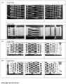
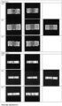
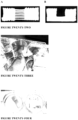
- the morphology of the scaffolds was investigated by SEM and this revealed the production of two architecturally highly distinct components within the construct; the two extremities (bone compartments) of the BLB-scaffold consisted of the typical alternating 0/90 angle polymeric struts layer ( Figure 2A and E ), whereas the ligament compartment, as per our printing design, was composed of aligned and superimposed struts creating macroscopic grooves ( Figure 2B and F ). While additive manufacturing theoretically allows the creation of various shapes and dimensions, it is challenging for the internal porous structure or strut layout to be varied throughout the scaffold printing.
- an in-house g-code production software was used to permit manual modification of the internal layout of the scaffold and resulted in the creation of aligned struts.
- the struts composing the ligament compartment are completely integrated within the bone compartments hence ensuring a mechanically strong cohesion, while maintaining a porous interface.
- the BLB-scaffold is a biphasic scaffold, having two architecturally different portions, but manufactured in a continuous manner similarly to a monolithic 3D printed construct.
- the mPCL struts in the ligament compartment were homogenous in width, although some sagging was observed ( Figure 2B and F , white arrows).
- the mechanical properties of the construct were assessed in both tension and compression, under quasi-static and cyclic loading.
- the 3D-printed scaffold displayed a typical force-strain curve of polymeric material composed of an initial linear region followed by a yield point and a plastic deformation region ( Figure 4A ).
- the 350 ⁇ m BLB-scaffold was the strongest construct with a yield force around 65N and an ultimate force of 70N, whereas the 600 ⁇ m displayed a yield and ultimate force of 40N and 50N respectively.
- the less porous 350 ⁇ m BLB-scaffold was significantly stiffer than the 600 ⁇ m BLB scaffold with a stiffness of 130 N.mm-1 and 90 N.mm-1 respectively.
- Table 1 lists the mechanical parameters measured for the quasi-static tensile test and compares them to the properties of the human native tissue. It is evident that the ultimate force of both constructs are substantially lower than the anatomic SLIL complex. However the physiological load withstood by the ligament is in the range of 20N which is still in the elastic range of the BLB-scaffold. Table 1: Tensile testing results of the 350 ⁇ m / 600 ⁇ m scaffold as compared to the native dorsal SLIL.
- the mechanical behaviour was investigated under dynamic conditions.
- the testing parameters 5 and 10% strain at 0.5Hz, were selected as they represented, to some extent, the physiological extension that the native tissue withstands during normal motion.
- the scaffolds displayed a high level of recovery as shown in Figure 6 by the superimposition of the deformation hysteresis. No accumulation of plastic deformation was noted over the 1000 cycles of the experiment.
- the level of compression did not change for a given design regardless of the stain applied (5 or 10%), however the stiffer construct (350 ⁇ m) experienced a higher level of compression.
- the evolution of the force at peak was plotted against cycle number and this demonstrated that the force at peak sharply decreased after the first cycle and remained relatively constant thereafter, indicating that strain-induced relaxation occurred in the BLB construct ( Figure 7 ).
- the construct stiffness was evaluated and this revealed a differential behaviour between the 350 ⁇ m and 600 ⁇ m design ( Figure 7 ). Indeed, the 350 ⁇ m design, which is the stiffer construct, could better retain of its stiffness as the percentage decrease in this parameter was less at both 5% and 10% strain (Table 2).
- cell sheet technology facilitates maturation and increased deposition of the cell's own ECM prior to implantation.
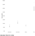
- the cell sheet utilised in the current study were characterised by measuring DNA content as an indicator of relative cell number and collagen deposition ( Figure 8 ).
- Total DNA content of and collagen synthesis by the human bmMSC cell sheets cultured in ascorbic acid supplemented medium were significantly higher than that of control cells cultured in expansion media.
- the low amount of DNA detected in the control samples can be attributed to decreased proliferation and increased cell death over the 21-day culture period with DNA being discarded with each media change.
- Live dead assay was conducted to evaluate the impact of the assembly method, utilising mechanical harvesting and rolling of the cell sheets, on cell viability ( Figure 10 ).
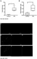
- Figure 10A-C presents the confocal laser microscopy images indicating that most of the cells were alive (green staining). While the non-harvested control cell sheets had the highest cell viability with over 90% live cells, this only decreased slightly to approximately 80% live cells in the seeded cell sheets of both designs. There was no statistically significant difference in cell viability between the bundled cell sheets and the flat cell sheet placed on the dorsal surface of the ligament compartment, and this was observed regardless of the design. There was a statistically significant difference in cell viability between the cell sheets in the constructs and the control cell sheets (p ⁇ 0.05) ( Figure 10D ).
- the regenerative performance of the BLB construct was assessed ectopically in athymic rats using an ectopic model.
- the cellularised constructs were implanted six hours post-assembly and the bone formation along with soft tissue regeneration were measured at 2 and 8 weeks post-implantation.
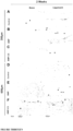
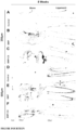
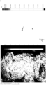
- the ligament compartment was vascularised rapidly and remained vascularised as indicated by the presence of vessel structures with erythrocytes in week 2 and 8 H&E sections ( Figure 13 and 14 , indicated by *).
- the presence of the cell sheets had a significant impact on the morphology of the regenerated tissue which was denser, enriched in collagen and more structured as shown in Figures 13 and 14 .
- TEC Tissue Engineered Construct
- This study features the development of a multiphasic BLB multiphasic construct, combining additive manufacturing and cell sheet technology for the reconstruction of the dorsal SLIL.
- the potential utilisation of this scaffold strongly aligns with clinical practice as reconstruction procedures aim to replace the dorsal segment of the SLIL, the main contributor to carpal stability.
- using a tissue engineering approach as a replacement for grafts mitigates the issue of donor site morbidity.
- the clinical translatability and broader number of patients that could benefit from this novel approach justifies the creation and utilisation of a synthetic BLB scaffold.
- TECs for other orthopaedic applications
- tissue engineering for the regeneration of bone, ligament and tendon in the hand and wrist highlighting the challenges present in this particular area.
- Decellularised cadaveric allografts of the proximal interphalangeal joint collateral ligament have limitations including graft availability, differences in the anatomical dimensions and potential disease transmission.
- the regenerative performance and re-cellularisation ability of such allografts are not known.
- the implantation of cells within a mature ECM network, developed during in vitro culture, can significantly circumvent limitations related to host cell infiltration and differentiation within the construct.
- Cell sheet technology is an efficient manner to deliver cells committed towards a specific differentiation lineage by a prolonged in vitro maturation step whereby the deposition of the cell's own extracellular matrix improves the quality of the regenerated tissue.
- This study showed a significant remodelling of the cell sheet in the early period of the implantation, featuring a cell death peak at 3 days post-implantation concomitant with an increase in neutrophil infiltration followed by collagen densification at a later stage, consistent with tissue maturation.
- the design of the scaffold demonstrated tissue-guiding properties, as shown by the slight increase in tissue alignment within the ligament compartment for the specimen without cell sheets.
- the architecture of the scaffold plays a significant role in guiding tissue regeneration. Indeed, the development of advanced scaffold manufacturing technologies has enabled the fabrication of complex multiphasic scaffolds whereby the internal porous organisation can be controlled and tailored to address tissue-specific needs. Traditionally, the compartments of these multiphase scaffolds are fabricated separately and subsequently assembled using various methodologies. This may result in weak cohesion at the interface of the different compartmentsand affect the ability of the multiphasic construct to withstand physiological loads without excessive biomechanical fatigue.
- the bone and ligament compartments are created via a continuous additive manufacturing process enabling the formation of a porous yet fully integrated interface.
- This strategy ensures that the polymeric filaments of the ligament compartment are entirely anchored in the adjacent layers of the bone compartment, allowing a high number of fusion points and thus providing excellent biomechanical stability. This was best illustrated by the ability of the BLB scaffold to withstand repetitive loads without excessive fatigue or delamination at the bone-ligament interface under repeated mechanical cycles.
- Another important aspect of the BLB design originates from the creation of a porous interface between the compartments resulting from the continuous printing, which had a significant impact on the integration of the ligament into the bone compartment.
- the collagen fibres are transversely oriented and inserted into both the lunate and scaphoid bone enabling the transmission of force during flexion and torsion of the wrist.
- Such an organisation was partially observed in our construct with the formation of collagenous tissue aligned in the long axis of the BLB and inserted into the bone compartment. Although no direct insertion into the newly formed bone was observed due to the small volume of bone formed, these results illustrate the potential of the BLB construct to guide tissue regeneration in the ligament compartment while allowing cross-communication between the hard and soft tissues at the compartment interface.
- the rabbit medial collateral ligament has similar anatomical properties as the dorsal SLIL and thus, can be used as an animal model for testing this novel scaffold in vivo.
- This scaffold will facilitate regeneration of composite tissue and can be implanted for clinical use.
- Multiphasic bone-ligament-bone scaffolds modeled from the dorsal component of the SLIL were 3D-printed with medical grade polycaprolactone (PCL). These simulated a BLB construct with two bone compartments bridged by aligned PCL fibres mimicking the architecture of the native ligament studied from cadaveric specimens.
- PCL polycaprolactone
- the native MCL of the rabbit was removed with holes drilled into insertion and origin points of the ligament on the femur and tibia using a 5mm trephine.
- the bone compartments of the scaffold was press-fitted into the cavities and stapled in place.
- the scaffold After 4 and 8 weeks in vivo, the scaffold remained intact. Mechanical testing of the BLB scaffolds showed that they were capable of withstanding normal SLIL physiological forces. After 4 weeks of mobilization of the knee joint, the scaffolds improved in strength. In vivo study in the rabbits demonstrated that the scaffold was biocompatible and displayed good tissue integration and vascularization. At 4 and 8 weeks, bone formation and ligament remodeling was observed in the corresponding compartments.
- Rabbit scaffolds ligament compartment is 10mm (elongation necessary to bridge knee joint) ( Figure 22 ).
- Figure 26 demonstrates new bone mineralization in a rabbit knee joint (MCL removed and multiphasic bone-ligament-bone scaffold implanted) under K-wire fixation.
- Figure 27 further demonstrates bone regeneration/mineralisation in the joint at 4 and 8 weeks.
- Strain maps ( Figure 30 ) illustrate that the native MCL had much more concentrated regions of increased strain. This can be compared to the strain map of the control scaffold which distributed the force over a larger surface area. The failure point for the control scaffold is within the ligament compartment compared to that of the native MCL at the bone-ligament interphase. This suggests that the bone-ligament interphase of the scaffold is strong enough to withstand considerable force.
Landscapes
- Health & Medical Sciences (AREA)
- Life Sciences & Earth Sciences (AREA)
- Chemical & Material Sciences (AREA)
- Engineering & Computer Science (AREA)
- Biomedical Technology (AREA)
- Animal Behavior & Ethology (AREA)
- Transplantation (AREA)
- Oral & Maxillofacial Surgery (AREA)
- Veterinary Medicine (AREA)
- Public Health (AREA)
- General Health & Medical Sciences (AREA)
- Epidemiology (AREA)
- Medicinal Chemistry (AREA)
- Dermatology (AREA)
- Orthopedic Medicine & Surgery (AREA)
- Cell Biology (AREA)
- Chemical Kinetics & Catalysis (AREA)
- Vascular Medicine (AREA)
- Zoology (AREA)
- Botany (AREA)
- Heart & Thoracic Surgery (AREA)
- Cardiology (AREA)
- Rheumatology (AREA)
- Rehabilitation Therapy (AREA)
- Urology & Nephrology (AREA)
- Manufacturing & Machinery (AREA)
- Materials Engineering (AREA)
- Biophysics (AREA)
- Developmental Biology & Embryology (AREA)
- Dispersion Chemistry (AREA)
- Hematology (AREA)
- Materials For Medical Uses (AREA)
- Prostheses (AREA)
Claims (12)
- Échafaudage tissulaire synthétique multiphasique tridimensionnel comprenant des premier, deuxième et troisième compartiments, dans lequel :chaque dit compartiment comprend des propriétés microstructurales, et/ou chimiques, et/ou mécaniques distinctes, et est relié à au moins un autre compartiment de l'échafaudage par une interface continue ;l'échafaudage tissulaire est poreux ;la morphologie externe de l'échafaudage tissulaire imite celle d'une articulation de mammifère ou d'un composant de celle-ci ;l'un quelconque ou plusieurs des premier, deuxième et/ou troisième compartiments comprend ou consiste en une série de fibres, dans lequel la série de fibres comprend de multiples couches de fibres ;le deuxième compartiment comprend une série de fibres imitant la morphologie externe d'une série de ligaments, et ladite série de fibres sont situées entre et reliant les premier et troisième compartiments ;les multiples couches de fibres comprennent des première et deuxième couches de fibres,la première couche de fibres est alignée le long d'un premier axe,la deuxième couche de fibres est alignée le long d'un deuxième axe, etle deuxième axe est tourné à un angle par rapport au premier axe.
- Échafaudage tissulaire synthétique multiphasique tridimensionnel selon la revendication 1, dans lequel le deuxième axe est tourné d'un angle de :- au moins : 2°, 3°, 4°, 5°, 6°, 7°, 8°, 9°, 10°, 11°, 12°, 13°, 14°, 15°, 16°, 17°, 18°, 19° ou 20° par rapport au premier axe ; ou- entre 20° et 50°, entre 25° et 45°, entre 30° et 40°, entre 25° et 35°, ou environ 35°, par rapport au premier axe.
- Échafaudage tissulaire synthétique multiphasique tridimensionnel selon la revendication 1 ou la revendication 2, dans lequel les multiples couches de fibres comprennent une troisième couche alignée le long d'un troisième axe, et le troisième axe est ;- tourné à un angle par rapport à l'un ou l'autre ou aux deux du premier axe et/ou deuxième axe ; ou- tourné à un angle égal ou sensiblement égal par rapport au premier axe et/ou deuxième axe, ou- tourné dans le sens contraire des aiguilles d'une montre par rapport au premier axe, et est tourné dans un sens horaire par rapport au deuxième axe.
- Échafaudage tissulaire synthétique multiphasique tridimensionnel selon l'une quelconque des revendications 1 à 3, dans lequel :- la morphologie externe de chacun des premier et troisième compartiments imite celle d'un os, et la morphologie externe du deuxième compartiment imite celle d'une série de ligaments situés entre et reliant les premier et troisième compartiments ; ou- la morphologie externe de l'échafaudage tissulaire imite celle d'une articulation scapho-lunaire ou d'un composant de celle-ci ; ou- la morphologie externe du premier compartiment imite celle d'un scaphoïde, la morphologie externe du troisième compartiment imite celle d'un semi-lunaire, et la morphologie externe du deuxième compartiment imite celle d'une série de ligaments scapho-lunaires.
- Échafaudage tissulaire synthétique multiphasique tridimensionnel selon la revendication 4, dans lequel :- la série de ligaments scapho-lunaires sont des ligaments scapho-lunaires dorsaux, et ne sont pas des ligaments scapho-lunaires proximaux ou palmaires ; et/ou- l'échafaudage comprend des pores d'une largeur maximale comprise entre 100 µm et 1000 µm, entre 100 µm et 800 µm, entre 100 µm et 600 µm, entre 200 µm et 600 µm, entre 200 µm et 500 µm, entre 300 µm et 600 µm, entre 300 µm et 500 µm, entre 350 µm et 600 µm, entre 350 µm et 500 µm, entre 400 µm et 600 µm, ou entre 400 µm et 500 µm.
- Échafaudage tissulaire synthétique multiphasique tridimensionnel selon l'une quelconque des revendications 1 à 5, comprenant en outre des cellules de mammifère, de préférence les cellules de mammifère sont choisies dans le groupe consistant en : cellules souches dérivées de ligaments, cellules souches de cartilage, cellules souches mésenchymateuses telles que cellules stromales de mœlle osseuse, cellules stromales dérivées d'adipeux telles que cellules souches mésenchymateuses dérivées de mœlle osseuse, cellules souches mésenchymateuses d'origine adipeuse, ostéoblastes, cellules de type ostéoblaste, cellules souches, cellules progénitrices, ou toute combinaison de celles-ci ; ou de préférence dans lequel les cellules de mammifère sont des cellules souches mésenchymateuses humaines ou des cellules souches mésenchymateuses de mœlle osseuse humaine.
- Échafaudage tissulaire synthétique multiphasique tridimensionnel selon la revendication 6, dans lequel les cellules de mammifère sont à l'intérieur d'une feuille cellulaire enveloppée autour de et/ou insérée dans un ou plusieurs des compartiments.
- Échafaudage tissulaire synthétique multiphasique tridimensionnel selon l'une quelconque des revendications 1 à 7, dans lequel l'un quelconque ou plusieurs des premier, deuxième et/ou troisième compartiments comprend un matériau polymère, de préférence choisi dans le groupe consistant en: collagène, chitosan, acide hyaluronique, alginate, gélatine, diméthacrylate de polyéthylène glycol (PEG), gélatine méthacryloyl, matrigel, fibrinogène, agarose, et polycaprolactone ; ou de préférence dans lequel le matériau polymère est la polycaprolactone ou le polyuréthane.
- Échafaudage tissulaire synthétique multiphasique tridimensionnel selon l'une quelconque des revendications 1 à 8, dans lequel l'échafaudage tissulaire est produit à l'aide d'une bioimprimante tridimensionnelle (3D) pour déposer en continu le matériau polymère sur une plateforme jusqu'à ce que l'échafaudage tissulaire soit produit dans son intégralité.
- Procédé de production de l'échafaudage tissulaire synthétique multiphasique tridimensionnel selon l'une quelconque des revendications 1 à 9, le procédé comprenant l'utilisation d'une bioimprimante tridimensionnelle (3D) pour déposer en continu un matériau polymère sur une plateforme jusqu'à ce que l'échafaudage tissulaire soit produit dans son intégralité.
- Échafaudage tissulaire synthétique tridimensionnel selon l'une quelconque des revendications 1 à 9 pour une utilisation dans le traitement d'un ligament lésé chez un sujet.
- Échafaudage tissulaire synthétique multiphasique tridimensionnel pour une utilisation selon la revendication 11, dans lequel :- le ligament est un ligament interosseux scapho-lunaire ou son équivalent mammifère ; et/ou- le sujet est un humain.
Applications Claiming Priority (2)
| Application Number | Priority Date | Filing Date | Title |
|---|---|---|---|
| AU2017903229A AU2017903229A0 (en) | 2017-08-11 | Multiphasic tissue scaffold constructs | |
| PCT/AU2018/000133 WO2019028494A1 (fr) | 2017-08-11 | 2018-08-13 | Constructions d'échafaudage tissulaire multiphasique |
Publications (4)
| Publication Number | Publication Date |
|---|---|
| EP3664746A1 EP3664746A1 (fr) | 2020-06-17 |
| EP3664746A4 EP3664746A4 (fr) | 2021-05-19 |
| EP3664746B1 true EP3664746B1 (fr) | 2025-04-23 |
| EP3664746C0 EP3664746C0 (fr) | 2025-04-23 |
Family
ID=65272976
Family Applications (1)
| Application Number | Title | Priority Date | Filing Date |
|---|---|---|---|
| EP18845151.2A Active EP3664746B1 (fr) | 2017-08-11 | 2018-08-13 | Constructions d'échafaudage tissulaire multiphasique |
Country Status (6)
| Country | Link |
|---|---|
| US (1) | US11752002B2 (fr) |
| EP (1) | EP3664746B1 (fr) |
| JP (1) | JP7534752B2 (fr) |
| AU (1) | AU2018315622B2 (fr) |
| ES (1) | ES3041158T3 (fr) |
| WO (1) | WO2019028494A1 (fr) |
Families Citing this family (3)
| Publication number | Priority date | Publication date | Assignee | Title |
|---|---|---|---|---|
| EP3752049B1 (fr) * | 2018-02-12 | 2025-10-22 | Massachusetts Institute of Technology | Cadre de fabrication et de conception quantitative pour une interface biomécanique en contact avec un segment corporel biologique |
| KR20240013753A (ko) * | 2021-05-06 | 2024-01-30 | 렁 바이오테크놀로지 피비씨 | 변형된 3d-프린팅된 물체 및 이들의 용도 |
| US20230082358A1 (en) * | 2021-09-08 | 2023-03-16 | University Of Maryland, College Park | 3d printed uv crosslinking masks |
Citations (1)
| Publication number | Priority date | Publication date | Assignee | Title |
|---|---|---|---|---|
| WO2017050837A1 (fr) * | 2015-09-23 | 2017-03-30 | Novus Scientific Ab | Implant médical en trois dimensions pour la régénération des tissus mous |
Family Cites Families (14)
| Publication number | Priority date | Publication date | Assignee | Title |
|---|---|---|---|---|
| US6530956B1 (en) * | 1998-09-10 | 2003-03-11 | Kevin A. Mansmann | Resorbable scaffolds to promote cartilage regeneration |
| US9943410B2 (en) * | 2011-02-28 | 2018-04-17 | DePuy Synthes Products, Inc. | Modular tissue scaffolds |
| US8753391B2 (en) * | 2007-02-12 | 2014-06-17 | The Trustees Of Columbia University In The City Of New York | Fully synthetic implantable multi-phased scaffold |
| US20100310623A1 (en) | 2009-06-05 | 2010-12-09 | Laurencin Cato T | Synergetic functionalized spiral-in-tubular bone scaffolds |
| US20120095482A1 (en) | 2010-05-11 | 2012-04-19 | Dale Peterson | Endoprosthetic textile scaffold |
| WO2013086404A1 (fr) | 2011-12-09 | 2013-06-13 | The Board Of Trustees Of The Leland Stanford Junior University | Bio-échafaudages de tissu composite décellularisés pour reconstruction d'interface de tissu musculosquelettique et procédés de production |
| WO2013128434A1 (fr) | 2012-02-28 | 2013-09-06 | Incorpracyl Technologies Ltd. | Structure de support de tissu |
| US9101606B2 (en) * | 2012-04-19 | 2015-08-11 | Warsaw Orthopedic, Inc. | Bone delivery system having a therapeutic agent |
| US8771368B2 (en) | 2012-04-24 | 2014-07-08 | William F. McKay | Interspinous bone implant device |
| CN105473103B (zh) * | 2013-05-31 | 2018-06-12 | 新加坡国立大学 | 一种人工气管假体和/或发音假体材料及其制造方法 |
| GB201315074D0 (en) * | 2013-08-23 | 2013-10-02 | Univ Singapore | 3-Dimensional Bioscaffolds |
| JP2015089433A (ja) | 2013-11-06 | 2015-05-11 | 学校法人慈恵大学 | 組織再生体およびその製造方法、並びに組織再生体用足場 |
| WO2016094669A1 (fr) * | 2014-12-11 | 2016-06-16 | Tepha, Inc. | Procédés d'orientation de fil multifilament et de monofilaments de poly-4-hydroxybutyrate et de copolymères de celui-ci |
| US11045585B2 (en) | 2015-04-08 | 2021-06-29 | The Trustees Of Columbia University In The City Of New York | Spatiotemporal delivery system embedded in 3D-printing |
-
2018
- 2018-08-13 JP JP2020529780A patent/JP7534752B2/ja active Active
- 2018-08-13 ES ES18845151T patent/ES3041158T3/es active Active
- 2018-08-13 US US16/636,706 patent/US11752002B2/en active Active
- 2018-08-13 EP EP18845151.2A patent/EP3664746B1/fr active Active
- 2018-08-13 AU AU2018315622A patent/AU2018315622B2/en active Active
- 2018-08-13 WO PCT/AU2018/000133 patent/WO2019028494A1/fr not_active Ceased
Patent Citations (1)
| Publication number | Priority date | Publication date | Assignee | Title |
|---|---|---|---|---|
| WO2017050837A1 (fr) * | 2015-09-23 | 2017-03-30 | Novus Scientific Ab | Implant médical en trois dimensions pour la régénération des tissus mous |
Also Published As
| Publication number | Publication date |
|---|---|
| AU2018315622B2 (en) | 2024-05-16 |
| WO2019028494A1 (fr) | 2019-02-14 |
| JP2020530375A (ja) | 2020-10-22 |
| AU2018315622A1 (en) | 2020-02-27 |
| ES3041158T3 (en) | 2025-11-07 |
| US11752002B2 (en) | 2023-09-12 |
| EP3664746A4 (fr) | 2021-05-19 |
| EP3664746C0 (fr) | 2025-04-23 |
| US20200237520A1 (en) | 2020-07-30 |
| JP7534752B2 (ja) | 2024-08-15 |
| EP3664746A1 (fr) | 2020-06-17 |
Similar Documents
| Publication | Publication Date | Title |
|---|---|---|
| Baldino et al. | Regeneration techniques for bone-to-tendon and muscle-to-tendon interfaces reconstruction | |
| Li et al. | Scaffold-based tissue engineering strategies for soft–hard interface regeneration | |
| Ma et al. | Rational design of nanofiber scaffolds for orthopedic tissue repair and regeneration | |
| EP2097116B1 (fr) | Système in situ pour une réparation du tissu chondral et osseux intra-articulaire | |
| US20230079113A1 (en) | Biomaterial for Articular Cartilage Maintenance and Treatment of Arthritis | |
| Lui et al. | Additively manufactured multiphasic bone–ligament–bone scaffold for scapholunate interosseous ligament reconstruction | |
| Zhang et al. | Three-dimensional bioprinting of a structure-, composition-, and mechanics-graded biomimetic scaffold coated with specific decellularized extracellular matrix to improve the tendon-to-bone healing | |
| CN101332134B (zh) | I型胶原人工肌腱、韧带的制备及应用 | |
| Luo et al. | Advanced strategies for constructing interfacial tissues of bone and tendon/ligament | |
| EP3664746B1 (fr) | Constructions d'échafaudage tissulaire multiphasique | |
| Mahmoud et al. | Treatment strategies for meniscal lesions: from past to prospective therapeutics | |
| Grogan et al. | Meniscal tissue repair with nanofibers: future perspectives | |
| CN114533959B (zh) | 一种肌腱修复材料、制备方法及在制备肌腱修复产品中的应用 | |
| Semitela et al. | Strategies to engineer articular cartilage with biomimetic zonal features: a review | |
| Manoukian et al. | Nanomaterials/nanocomposites for osteochondral tissue | |
| Jiang et al. | Fabrication of collagen-based biomaterials for sports medicine application | |
| Sbirkov et al. | Rotator Cuff Repair: Lessons from Immune Strategies, 3D Biofabrication and In Vivo Testing | |
| Huang et al. | A Braided Scaffold for Rotator Cuff Tissue Engineering | |
| Yi et al. | Extracellular Matrix in Peripheral Nerve Regeneration | |
| Lee et al. | Knee—Meniscus and Cartilage Regeneration | |
| Mallick et al. | 3D-Bioprinted Scaffold Technology to Promote Articular Cartilage Regeneration in Osteoarthritis: An Example of Challenges of 3D Bioprinting | |
| Thomas | The Development of a Novel Cartilage Analog Scaffold for Chondral Regeneration | |
| Ozmen et al. | Biomimetic Constructs for Achilles Tendon Regeneration and their Translation to Human Medicine | |
| Jiang | The Rotator Cuff Tendon-to-Bone Interface: Maturation, Aging, and 3D Bioprinting for Regeneration | |
| James et al. | Innovative regenerative engineering technologies for soft tissue regeneration |
Legal Events
| Date | Code | Title | Description |
|---|---|---|---|
| STAA | Information on the status of an ep patent application or granted ep patent |
Free format text: STATUS: THE INTERNATIONAL PUBLICATION HAS BEEN MADE |
|
| PUAI | Public reference made under article 153(3) epc to a published international application that has entered the european phase |
Free format text: ORIGINAL CODE: 0009012 |
|
| STAA | Information on the status of an ep patent application or granted ep patent |
Free format text: STATUS: REQUEST FOR EXAMINATION WAS MADE |
|
| 17P | Request for examination filed |
Effective date: 20200304 |
|
| AK | Designated contracting states |
Kind code of ref document: A1 Designated state(s): AL AT BE BG CH CY CZ DE DK EE ES FI FR GB GR HR HU IE IS IT LI LT LU LV MC MK MT NL NO PL PT RO RS SE SI SK SM TR |
|
| AX | Request for extension of the european patent |
Extension state: BA ME |
|
| DAV | Request for validation of the european patent (deleted) | ||
| DAX | Request for extension of the european patent (deleted) | ||
| A4 | Supplementary search report drawn up and despatched |
Effective date: 20210419 |
|
| RIC1 | Information provided on ipc code assigned before grant |
Ipc: A61F 2/08 20060101AFI20210413BHEP Ipc: A61F 2/30 20060101ALI20210413BHEP Ipc: A61L 27/56 20060101ALI20210413BHEP |
|
| STAA | Information on the status of an ep patent application or granted ep patent |
Free format text: STATUS: EXAMINATION IS IN PROGRESS |
|
| 17Q | First examination report despatched |
Effective date: 20240729 |
|
| GRAP | Despatch of communication of intention to grant a patent |
Free format text: ORIGINAL CODE: EPIDOSNIGR1 |
|
| STAA | Information on the status of an ep patent application or granted ep patent |
Free format text: STATUS: GRANT OF PATENT IS INTENDED |
|
| INTG | Intention to grant announced |
Effective date: 20241217 |
|
| GRAS | Grant fee paid |
Free format text: ORIGINAL CODE: EPIDOSNIGR3 |
|
| GRAA | (expected) grant |
Free format text: ORIGINAL CODE: 0009210 |
|
| STAA | Information on the status of an ep patent application or granted ep patent |
Free format text: STATUS: THE PATENT HAS BEEN GRANTED |
|
| AK | Designated contracting states |
Kind code of ref document: B1 Designated state(s): AL AT BE BG CH CY CZ DE DK EE ES FI FR GB GR HR HU IE IS IT LI LT LU LV MC MK MT NL NO PL PT RO RS SE SI SK SM TR |
|
| REG | Reference to a national code |
Ref country code: GB Ref legal event code: FG4D |
|
| REG | Reference to a national code |
Ref country code: CH Ref legal event code: EP |
|
| REG | Reference to a national code |
Ref country code: DE Ref legal event code: R096 Ref document number: 602018081375 Country of ref document: DE |
|
| REG | Reference to a national code |
Ref country code: IE Ref legal event code: FG4D |
|
| U01 | Request for unitary effect filed |
Effective date: 20250522 |
|
| U07 | Unitary effect registered |
Designated state(s): AT BE BG DE DK EE FI FR IT LT LU LV MT NL PT RO SE SI Effective date: 20250530 |
|
| U20 | Renewal fee for the european patent with unitary effect paid |
Year of fee payment: 8 Effective date: 20250821 |
|
| PGFP | Annual fee paid to national office [announced via postgrant information from national office to epo] |
Ref country code: ES Payment date: 20250905 Year of fee payment: 8 |
|
| PG25 | Lapsed in a contracting state [announced via postgrant information from national office to epo] |
Ref country code: GR Free format text: LAPSE BECAUSE OF FAILURE TO SUBMIT A TRANSLATION OF THE DESCRIPTION OR TO PAY THE FEE WITHIN THE PRESCRIBED TIME-LIMIT Effective date: 20250724 Ref country code: NO Free format text: LAPSE BECAUSE OF FAILURE TO SUBMIT A TRANSLATION OF THE DESCRIPTION OR TO PAY THE FEE WITHIN THE PRESCRIBED TIME-LIMIT Effective date: 20250723 |
|
| PG25 | Lapsed in a contracting state [announced via postgrant information from national office to epo] |
Ref country code: PL Free format text: LAPSE BECAUSE OF FAILURE TO SUBMIT A TRANSLATION OF THE DESCRIPTION OR TO PAY THE FEE WITHIN THE PRESCRIBED TIME-LIMIT Effective date: 20250423 |
|
| PGFP | Annual fee paid to national office [announced via postgrant information from national office to epo] |
Ref country code: GB Payment date: 20250814 Year of fee payment: 8 |
|
| PG25 | Lapsed in a contracting state [announced via postgrant information from national office to epo] |
Ref country code: HR Free format text: LAPSE BECAUSE OF FAILURE TO SUBMIT A TRANSLATION OF THE DESCRIPTION OR TO PAY THE FEE WITHIN THE PRESCRIBED TIME-LIMIT Effective date: 20250423 |
|
| PGFP | Annual fee paid to national office [announced via postgrant information from national office to epo] |
Ref country code: CH Payment date: 20250901 Year of fee payment: 8 |
|
| PG25 | Lapsed in a contracting state [announced via postgrant information from national office to epo] |
Ref country code: RS Free format text: LAPSE BECAUSE OF FAILURE TO SUBMIT A TRANSLATION OF THE DESCRIPTION OR TO PAY THE FEE WITHIN THE PRESCRIBED TIME-LIMIT Effective date: 20250723 |
|
| PG25 | Lapsed in a contracting state [announced via postgrant information from national office to epo] |
Ref country code: IS Free format text: LAPSE BECAUSE OF FAILURE TO SUBMIT A TRANSLATION OF THE DESCRIPTION OR TO PAY THE FEE WITHIN THE PRESCRIBED TIME-LIMIT Effective date: 20250823 |
|
| REG | Reference to a national code |
Ref country code: ES Ref legal event code: FG2A Ref document number: 3041158 Country of ref document: ES Kind code of ref document: T3 Effective date: 20251107 |
|
| PG25 | Lapsed in a contracting state [announced via postgrant information from national office to epo] |
Ref country code: SM Free format text: LAPSE BECAUSE OF FAILURE TO SUBMIT A TRANSLATION OF THE DESCRIPTION OR TO PAY THE FEE WITHIN THE PRESCRIBED TIME-LIMIT Effective date: 20250423 |
|
| PG25 | Lapsed in a contracting state [announced via postgrant information from national office to epo] |
Ref country code: CZ Free format text: LAPSE BECAUSE OF FAILURE TO SUBMIT A TRANSLATION OF THE DESCRIPTION OR TO PAY THE FEE WITHIN THE PRESCRIBED TIME-LIMIT Effective date: 20250423 |
|
| PG25 | Lapsed in a contracting state [announced via postgrant information from national office to epo] |
Ref country code: SK Free format text: LAPSE BECAUSE OF FAILURE TO SUBMIT A TRANSLATION OF THE DESCRIPTION OR TO PAY THE FEE WITHIN THE PRESCRIBED TIME-LIMIT Effective date: 20250423 |
|
| PLBE | No opposition filed within time limit |
Free format text: ORIGINAL CODE: 0009261 |
|
| STAA | Information on the status of an ep patent application or granted ep patent |
Free format text: STATUS: NO OPPOSITION FILED WITHIN TIME LIMIT |
|
| REG | Reference to a national code |
Ref country code: CH Ref legal event code: L10 Free format text: ST27 STATUS EVENT CODE: U-0-0-L10-L00 (AS PROVIDED BY THE NATIONAL OFFICE) Effective date: 20260304 |
|
| PG25 | Lapsed in a contracting state [announced via postgrant information from national office to epo] |
Ref country code: MC Free format text: LAPSE BECAUSE OF FAILURE TO SUBMIT A TRANSLATION OF THE DESCRIPTION OR TO PAY THE FEE WITHIN THE PRESCRIBED TIME-LIMIT Effective date: 20250423 |
|
| 26N | No opposition filed |
Effective date: 20260126 |