EP3712845A1 - Bildidentifizierungsverfahren und bildidentifizierungsvorrichtung - Google Patents
Bildidentifizierungsverfahren und bildidentifizierungsvorrichtung Download PDFInfo
- Publication number
- EP3712845A1 EP3712845A1 EP19189119.1A EP19189119A EP3712845A1 EP 3712845 A1 EP3712845 A1 EP 3712845A1 EP 19189119 A EP19189119 A EP 19189119A EP 3712845 A1 EP3712845 A1 EP 3712845A1
- Authority
- EP
- European Patent Office
- Prior art keywords
- image
- pixels
- sub
- images
- obi
- Prior art date
- Legal status (The legal status is an assumption and is not a legal conclusion. Google has not performed a legal analysis and makes no representation as to the accuracy of the status listed.)
- Granted
Links
Images

Classifications
-
- G—PHYSICS
- G06—COMPUTING OR CALCULATING; COUNTING
- G06T—IMAGE DATA PROCESSING OR GENERATION, IN GENERAL
- G06T7/00—Image analysis
- G06T7/0002—Inspection of images, e.g. flaw detection
- G06T7/0012—Biomedical image inspection
-
- A—HUMAN NECESSITIES
- A61—MEDICAL OR VETERINARY SCIENCE; HYGIENE
- A61B—DIAGNOSIS; SURGERY; IDENTIFICATION
- A61B6/00—Apparatus or devices for radiation diagnosis; Apparatus or devices for radiation diagnosis combined with radiation therapy equipment
- A61B6/02—Arrangements for diagnosis sequentially in different planes; Stereoscopic radiation diagnosis
- A61B6/03—Computed tomography [CT]
- A61B6/032—Transmission computed tomography [CT]
-
- A—HUMAN NECESSITIES
- A61—MEDICAL OR VETERINARY SCIENCE; HYGIENE
- A61B—DIAGNOSIS; SURGERY; IDENTIFICATION
- A61B6/00—Apparatus or devices for radiation diagnosis; Apparatus or devices for radiation diagnosis combined with radiation therapy equipment
- A61B6/52—Devices using data or image processing specially adapted for radiation diagnosis
- A61B6/5205—Devices using data or image processing specially adapted for radiation diagnosis involving processing of raw data to produce diagnostic data
-
- G—PHYSICS
- G06—COMPUTING OR CALCULATING; COUNTING
- G06T—IMAGE DATA PROCESSING OR GENERATION, IN GENERAL
- G06T5/00—Image enhancement or restoration
- G06T5/40—Image enhancement or restoration using histogram techniques
-
- G—PHYSICS
- G06—COMPUTING OR CALCULATING; COUNTING
- G06T—IMAGE DATA PROCESSING OR GENERATION, IN GENERAL
- G06T5/00—Image enhancement or restoration
- G06T5/90—Dynamic range modification of images or parts thereof
-
- G—PHYSICS
- G06—COMPUTING OR CALCULATING; COUNTING
- G06T—IMAGE DATA PROCESSING OR GENERATION, IN GENERAL
- G06T5/00—Image enhancement or restoration
- G06T5/90—Dynamic range modification of images or parts thereof
- G06T5/92—Dynamic range modification of images or parts thereof based on global image properties
-
- G—PHYSICS
- G06—COMPUTING OR CALCULATING; COUNTING
- G06T—IMAGE DATA PROCESSING OR GENERATION, IN GENERAL
- G06T2207/00—Indexing scheme for image analysis or image enhancement
- G06T2207/10—Image acquisition modality
- G06T2207/10072—Tomographic images
- G06T2207/10081—Computed x-ray tomography [CT]
-
- G—PHYSICS
- G06—COMPUTING OR CALCULATING; COUNTING
- G06T—IMAGE DATA PROCESSING OR GENERATION, IN GENERAL
- G06T2207/00—Indexing scheme for image analysis or image enhancement
- G06T2207/20—Special algorithmic details
- G06T2207/20021—Dividing image into blocks, subimages or windows
-
- G—PHYSICS
- G06—COMPUTING OR CALCULATING; COUNTING
- G06T—IMAGE DATA PROCESSING OR GENERATION, IN GENERAL
- G06T2207/00—Indexing scheme for image analysis or image enhancement
- G06T2207/20—Special algorithmic details
- G06T2207/20081—Training; Learning
-
- G—PHYSICS
- G06—COMPUTING OR CALCULATING; COUNTING
- G06T—IMAGE DATA PROCESSING OR GENERATION, IN GENERAL
- G06T2207/00—Indexing scheme for image analysis or image enhancement
- G06T2207/20—Special algorithmic details
- G06T2207/20084—Artificial neural networks [ANN]
-
- G—PHYSICS
- G06—COMPUTING OR CALCULATING; COUNTING
- G06T—IMAGE DATA PROCESSING OR GENERATION, IN GENERAL
- G06T2207/00—Indexing scheme for image analysis or image enhancement
- G06T2207/30—Subject of image; Context of image processing
- G06T2207/30004—Biomedical image processing
- G06T2207/30056—Liver; Hepatic
-
- G—PHYSICS
- G06—COMPUTING OR CALCULATING; COUNTING
- G06T—IMAGE DATA PROCESSING OR GENERATION, IN GENERAL
- G06T2207/00—Indexing scheme for image analysis or image enhancement
- G06T2207/30—Subject of image; Context of image processing
- G06T2207/30004—Biomedical image processing
- G06T2207/30096—Tumor; Lesion
Definitions
- the disclosure relates to an identifying method and an identifying device, and particularly relates to an image identifying method and an image identifying device.
- liver cancer hepatocellular carcinoma
- the disclosure provides an image identifying method and an image identifying device for addressing the above technical issues.
- An embodiment of the disclosure provides an image identifying method.
- the image identifying method includes: retrieving a first computer tomographic (CT) image, wherein the first CT image includes a plurality of first pixels, each of the first pixels has a channel value, and the channel value of each of the first pixels falls within a first range; retrieving a plurality of second pixels from the first pixels, and generating a second CT image based on the second pixels, wherein the channel value of each of the second pixels falls within a second range, and the second range is smaller than the first range; performing a contrast enhancement algorithm on the second CT image, so as to adjust a contrast of the second CT image; identifying a first image region in the second CT image to generate a third CT image, wherein the first image region includes a first object; dividing the third CT image into a plurality of sub-images, and only reserving a specific image region corresponding to the first object in each sub-image; combining the sub-images into a fourth CT image; and identifying an object type of
- An embodiment of the disclosure provides an image identifying device.
- the image identifying device includes a storage circuit and a processor.
- the storage circuit stores a plurality of modules.
- the processor is coupled to the storage circuit and accesses the modules to perform the following: retrieving a first computer tomographic (CT) image, wherein the first CT image includes a plurality of first pixels, each of the first pixels has a channel value, and the channel value of each of the first pixels falls within a first range; retrieving a plurality of second pixels from the first pixels, and generating a second CT image based on the second pixels, wherein the channel value of each of the second pixels falls within a second range, and the second range is smaller than the first range; performing a contrast enhancement algorithm on the second CT image, so as to adjust a contrast of the second CT image; identifying a first image region in the second CT image to generate a third CT image, wherein the first image region includes a first object; dividing the third CT image into a plurality of sub-images, and only
- the image identifying method and the image identifying device are capable of sequentially performing identification to find the first image region, the specific image region corresponding to the first object, and the object type of the first object after excluding the pixels whose channel values do not fall within the first range and performing the contrast enhancement algorithm. Accordingly, the performance and accuracy of identification can be more favorable.
- an image identifying device is capable of performing identification on a specific computer tomographic (CT) image to find a specific object in the specific image and further identifying an object type of the specific object after retrieving the specific CT image.
- the image identifying device of the disclosure is capable of performing identification on a CT image to find a tumor in the CT image and further determining a tumor type of the tumor after retrieving the CT image.
- CT computer tomographic
- the target on which the image identifying device performs the image identification is an abdominal CT image captured from a patient, and relevant identifying operations performed by the image identifying device based on the abdominal CT image will be described in detail in the following.
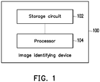
- FIG. 1 is a schematic view illustrating an image identifying device according to an embodiment of the disclosure.
- an image identifying device 100 includes a storage circuit 102 and a processor 104.
- the image identifying device 100 may be a personal computer, a laptop computer, a workstation, a sever, a smart device, or other devices capable of analyzing and processing a CT image.
- the disclosure is not limited thereto.
- the storage circuit 102 may be, for example, any type of fixed or movable random access memories (RAMs), read-only memories (ROMs), flash memories, hard drives, other similar devices, or a combination of these devices, and the storage circuit 102 may serve to record a plurality of program codes or modules.
- RAMs random access memories
- ROMs read-only memories
- flash memories hard drives, other similar devices, or a combination of these devices, and the storage circuit 102 may serve to record a plurality of program codes or modules.
- the processor 104 is coupled to the storage circuit 102, and may be a general-purpose processor, a specific-purpose processor, a traditional processor, a digital signal processor, a plurality of microprocessors, one or more microprocessors incorporating a digital signal processor core, a controller, a microcontroller, an application specific integrated circuit (ASIC), a field programmable gate array (FPGA), any other type of integrated circuits, state machines, advanced RISC machine (ARM)-based processors, or similar products.
- ASIC application specific integrated circuit
- FPGA field programmable gate array
- the processor 104 may access the modules and program codes recorded in the storage circuit 102 to realize the image identifying method of the disclosure. Details in this regard will be described in the following.
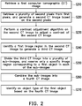
- FIG. 2 is a flowchart illustrating an image identifying method according to an embodiment of the disclosure.
- the method of the embodiment may be carried out by the image identifying device 100 of FIG. 1 .
- details of the respective steps shown in FIG. 2 are described with reference to the components shown in FIG. 1 .
- the description is also made with reference to the scenario shown in FIG. 3 .
- FIG. 3 merely serves as an example and shall not be construed as a limitation on possible embodiments of the disclosure.
- the processor 104 may retrieve a first CT image 310.
- the first CT image 310 is, for example, an abdominal CT image captured from the abdominal cavity of a patient.
- the image identifying device 100 may, after performing specific pre-processing on the first CT image 310, find the liver, a tumor in the liver, and a tumor type of the tumor in a pre-processed image by using different deep-learning models.
- the disclosure is not limited thereto.
- the first CT image 310 may include a plurality of first pixels.
- each of the first pixels has a channel value, and the channel value falls within a first range.
- the channel value is, for example, a Hounsfield unit (HU) value, and an HU value normally falls between -2048 and 30000 (i.e., the first range).
- HU Hounsfield unit
- the disclosure is not limited thereto.
- the processor 104 may exclude some of the first pixels through Step S220, so as to improve the performance of the subsequent identification process.
- the processor 104 may retrieve a plurality of second pixels from the first pixels, and generate a second CT image based on the second pixels.
- the channel value of each of the second pixels falls within a second range, and the second range is smaller than the first range.
- the processor 104 may find a plurality of third pixels from the first pixels, and the channel value of each of the third pixels falls outside the second range.
- the processor 104 removes the third pixels from the first CT image to generate the second CT image 320 only including the second pixels.
- the processor 104 may remove the third pixels whose channel values do not fall within the second range in the first CT image 310, so that only the second pixels whose channel values fall within the second range are retained in the first CT image, thereby forming the second CT image 320.
- the second range may be set by the designer based on needs.
- the processor 104 since the HU value corresponding to a liver and a tumor normally falls between -70 and 200, the processor 104 may define the second range as ranging between -70 and 200.
- the disclosure is not limited thereto.
- the processor 104 may also be configured to generate other second CT images based on one or more other second ranges. In other words, each of these other second CT images may only include pixels whose channel values fall within a specific range.
- the other one or more second ranges may be determined according to a HU value which makes the liver and a tumor more identifiable after the practical modifications and testing of a radiologist. However, the disclosure is not limited thereto.
- the processor 104 may perform a contrast enhancement algorithm on the second CT image 320, so as to adjust the contrast of the second CT image 320.
- the contrast enhancement algorithm may include a histogram equalization algorithm or other similar algorithms, for example.
- the disclosure is not limited thereto. Specifically, after the contrast enhancement algorithm is performed on the second CT image 320, the contrast between image regions corresponding to the liver and the tumor may be increased, so that the tumor becomes more identifiable.
- the disclosure is not limited thereto.
- the performance of the image identification (e.g., tumor identification) performed subsequently is facilitated through pre-processing such as removing the pixels whose channel values do not fall within the first range and performing the contrast enhancement algorithm.
- the processor 104 may identify a first image region 330a in the second CT image 320, so as to generate a third CT image 330.
- the first image region 330a may be, for example, an image region of the liver including a tumor, and the first image region 330a may include a first object OB1.
- the first object OBI is, for example, a tumor located in the liver.
- the processor 104 may input the second CT image 320 to a first deep learning model to find the first image region 330a in the second CT image 320 according to the first deep learning model. Then, the processor 104 may remove the second pixels not belonging to the first image region 330a from the second CT image 320, so as to generate the third CT image 330.
- the first deep learning model may be realized by adopting the Unet model under the framework of the convolutional neural network (CNN) or other similar models.
- CNN convolutional neural network
- the disclosure is not limited thereto.
- the first deep learning model may be firstly trained with training images in which specific regions of interest (ROIs) are labeled, so as to learn the features of the ROIs through the training images.
- ROIs regions of interest
- the first deep learning model in order for the first deep learning model to be capable of identifying the first image region 330a (i.e., the image region of the liver having the tumor), the first deep learning model may firstly learn based on a plurality of training images which are images of livers having tumors as the ROIs, so as to find the features of the image region of the liver having a tumor.
- the processor 104 may divide the third CT image 330 into a plurality of sub-images 330b, and reserve only a specific image region corresponding to the first object OBI in each of the sub-images 330b.
- the processor 104 may input a first sub-image of the sub-images into a second deep learning model, so as to find the specific image region corresponding to the first object OBI in the first sub-image based on the second deep learning model.
- each of the sub-images includes a plurality of specific pixels. Then, the processor 104 may remove the specific pixels not belonging to the specific image region from the first sub-image, so that the first sub-image only includes the specific image region corresponding to the first object OB1.
- the processor 104 may evenly divide the third CT image 330 into the sub-images 330b according to a predetermined size. For example, assuming that the size of the third CT image 330 is 512x512 and the predetermined size is 64x64, the processor 104 may evenly divide the third CT image 330 into 64 sub-images 330b according to the predetermined size. The size of each of the sub-images 330b is the predetermined size (i.e., 64x64). However, the disclosure is not limited thereto.
- the processor 104 may further record the distributed locations of the sub-images 330b in the third CT image 330. For example, the distributed location of each of the sub-images 330b may be represented as coordinates of each of the sub-images 330b in the third CT image 330. However, the disclosure is not limited thereto.
- the processor 104 may input the respective 64 sub-images 330b to the second deep learning model, so as to find the specific image region corresponding to the first object OBI in each of the sub-images 330b according to the second deep learning model. Then, the processor 104 may remove the specific pixels not corresponding to the first object OBI from each of the sub-images 330b, so that only the specific image region corresponding to the first object OBI is retained in each of the sub-images 330b. In short, the processor 104 may remove liver regions in which the tumor is not found from each of the sub-images 330b. However, the disclosure is not limited thereto.
- the second deep learning model may be realized by adopting the Unet model under the CNN framework or other similar models.
- the disclosure is not limited thereto.
- the second deep learning model may be firstly trained with training images in which specific regions of interest (ROIs) are labeled, so as to learn the features of the ROIs through the training images.
- ROIs regions of interest
- the second deep learning model in order for the second deep learning model to be capable of finding the specific image region corresponding to the first object OBI (e.g., the image region of the tumor), the second deep learning model may firstly learn based on training sub-images in which tumors are set as the ROIs (the size of the training sub-image may correspond to the size of the sub-image 330b), so as to find the features of image regions having tumors.
- the training sub-images may be obtained by evenly dividing up a liver training image in which the ROI (e.g., a tumor) is labeled, for example.
- the volume of data for the training of the second deep learning model may be increased, and the learning effect may thus be more favorable.
- the size of each of the training sub-images is smaller, the parameters required for the second deep learning model during the training are also reduced. Accordingly, the training difficulty is reduced, and the accuracy of identification is increased.
- the processor 104 may combine the sub-images 330b into a fourth CT image 340.
- the processor 104 may combine the sub-images 330b into the fourth CT image 340 according to the distributed locations of the sub-images 330b (including only the specific image regions corresponding to the first object OBI) in the third CT image 330.
- the distributed location of each of the sub-images 330b in the third CT image 330 corresponds to the distributed location in the fourth CT image 340.
- the fourth CT image 340 since the sub-image 330b under consideration only includes the specific image region corresponding to the first object OBI, after the respective sub-images 330b are combined into the fourth CT image 340, the fourth CT image 340 only includes the first object OBI (i.e., the tumor).
- the processor 104 may identify the object type of the first object OBI based on the fourth CT image 340.
- the processor 104 may input the fourth CT image 340 into the third deep learning model, so as to identify the object type of the first object OBI (e.g., the tumor type of the tumor) according to the third deep learning model.
- the third deep learning model may be realized by adopting the NASnet model under the CNN framework or other similar models.
- the disclosure is not limited thereto.
- the third deep learning model may be firstly trained with training images in which specific regions of interest (ROI) are labeled, so as to learn the features of the ROIs through the training images.
- ROI regions of interest
- the third deep learning model in order for the third deep learning model to be capable of identifying the object type of the first object OBI (e.g., the tumor type of the tumor), the third deep learning model may firstly learn based on tumor images corresponding one or more specific tumor types (e.g., hepatocellular carcinoma (HCC) tumors, hemangioma tumors, or focal nodular hyperplasia (FNH) tumors) as training images, so as to learn the corresponding features.
- specific tumor types e.g., hepatocellular carcinoma (HCC) tumors, hemangioma tumors, or focal nodular hyperplasia (FNH) tumors
- the image identifying method and the image identifying device are capable of sequentially performing identification to find the first image region (e.g., the liver image region including the tumor), the specific image region corresponding to the first object (e.g., the tumor region located in the liver image region), and the object type of the first object (e.g., the tumor type of the tumor) according to different deep learning models after the pre-processing operations on the CT images. Accordingly, the performance and accuracy of identification can be more favorable.
- the first image region e.g., the liver image region including the tumor
- the specific image region corresponding to the first object e.g., the tumor region located in the liver image region
- the object type of the first object e.g., the tumor type of the tumor
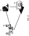
- FIG. 4 is a view illustrating another usage scenario according to an embodiment of the disclosure.
- a server 410 is a medical picture archiving and communication system (PACS), for example, and capable of storing various medical images such as the first CT image 310 shown in FIG. 3 .
- PACS medical picture archiving and communication system
- the doctor 420 may send a corresponding request to the server 410 through a client computer thereof. Then, in response to the request, the server 410 may transmit the first CT image 310 to the image identifying device 100, and the image identifying device 100 may perform relevant operations, such as pre-processing and identification, etc., on the first CT image 310 according to the teaching of the embodiments, so as to learn the object type (e.g., the tumor type) of the first object OBI (e.g., the tumor). Then, the image identifying device 100 may return the original first CT image 310 and relevant identification results to the client computer for the doctor 420 to consider when making a diagnosis.
- a specific disease e.g., HCC, FNH, etc.
- the image identifying device 100 may further overlay the first object OB1 as a semi-transparent image layer on the original first CT image 310, so that the doctor 420 may check the images more conveniently.
- the disclosure is not limited thereto.
- the image identifying method and the image identifying device are capable of sequentially performing identification to find the first image region (e.g., the liver image region including the tumor), the specific image region corresponding to the first object (e.g., the tumor region located in the liver image region), and the object type of the first object (e.g., the tumor type of the tumor) according to different deep learning models after the pre-processing operations on the CT images (e.g., excluding the pixels whose channel values do not fall within the first range and performing the contrast enhancement algorithm). Accordingly, the performance and accuracy of identification can be more favorable.
- the first image region e.g., the liver image region including the tumor
- the specific image region corresponding to the first object e.g., the tumor region located in the liver image region
- the object type of the first object e.g., the tumor type of the tumor
- the tumor identified through the embodiments of the disclosure may be further provided for relevant doctors to consider. Therefore, the disease diagnosis efficiency can be further facilitated.
Landscapes
- Engineering & Computer Science (AREA)
- Health & Medical Sciences (AREA)
- Physics & Mathematics (AREA)
- Medical Informatics (AREA)
- Theoretical Computer Science (AREA)
- Life Sciences & Earth Sciences (AREA)
- General Physics & Mathematics (AREA)
- General Health & Medical Sciences (AREA)
- Radiology & Medical Imaging (AREA)
- Nuclear Medicine, Radiotherapy & Molecular Imaging (AREA)
- Computer Vision & Pattern Recognition (AREA)
- Quality & Reliability (AREA)
- Surgery (AREA)
- Public Health (AREA)
- Heart & Thoracic Surgery (AREA)
- Molecular Biology (AREA)
- High Energy & Nuclear Physics (AREA)
- Animal Behavior & Ethology (AREA)
- Biophysics (AREA)
- Biomedical Technology (AREA)
- Veterinary Medicine (AREA)
- Optics & Photonics (AREA)
- Pathology (AREA)
- Pulmonology (AREA)
- Apparatus For Radiation Diagnosis (AREA)
- Image Analysis (AREA)
Applications Claiming Priority (1)
| Application Number | Priority Date | Filing Date | Title |
|---|---|---|---|
| TW108109259A TWI714025B (zh) | 2019-03-19 | 2019-03-19 | 影像辨識方法及影像辨識裝置 |
Publications (3)
| Publication Number | Publication Date |
|---|---|
| EP3712845A1 true EP3712845A1 (de) | 2020-09-23 |
| EP3712845B1 EP3712845B1 (de) | 2024-03-27 |
| EP3712845C0 EP3712845C0 (de) | 2024-03-27 |
Family
ID=67513390
Family Applications (1)
| Application Number | Title | Priority Date | Filing Date |
|---|---|---|---|
| EP19189119.1A Active EP3712845B1 (de) | 2019-03-19 | 2019-07-30 | Bildidentifizierungsverfahren und bildidentifizierungsvorrichtung |
Country Status (5)
| Country | Link |
|---|---|
| US (1) | US11010893B2 (de) |
| EP (1) | EP3712845B1 (de) |
| JP (1) | JP6810212B2 (de) |
| CN (1) | CN111738975B (de) |
| TW (1) | TWI714025B (de) |
Families Citing this family (6)
| Publication number | Priority date | Publication date | Assignee | Title |
|---|---|---|---|---|
| US20230218169A1 (en) * | 2018-03-30 | 2023-07-13 | A-Moy Limited | Brain imaging system and brain imaging method |
| JP7608119B2 (ja) * | 2020-11-12 | 2025-01-06 | キヤノン株式会社 | 画像処理装置、画像処理方法、撮像装置、プログラムおよび記録媒体 |
| KR102317857B1 (ko) * | 2020-12-14 | 2021-10-26 | 주식회사 뷰노 | 병변 판독 방법 |
| US11610306B2 (en) | 2020-12-16 | 2023-03-21 | Industrial Technology Research Institute | Medical image analysis method and device |
| TWI836280B (zh) * | 2020-12-16 | 2024-03-21 | 財團法人工業技術研究院 | 醫學影像分析方法與裝置 |
| KR102900354B1 (ko) * | 2022-11-14 | 2025-12-15 | 성균관대학교산학협력단 | 이미지 처리 방법 및 장치 |
Citations (1)
| Publication number | Priority date | Publication date | Assignee | Title |
|---|---|---|---|---|
| CN109102506A (zh) * | 2018-08-20 | 2018-12-28 | 东北大学 | 一种基于三级级联网络进行腹部ct肝脏病变图像的自动分割方法 |
Family Cites Families (23)
| Publication number | Priority date | Publication date | Assignee | Title |
|---|---|---|---|---|
| JPH07129751A (ja) * | 1993-10-29 | 1995-05-19 | Hitachi Medical Corp | 医用画像処理装置 |
| JP4393016B2 (ja) * | 2000-06-30 | 2010-01-06 | 株式会社日立メディコ | 画像診断支援装置 |
| US7289841B2 (en) * | 2002-10-25 | 2007-10-30 | Koninklijke Philips Electronics N.V. | Method and apparatus for volumetric cardiac computed tomography imaging |
| US6999549B2 (en) * | 2002-11-27 | 2006-02-14 | Ge Medical Systems Global Technology, Llc | Method and apparatus for quantifying tissue fat content |
| JP2004222864A (ja) * | 2003-01-21 | 2004-08-12 | Mitsubishi Research Institute Inc | 診断支援システム、診断支援方法及び診断支援プログラム |
| GB2416223A (en) | 2004-07-15 | 2006-01-18 | Medicsight Plc | Quantification of coronary artery calcification |
| CN101987019A (zh) * | 2009-08-03 | 2011-03-23 | 徐子海 | 基于小波变换的pet图像和ct图像异机融合方法 |
| JP5486364B2 (ja) * | 2009-09-17 | 2014-05-07 | 富士フイルム株式会社 | 読影レポート作成装置および方法並びにプログラム |
| US9454830B2 (en) * | 2012-02-02 | 2016-09-27 | Toshiba Medical Systems Corporation | Medical image-processing apparatus, X-ray CT apparatus, and a medical image-processing method |
| RU2014138042A (ru) * | 2012-02-22 | 2016-04-10 | Конинклейке Филипс Н.В. | Способ и система уменьшения локализованных артефактов в данных визуализации |
| US8958618B2 (en) * | 2012-06-28 | 2015-02-17 | Kabushiki Kaisha Toshiba | Method and system for identification of calcification in imaged blood vessels |
| WO2016080813A1 (en) * | 2014-11-21 | 2016-05-26 | Samsung Electronics Co., Ltd. | Method and apparatus for processing medical image |
| CN104574507A (zh) * | 2015-01-14 | 2015-04-29 | 清华大学 | 基于多幅断层扫描图像的三维实体构建方法 |
| CN105488781B (zh) * | 2015-06-01 | 2019-04-30 | 深圳市第二人民医院 | 一种基于ct影像肝脏肿瘤病灶的分割方法 |
| US9978150B2 (en) * | 2015-08-05 | 2018-05-22 | Algotec Systems Ltd. | Method and system for spatial segmentation of anatomical structures |
| US10685429B2 (en) * | 2017-02-22 | 2020-06-16 | Siemens Healthcare Gmbh | Denoising medical images by learning sparse image representations with a deep unfolding approach |
| EP3602125A4 (de) * | 2017-03-31 | 2020-03-18 | Konica Minolta Laboratory U.S.A., Inc. | 3d-bildgebung durch mehrere sensoren während des 3d-drucks |
| TWI640929B (zh) * | 2017-04-18 | 2018-11-11 | Gingy Technology Inc. | 指紋辨識方法以及指紋辨識裝置 |
| JP7005191B2 (ja) * | 2017-06-30 | 2022-01-21 | キヤノンメディカルシステムズ株式会社 | 画像処理装置、医用画像診断装置、及びプログラム |
| CN108319894A (zh) * | 2017-12-28 | 2018-07-24 | 杭州乔戈里科技有限公司 | 基于深度学习的水果识别方法及装置 |
| CN108288496A (zh) * | 2018-01-26 | 2018-07-17 | 中国人民解放军总医院 | 肿瘤容积智能勾画方法和装置 |
| CN108470359A (zh) * | 2018-02-11 | 2018-08-31 | 艾视医疗科技成都有限公司 | 一种糖尿病性视网膜眼底图像病变检测方法 |
| CN108717554A (zh) * | 2018-05-22 | 2018-10-30 | 复旦大学附属肿瘤医院 | 一种甲状腺肿瘤病理组织切片图像分类方法及其装置 |
-
2019
- 2019-03-19 TW TW108109259A patent/TWI714025B/zh active
- 2019-04-10 CN CN201910284201.1A patent/CN111738975B/zh active Active
- 2019-06-17 US US16/443,787 patent/US11010893B2/en active Active
- 2019-07-30 JP JP2019139550A patent/JP6810212B2/ja active Active
- 2019-07-30 EP EP19189119.1A patent/EP3712845B1/de active Active
Patent Citations (1)
| Publication number | Priority date | Publication date | Assignee | Title |
|---|---|---|---|---|
| CN109102506A (zh) * | 2018-08-20 | 2018-12-28 | 东北大学 | 一种基于三级级联网络进行腹部ct肝脏病变图像的自动分割方法 |
Non-Patent Citations (2)
| Title |
|---|
| PATRICK FERDINAND CHRIST ET AL: "Automatic Liver and Lesion Segmentation in CT Using Cascaded Fully Convolutional Neural Networks and 3D Conditional Random Fields", 7 October 2016 (2016-10-07), XP055539508, Retrieved from the Internet <URL:https://arxiv.org/pdf/1610.02177> [retrieved on 20190109], DOI: 10.1007/978-3-319-46723-8_48 * |
| WEN LI ET AL: "Automatic Segmentation of Liver Tumor in CT Images with Deep Convolutional Neural Networks", JOURNAL OF COMPUTER AND COMMUNICATIONS, vol. 03, no. 11, November 2015 (2015-11-01), pages 146 - 151, XP055634172, ISSN: 2327-5219, DOI: 10.4236/jcc.2015.311023 * |
Also Published As
| Publication number | Publication date |
|---|---|
| CN111738975B (zh) | 2023-04-14 |
| TWI714025B (zh) | 2020-12-21 |
| JP2020151450A (ja) | 2020-09-24 |
| US11010893B2 (en) | 2021-05-18 |
| JP6810212B2 (ja) | 2021-01-06 |
| TW202036592A (zh) | 2020-10-01 |
| US20200302597A1 (en) | 2020-09-24 |
| EP3712845B1 (de) | 2024-03-27 |
| CN111738975A (zh) | 2020-10-02 |
| EP3712845C0 (de) | 2024-03-27 |
Similar Documents
| Publication | Publication Date | Title |
|---|---|---|
| US11010893B2 (en) | Image identifying method and image identifying device | |
| US11900647B2 (en) | Image classification method, apparatus, and device, storage medium, and medical electronic device | |
| US20190102878A1 (en) | Method and apparatus for analyzing medical image | |
| WO2020228570A1 (zh) | 乳腺钼靶图像处理方法、装置、系统及介质 | |
| CN110852384B (zh) | 医学影像质量检测方法、装置和存储介质 | |
| KR101898580B1 (ko) | 영상의 열람을 지원하는 방법 및 이를 이용한 장치 | |
| JP7828486B2 (ja) | 情報処理装置、情報処理方法及び情報処理プログラム | |
| WO2021197176A1 (en) | Systems and methods for tumor characterization | |
| WO2022138277A1 (ja) | 学習装置、方法及びプログラム並びに医用画像処理装置 | |
| TWI839758B (zh) | 醫療影像的處理方法及處理醫療影像的運算裝置 | |
| WO2022033598A1 (zh) | 乳房x射线图像获取方法、装置、计算机设备和存储介质 | |
| WO2023274599A1 (en) | Methods and systems for automated follow-up reading of medical image data | |
| CN111862259A (zh) | 医学灌注图像处理方法和医学成像设备 | |
| CN116869555A (zh) | 扫描协议调节方法、装置以及存储介质 | |
| CN113160199A (zh) | 影像识别方法、装置、计算机设备和存储介质 | |
| CN111462203B (zh) | Dr病灶演化分析装置和方法 | |
| CN115035133B (zh) | 一种模型训练方法、图像分割方法及相关装置 | |
| KR20250015796A (ko) | 영상 및 비영상 데이터를 이용한 종양 예후 통합 예측 방법, 장치 및 컴퓨터 프로그램 | |
| CN110415239B (zh) | 图像处理方法、装置、设备、医疗电子设备以及介质 | |
| JP7697794B2 (ja) | 情報処理装置、情報処理方法及び情報処理プログラム | |
| US20240331335A1 (en) | Image processing apparatus, image processing method, and image processing program | |
| HK40041503B (zh) | 头部ct图像检测装置、方法、电子设备及存储介质 | |
| HK40032381B (zh) | 图像处理方法、装置、计算机设备和存储介质 | |
| EP4728472A2 (de) | Ki-gesteuerte analyse und erklärung medizinischer bilder | |
| WO2024147255A1 (ja) | 画像探索方法及び画像探索プログラム |
Legal Events
| Date | Code | Title | Description |
|---|---|---|---|
| PUAI | Public reference made under article 153(3) epc to a published international application that has entered the european phase |
Free format text: ORIGINAL CODE: 0009012 |
|
| STAA | Information on the status of an ep patent application or granted ep patent |
Free format text: STATUS: REQUEST FOR EXAMINATION WAS MADE |
|
| 17P | Request for examination filed |
Effective date: 20200304 |
|
| AK | Designated contracting states |
Kind code of ref document: A1 Designated state(s): AL AT BE BG CH CY CZ DE DK EE ES FI FR GB GR HR HU IE IS IT LI LT LU LV MC MK MT NL NO PL PT RO RS SE SI SK SM TR |
|
| AX | Request for extension of the european patent |
Extension state: BA ME |
|
| STAA | Information on the status of an ep patent application or granted ep patent |
Free format text: STATUS: EXAMINATION IS IN PROGRESS |
|
| 17Q | First examination report despatched |
Effective date: 20210526 |
|
| GRAP | Despatch of communication of intention to grant a patent |
Free format text: ORIGINAL CODE: EPIDOSNIGR1 |
|
| STAA | Information on the status of an ep patent application or granted ep patent |
Free format text: STATUS: GRANT OF PATENT IS INTENDED |
|
| GRAS | Grant fee paid |
Free format text: ORIGINAL CODE: EPIDOSNIGR3 |
|
| INTG | Intention to grant announced |
Effective date: 20240122 |
|
| GRAA | (expected) grant |
Free format text: ORIGINAL CODE: 0009210 |
|
| STAA | Information on the status of an ep patent application or granted ep patent |
Free format text: STATUS: THE PATENT HAS BEEN GRANTED |
|
| AK | Designated contracting states |
Kind code of ref document: B1 Designated state(s): AL AT BE BG CH CY CZ DE DK EE ES FI FR GB GR HR HU IE IS IT LI LT LU LV MC MK MT NL NO PL PT RO RS SE SI SK SM TR |
|
| REG | Reference to a national code |
Ref country code: GB Ref legal event code: FG4D |
|
| REG | Reference to a national code |
Ref country code: CH Ref legal event code: EP |
|
| REG | Reference to a national code |
Ref country code: DE Ref legal event code: R096 Ref document number: 602019048904 Country of ref document: DE |
|
| REG | Reference to a national code |
Ref country code: IE Ref legal event code: FG4D |
|
| U01 | Request for unitary effect filed |
Effective date: 20240327 |
|
| U07 | Unitary effect registered |
Designated state(s): AT BE BG DE DK EE FI FR IT LT LU LV MT NL PT SE SI Effective date: 20240405 |
|
| PG25 | Lapsed in a contracting state [announced via postgrant information from national office to epo] |
Ref country code: GR Free format text: LAPSE BECAUSE OF FAILURE TO SUBMIT A TRANSLATION OF THE DESCRIPTION OR TO PAY THE FEE WITHIN THE PRESCRIBED TIME-LIMIT Effective date: 20240628 |
|
| PG25 | Lapsed in a contracting state [announced via postgrant information from national office to epo] |
Ref country code: HR Free format text: LAPSE BECAUSE OF FAILURE TO SUBMIT A TRANSLATION OF THE DESCRIPTION OR TO PAY THE FEE WITHIN THE PRESCRIBED TIME-LIMIT Effective date: 20240327 Ref country code: RS Free format text: LAPSE BECAUSE OF FAILURE TO SUBMIT A TRANSLATION OF THE DESCRIPTION OR TO PAY THE FEE WITHIN THE PRESCRIBED TIME-LIMIT Effective date: 20240627 |
|
| PG25 | Lapsed in a contracting state [announced via postgrant information from national office to epo] |
Ref country code: RS Free format text: LAPSE BECAUSE OF FAILURE TO SUBMIT A TRANSLATION OF THE DESCRIPTION OR TO PAY THE FEE WITHIN THE PRESCRIBED TIME-LIMIT Effective date: 20240627 Ref country code: NO Free format text: LAPSE BECAUSE OF FAILURE TO SUBMIT A TRANSLATION OF THE DESCRIPTION OR TO PAY THE FEE WITHIN THE PRESCRIBED TIME-LIMIT Effective date: 20240627 Ref country code: HR Free format text: LAPSE BECAUSE OF FAILURE TO SUBMIT A TRANSLATION OF THE DESCRIPTION OR TO PAY THE FEE WITHIN THE PRESCRIBED TIME-LIMIT Effective date: 20240327 Ref country code: GR Free format text: LAPSE BECAUSE OF FAILURE TO SUBMIT A TRANSLATION OF THE DESCRIPTION OR TO PAY THE FEE WITHIN THE PRESCRIBED TIME-LIMIT Effective date: 20240628 |
|
| U20 | Renewal fee for the european patent with unitary effect paid |
Year of fee payment: 6 Effective date: 20240725 |
|
| PG25 | Lapsed in a contracting state [announced via postgrant information from national office to epo] |
Ref country code: IS Free format text: LAPSE BECAUSE OF FAILURE TO SUBMIT A TRANSLATION OF THE DESCRIPTION OR TO PAY THE FEE WITHIN THE PRESCRIBED TIME-LIMIT Effective date: 20240727 |
|
| PG25 | Lapsed in a contracting state [announced via postgrant information from national office to epo] |
Ref country code: SM Free format text: LAPSE BECAUSE OF FAILURE TO SUBMIT A TRANSLATION OF THE DESCRIPTION OR TO PAY THE FEE WITHIN THE PRESCRIBED TIME-LIMIT Effective date: 20240327 |
|
| PG25 | Lapsed in a contracting state [announced via postgrant information from national office to epo] |
Ref country code: ES Free format text: LAPSE BECAUSE OF FAILURE TO SUBMIT A TRANSLATION OF THE DESCRIPTION OR TO PAY THE FEE WITHIN THE PRESCRIBED TIME-LIMIT Effective date: 20240327 |
|
| PG25 | Lapsed in a contracting state [announced via postgrant information from national office to epo] |
Ref country code: CZ Free format text: LAPSE BECAUSE OF FAILURE TO SUBMIT A TRANSLATION OF THE DESCRIPTION OR TO PAY THE FEE WITHIN THE PRESCRIBED TIME-LIMIT Effective date: 20240327 |
|
| PG25 | Lapsed in a contracting state [announced via postgrant information from national office to epo] |
Ref country code: PL Free format text: LAPSE BECAUSE OF FAILURE TO SUBMIT A TRANSLATION OF THE DESCRIPTION OR TO PAY THE FEE WITHIN THE PRESCRIBED TIME-LIMIT Effective date: 20240327 |
|
| PG25 | Lapsed in a contracting state [announced via postgrant information from national office to epo] |
Ref country code: SK Free format text: LAPSE BECAUSE OF FAILURE TO SUBMIT A TRANSLATION OF THE DESCRIPTION OR TO PAY THE FEE WITHIN THE PRESCRIBED TIME-LIMIT Effective date: 20240327 |
|
| PG25 | Lapsed in a contracting state [announced via postgrant information from national office to epo] |
Ref country code: SM Free format text: LAPSE BECAUSE OF FAILURE TO SUBMIT A TRANSLATION OF THE DESCRIPTION OR TO PAY THE FEE WITHIN THE PRESCRIBED TIME-LIMIT Effective date: 20240327 Ref country code: SK Free format text: LAPSE BECAUSE OF FAILURE TO SUBMIT A TRANSLATION OF THE DESCRIPTION OR TO PAY THE FEE WITHIN THE PRESCRIBED TIME-LIMIT Effective date: 20240327 Ref country code: RO Free format text: LAPSE BECAUSE OF FAILURE TO SUBMIT A TRANSLATION OF THE DESCRIPTION OR TO PAY THE FEE WITHIN THE PRESCRIBED TIME-LIMIT Effective date: 20240327 Ref country code: PL Free format text: LAPSE BECAUSE OF FAILURE TO SUBMIT A TRANSLATION OF THE DESCRIPTION OR TO PAY THE FEE WITHIN THE PRESCRIBED TIME-LIMIT Effective date: 20240327 Ref country code: IS Free format text: LAPSE BECAUSE OF FAILURE TO SUBMIT A TRANSLATION OF THE DESCRIPTION OR TO PAY THE FEE WITHIN THE PRESCRIBED TIME-LIMIT Effective date: 20240727 Ref country code: ES Free format text: LAPSE BECAUSE OF FAILURE TO SUBMIT A TRANSLATION OF THE DESCRIPTION OR TO PAY THE FEE WITHIN THE PRESCRIBED TIME-LIMIT Effective date: 20240327 Ref country code: CZ Free format text: LAPSE BECAUSE OF FAILURE TO SUBMIT A TRANSLATION OF THE DESCRIPTION OR TO PAY THE FEE WITHIN THE PRESCRIBED TIME-LIMIT Effective date: 20240327 |
|
| REG | Reference to a national code |
Ref country code: DE Ref legal event code: R097 Ref document number: 602019048904 Country of ref document: DE |
|
| PG25 | Lapsed in a contracting state [announced via postgrant information from national office to epo] |
Ref country code: MC Free format text: LAPSE BECAUSE OF FAILURE TO SUBMIT A TRANSLATION OF THE DESCRIPTION OR TO PAY THE FEE WITHIN THE PRESCRIBED TIME-LIMIT Effective date: 20240327 |
|
| PLBE | No opposition filed within time limit |
Free format text: ORIGINAL CODE: 0009261 |
|
| STAA | Information on the status of an ep patent application or granted ep patent |
Free format text: STATUS: NO OPPOSITION FILED WITHIN TIME LIMIT |
|
| REG | Reference to a national code |
Ref country code: CH Ref legal event code: PL |
|
| 26N | No opposition filed |
Effective date: 20250103 |
|
| PG25 | Lapsed in a contracting state [announced via postgrant information from national office to epo] |
Ref country code: CH Free format text: LAPSE BECAUSE OF NON-PAYMENT OF DUE FEES Effective date: 20240731 |
|
| PG25 | Lapsed in a contracting state [announced via postgrant information from national office to epo] |
Ref country code: IE Free format text: LAPSE BECAUSE OF NON-PAYMENT OF DUE FEES Effective date: 20240730 |
|
| U20 | Renewal fee for the european patent with unitary effect paid |
Year of fee payment: 7 Effective date: 20250725 |
|
| PGFP | Annual fee paid to national office [announced via postgrant information from national office to epo] |
Ref country code: GB Payment date: 20250710 Year of fee payment: 7 |
|
| PG25 | Lapsed in a contracting state [announced via postgrant information from national office to epo] |
Ref country code: CY Free format text: LAPSE BECAUSE OF FAILURE TO SUBMIT A TRANSLATION OF THE DESCRIPTION OR TO PAY THE FEE WITHIN THE PRESCRIBED TIME-LIMIT; INVALID AB INITIO Effective date: 20190730 |
|
| PG25 | Lapsed in a contracting state [announced via postgrant information from national office to epo] |
Ref country code: HU Free format text: LAPSE BECAUSE OF FAILURE TO SUBMIT A TRANSLATION OF THE DESCRIPTION OR TO PAY THE FEE WITHIN THE PRESCRIBED TIME-LIMIT; INVALID AB INITIO Effective date: 20190730 |