EP3723069A1 - Systèmes et procédés pour simuler des procédures chirurgicales - Google Patents
Systèmes et procédés pour simuler des procédures chirurgicales Download PDFInfo
- Publication number
- EP3723069A1 EP3723069A1 EP20168236.6A EP20168236A EP3723069A1 EP 3723069 A1 EP3723069 A1 EP 3723069A1 EP 20168236 A EP20168236 A EP 20168236A EP 3723069 A1 EP3723069 A1 EP 3723069A1
- Authority
- EP
- European Patent Office
- Prior art keywords
- surgical device
- visual representation
- anatomical feature
- display
- workstation
- Prior art date
- Legal status (The legal status is an assumption and is not a legal conclusion. Google has not performed a legal analysis and makes no representation as to the accuracy of the status listed.)
- Withdrawn
Links
- 238000001356 surgical procedure Methods 0.000 title claims abstract description 43
- 238000000034 method Methods 0.000 title claims description 34
- 230000000007 visual effect Effects 0.000 claims abstract description 84
- 230000000694 effects Effects 0.000 claims abstract description 43
- 210000004072 lung Anatomy 0.000 claims abstract description 33
- 238000003384 imaging method Methods 0.000 claims description 63
- 210000001519 tissue Anatomy 0.000 claims description 28
- 230000015654 memory Effects 0.000 claims description 25
- 230000008859 change Effects 0.000 claims description 17
- 238000004891 communication Methods 0.000 claims description 13
- 210000000115 thoracic cavity Anatomy 0.000 claims description 8
- 238000006073 displacement reaction Methods 0.000 claims description 7
- 238000004458 analytical method Methods 0.000 claims description 6
- 210000000621 bronchi Anatomy 0.000 claims description 4
- 238000002059 diagnostic imaging Methods 0.000 claims description 4
- 206010028980 Neoplasm Diseases 0.000 claims description 3
- 210000000845 cartilage Anatomy 0.000 claims description 3
- 238000006243 chemical reaction Methods 0.000 claims description 3
- 210000003205 muscle Anatomy 0.000 claims description 3
- 210000005166 vasculature Anatomy 0.000 claims description 3
- 238000012549 training Methods 0.000 description 22
- 210000000056 organ Anatomy 0.000 description 5
- 238000004088 simulation Methods 0.000 description 5
- 241001465754 Metazoa Species 0.000 description 4
- 210000003484 anatomy Anatomy 0.000 description 4
- 238000013170 computed tomography imaging Methods 0.000 description 3
- 238000010586 diagram Methods 0.000 description 3
- 238000012830 laparoscopic surgical procedure Methods 0.000 description 3
- 238000002595 magnetic resonance imaging Methods 0.000 description 3
- 230000008569 process Effects 0.000 description 3
- 230000004044 response Effects 0.000 description 3
- 230000009471 action Effects 0.000 description 2
- 210000004204 blood vessel Anatomy 0.000 description 2
- 238000002591 computed tomography Methods 0.000 description 2
- 238000004590 computer program Methods 0.000 description 2
- 238000007408 cone-beam computed tomography Methods 0.000 description 2
- 230000006870 function Effects 0.000 description 2
- 230000003902 lesion Effects 0.000 description 2
- 210000004185 liver Anatomy 0.000 description 2
- 238000005259 measurement Methods 0.000 description 2
- 238000012986 modification Methods 0.000 description 2
- 230000004048 modification Effects 0.000 description 2
- 230000003287 optical effect Effects 0.000 description 2
- 238000012634 optical imaging Methods 0.000 description 2
- 238000002600 positron emission tomography Methods 0.000 description 2
- 210000002345 respiratory system Anatomy 0.000 description 2
- 238000011477 surgical intervention Methods 0.000 description 2
- 210000003437 trachea Anatomy 0.000 description 2
- 238000002604 ultrasonography Methods 0.000 description 2
- 241000283690 Bos taurus Species 0.000 description 1
- 210000001015 abdomen Anatomy 0.000 description 1
- 238000013459 approach Methods 0.000 description 1
- 230000008901 benefit Effects 0.000 description 1
- 230000005540 biological transmission Effects 0.000 description 1
- 239000010796 biological waste Substances 0.000 description 1
- 210000004556 brain Anatomy 0.000 description 1
- 238000004364 calculation method Methods 0.000 description 1
- 210000000038 chest Anatomy 0.000 description 1
- 238000010276 construction Methods 0.000 description 1
- 201000010099 disease Diseases 0.000 description 1
- 208000037265 diseases, disorders, signs and symptoms Diseases 0.000 description 1
- 238000002594 fluoroscopy Methods 0.000 description 1
- 230000002496 gastric effect Effects 0.000 description 1
- 238000002357 laparoscopic surgery Methods 0.000 description 1
- 230000001926 lymphatic effect Effects 0.000 description 1
- 238000012423 maintenance Methods 0.000 description 1
- 230000007246 mechanism Effects 0.000 description 1
- 230000005055 memory storage Effects 0.000 description 1
- 230000037361 pathway Effects 0.000 description 1
- 210000004197 pelvis Anatomy 0.000 description 1
- 238000012545 processing Methods 0.000 description 1
- 238000002271 resection Methods 0.000 description 1
- 230000005477 standard model Effects 0.000 description 1
- 230000001954 sterilising effect Effects 0.000 description 1
- 238000004659 sterilization and disinfection Methods 0.000 description 1
- 238000007920 subcutaneous administration Methods 0.000 description 1
- 238000003325 tomography Methods 0.000 description 1
- 230000009466 transformation Effects 0.000 description 1
- 230000002792 vascular Effects 0.000 description 1
- 208000025247 virus-associated trichodysplasia spinulosa Diseases 0.000 description 1
- 238000007794 visualization technique Methods 0.000 description 1
Images



Classifications
-
- G—PHYSICS
- G09—EDUCATION; CRYPTOGRAPHY; DISPLAY; ADVERTISING; SEALS
- G09B—EDUCATIONAL OR DEMONSTRATION APPLIANCES; APPLIANCES FOR TEACHING, OR COMMUNICATING WITH, THE BLIND, DEAF OR MUTE; MODELS; PLANETARIA; GLOBES; MAPS; DIAGRAMS
- G09B23/00—Models for scientific, medical, or mathematical purposes, e.g. full-sized devices for demonstration purposes
- G09B23/28—Models for scientific, medical, or mathematical purposes, e.g. full-sized devices for demonstration purposes for medicine
-
- A—HUMAN NECESSITIES
- A61—MEDICAL OR VETERINARY SCIENCE; HYGIENE
- A61B—DIAGNOSIS; SURGERY; IDENTIFICATION
- A61B34/00—Computer-aided surgery; Manipulators or robots specially adapted for use in surgery
- A61B34/10—Computer-aided planning, simulation or modelling of surgical operations
-
- A—HUMAN NECESSITIES
- A61—MEDICAL OR VETERINARY SCIENCE; HYGIENE
- A61B—DIAGNOSIS; SURGERY; IDENTIFICATION
- A61B34/00—Computer-aided surgery; Manipulators or robots specially adapted for use in surgery
- A61B34/20—Surgical navigation systems; Devices for tracking or guiding surgical instruments, e.g. for frameless stereotaxis
-
- G—PHYSICS
- G09—EDUCATION; CRYPTOGRAPHY; DISPLAY; ADVERTISING; SEALS
- G09B—EDUCATIONAL OR DEMONSTRATION APPLIANCES; APPLIANCES FOR TEACHING, OR COMMUNICATING WITH, THE BLIND, DEAF OR MUTE; MODELS; PLANETARIA; GLOBES; MAPS; DIAGRAMS
- G09B23/00—Models for scientific, medical, or mathematical purposes, e.g. full-sized devices for demonstration purposes
- G09B23/28—Models for scientific, medical, or mathematical purposes, e.g. full-sized devices for demonstration purposes for medicine
- G09B23/285—Models for scientific, medical, or mathematical purposes, e.g. full-sized devices for demonstration purposes for medicine for injections, endoscopy, bronchoscopy, sigmoidscopy, insertion of contraceptive devices or enemas
-
- G—PHYSICS
- G09—EDUCATION; CRYPTOGRAPHY; DISPLAY; ADVERTISING; SEALS
- G09B—EDUCATIONAL OR DEMONSTRATION APPLIANCES; APPLIANCES FOR TEACHING, OR COMMUNICATING WITH, THE BLIND, DEAF OR MUTE; MODELS; PLANETARIA; GLOBES; MAPS; DIAGRAMS
- G09B23/00—Models for scientific, medical, or mathematical purposes, e.g. full-sized devices for demonstration purposes
- G09B23/28—Models for scientific, medical, or mathematical purposes, e.g. full-sized devices for demonstration purposes for medicine
- G09B23/286—Models for scientific, medical, or mathematical purposes, e.g. full-sized devices for demonstration purposes for medicine for scanning or photography techniques, e.g. X-rays, ultrasonics
-
- G—PHYSICS
- G09—EDUCATION; CRYPTOGRAPHY; DISPLAY; ADVERTISING; SEALS
- G09B—EDUCATIONAL OR DEMONSTRATION APPLIANCES; APPLIANCES FOR TEACHING, OR COMMUNICATING WITH, THE BLIND, DEAF OR MUTE; MODELS; PLANETARIA; GLOBES; MAPS; DIAGRAMS
- G09B23/00—Models for scientific, medical, or mathematical purposes, e.g. full-sized devices for demonstration purposes
- G09B23/28—Models for scientific, medical, or mathematical purposes, e.g. full-sized devices for demonstration purposes for medicine
- G09B23/30—Anatomical models
-
- G—PHYSICS
- G09—EDUCATION; CRYPTOGRAPHY; DISPLAY; ADVERTISING; SEALS
- G09B—EDUCATIONAL OR DEMONSTRATION APPLIANCES; APPLIANCES FOR TEACHING, OR COMMUNICATING WITH, THE BLIND, DEAF OR MUTE; MODELS; PLANETARIA; GLOBES; MAPS; DIAGRAMS
- G09B5/00—Electrically-operated educational appliances
- G09B5/02—Electrically-operated educational appliances with visual presentation of the material to be studied, e.g. using film strip
-
- A—HUMAN NECESSITIES
- A61—MEDICAL OR VETERINARY SCIENCE; HYGIENE
- A61B—DIAGNOSIS; SURGERY; IDENTIFICATION
- A61B34/00—Computer-aided surgery; Manipulators or robots specially adapted for use in surgery
- A61B34/10—Computer-aided planning, simulation or modelling of surgical operations
- A61B2034/101—Computer-aided simulation of surgical operations
- A61B2034/102—Modelling of surgical devices, implants or prosthesis
- A61B2034/104—Modelling the effect of the tool, e.g. the effect of an implanted prosthesis or for predicting the effect of ablation or burring
-
- A—HUMAN NECESSITIES
- A61—MEDICAL OR VETERINARY SCIENCE; HYGIENE
- A61B—DIAGNOSIS; SURGERY; IDENTIFICATION
- A61B34/00—Computer-aided surgery; Manipulators or robots specially adapted for use in surgery
- A61B34/10—Computer-aided planning, simulation or modelling of surgical operations
- A61B2034/101—Computer-aided simulation of surgical operations
- A61B2034/105—Modelling of the patient, e.g. for ligaments or bones
-
- A—HUMAN NECESSITIES
- A61—MEDICAL OR VETERINARY SCIENCE; HYGIENE
- A61B—DIAGNOSIS; SURGERY; IDENTIFICATION
- A61B34/00—Computer-aided surgery; Manipulators or robots specially adapted for use in surgery
- A61B34/20—Surgical navigation systems; Devices for tracking or guiding surgical instruments, e.g. for frameless stereotaxis
- A61B2034/2046—Tracking techniques
- A61B2034/2048—Tracking techniques using an accelerometer or inertia sensor
-
- A—HUMAN NECESSITIES
- A61—MEDICAL OR VETERINARY SCIENCE; HYGIENE
- A61B—DIAGNOSIS; SURGERY; IDENTIFICATION
- A61B34/00—Computer-aided surgery; Manipulators or robots specially adapted for use in surgery
- A61B34/20—Surgical navigation systems; Devices for tracking or guiding surgical instruments, e.g. for frameless stereotaxis
- A61B2034/2046—Tracking techniques
- A61B2034/2051—Electromagnetic tracking systems
Definitions
- the diseased or malfunctioning lung tissue may be removed or resected.
- a surgical instrument such as a surgical stapler, an electrosurgical forceps, or the like, may be utilized to ligate the lung tissue and effectuate a seal.
- a physician may undergo training on these procedures by performing a simulated laparoscopic surgical procedure on either a live animal or ex-vivo tissue.
- a system for simulating thoracoscopic lung surgery includes a simulator and a workstation in electrical communication with the simulator.
- the workstation includes a display, a processor coupled to the display, and a memory coupled to the processor.
- the memory has instructions stored thereon which, when executed by the processor, cause the workstation to receive position information of a surgical device from the simulator, generate on the display a visual representation of the surgical device relative to a visual representation of an anatomical feature, and simulate, on the display, an effect a manipulation of the surgical device has on the visual representation of the anatomical feature.
- the system may further include an EM sensor associated with the surgical device.
- Receiving position information of the surgical device may include receiving position information from the EM sensor, and the position information may indicate a position of the surgical device in space.
- the surgical device may be a working surgical device, a control representative of a working surgical device, or a virtual surgical device.
- the workstation may predict the effect on the visual representation of the anatomical feature based on an analysis of the position information of the surgical device.
- the workstation may predict the effect on the visual representation of the anatomical feature based on a type of actuation of the surgical device.
- the type of actuation of the surgical device may include clamping, stapling, and/or cutting.
- simulating, on the display, the effect the manipulation of the surgical device has on the visual representation of the anatomical feature may include generating on the display a change in state of the visual representation of the anatomical feature.
- the change in state of the visual representation of the anatomical feature may be displayed as a movement of a piece of virtual tissue of the visual representation of the anatomical feature.
- the instructions stored on the memory when executed by the processor, may cause the workstation to generate on the display a type of actuation of the surgical device.
- the system may further include a housing defining an internal volume representative of a thoracic cavity.
- the surgical device may be movably coupled to the housing.
- the visual representation of the anatomical feature may be a generated model based on medical imaging data of the anatomical feature of a patient.
- the medical imaging data may be computerized tomography (CT) scan data of the patient's anatomical feature.
- CT computerized tomography
- the patient's anatomical features may be segmented to assign specific tissue properties (e.g., density, elastic modulus, Poisson's ratio) as needed to perform deflection calculations of the entire organ or anatomic region, including collapse based on applied pressure or regional tissue and organ deflections based on locally induced virtual deflections from a surgical device.
- tissue properties e.g., density, elastic modulus, Poisson's ratio
- they system may further include an imaging device configured to image the surgical device to gather the position information of the surgical device.
- the visual representation of the anatomical feature may include virtual tissue.
- the position information of the surgical device may be used to apply local displacements to the virtual tissue.
- a reaction of the virtual tissue to the applied local displacement may be calculated from mechanical properties assigned to structures in the virtual tissue.
- the mechanical properties may be assigned by tissue type.
- tissue type may include parenchyma, vasculature, bronchi, tumor, cartilage, and muscle.
- a system for simulating thoracoscopic lung surgery includes a surgical device, an imaging device configured to capture images including a portion of the surgical device, and a workstation in electrical communication with the surgical device and/or the imaging device.
- the workstation includes a display, a processor coupled to the display, and a memory coupled to the processor.
- the memory has instructions stored thereon which, when executed by the processor, cause the workstation to receive image data from the imaging device, analyze the image data to determine position information of the surgical device, generate on the display a visual representation of the surgical device relative to a visual representation of an anatomical feature based on the determined position information of the surgical device, and simulate, on the display, an effect a manipulation of the surgical device has on the visual representation of the anatomical feature.
- the workstation may predict the effect on the visual representation of the anatomical feature based on an analysis of the position information of the surgical device.
- the workstation may predict the effect on the visual representation of the anatomical feature based on a type of actuation of the surgical device.
- simulating, on the display, the effect the manipulation of the surgical device has on the visual representation of the anatomical feature may include generating on the display a change in state of the visual representation of the anatomical feature based on displacement of the surgical device and tissue properties being acted on by the surgical device.
- the surgical device may be a virtual representation of a surgical device.
- a method of simulating thoracoscopic lung surgery includes receiving position information of the surgical device, generating on the display a visual representation of the surgical device relative to a visual representation of an anatomical feature, predicting an effect a manipulation of the surgical device would have on the anatomical feature, and generating on the display a change in state of the visual representation of the anatomical feature.
- the change in state may correspond to the predicted effect on the anatomical feature.
- the method may further include displaying on the display a movement of a piece of virtual tissue of the visual representation of the anatomical feature.
- the method may further include generating on the display a type of actuation of the surgical device.
- Simulated surgical procedures for training purposes are traditionally performed on either a live animal or ex-vivo tissue (e.g., harvested organs such as a bovine or pig lung, liver, etc.).
- a live animal or ex-vivo tissue e.g., harvested organs such as a bovine or pig lung, liver, etc.
- the tools Prior to training, the tools are set-up in a training surgical suite or an operational surgical suite, sometimes a working suite taken out of service.
- the use of industry training facilities adds additional costs such as maintenance of the facility and transportation of personnel and/or equipment to and from the facility. Once training has finished, placing the operational surgical suite back in service requires sterilization and replacement of suite equipment.
- Known systems and methods of training which include the use of live animals or ex-vivo tissue additionally require disposal of biological waste.
- the training systems include a workstation (e.g., a computer and a display) and a simulator (e.g., one or more surgical devices operably coupled to a housing defining an internal space or a virtual representation of one or more surgical devices).
- the workstation receives signals from a laparoscopic surgical device (inoperable or fully operable) or a control that simulates a working surgical device, and a position tracker associated with the surgical device for tracking a position of the surgical device during its use.
- the surgical device or a virtual representation of a surgical device is mapped on the display of the workstation over an actual patient anatomy reconstructed from CT, PET, or MRI data, whereby the simulated surgical procedure being performed is displayed as if the patient from which the imaging data is taken was being operated on rather than the internal space of the housing.
- a pre-set anatomy e.g., a simulation of a collapsed lung within a thoracic cavity
- signals may be received by the workstation from known imaging devices, such as computed tomography (CT) imaging devices, cone-beam CT imaging devices, magnetic resonance imaging (MRI) devices, and fluoroscopy imaging devices, which indicate the position of the respective surgical device and/or imaging device in three-dimensional space.
- CT computed tomography
- MRI magnetic resonance imaging
- fluoroscopy imaging devices which indicate the position of the respective surgical device and/or imaging device in three-dimensional space.
- the housing may be a phantom including synthetic tissue mass (e.g., a synthetic liver, synthetic torso, and the like).
- the phantom may simulate the function of a chest cavity by being transitionable between contracted and expanded states, and may be equipped with rib-like structures (not shown) to enhance the lifelike appearance of the phantom.
- a clinician may manipulate a working surgical device, a replica of a surgical device, or a hand-control that simulates a working surgical device.
- the workstation and simulator as well as the associated components thereof, may be directly or indirectly in electrical communication (either via wired or wireless connection) with the workstation, or to one another.
- the clinician causes the surgical device and the imaging device to be passed through ports along the exterior surface of a housing.
- the simulator may include an electromagnetic (EM) field generator forming part of an EM tracking system which tracks the position and orientation (also commonly referred to as the "pose") of EM sensors disposed on the surgical device and the imaging device. Additionally, or alternatively, the simulator may include an imaging device located away from the simulator, the imaging device configured to capture images of the simulator when acted upon by a clinician with the surgical device and the imaging device for the purpose of tracking the devices in space. The simulator then transmits the information received by the EM tracking system and/or the imaging device to the workstation, which, in turn, determines the pose of the instruments in three-dimensional space.
- EM electromagnetic
- IMUs inertial measurement units
- accelerometers and/or gyroscopes e.g., accelerometers and/or gyroscopes
- acoustic tracking e.g., ultrasonic sensors
- other known tracking systems and sensors e.g., ultrasonic sensors
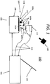
- FIG. 1 illustrates a schematic diagram of a laparoscopic surgical training system 100 configured to be used by one or more clinicians during a simulated surgical procedure to enable performance of a video assisted surgical procedure ("VATS").
- the training system 100 includes an operation simulator 106 in communication with a workstation 102 (via either wired or wireless communication).
- the operation simulator 106 includes a laparoscopic surgical device 112, a laparoscopic imaging device 114 coupled to the surgical device 112, and a housing 110.
- the surgical device 112 and the imaging device 114 may be coupled to the workstation 102 via one or more wired or wireless connections 116.
- the simulator 106 includes a base 108 having the housing 110 disposed thereon.
- the base 108 may include connectivity ports (not explicitly shown) which couple to the connections 116 associated with the surgical device 112, the imaging device 114, and/or the workstation 102.
- the housing 110 supports the surgical device 112 (e.g., an actual surgical device or a control knob, glove, mouse, or the like manipulatable in a similar manner as an actual surgical device) and an imaging device (e.g., a video imaging device configured to image an interior portion of the body of a patient) thereon.
- the surgical device may be a virtual surgical device displayed and manipulatable on a display 104.
- the housing 110 may be formed in the shape of a lung (in a normal or collapsed configuration) to approximate corresponding visual representations of the internal structure of the anatomic feature displayed by the workstation 102, which anatomically approximate living organs.
- the housing 110 may further include a bellow 110c ( FIG. 1 ) or other such suitable components capable of manipulating the housing 110 (e.g., expanding and contracting the exterior surface of the housing 110) which, during operation, cause the housing 110 to move during simulated surgeries.
- the housing 110 may be equipped with force sensors positioned at selected locations within the housing 110 and in communication with the workstation 110. The force sensors transmit data (e.g., forces exerted on the housing 110 by the surgical devices 112).
- the housing 110 further includes ports 118a, 118b, configured to receive the surgical device 112 and the imaging device 114, respectively.
- the ports 118a, 118b enable distal portions of the surgical device 112 and the imaging device 114 to pass through a surface of the housing 110 as would traditionally occur during a typical laparoscopic surgical procedure.
- the ports 118a, 118b may be rigid or semi-rigid, to represent the elasticity of the tissue of a patient.
- An EM field generator 110a may be disposed either in or on the base 108 or beneath the housing 110 so as to generate an EM field for capturing the position of one or more EM sensors in proximity to, or disposed on, the simulator 106.
- the housing 110 may also have one or more EM reference sensors 110b disposed either internal or external to the housing 110 which capture the pose of the housing 110 intermittently or continuously during the simulated surgical procedure.
- a tracking module (not explicitly shown) may receive signals from each of the EM reference sensors 110b, 112a, 114a and, based on the signals, derive the location of each EM reference sensor 110b, 112a, 114a, as well as their position along the device to which they are coupled in six degrees of freedom.
- one or more reference sensors may be disposed in fixed relation to the housing 110. Signals transmitted by the reference sensors to the tracking module may subsequently be used to calculate a patient coordinate frame of reference. Registration is generally performed by identifying select locations in both the stored representation of the anatomical feature associated with the housing 110 and the reference sensors disposed along the housing 110.
- a surgical device EM sensor 112a and an imaging device EM sensor 114a are disposed on the surgical device 112 and the imaging device 114, respectively. Additionally, the surgical device EM sensor 112a and the imaging device EM sensor 114a may include an array of EM sensors (not explicitly shown) disposed along the respective device in a predetermined pattern, so as to provide a more accurate positional measurement of the device. Collectively, the EM components disclosed herein will be referred to as the EM tracking system 109.
- the workstation 102 of the training system 100 may have training software stored as an application 208 in a memory 204 of a computing device 200.
- the workstation 102 may have additional software or instructions stored therein which may be executed while the workstation 102 is in use.
- the application 208 is executed by the processor 202 of the workstation 102, the application may control a display 104 of the workstation 102 to cause the display 104 to output one or more visual and/or audible outputs (e.g., a series of images, a 2D or 3D video stream, or sound to speakers integrated into the display 104 (not explicitly shown)).
- the images to be displayed may include, without limitation, ultrasound images, simulated ultrasound images, CT images, simulated CT images, 3D models, and other predetermined user-interfaces for simulating video assisted thoracic surgeries.
- the visual and/or audible output may be transmitted by the workstation 102 for display on the display 104 in response to input, such as positional data and/or a device state or configuration of either the surgical device 112 and/or the imaging device 114.
- the computing device 200 may represent one or more components (e.g., workstation 102, simulator 106, surgical device 112, simulated imaging device 114, etc.) of the training system 100.
- the computing device 200 may include one or more processors 202, memories 204, display devices or displays 212, input modules, 214, output modules 216, and/or network interfaces 218, or any suitable subset of components thereof.
- the memory 204 includes non-transitory computer readable storage media for storing data and/or software having instructions that may be executed by the one or more processors 202 and which, when executed, control operation of the computing device 200, as well as various other devices in communication with the computing device 200.
- the memory 204 stores data 206 and/or one or more applications 208.
- Such applications 208 may include instructions which are executed on the one or more processors 202 of the computing device 200.
- the application 208 may include instructions which cause a user interface component 210 to control the display 212 such that a user interface 210 is displayed (e.g., a graphical user interface (GUI)).
- GUI graphical user interface
- the workstation 102 may display multiple views such as, for example, a pre-scanned CT image and a simulated CT image on the display 104 of the workstation 102 to assist the clinician during the performance of a simulated surgical procedure.
- the workstation 102 may display navigational aids or visual cues, surgery specific data, information input during pre-operative planning (e.g., directions to a target area of tissue where a growth targeted for treatment is located), and the like.
- the workstation 102 may, similar to the simulated surgical device 112 and the simulated imaging device 114, be in either wired or wireless electrical communication via a connection 116 with the simulator 106. While the surgical device 112 and the imaging device 114 are shown as connected to the workstation 102 via connections 116, the surgical device 112 and the imaging device 114 may be operably coupled to the workstation 102 via connection to the simulator 106.
- the simulator 106 may include one or more applications 208 stored in the memory 204 of the simulator 106 which, when executed on the processor 202 of the simulator 106, control the transmission of data to or from the simulator 106 to the workstation 102.
- the workstation 102 may be integrated, either in whole or in part, into the simulator 106 such that the simulator 106 displays outputs similar to those described above during the simulated surgical procedures.
- the EM tracking system 109 transmits signals to the workstation 102 to indicate the pose of any one of the EM reference sensors 110b, the surgical device EM sensor 112a, and the imaging device EM sensor 114a.
- the workstation 102 in response to receiving signals from the EM tracking system 109, determines a pose for each of the instruments associated with particular EM sensors.
- the EM tracking system 109 may measure or determine the position of any of the included instruments within three-dimensional space and further within proximity of the EM field generator 110a, thereby enabling the EM tracking system 109 to determine the position and orientation of the relevant components to the internal space within the housing 110 during the simulated surgical procedure.
- the workstation 102 displays a series of images or video stream of the surgical site and/or CT images on the display 104, similar to those expected during a typical surgical procedure. For example, based on the determined pose of the housing 110, the surgical device 112, and the imaging device 114 relative to one another, a simulated surgical application 208 may display the position of the distal portion of the surgical device 112 relative to a visual representation of an anatomic feature "AF" ( FIG. 4 ).
- AF anatomic feature
- the application 208 may simulate the various phases of a surgical procedure, including the generation of one or more 3D models during a planning phase or during the simulated surgical procedure, (e.g., identifying target locations and planning a pathway to the target locations as well as surgical interventions such as tissue resection to be performed), registering either stored or generated 3D models (e.g., calibrating the simulator for use with a phantom lung), navigation during a simulated surgical procedure to the target location or locations, performance of the planned surgical intervention at the target location, and the like.
- Models of anatomical features may be generated and stored either in a library of standard models (which include an average representation of an anatomical feature).
- 3D models may be generated by the workstation 102 prior to or during a simulated surgical procedure so as to simulate a surgical procedure on the scanned anatomical feature.
- pre-operative scan data such as CT, magnetic resonance imaging (MRI), X-ray, cone beam computed tomography (CBCT), and/or positron emission tomography (PET) scan data
- 3D models may be generated by the workstation 102 prior to or during a simulated surgical procedure so as to simulate a surgical procedure on the scanned anatomical feature.
- the application 208 may cause the display 104 to illustrate the position of the distal portion or distal tip of the surgical device 112 (e.g., a surgical stapler) relative to the target location 402 ( FIG. 4 ) of an anatomical feature "AF" as would be illustrated during typical percutaneous and/or subcutaneous procedures.
- the workstation 102 may continuously superimpose the position of the surgical device 112 onto a 3D model of the visual representation of the anatomical feature.
- the visual representation of the anatomical feature as well as the position of the surgical device 112 relative to the visual representation of the anatomical feature may be updated in the memory 204 and on the display 104 of the workstation 102 without reflecting any gaps, or other imperfections in the sensor data associated with the visual representation anatomical feature and/or the simulated surgical device 112. Where gaps become too great (e.g., a positional signal is not received for a predetermined period), the application 208 may cause the display 104 of the workstation 102 to display an alert (e.g., "SIGNAL ERROR") to warn the clinician that such a condition exists. Similarly, during a simulated surgical procedure, the application 208 may simulate such conditions (e.g., signal loss, signal errors, etc.) and cause the display to output information indicating such conditions.
- an alert e.g., "SIGNAL ERROR”
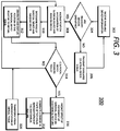
- FIG. 3 illustrates a flowchart depicting an illustrative method 300 for simulating surgical procedures with the training system 100 ( FIG. 1 ).
- the method 300 and associated techniques described herein enable visual simulation of a simulated surgical procedure via the display 104 of the training system 100 ( FIG. 1 ). While the method 300 is described with reference to a particular sequence of steps, it will be apparent to one skilled in the art that certain steps described may be concurrently executed, or executed out of the sequence explicitly disclosed herein, without departing from the scope of the disclosure.
- the simulated procedure may begin with the workstation 102 receiving information from devices (e.g., the simulator 106, the surgical device 112, and the imaging device 114) such as a device ID or other information to identify the devices, as well as the operational state of the devices (e.g., operational, low battery, non-functional, etc.) (block 302).
- devices e.g., the simulator 106, the surgical device 112, and the imaging device 114.
- the workstation 102 determines whether the necessary devices for performing the simulated procedure are connected and operational (block 304).
- the workstation 102 causes the display 104 to output a relevant error message, including a message that certain devices are not connected, or are not operating properly (block 306).
- the method 300 may reiterate this process until it is determined that the training system 100 is ready for use.
- the method 300 continues and the workstation 102 receives information on the position of the surgical device 112 and the imaging device 114 relative to one another (block 308). More particularly, as discussed above, the EM tracking system 109 may capture signals from the EM reference sensors 110b, the surgical device EM sensor 112a, and the imaging device EM sensor 114a based on operation of the EM tracking system 109, thereby indicating the position of the surgical device 112 and imaging device 114 relative to the EM field generator 110a. Based on the position information, the workstation 102 may determine the pose of the surgical device 112 and/or the imaging device 114 (block 310).
- the workstation 102 may receive sensor information from any of the earlier instrument tracking systems mentioned or an imaging device 120. Images captured by the imaging device 120 may capture optical and/or depth image data which, when transmitted to the workstation 102, enable the workstation 102 to determine the position of the surgical device 112 and/or the imaging device 114 relative to one another (see block 308). For example, one or more optical imaging sensors and/or infrared (IR) or depth sensors may be positioned to image the simulator 106 as well as devices engaging the simulator 106 during simulated surgical procedures.
- IR infrared
- the optical imaging sensors may identify the pose of the surgical device 112 and/or the imaging device 114 and, based on the identification, transmit sensor signals to the workstation 102 indicative of the pose of the surgical device 112 and/or the imaging device 114 in three-dimensional space.
- Imaging devices may capture position-identifying information such as, without limitation, markers disposed about the housing 110, the surgical device 112, and/or the imaging device 114. The imaging devices may then transmit the captured image information to the workstation 102 which registers the position of the markers, and their respective device, to determine the pose of each device in three-dimensional space.
- the workstation 102 generates an image or images to be displayed on the display 104 indicative of the positions of the surgical device 112 and the imaging device 114 (block 312).
- the visual representation of the anatomical feature displayed on the display 104 is generated and displayed relative to a visual representation of a pre-selected starting position of the surgical device 112.
- the workstation 102 depicts on the display 104 a movement of the image of the surgical device 112 relative to the image of the anatomical feature based on the determined change in position and/or orientation of the surgical device 112 from the starting position.
- the visual representation of the surgical device 112 and the anatomical feature may be shown from the point of view of the imaging device 114.
- the workstation 102 determines that the surgical device 112 has been moved to a position in which the virtual surgical device 112 engages virtual tissue of the visual representation of the anatomical feature ("YES" at block 314), the workstation 102 predicts an effect the surgical device 112 would have on the anatomical feature (block 316).
- the workstation 102 predicts the effect based on known characteristics of the actual tissue being represented on the display 104 and based on the determined force with which the surgical device 112 is moved, the determined direction the surgical device 112 is moved in, the distance the surgical device 112 is moved. Other factors may be considered in predicting the effect, such as, for example, the speed of the surgical device 112, vibrations 112 of the surgical device 112, or the like.
- the workstation 102 Based on the predicted effect, the workstation 102 generates on the display 104 a change in state of the visual representation of the anatomical feature (block 318). In particular, the virtual tissue is shown being moved in a manner as would have occurred if actual tissue were being engaged by the surgical device 112. Alternatively, if the workstation 102 determines that the anatomical representation was not engaged ("NO" at block 314), process 300 returns to block 308.
- the workstation 102 may overlay elements onto the generated display such as navigational aid 404 ( FIG. 4 ) which assists the clinician in advancing the surgical device 112 to the target site.
- the navigational aid 404 may be displayed until the surgical device 112 is in position, e.g., is at a target region.
- the clinician may input information which is received by the surgical device 112 that is subsequently transmitted to the workstation 102.
- the information received by the surgical device 112 may include selection of a power setting for electrical cutting of tissue during simulated surgeries, selection of a stapler configuration (e.g., switching between grabbing or cutting), or other known setting for a particular surgical device normally adjustable by the clinician during surgical procedures.
- the workstation 102 may generate images illustrating a transformation of the anatomical feature (block 320). For example, as the user actuates the surgical device 112 to effect treatment on the virtual tissue, the workstation 102 may generate images to visually represent the type of actuation of the surgical device 112 (e.g., cutting, ablating, stapling, etc.), and visually represent the effect the actuation has on the virtual tissue. In particular, the virtual tissue may be displayed as being cut, scored, ablated, or stapled depending on the type of actuation of the surgical device 112.
- the type of actuation of the surgical device 112 e.g., cutting, ablating, stapling, etc.
- the virtual tissue may be displayed as being cut, scored, ablated, or stapled depending on the type of actuation of the surgical device 112.
- any determined effect the actions of the surgical device 112 have on the virtual tissue will be illustrated on the display 104 as happening to the anatomy shown on the display 104 (e.g., an image of a lung taken from a CT scan). For example, if the system 100 determines that the surgical device 112 is pulling on a location of the virtual tissue, the image of the lung shown on the display 104 will be illustrated as being pulled at the corresponding location. Once the images are generated, process 300 may be repeated by returning to block 308 and advancing the surgical device 112 to a different target site.
- FIG. 4 illustrates a user interface which may be displayed on the display 104 ( FIG. 1 ) of the workstation 102 during simulated procedures.
- FIG. 4 shows an image generated by the laparoscopic surgical training system 100 and, more specifically, a visual representation of a distal portion of the surgical device 112 (e.g., a surgical stapler) imaged within the thoracic cavity of a patient with the distal portion of the surgical device 112 displayed as if imaged by an imaging device within the thoracic cavity.
- the surgical device 112 e.g., a surgical stapler
- the system 100 may provide tactile feedback to the clinician as the clinician manipulates the surgical device in the simulated space.
- the tactile feedback simulates a predicted resistance to movement of the surgical device as if the surgical device were encountering actual tissue.
- processor 202 may manipulate the virtual tissue by applying a deflection to the virtual model using computational mechanics (finite element simulation) based on instrument tracking.
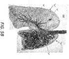
- FIGS. 5A and 5B illustrate an exemplary computer generated model 500 of parts of a respiratory system for display by the workstation 102.
- the model 500 includes a trachea "T” and left and right lungs “LL,” “RL.”
- the model 500 also shows airways “A” (e.g., bronchi) and blood vessels “V” within the lungs “LL,” “RL.”
- airways “A” e.g., bronchi
- V blood vessels
- L shown in the upper left quadrant of the left lung "LL.”
- a user may actuate the surgical device 112 ( FIG. 1 ) to effect treatment on virtual tissue, whereby the workstation 102 may visually represent the effect the actuation of the surgical device 112 has on the model 500.
- any determined effect the actions of the surgical device 112 would have on tissue may be shown on the display 104 by depicting the determined effect on the model 500 shown on the display 104.
- the system 100 determines that the surgical device 112 is pulling on virtual lung tissue, e.g., the right lung "RL,” the appropriate portion of the right lung "RL” of the model 500 shown on the display 104 will be depicted as being pulled.
- the system 100 determines that the user intended to cut a portion of virtual lung tissue, e.g., the left lung "LL,” the portion of the left lung “LL” of the model 500 shown on the display 104 will be depicted as being cut.
- a portion of virtual lung tissue e.g., the left lung "LL”
- proximal refers to the portion of a device or component which is closer to the clinician whereas the term “distal” refers to the portion of the device or component which is further from the clinician.
- distal refers to the portion of the device or component which is further from the clinician.
- front, rear, upper, lower, top, bottom, and other such directional terms are used to aid in the description of the disclosed embodiments and are not intended to limit the disclosure. Well-known functions or constructions are not described in detail so as to avoid obscuring the disclosure unnecessarily.
- programming language and "computer program,” as used herein, each include any language used to specify instructions to a computer, and include (but is not limited to) the following languages and their derivatives: Assembler, Basic, Batch files, BCPL, C, C+, C++, Delphi, Fortran, Java, JavaScript, machine code, operating system command languages, Pascal, Perl, PL1, scripting languages, Visual Basic, metalanguages which themselves specify programs, and all first, second, third, fourth, fifth, or further generation computer languages. Also included are database and other data schemas, and any other meta-languages.
- any of the herein described methods, programs, algorithms or codes may be contained on one or more machine-readable media or memory.
- the term "memory" may include a mechanism that provides (e.g., stores and/or transmits) information in a form readable by a machine such a processor, computer, or a digital processing device.
- a memory may include a read only memory (ROM), random access memory (RAM), magnetic disk storage media, optical storage media, flash memory devices, or any other volatile or non-volatile memory storage device.
- Code or instructions contained thereon can be represented by carrier wave signals, infrared signals, digital signals, and by other like signals. It should be understood that the foregoing description is only illustrative of the disclosure.
Landscapes
- Engineering & Computer Science (AREA)
- Health & Medical Sciences (AREA)
- Physics & Mathematics (AREA)
- General Physics & Mathematics (AREA)
- General Health & Medical Sciences (AREA)
- Medical Informatics (AREA)
- Theoretical Computer Science (AREA)
- Educational Technology (AREA)
- Educational Administration (AREA)
- Business, Economics & Management (AREA)
- Life Sciences & Earth Sciences (AREA)
- Surgery (AREA)
- Mathematical Physics (AREA)
- Mathematical Analysis (AREA)
- Pure & Applied Mathematics (AREA)
- Computational Mathematics (AREA)
- Algebra (AREA)
- Medicinal Chemistry (AREA)
- Chemical & Material Sciences (AREA)
- Mathematical Optimization (AREA)
- Nuclear Medicine, Radiotherapy & Molecular Imaging (AREA)
- Radiology & Medical Imaging (AREA)
- Robotics (AREA)
- Biomedical Technology (AREA)
- Heart & Thoracic Surgery (AREA)
- Molecular Biology (AREA)
- Animal Behavior & Ethology (AREA)
- Public Health (AREA)
- Veterinary Medicine (AREA)
- Pulmonology (AREA)
- Apparatus For Radiation Diagnosis (AREA)
Applications Claiming Priority (2)
| Application Number | Priority Date | Filing Date | Title |
|---|---|---|---|
| US201962830605P | 2019-04-08 | 2019-04-08 | |
| US16/813,866 US20200320900A1 (en) | 2019-04-08 | 2020-03-10 | Systems and methods for simulating surgical procedures |
Publications (1)
| Publication Number | Publication Date |
|---|---|
| EP3723069A1 true EP3723069A1 (fr) | 2020-10-14 |
Family
ID=70224261
Family Applications (1)
| Application Number | Title | Priority Date | Filing Date |
|---|---|---|---|
| EP20168236.6A Withdrawn EP3723069A1 (fr) | 2019-04-08 | 2020-04-06 | Systèmes et procédés pour simuler des procédures chirurgicales |
Country Status (2)
| Country | Link |
|---|---|
| US (1) | US20200320900A1 (fr) |
| EP (1) | EP3723069A1 (fr) |
Families Citing this family (1)
| Publication number | Priority date | Publication date | Assignee | Title |
|---|---|---|---|---|
| US12315160B2 (en) * | 2022-03-16 | 2025-05-27 | Optellum Limited | Method and apparatus to predict feature state change within an image |
Citations (3)
| Publication number | Priority date | Publication date | Assignee | Title |
|---|---|---|---|---|
| WO2006060406A1 (fr) * | 2004-11-30 | 2006-06-08 | The Regents Of The University Of California | Systeme de formation d'un acte medical multimode |
| WO2015095715A1 (fr) * | 2013-12-20 | 2015-06-25 | Intuitive Surgical Operations, Inc. | Système simulateur pour apprentissage de procédure médicale |
| US20180286287A1 (en) * | 2017-03-28 | 2018-10-04 | Covidien Lp | System and methods for training physicians to perform ablation procedures |
Family Cites Families (2)
| Publication number | Priority date | Publication date | Assignee | Title |
|---|---|---|---|---|
| US10939963B2 (en) * | 2016-09-01 | 2021-03-09 | Covidien Lp | Systems and methods for providing proximity awareness to pleural boundaries, vascular structures, and other critical intra-thoracic structures during electromagnetic navigation bronchoscopy |
| US10806532B2 (en) * | 2017-05-24 | 2020-10-20 | KindHeart, Inc. | Surgical simulation system using force sensing and optical tracking and robotic surgery system |
-
2020
- 2020-03-10 US US16/813,866 patent/US20200320900A1/en not_active Abandoned
- 2020-04-06 EP EP20168236.6A patent/EP3723069A1/fr not_active Withdrawn
Patent Citations (3)
| Publication number | Priority date | Publication date | Assignee | Title |
|---|---|---|---|---|
| WO2006060406A1 (fr) * | 2004-11-30 | 2006-06-08 | The Regents Of The University Of California | Systeme de formation d'un acte medical multimode |
| WO2015095715A1 (fr) * | 2013-12-20 | 2015-06-25 | Intuitive Surgical Operations, Inc. | Système simulateur pour apprentissage de procédure médicale |
| US20180286287A1 (en) * | 2017-03-28 | 2018-10-04 | Covidien Lp | System and methods for training physicians to perform ablation procedures |
Also Published As
| Publication number | Publication date |
|---|---|
| US20200320900A1 (en) | 2020-10-08 |
Similar Documents
| Publication | Publication Date | Title |
|---|---|---|
| US11341692B2 (en) | System and method for identifying, marking and navigating to a target using real time two dimensional fluoroscopic data | |
| US20180286287A1 (en) | System and methods for training physicians to perform ablation procedures | |
| JP7221862B2 (ja) | 医療器具の位置計画及び器具誘導のための解剖学的モデル | |
| CN108024838B (zh) | 用于在图像引导手术中使用配准荧光透视图像的系统和方法 | |
| EP3506848B1 (fr) | Planification de chemin pour utilisation avec un système de planification de navigation et de procédure | |
| EP3689223B1 (fr) | Systèmes et procédés pour fournir une sensibilisation de proximité des limites pleurales, structures vasculaires et autres structures intra-thoraciques critiques pendant une bronchoscopie par navigation électromagnétique | |
| CN108451639B (zh) | 用于定位与导航的多数据源集成 | |
| JP5121401B2 (ja) | 埋植物距離測定のシステム | |
| JP5328137B2 (ja) | 用具又は埋植物の表現を表示するユーザ・インタフェイス・システム | |
| JP2021030073A (ja) | 初期位置合わせのための透視ct撮像のシステムおよび方法 | |
| JP7182126B2 (ja) | ロボット手術支援装置、ロボット手術支援方法、及びプログラム | |
| US11625825B2 (en) | Method for displaying tumor location within endoscopic images | |
| US20070016008A1 (en) | Selective gesturing input to a surgical navigation system | |
| JP2020511239A (ja) | ナビゲーション手術における拡張現実ディスプレイのためのシステム及び方法 | |
| ITBO20090470A1 (it) | Apparecchiatura per procedure chirurgiche minimamente invasive | |
| JP2013513462A (ja) | 視覚化によりガイドされるacl位置特定システム | |
| JP2009078144A (ja) | 副鼻洞形成術ナビゲーションのためのフルオロスコープと計算機式断層写真法との位置揃えのシステム及び利用方法 | |
| WO2017086789A1 (fr) | Procédé et système de fourniture d'informations visuelles concernant le siège d'une tumeur sous une surface corporelle d'un corps humain ou animal, programme informatique, et produit-programme informatique | |
| US20240238049A1 (en) | Medical instrument guidance systems and associated methods | |
| EP3398552A1 (fr) | Commande de visionneuse d'images médicales à partir de la caméra du chirurgien | |
| EP3723069A1 (fr) | Systèmes et procédés pour simuler des procédures chirurgicales | |
| JP7182127B2 (ja) | ロボット手術支援装置、情報出力方法、及びプログラム | |
| JP7495216B2 (ja) | 鏡視下手術支援装置、鏡視下手術支援方法、及びプログラム |
Legal Events
| Date | Code | Title | Description |
|---|---|---|---|
| PUAI | Public reference made under article 153(3) epc to a published international application that has entered the european phase |
Free format text: ORIGINAL CODE: 0009012 |
|
| STAA | Information on the status of an ep patent application or granted ep patent |
Free format text: STATUS: THE APPLICATION HAS BEEN PUBLISHED |
|
| AK | Designated contracting states |
Kind code of ref document: A1 Designated state(s): AL AT BE BG CH CY CZ DE DK EE ES FI FR GB GR HR HU IE IS IT LI LT LU LV MC MK MT NL NO PL PT RO RS SE SI SK SM TR |
|
| AX | Request for extension of the european patent |
Extension state: BA ME |
|
| STAA | Information on the status of an ep patent application or granted ep patent |
Free format text: STATUS: REQUEST FOR EXAMINATION WAS MADE |
|
| 17P | Request for examination filed |
Effective date: 20210412 |
|
| RBV | Designated contracting states (corrected) |
Designated state(s): AL AT BE BG CH CY CZ DE DK EE ES FI FR GB GR HR HU IE IS IT LI LT LU LV MC MK MT NL NO PL PT RO RS SE SI SK SM TR |
|
| STAA | Information on the status of an ep patent application or granted ep patent |
Free format text: STATUS: EXAMINATION IS IN PROGRESS |
|
| 17Q | First examination report despatched |
Effective date: 20220321 |
|
| STAA | Information on the status of an ep patent application or granted ep patent |
Free format text: STATUS: THE APPLICATION IS DEEMED TO BE WITHDRAWN |
|
| 18D | Application deemed to be withdrawn |
Effective date: 20220802 |