EP3725876A1 - Vorrichtung - Google Patents
Vorrichtung Download PDFInfo
- Publication number
- EP3725876A1 EP3725876A1 EP18879721.1A EP18879721A EP3725876A1 EP 3725876 A1 EP3725876 A1 EP 3725876A1 EP 18879721 A EP18879721 A EP 18879721A EP 3725876 A1 EP3725876 A1 EP 3725876A1
- Authority
- EP
- European Patent Office
- Prior art keywords
- cell device
- neurons
- nerve cell
- cells
- cell
- Prior art date
- Legal status (The legal status is an assumption and is not a legal conclusion. Google has not performed a legal analysis and makes no representation as to the accuracy of the status listed.)
- Withdrawn
Links
Images



Classifications
-
- C—CHEMISTRY; METALLURGY
- C12—BIOCHEMISTRY; BEER; SPIRITS; WINE; VINEGAR; MICROBIOLOGY; ENZYMOLOGY; MUTATION OR GENETIC ENGINEERING
- C12N—MICROORGANISMS OR ENZYMES; COMPOSITIONS THEREOF; PROPAGATING, PRESERVING, OR MAINTAINING MICROORGANISMS; MUTATION OR GENETIC ENGINEERING; CULTURE MEDIA
- C12N5/00—Undifferentiated human, animal or plant cells, e.g. cell lines; Tissues; Cultivation or maintenance thereof; Culture media therefor
- C12N5/0068—General culture methods using substrates
-
- C—CHEMISTRY; METALLURGY
- C12—BIOCHEMISTRY; BEER; SPIRITS; WINE; VINEGAR; MICROBIOLOGY; ENZYMOLOGY; MUTATION OR GENETIC ENGINEERING
- C12M—APPARATUS FOR ENZYMOLOGY OR MICROBIOLOGY; APPARATUS FOR CULTURING MICROORGANISMS FOR PRODUCING BIOMASS, FOR GROWING CELLS OR FOR OBTAINING FERMENTATION OR METABOLIC PRODUCTS, i.e. BIOREACTORS OR FERMENTERS
- C12M23/00—Constructional details, e.g. recesses, hinges
- C12M23/02—Form or structure of the vessel
- C12M23/12—Well or multiwell plates
-
- C—CHEMISTRY; METALLURGY
- C12—BIOCHEMISTRY; BEER; SPIRITS; WINE; VINEGAR; MICROBIOLOGY; ENZYMOLOGY; MUTATION OR GENETIC ENGINEERING
- C12M—APPARATUS FOR ENZYMOLOGY OR MICROBIOLOGY; APPARATUS FOR CULTURING MICROORGANISMS FOR PRODUCING BIOMASS, FOR GROWING CELLS OR FOR OBTAINING FERMENTATION OR METABOLIC PRODUCTS, i.e. BIOREACTORS OR FERMENTERS
- C12M25/00—Means for supporting, enclosing or fixing the microorganisms, e.g. immunocoatings
- C12M25/14—Scaffolds; Matrices
-
- C—CHEMISTRY; METALLURGY
- C12—BIOCHEMISTRY; BEER; SPIRITS; WINE; VINEGAR; MICROBIOLOGY; ENZYMOLOGY; MUTATION OR GENETIC ENGINEERING
- C12N—MICROORGANISMS OR ENZYMES; COMPOSITIONS THEREOF; PROPAGATING, PRESERVING, OR MAINTAINING MICROORGANISMS; MUTATION OR GENETIC ENGINEERING; CULTURE MEDIA
- C12N11/00—Carrier-bound or immobilised enzymes; Carrier-bound or immobilised microbial cells; Preparation thereof
- C12N11/02—Enzymes or microbial cells immobilised on or in an organic carrier
- C12N11/08—Enzymes or microbial cells immobilised on or in an organic carrier the carrier being a synthetic polymer
- C12N11/082—Enzymes or microbial cells immobilised on or in an organic carrier the carrier being a synthetic polymer obtained by reactions only involving carbon-to-carbon unsaturated bonds
-
- C—CHEMISTRY; METALLURGY
- C12—BIOCHEMISTRY; BEER; SPIRITS; WINE; VINEGAR; MICROBIOLOGY; ENZYMOLOGY; MUTATION OR GENETIC ENGINEERING
- C12N—MICROORGANISMS OR ENZYMES; COMPOSITIONS THEREOF; PROPAGATING, PRESERVING, OR MAINTAINING MICROORGANISMS; MUTATION OR GENETIC ENGINEERING; CULTURE MEDIA
- C12N11/00—Carrier-bound or immobilised enzymes; Carrier-bound or immobilised microbial cells; Preparation thereof
- C12N11/02—Enzymes or microbial cells immobilised on or in an organic carrier
- C12N11/08—Enzymes or microbial cells immobilised on or in an organic carrier the carrier being a synthetic polymer
- C12N11/089—Enzymes or microbial cells immobilised on or in an organic carrier the carrier being a synthetic polymer obtained otherwise than by reactions only involving carbon-to-carbon unsaturated bonds
- C12N11/096—Polyesters; Polyamides
-
- C—CHEMISTRY; METALLURGY
- C12—BIOCHEMISTRY; BEER; SPIRITS; WINE; VINEGAR; MICROBIOLOGY; ENZYMOLOGY; MUTATION OR GENETIC ENGINEERING
- C12N—MICROORGANISMS OR ENZYMES; COMPOSITIONS THEREOF; PROPAGATING, PRESERVING, OR MAINTAINING MICROORGANISMS; MUTATION OR GENETIC ENGINEERING; CULTURE MEDIA
- C12N5/00—Undifferentiated human, animal or plant cells, e.g. cell lines; Tissues; Cultivation or maintenance thereof; Culture media therefor
- C12N5/06—Animal cells or tissues; Human cells or tissues
- C12N5/0602—Vertebrate cells
- C12N5/0618—Cells of the nervous system
- C12N5/0619—Neurons
-
- G—PHYSICS
- G01—MEASURING; TESTING
- G01N—INVESTIGATING OR ANALYSING MATERIALS BY DETERMINING THEIR CHEMICAL OR PHYSICAL PROPERTIES
- G01N33/00—Investigating or analysing materials by specific methods not covered by groups G01N1/00 - G01N31/00
- G01N33/48—Biological material, e.g. blood, urine; Haemocytometers
- G01N33/483—Physical analysis of biological material
- G01N33/4833—Physical analysis of biological material of solid biological material, e.g. tissue samples, cell cultures
- G01N33/4836—Physical analysis of biological material of solid biological material, e.g. tissue samples, cell cultures using multielectrode arrays
-
- G—PHYSICS
- G01—MEASURING; TESTING
- G01N—INVESTIGATING OR ANALYSING MATERIALS BY DETERMINING THEIR CHEMICAL OR PHYSICAL PROPERTIES
- G01N33/00—Investigating or analysing materials by specific methods not covered by groups G01N1/00 - G01N31/00
- G01N33/48—Biological material, e.g. blood, urine; Haemocytometers
- G01N33/50—Chemical analysis of biological material, e.g. blood, urine; Testing involving biospecific ligand binding methods; Immunological testing
- G01N33/5005—Chemical analysis of biological material, e.g. blood, urine; Testing involving biospecific ligand binding methods; Immunological testing involving human or animal cells
- G01N33/5008—Chemical analysis of biological material, e.g. blood, urine; Testing involving biospecific ligand binding methods; Immunological testing involving human or animal cells for testing or evaluating the effect of chemical or biological compounds, e.g. drugs, cosmetics
- G01N33/5044—Chemical analysis of biological material, e.g. blood, urine; Testing involving biospecific ligand binding methods; Immunological testing involving human or animal cells for testing or evaluating the effect of chemical or biological compounds, e.g. drugs, cosmetics involving specific cell types
- G01N33/5058—Neurological cells
-
- G—PHYSICS
- G01—MEASURING; TESTING
- G01N—INVESTIGATING OR ANALYSING MATERIALS BY DETERMINING THEIR CHEMICAL OR PHYSICAL PROPERTIES
- G01N33/00—Investigating or analysing materials by specific methods not covered by groups G01N1/00 - G01N31/00
- G01N33/48—Biological material, e.g. blood, urine; Haemocytometers
- G01N33/50—Chemical analysis of biological material, e.g. blood, urine; Testing involving biospecific ligand binding methods; Immunological testing
- G01N33/5005—Chemical analysis of biological material, e.g. blood, urine; Testing involving biospecific ligand binding methods; Immunological testing involving human or animal cells
- G01N33/5008—Chemical analysis of biological material, e.g. blood, urine; Testing involving biospecific ligand binding methods; Immunological testing involving human or animal cells for testing or evaluating the effect of chemical or biological compounds, e.g. drugs, cosmetics
- G01N33/5044—Chemical analysis of biological material, e.g. blood, urine; Testing involving biospecific ligand binding methods; Immunological testing involving human or animal cells for testing or evaluating the effect of chemical or biological compounds, e.g. drugs, cosmetics involving specific cell types
- G01N33/5073—Stem cells
-
- G—PHYSICS
- G01—MEASURING; TESTING
- G01N—INVESTIGATING OR ANALYSING MATERIALS BY DETERMINING THEIR CHEMICAL OR PHYSICAL PROPERTIES
- G01N33/00—Investigating or analysing materials by specific methods not covered by groups G01N1/00 - G01N31/00
- G01N33/48—Biological material, e.g. blood, urine; Haemocytometers
- G01N33/50—Chemical analysis of biological material, e.g. blood, urine; Testing involving biospecific ligand binding methods; Immunological testing
- G01N33/5005—Chemical analysis of biological material, e.g. blood, urine; Testing involving biospecific ligand binding methods; Immunological testing involving human or animal cells
- G01N33/5008—Chemical analysis of biological material, e.g. blood, urine; Testing involving biospecific ligand binding methods; Immunological testing involving human or animal cells for testing or evaluating the effect of chemical or biological compounds, e.g. drugs, cosmetics
- G01N33/5082—Supracellular entities, e.g. tissue, organisms
-
- C—CHEMISTRY; METALLURGY
- C12—BIOCHEMISTRY; BEER; SPIRITS; WINE; VINEGAR; MICROBIOLOGY; ENZYMOLOGY; MUTATION OR GENETIC ENGINEERING
- C12N—MICROORGANISMS OR ENZYMES; COMPOSITIONS THEREOF; PROPAGATING, PRESERVING, OR MAINTAINING MICROORGANISMS; MUTATION OR GENETIC ENGINEERING; CULTURE MEDIA
- C12N2533/00—Supports or coatings for cell culture, characterised by material
- C12N2533/30—Synthetic polymers
Definitions
- the present invention relates to a novel nerve cell device for measuring neural activity.
- neurotoxicity As well as cardiac toxicity and hepatotoxicity, is one of main causes by which drug development is discontinued. Therefore, it is necessary to evaluate accurately the neurotoxicity from the initial stage of the development of the medicine, but the evaluation of neurotoxicity in non-clinical studies is mainly based on an in vivo evaluation system such as symptom observation by animal experiments and evaluation of the pathological tissue of the brain, and no convenient in vitro evaluation system for assessing neurotoxicity has not been established.
- Neural activity is based on the electric activity expressed by the interaction of a plurality of neurons.
- a method for observing the activity of neurons in which neurons derived from isolated neurons or pluripotent stem cells are dispersed onto and cultured on a multi-electrode array (hereinafter abbreviated as MEA) to reconstruct a neural network, so as to observe the neural activity by measuring extracellular field potential (non-patent documents 1), and the studies evaluating neurotoxicity using the method have been reported (non-patent documents 2 and 3).
- MEA multi-electrode array
- non-patent document 7 There are several reports relating to scaffold materials used as a cell scaffold for the purpose of application to medical materials and the like.
- a cell culture or tissue regeneration is effectively performed by using a cell scaffold material composed of a nano-fiber, for example, polyolefin, polyamide, polyurethane, polyester, fluorine-based polymer, polylactic acid, polyvinyl alcohol, or the like, or the nanofiber obtained by adsorbing a protein component (patent document 1) ; by using a three-dimensional cell culture using a cell scaffold material having a hollow fiber membrane mesh and a nano-fiber layer, nutrients and oxygen are supplied to the cultured cells and metabolite wastes are removed from the cultured cells at high efficiency (patent document 2); by using a cell scaffold material composed of a nano-fiber containing gelatin, collagen or cellulose or the nano-fiber which is crosslinked, a large amount of pluripotent stem cells are supplied and cell death is suppressed (patent document 3) ; by using a cell scaffold material in which polyglycolic acid is used as a support and a nanofiber composed of polyglycolic acid,
- the purpose of the present invention is to overcome the problems of the conventional technique.
- the present inventors have found that a nerve cell device for culturing neurons on a cell scaffold can be provided as a result of intensive studies in order to overcome the problem of the conventional technique, and the present invention has been completed.
- the nerve cell device of the present invention by culturing neurons using a cell scaffold, cell adhesiveness is improved, and stable culture including reduction of detachment of the cells during culture or the like has become possible.
- a period until a neural activity such as spikes, bursts, or the like is to be measured can be shortened without causing the detachment of seeded neurons from an electrode of a multi-electrode array.
- aggregation of neurons is suppressed, high-frequency spikes can be observed.
- intracellular signal imaging has also become possible.
- the nerve cell device is useful for neurotoxicity evaluation and drug screening for neurological diseases.
- the cell scaffold used in a nerve cell device of this invention is composed of a fiber produced from a polymeric material.
- the cell scaffold is preferably a fiber sheet comprising a shape of a sheet in which fibers are integrated.
- the fiber sheet can have an orientated structure, a non-oriented structure, or a mixed structure of orientation and non-orientation.
- the oriented structure is a structure in which fibers constituting a fiber sheet are aligned in one direction, and when the angle in one direction is set to 0°, 80% or more of fibers are present in a range of ⁇ 30°.
- the distance between the fibers is not particularly limited, but is preferably 5 to 50 ⁇ m.
- the non-oriented structure is a structure in which the directions of fibers are randomly arranged.
- the polymeric material constituting the fiber is preferably a biodegradable or non-biodegradable polymeric material, and examples thereof include, but are not limited to, PLGA (polylactic acid polyglycolic acid), polystyrene (PS), polysulfone (PSU), and polytetrafluoroethylene (PTFE).
- the diameter of the cross section of the fiber constituting the fiber sheet is not particularly limited, but is, for example, 0.1 to 8 ⁇ m, preferably 0.5 to 7 ⁇ m, and more preferably 1 to 6 ⁇ m.
- the thickness of the fiber sheet is, for example, 1 to 40 ⁇ m, preferably 5 to 35 ⁇ m, and more preferably 10 to 30 ⁇ m.
- the opening ratio of the fibers constituting the fiber sheet can vary depending on the polymeric material used.
- the opening ratio is not particularly limited, but is, for example, 10 to 50%, preferably 15 to 45%, and more preferably 20 to 40%.
- the opening ratio refers to a ratio of an area in which no fiber exists with respect to a fixed area of a fiber sheet plane.
- the fiber sheet can be produced, for example, by using an electrospinning method from a solution containing a polymeric material.
- a fiber sheet having an oriented structure a fiber sheet can be produced by, for example, using a rotary drum, spraying a solution containing a polymer material to a rotary surface of the drum from a nozzle while rotating the drum, and winding the fiber formed on the rotary drum.
- a fiber sheet having a non-oriented structure a fiber sheet can be produced by spraying a solution containing a polymeric material onto a flat plate.
- the fiber sheet having an oriented structure and a non-oriented structure can be produced by combining the above production methods.
- a polytetrafluoroethylene (PTFE) sheet for example, a commercially available pore chlorofluorocarbon (registered trademark) of Sumitomo Electric Industries, Ltd. can be used.
- an organic solvent in which the polymer material to be used is dissolved at 10 to 30 wt. % at room temperature may be used.
- HFIP 1,1,1,3,3,3-hexafluoro-2-propanol
- DMF N,N-dimethylformamide
- the fiber sheet is fixed or held at periphery with a frame.
- a frame is fixed or held on the fiber sheet, there is no particular limitation as long as it does not affect cell culture.
- a commercially available biocompatible adhesive for example, a silicone one-liquid condensation type RVT rubber (Shin-Etsu Chemical Co., Ltd., catalog number KE -45) can be used to bond the frame to the fiber sheet.
- the material of the frame is not particularly limited as long as it does not affect cell culture.
- a polydimethylsiloxane (PDMS), PS, polycarbonate, stainless steel or the like is exemplified.
- the thickness of the frame is not particularly limited, but is 0.1-4 mm, preferably 0.25-3 mm, and more preferably 0.5-2 mm.
- the shape of the frame can be changed depending on the purpose of use, and its longitudinal length ⁇ its lateral length is preferably 2 mm ⁇ 2 mm to 15 mm ⁇ 15 mm, respectively, and is circular or polygonal.
- the nerve cell device using the fiber sheet or the fiber sheet by fixing or holding the periphery of the fiber sheet with a frame as a cell scaffold can be arranged as it is in at least one of wells included in a cell culture dish or a multi-well plate having a plurality of wells.
- a nerve cell means a nerve unit composed of a cell body, dendrites, and axons, and is also referred to as a neuron.
- the neuron can be classified according to a difference in neurotransmitters produced by the neurons.
- the neurotransmitters include: monoamines such as dopamine, noradrenergic, adrenergic and serotonin; non-peptide neurotransmitters such as acetylcholine, gamma-aminobutyric acid and glutamic acid; peptide nerve transfer substance such as adrenal cortex stimulation hormones (ACTH), alpha-endorphins, beta-endorphins, gamma-endorphins, and vasopressin.
- neurons using dopamine, acetylcholine, and glutamic acid as transfer substances are referred to as dopaminergic neurons, cholinergic neurons, and glutamic acid-activated neurons, respectively.
- primary cultured cells can be used as the neuron.
- Primary cultured cells are important as a system for evaluating the effect of drugs or the like in a living organism because the primary cultured cells have many intrinsic cell functions in a living organism.
- neural cells in central nervous system and peripheral nervous system of a mammalian for example, rodents as mouse or, rat, or primates as monkey or human can be used.
- an animal anatomical method, a tissue collecting method, and a nerve separation/isolation method, the culture medium for culturing neurons, culture conditions and the like can be selected from known methods depending on the type of cells to be cultured and the purpose of cells.
- a commercially available primary cultured neural cell product for example, rat cerebral neurons of Lonza Bioscience and human cerebral neurons of ScienCell Research Laboratories, Inc. can be used.
- neural cells derived from pluripotent stem cells can be used.
- pluripotent stem cells include embryonic stem cells (ES cells) and iPS cells.
- ES cells embryonic stem cells
- iPS cells iPS cells.
- a variety of types of neural cells can be obtained by differentiating pluripotent stem cells using a known neural differentiation induction method.
- neural cells can be obtained by a differentiation inducing method using the low-molecular compound described in the literature ( Honda et al. Biochemical and biophysical Research Communications 469 (2016) 587-592 ).
- a neural cell product derived from a commercially available pluripotent stem cell for example, iCell NEURON of Cellular Dynamics International, Inc. and XCL-1 NEURONS of XCell Science Inc. can also be used. These commercially available neural cells are cultured in the medium attached.
- the neurons can be co-cultured with astrocytes derived from the brain of mammals.
- the neuron can be cultured by adding medium after culturing astrocytes (an astrocyte culture supernatant) to the medium for neurons at a final concentration of 5-30%.
- neural cells suspended in the medium are seeded at a density of 1 ⁇ 10 4 cells/cm 2 to 4 ⁇ 10 6 cells/cm 2 , preferably 5 ⁇ 10 4 cells/cm 2 to 3 ⁇ 10 6 cells/cm 2 , more preferably 1 ⁇ 10 5 cells/cm 2 to 2 ⁇ 10 6 /cm 2 and are cultured for 7 to 14 days while exchanging the medium at 1 to 7 day intervals, therefore the nerve cell device in which the neurons uniformly formed in a three-dimensional structure can be obtained.
- the formation of a three-dimensional structure means to a state in which neurons adhere along fibers constituting the fiber sheet, penetrate into one side or both sides of the fiber sheet and are glowing.
- the cells When neural cells are seeded on a fiber sheet and cultured, the cells are maintained in an uniformly spread state in the culture with little detachment or aggregation of cells which are observed when the neurons are directly seeded on the culture petri dish without using a fiber sheet.
- the nerve cell device of the present invention is brought into contact with a multi-electrode array for measuring the extracellular field potential of the neurons included in the nerve cell device under an environment of 5% CO 2 and 37°C.
- the multi-electrode array has multiple planar microelectrodes arranged on a substrate allowing simultaneous observation of electric signals from a plurality of cells.
- a fluorescent calcium indicator such as a calcium-sensitive dye and a calcium-sensitive fluorescent protein
- a fluorescent potential indicator such as a voltage-sensitive dye and a voltage-sensitive fluorescent protein
- the fluorescent calcium indicator examples include a calcium-sensitive dye such as Quin-2, Fura-2, Fluo-3, -4 and -8, Indo-1, Rhod-2 and - 3, X-Rhod-1, Cal-520, Calbryte (TradeMark), and CaTM. They are usually used as an acetoxymethyl ester for the purpose of imparting permeability to neurons. The acetoxymethyl group is subjected to hydrolysis and is cleaved by intracellular esterase. Further, as the calcium-sensitive fluorescent protein, a gene-code type calcium probe such as Camgaroo-1 and -2, GCaMP-2, -3, -5 and -6, CaMPARI, and Case 12 is known.
- a gene-code type calcium probe such as Camgaroo-1 and -2, GCaMP-2, -3, -5 and -6, CaMPARI, and Case 12 is known.
- the fluorescent voltage indicator examples include voltage-sensitive dyes such as Merocyanine 540, Rh1692, Di-4ANEPPS, JPW114, ANNINE-6, Indocyanine Green, Dipicrylamine, FluoVolt (TradeMark).
- a potential sensitive fluorescent protein a gene code type membrane potential probe such as VSFP-1 and -2, FlaSh, SPARC based on green fluorescent protein (GFP) is known ( Siegel, M. S. and Isacoff, E. Y. Neuron 19, 735-741, 1997 ; SaKai, R., Repunte-Canonigo, V., et al. Eur. J Neurosci. 13, 2314-2318, 2001 ; Ataka, K. and Pieribone, V. A., Biophys J. 82, 509-516, 2002 ; Akemann, W. Mutoh, H., et al. Nature Methods, 7, 643-649, 2010 ).
- PLGA SIGMAP1941
- PSU SIGMA 182443
- HFIP HFIP
- the solution was filled into a syringe (Norm-Ject Syringes 5 ml capacity, Osaka chemical), and the syringe was set to a nano-fiber electric field spinning device NANON-03 (MECC CO., LTD.) and then the fiber sheet was produced on a plate collector under the conditions of, when using PLGA, a needle diameter of 22 G, a voltage of 20 kV and a feeding speed of 1 ml/h, and when using PSU, a needle diameter of 27 G, a voltage of 15 kV and a feeding speed of 1 ml/h.
- PLGA (SIGMA P1941) was dissolved in HFIP(wako 089-04233) at room temperature so as to have a concentration of 20 wt. %, the solution was filled into a syringe (Norm-Ject Syringes 5 ml capacity, Osaka chemical), the syringe was set to a nano-fiber electric field spinning device NANON-03 (MECC CO., LTD.), and then the PLGA fiber sheet was produced on a drum collector under the condition of a needle diameter of 22 G, a voltage of 20 kV, a feeding speed of 1 ml/h, and a rotation speed of 750 rpm.
- syringe Normal-Ject Syringes 5 ml capacity, Osaka chemical
- NANON-03 MECC CO., LTD.
- PS Factor 1
- DMF N,N-dimethylformamide, wako pure chemical
- the dissolved solution is filled into a syringe (Norm-Ject Syringes 5 ml capacity, Osaka chemical)
- the syringe was set to a nano-fiber electric field spinning device NANON-03 (MECC CO., LTD.)
- the PSU fiber sheet was produced on a drum collector under conditions of a needle diameter of 25 G, a voltage of 10 kV, a feeding speed of 1.5 ml/h, and a rotation speed of 2,000 rpm.
- a polycarbonate frame (15 mm ⁇ 15 mm) or a stainless steel circular frame (outer diameter 6 mm, inner diameter 3 mm) was adhered to the prepared fiber sheet by using a silicone one-component condensation type RVT rubber (Shin-Etsu Chemical Co., Ltd., catalog number KE-45) to produce the fiber device.
- a silicone one-component condensation type RVT rubber Shin-Etsu Chemical Co., Ltd., catalog number KE-45
- Neurons differentiated from human iPS cells 253G1 in accordance with the previous report ( Honda et al. Biochemical and Biophysical Research Communications 469 (2016) 587-592 ) were used.
- the undifferentiated hiPS cells were seeded on a culture dish coated with poly-L-lysine (PLL, Sigma-Aldrich) and laminin111 (LM, Sigma-Aldrich), and cultured for 7-10 days in a N2B27 nerve medium (medium in which a DMEM/F-12 medium containing a N2 supplement (GIBCO, 17502048) and a Neurobasal medium containing a B27 minus vitamin A supplement is (GIBCO,12587001) is mixed at 1:1) containing 100 nM LDN193189 (Cellagen Technology) and 1 ⁇ M SB431542 (Sigma-Aldrich).
- PLL poly-L-lysine
- LM laminin111
- N2B27 nerve medium medium in which a DMEM/F-12
- the cell colonies were dissociated into small cell clusters by collagenase (GIBCO) and cultured for further 7 to 10 days in a N2B27 nerve medium containing only the LDN1939189, and the resultant cell colonies were again dissociated into cell clumps with collagenase, followed by the continuous culture in a N2B27 medium.
- GEBCO collagenase
- neurons which become available for the following experiments are obtained.
- the obtained neurons can be cryopreserved, and the cryopreserved neurons can also be used.
- neural aggregates were dissociated into a single cell with Accutase (Innovative Cell Technologies), suspended in a Bambanker (NIPPON genetics Co., Ltd.), frozen at -80°C and stored in a ultra-low temperature freezer at -150°C for long-term storage.
- a commercially available hiPS cell-derived cryopreserved neurons XCL-1 NEURONS (XCell Science Inc., catalog number XN-001-1V) were thawed according to the protocol of XCell science Inc., to obtain a neuron suspension.
- iPS cell-derived neurons suspended in a N2B27 medium were seeded on oriented PLGA, random PLGA, random PSU fiber sheet, and PTFE sheet (Sumitomo Electric Industries, Ltd.) at a density of 1.3 ⁇ 10 6 cells/cm 2 , and cultured in an incubator at 5% CO 2 and 37°C for 6 days.
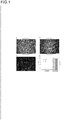
- the results of the obtained cell sheet (nerve cell device) observed by an optical microscope are shown in Fig. 1a (using the oriented PLGA fiber sheet) and Fig. 1b (using the random PSU fiber sheet).
- iPS cell-derived neurons were similarly seeded on a culture dish (Nunc (registered trademark) Cell-Culture Treated Multidishes) or an electrode probe commonly used (Alpha MED Scientific Inc. / MED probe 16 / MED-RG515A), and cultured for 1-2 days.
- the results of the obtained cells observed with an optical microscope are shown in Fig. 2a (on the culture dish) and Fig. 2b (on the electrode).
- Fig. 2a on the culture dish
- Fig. 2b on the electrode
- the cells were fixed with 4% paraformaldehyde (NACALAI TESQUE, INC.) at room temperature for 15 to 30 minutes. After washing with PBS, the cells were treated with 0.2% TritonX-100 (Sigma) at room temperature. After 5 minutes, cells were washed with PBS and blocked with 1% bovine serum albumin for 1 hour at room temperature. Then, the cells were washed with PBS and treated with an anti-MAP2 antibody (Abcam). After 1 hour, the cells were washed with PBS and treated with Alexa Fluor 546-anti-rabbit IgG antibody (Thermo Fisher Scientific K. K.) for one hour.
- NACALAI TESQUE NACALAI TESQUE, INC.
- Fig. 1c After washing with PBS, it was observed by a fluorescence microscope (KEYENCE CORPORATION) ( Fig. 1c ). As shown in Fig. 1c , aligned neurons are recognized by staining with a MAP 2 antibody that can stain neuronal axons.
- Fig. 1d shows the results of MEA measurement detecting the number of spikes and bursts of neurons after 4 weeks from seeding on the random PLGA fiber sheet and the oriented PLGA fiber sheet. It has been found from Fig. 1d that the oriented fiber sheet has an effect in increasing the number of spikes and the number of bursts.
- N2B27 medium containing 20% Astrocyte Conditioned Medium (ACM) (Astrocyte Conditioned Medium-Serum Free (ACM-sf) / 11811-sf, ScienCell Research Laboratries Inc.) (Medium A), Neurobasal (registered trademark) medium containing B27 minus vitamin A supplement (Medium B), and BrainPhys (registered trademark) Neuronal Medium (05792, STEMCELL TECHNOLOGIES) containing 20% ACM (Medium C), 253G1 iPS cell-derived neurons and XCL-1 NEURONS (XCell Science, catalog number XN-001-1V) are seeded at a density of 1.2 ⁇ 10 6 cells/cm 2 on a cell scaffold and performed cultural observation.
- ACM Astrocyte Conditioned Medium
- ACM-sf Astrocyte Conditioned Medium-Serum Free
- 11811-sf ScienCell Research Laboratries Inc.
- Medium A Neurobasal (register
- the oriented PLGA fibers coated with 0.002% poly-L-lysine and 20 ⁇ g/ml laminin in the case of 253G1 iPS cell-derived neurons and the oriented PLGA fibers coated with 0.002% poly-D-lysine and 10 ⁇ g/ml laminin in the case of XCL-1 NEURONS are used.
- the seeded cells are cultured for 1-3 weeks in an incubator at 5% CO 2 , 37°C.
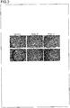
- the results of the obtained cell sheet (nerve cell device) observed by an optical microscope are shown in Figs. 3a, 3b, and 3c .
- neurons were seeded on probes (electrodes) immersed in 10% bovine serum-containing culture medium based on conventional methods, followed by coating with 0.003% poly-L-lysine (Sigma-Aldrich) and 20 ⁇ g/ml laminin (Sigma-Aldrich) in the case of the neuron derived from 253G1 iPS cell and, or by coating with 0.02% poly-D-lysine and 10 ⁇ g/ml laminin in the case of XCL-1 NEURONS, and then cultured for 1-3 weeks.
- the results of the obtained cell sheet observed by an optical microscope are shown in Figs. 3d, 3e and 3f .
- the seeding density of the cells is 1.3 ⁇ 10 6 cells/cm 2 ( Fig. 3d) or 3 ⁇ 10 5 cells/cm 2 ( Fig. 3e and f ).
- Basal 3d As a result in the control group using Medium A, it was observed that neurons detached from the electrodes and the detached neurons formed cell aggregates. Cells formed small colonies in the control groups using Medium B, and detachment of the cells were observed in the periphery of the region where the cells were seeded in the control groups using Medium C.
- the cell sheet was uniformly maintained regardless of the type of the culture media ( Fig. 3 ).
- iPS cell-derived neurons XCL-1 NEURONS (XCell Science, catalog number XN-001-1V) were seeded on the oriented PLGA fiber sheet as a cell scaffold at 3 ⁇ 10 5 cells/cm 2 (low-density seeding condition) or 12 ⁇ 10 5 cells/cm 2 (high-density seeding condition), and cultured with BrainPhys (registered trademark) Neuronal Medium (05792, STEMCELL TECH NOLOGIES) with or without 20% ACM in an incubator at 37°C, 5% CO 2 for 2, 4 and 6 weeks.
- BrainPhys registered trademark
- the fiber was placed on a multi-electrode array (MEA) probe (MED64 system, Alpha MED Scientific Inc.) to make the cells contact with the electrodes of MEA probe to measure neural activity (spike and burst).
- MEA multi-electrode array
- the neurons were seeded directly on the MEA probe at 3 ⁇ 10 5 cells/cm 2 , and cultured in an incubator at 37°C, 5% CO 2 for 2, 4 and 6 weeks, and the neural activity was similarly measured.
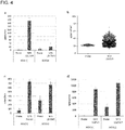
- the results are shown in Fig. 4 .
- a high frequency spikes were detected in the cells seeded at a high density on an oriented PLGA fiber sheet at an early stage after 2 week-culture ( Fig.
- Rat cerebral cortex neurons (Thermo Fisher Scientific K. K., product number A10840), which are primary cultured neurons, were used as neurons, and seeded on the aligned PLGA fiber sheet as a cell scaffold at a cell density of 1.0 ⁇ 10 5 cells/cm 2 , 3.0 ⁇ 10 5 cells/cm 2 , or 9.0 ⁇ 10 5 cells/cm 2 .
- the cells were cultured with Neurobasal (registered trademark) Medium (GIBCO, catalog number 21103-049) containing 1 ⁇ B-27 (registered trademark) Supplement / serum-free (GIBCO, catalog number 17504-044) in an incubator at 37°C, 5% CO 2 for 1, 2, and 4 weeks.
- the obtained cell sheet (nerve cell device) was placed on the MEA probe (MED64 system, Alpha MED Scientific Inc.) to make the cells contact with the electrodes of the MEA probe to measure neural activity (spikes and bursts).
- the MEA probe MED64 system, Alpha MED Scientific Inc.
- spikes and bursts were detected in a nerve cell device cultured 2 and 4 weeks.
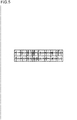
- the results of the MEA measurement detecting extracellular field potential evoked by the cell sheet (nerve cell device) seeded with 9.0 ⁇ 10 5 cells/cm 2 and cultured for 4 weeks are shown in Fig. 5 . In each electrode, spikes and synchronous bursts were observed.
- iPS cell-derived neurons iCell registered trademark
- GlutaNeurons glutamategic neuron, Cellular Dynamics International Inc., product number GNC-301-030-000.5
- iCell glutamatergic neuron medium Cellular Dynamics International Inc.
- FIG. 6a The photographic images of the cell sheet (nerve cell device) seeded at 12 ⁇ 10 5 cells/cm 2 and cultured with iCell glutamatergic neuron medium without ACM for 4 weeks, which were acquired by an optic microscope, are shown in Fig. 6a .
- Fig. 6a neurons are successfully attached to the polystyrene fiber sheet, and an uniform cell sheet without cell detachment is maintained even when culturing is continued for 4 weeks. Similar results were obtained in the case of a medium containing 20% ACM.
- a cell sheet (nerve cell device) seeded with 12 ⁇ 10 5 cells/cm 2 and cultured for 2 weeks in an iCell glutamatergic neuron medium with 20% ACM was placed on an MEA probe (MED64 system, Alpha MED Scientific Inc.), and made the cells contact with the electrodes of the MEA probe to measure neural activity (spikes and synchronous bursts).
- the detection of the spikes and the synchronous bursts were analyzed by a raster plot using a MED64 Burstscope (Alpha MED Scientific Inc.). The results are shown in Fig. 6b . Even in 2 weeks after culturing, the synchronous bursts were confirmed.
- iPS cell-derived neurons (253G1 hiPS cell-derived neuron or XCL-1 NEURONS (XCell Science, catalog number XN-001-1V)) were seeded on the oriented PLGA fiber sheet as a cell scaffold at 13 ⁇ 10 5 cells/cm 2 .
- 253G1 hiPS cell-derived neurons were cultured in N2B27 medium with or without 20% ACM, and the XCL-1 NEURONS are cultured in BrainPhys (registered trademark) Neuronal Medium (05792, STEMCELL TECHNOLOGIES) with or without 20% ACM in an incubator at 5%CO 2 and 37°C for 4 or 6 weeks.
- the fiber sheet was placed on an MEA probe (MED64 system, Alpha MED Scientific Inc.), and made the cells contact with the electrodes of MEA probe to measure neural activity (spikes).
- MEA probe MED64 system, Alpha MED Scientific Inc.
- neurons were similarly seeded directly on a probe (electrodes) based on a conventional method, and cultured in an incubator at 5%CO 2 and 37°C for 4 or 6 weeks.
- the obtained results were compared with those given by a conventional method (direct seeding to a probe).
- a larger number of spikes were detected in 253G1 hiPS cell-derived neurons on the fiber sheet at 4-week culture ( Fig. 7a ).
- the increase in the number of spikes was 6-fold in the neurons cultured on the fiber sheet of the present invention, and 9-fold in the fiber sheets cultured in the medium with 20% ACM, which means that the number of spikes was significantly increased by using the fiber sheet.
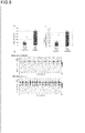
- 4-aminopyridine (4-AP) known to have a function of inhibiting various potassium channels in neurons and maintaining neuronal action potential, was added to the neurons cultured for 4 weeks at a final concentration of 100 ⁇ M, and then the spikes were measured.
- the number of spikes was increased both in the control group and in the fiber sheet group by the addition of 4-aminopyridine, and appropriate drug response was also confirmed ( Fig. 7a ).
- the increase in the number of spikes was 167% in the fiber sheet group of the present invention, compared to 156% in the control group cultured on the probe, and 194% in the fiber sheet group cultured in the medium with 20% ACM.
- XCL-1 NEURONS seeded on the fiber sheet at a density of 12 ⁇ 10 5 cells/cm 2 and cultured for 6 weeks in the medium with ACM were treated with 4-AP having a final concentration of 100 ⁇ M, which resulted in a 2.6-fold increase in the number of bursts ( Fig. 7b ).
- iPS cell-derived neurons XCL-1 NEURONS (XCell Science, catalog number XN-001-1V)) were seeded on the oriented PLGA fiber sheet as a cell scaffold at 24 ⁇ 10 5 cells/cm 2 , twice as high as in the case of Example 7.
- the cells were cultured in BrainPhys (registered trademark) Neuronal Medium (05792, STEMCELL TECHNOLOGIES) containing 20% ACM in an incubator at 5%CO 2 and 37°C for 2, 4, and 6 weeks.
- the obtained cell sheet (nerve cell device) was placed on an MEA probe (MED64 system, Alpha MED Scientific Inc.) and made the cells contact with the electrodes of MEA probe to measure neural activity (spikes and bursts).
- MEA probe MED64 system, Alpha MED Scientific Inc.
- 4-aminopyridine (4-AP), known to maintain a neural action potential was added at a final concentration of 100 ⁇ M to examine the drug response of neurons.
- MED 64 Burstscope (Alpha MED Scientific Inc.) was used for analyzing neural activity.
- Rat cerebral cortex neurons (Thermo Fisher Scientific K. K., product number A10840), which are primary cultured neurons, were seeded on the oriented polystyrene fiber sheet as a cell scaffold at a cell density of 1.0 ⁇ 10 5 cells/cm 2 , 3.0 ⁇ 10 5 cells/cm 2 , or 9.0 ⁇ 10 5 cells/cm 2 .
- the cells were cultured in Neurobasal (registered trademark) Medium (GIBCO, catalog number 21103-049) containing B-27 (registered trademark) Supplement ( ⁇ 50) / serum-free (GIBCO, catalog number 17504-044) in an incubator at 5% CO 2 and 37°C for 4 weeks.
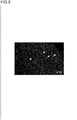
- the obtained cell sheet (nerve cell device) was transferred into medium containing 5 ⁇ M Fluo-8-AM (ATT Bioquest, catalog number 21081), a calcium indicator reagent, and cultured for 1 hour in an incubator at 5% CO 2 and 37°C.
- the Fluo-8-AM dye-incorporated cell sheet was set to a heat insulating plate attached to a fluorescence microscope (Olympus IX73), and fluorescence of Fluo-8-AM were observed.
- the change in fluorescence intensity due to a change in the calcium ion concentration is observed as a synchronized blinking (a blinking cycle of about 1.83 seconds) among the individual neurons.
- Fig. 9 shows the observed image by a fluorescence microscope (magnification ⁇ 10). It is shown that a change in intracellular calcium concentration of neurons can be visualized (imaged) in the nerve cell device of the present invention.
Landscapes
- Health & Medical Sciences (AREA)
- Engineering & Computer Science (AREA)
- Life Sciences & Earth Sciences (AREA)
- Biomedical Technology (AREA)
- Chemical & Material Sciences (AREA)
- Bioinformatics & Cheminformatics (AREA)
- Wood Science & Technology (AREA)
- Organic Chemistry (AREA)
- Zoology (AREA)
- Genetics & Genomics (AREA)
- Biotechnology (AREA)
- Biochemistry (AREA)
- General Health & Medical Sciences (AREA)
- Microbiology (AREA)
- Immunology (AREA)
- General Engineering & Computer Science (AREA)
- Cell Biology (AREA)
- Urology & Nephrology (AREA)
- Hematology (AREA)
- Molecular Biology (AREA)
- Physics & Mathematics (AREA)
- Neurology (AREA)
- Medicinal Chemistry (AREA)
- Analytical Chemistry (AREA)
- General Physics & Mathematics (AREA)
- Food Science & Technology (AREA)
- Pathology (AREA)
- Neurosurgery (AREA)
- Toxicology (AREA)
- Tropical Medicine & Parasitology (AREA)
- Sustainable Development (AREA)
- Biophysics (AREA)
- Clinical Laboratory Science (AREA)
- Developmental Biology & Embryology (AREA)
- Optics & Photonics (AREA)
- Micro-Organisms Or Cultivation Processes Thereof (AREA)
- Apparatus Associated With Microorganisms And Enzymes (AREA)
- Measuring Or Testing Involving Enzymes Or Micro-Organisms (AREA)
Applications Claiming Priority (3)
| Application Number | Priority Date | Filing Date | Title |
|---|---|---|---|
| JP2017220526 | 2017-11-16 | ||
| JP2018116818 | 2018-06-20 | ||
| PCT/JP2018/042207 WO2019098256A1 (ja) | 2017-11-16 | 2018-11-15 | デバイス |
Publications (2)
| Publication Number | Publication Date |
|---|---|
| EP3725876A1 true EP3725876A1 (de) | 2020-10-21 |
| EP3725876A4 EP3725876A4 (de) | 2021-08-04 |
Family
ID=66539553
Family Applications (1)
| Application Number | Title | Priority Date | Filing Date |
|---|---|---|---|
| EP18879721.1A Withdrawn EP3725876A4 (de) | 2017-11-16 | 2018-11-15 | Vorrichtung |
Country Status (4)
| Country | Link |
|---|---|
| US (1) | US11543404B2 (de) |
| EP (1) | EP3725876A4 (de) |
| JP (1) | JP7662169B2 (de) |
| WO (1) | WO2019098256A1 (de) |
Cited By (1)
| Publication number | Priority date | Publication date | Assignee | Title |
|---|---|---|---|---|
| EP3822358A4 (de) * | 2018-07-12 | 2022-04-06 | Tohoku Institute of Technology | Messverfahren |
Families Citing this family (4)
| Publication number | Priority date | Publication date | Assignee | Title |
|---|---|---|---|---|
| JPWO2020013269A1 (ja) * | 2018-07-12 | 2021-10-14 | 学校法人東北工業大学 | 神経細胞の機能的成熟化法 |
| JPWO2021045095A1 (de) * | 2019-09-06 | 2021-03-11 | ||
| US20210301240A1 (en) * | 2020-03-30 | 2021-09-30 | Ricoh Company, Ltd. | Cell-containing container and method for producing same |
| JP2021185780A (ja) * | 2020-05-27 | 2021-12-13 | 株式会社リコー | 細胞含有容器の製造方法 |
Family Cites Families (10)
| Publication number | Priority date | Publication date | Assignee | Title |
|---|---|---|---|---|
| JP2006254722A (ja) | 2005-03-15 | 2006-09-28 | Toray Ind Inc | 細胞足場材料 |
| JP5249785B2 (ja) * | 2006-01-27 | 2013-07-31 | ザ リージェンツ オブ ザ ユニバーシティ オブ カリフォルニア | 生体模倣足場材 |
| TW200848054A (en) * | 2007-02-28 | 2008-12-16 | Eisai R&D Man Co Ltd | Two cyclic oxomorpholine derivatives |
| US20110125170A1 (en) * | 2008-01-25 | 2011-05-26 | The Johns Hopkins University | Hydrogel-grafted degradable nerve guides |
| JP5691240B2 (ja) | 2010-05-21 | 2015-04-01 | 三菱レイヨン株式会社 | 細胞培養用モジュール |
| JP6024047B2 (ja) | 2012-06-04 | 2016-11-09 | 国立大学法人京都大学 | 多能性幹細胞の培養方法及びそのための基材 |
| US10444227B2 (en) | 2014-08-19 | 2019-10-15 | FUJIFILM Cellular Dynamics, Inc. | Neural networks formed from cells derived from pluripotent stem cells |
| WO2016068266A1 (ja) | 2014-10-31 | 2016-05-06 | 国立大学法人京都大学 | 生分解性ポリマーを用いた3次元培養方法、及び細胞移植を可能にする培養基材 |
| JP6602047B2 (ja) | 2015-05-20 | 2019-11-06 | 株式会社幹細胞&デバイス研究所 | 細胞培養担体及びこれを備える細胞シート |
| JP6530638B2 (ja) | 2015-05-20 | 2019-06-12 | 株式会社幹細胞&デバイス研究所 | 細胞培養担体及びこれを備える細胞シート |
-
2018
- 2018-11-15 EP EP18879721.1A patent/EP3725876A4/de not_active Withdrawn
- 2018-11-15 JP JP2019554268A patent/JP7662169B2/ja active Active
- 2018-11-15 WO PCT/JP2018/042207 patent/WO2019098256A1/ja not_active Ceased
- 2018-11-15 US US16/763,944 patent/US11543404B2/en active Active
Cited By (1)
| Publication number | Priority date | Publication date | Assignee | Title |
|---|---|---|---|---|
| EP3822358A4 (de) * | 2018-07-12 | 2022-04-06 | Tohoku Institute of Technology | Messverfahren |
Also Published As
| Publication number | Publication date |
|---|---|
| EP3725876A4 (de) | 2021-08-04 |
| WO2019098256A1 (ja) | 2019-05-23 |
| JPWO2019098256A1 (ja) | 2020-12-03 |
| JP7662169B2 (ja) | 2025-04-15 |
| US11543404B2 (en) | 2023-01-03 |
| US20210025870A1 (en) | 2021-01-28 |
Similar Documents
| Publication | Publication Date | Title |
|---|---|---|
| US20210130757A1 (en) | Tissue fragment | |
| US11543404B2 (en) | Nerve cell device and method for evaluating neural activity | |
| Yan et al. | 3D bioprinting of human neural tissues with functional connectivity | |
| Jakobsson et al. | Three-dimensional functional human neuronal networks in uncompressed low-density electrospun fiber scaffolds | |
| Cantley et al. | Functional and sustainable 3D human neural network models from pluripotent stem cells | |
| EP3402874A1 (de) | Unterstützte in-vitro-gezüchtete gewebekultur und anzuchtverfahren | |
| US11407977B2 (en) | Tissue scaffolds for electrically excitable cells | |
| CA2790873A1 (en) | Methods of generating engineered innervated tissue and uses thereof | |
| JP7159055B2 (ja) | ヒト神経細胞及びグリア細胞の機能的ネットワークを形成する方法 | |
| WO2020013269A1 (ja) | 神経細胞の機能的成熟化法 | |
| Iwoń et al. | Maturation of human cardiomyocytes derived from induced pluripotent stem cells (iPSC-CMs) on polycaprolactone and polyurethane nanofibrous mats | |
| JP2022135969A (ja) | 神経細胞デバイス、その製造方法、及び神経活動評価方法 | |
| WO2019078278A1 (ja) | 心筋細胞に分化させるための多能性幹細胞の製造方法 | |
| US20210238528A1 (en) | Cell scaffold material | |
| Phillips et al. | Developing HiPSC derived serum free embryoid bodies for the interrogation of 3-D stem cell cultures using physiologically relevant assays | |
| US20190365951A1 (en) | Human cardiac tissue construct, related methods and uses | |
| JP7374422B2 (ja) | 計測方法 | |
| JP2021514203A (ja) | 生体適合性三次元ネットワークおよび細胞支持体としてのその使用 | |
| JP7721879B2 (ja) | 神経細胞ネットワーク構造体の形成方法、及びインビトロで神経細胞を評価する方法 | |
| Muzzi | Development of engineered human-derived brain-on-a-chip models for electrophysiological recording | |
| EP4159844B1 (de) | Zellhaltige struktur | |
| WO2021166984A1 (ja) | ミエリン化神経細胞を含む神経細胞デバイスの製造方法 | |
| Kundu et al. | Enhancement of Neuroglial Extracellular Matrix Formation and Physiological Activity of Dopaminergic Neural Cocultures by Macromolecular Crowding | |
| WO2023199952A1 (ja) | 薬剤の有効性の評価方法、神経細胞デバイス及びその製造方法、疾患モデル及びその作製方法、並びに成熟したプルキンエ細胞の作製方法 | |
| TW202115237A (zh) | 肝細胞培養體之製造方法、含細胞之多孔膜及肝細胞或肝前驅細胞的功能提高或功能維持方法 |
Legal Events
| Date | Code | Title | Description |
|---|---|---|---|
| STAA | Information on the status of an ep patent application or granted ep patent |
Free format text: STATUS: THE INTERNATIONAL PUBLICATION HAS BEEN MADE |
|
| PUAI | Public reference made under article 153(3) epc to a published international application that has entered the european phase |
Free format text: ORIGINAL CODE: 0009012 |
|
| STAA | Information on the status of an ep patent application or granted ep patent |
Free format text: STATUS: REQUEST FOR EXAMINATION WAS MADE |
|
| 17P | Request for examination filed |
Effective date: 20200603 |
|
| AK | Designated contracting states |
Kind code of ref document: A1 Designated state(s): AL AT BE BG CH CY CZ DE DK EE ES FI FR GB GR HR HU IE IS IT LI LT LU LV MC MK MT NL NO PL PT RO RS SE SI SK SM TR |
|
| AX | Request for extension of the european patent |
Extension state: BA ME |
|
| DAV | Request for validation of the european patent (deleted) | ||
| DAX | Request for extension of the european patent (deleted) | ||
| A4 | Supplementary search report drawn up and despatched |
Effective date: 20210705 |
|
| RIC1 | Information provided on ipc code assigned before grant |
Ipc: C12N 5/0793 20100101AFI20210629BHEP Ipc: C12M 1/22 20060101ALI20210629BHEP Ipc: C12M 3/00 20060101ALI20210629BHEP Ipc: C12N 11/08 20200101ALI20210629BHEP Ipc: C12M 1/12 20060101ALI20210629BHEP Ipc: G01N 33/50 20060101ALI20210629BHEP Ipc: C12N 5/00 20060101ALI20210629BHEP |
|
| STAA | Information on the status of an ep patent application or granted ep patent |
Free format text: STATUS: EXAMINATION IS IN PROGRESS |
|
| 17Q | First examination report despatched |
Effective date: 20220617 |
|
| STAA | Information on the status of an ep patent application or granted ep patent |
Free format text: STATUS: THE APPLICATION IS DEEMED TO BE WITHDRAWN |
|
| 18D | Application deemed to be withdrawn |
Effective date: 20240423 |