EP3733704A1 - Anticorps anti-pd-l1 et ses applications - Google Patents
Anticorps anti-pd-l1 et ses applications Download PDFInfo
- Publication number
- EP3733704A1 EP3733704A1 EP18894248.6A EP18894248A EP3733704A1 EP 3733704 A1 EP3733704 A1 EP 3733704A1 EP 18894248 A EP18894248 A EP 18894248A EP 3733704 A1 EP3733704 A1 EP 3733704A1
- Authority
- EP
- European Patent Office
- Prior art keywords
- antibody
- amino acid
- seq
- acid sequence
- chain variable
- Prior art date
- Legal status (The legal status is an assumption and is not a legal conclusion. Google has not performed a legal analysis and makes no representation as to the accuracy of the status listed.)
- Pending
Links
Images


Classifications
-
- C—CHEMISTRY; METALLURGY
- C07—ORGANIC CHEMISTRY
- C07K—PEPTIDES
- C07K16/00—Immunoglobulins [IG], e.g. monoclonal or polyclonal antibodies
- C07K16/18—Immunoglobulins [IG], e.g. monoclonal or polyclonal antibodies against material from animals or humans
- C07K16/28—Immunoglobulins [IG], e.g. monoclonal or polyclonal antibodies against material from animals or humans against receptors, cell surface antigens or cell surface determinants
-
- A—HUMAN NECESSITIES
- A61—MEDICAL OR VETERINARY SCIENCE; HYGIENE
- A61K—PREPARATIONS FOR MEDICAL, DENTAL OR TOILETRY PURPOSES
- A61K39/00—Medicinal preparations containing antigens or antibodies
- A61K39/395—Antibodies; Immunoglobulins; Immune serum, e.g. antilymphocytic serum
- A61K39/39533—Antibodies; Immunoglobulins; Immune serum, e.g. antilymphocytic serum against materials from animals
- A61K39/39558—Antibodies; Immunoglobulins; Immune serum, e.g. antilymphocytic serum against materials from animals against tumor tissues, cells, antigens
-
- C—CHEMISTRY; METALLURGY
- C07—ORGANIC CHEMISTRY
- C07K—PEPTIDES
- C07K16/00—Immunoglobulins [IG], e.g. monoclonal or polyclonal antibodies
- C07K16/18—Immunoglobulins [IG], e.g. monoclonal or polyclonal antibodies against material from animals or humans
- C07K16/28—Immunoglobulins [IG], e.g. monoclonal or polyclonal antibodies against material from animals or humans against receptors, cell surface antigens or cell surface determinants
- C07K16/2803—Immunoglobulins [IG], e.g. monoclonal or polyclonal antibodies against material from animals or humans against receptors, cell surface antigens or cell surface determinants against the immunoglobulin superfamily
- C07K16/2827—Immunoglobulins [IG], e.g. monoclonal or polyclonal antibodies against material from animals or humans against receptors, cell surface antigens or cell surface determinants against the immunoglobulin superfamily against B7 molecules, e.g. CD80, CD86
-
- A—HUMAN NECESSITIES
- A61—MEDICAL OR VETERINARY SCIENCE; HYGIENE
- A61K—PREPARATIONS FOR MEDICAL, DENTAL OR TOILETRY PURPOSES
- A61K31/00—Medicinal preparations containing organic active ingredients
- A61K31/70—Carbohydrates; Sugars; Derivatives thereof
- A61K31/7088—Compounds having three or more nucleosides or nucleotides
-
- A—HUMAN NECESSITIES
- A61—MEDICAL OR VETERINARY SCIENCE; HYGIENE
- A61K—PREPARATIONS FOR MEDICAL, DENTAL OR TOILETRY PURPOSES
- A61K39/00—Medicinal preparations containing antigens or antibodies
- A61K39/395—Antibodies; Immunoglobulins; Immune serum, e.g. antilymphocytic serum
-
- A—HUMAN NECESSITIES
- A61—MEDICAL OR VETERINARY SCIENCE; HYGIENE
- A61K—PREPARATIONS FOR MEDICAL, DENTAL OR TOILETRY PURPOSES
- A61K45/00—Medicinal preparations containing active ingredients not provided for in groups A61K31/00 - A61K41/00
- A61K45/06—Mixtures of active ingredients without chemical characterisation, e.g. antiphlogistics and cardiaca
-
- A—HUMAN NECESSITIES
- A61—MEDICAL OR VETERINARY SCIENCE; HYGIENE
- A61P—SPECIFIC THERAPEUTIC ACTIVITY OF CHEMICAL COMPOUNDS OR MEDICINAL PREPARATIONS
- A61P35/00—Antineoplastic agents
-
- A—HUMAN NECESSITIES
- A61—MEDICAL OR VETERINARY SCIENCE; HYGIENE
- A61P—SPECIFIC THERAPEUTIC ACTIVITY OF CHEMICAL COMPOUNDS OR MEDICINAL PREPARATIONS
- A61P35/00—Antineoplastic agents
- A61P35/02—Antineoplastic agents specific for leukemia
-
- C—CHEMISTRY; METALLURGY
- C07—ORGANIC CHEMISTRY
- C07K—PEPTIDES
- C07K16/00—Immunoglobulins [IG], e.g. monoclonal or polyclonal antibodies
- C07K16/18—Immunoglobulins [IG], e.g. monoclonal or polyclonal antibodies against material from animals or humans
- C07K16/28—Immunoglobulins [IG], e.g. monoclonal or polyclonal antibodies against material from animals or humans against receptors, cell surface antigens or cell surface determinants
- C07K16/2803—Immunoglobulins [IG], e.g. monoclonal or polyclonal antibodies against material from animals or humans against receptors, cell surface antigens or cell surface determinants against the immunoglobulin superfamily
-
- G—PHYSICS
- G01—MEASURING; TESTING
- G01N—INVESTIGATING OR ANALYSING MATERIALS BY DETERMINING THEIR CHEMICAL OR PHYSICAL PROPERTIES
- G01N33/00—Investigating or analysing materials by specific methods not covered by groups G01N1/00 - G01N31/00
- G01N33/48—Biological material, e.g. blood, urine; Haemocytometers
- G01N33/50—Chemical analysis of biological material, e.g. blood, urine; Testing involving biospecific ligand binding methods; Immunological testing
- G01N33/68—Chemical analysis of biological material, e.g. blood, urine; Testing involving biospecific ligand binding methods; Immunological testing involving proteins, peptides or amino acids
-
- A—HUMAN NECESSITIES
- A61—MEDICAL OR VETERINARY SCIENCE; HYGIENE
- A61K—PREPARATIONS FOR MEDICAL, DENTAL OR TOILETRY PURPOSES
- A61K39/00—Medicinal preparations containing antigens or antibodies
- A61K2039/505—Medicinal preparations containing antigens or antibodies comprising antibodies
-
- A—HUMAN NECESSITIES
- A61—MEDICAL OR VETERINARY SCIENCE; HYGIENE
- A61K—PREPARATIONS FOR MEDICAL, DENTAL OR TOILETRY PURPOSES
- A61K39/00—Medicinal preparations containing antigens or antibodies
- A61K2039/505—Medicinal preparations containing antigens or antibodies comprising antibodies
- A61K2039/507—Comprising a combination of two or more separate antibodies
-
- C—CHEMISTRY; METALLURGY
- C07—ORGANIC CHEMISTRY
- C07K—PEPTIDES
- C07K2317/00—Immunoglobulins specific features
- C07K2317/20—Immunoglobulins specific features characterized by taxonomic origin
- C07K2317/24—Immunoglobulins specific features characterized by taxonomic origin containing regions, domains or residues from different species, e.g. chimeric, humanized or veneered
-
- C—CHEMISTRY; METALLURGY
- C07—ORGANIC CHEMISTRY
- C07K—PEPTIDES
- C07K2317/00—Immunoglobulins specific features
- C07K2317/30—Immunoglobulins specific features characterized by aspects of specificity or valency
- C07K2317/31—Immunoglobulins specific features characterized by aspects of specificity or valency multispecific
-
- C—CHEMISTRY; METALLURGY
- C07—ORGANIC CHEMISTRY
- C07K—PEPTIDES
- C07K2317/00—Immunoglobulins specific features
- C07K2317/50—Immunoglobulins specific features characterized by immunoglobulin fragments
- C07K2317/56—Immunoglobulins specific features characterized by immunoglobulin fragments variable (Fv) region, i.e. VH and/or VL
- C07K2317/565—Complementarity determining region [CDR]
-
- C—CHEMISTRY; METALLURGY
- C07—ORGANIC CHEMISTRY
- C07K—PEPTIDES
- C07K2317/00—Immunoglobulins specific features
- C07K2317/70—Immunoglobulins specific features characterized by effect upon binding to a cell or to an antigen
- C07K2317/76—Antagonist effect on antigen, e.g. neutralization or inhibition of binding
-
- C—CHEMISTRY; METALLURGY
- C07—ORGANIC CHEMISTRY
- C07K—PEPTIDES
- C07K2317/00—Immunoglobulins specific features
- C07K2317/90—Immunoglobulins specific features characterized by (pharmaco)kinetic aspects or by stability of the immunoglobulin
- C07K2317/92—Affinity (KD), association rate (Ka), dissociation rate (Kd) or EC50 value
Definitions
- the invention relates to a novel antibody and an antibody fragment thereof that specifically bind to PD-L1 and a composition comprising the antibody or the antibody fragment.
- the invention relates to a nucleic acid encoding the antibody or the antibody fragment thereof, a host cell comprising the nucleic acid, and related uses.
- the invention relates to therapeutic and diagnostic uses of these antibodies and antibody fragments.
- the invention relates to combination therapies of these antibodies and antibody fragments with other therapies, such as therapeutic modalities or agents.
- Programmed death-ligand 1 is a protein involved in suppressing immune system responses during chronic infections, pregnancy, tissue allografts, autoimmune diseases, and cancer. PD-L1 regulates immune responses by binding to an inhibitory receptor called programmed death 1 (PD-1) which is expressed on the surface of T cells, B cells, and monocytes. PD-L1 also negatively regulates T cell functions through interaction with another receptor B7.1 (also known as B7-1 or CD80). The formation of PD-L1/PD-1 and PD-L1/B7.1 complexes negatively regulate T cell receptor signaling, leading to subsequent down regulation of T cell activation and inhibition of anti-tumor immune activity.
- PD-1 an inhibitory receptor
- B7.1 also known as B7-1 or CD80
- PD-L1 is overexpressed in many cancers, including a variety of solid tumors, such as bladder tumors, breast tumors, colon tumors, lung tumors, melanomas, ovarian tumors, salivary tumors, stomach tumors, and thyroid tumors.
- solid tumors such as bladder tumors, breast tumors, colon tumors, lung tumors, melanomas, ovarian tumors, salivary tumors, stomach tumors, and thyroid tumors.
- PD-L1 overexpression in tumor cells could promote tumor invasion and is often associated with poor prognosis.
- a nucleic acid encoding the antibody or the antibody fragment thereof, and an expression vector, a host cell, and a method for producing the antibody molecule are also provided.
- an immunoconjugate comprising an anti-PD-Ll antibody molecule, a multispecific or bispecific antibody molecule, and a pharmaceutical composition.
- the anti-PD-Ll antibody molecule disclosed herein can be used alone or in combination with other therapies, such as therapeutic agents or modalities, to treat, prevent and/or diagnose tumor diseases and infectious diseases.
- a composition and a method for detecting PD-L1, and a method for preventing or treating a variety of diseases including tumors and/or infectious diseases using the anti-PD-Ll antibody molecule are also provided.
- the antibody or the fragment thereof of the invention (specifically) binds to PD-L1. In some embodiments, the antibody or the fragment thereof of the invention (specifically) binds to human PD-L1.
- the anti-PD-Ll antibody or the fragment thereof of the invention binds to PD-L1 (e.g., human PD-L1) with high affinity, for example, binds to PD-L1 with an equilibrium dissociation constants (K D ) of less than about 50 nM, preferably less than or equal to about 20 nM, more preferably less than or equal to about 15 nM, more preferably less than or equal to about 10 nM, 9 nM, 8 nM, 7 nM, 6 nM, 5 nM, 4 nM, 3 nM, or 2 nM, and most preferably, less than or equal to about 1.5 nM, 1.4 nM, 1.3 nM, 1.2 nM, 1.1 nM, 1 nM, 0.9 nM, or 0.8 nM.
- K D equilibrium dissociation constants
- the anti-PD-Ll antibody of the invention binds to PD-L1 with a K D of 0.1-10 nM, preferably 0.5-10 nM, more preferably 0.6-10 nM, 0.7-8 nM, and 0.7-5 nM, and most preferably 0.5-1.5 nM, 0.7-1.5 nM, and 0.7-1 nM.
- the PD-L1 is a human PD-L1.
- the antibody binding affinity is determined using biological optical interferometry (e.g., Fortebio affinity measurement).
- the antibody or the fragment thereof of the invention binds to cells expressing human PD-L1, for example, with an EC 50 of less than or equal to about 4 nM, 3.5 nM, 3 nM, 2.9 nM, 2.8 nM, 2.7 nM, 2.6 nM, 2.5 nM, 2.4 nM, 2.3 nM, 2.2 nM, 2.1 nM, 2 nM, 1.9 nM, 1.8 nM, 1.7 nM, or 1.6 nM.
- the binding is determined using flow cytometry (e.g., FACS).
- the cell expressing human PD-L1 is a CHO cell expressing human PD-L1.
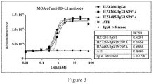
- the antibody or the fragment thereof of the invention blocks relevant activities of PD-L1, for example, with an EC 50 of less than or equal to about 10 nM, 5 nM, 4 nM, 3 nM, 2 nM, 1 nM, 0.9 nM, 0.8 nM, or 0.7 nM, and preferably 0.1-1 nM, 0.5-1 nM, 0.6-1 nM, 0.6 nM, 0.7 nM, 0.8 nM, 0.9 nM, or 1 nM.
- the relevant activity of PD-L1 is the binding of PD-L1 to PD-1.
- the antibody or the fragment thereof of the invention inhibits the binding of PD-L1 to PD-1 with an EC 50 of less than or equal to about 10 nM, 5 nM, 4 nM, 3 nM, 2 nM, 1 nM, 0.9 nM, 0.8 nM, or 0.7 nM, and preferably 0.1-1 nM, 0.5-1 nM, 0.6-1 nM, 0.6 nM, 0.7 nM, 0.8 nM, 0.9 nM, or 1 nM in an MOA assay.
- the cell is a CHO cell.
- the antibody or the fragment thereof of the invention increases T cell functions. In some embodiments, the antibody or the fragment thereof of the invention increases T cell proliferation. In some embodiments, the antibody or the fragment thereof of the invention increases IFN- ⁇ secretion. In some embodiments, the antibody or the fragment thereof of the invention increases IL-2 secretion. In some embodiments, the antibody or the fragment thereof of the invention increases both IFN- ⁇ secretion and IL-2 secretion. In some embodiments, the increase is determined in a mixed lymphocyte reaction (MLR). In some embodiments, the ability of the antibody or the fragment thereof of the invention to activate T cells is superior to known anti-PD-Ll antibodies, such as Tecentriq.
- MLR mixed lymphocyte reaction
- the antibody or the fragment thereof of the invention is less viscous than known anti-PD-Ll antibody (e.g., Tecentriq), and therefore has better druggability.
- the antibody or the fragment thereof of the invention has a retention time (RT) of less than about 10 minutes, about 9 minutes, or about 8 minutes, preferably, about 7-9 minutes, and preferably about 7-8.5 minutes, about 7.5-8.5 minutes, about 7-8 minutes, or about 7.5-8 minutes, such as about 7.5 minutes, 7.6 minutes, 7.7 minutes, 7.8 minutes, 7.9 minutes, 8 minutes, 8.1 minutes, 8.2 minutes, 8.3 minutes, 8.4 minutes, 8.5 minutes, in a Zenix column chromatography.
- RT retention time
- the antibody or the fragment thereof of the invention inhibits one or more activities of PD-L1, for example, causing one or more of the following: increased tumor-infiltrating lymphocytes, increased T cell receptor-mediated proliferation, or decreased immune evasion of cancer cells.
- the anti-PD-Ll antibody or the fragment thereof of the invention can induce antibody-dependent cell-mediated cytotoxicity (ADCC).
- ADCC antibody-dependent cell-mediated cytotoxicity
- the anti-PD-Ll antibody of the invention is effective in treating tumors (e.g., cancer) or infections (e.g., chronic infection).
- tumors e.g., cancer
- infections e.g., chronic infection
- the tumor is a tumor immune escape.
- the tumor is cancer.
- the tumor is a gastrointestinal tumor.
- the cancer is colon cancer.
- the heavy chain and/or light chain of the anti-PD-Ll antibody or the fragment thereof of the invention further comprises a signal peptide sequence, such as METDTLLLWVLLLWVPGSTG (SEQ ID NO: 68).
- the antibody of the invention also encompasses variants of the amino acid sequence of the anti-PD-Ll antibody, as well as antibodies that bind to the same epitope as any of the anti-PD-Ll antibodies or the fragments thereof described above.
- the anti-PD-Ll antibody of the invention further comprises a human or murine constant region.
- the anti-PD-Ll antibody of the invention is an antibody in the form of IgG1, IgG2, IgG3, IgG4, IgM, IgAl, IgA2, IgD, or IgE.
- the anti-PD-Ll antibody of the invention comprises a heavy chain constant region selected from the group consisting of heavy chain constant regions of, for example, IgG1, IgG2, IgG3, IgG4, IgM, IgAl, IgA2, IgD, and IgE; particularly, selected from the group consisting of heavy chain constant regions of, for example, IgG1, IgG2, IgG3, and IgG4, and more particularly, a heavy chain constant region of IgG1 or IgG4, such as a heavy chain constant region of human IgG1 or IgG4.
- the heavy chain constant region is a heavy chain constant region of human IgG1 or human IgG4.
- the murine constant region comprised in the anti-PD-Ll antibody of the invention is selected from the group consisting of IgG1, IgG2A, IgG2B, and IgG3.
- the anti-PD-Ll antibody molecule of the invention has a light chain constant region selected from, e.g., a kappa or lambda light chain constant region, and preferably a kappa (e.g., human kappa) light chain constant region.
- the anti-PD-Ll antibody molecule comprises a heavy chain constant region of IgG4 (e.g., human IgG4).
- the human IgG4 comprises a substitution at position 228 according to EU numbering (e.g., a substitution of Ser to Pro).
- the human IgG4 contains a mutation to AA at positions 114-115 (EU numbering) ( Armour KL1, Clark MR, Hadley AG, Williamson LM, Eur J Immunol., Aug., 1999; 29(8):2613-24 , Recombinant human IgG molecules lacking Fcgamma receptor I binding and monocyte triggering activities).
- the anti-PD-Ll antibody molecule comprises a heavy chain constant region of IgG1 (e.g., human IgG1).
- the human IgG1 comprises a substitution at position 297 according to EU numbering (e.g., a substitution of Asn to Ala).
- the human IgG1 comprises a substitution at position 265 according to EU numbering, a substitution at position 329 according to EU numbering, or both (e.g., a substitution of Asp to Ala at position 265 according to EU numbering and/or a substitution of Pro to Ala at position 329 according to EU numbering).
- the human IgG1 comprises a substitution at position 234 according to EU numbering, a substitution at position 235 according to EU numbering, or both (e.g., a substitution of Leu to Ala at position 234 according to EU numbering and/or a substitution of Leu to Ala at position 235 according to EU numbering).
- the heavy chain constant region comprises an amino acid sequence as shown in SEQ ID NOs: 64, 65, or 66, or a sequence having at least 80%, 85%, 90%, 91%, 92%, 93%, 94%, 95%, 96%, 97%, 98%, 99% or more identity to the same, or consists of the sequence.
- the anti-PD-Ll antibody molecule comprises a kappa light chain constant region, e.g., a human kappa light chain constant region.
- the light chain constant region comprises an amino acid sequence as shown in SEQ ID NO: 67, or a sequence having at least 80%, 85%, 90%, 91%, 92%, 93%, 94%, 95%, 96%, 97%, 98%, 99%, or more identity to the same, or consists of the sequence.
- the anti-PD-Ll antibody molecule comprises a heavy chain constant region of IgG1 (e.g., a heavy chain constant region of human IgG1) and a kappa light chain constant region (e.g., a human kappa light chain constant region).
- the human IgG1 comprises a substitution at position 297 according to EU numbering (e.g., a substitution of Asn to Ala).
- the IgG1 heavy chain constant region comprises an amino acid sequence as shown in SEQ ID NOs: 64 or 65, or a sequence having at least 80%, 85%, 90%, 91%, 92%, 93%, 94%, 95%, 96%, 97%, 98%, 99% or more identity to the same, or consists of the sequence.
- the human kappa light chain constant region comprises an amino acid sequence as shown in SEQ ID NO: 67, or a sequence having at least 80%, 85%, 90%, 91%, 92%, 93%, 94%, 95%, 96%, 97%, 98%, 99% or more identity to the same, or consists of the sequence.
- the anti-PD-Ll antibody molecule comprises a heavy chain constant region of IgG4 (e.g., a human IgG4 heavy chain constant region) and a kappa light chain constant region (e.g., a human kappa light chain constant region).
- the constant region is a mutated IgG4 constant region, e.g., a mutated human IgG4 constant region (e.g., having a mutation at position 228 according to EU numbering, such as S228P mutation, and/or having a mutation to AA at positions 114-115 (EU numbering)).
- the IgG4 heavy chain constant region comprises an amino acid sequence as shown in SEQ ID NO: 66, or a sequence having at least 80%, 85%, 90%, 91%, 92%, 93%, 94%, 95%, 96%, 97%, 98%, 99% or more identity to the same, or consists of the sequence.
- the human kappa light chain constant region comprises an amino acid sequence as shown in SEQ ID NO: 67, or a sequence having at least 80%, 85%, 90%, 91%, 92%, 93%, 94%, 95%, 96%, 97%, 98%, 99% or more identity to the same, or consists of the sequence.
- the anti-PD-Ll antibody molecule is isolated or recombinant.
- the anti-PD-Ll antibody is a monoclonal antibody or an antibody of monospecificity.
- the anti-PD-Ll antibody molecule may also be a humanized, chimeric, or human antibody molecule.
- the anti-PD-Ll antibody is a chimeric antibody.
- the anti-PD-Ll antibody is a humanized antibody.
- the anti-PD-L1 antibody is a human antibody.
- at least a portion of the framework sequence of the anti-PD-Ll antibody is a human consensus framework sequence.
- the anti-PD-Ll antibody of the invention also comprises an antibody fragment thereof, preferably an antibody fragment selected from the group consisting of: Fab, Fab', Fab'-SH, Fv, single-chain variable fragment (e.g., scFv) or (Fab') 2 , single-domain antibody, diabody (dAb), or linear antibody.
- an antibody fragment selected from the group consisting of: Fab, Fab', Fab'-SH, Fv, single-chain variable fragment (e.g., scFv) or (Fab') 2 , single-domain antibody, diabody (dAb), or linear antibody.
- the anti-PD-Ll antibody molecule is in the form of a bispecific or multispecific antibody molecule.
- the bispecific antibody molecule has a first binding specificity for PD-L1 and a second binding specificity for LAG-3.
- the bispecific antibody molecule binds to PD-L1 and LAG-3.
- Multispecific antibody molecule may have any combinations of binding specificities for PD-L1 and other targets.
- the invention provides a nucleic acid encoding any of the above anti-PD-Ll antibodies or fragments thereof.
- a vector comprising the nucleic acid is provided.
- the vector is an expression vector.
- a host cell comprising the nucleic acid or the vector is provided.
- the host cell is eukaryotic.
- the host cell is selected from yeast cells, mammalian cells (e.g., CHO cells or 293 cells), or other cells suitable for preparation of an antibody or antigen-binding fragment thereof.
- the host cell is prokaryotic, such as an E. coli cell.
- the invention provides a method for preparing the anti-PD-Ll antibody or fragment thereof (preferably antigen-binding fragment), wherein the method comprises incubating the host cell under conditions suitable for the expression of nucleic acid encoding the antibody or fragment thereof (preferably antigen-binding fragment), and optionally isolating the antibody or fragment thereof (preferably antigen-binding fragment). In a certain embodiment, the method further comprises recovering the anti-PD-Ll antibody or fragment thereof (preferably antigen-binding fragment) from the host cell.
- the invention provides an immunoconjugate comprising any of the anti-PD-Ll antibodies provided herein and other substances, such as a cytotoxic agent or a marker.
- the immunoconjugate is used to prevent or treat tumors (e.g., cancer) or infectious diseases.
- the tumor is a tumor immune escape.
- the tumor is a gastrointestinal tumor (e.g., cancer), such as colon cancer.
- the infectious disease is a chronic infection.
- the invention provides a composition comprising any of the anti-PD-Ll antibodies or the fragments thereof (preferably the antigen-binding fragments), or the immunoconjugates thereof described herein, wherein, preferably, the composition is a pharmaceutical composition.
- the composition further comprises pharmaceutical supplementary materials.
- the composition e.g., the pharmaceutical composition, comprises the anti-PD-Ll antibody or fragment thereof of the invention or the immunoconjugate thereof, and a combination of one or more other therapeutic agents (e.g., chemotherapeutic agents, other antibodies, cytotoxic agents, vaccines, active anti-infective agents, or immunomodulators such as activators of co-stimulatory molecules or inhibitors of immune checkpoint molecules).
- the pharmaceutical composition is used to prevent or treat tumors (e.g., cancer) or infections.
- the tumor is a tumor immune escape.
- the tumor is a gastrointestinal tumor (e.g., cancer), such as colon cancer.
- the infectious disease is a chronic infection.
- the invention relates to a method for preventing or treating a tumor (e.g., cancer) or an infectious disease in a subject or an individual, wherein the method comprises administering to the subject an effective amount of any anti-PD-Ll antibodies or fragments thereof, pharmaceutical compositions, or immunoconjugates described herein.
- the tumor is a tumor immune escape.
- the tumor is a gastrointestinal tumor (e.g., cancer), such as colon cancer.
- the infectious disease is a chronic infection.
- the invention also relates to use of any of the anti-PD-Ll antibodies or fragments thereof described herein for preparing a medicament for treating tumors (e.g., cancer) or infections in a subject.
- the tumor is a tumor immune escape.
- the tumor is a gastrointestinal tumor (e.g., cancer), such as colon cancer.
- the infectious disease is a chronic infection.
- the method of prevention or treatment described herein further comprises administering one or more therapies (e.g., therapeutic modalities and/or other therapeutic agents) to the subject or individual.
- the therapeutic modality includes surgical treatments and/or radiation therapies.
- other therapeutic agents are selected from the group consisting of chemotherapeutic agents, cytotoxic agents, vaccines, anti-infective active agents, other antibodies, and immunomodulators (e.g., co-stimulatory molecule activators or immune checkpoint molecule inhibitors).
- the subject or individual is a non-human animal, such as a mammal, preferably a human.
- the invention relates to a method for detecting PD-L1 in a sample, comprising (a) contacting the sample with any of the anti-PD-L1 antibodies or fragments thereof described herein; and (b) detecting the formation of the complex of the anti-PD-Ll antibodies or fragments thereof and PD-L1.
- the anti-PD-Ll antibody is detectably labeled.
- the invention relates to a kit or artifact comprising the anti-PD-Ll antibody or fragment thereof described herein.
- the kit or artifact comprises the anti-PD-Ll antibody or fragment thereof described herein and optional pharmaceutical supplementary materials.
- the kit or artifact further comprises instructions for administering the drug to treat a tumor or infection.
- the invention also encompasses any combination of any of the embodiments described herein. Any of the embodiments described herein or any combination thereof are/is applicable to any and all of the anti-PD-L1 antibodies or fragments thereof, the methods, and the uses of the invention described herein.
- Binding affinity refers to intrinsic binding affinity that reflects a 1:1 interaction between members of a binding pair (e.g., an antibody and an antigen).
- the affinity of a molecule X for its partner Y can generally be represented by the equilibrium dissociation constant (K D ). Affinity can be measured by common methods known in the art, including those known in the prior art and described herein.
- programmed cell death 1 ligand 1 refers to any natural PD-L1 from any vertebrate source including mammals such as primates (e.g., humans) and rodents (e.g., mice and rats).
- the terms encompass "full length”, unprocessed PD-L1, and PD-L1 of any form that results from processing in the cell.
- PD-L1 may present as a transmembrane protein or as a soluble protein.
- the terms also encompass variants of naturally occurring PD-L1, such as splicing variants or allelic variants.
- the basic structure of PD-L1 includes four domains: an extracellular Ig-like V-type domain and an Ig-like C2-type domain, a transmembrane domain and a cytoplasmic domain. Additional information about the human PD-L1 gene including genomic DNA sequences can be found under NCBI Gene ID No. 29126. Additional information about the human PD-L1 gene including genomic DNA sequences can be found under NCBI Gene ID No. 60533.
- amino acid sequence of an exemplary full-length human PD-L1 protein can be found, e.g., under NCBI accession number NP_001254653 or UniProt accession number Q9NZQ7, and the sequence of an exemplary full-length mouse PD-L1 protein can be found, e.g., under NCBI accession number NP_068693 or Uniprot accession number Q9EP73.
- anti-PD-L1 antibody refers to antibodies capable of binding PD-L1 protein with sufficient affinity, or fragments thereof.
- the extent to which the anti-PD-Ll antibody binds to a non-PD-Ll protein is less than about 10%, about 20%, about 30%, about 40%, about 50%, about 60%, about 70%, about 80%, or about 90% or more of the extent to which the antibody binds to a PD-L1 protein, as measured, e.g., by radioimmunoassay (RIA) or bio-optical interference assay or MSD assay.
- RIA radioimmunoassay
- MSD bio-optical interference assay
- monoclonal antibody or “mAb” or “Mab” refers to an antibody of a single copy or clone derived from, e.g., a eukaryotic, prokaryotic, or phage clone, and not the method by which they are produced.
- Monoclonal antibodies or antigen-binding fragments thereof can be produced, e.g., by hybridoma technology, recombinant technology, phage display technology, synthetic technology such as CDR grafting, or a combination of such or other techniques known in the art.
- “Native antibody” refers to naturally occurring immunoglobulin molecules of different structures.
- a native IgG antibody is a heterotetrameric glycoprotein of about 150,000 Daltons, consisting of two identical light chains and two identical heavy chains bonded with a disulfide bond.
- VH variable region
- CH2 constant domains
- VL variable region
- CL constant light
- the light chain of antibody can be assigned to one of two types, called kappa ( ⁇ ) and lambda ( ⁇ ), based on the amino acid sequence of its constant domain.
- "Fc region of native sequence” includes amino acid sequence identical to the amino acid sequence of Fc regions found in nature.
- Human Fc region of native sequence includes: human IgG1 Fc region of native sequence (non-A and A allotypes), human IgG2 Fc region of native sequence, human IgG3 Fc region of native sequence, and human IgG4 Fc region of native sequence, and naturally occurring variants of the foregoing.
- Antibody fragment refers to a molecule different from an intact antibody, which comprises a portion of the intact antibody and binding to an antigen to which the intact antibody binds.
- antibody fragments include but are not limited to Fv, Fab, Fab', Fab'-SH, F(ab') 2 ; diabodies; linear antibodies; single chain antibodies (e.g., scFv); single domain antibodies; bivalent or bispecific antibodies or fragments thereof; camelid antibodies; and bispecific antibodies or multispecific antibodies formed from the antibody fragments.
- epitope refers to moieties of an antigen (e.g., human PD-L1) that specifically interact with an antibody molecule.
- Such moieties (referred to herein as an antigenic determinant) generally comprise, or are part of, elements such as amino acid side chains or sugar side chains.
- Antigenic determinant can be defined using methods known in the art or disclosed herein (e.g., by crystallography or by hydrogen-deuterium exchange).
- At least one or some moieties of the antibody molecule that specifically interact with the antigenic determinant are generally located within the CDRs.
- an epitope has specific three-dimensional structural characteristics.
- an epitope has specific charge characteristics. Some epitopes are linear epitopes, while others are conformational epitopes.
- Antibody that binds to the same or overlapping epitope refers to an antibody that blocks 50%, 60%, 70%, 80%, 90%, or 95% or more of the binding of the reference antibody to its antigen in a competition assay, or conversely, the reference antibody blocking 50%, 60%, 70%, 80%, 90%, or 95% or more of the binding of the antibody to its antigen in a competition assay.
- An antibody that competes with a reference antibody to bind to its antigen refers to an antibody that blocks 50%, 60%, 70%, 80%, 90%, or 95% or more of the binding of the reference antibody to its antigen in a competition assay. Conversely, the reference antibody blocks 50%, 60%, 70%, 80%, 90%, or 95% or more of the binding of the antibody to its antigen in a competition assay.
- Numerous types of competitive binding assays can be used to determine whether an antibody competes with another, such as direct or indirect solid-phase radioimmunoassay (RIA), direct or indirect solid-phase enzyme immunoassay (EIA), sandwich competition assay (see, e.g., Stahli et al., 1983, Methods in Enzymology 9:242-253 ).
- An antibody that inhibits (e.g., competitively inhibits) the binding of a reference antibody to its antigen refers to an antibody that inhibits 50%, 60%, 70%, 80%, 90%, or 95% or more of the binding of the reference antibody to its antigen. Conversely, the reference antibody inhibits 50%, 60%, 70%, 80%, 90%, or 95% or more of the binding of the antibody to its antigen.
- the binding of an antibody to its antigen can be measured by affinity (e.g., equilibrium dissociation constant). Methods for determining affinity are known in the art.
- An antibody that shows the same or similar binding affinity and/or specificity as a reference antibody refers to an antibody that is capable of having at least 50%, 60%, 70%, 80%, 90%, or 95% or more of the binding affinity and/or specificity of the reference antibody. This can be determined by any methods known in the art for determining binding affinity and/or specificity.
- CDR region is a region in an antibody variable domain that is highly variable in sequence and forms a structurally defined loop ("hypervariable loop") and/or comprises antigen-contacting residues ("antigen contact point").
- CDRs are primarily responsible for binding to epitopes.
- the CDRs of the heavy and light chains are generally referred to as CDR1, CDR2, and CDR3, and are numbered sequentially from the N-terminus.
- the CDRs located in the variable domain of the antibody heavy chains are referred to as HCDR1, HCDR2, and HCDR3, while the CDRs located in the variable domain of the antibody light chains are referred to as LCDR1, LCDR2, and LCDR3.
- each CDR can be determined using any one or a combination of many well-known antibody CDR assignment systems including, e.g., Chothia based on the three-dimensional structure of antibodies and the topology of the CDR loops ( Chothia et al.
- each CDR is as follows.
- CDR Kabat scheme AbM scheme Chothia scheme Contact scheme LCDR1 L24-L34 L24-L34 L26-L32 L30-L36 LCDR2 L50-L56 L50-L56 L50-L52 L46-L55 LCDR3 L89-L97 L89-L97 L91-L96 L89-L96 HCDR1 H31-H35B H26-H35B H26-H32 H30-H35B (Kabat numbering system) HCDR1 H31-H35 H26-H35 H26-H32 H30-H35 (Chothia Numbering System) HCDR2 H50-H65 H50-H58 H53-H55 H47-H58 HCDR3 H95-H102 H95-H102 H96-H101 H93-H101 (Kabat numbering system)
- CDRs can also be determined based on having the same Kabat numbering positions as a reference CDR sequence (e.g., any of the exemplary CDRs of the invention).
- the boundaries of the CDRs of the antibodies of the invention are determined by Chothia rules or Kabat rules, e.g., antibodies of sequences shown in Table 1.
- Antibodies with different specificities have different CDRs (under the same assignment system).
- CDRs differ from antibody to antibody, only a limited number of amino acid positions within the CDRs are directly involved in antigen binding. The smallest overlapping region can be determined using at least two of the Kabat, Chothia, AbM, Contact, and North methods, thereby providing a "minimal binding unit" for antigen binding.
- the minimal binding unit may be a sub-portion of the CDR.
- residues of the rest CDR sequences can be determined by antibody structure and protein folding. Therefore, any variants of the CDRs given herein will also be considered in the invention.
- the amino acid residues in the minimal binding unit may remain unchanged, while the other CDR residues defined by Kabat or Chothia may be substituted by conservative amino acid residues.
- IgA immunoglobulin A
- IgD immunoglobulin D
- IgE immunoglobulin D
- IgG immunoglobulin G
- IgM immunoglobulin M
- subclasses e.g., IgG1, IgG2, IgG3, IgG4, IgAl, and IgA2.
- the heavy chain constant domains that correspond to different classes of immunoglobulins are referred to as ⁇ , ⁇ , ⁇ , ⁇ , and ⁇ , respectively.
- Antibody in IgG form refers to the IgG form to which the heavy chain constant region of an antibody belongs. Heavy chain constant regions of all antibodies of the same type are identical, and heavy chain constant regions of antibodies of different types are different. For example, an antibody in the form of IgG1 refers to the Ig domain of its heavy chain constant region being an Ig domain of an IgG1.
- ADCC antibody-dependent cell-mediated cytotoxicity
- cytotoxic cells e.g., NK cells, neutrophils and macrophages
- NK cells e.g., NK cells, neutrophils and macrophages
- cytotoxic effector cells bind specifically to antigen-bearing target cells and subsequently kill the target cells with cytotoxin.
- an in vitro ADCC assay can be performed, such as described in US Patent No. 5,500,362 or 5,821,337 , or US Patent No. 6,737,056 (Presta ). Effector cells useful for such assays include PBMC and NK cells.
- the ADCC activity of the molecule of interest can be assessed in vivo, e.g., in an animal model, such as that disclosed in Clynes et al., PNAS (USA) 95:652-656 (1998 ).
- cytotoxic agent or "cytotoxic factor” used in the invention refers to substances that inhibit or prevent cell function and/or causes cell death or destruction.
- examples of cytotoxic agents are those disclosed in WO2015/153513 , WO2016/028672 , WO2015/138920 , and WO2016/007235 .
- therapeutic agent encompasses any substance effective in preventing or treating tumors (such as cancer) and infections (such as chronic infections), including chemotherapeutic agents, cytotoxic agents, vaccines, other antibodies, active anti-infective agents, immunomodulators, such as any of the substances disclosed in WO2016/007235 or WO2010/077634 or US60/696426 that can be used in combination with anti-PD-L1 antibodies.
- “Chemotherapeutic agents” include chemical compounds useful in treatment of cancer. Examples of chemotherapeutic agents are those disclosed in WO2016/007235 , WO2010/077634 , US60/696426 or WO2016/061142 , US61/264061 , or WO2016/007235 .
- cytokine is a general term for proteins that are released by a cell population and act as intercellular mediators on another cell.
- cytokines are lymphokines and monokines; interleukins (IL), such as IL-1, IL-1 ⁇ , IL-2, IL-3, IL-4, IL-5, IL-6, IL-7, IL-8, IL-9, IL-11, IL-12, and IL-15; tumor necrosis factor, such as TNF- ⁇ or TNF- ⁇ ; and other polypeptide factors, including LIF and kit ligand (KL) and ⁇ -interferon.
- cytokine includes proteins from natural sources or from recombinant cell cultures and biologically active equivalents of cytokines of native sequence, including small molecule entities produced by artificial synthesis, and pharmacologically acceptable derivatives and salts thereof.
- co-stimulatory molecule refers to a relevant binding partner that specifically binds to a co-stimulatory ligand on a T cell and thus allows a T-cell-mediated co-stimulatory response (for example, but not limited to, proliferation).
- Co-stimulatory molecules are cell surface molecules other than antigen receptors or ligands thereof required for a highly efficient immune response.
- Co-stimulatory molecules include, but are not limited to: MHC Class I molecules, TNF receptor proteins, immunoglobulin-like proteins, cytokine receptors, integrins, signaling lymphocytic activation molecules (SLAM proteins), NK cell activating receptor, BTLA, Toll ligand receptor, OX40, CD2, CD7, CD27, CD28, CD30, CD40, CDS, ICAM-1, LFA-1 (CD11a/CD18), 4-1BB (CD137), B7-H3, CDS, ICAM-1, ICOS (CD278), GITR, BAFFR, LIGHT, HVEM (LIGHTR), KIRDS2, SLAMF7, NKp80 (KLRF1), NKp44, NKp30, NKp46, CD19, CD4, CD8 ⁇ , CD8 ⁇ , IL2R ⁇ , IL2R ⁇ , IL7R ⁇ , ITGA4, VLA1, CD49a, ITGA4, IA4, CD49D, ITGA6, VLA-6
- activator or "agonist” includes substances that increase certain parameters (e.g., activity) of a given molecule (e.g., a co-stimulatory molecule).
- this term includes substances that increase the activity (e.g., co-stimulatory activity) of a given molecule by at least 5%, 10%, 25%, 50%, 75%, or more.
- Immune checkpoint molecule refers to the group of molecules on the cell surface of CD4 T cells and CD8 T cells. These molecules can effectively act as “brakes” that down-regulate or suppress anti-tumor immune responses. Immune checkpoint molecules include, but are not limited to, programmed death receptor 1 (PD-1), cytotoxic T lymphocyte antigen 4 (CTLA-4), B7H1, B7H4, OX-40, CD137, CD40, and LAG-3, which directly inhibit immune cells.
- PD-1 programmed death receptor 1
- CTL-4 cytotoxic T lymphocyte antigen 4
- B7H1, B7H4, OX-40 CD137, CD40, and LAG-3
- inhibitor or "antagonist” includes substance that reduce certain parameters (e.g., activity) of a given molecule (e.g., an immune checkpoint inhibitory protein).
- this term includes substances that inhibit the activity (e.g., LAG-3 activity) of a given molecule by at least 5%, 10%, 20%, 30%, 40% or more. Therefore, the inhibitory effect need not be 100%.
- diabody refers to an antibody fragment having two antigen binding sites, the fragment comprising a heavy chain variable domain (VH) linked to a light chain variable domain (VL) in one polypeptide chain (VH-VL).
- VH heavy chain variable domain
- VL light chain variable domain
- linker that is too short to pair between the two domains in each chain, the domains are forced to pair with the complementary domains of another chain to form two antigen-binding sites.
- Diabodies can be bivalent or bispecific. Diabodies are described in greater detail in, e.g., EP 404,097 ; WO 1993/01161 ; Hudson et al., Nat. Med. 9:129-134 (2003 ); and Hollinger et al., Proc. Natl. Acad. Sci. USA, 90:6444-6448 (1993 ). Tribodies and tetrabodies are also described in Hudson et al., Nat. Med. 9:129-134 (2003 ).
- Functional Fc region possesses the “effector functions” of Fc regions of native sequences.
- exemplary “effector functions” include C1q binding, CDC, Fc receptor binding, ADCC, phagocytosis, cell surface receptors (e.g., B cell receptors, or BCRs) down-regulation, and the like.
- Such effector functions generally require that the Fc region is associated with a binding domain (e.g., an antibody variable domain) and can be assessed using a variety of assays, such as those disclosed herein.
- Antibody function refers to biological activities which can be attributed to the antibody Fc region and vary with the antibody isotype. Examples of antibody effector functions include: C1q binding and complement-dependent cytotoxicity (CDC), Fc receptor binding, antibody-dependent cell-mediated cytotoxicity (ADCC), phagocytosis, cell surface receptors (e.g., B cell receptors) down-regulation, and B cell activation.
- Human effector cell refers to a leukocyte that expresses one or more FcRs and executes effector functions. In certain embodiments, the cell expresses at least FcyRIII and executes effector function of ADCC. Examples of human leukocytes mediating ADCC include peripheral blood mononuclear cells (PBMC), natural killer (NK) cells, monocytes, cytotoxic T cells, and neutrophils. Effector cells can be isolated from their natural sources, such as blood.
- PBMC peripheral blood mononuclear cells
- NK natural killer cells
- monocytes cytotoxic T cells
- neutrophils effector cells can be isolated from their natural sources, such as blood.
- the term "effective amount” refers to an amount or dosage of the antibody or fragment or conjugate or composition of the invention which generates expected effects in a patient in need of treatment or prevention after administered to the patient in a single or multiple doses.
- the effective amount can be easily determined by an attending physician as a person skilled in the art by considering a variety of factors as follows: species such as mammals; its size, age, and general health; the specific disease involved; the extent or severity of the disease; response in an individual patient; specific antibody administered; route of administration; bioavailability characteristics of the administered formulation; selected dose regimen; and use of any concomitant therapy.
- Therapeutically effective amount refers to an amount effective to achieve a desired therapeutic outcome at a required dosage for a desired period of time.
- the therapeutically effective amount of an antibody or an antibody fragment, or conjugate or composition thereof can vary depending on a variety of factors such as morbid state, age, sex, and weight of an individual, and the ability of the antibody or antibody portion to elicit a desired response in the individual.
- the therapeutically effective amount is also such an amount in which any toxic or undesired effect of the antibody or antibody fragment, or conjugate or composition thereof is inferior to the therapeutically beneficial effect.
- “Therapeutically effective amount” preferably inhibits a measurable parameter (e.g., tumor growth rate) by at least about 20%, more preferably at least about 40%, even more preferably at least about 50%, 60%, or 70%, and still more preferably at least about 80% or 90%, relative to untreated subjects.
- a measurable parameter e.g., tumor growth rate
- the ability of a compound to inhibit a measurable parameter can be evaluated in an animal model system that predicts efficacy in human tumors.
- such property of a composition can be evaluated by examining the inhibition ability of the compound, which can be measured in vitro by assays known to those skilled.
- prophylactically effective amount refers to an amount effective to achieve a desired prophylactic outcome at a required dosage for a desired period of time. Generally, since a prophylactic dose is administered in a subject before or at an earlier stage of a disease, a prophylactically effective amount will be less than a therapeutically effective amount.
- Antibodies and antigen-binding fragments thereof suitable for the invention include, but are not limited to, polyclonal, monoclonal, monovalent, bispecific, heteroconjugate, multispecific, recombinant, heterologous, heterohybrid, chimeric, humanized (especially CDR-grafted), deimmunized, or human antibody, Fab fragment, Fab' fragment, F (ab') 2 fragment, fragment generated from the Fab expression library, Fd, Fv, disulfide-stablized Fv (dsFv), single chain antibody (e.g., scFv), diabody or tetrabody ( Holliger P. et al. (1993) Proc. Natl. Acad. Sci.
- nanobody also referred to as single domain antibody
- anti-idiotypic (anti-Id) antibody including, e.g., anti-Id antibody against the antibody of the invention
- epitope binding fragment of any of the above.
- Fab fragment includes a heavy chain variable domain and a light chain variable domain, and also includes the constant domain of the light chain and the first constant domain (CH1) of the heavy chain.
- An Fab' fragment differs from the Fab fragment due to addition of some residues (including one or more cysteine from an antibody hinge region) to the carboxyl terminal of the heavy chain CH1 domain.
- Fab'-SH refers to an Fab' in which the cysteine residue of the constant domain carries a free thiol group.
- An F(ab') 2 antibody fragment was originally generated as paired Fab' fragments with hinge cysteines between the Fab' fragments. Other chemical couplings of antibody fragments are also known.
- Fc region is used herein to define a C-terminal region of an immunoglobulin heavy chain, comprising at least a portion of a constant region.
- the term includes Fc regions and variant Fc regions of native sequences.
- a human IgG heavy chain Fc region extends from Cys226 or Pro230 to the carbonyl end of the heavy chain.
- the C-terminal lysine (Lys447) of the Fc region may or may not be present.
- the numbering of amino acid residues in the Fc region or constant region is based on an EU numbering system, which is also called EU index as described in Kabat et al., Sequences of Proteins of Immunological Interest, 5th Ed. Public Health Service, National Institutes of Health, Bethesda, MD, 1991 .
- variable region refers to a domain of a heavy or light chain of an antibody involved in the binding of the antibody to an antigen.
- Variable domains of heavy and light chains of native antibodies often have similar structures, wherein each domain contains four conserved framework regions (FR) and three complementarity determining regions (CDR).
- FR conserved framework regions
- CDR complementarity determining regions
- a single VH or VL domain may be sufficient to provide antigen-binding specificity.
- VH or VL domain from an antibody binding to a particular antigen can be used to isolate antibodies that bind to the antigen, so as to screen libraries of complementary VL or VH domains, respectively. See, e.g., Portolano et al., J. Immunol., 150:880-887 (1993 ); Clarkson et al., Nature, 352:624-628 (1991 ).
- FR Framework or “FR” refers to variable domain residues other than complementarity determining region CDR residues.
- An FR of a variable domain generally consists of four FR domains: FR1, FR2, FR3, and FR4. Therefore, CDR and FR sequences generally appear in the following sequence of heavy chain variable domain (VH) (or light chain variable domain (VL)): FR1-HCDR1 (LCDR1) -FR2-HCDR2 (LCDR2)-FR3-HCDR3 (LCDR3) -FR4.
- VH heavy chain variable domain
- VL light chain variable domain
- EU numbering system which is also called EU index as described in Kabat et al., Sequences of Proteins of Immunological Interest, 5th Ed. Public Health Service, National Institutes of Science Health, Bethesda, MD, 1991 .
- full-length antibody “whole antibody” and “intact antibody” are used interchangeably herein to refer to an antibody having a substantially similar structure to a native antibody structure or an antibody having a heavy chain that contains an Fc region as defined herein.
- Fv is the smallest antibody fragment that contains an intact antigen-binding site.
- a double-chain Fv type consists of one heavy chain variable domain and one light chain variable domain in a tight, non-covalently associated dimer.
- scFv single-chain Fv
- one heavy chain variable domain and one light chain variable domain can be covalently linked through a flexible peptide linker so that the light chain and heavy chain can be associated with a structure similar to the "Dimer" structure of the double-chain type.
- it is the three CDRs of each variable domain that define the antigen-binding site on the surface of the VH-VL dimer. To summarize, the six CDRs impart antigen-binding specificity to the antibody.
- variable domain or containing only half Fv of the three CDRs specific to the antigen
- affinity is lower than an intact binding site.
- host cell refers to cells into which an exogenous nucleic acid is introduced, including generations of such cells.
- Host cells include “transformants” and “transformed cells”, which include primary transformed cells and generations derived therefrom, regardless of the number of passages. A generation may not be completely identical in nucleic acid content to the parent cell, but may contain mutations. Mutant generations having the same function or biological activity that are screened or selected from the initially transformed cells are included herein.
- Human antibody refers to an antibody having an amino acid sequence which corresponds to the amino acid sequence of an antibody generated by a human or human cell or derived from a non-human source that utilizes human antibody libraries or other human antibody encoding sequences. This definition of a human antibody explicitly excludes humanized antibodies containing non-human antigen-binding residues.
- Human consensus framework refers to a framework that represents the most frequently occurring amino acid residues in the selection of human immunoglobulin VL or VH framework sequences.
- the selection of human immunoglobulin VL or VH sequences is a selection from a subtype of a variable domain sequence.
- the subtype of the sequence is a subtype disclosed in Kabat et al., Sequences of Proteins of Immunological Interest, Fifth Edition, NIH Public Publication 91-3242, Bethesda MD (1991), Volumes 1-3 .
- the subtype is the subtype kappa I as in Kabat et al. (see above).
- the subtype is the subtype III as in Kabat et al. (see above).
- Humanized antibody refers to a chimeric antibody comprising amino acid residues from non-human CDRs and amino acid residues from human FRs.
- a humanized antibody will comprise substantially all of at least one, typically two variable domains, wherein all or substantially all CDRs (e.g., CDRs) correspond to those of a non-human antibody, and all or substantially all FRs correspond to those of a human antibody.
- a humanized antibody may optionally comprise at least a portion of an antibody constant region derived from a human antibody.
- the "humanized form" of an antibody (e.g., a non-human antibody) refers to an antibody that has been humanized.
- cancer and “cancerous” refer to or describe a physiological disease in mammals that is typically characterized by unregulated cell growth.
- cancers include, but are not limited to, carcinomas, lymphomas, blastomas, sarcomas, and leukemias or lymphoid malignancies.
- cancers include, but are not limited to, squamous cell carcinoma (e.g., epithelial squamous cell carcinoma), lung cancer (including small cell lung cancer, non-small cell lung cancer, lung adenocarcinoma, and lung squamous cell carcinoma), peritoneal cancer, hepatocellular carcinoma, gastric cancer (including gastrointestinal cancer and gastrointestinal stromal cancer), pancreatic cancer, glioblastoma, cervical cancer, ovarian cancer, liver cancer, bladder cancer, urinary tract cancer, liver tumor, breast cancer, colon cancer, rectal cancer, colorectal cancer, endometrial or uterine cancer, salivary adenocarcinoma, kidney cancer, prostate cancer, vulvar cancer, thyroid cancer, liver cancer, anal cancer, penile cancer, melanoma, superficial diffuse melanoma, lentigo maligna melanoma, acral melanoma, nodular melanoma, multiple myeloma
- cancers suitable for treatment by the antibodies of the invention include non-small cell lung cancer, squamous cell carcinoma, small cell lung cancer, peritoneal cancer, hepatocellular carcinoma, gastrointestinal cancer, pancreatic cancer, neuroglioma, cervical cancer, ovarian cancer, liver cancer, bladder cancer, hepatocellular carcinoma, breast cancer, colon cancer, colorectal cancer, endometrial cancer or uterine cancer, salivary gland cancer, kidney cancer, liver cancer, prostate cancer, vulvar cancer, thyroid cancer, liver cancer, leukemia, and head and neck cancer, including the metastatic forms of those cancers.
- cell proliferative disorder and “proliferative disorder” refer to disorders associated with a certain extent of abnormal cell proliferation. In one embodiment, the cell proliferative disorder refers to cancer.
- tumor refers to all neoplastic cell growth and proliferation regardless of whether malignant or benign, and all pre-cancerous and cancerous cells and tissues.
- cancer cancer
- cancer cancer
- cancer cancer
- cancer cancer
- cancer cancer
- cancer cancer
- cancer cancer
- cancer cancer
- cancer cancer
- cancer cancer
- cancer cancer
- cancer cancer
- cancer cancer
- cancer cancer
- cancer cancer
- cancer cancer
- cancer cancer
- cancer cancer
- cancer cancer
- cancer cancer
- cancer cancer
- cancer cancer
- cancer cancer
- cancer cancer
- cancer cancer
- cancer cancer
- cancer cancer
- cancer cancer
- cancer cancer
- cancer cancer
- cancer cancer
- cancer cancer
- cancer cancer
- cancer cancer
- cancer cancer
- cancer cancer
- infectious disease refers to a disease caused by a pathogen, including, for example, viral infection, bacterial infection, fungal infection, or protozoan such as parasitic infection.
- tumor immune escape refers to tumors evading immune recognition and clearance. Therefore, as a concept of treatment, tumor immunity is “treated” and the tumor is recognized and attacked by the immune system when the escape is weakened. Examples of tumor recognition include tumor binding, tumor shrinkage, and tumor clearance.
- chronic infection refers to such an infection in which an infectious agent (e.g., a pathogen such as a virus, bacteria, protozoa such as a parasite, fungus, or the like) has induced an immune response in an infected host, but has not been cleared or eliminated from the host as in acute infections.
- Chronic infections can be persistent, latent or slow. While acute infections are generally resolved by immune system within days or weeks (such as the flu), persistent infections can last for months, years, decades, or a lifetime at relatively lower levels (e.g., hepatitis B).
- latent infections are characterized by long-term asymptomatic activity, interrupted at times by rapidly increasing hyperinfections and elevated pathogen levels (such as herpes simplex).
- slow infections are characterized by gradual and continuous progressions of disease symptoms, such as a long incubation period followed by prolonged and progressive clinical processes after the onset of clinical symptoms.
- chronic infections may not begin in the acute phase of virus proliferation (e.g., picornaviruses infection, visna virus, scrapie, Creutzfeldt-Jakob disease).
- infectious agents capable of inducing chronic infections include viruses (e.g., cytomegalovirus, EB virus, hepatitis B virus, hepatitis C virus, herpes simplex virus types I and II, human immunodeficiency virus types 1 and 2, human papilloma virus, human T lymphocyte virus types 1 and 2, varicella-zoster virus, etc.), bacteria (e.g., Mycobacterium tuberculosis, Listeria spp., Klebsiella pneumoniae, Streptococcus pneumoniae, Staphylococcus aureus, Borrelia spp., Helicobacter pylori, etc.), protozoa such as parasites (e.g., Leishmania spp., Plasmodium falciparum, Schistosoma spp., Toxoplasma spp., Trypanosoma spp., Taenia carssiceps, etc.), and fungi (e.g., Asper
- Additional infectious agents include prions or misfolded proteins which affect brain or neuron structure by further propagating protein misfolding in these tissues, leading to the formation of amyloid plaques (which cause cell death, tissue damage, and eventually, death).
- diseases caused by prion infections include Creutzfeldt-Jakob disease and its varieties, Gerstmann-Straussler-Scheinker syndrome (GSS), fatal familial insomnia (sFI), kuru, scrapie, bovine spongiform encephalopathy (BSE) in cattle (aka "mad cow” disease), and various other encephalopathy in various animal forms [e.g., transmissible mink encephalopathy (TME), chronic wasting disease (CWD) in white-tailed deer, elk and mule deer, feline spongiform encephalopathy, exoticungulate encephalopathy(EUE) in nyala, oryx and greater kudu, spongiform encephalopathy of the ostrich].
- TAE transmis
- Immunoconjugate is an antibody which is conjugated to one or more other substances, including but not limited to cytotoxic agents or labels.
- label refers to a compound or composition which is directly or indirectly conjugated or fused to an agent, such as a polynucleotide probe or an antibody, and facilitates the detection of the agent to which it is conjugated or fused.
- the label itself can be detectable (e.g., a radioisotope label or a fluorescent label) or can catalyze a chemical change of a detectable substrate compound or composition in the case of enzymatic labeling.
- the term is intended to encompass direct labeling of a probe or an antibody by coupling (i.e., physical linking) a detectable substance to the probe or antibody and indirect labeling of a probe or an antibody by reacting with another reagent which is directly labeled. Examples of indirect labeling include detection of a primary antibody using a fluorescently labeled secondary antibody, and end labeling of a biotinylated DNA probe such that it can be detected with a fluorescently labeled streptavidin.
- “Individual” or “subject” includes mammals. Mammals include, but are not limited to, domestic animals (e.g., cattle, goat, cat, dog, and horse), primates (e.g., human and non-human primates such as monkey), rabbit, and rodents (e.g., mouse and rat). In some embodiments, the individual or subject is human.
- an “isolated” antibody is one that has been separated from components of its natural environment.
- the antibody is purified to a purity greater than 95% or 99% as determined by, e.g., electrophoresis [e.g., SDS-PAGE, isoelectric focusing (IEF), capillary electrophoresis] or chromatography (e.g., ion exchange or reversed phase HPLC).
- electrophoresis e.g., SDS-PAGE, isoelectric focusing (IEF), capillary electrophoresis
- chromatography e.g., ion exchange or reversed phase HPLC
- isolated nucleic acid refers to a nucleic acid molecule which has been separated from components of its natural environment.
- the isolated nucleic acid includes a nucleic acid molecule contained in a cell that normally contains the nucleic acid molecule, but present extrachromosomally or at a chromosomal location that is different from its natural chromosomal location.
- nucleic acid encoding an anti-PD-Ll antibody or a fragment thereof refers to one or more nucleic acid molecules encoding the antibody heavy or light chain (or fragment thereof), including such nucleic acid molecules in a single vector or separate vectors, and such nucleic acid molecules present at one or more locations in a host cell.
- nucleic acid refers to nucleotides (deoxyribonucleotides or ribonucleotides) of any length in polymer form or analogs thereof.
- a polynucleotide may be single-stranded or double-stranded, and if single-stranded, may be a coding or non-coding (antisense) strand.
- Polynucleotides can include modified nucleotides, such as methylated nucleotides and nucleotide analogs.
- the sequence of nucleotide can be interrupted by non-nucleotide components.
- the polynucleotide may be further modified after polymerization, such as by conjugation with a labeling component.
- a nucleic acid can be a recombinant polynucleotide or a polynucleotide which does not exist in nature, or is linked in an unnatural layout to another polynucleotide from genomic source, cDNA source, semi-synthetic source, or synthetic source.
- polypeptide if in single chain
- the terms “polypeptide”, “peptide” and “protein” are used interchangeably herein and refer to amino acid polymers of any length.
- the polymer can be linear or branched, can comprise modified amino acids, and can be interrupted by non-amino acids.
- the term also includes amino acid polymers which have been modified (e.g., formation of disulfide bond, glycosylation, lipidation, acetylation, phosphorylation, or any other operation such as conjugation with a labeling component).
- Polypeptides can be isolated from natural sources, can be produced from eukaryotic or prokaryotic hosts via recombinant techniques, and can be the products of synthetic methods.
- the sequences are aligned for optimal comparison purposes (e.g., for optimal alignment, gaps can be introduced in the first and second amino acid sequences or in one or both of nucleic acid sequences, or non-homologous sequences can be discarded for comparison purposes).
- the length of the aligned reference sequence is at least 30%, preferably at least 40%, more preferably at least 50%, 60%, and even more preferably at least 70%, 80%, 90%, 100% of the length of the reference sequence.
- Amino acid residues or nucleotides at corresponding amino acid positions or nucleotide positions are then compared. When a position in the first sequence is occupied by the same amino acid residue or nucleotide at the corresponding position in the second sequence, then the molecules are identical at this position.
- a mathematical algorithm can be used to achieve the sequence comparison and calculation of percent identity between two sequences.
- the percent identity between two amino acid sequences is determined with the Needlema and Wunsch [(1970) J. Mol. Biol., 48:444-453 ] algorithm (available at http://www.gcg.com) which has been integrated into the GAP program of the GCG software package, using the Blossom 62 matrix or PAM250 matrix and gap weights of 16, 14, 12, 10, 8, 6, or 4 and length weights of 1, 2, 3, 4, 5, or 6.
- the percent identity between two nucleotide acid sequences is determined with the GAP program (available at http://www.gcg.com) of the GCG software package, using the NWSgapdna.CMP matrix and gap weight of 40, 50, 60, 70, or 80 and length weight of 1, 2, 3, 4, 5 or 6.
- a particularly preferred parameter set (and one that should be used unless otherwise stated) is a Blossom 62 scoring matrix with a gap penalty of 12, a gap extension penalty of 4, and a frameshift gap penalty of 5.
- the percent identity between two amino acid sequences or nucleotide sequences can also be determined with PAM120 weighted remainder table, gap length penalty of 12 and gap penalty of 4, using the E. Meyers and W. Miller algorithms which have been incorporated into the ALIGN program (version 2.0) (( 1989) CABIOS, 4:11-17 ).
- nucleic acid sequences and protein sequences described herein can be further used as "query sequences" to perform searches against public databases to, e.g., identify other family member sequences or related sequences.
- search can be performed using the NBLAST and XBLAST programs of Altschul et al., (1990) J. Mol. Biol., 215:403-10 .
- gapped BLAST can be used as described in Altschul et al. (1997) Nucleic Acids Res. 25:3389-3402 .
- the default parameters of the respective programs e.g., XBLAST and NBLAST
- XBLAST and NBLAST can be used. See http://www.ncbi.nlm.nih.gov.
- hybridization under conditions of low stringency, medium stringency, high stringency, or extreme stringency describes hybridization and washing conditions. Instructions for performing hybridization reactions can be found in Current Protocols in Molecular Biology, John Wiley & Sons, N.Y. (1989), 6.3.1-6.3.6 , which is incorporated by reference. Aqueous and non-aqueous methods are described in the references and either method can be used.
- low stringency hybridization conditions are in 6 X sodium chloride/sodium citrate (SSC) at about 45 °C, followed by two washes in 0.2 X SSC, 0.1% SDS at least at 50 °C (for low stringency conditions, the temperature of the washes can be increased to 55 °C); 2) medium stringency hybridization conditions are in 6 X SSC at about 45 °C, followed by one or more washes in 0.2 X SSC, 0.1% SDS at about 60 °C; 3) high stringency hybridization conditions are in 6 X SSC at about 45 °C, followed by one or more washes in 0.2 X SSC, 0.1% SDS at 65 °C; and preferably 4) extreme stringency hybridization conditions are in 0.5 M sodium phosphate, 7% SDS at 65 °C, followed by one or more washes in 0.2 X SSC, 0.1% SDS at 65 °C.
- Extreme stringency condition (4) is a
- composition refers to such a composition that exists in a form which allows the biological activity of the active ingredient contained therein to be effective, and does not contain additional ingredients having unacceptable toxicity to a subject to which the composition is administered.
- pharmaceutical supplementary materials refers to diluents, adjuvants (e.g., Freund's adjuvants (complete and incomplete)), excipients, carriers or stabilizers, etc., which are administered with the active substance.
- adjuvants e.g., Freund's adjuvants (complete and incomplete)
- excipients e.g., carriers or stabilizers, etc.
- treatment refers to slowing, interrupting, arresting, alleviating, stopping, reducing, or reversing the progression or severity of an existing symptom, disorder, condition, or disease.
- prevention includes the inhibition of the onset or progression of a disease or disorder or a symptom of a particular disease or disorder.
- subjects with family history of cancer are candidates for preventive regimens.
- prevention refers to the administration of a drug prior to the onset of signs or symptoms of a cancer, particularly in subjects at risk of cancer.
- anti-infective agent includes any molecule that specifically inhibits or eliminates the growth of microorganisms such as viruses, bacteria, fungi, or protozoa, e.g., parasites, and is not lethal to the host, at the administration concentration and interval of administration.
- anti-infective agent includes antibiotics, antibacterials, antivirals, antifungals, and antiprotozoals.
- the anti-infective agent is non-toxic to the host at the administration concentration and interval of administration.
- Antibacterial anti-infective agents or antibacterial agents can be broadly classified into bactericidal (i.e., directly killing) or bacteriostatic (i.e., preventing division). Antibacterial anti-infective agents can be further classified into narrow-spectrum antibacterial agents (i.e., affecting only limited bacterial subtypes, such as Gram-negative, etc.) or broad-spectrum antibacterial agents (i.e., affecting a wide range of species).
- Examples include amikacin, gentamicin, geldanamycin, herbimycin, mupirocin, furantoin, pyrazinamide, quinupristin/dalfopristin, rifampicin/rifampin, isoniazid and pyrazinamide tablets, or tinidazole.
- antiviral includes any substance that inhibits or eliminates the growth, pathogenicity, and/or survival of a virus. This includes, e.g., acyclovir, cidofovir, zidovudine, didanosine (ddI, VIDEX), zalcitabine (ddC, HIVID), stavudine (d4T, ZERIT), lamivudine (3TC, EPIVIR), abacavir (ZIAGEN), emtricitabine (EMTRIVA), etc.
- antifungal includes any substance that inhibits or eliminates the growth, pathogenicity, and/or survival of a fungus. This includes, e.g., natamycin, rimocidin, filipin, nystatin, amphotericin B, candicin, patchouli, neem seed oil, coconut oil, etc.
- antiprotozoal includes any substance that inhibits or eliminates the growth, pathogenicity, and/or survival of protozoan organisms (e.g., parasites).
- antiprotozoal agents include antimalarials such as quinine, quinidine, etc.
- antibacterials see, e.g., WO2010/077634 and the like.
- anti-infective agents see also, e.g., WO2014/008218 , WO2016/028672 , WO2015/138920 or WO2016/061142 .
- vector refers to a nucleic acid molecule capable of proliferating another nucleic acid to which it is linked.
- the term includes vectors that serve as self-replicating nucleic acid structures as well as vectors binding to the genome of a host cell into which they have been introduced. Some vectors are capable of directing the expression of a nucleic acid to which they are operably linked. Such vectors are called "expression" vectors herein.
- Subject/patient sample refers to a collection of cells or fluids obtained from a patient or subject.
- the source of the tissue or cell samples can be solid tissues, e.g., from fresh, frozen and/or preserved organ or tissue samples or biopsy samples or puncture samples; blood or any blood component; body fluids such as cerebrospinal fluids, amniotic fluids, peritoneal fluids, or interstitial fluids; cells from a subject at any time during pregnancy or development.
- Tissue samples may comprise compounds which are naturally not mixed with tissues, such as preservatives, anticoagulants, buffers, fixatives, nutrients, antibiotics, and the like.
- tumor samples include but are not limited to tumor biopsies, fine needle aspirates, bronchial lavage fluids, pleural fluids, sputa, urine, surgical specimens, circulating tumor cells, serum, plasma, circulating plasma proteins, ascites, primary cell cultures or cell lines derived from tumors or exhibiting tumor-like properties, and preserved tumor samples such as formalin-fixed, paraffin-embedded tumor samples or frozen tumors samples.
- package insert is used to refer to the instructions generally contained in the commercial package of therapeutic products, which contain information about indications, usage, dosage, administration, combination therapies, contraindications and/or warnings related to the application of such therapeutic products.
- the antibody or fragment thereof of the invention binds to PD-L1.
- the antibody or fragment thereof of the invention binds to mammalian PD-L1, such as human PD-L1.
- the antibody molecule specifically binds to an epitope (e.g., a linear or conformational epitope) of PD-L1.
- the antibody molecule binds to one or more extracellular domains of PD-L1.
- the anti-PD-Ll antibody or fragment thereof of the invention has one or more of the following properties:
- the anti-PD-Ll antibody or the antigen-binding fragment thereof of the invention has one or more of the following characteristics:
- the anti-PD-Ll antibody or the antigen-binding fragment thereof of the invention comprises a heavy chain variable region (VH), wherein the VH comprises:
- the anti-PD-Ll antibody or the antigen-binding fragment thereof of the invention comprises a light chain variable region (VL), wherein the VL comprises:
- the anti-PD-Ll antibody or antigen-binding fragment thereof of the invention comprises a heavy chain variable region VH and a light chain variable region VL, wherein
- the VH comprises or consists of an amino acid sequence selected from the group consisting of SEQ ID NO: 26, 27, 28, 29, 30, or 31.
- the VL comprises or consists of an amino acid sequence selected from the group consisting of SEQ ID NO: 32, 33, 34, 35, 36, or 37.
- the anti-PD-Ll antibody or the antigen-binding fragment thereof of the invention comprises:
- the anti-PD-Ll antibody or the antigen-binding fragment thereof of the invention comprises a heavy chain variable region (VH) and/or a light chain variable region (VL), wherein
- the invention provides an anti-PD-Ll antibody or an antigen-binding fragment thereof comprising a heavy chain variable region (VH) and/or a light chain variable region (VL), wherein
- the invention provides an anti-PD-Ll antibody or an antigen-binding fragment thereof, comprising a heavy chain variable region (VH) and a light chain variable region (VL), wherein the VH comprises complementary determining regions (CDRs), HCDR1, HCDR2, and HCDR3, and the VL comprises CDRs, LCDR1, LCDR2, and LCDR3, wherein combinations of HCDR1, HCDR2, HCDR3, LCDR1, LCDR2, and LCDR3 contained in the antibody or the antigen-binding fragment thereof are shown in the following table (Table A): Table A: Exemplary combinations of HCDR1, HCDR2, HCDR3, LCDR1, LCDR2, and LCDR3 in the antibody or the antigen-binding fragment thereof of the invention Combinations HCDR1, which comprises or consists of an amino acid sequence shown in the following SEQ ID NOs HCDR2, which comprises or consists of an amino acid sequence shown in the following SEQ ID NOs HCDR3, which comprises or consists of an amino acid sequence shown in
- the anti-PD-Ll antibody or the antigen-binding fragment thereof of the invention comprises a heavy chain variable region VH and/or a light chain variable region VL, wherein,
- the invention provides an anti-PD-Ll antibody or an antigen-binding fragment thereof, comprising a heavy chain variable region (VH) and a light chain variable region (VL), wherein combinations of the heavy chain variable region VH and light chain variable region VL contained in the antibody or the antigen-binding fragment thereof are shown in the following table (Table B): Table B: Exemplary combinations of heavy chain variable region VH and light chain variable region VL in the antibody or the antigen-binding fragment thereof of the invention Combinations VH, which comprises or consists of the amino acid sequence shown in the following SEQ ID NO VL, which comprises or consists of the amino acid sequence shown in the following SEQ ID NO (1) SEQ ID NO: 26 SEQ ID NO: 32 (2) SEQ ID NO: 27 SEQ ID NO: 33 (3) SEQ ID NO: 28 SEQ ID NO: 34 (4) SEQ ID NO: 29 SEQ ID NO: 35 (5) SEQ ID NO: 30 SEQ ID NO: 36 (6) SEQ ID NO: 31 SEQ ID NO: 37
- the anti-PD-Ll antibody or the antigen-binding fragment thereof of the invention comprises a heavy chain and/or a light chain, wherein
- the invention provides an anti-PD-Ll antibody or an antigen-binding fragment thereof, comprising a heavy chain and a light chain, wherein the combinations of the heavy and light chains contained in the antibody or the antigen-binding fragment thereof are shown in the following table (Table C): Table C: Exemplary combinations of heavy chain and light chain in the antibody or the antigen-binding fragment thereof of the invention
- Combinations HC which comprises or consists of the amino acid sequence shown in the following SEQ ID NO LC, which comprises or consists of the amino acid sequence shown in the following SEQ ID NO (1) SEQ ID NO: 38 SEQ ID NO: 46 (2) SEQ ID NO: 39 SEQ ID NO: 47 (3) SEQ ID NO: 40 SEQ ID NO: 48 (4) SEQ ID NO: 41 SEQ ID NO: 49 (5) SEQ ID NO: 42 SEQ ID NO: 50 (6) SEQ ID NO: 43 SEQ ID NO: 50 (7) SEQ ID NO: 44 SEQ ID NO: 50 (8) SEQ ID NO: 45 SEQ ID NO: 51
- the heavy chain and/or light chain of the anti-PD-Ll antibody or the fragment thereof of the invention further comprises a signal peptide sequence, such as METDTLLLWVLLLWVPGSTG (SEQ ID NO: 68).
- the amino acid alternation described herein includes amino acid substitution, insertion, or deletion.
- the amino acid alternation described herein is amino acid substitution, preferably conservative substitution.
- the amino acid alteration described herein occurs in regions outside the CDRs (e.g., in FRs). More preferably, the amino acid alternation described herein occurs in regions outside the heavy chain variable region and/or outside the light chain variable region.
- the anti-PD-Ll antibody of the invention comprises post-translational modifications to the light chain variable region, the heavy chain variable region, the light chain, or the heavy chain.
- post-translational modifications include disulfide bond formation, glycosylation, lipidation, acetylation, phosphorylation, or any other operations, such as conjugation with a labeling component.
- the substitution is a conservative substitution.
- a conservative substitution refers to a replacement of an amino acid by another amino acid of the same class, e.g., an acidic amino acid replacement by another acidic amino acid, a basic amino acid replacement by another basic amino acid, or a neutral amino acid replacement by another neutral amino acid.
- substitutions are shown in Table D below: Table D Original residue Exemplary substitution Preferred substitution Ala (A) Val; Leu; Ile Val Arg (R) Lys; Gln; Asn Lys Asn (N) Gln; His: Asp, Lys; Arg Gln Asp (D) Glu; Asn Glu Cys (C) Ser; Ala Ser Gln (Q) Asn; Glu Asn Glu (E) Asp; Gln Asp Gly (G) Ala Ala His (H) Asn; Gln; Lys; Arg Arg Ile (I) Leu; Val; Met; Ala; Phe; norleucine Leu Leu (L) norleucine; Ile; Val; Met; Ala: Phe Ile Lys ( K ) Arg; Gln; Asn Arg Met (M) Leu; Phe; Ile Leu Phe (F) Trp; Leu; Val; Ile; Ala; Tyr Tyr Pro (P) Ala Ala
- the antibody provided herein is altered to increase or decrease the extent to which the antibody is glycosylated.
- Addition or deletion of glycosylation sites to an antibody can be conveniently achieved by altering the amino acid sequence such that one or more glycosylation sites are created or removed.
- one or more amino acid substitutions can be performed to eliminate one or more variable region framework glycosylation sites, thereby eliminating glycosylation at that site.
- aglycosylation can increase the affinity of an antibody for an antigen. See, e.g., US Patent Nos 5,714,350 and 6,350,861 .
- Antibodies with altered classes of glycosylation can be prepared, such as low fucosylated antibodies with reduced amounts of fucosyl residues or antibodies with increased bisecting GlcNac structures. Such altered glycosylation patterns have shown the ability to increase ADCC of antibodies.
- Such carbohydrate modifications can be achieved by, e.g., expressing antibodies in host cells with altered glycosylation systems.
- cell lines with altered glycosylation systems have been described in the art and can be used as host cells in which the antibodies of the invention are expressed to thereby produce antibodies with altered glycosylation.
- cell lines Ms704, Ms705, and Ms709 lack the fucosyltransferase gene FUT8 ( ⁇ (1,6)-fucosyltransferase), such that antibodies expressed in cell lines Ms704, Ms705, and Ms709 lack fucose in their carbohydrates.
- Cell lines Ms704, Ms705, and Ms709FUT8-/- are created by using two alternative vectors to target the FUT8 gene in the CHO/DG44 cells for disruption (see US Patent Publication No. 20040110704 and Yamane-Ohnuki et al.
- EP 1,176,195 describes cell lines with a functionally disrupted FUT8 gene encoding a fucosyltransferase, such that antibodies expressed in the cell lines exhibit low fucosylation by reducing or eliminating ⁇ -1,6 bond-related enzymes.
- EP 1,176,195 also describes cell lines with low or no enzymatic activity which adds fucose to N-acetylglucosamine binding to Fc regions of an antibody, such as rat myeloma cell line YB2/0 (ATCC CRL 1662).
- PCT publication WO03/035835 describes a variant CHO cell line Lec13 cells, in which the ability of attaching fucose to Asn (297)-linked carbohydrates is reduced, thereby also leading to low fucosylation of antibodies expressed in the host cell (see also Shields et al. (2002) J. Biol. Chem. 277:26733-26740 ).
- Antibodies with modified glycosylation profiles can also be produced in eggs, as described in PCT publication WO06/089231 .
- antibodies with modified glycosylation profiles can be produced in plant cells, such as Lemna.
- a method for producing antibodies in plant systems is disclosed in a US patent application filed on August 11, 2006, corresponding to Alston and Bird LLP. Attorney Docket No.
- PCT publication WO99/54342 describes a cell line engineered to express a glycoprotein modifying glycosyltransferase [such as ⁇ (1,4)-N-acetylglucosaminyltransferase III (GnTIII)], thereby antibodies expressed in the engineered cell line exhibit increased bisecting GlcNac structures, which results in increased ADCC activity of the antibody [see also Umana et al. (1999) Nat. Biotech. 17:176-180 ].
- fucosidase can be used to cut fucosyl residues off the antibody; for example, ⁇ -L-fucosidase removes fucosyl residues from the antibody [ Tarentino et al. (1975) Biochem. 14:5516-23 ].
- the antibody or the fragment of the invention is glycosylated with an engineered yeast N-linked glycan or CHO N-linked glycan.
- PEGylation Another modification encompassed by the invention to the antibody or the fragment thereof described herein is PEGylation.
- An antibody can be PEGylated to, e.g. increase the biological (e.g., serum) half-life of the antibody.
- the antibody or the fragment thereof typically reacts with polyethylene glycol (PEG) (such as a reactive ester or aldehyde derivative of PEG) in a condition where one or more PEG groups become attached to the antibody or antibody fragment.
- PEGylation is performed via an acylation reaction or an alkylation reaction using a reactive PEG molecule (or a similar reactive water-soluble polymer).
- polyethylene glycol is intended to encompass any form of PEG which has been used to derivatize other proteins, such as mono (C1-C10) alkoxy- or aryloxy polyethylene glycol or polyethylene glycol-maleimide.
- the antibody to be PEGylated is an aglycosylated antibody. Methods for PEGylation of proteins are known in the art and can be applied to the antibody of the invention, see for example EP 0154316 and EP 0401384 .
- one or more amino acid modifications can be introduced into an Fc region of an antibody provided herein, thereby producing an Fc region variant such that, for example, the efficacy of the antibody in treating cancer or a cell proliferative disease is enhanced.
- the anti-PD-Ll antibody e.g., humanized or chimeric antibody
- the antigen-binding fragment thereof disclosed herein also include antibodies and fragments having modified (or closed) Fc regions to provide altered effector functions. See, e.g., US Patent No. 5,624,821 , WO2003/086310 , WO2005/120571 , WO2006/0057702 .
- modifications can be used to enhance or suppress various responses of the immune system and may have beneficial effects in diagnosis and treatment.
- Modifications of the Fc region include amino acid alternation (substitution, deletions and insertion), glycosylation or deglycosylation, and addition of multiple Fc. Modifications to Fc can also alter the half-life of the antibody in the therapeutic antibodies, thereby enabling less frequent administrations, and thus increased convenience and reduced material use. See Presta (2005) J. Allergy Clin. Immunol. 116:731, pages 734-735 .
- the number of cysteine residues of an antibody can be altered to modify antibody properties.
- the hinge region of CHI is modified to change (e.g., increase or decrease) the number of cysteine residues in the hinge region. This method is further described in US Patent No. 5,677,425 .
- the number of cysteine residues in the hinge region of CHI can be changed to, e.g., facilitate assembly of the light and heavy chains, or increase or decrease the stability of the antibody.
- the antibodies provided herein can be further modified to contain other non-protein portions known in the art and readily available.
- Suitable portions for antibody derivatization include, but are not limited to, water-soluble polymers.
- water-soluble polymers include, but are not limited to, polyethylene glycol (PEG), ethylene glycol/propylene glycol copolymers, carboxymethyl cellulose, dextran, polyvinyl alcohol, polyvinylpyrrolidone, poly-1,3-dioxane, poly-1,3,6-trioxane, ethylene/maleic anhydride copolymer, polyamino acid (homopolymer or random copolymer), and dextran or poly(n-ethylene methylpyrrolidone) polyethylene glycol, propylene glycol homopolymer, polypropylene oxide/ethylene oxide copolymer, polyoxyethylated polyol (such as glycerol), polyvinyl alcohol, and mixtures thereof.
- PEG polyethylene glycol
- the polymer may have any molecular weight and may be branched or unbranched.
- the number of polymers attached to the antibody can vary, and if more than one polymer is attached, they can be the same or different molecules. In general, the number and/or type of polymers used for derivatization can be determined based on considerations including, but not limited to, the specific characteristics or functions of the antibody to be improved, whether the antibody derivative will be used in therapy in defined conditions, etc.
- the invention encompasses fragments of the anti-PD-Ll antibodies.
- antibody fragments include but are not limited to Fv, Fab, Fab', Fab'-SH, F(ab') 2 , diabodies, linear antibodies, single chain antibodies (e.g., scFv), single domain antibodies, and bispecific antibodies formed from the antibody fragments.
- the antibody molecule may comprise a heavy chain (HC) variable domain sequence and a light chain (LC) variable domain sequence.
- the antibody molecule (referred to herein as an incomplete antibody) comprises or consists of a heavy chain and a light chain.
- the antibody molecule contains two heavy chain variable domain sequences and two light chain variable domain sequences, thereby forming two antigen-binding sites.
- scFv single chain antibodies
- Dab diabody
- chimeric antibodies e.g., humanized antibodies
- Antibodies and antibody fragments can be from any antibody class, including but not limited to, IgG, IgA, IgM, IgD, and IgE, and from any antibody subclass (e.g., IgG1, IgG2, IgG3, and IgG4).
- the preparation of antibody molecules can be monoclonal or polyclonal.
- the antibody may be a human antibody, a humanized antibody, a chimeric antibody, a CDR-grafted antibody, or an antibody produced in vitro.
- the antibody may have, e.g., a heavy chain constant region selected from IgG1, IgG2, IgG3, or IgG4.
- the antibody may also have a light chain selected from, e.g., kappa or lambda.
- the antibody of the invention may also be a single-domain antibody.
- the single-domain antibody can include antibody whose complementary determining regions are part of a single-domain polypeptide. Examples include, but are not limited to, heavy chain antibodies, antibodies naturally devoid of light chains, single-domain antibodies or engineered antibodies derived from conventional 4-chain antibody.
- the single-domain antibody can be any antibody of the prior art, or any single-domain antibody in the future.
- the single-domain antibody can be derived from any species, including but not limited to, mouse, human, camel, alpaca, fish, shark, goat, rabbit, and cattle.
- the single-domain antibody is a naturally occurring single domain-antibody, which is referred to as a heavy chain antibody devoid of light chains.
- a single domain antibody or nanobody can be an antibody produced from camelidae species, such as camel, alpaca, dromedary, llama, and guanaco. Species other than camel can produce heavy chain antibodies naturally devoid of light chains; such single domain antibodies are within the scope of the invention.
- the anti-PD-Ll antibody of the invention is a humanized antibody.
- Different methods for humanizing antibodies are known to those skilled, as summarized by Almagro & Fransson, the contents of which are fully incorporated herein by reference [ Almagro J.C. and Fransson J (2008) Frontiers in Bioscience 13:1619-1633 ].
- Almagro & Fransson distinguishes between rational approach and empirical approach. The rational approach is characterized by generating a small number of engineered antibody variants and assessing their binding or any other characteristics of interest. If variants of the design does not produce the expected results, a new round of design and integration evaluation starts. The rational approach includes CDR grafting, resurfacing, superhumanization, and human string content optimization.
- the empirical method is based on generating large humanized variant libraries, and selects the best clones using enrichment techniques or high-throughput screening.
- the empirical approach depends on a reliable selection and/or screening system capable of searching a large number of antibody variants.
- In vitro display technologies such as phage display and ribosome display meet these requirements and are well known to those skilled.
- the Empirical approach includes FR library construction, guided selection, framework-shuffling, and humaneering.
- the anti-PD-Ll antibody is a human antibody.
- the human antibody can be prepared through a variety of techniques known in the art. The human antibody is generally described in van Dijk and van de Winkel, Curr. Opin. Pharmacol 5:368-74(2001 ) and Lonberg, Curr. Opin. Immunol 20:450-459(2008 ).
- a transgenic mouse carrying human immunoglobulin genes rather than a mouse system can be used for producing human monoclonal antibodies (see, e.g., Wood et al., International Application WO 91/00906 ; Kucherlapati et al., PCT Publication WO 91/10741 ; Lonberg et al., International Application WO 92/03918 ; Kay et al., International Application 92/03917 ; Lonberg, N. et al., 1994 Nature 368:856-859 ; Green, L.L. et al., 1994 Nature Genet. 7:13-21 ; Morrison, S.L. et al., 1994 Proc. Natl. Acad.
- the anti-PD-Ll antibody of the invention is a non-human antibody, such as a rodent (mouse or rat) antibody, a goat antibody, a primate (e.g., monkey) antibody and a camelid antibody.
- the non-human antibody is a rodent (mouse or rat) antibody.
- a method for producing the rodent antibody is known in the art.
- the antibody of the invention is a chimeric antibody.
- Some chimeric antibodies are described in, for example, U.S. Patent No. 4,816,567 ; and Morrison et al., Proc. Natl. Acad. Sci. USA. 81:6851-6855, 1984 .
- the chimeric antibody comprises a non-human variable region (e.g., a variable region derived from mice, rats, hamsters, rabbits or non-human primates such as monkeys) and a human constant region.
- the chimeric antibody is a "class-switched" antibody of which the class or subclass has been altered compared to the class or subclass of parent antibody.
- the chimeric antibody includes antigen-binding fragments thereof.
- the chimeric antibody is a humanized antibody.
- a non-human antibody is humanized to reduce its immunogenicity in human while retaining the specificity and affinity of its parent non-human antibody.
- the humanized antibody comprises one or more variable domains, wherein, for example, a CDR (or a part thereof) is derived from the non-human antibody and an FR (or a part thereof) is derived from a human antibody sequence.
- the humanized antibody further comprises at least part of the human constant region.
- some FR residues in the humanized antibody are replaced by corresponding residues of a non-human antibody (e.g., the antibody from which CDR residues are derived), e.g., to restore or improve the antibody specificity or affinity.
- a non-human antibody e.g., the antibody from which CDR residues are derived
- the antibody of the invention can be isolated by screening a combinatorial library for antibodies having desired activities.
- various methods are known in the art for generating phage display libraries and screening the libraries for the antibodies with desired binding characteristics. The methods are described in, for example, Hoogenboom et al., Methods in Molecular Biology 178:1-37 (compiled by O' Brien et al., Human Press, Totowa, NJ, 2001 ), and further described in, for example, McCafferty et al., Nature 348:552-554 ; Clackso et al., Nature 352:624-628 (1991 ); Marks et al., J.Mol.Biol.
- the antibody molecule is a monospecific antibody molecule and binds to a single epitope.
- the monospecific antibody molecule includes a plurality of immunoglobulin variable domain sequences that each binds to the same epitope.
- the antibody molecule is a multispecific antibody molecule, for example, the antibody molecule comprises a plurality of immunoglobulin variable domain sequences, wherein a first immunoglobulin variable domain sequence of the plurality of immunoglobulin variable domain sequences has binding specificity for a first epitope, and a second immunoglobulin variable domain sequence of the plurality of immunoglobulin variable domain sequences has binding specificity for a second epitope.
- the first and second epitopes are on the same antigen (e.g., the same protein or the same subunit of a multimeric protein).
- the first and second epitopes overlap. In one embodiment, the first and second epitopes do not overlap.
- the first and second epitopes are located on different antigens (e.g., different proteins or different subunits of a multimeric protein).
- the multispecific antibody molecule comprises a third, fourth or fifth immunoglobulin variable domain.
- the multispecific antibody molecule is a bispecific antibody molecule or a trispecific antibody molecule or a tetraspecific antibody molecule.
- the multispecific antibody molecule is a bispecific antibody molecule.
- a bispecific antibody is specific for no more than two antigens.
- a bispecific antibody molecule is characterized by a first immunoglobulin variable domain sequence having binding specificity for a first epitope and a second immunoglobulin variable domain sequence having binding specificity for a second epitope.
- the first and second epitopes are on the same antigen (e.g., the same protein or the same subunit of a multimeric protein).
- the first and second epitopes overlap. In one embodiment, the first and second epitopes do not overlap.
- the first and second epitopes are located on different antigens (e.g., different proteins or different subunits of a multimeric protein).
- a bispecific antibody molecule comprises heavy and light chain variable domain sequences having binding specificity for a first epitope, and heavy and light chain variable domain sequences having binding specificity for a second epitope.
- a bispecific antibody molecule comprises an incomplete antibody having binding specificity for a first epitope, and an incomplete antibody having binding specificity for a second epitope.
- a bispecific antibody molecule comprises an incomplete antibody or a fragment thereof having binding specificity for a first epitope, and an incomplete antibody or a fragment thereof having binding specificity for a second epitope.
- the bispecific antibody molecule comprises a scFv or a fragment thereof having binding specificity for a first epitope, and a scFv or a fragment thereof having binding specificity for a second epitope.
- the first epitope is on PD-L1 and the second epitope is on LAG-3, OX40, TIM-3, CEACAM (e.g., CEACAM-1 and/or CEACAM-5), or PD-L2.
- the invention further provides an anti-PD-Ll monoclonal antibody ("an immunoconjugate") conjugated to other substances, e.g., a therapeutic moiety or marker, such as a cytotoxic agent or an immunomodulator.
- an immunoconjugate conjugated to other substances, e.g., a therapeutic moiety or marker, such as a cytotoxic agent or an immunomodulator.
- the cytotoxic agent includes any agent that is harmful to cells. Examples of cytotoxic agent (e.g. a chemotherapeutic agent) suitable for forming the immunoconjugate are known in the art, see e.g. WO2015/153513 or WO2015/138920 or the like.
- the cytotoxic agent includes, but is not limited to, radioisotopes, e.g., iodine ( 131 I or 125 I), yttrium ( 90 Y), lutetium ( 177 Lu), actinium ( 225 Ac), praseodymium, astatine ( 211 At), rhenium ( 186 Re), bismuth ( 212 Bi or 213 Bi), indium ( 111 In), technetium ( 99m Tc), phosphorus ( 32 P), rhodium ( 188 Rh), sulfur ( 15 S), carbon ( 14 C), tritium ( 3 H), chromium ( 51 Cr), chlorine ( 16 Cl), cobalt ( 51 Co or 58 Co), iron ( 59 Fe), selenium ( 75 Se), or gallium ( 67 Ga).
- radioisotopes e.g., iodine ( 131 I or 125 I), yttrium ( 90 Y), lutetium ( 177 Lu), actinium ( 225 Ac), p
- cytotoxic agents further include chemotherapeutic agents or other therapeutic substance, e.g., paclitaxel, cytochalasin B, gramicidin D, ethidium bromide, emetine, mitomycin, etoposide, teniposide, vincristine, vinblastine, colchicine, doxorubicin, daunomycin, dihydroxyanthracine diketone, mitoxantrone, mithramycin, actinomycin D, 1-dihydrotestosterone, glucocorticoids, procaine, tetracaine, lidocaine, propranolol, and puromycin and analogs or homologs of all above therapeutic agents.
- chemotherapeutic agents or other therapeutic substance e.g., paclitaxel, cytochalasin B, gramicidin D, ethidium bromide, emetine, mitomycin, etoposide, teniposide, vincristine
- Cytotoxic agents further include, for example, antimetabolites (e.g., methotrexate, 6-mercaptopurine, 6-thioguanine, cytarabine, 5-fluorouracil, and dacarbazine), alkylating agents (e.g., mechlorethamine, thioephaloramucil, melphalan, carmustine (BSNU), lomustine (CCNU), cyclophosphamide, busulfan, dibromomannitol, streptozotocin, mitomycin C, and cis-dichlorodiammineplatinum (II) (DDP) cisplatin), anthramycins (e.g., daunorubicin (formerly known as daunomycin) and doxorubicin), antibiotics (e.g., actinomycin D (formerly known as actinomycin), bleomycin, mithramycin, and anthramycin (AMC)), and antimitotics
- Substances that can be conjugated/coupled to anti-PD-Ll antibody can be seen in, for example, WO2010/077634 or US60/696426 or WO2016/007235 or WO2016/061142 or the like.
- the invention provides a nucleic acid encoding any of the above anti-PD-Ll antibodies or fragments thereof.
- the nucleic acid can encode an amino acid sequence comprising the light chain variable region and/or the heavy chain variable region of the antibody, or an amino acid sequence comprising the light chain and/or the heavy chain of the antibody.
- an exemplary nucleic acid of the invention includes a nucleic acid encoding an amino acid sequence selected from any one of SEQ ID NOs: 26 to 51, or a nucleic acid encoding an amino acid sequence having at least 85%, 90%, 91%, 92%, 93%, 94%, 95%, 96%, 97%, 98%, or 99% identity to an amino acid sequence selected from any one of SEQ ID NOs: 26 to 51.
- the invention further provides a nucleic acid that is hybridized under a stringent condition with the following nucleic acid or a variation of the following nucleic acid having one or more substitutions (e.g., conservative substitutions), deletions or insertions: a nucleic acid encoding an amino acid sequence selected from any one of SEQ ID NOs: 26 to 51; or a nucleic acid encoding an amino acid sequence having at least 85%, 90%, 91%, 92%, 93%, 94%, 95%, 96%, 97%, 98%, or 99% identity to an amino acid sequence selected from any one of SEQ ID NOs: 26 to 51.
- substitutions e.g., conservative substitutions
- one or more vectors containing the nucleic acid are provided.
- the vector is an expression vector, such as an eukaryotic expression vector.
- the vector includes, but is not limited to, a virus, a plasmid, a cosmid, a lambda phage or a yeast artificial chromosome (YAC).
- YAC yeast artificial chromosome
- Numerous vector systems can be used.
- one class of vectors utilizes DNA elements derived from animal viruses such as bovine papillomaviruses, polyomaviruses, adenoviruses, vaccinia virus, baculoviruses, retroviruses (Rous sarcoma virus or MMTV or MOMLV) and SV40 virus.
- RNA elements derived from RNA viruses such as Semliki Forest virus, eastern equine encephalitis virus and flaviviruses.
- the expression vector of the present invention is a pTT5 expression vector.
- cells having stably incorporated DNA in chromosomes thereof can be selected by introducing one or more markers permitting the selection of transfected host cells.
- the markers may, for example, provide prototrophy, biocidal (e.g., antibiotics) resistance, or heavy metal (e.g., copper) resistance, etc., for an auxotrophic host.
- Selectable marker genes may be linked directly to a DNA sequence to be expressed or introduced through co-transformation into the same cell. Additional elements may also be required for optimal synthesis of mRNA.
- the elements may include splicing signals, transcriptional promoters, enhancers, and termination signals.
- the expression vector can be transfected or introduced into suitable host cells.
- suitable host cells for example, protoplast fusion, calcium phosphate precipitation, electroporation, retroviral transduction, viral transfection, biolistics, lipid-based transfection, or other conventional techniques.
- protoplast fusion cells are incubated in a culture medium and screened for appropriate activity. Methods and conditions for incubating the resulting transfected cell and for recovering the resulting antibody molecules are known to those skilled in the art and may be varied or optimized according to the particular expression vector and the particular mammalian host cell used based on the present description and methods known in the art.
- a host cell containing a nucleic acid encoding the antibody molecule described herein or the vector described herein is provided.
- Suitable host cells for cloning or expressing the nucleic acid encoding the antibody or the vector include prokaryotic or eukaryotic cells as described herein.
- the antibody can be produced, for example, in bacteria, particularly when glycosylation and Fc effector functions are not required. Expression of an antibody fragment and a polypeptide in bacteria is described in, for example, U.S. Pat. Nos. 5648237 , 5789199 and 5840523 , and also described in Charlton, Methods in Molecular Biology, Vol. 248 (B. K. C.
- the antibody can be isolated from bacterial paste in soluble fraction and can be further purified.
- the host cell is E. coli.
- the host cell is eukaryotic.
- the host cell is selected from the group consisting of yeasts, mammalian cells (e.g., a human cell), insect cells, plant cells, or other cells suitable for preparation of an antibody or an antigen-binding fragment thereof.
- yeasts such as filamentous fungi or yeast are suitable cloning or expression hosts for the vector encoding the antibody, including fungus and yeast strains in which a glycosylation pathway has been "humanized” resulting in production of an antibody having a partially or fully human glycosylation pattern, referring to Gerngross, Nat. Biotech. 22: 1409-1414(2004 ), and Nat. Biotech. 24:210-215 (2006 ).
- Host cells suitable for expressing a glycosylated antibody are also derived from multicellular organisms (invertebrates and vertebrates).
- Vertebrate cells may also be used as host.
- a mammalian cell line engineered to be suitable for suspension growth may be used.
- Other examples of useful mammalian host cell lines are monkey kidney CV1 lines (COS-7) transformed with SV40, human embryonic kidney lines (293HEK or 293 cells, as described in, e.g., Graham et al., J. Gen Virol. 36: 59 (1977 )) and the like.
- Other useful mammalian host cell lines include Chinese hamster ovary (CHO) cells including DHFR - CHO cells ( Urlaub et al., Proc. Natl. Acad. Sci. USA 77: 216 (1980 )), and myeloma cell lines such as Y0, NS0, and Sp2/0.
- Suitable host cells include, but are not limited to, Vero cells, Hela cells, COS cells, CHO cells, HEK293 cells, BHK cells, MDCKII cells, oocytes from PerC6 cell lines (e.g., PERC6 cells from Crucell), and cells from transgenic animals, such as mammary epithelial cells.
- Suitable insect cells include, but are not limited to, Sf9 cells.
- the anti-PD-Ll antibody disclosed herein can be recombinantly produced. There are several methods known in the art for the production of recombinant antibodies. An example of methods for recombinant production of antibodies is disclosed in U.S. Patent No. 4,816,567 .
- a method for preparing an anti-PD-Ll antibody comprises steps of incubating host cells containing an nucleic acid encoding the antibody, as provided above, under conditions suitable for expressing antibodies, and optionally, recovering the antibody from the host cells (or the host cell cultures).
- a nucleic acids encoding the antibody e.g., the antibody described above
- the nucleic acid is readily isolated and sequenced by using conventional procedures (e.g., by using oligonucleotide probes that are capable of binding specifically to genes encoding heavy and light chains of antibodies).
- the host cell contains a vector including a nucleic acid encoding an amino acid sequence of a VL of the antibody and a nucleic acid encoding an amino acid sequence of a VH of the antibody. In one embodiment, the host cell contains a first vector comprising a nucleic acid encoding an amino acid sequence of a VL of the antibody and a second vector comprising a nucleic acid encoding an amino acid sequence of a VH of the antibody.
- the anti-PD-Ll antibody provided herein can be identified, screened, or characterized for its physical/chemical properties and/or biological activity through a variety of assays known in the art.
- the antigen-binding activity of the antibody of the invention is tested, for example, by known methods such as ELISA, Western blotting, flow cytometry, and magnetic beads coated with antibody molecules.
- PD-L1 binding can be determined by methods known in the art, and exemplary methods are disclosed herein.
- a biological optical interferometry e.g., Fortebio affinity measurement
- MSD assay flow cytometry
- a competitive binding assay can be used for identifying an antibody that competes for binding to PD-L1 with any of the anti-PD-Ll antibodies disclosed herein.
- such competitive antibodies bind to the same epitope (e.g., a linear or conformational epitope) as any of the anti-PD-Ll antibodies disclosed herein.
- epitope e.g., a linear or conformational epitope
- a detailed exemplary method for locating an antibody binding epitope is described in Morris (1996) "Epitope Mapping Protocols", Methods in Molecular Biology vol. 66 (Humana Press, Totowa, NJ ).
- the invention further provides an assay for identifying an anti-PD-Ll antibody having one or more of the properties described above. Further provided is an antibody having such biological activities in vivo and/or in vitro.
- the antibody of the invention is tested for one or more of the properties described above.
- Cells for use in any of said in-vitro assays include cells or cell lines that naturally express PD-L1 or that are engineered to express PD-L1.
- any of said assays can be performed by using the immunoconjugate of the invention in place of or in addition to the anti-PD-Ll antibody.
- any of said assays can be performed by using the anti-PD-L1 antibody and other therapeutic agents.
- the invention further provides a composition (including a pharmaceutical composition or a pharmaceutical preparation) comprising an anti-PD-Ll antibody or a fragment of the antibody or a immunoconjugate thereof, and a composition comprising the nucleic acid encoding the anti-PD-Ll antibody or the fragment thereof.
- the compositions comprise one or more antibodies that bind to PD-L1 or fragments of the antibodies or immunoconjugates thereof, or one or more nucleic acids encoding the one or more antibodies that bind PD-L1 or the fragments thereof.
- Such compositions may further contain suitable pharmaceutical supplementary materials such as a pharmaceutical carrier, an excipient, and the like known in the art, including buffers.
- the pharmaceutical carrier suitable for use in the invention can be sterile liquid, such as water and oil, including petroleum, or oil of animal, vegetable, or synthetic source, e.g., peanut oil, soybean oil, mineral oil, sesame oil, and the like. Water is a preferred carrier when the pharmaceutical composition is administered intravenously. Saline solutions and aqueous dextrose and glycerol solutions can also be employed as liquid carriers, particularly used for injectable solutions.
- Suitable excipients includes starch, glucose, lactose, sucrose, gelatin, malt, rice, flour, chalk, silica gel, sodium stearate, glycerol monostearate, talc, sodium chloride, dried skim milk, glycerol, propylene, glycol, water, ethanol, and the like.
- excipients see " Handbook of Pharmaceutical Excipients", the fifth edition, R. C. Rowe, P. J. Seskey and S. C. Owen, Pharmaceutical Press, London, Chicago .
- composition may further contain a small quantity of wetting agent or emulsifier, or pH buffer, if desired.
- compositions may take the form of a solution, a suspension, an emulsion, a tablet, a pill, a capsule, a powder, a sustained release preparation, and the like.
- Oral preparations may contain standard carriers and/or excipients such as pharmaceutical-grade mannitol, lactose, starch, magnesium stearate and saccharin.
- the pharmaceutical preparation preferably in the form of a lyophilized preparation or an aqueous solution, comprising the anti-PD-Ll antibody described herein can be prepared by mixing the anti-PD-Ll antibody of desired purity with one or more optional pharmaceutical supplementary materials [ Remington's Pharmaceutical Sciences, the 16th edition, Osol, A. ed. (1980 )].
- aqueous antibody preparation includes those described in U.S. Pat. Nos. 6171586 and WO2006/044908 , and the latter preparation comprises a histidine-acetate buffer.
- the pharmaceutical composition or preparation of the invention may also contain more than one active ingredient required by a treated particular indication, preferably active ingredients having complementary activities without adversely affecting one another.
- active ingredients may be suitably combined in an amount effective for an intended purpose.
- the active ingredients may be any substance known in the art and capable of being combined with an anti-PD-Ll antibody, including chemotherapeutic agents, other antibodies, and other therapeutic agents. Examples of the active ingredients can be seen in, for example, WO2010/077634 , WO2016/061142 , US61/264061 , US60/696426 , WO2016/007235 , and the like.
- the active ingredient is an anti-LAG-3 antibody, e.g., an anti-LAG-3 antibody that binds to a human antigen, preferably, the anti-LAG-3 antibody is humanized.
- a sustained release preparation can be prepared.
- suitable examples of the sustained release preparation include a semipermeable matrix of a solid hydrophobic polymer containing an antibody, the matrix is in the form of a shaped article, e.g., a film or a microcapsule.
- the invention relates to a method for modulating an immune response in a subject.
- the method comprises administering to the subject an effective amount of the antibody molecule (e.g., the anti-PD-Ll antibody) or the pharmaceutical composition or the immunoconjugate disclosed herein, thereby modulating the immune response in the subject.
- the antibody molecule e.g., a therapeutically effective amount of the anti-PD-Ll antibody molecule
- the pharmaceutical composition or the immunoconjugate disclosed herein restores, enhances, stimulates or increases the immune response in the subject.
- the invention in another aspect, relates to a method for preventing or treating a tumor (e.g., cancer) in a subject, wherein the method comprises administering to the subject an effective amount of the antibody molecule (e.g., the anti-PD-Ll antibody) or the pharmaceutical composition or the immunoconjugate disclosed herein.
- the tumor is a tumor immune escape.
- the tumor is a gastrointestinal tumor (e.g., cancer), such as colon cancer.
- the invention relates to a method for preventing or treating an infectious disease in a subject, wherein the method comprises administering to the subject an effective amount of the antibody molecule (e.g., the anti-PD-Ll antibody) or the pharmaceutical composition or the immunoconjugate disclosed herein.
- the infectious disease is a chronic infection.
- the invention in another aspect, relates to a method for inducing antibody-dependent cell-mediated cytotoxicity in the subject, and the method comprises administering to the subject an effective amount of the antibody molecule (e.g., the anti-PD-Ll antibody) or the pharmaceutical composition or the immunoconjugate disclosed herein.
- the antibody molecule e.g., the anti-PD-Ll antibody
- the pharmaceutical composition or the immunoconjugate disclosed herein comprises administering to the subject an effective amount of the antibody molecule (e.g., the anti-PD-Ll antibody) or the pharmaceutical composition or the immunoconjugate disclosed herein.
- the subject can be a mammal, e.g., a primate, preferably a higher primate, e.g., a human (e.g., a patient having or at risk of having the disease described herein).
- the subject is in need of an enhanced immune response.
- the anti-PD-Ll antibody molecule described herein promotes T cell proliferation.
- the anti-PD-Ll antibody molecule described herein restores, enhances or stimulates an antigen-specific T cell response in the subject, e.g., interleukin-2 (IL-2) or interferon-gamma (IFN-gamma) production in the antigen-specific T cell response.
- IL-2 interleukin-2
- IFN-gamma interferon-gamma
- the immune response is an anti-tumor response.
- the subject has or is at risk of having the disease described herein (e.g., the tumor or infectious disease as described herein).
- the subject is immunocompromised or at risk of immunocompromising.
- the subject receives or has received chemotherapy and/or radiation therapy.
- the subject is immunocompromised due to infection or is at risk of being immunocompromised due to infection.
- the tumor e.g., cancer
- the tumor includes, but is not limited to, a solid tumor, a hematological cancer, a soft tissue tumor, and a metastatic lesion.
- the solid tumor examples include a malignant tumor, e.g., sarcomas and carcinomas (including adenocarcinomas and squamous cell carcinomas) of various organ systems, such as carcinomas that affect liver, lungs, breasts, lymph, gastrointestinal tract (e.g., the colon), genitourinary tract (e.g., kidney and bladder epithelial cells), prostate, and pharynx.
- adenocarcinomas include malignant tumors such as most colon cancers, rectal cancers, renal cell carcinomas, liver cancers, non-small cell carcinomas in lung cancers, small intestine cancers, and esophageal cancers.
- the squamous cell carcinomas include malignant tumors, such as carcinomas in lungs, esophagus, skin, head and neck regions, oral cavity, anus, and cervix.
- the cancer is melanoma, e.g., advanced melanomas.
- the cancer is gastrointestinal cancer, such as the colon cancer. Metastatic lesions of the aforementioned carcinomas can also be treated or prevented by using the method and composition of the invention.
- Non-limiting examples of preferred cancers to be treated include lymphoma (e.g., diffuse large B-cell lymphoma, Hodgkin's lymphoma and non-Hodgkin's lymphoma), breast cancer (e.g., metastatic breast cancer), lung cancer (e.g., non-small cell lung cancer (NSCLC), e.g., stage IV or recurrent non-small cell lung cancer, NSCLC adenocarcinoma, or NSCLC squamous cell carcinoma), myelomas (e.g., multiple myelomas), leukemias (e.g., chronic myelogenous leukemias), skin cancer (e.g., melanomas (e.g., stage III or IV melanoma) or Merkel cell carcinoma), head and neck cancers (e.g., head and neck squamous cell carcinomas (HNSCC)), myelodysplastic syndromes, bladder cancers (e.g., transitional cell carcinomas
- Examples of other cancers that may be treated include osteocarcinoma, pancreatic cancer, skin cancer, hear or neck cancer, cutaneous or intraocular melanomas, uterine cancer, ovarian cancer, rectal cancer, anal cancer, gastro-esophageal cancer, gastric cancer, testicular cancer, uterine cancer, fallopian tube cancer, endometrial cancer, cervical cancer, vaginal cancer, vulval cancer, Merkel cell carcinoma, Hodgkin's lymphoma, non-Hodgkin's lymphoma, esophageal cancer, small intestine cancer, endocrine carcinoma, thyroid cancer, parathyroid cancer, adrenal carcinoma, soft tissue sarcoma, urethral carcinoma, penis carcinoma, chronic or acute leukemia (including acute myeloid leukemia, chronic myeloid leukemia, acute lymphoblastic leukemia, and chronic lymphocytic leukemia), pediatric solid tumors, lymphocytic lymphoma, bladder cancer, multiple myeloma, my
- metastatic cancer for example, metastatic cancer expressing PD-L1 ( Iwai et al., (2005) Int. Immunol. 17:133-144 ), can be implemented by using the antibody molecule described herein.
- Tumor immune escape may also be treated by using the antibody molecule described herein.
- the tumor is a cancer that expresses elevated levels of PD-L1.
- the cancer described herein is colon cancer and metastatic cancer thereof.
- the infection is acute or chronic.
- the chronic infection is a persistent infection, a latent infection or a slow infection.
- the chronic infection is caused by a pathogen selected from the group consisting of bacteria, viruses, fungi, and protozoa.
- the pathogen is a bacterium.
- the bacterium is selected from the group consisting of Mycobacterium species ( Mycobacterium spp.), Salmonella species ( Salmonella spp.), Listeria species ( Listeria spp.), Streptococcus species ( Streptococcus spp.), Haemophilus species ( Haemophilus spp.), Neisseria species ( Neisseria spp.), Klebsiella species ( Klebsiella spp.), Borrelia species ( Borrelia spp.), Bacterioides fragilis, Treponema species ( Treponema spp.), and Helicobacter pylori.
- the pathogen is a virus.
- the virus is selected from the group consisting of infectious viruses, for example, hepatitis B virus or hepatitis C virus (HBV or HCV), herpes simplex virus I or II, human immunodeficiency virus I or II, cytomegavirus, epstein barr virus (EBV), human papillomavirus, human T lymphotropic leukemia virus I or II, and varicella zoster.
- infectious viruses for example, hepatitis B virus or hepatitis C virus (HBV or HCV), herpes simplex virus I or II, human immunodeficiency virus I or II, cytomegavirus, epstein barr virus (EBV), human papillomavirus, human T lymphotropic leukemia virus I or II, and varicella zoster.
- the pathogen is a fungus.
- a fungal-induced disorder is selected from the group consisting of aspergilosis, blastomycosis, candidiasis albicans, coccidioiodmycosis immitis, histoplasmosis, paracoccidioiomycosis, and microsporidiosis.
- the pathogen is a protozoon such as a parasite.
- the disorder caused by the protist is selected from the group consisting of leishmaniasis, plasmodiosis (i.e., malaria), cryptosporidiosis, toxoplasmosis, trypanosomiasis and helminth infection, including diseases caused by trematodes (e.g., schistosomiasis), cestodes (e.g., echinococcosis) and nematodes (e.g., trichinosis), ascariasis, filariasis and strongyloidosis.
- trematodes e.g., schistosomiasis
- cestodes e.g., echinococcosis
- nematodes e.g., trichinosis
- the infection is a hepatitis infection, e.g., a hepatitis B or hepatitis C infection.
- the anti-PD-Ll antibody molecule may be combined with conventional treatments for hepatitis B infection or hepatitis C infection for therapeutic advantages.
- the anti-PD-Ll antibody molecule is administered in combination with a hepatitis B antigen (e.g., Engerix B) or vaccine, and optionally in combination with an aluminum-containing adjuvant.
- the infectious disease is influenza.
- the anti-PD-Ll antibody molecule is administered in combination with an influenza antigen or vaccine.
- the invention provides uses of the anti-PD-L1 antibody or the fragment thereof or the immunoconjugate thereof in manufacture or preparation of a drug for prevention or treatment of the above-mentioned related diseases or conditions.
- the antibody or the antibody fragment or the immunoconjugate of the invention delays the onset of the conditions and/or symptoms associated with the conditions.
- the prevention or treatment method described herein further comprises administering to the subject or individual the antibody molecule (e.g., the anti-PD-Ll antibody or the fragment thereof) or the pharmaceutical composition or the immunoconjugate disclosed herein in combination with one or more other therapies, e.g., therapeutic modalities and/or other therapeutic agents.
- the antibody molecule e.g., the anti-PD-Ll antibody or the fragment thereof
- the pharmaceutical composition or the immunoconjugate disclosed herein in combination with one or more other therapies, e.g., therapeutic modalities and/or other therapeutic agents.
- the therapeutic modality includes surgery (e.g., tumor resection), a radiation therapy (e.g., an external beam therapy that involves a three-dimensional conformal radiation therapy in which an irradiation region is designed), partial irradiation (e.g., irradiation directed to a preselected target or organ), focused irradiation, and the like.
- the focused irradiation may be selected from the group consisting of stereotactic radiosurgery, fractionated stereotactic radiosurgery, and intensity-modulated radiotherapy.
- the focused irradiation may have a radiation source selected from the group consisting of particle beams (protons), cobalt-60 (photons), and linear accelerators (X-rays), for example, as described in WO 2012/177624 .
- the radiation therapy can be conducted through one or combination of methods including, but not limited to, external beam therapy, internal radiation therapy, implant irradiation, stereotactic radiosurgery, systemic radiotherapy, radiotherapy and permanent or transient interstitial brachytherapy.
- brachytherapy refers to radiation therapy delivered by a spatially confined radioactive substance inserted into the body at or near the sites of tumors or other proliferative tissue diseases.
- the term is intended to include but not be limited to exposure to radioisotopes (e.g., At-211, 1-131,1-125, Y-90, Re-186, Re-188, Sm-153, Bi-212, P-32, and radioactive isotopes of Lu).
- Suitable radiation sources includes solids and liquids.
- the radiation source may be a radionuclide, such as 1-125, 1-131, Yb-169 and Ir-192 as a solid source, 1-125 as a solid source, or other radionuclides that emit photons, beta particles, gamma radiation or other therapeutic rays.
- the radioactive substance may also be a fluid made from any radionuclide solution, for example, an 1-125 or 1-131 solution, or a radioactive fluid can be produced by using a slurry of a suitable fluid containing small particles of a solid radionuclide (e.g., Au-198 and Y-90).
- the radionuclide may be contained in gel or radioactive microspheres.
- the therapeutic agent is selected from the group consisting of chemotherapeutic agents, cytotoxic agents, vaccines, other antibodies, anti-infective active agents, and immunomodulators (e.g., co-stimulatory molecule activators or immune checkpoint molecule inhibitors).
- chemotherapeutic agents e.g., cytotoxic agents, vaccines, other antibodies, anti-infective active agents, and immunomodulators (e.g., co-stimulatory molecule activators or immune checkpoint molecule inhibitors).
- cytotoxic agents include anti-microtubule drugs, topoisomerase inhibitors, antimetabolites, mitotic inhibitors, alkylating agents, anthracyclines, vinca alkaloids, intercalating agents, active agents capable of interfering with signal transduction pathways, active apoptotic agents, proteasome inhibitors, and irradiation (e.g., partial or total body irradiation (e.g., gamma radiation)).
- irradiation e.g., partial or total body irradiation (e.g., gamma radiation)
- exemplary antibodies include, but are not limited to, immune checkpoint inhibitors (e.g., anti-CTLA-4, anti-TIM-3, anti-CEACAM or anti-LAG-3), antibodies (e.g., agonistic GITR antibodies or CD137 antibodies) that stimulate immune cells, anti-cancer antibodies (e.g., rituximab (Rituxan® or MabThera®), trastuzumab (Herceptin®), tositumomab (Bexxar®), ibritumomab (Zevalin®), alemtuzumab (Campath®), epratuzumab (Lymphocide®), bevacizumab (Avastin®), erlotinib (Tarceva®), cetuximab (Erbitux®), and the like.
- immune checkpoint inhibitors e.g., anti-CTLA-4, anti-TIM-3, anti-CEACAM or anti-LAG-3
- antibodies e.g
- chemotherapeutic agents include, but are not limited to, anastrozole (Arimitex®), bicalutamide (Casodex®), bleomycin sulfate (Blenoxane®), busulfan (Myleran®), busulfan injection (Busulfex®), capecitabine (Xeloda®), N4-pentyloxycarbonyl-5-deoxy-5-fluorocytidine, carboplatin (Parapalatin®), carmustine (BiCNU®), chlorambucil (Leukeran®), cisplatin (Platinol®), cladribine (Leustatin®), cyclophosphamide (Cytoxan® or Neosar®), cytosine arabinoside, cytosine arabinoside (Cytosar-U®), depocyt (DepoCyt®), dacarbazine (DTIC-Dome®), dactinomycin (
- Exemplary vaccines include, but are not limited to, cancer vaccines.
- the vaccine may be a DNA-based vaccine, a RNA-based vaccine or a virus transduction-based vaccine.
- Cancer vaccines can be prophylactic or therapeutic.
- the cancer vaccine is a peptide cancer vaccine which is a personalized peptide vaccine in some embodiments.
- the peptide cancer vaccine is multivalent long peptide, multiple peptide, peptide mixture, hybrid peptide, or peptide-pulsed dendritic cell vaccine (see, e.g., Yamada et al., Cancer Sci, 104:14-21, 2013 ).
- active anti-infective agents include, but are not limited to, antivirals, antifungals, antiprotozoals and antibacterials, such as nucleoside analogs (zidovudine (AST), ganciclovir, foscarnet, or cidovir), as described above.
- Immunomodulators include immune checkpoint molecule inhibitors and co-stimulatory molecule activators.
- the immune checkpoint molecule inhibitor is an inhibitor of PD-1, PD-L2, CTLA-4, TIM-3, LAG-3, CEACAM (e.g., CEACAM-1, -3, and/or -5), VISTA, BTLA, TIGIT, LAIR1, CD160, 2B4 and/or TGFR beta. Inhibition of the molecule may occur at DNA, RNA, or protein level.
- inhibitory nucleic acids e.g., dsRNA, siRNA, or shRNA
- immune checkpoint molecule inhibitors are polypeptides that bind to the immune checkpoint molecule such as a soluble ligand (e.g., PD-1-Ig or CTLA-4Ig), or an antibody or an antigen-binding fragment thereof, for example, the antibody or the fragment thereof that binds to PD-1, PD-L2, CEACAM (e.g., CEACAM-1, -3, and/or -5), CTLA-4, TIM-3, LAG-3, VISTA, BTLA, TIGIT, LAIR1, CD160, 2B4, and/or TGFR beta or a combination thereof.
- a soluble ligand e.g., PD-1-Ig or CTLA-4Ig
- CEACAM e.g., CEACAM-1, -3, and/or -5
- CTLA-4 TIM-3
- LAG-3 VISTA
- BTLA TIGIT
- LAIR1 CD160
- 2B4 TIGIT
- TGFR beta a combination
- the immunomodulator is an inhibitor of CEACAM (e.g., CEACAM-1, -3 and/or -5) (e.g., human CEACAM (e.g., CEACAM-1, -3 and/or -5)).
- the immunomodulator is an inhibitor of LAG-3 (e.g., human LAG-3).
- the inhibitor of LAG-3 is an antibody molecule against LAG-3, e.g., an anti-LAG-3 antibody that binds to human, preferably the anti-LAG-3 antibody being humanized.
- the immunomodulator is an inhibitor of TIM-3 (e.g., human TIM-3).
- the inhibitor of TIM-3 is an antibody molecule against TIM-3.
- the immunomodulator is a co-stimulatory molecule activator or agonist.
- the co-stimulatory molecule agonist is an agonist (e.g., an agonistic antibody or an antigen-binding fragment thereof, or a soluble fusion) of a molecule selected from the group consisting of OX40, CD2, CD27, CD28, CDS, ICAM-1, LFA-1 (CD11a/CD18), ICOS (CD278), 4-1BB (CD137), GITR, CD30, CD40, BAFFR, HVEM, CD7, LIGHT, NKG2C, SLAMF7, NKp80, CD160, B7-H3, or CD83 ligand.
- the immunomodulator is a GITR agonist. In one embodiment, the GITR agonist is an antibody molecule against GITR. In other embodiments, the immunomodulator is an OX40 agonist. In one embodiment, the OX40 agonist is an antibody molecule against OX40.
- the anti-PD-Ll antibody or the fragment thereof can also be used in combination with a tyrosine kinase inhibitor (e.g., a receptor tyrosine kinase (RTK) inhibitor).
- a tyrosine kinase inhibitor e.g., a receptor tyrosine kinase (RTK) inhibitor.
- exemplary tyrosine kinase inhibitors include, but are not limited to, epidermal growth factor (EGF) pathway inhibitors (e.g., epidermal growth factor receptor (EGFR) inhibitor), vascular endothelial growth factor (VEGF) pathway inhibitors (e.g., vascular endothelial growth factor receptor (VEGFR) inhibitor, for example, VEGFR-1 inhibitor, VEGFR-2 inhibitor, and VEGFR-3 inhibitor), platelet-derived growth factor (PDGF) pathway inhibitors (e.g., platelet-derived growth factor receptor (PDGFR) inhibitor, for example, PDGFR-bet
- the anti-PD-Ll antibody or the fragment thereof of the invention can also be used in combination with a PI3K inhibitor, an mTOR inhibitor, a BRAF inhibitor, an MEK inhibitor, a JAK2 inhibitor, and/or the like.
- administration of the anti-PD-L1 antibody or the fragment thereof of the invention is combined with administration of an antigen.
- the antigen may be, for example, a tumor antigen, a viral antigen, a bacterial antigen, or an antigen from a pathogen.
- the tumor antigen comprises a protein.
- the tumor antigen comprises a nucleic acid.
- the tumor antigen is a tumor cell.
- the anti-PD-Ll antibody or the fragment thereof of the invention can be administered in combination with a therapy comprising adoptive transfer of T cells (e.g., cytotoxic T lymphocytes or CTLs) expressing a chimeric antigen receptor (CAR).
- T cells e.g., cytotoxic T lymphocytes or CTLs
- CAR chimeric antigen receptor
- the anti-PD-Ll antibody or the fragment thereof of the invention can be administered in combination with an anti-tumor agent.
- the anti-PD-Ll antibody or the fragment thereof of the invention can be administered in combination with an oncolytic virus.
- the oncolytic virus is capable of selectively replicating in cancer cells and triggering cancer cell death or delaying the growth of cancer cells. In some cases, the oncolytic virus has no effect or minimal effect on non-cancer cells.
- the oncolytic virus includes, but is not limited to, oncolytic adenovirus, oncolytic herpes simplex virus, oncolytic retrovirus, oncolytic parvovirus, oncolytic vaccinia virus, oncolytic Sindbis virus, oncolytic influenza virus, or oncolytic RNA virus (e.g., oncolytic reovirus, oncolytic Newcastle disease virus (NDV), oncolytic measles virus, or oncolytic vesicular stomatitis virus (VSV)).
- the oncolytic virus is a virus described in US 2010/0178684 A1 , for example, a recombinant oncolytic virus.
- the anti-PD-Ll antibody or the fragment thereof of the invention can be administered in combination with a cytokine.
- the antibody or the fragment thereof of the invention can be combined with a conventional cancer therapy in the art including, but not limited to, (i) radiation therapies (e.g., radiation therapy, X-ray therapy and irradiation) or ionizing radiation to kill cancer cells and reduce tumors, wherein the radiation therapy can be conducted through external beam radiation therapy (EBRT) or internal brachytherapy; (ii) chemotherapies, or application of cytotoxic drugs, which generally affect cells that are rapidly dividing; (iii) targeted therapies, or agents (e.g., tyrosine kinase inhibitor such as imatinib and gefitinib; monoclonal antibody; photodynamic therapy) that specifically affect the deregulation of cancer cell proteins; (iv) immunotherapies, or enhanced host immune responses (e.g., vaccine); (v) hormone therapies, or blocking hormones (e.g., when the tumors are sensitive to hormones); (vi) angiogenesis inhibitors, or therapies blocking angiogenesis and growth; and (i) radiation therapies
- the antibody or the fragment thereof of the invention can be combined with a conventional method enhancing host immune functions.
- the conventional method includes, but is not limited to: (i) APC enhancement by, for example, (a) injecting a DNA encoding a heterologous MHC alloantigen to a tumor, or (b) transfecting a biopsied tumor cell with a gene that increases the possibility of immune antigen recognition (e.g., immunostimulatory cytokine, GM-CSF, co-stimulatory molecule B7.1, and co-stimulatory molecule B7.2), (ii) adoptive cellular immunotherapy, or activated tumor-specific T cell therapy.
- APC enhancement by, for example, (a) injecting a DNA encoding a heterologous MHC alloantigen to a tumor, or (b) transfecting a biopsied tumor cell with a gene that increases the possibility of immune antigen recognition (e.g., immunostimulatory cytokine, GM-CSF, co-sti
- the adoptive cell immunotherapy includes isolating tumor-infiltrating host T lymphocytes, and stimulating the expansion of the population in vitro, for example, through IL-2 or tumor or both; in addition, isolated dysfunctional T cells can be activated through in-vitro application of the antibody of the invention, and then the activated T cells can be re-administered to the host.
- Such combination therapies encompass both co-administration (wherein two or more therapeutic agents are contained in the same formulation or separate formulations), and separate administration, in which administration of the antibody of the invention can occur prior to, concurrently with, and/or after the administration of other therapies, e.g., therapeutic modalities or therapeutic agents.
- the antibody molecule and/or other therapies, e.g., therapeutic agents or therapeutic modalities can be administered during active diseases or in the period of remission or less active diseases.
- the antibody molecule may be administered prior to other therapies, concurrently with other therapies, after other therapies, or during remission of diseases.
- administration of the anti-PD-Ll antibody and administration of other therapies occur within about one month, or within about one, two, or three weeks, or within about 1, 2, 3, 4, 5 or 6 days from each other.
- the antibody combinations described herein can be administered separately (e.g., as separate antibodies) or in linkage (e.g., as a bispecific or trispecific antibody molecule).
- any therapy can be performed by using the immunoconjugate of the invention in place of or in addition to the anti-PD-Ll antibody.
- the anti-PD-Ll antibody of the invention may be used for treatment in combination with an anti-LAG-3 antibody.
- the anti-LAG-3 antibody of the invention is an anti-human LAG-3 antibody.
- the anti-LAG-3 antibody of the invention is an IgG1 antibody or an IgG2 antibody or an IgG4 antibody.
- the anti-LAG-3 antibody is a monoclonal antibody.
- the anti-LAG-3 antibody is humanized.
- the anti-LAG-3 antibody is a chimeric antibody.
- at least a portion of the framework sequence of the anti-LAG-3 antibody is a human consensus framework sequence.
- the anti-LAG-3 antibody of the invention also comprises an antibody fragment thereof, preferably an antibody fragment selected from the group consisting of: Fab, Fab', Fab'-SH, Fv, single-chain variable fragment (e.g., scFv) or (Fab') 2 , single-domain antibody, diabody (dAb), or linear antibody.
- an antibody fragment selected from the group consisting of: Fab, Fab', Fab'-SH, Fv, single-chain variable fragment (e.g., scFv) or (Fab') 2 , single-domain antibody, diabody (dAb), or linear antibody.
- the anti-LAG-3 antibody or the antigen-binding fragment thereof of the invention comprises:
- the anti-LAG-3 antibody or the antigen-binding fragment thereof of the invention comprises a heavy chain variable region (VH) and/or a light chain variable region (VL), wherein
- the anti-LAG-3 antibody or the antigen-binding fragment thereof of the invention comprises a heavy chain variable region VH and/or a light chain variable region VL, wherein
- the anti-LAG-3 antibody or the antigen-binding fragment thereof of the invention comprises a heavy chain and/or a light chain, wherein
- modifications for the anti-PD-Ll antibody of the invention also apply to the anti-LAG-3 antibody.
- the antibody of the invention (or the pharmaceutical composition or the immunoconjugate containing the antibody, or any other therapeutic agents) can be administered by any suitable means, including parenteral administration, intrapulmonary administration, intranasal administration, and intralesional administration if needed by local treatment.
- Parenteral infusion includes intramuscular, intravenous, intraarterial, intraperitoneal, or subcutaneous administration.
- the drugs may be administered by any suitable means, such as injection, e.g., intravenous or subcutaneous injection, to some extent depending on short-term or long-term treatment.
- Various dosing schedules are contemplated herein, including, but not limited to, single administration or multiple administrations, bolus injections, and pulse infusions at multiple time points.
- the appropriate dosage (when used alone or in combination with one or more of other therapeutic agents) of the antibody of the invention will depend on the type of the disease to be treated, the type of the antibody, the severity and progression of the disease, whether the antibody is administered for prophylactic or therapeutic purposes, previous treatments, clinical history of a patient and responses to the antibody, and the discretion of an attending physician.
- the antibody is suitably administered to a patient through a single dose or through a series of treatments.
- the dose regimen is adjusted to provide the optimal desired response (for example, a therapeutic response).
- a single bolus injection may be given, several separate doses may be administered over time, or a dose may be proportionally reduced or increased as indicated by the criticality of the treatment condition.
- the dosage unit form as used herein refers to physically separated units suitable as unitary doses for subjects to be treated; each unit contains a predetermined quantity of active compound, which is calculated to produce a desired therapeutic effect in combination with a required pharmaceutical carrier.
- the specification of the dosage unit form of the invention is directly dependent on (a) the unique characteristics of the active compound and the particular therapeutic effect to be achieved, and (b) limitations which are unique in the field of combining the active compound for a sensitive therapy in an individual.
- the dosage and regimen of the anti-PD-Ll antibody molecule can be determined by those skilled.
- the anti-PD-Ll antibody molecule is administered (e.g., subcutaneously or intravenously) through injection at a dose of about 1 to 30 mg/kg, e.g., about 5 to 25 mg/kg, about 10 to 20 mg/kg, about 1 to 5 mg/kg, or about 3 mg/ kg.
- the regimen may range from, for example, once a week to once every 2, 3, or 4 weeks.
- the anti-PD-Ll antibody molecule is administered every other week at a dose of about 10 to 20 mg/kg.
- the anti-PD-Ll antibody molecule is administered at a dose of less than or equal to about 5 mg/kg, less than or equal to about 4 mg/kg, less than or equal to about 3 mg/kg, less than or equal to about 2 mg/kg, or less than or equal to about 1 mg/kg every other week, alone or in combination (e.g., in combination with an anti-LAG-3 antibody molecule).
- the anti-PD-L1 antibody molecule is administered at a dose of about 1 to 5 mg/kg every other week, at a dose of about 1 to 4 mg/kg every other week, at a dose of about 1 to 3 mg/kg every other week, or at a dose of about 1 to 2 mg/kg every other week.
- the anti-PD-Ll antibody molecule is administered alone or in combination (e.g., in combination with an anti-LAG-3 antibody molecule) at a dose of about 1 to 5 mg/kg every other week, at a dose of about 1 to 4 mg/kg every other week, at a dose of about 1 to 3 mg/kg every other week, or at a dose of about 1 to 2 mg/kg every other week.
- any of the anti-PD-Ll antibodies or the antigen-binding fragments thereof provided herein can be used for detection of the presence of PD-L1 in a biological sample.
- detection includes quantitative or qualitative detection, and exemplary detections may involve immunohistochemistry, immunocytochemistry, flow cytometry (e.g., FACS), magnetic beads complexed with antibody molecules, ELISA, and PCR techniques (e.g., RT-PCR).
- the biological sample is blood, serum, or other fluid sample of biological source.
- the biological sample includes cells or tissues.
- the biological sample is derived from a proliferative or cancerous lesion.
- an anti-PD-Ll antibody for use in a diagnostic or detection method.
- a method for detecting the presence of PD-L1 in a biological sample comprises detecting the presence of PD-L1 proteins in a biological sample.
- the PD-L1 is human PD-L1.
- the method comprises contacting the biological sample with the anti-PD-Ll antibody as described herein in a condition that allows the anti-PD-Ll antibody to bind to PD-L1, and detecting whether a complex is formed by the anti-PD-Ll antibody and PD-L1. The formation of the complex indicates the presence of PD-L1.
- the method may be an in-vitro or in-vivo method.
- the anti-PD-Ll antibody is used to select a subject suitable for treatment with the anti-PD-Ll antibody, e.g., wherein PD-L1 is a biomarker for selecting the subject.
- the antibody of the invention can be used to diagnose cancers or tumors, e.g., to assess (e.g., monitor) the treatment or progression, diagnosis and/or staging of a disease (e.g., the hyperproliferative or cancerous disease) described herein in a subject.
- a labeled anti-PD-Ll antibody is provided.
- the label includes, but is not limited to, a label or moiety (e.g., a fluorescent label, a chromophoric label, an electron-dense label, a chemiluminescent label, and a radioactive label) that is detected directly, as well as a moiety that is detected indirectly, such as an enzyme or a ligand, for example, by an enzymatic reaction or a molecular interaction.
- a label or moiety e.g., a fluorescent label, a chromophoric label, an electron-dense label, a chemiluminescent label, and a radioactive label
- Exemplary labels include, but are not limited to, radioisotopes of 32 P, 14 C, 125 I, 3 H, and 131 I, fluorophores (e.g., rare earth chelates or luciferin and derivatives thereof), rhodamine and derivatives thereof, dansyl, umbelliferone, luceriferase [e.g., firefly luciferase and bacterial luciferase ( U.S. Pat. No.
- luciferin 2,3-dihydrophthalazinedione, horseradish peroxidase (HR), alkaline phosphatase, beta-galactosidase, glucoamylase, lysase, carbohydrate oxidases (e.g., glucose oxidase, galactose oxidase, and glucose-6-phosphate dehydrogenase), heterocyclic oxidases (e.g., uricase and xanthine oxidase), enzymes utilizing hydrogen peroxide to oxidize dye precursors (e.g., HR, lactoperoxidase, or microperoxidase), biotin/avidin, spin labels, phage labels, stable free radicals, and the like.
- HR horseradish peroxidase
- alkaline phosphatase beta-galactosidase
- glucoamylase lysase
- carbohydrate oxidases e.
- the sample is obtained prior to treatment with the anti-PD-Ll antibody. In some embodiments, the sample is obtained prior to treatment with an anti-cancer drug. In some embodiments, the sample is obtained after the cancer has metastasized. In some embodiments, the sample is a formalin-fixed, paraffin-embedded (FFPE) sample. In some embodiments, the sample is a biopsy (e.g., a core biopsy) specimen, a surgical specimen (e.g., a specimen from a surgical resection), or a fine-needle aspirate.
- FFPE formalin-fixed, paraffin-embedded
- PD-L1 is detected prior to treatment, e.g., prior to initial treatment or prior to a treatment after an interval from a certain treatment.
- a method for treating a tumor or infection comprises: detecting the presence of PD-L1 in a subject (for example, using a sample, e.g., a sample containing cancer cells of the subject), thereby determining a PD-L1 value; comparing the PD-L1 value to a control value; and if the PD-L1 value is greater than the control value, administering a therapeutically effective amount of an anti-PD-Ll antibody (e.g., the anti-PD-Ll antibody described herein), optionally in combination with one or more of other therapies, to the subject, thereby treating the tumor or infection.
- an anti-PD-Ll antibody e.g., the anti-PD-Ll antibody described herein
- a hybridoma technology is a method of fusing two cells while maintaining the main characteristics of both.
- the two cells refer to an antigen-immunized mouse splenocyte and a mouse myeloma cell.
- the mouse splenocyte (B lymphocyte) immunized by specific antigen is characterized by antibody secretion functions, but cannot be subcultured in vitro.
- the mouse myeloma cells can divide and proliferate indefinitely in culture conditions, i.e., are so-called immortalized.
- a selective medium only the hybrid cells formed by fusing the myeloma cells and the B cells may have the capacity of passaging, and a cell clone with both the antibody secretion function and immortality is formed.
- a mouse was immunized by hPD-L1 protein, and splenocytes and myeloma cells of the mouse were obtained and fused, so that the hybridoma cells capable of expressing positive antibodies were obtained.
- Mouse Balb/c (Beijing Vital River Laboratory Animal Technology Co., Ltd.) Immunizing antigen Human PD-L1 protein (extracellular domain), ACRO BIOSYSTEMS, Catalog No. PD1-H5229 Immunizing method 50 ⁇ g/mouse, subcutaneous (SC), 50 ⁇ L per spot, 4 spots Times of immunizing 5 Final booster immunization 50 ⁇ g of PD-L1 protein, intraperitoneal (IP), 3 days before fusion
- mice Isolation of splenocytes: the mouse was sacrificed by cervical dislocation, sterilized with 75% alcohol for 5 min, and immediately placed on a mouse dissecting plate in the ultra clean bench, in a left lateral decubitus position, and limbs of the mouse were fixed with No. 7 needles; the spleen was taken out after aseptically opening the abdominal cavity and then washed with basal medium (prepared as per the table below). Surrounding connective tissues were carefully removed.
- the spleen was then transferred to another dish containing the basal medium; the spleen was pressed with an elbow needle, perforated with a small needle, and then pressed with forceps to fully release the splenocytes so as to obtain splenocyte suspension; after being filtered through a 100- ⁇ M cell strainer, the cell suspension was washed once by 30 mL of basal medium and centrifuged at 1200 rpm for 6 min.
- Lysis of erythrocytes The supernatant was discarded and the cells were resuspended in 10 mL of a RBC lysis buffer (GIBCO). Then 20 mL of a RBC lysis buffer was added; the suspension was left to stand for 5 min and centrifuged at 1100 rpm for 6 min. After removing the supernatant, the cells were resuspended in 10 mL of basal medium. Then 30 mL of basal medium was added, and the cells were centrifuged at 1100 rpm for 6 min; after removal of the supernatant, the cells were resuspended in 20 mL of basal medium and counted.
- a RBC lysis buffer GBC lysis buffer
- SP2/0 cells mouse myeloma cells
- ATCC human myeloma cells
- the SP2/0 cells and the splenocytes were mixed in a ratio of 1:2 to 1:1, and centrifuged at 100 rpm for 6 min; after removal of the supernatant, the mixed cells were resuspended in 10 mL of fusion buffer (BTXpress); then, 15 mL of fusion buffer was added, and the mixture was centrifuged at 1000 rpm for 5 min with the supernatant discarded; after repeating the above steps, the cells were resuspended with an appropriate amount of fusion buffer solution and the mixed cell density was adjusted to 1 ⁇ 10 7 cells/mL.
- BTXpress fusion buffer
- the settings of the electroporation apparatus were as follows. 2 mL of cell suspension was added into each dish for electroporation. Condition Mouse (SP2/0-ECF-F) Alignment: 60v, 30 sec Membrane breaking: 1500V, 30 ⁇ s, 3X Post-fusion pulse: 60V, 3 sec
- Post-fusion plating The cells were left in the dish at room temperature for 5 min; the cells were transferred to a centrifuge tube and diluted to 1-2 ⁇ 10 4 cells/mL with selective medium (prepared as per the following table). 100 ⁇ L of cell suspension was added to each well of a 96-well plate. The selective medium was changed 7 days after the fusion. Cells were screened on Day 10 (or later, depending on the cell growth state) of incubation. The hybridoma cells expressing specific anti-PD-Ll antibodies were selected through FACS [FACS ARIA (BD Biosciences)]. Name Composition Preparation Selective medium RPMI-1640 (Hyclone) 90% FBS (Hyclone) 10% HAT medium (Gibco) 1 ⁇ GlutaMAXTM Supplement (Gibco) 1 ⁇
- Subcloning 200 ⁇ L of the basal medium described above was added into each well in the second to eighth columns of a 96-well plate; a suspension was made using the cells from the positive wells selected, and was added into the first column; 100 ⁇ L of the cell suspension in the first column was transferred into the second column, and 100 ⁇ L of the mixture was transferred into the next column after mixing well. The above step was repeated until a mixture of 300 ⁇ L was obatined in the last column. After sitting for 15 min, the cells were counted under a microscope. A corresponding volume containing about 100 cells was added to 20 mL of the basal medium described above for mixing and plating at 200 ⁇ L each well. After one week, cells were observed under the microscope. Monoclonal wells were marked, and positive wells were detected.
- Cell cryopreservation The cells were monitored for their states. Those growing well with viability over 90% were centrifuged at 1000 rpm for 5 min and the supernatant was discarded; The cells were resuspended to 1 ⁇ 10 7 cells/mL using cryoprotectant (45.5% of FBS, 44.5% of RPMI-1640 and 10% of DMSO), aliquoted into cryopreservation tubes, placed in programmed cooling containers, and cryopreserved at -80 °C.
- cryoprotectant 45.5% of FBS, 44.5% of RPMI-1640 and 10% of DMSO
- the invention produces antibody sequences in the anti-PD-Ll positive hybridoma cells and constructs a human-mouse chimeric antibody using the antibody sequences.
- RNA extraction Fresh cells were centrifuged at 300 g for 5 min, and the supernatant was discarded. 500 ⁇ L of LY buffer (Biomiga) (20 ⁇ L of betamercaptoethanol per 1 mL prior to use) was added to the precipitate, and was shaken until being clear. The mixture was transferred into a centrifugation tube and centrifuged at 13000 rpm for 2 min. The flow-through was collected. 100% ethanol was added to the flow-through in a ratio of 1/2, and the fluid was mixed for 5 times until a clear solution was obtained. The clear solution was added into an RNA collection tube and centrifuged at 13000 rpm for 1 min, and the liquid portion was discard.
- LY buffer Biomiga
- RNA washing buffer (Biomiga) (ethanol is added before use). The above process was repeated before further centrifugation and an evaporation for a complete removal of ethanol.
- the collection column was pre-treated with 30 ⁇ L of DEPC-treated water, and the mixture was centrifuged at 12000 g for 2 min. and the eluates were collected. The RNA concentration was measured.
- cDNA production through reverse transcription The reaction system I was configured as follows: Name Amount Oligo dT Primer* 1 ⁇ L dNTP* 1 ⁇ L Template RNA (RNA obtained above) 5 ug RNase free ddH 2 O* Make up to 10 ⁇ L *Derived from PrimeScript II 1 st Strand cDNA Synthesis Kit; purchased from Takara.
- reaction system I After incubation at 65 °C for 5 min, the system was rapidly cooled on ice; to the reaction system I the following reverse transcription system was added in a total amount of 20 ⁇ L: Name Amount Reaction system I 10 ⁇ L 5 ⁇ PrimeScript II Buffer* 4 ⁇ L RNase Inhibitor (40U/ ⁇ L)* 0.5 ⁇ L (20 U) PrimeScript II RTase (200U/ ⁇ L)* 1 ⁇ L (200U) RNase free ddH 2 O* Make up to 20 ⁇ L *Derived from PrimeScript II 1 st Strand cDNA Synthesis Kit; purchased from Takara.
- the heavy chain variable region and light chain variable region were amplified separately through PCR.
- the PCR reaction system comprised the following components: Name Amount TaKaRa EX Tag HS 0.25 ⁇ L Primer Mix 1 (following table 5) 1 ⁇ L Primer Mix 2 (following table 6) 1 ⁇ L cDNA (obtained as mentioned above) 1 ⁇ L 10 ⁇ Ex Tag buffer 5 ⁇ L dNTP Mixture (2.5 mM each) 4 ⁇ L RNase free ddH 2 O Make up to 50 ⁇ L
- PCR reaction conditions were as follows: 94 °C 5 min 94 °C 30 s 30 cycles 55 °C 30 s 72 °C 60 s 72 °C 5 min
- ligation product 0.5 ⁇ L of pMD20-T vector (Clontech) and 5 ⁇ L of Ligation Mighty Mix (Takara) were added to 4.5 ⁇ L of the PCR product from the PCR reaction. The mixture was gently mixed, and incubated at 37 °C for 2 h to obtain a ligation product.
- TOP10 competent cells [Tiangen Biotech (Beijing) Co., Ltd.] were taken from -80°C and thawed on ice. 5 ⁇ L of the obtained ligation product was added into the thawed TOP10 competent cells. The mixture was shaken and incubated on ice for 30 min. After heat shock at 42°C for 90 s, the obtained mixture was rapidly cooled on ice in 2 min. 900 ⁇ L of LB culture medium [Sangon Biotech (Shanghai) Co., Ltd.] was added into the EP tube, and the mixture was incubated at 37°C on a shaker at 220 rpm for 1 hour. The bacteria were centrifuged at 3000 g for 2 min. 800 ⁇ L of supernatant was removed, and the bacteria were resuspended with the remaining medium for inoculating on an ampicillin plate. The bacteria were incubated overnight at 37°C. Clones were separated for sequencing.
- VH and VL regions, which had been sequenced, of the mouse anti-PD-Ll antibody generated from the hybridomas of Example 1 were amplified by PCR: the sequences of upstream and downstream primers are shown in Tables 5 and 6. Table 5.
- Primer (Primer Mix 1) for heavy chain variable region (VH) of mouse anti-PD-Ll antibody Primer Sequence (5'-3') Ratio (%) OVH1 SAGGTCCAGCTGCAGCAGYYTGG 28.6
- VH2 CAGGTRCAGCTGAAGSAGTCAGG 10.7 OVH3 GAKGTGCAGCTTCAGCAGTCRGG 8.9 OVH5 GAVGTGAWGCTGGTGGAGTCTGR 7.1 OVH11 GAAGTGCAGCTGTTGGAGACTGG 3.6 OVH14 GAGGTTCAGCTGCAGCAGTCTGK 16.1 OVH15 CAGGTTCACCTACAACAGTCTGG 3.5
- REVESE-6 CTGAGGARACGGTGACCG 6 REVESE-4 CTGAGGAGACTGT
- Primer (Primer Mix 2) for light chain variable region (VL) of mouse anti-PD-Ll antibody Primer Sequence (5'-3') Ratio (%) P1 A total of ⁇ 75 OVK1 GATGYTKTKVTGACCCAAACTCC 21.3 OVK3 RACATTGTGCTGACMCAATCTCC 6.7 OVK4a SAAAWTGTKCTCWCCCAGTCTCC 5.97 OVK4b SAAAWTCTKCTCWCCCAGTCTCC 5.97 OVK4c SAAAWTTTKCTCWCCCAGTCTCC 5.97 OVK6a ARCATTGTGATGACCCAGWCTCA 2.63 OVK6b ARCATTGTGATGACCCAGWCTCC 2.63 OVK6c GRCATTGTGATGACCCAGWCTCA 2.63 OVK6d GRCATTGTGATGACCCAGWCTCC 2.63 OVK10 GATATCCAGATGACACAGACT
- Primer Mix 2 was used for subsequent VL PCR amplification.
- the PCR system was as follows: Name Amount 2 ⁇ Prime STAR HS (Premix) 25 ⁇ L Primer Mix* 2 ⁇ L Plasmid template 0.5 ⁇ L dNTP Mixture (2.5 mM each) 4 ⁇ L RNase free ddH 2 O Make up to 50 ⁇ L *For VH amplification, the Primer Mix 1 was applied; for VL amplification, the Primer Mix 2 was applied.
- the gel was cut for recovering the PCR amplification products.
- the homologous recombination system was configured as follows: Name Amount Recovering fragments 1 ⁇ L pTT5 vector 2 ⁇ L 5 ⁇ Buffer (Takara) 2 ⁇ L Homologous recombination enzyme (Takara) 1 ⁇ L ddH 2 O Make up to 10 ⁇ L
- Plasmids containing the anti-PD-L1 antibody sequence were extracted from the positive clones obtained above.
- 293F cells (Invitrogen) were passaged according to a desired transfection volume, and the cell density was adjusted to 1.5 ⁇ 10 6 cells/mL the day before transfection. The cell density on the day of transfection was approximately 3 ⁇ 10 6 cells/mL.
- An appropriate amount of plasmids was added to F17 culture medium (Gibco, A13835-01) with 1/10 of the final volume as transfection, and mixed.
- the cells After being resuspended with the DNA/PEI mixture, the cells were introduced at 36.5 °C, 8% CO 2 . After 24 h, the cells were supplemented with FEED (Sigma) with 2% of the transfection volume, and were incubated at 36.5 °C, 8% CO 2 at 120 rpm. On Day 6 days of subculture or until a viability fell below 60%, the cells were centrifuged and the supernatant was collected and purified.
- FEED Sigma
- the gravity column for purification was dried at 180 °C for 4 hours after being treated with 0.5 M NaOH overnight, resulting in a purification column. Glass containers were washed with distilled water and dried. Before purification, the collected cultures were centrifuged at 4500 rpm for 30 min, and the cells were discarded. The supernatant was filtered through a 0.22 ⁇ L filter. Each tube was filled with 1 mL of Protein A and equilibrated with 10 mL of binding buffer (sodium phosphate 20 mM, NaCl 150 mM, pH 7.0). The filtered supernatant was loaded to the purification column, which was then re-equilibrated with 15 mL of binding buffer.
- binding buffer sodium phosphate 20 mM, NaCl 150 mM, pH 7.0
- eluent buffer citric acid + sodium citrate 0.1 M, pH 3.5
- the eluate was collected, and 80 ⁇ L of Tris-HCl was added per mL of eluate.
- the buffer of the collected antibodies were changed into PBS (Gibco, 70011-044) by ultrafiltration/diafiltration, and the concentrations were measured.
- the reference antibody used in the invention is Roche's PD-L1 antibodies Atezolizumab (hereinafter referred to as ATE or Ate, or the trade name: Tecentriq), of which the amino acid sequences of the CDRs, the light and heavy chain variable regions, and the light and heavy chains are also shown in Tables 1 to 3 above.
- ATE Atezolizumab
- Tecentriq the amino acid sequences of the CDRs, the light and heavy chain variable regions, and the light and heavy chains are also shown in Tables 1 to 3 above.
- the equilibrium dissociation constant (K D ) for binding of the antibody of the invention to human PD-L1 was determined by biological optical interferometry (ForteBio).
- a ForteBio affinity assay of prior art was performed ( Estep, P., et al, High throughput solution based measurement of antibody-antigen affinity and epitope binning. MAbs, 2013.5(2):270-8 ).
- SD buffer, antibodies, and antigens (including human PD-L1, mouse PD-L1 and macaca fascicularis PD-L1, all purchased from Acrobiosystems) each of 100 ⁇ L were added to 96-well black polystyrene half area microplates (Greiner, 675076).
- the sensors were arranged according to the positions of the samples.
- the instrument settings were as follows: the operation procedures were Baseline, Loading ⁇ 1 nm, Baseline, Association, and Dissociation. The run time of each procedure was dependent on the rates of association and dissociation. The rotation speed was 400 rpm, and the temperature was 30 °C.
- the K D values were analyzed by ForteBio analysis software.
- the K D values of the chimeric antibodies 3-266.1, 4-79.2, 4-26.6, and 4-48.5 were 9.80E -10 M, 7.56E -09 M, 3.85E -09 M, and 1.23K -09 M respectively.
- the antibodies in this study had similar or superior K D values as compared with the reference.
- CHO-PDL1 overexpressing human PD-L1 were generated by transfecting CHO-S cells (Invitrogen, ExpiCHOTM Expression System Kit, Catalog No. A29133) with pCH01.0 vectors (Invitrogen) carrying human PD-L1 cDNA (Sino biological) cloned into multiple cloning sites (MCSs).
- CHO-PDL1 cells were counted, diluted to 1 ⁇ 10 6 cells/mL, and added to each well of a U-shaped bottom 96-well plates at 100 ⁇ L/well. The suspension was centrifuged at 400 g for 5 min, and the supernatant was discarded. The samples (chimeric antibodies 3-266.1, 4-79.2, 4-26.6 and 4-48.5 respectively, and the positive control Ate) (the antibodies were serially three-fold diluted from a concentration of 500 nM in PBS containing 0.1% bovine serum albumin (BSA) until the 8th concentration) were added into the U-shaped plates. The cells were resuspended to a volume of 100 ⁇ L/well, and then left on ice for 30 min.
- BSA bovine serum albumin
- the suspension was centrifuged at 400 g for 5 min, and the supernatant was discarded.
- the cells were washed once with PBS.
- the mixture was centrifuged at 400 g for 5 min, and the supernatant was discarded.
- 100 ⁇ L of FITC-labeled secondary antibody for anti-mouse-Fab (Jackson Immuno Research) (diluted in PBS in 1:500) was added per well, and 100 ⁇ L of FITC-labeled secondary antibody for anti-human-Fab (Jackson Immuno Research) was added to the cells to which the positive control antibody was added.
- the cells were incubated for 30 min on ice in the dark.
- the cells were centrifuged at 400 g for 5 min with the supernatant being discarded, and were washed once with a PBS.
- the cells were resuspended with 100 ⁇ L of 1 ⁇ PBS, and detected by FACS.
- the antibodies 3-266.1, 4-79.2, 4-26.6, and 4-48.5 all bound to human PD-L1 overexpressed on the CHO cells, and EC 50 was 2.139 nM, 2.598 nM, 1.985 nM, and 1.995 nM respectively.
- the antibodies 3-266.1, 4-79.2, 4-26.6, and 4-48.5 have superior affinities, and the binding capacities of some antibodies were more than twice of the reference antibody.
- the chimeric antibodies obtained in Example 1 were humanized.
- the antibody humanization was performed by using a SmrtMolHumanize proprietary software program from Macromoltek.
- the sequences were input into software, and the system generated three-dimensional models of the sequences.
- the sequences were then humanized via the following steps:
- the amino acid sequences of the CDRs, the light and heavy chain variable regions, and the light and heavy chains of the obtained 4 chimeric antibodies of the invention are shown in Tables 1 to 3 above.
- the equilibrium dissociation constant (K D ) binding of the humanized antibodies of different Fc subtypes of the invention to human PD-L1 was determined by ForteBio.
- the ForteBio affinity assay was shown in Example 2.
- the affinities of antibodies HZ3266-IgG1N297A, HZ3266-IgG1, HZ3266-IgG4PAK, and HZ4485-IgG1N297A are shown in Table 8: Table 8.
- the humanized antibodies HZ3266-IgG1N297A, HZ3266-IgG1, HZ3266-G4PAAK and HZ4485-IgG1N297A described herein had K D values of 7.24E -10 M, 9.35E -10 M, 1.32E -09 M, and 3.17E -09 M respectively, and the humanized antibodies in this study have similar or superior K D values compared to that of the reference.
- the binding conditions of the humanized antibodies HZ3266-IgG1N297A, HZ3266-IgG1, HZ3266-G4PAAK, HZ4485-IgG1N297A, and CHO-PDL1 are shown in FIG. 2 .
- the humanized antibodies HZ3266-IgG1, HZ3266-IgG1N297A, HZ3266-G4PAAK, and HZ4485-IgG1N297A bound to human PD-L1 over-expressed on the CHO cells, and the EC50 was 1.813 nM, 1.784 nM, 1.862 nM, and 1.561 nM respectively. Compared with the reference antibodies ATE, these antibodies had superior binding capacities.
- Anti-PD-1/PD-L1 antibodies can relieve the inhibitory effect on a downstream NFAT signal path by blocking the binding of PD-1 and PD-L1.
- a MOA detection system (PD-1/PD-L1 Blockade Bioassay, Cell Propagation Model, Catalog J1252) available from Promega, Inc. was utilized. According to the method provided in the product manual, the activation of NFAT signal was reflected by detecting the expression of fluorescent reporter genes, thereby detecting the inhibitory effects of the antibodies on PD-1 and PD-L1 binding.
- CHOK1-PDL1 cells (from the MOA detection system described above) were plated one day before the activity assay. The cells were passaged 1 to 2 days before plating. The culture supernatant was discarded and the cells were washed once with PBS (Gibco). An appropriate amount of Trypsin (Gibco) was added for digestion for 3-5 min at 37 °C, 5% CO 2 ; culture medium of a volume 4 times that of Trypsin was added, and the cells were transferred to 50-mL centrifuge tubes and counted. Cells of a desired volume were centrifuged at 230 g for 10 min. The cells were added in 1640 culture medium (Gibco) and were resuspended to 4 ⁇ 10 5 cells/mL.
- PBS Gibco
- the cells were added to 96-well white culture plates (Nunclon) at 100 ⁇ L/well. PBS was added into edge wells at 200 ⁇ L/well. The cells were incubated overnight in an incubator at 37 °C, 5% CO 2 .
- Jurkat-PDl cell from the MOA detection system described above: Cells were passaged two days before the activity assay. After counting, the cells of a desired volume were centrifuged at 170 g for 5 min; The cells were resuspended to 1.25 ⁇ 10 6 cells/mL with assay buffer [1640 culture medium (Gibco) + 1% FBS].
- Bio-GloTM buffer from the MOA detection system described above
- Bio-GloTM substrate from the MOA detection system described above
- Bio-GloTM reagent from the MOA detection system described above
- the antibodies HZ3266-IgG1, HZ3266-IgG1N297A, and HZ4485-IgG1N297A can effectively block the PD1/PD-L1 interaction.
- PBMC isolation 2.5-fold PBS was added into 50 mL of fresh blood from a donor. The mixture was gently added to FiColl (Thermo) and aliquoted into 4 tubes with 12.5 mL each tube. The samples were centrifuged at 400 g for 30 min before stopping at a deceleration of 0. The intermediate white strip was pipetted into PBS and washed twice with PBS.
- DC cell isolation The PBMC cells isolated above were incubated adherently for 2 hours at 37 °C, 6% CO 2 after adding 5 mL of T cell culture medium (prepared as per the following table). The cell suspension was pipetted for CD4+ cell isolation. 3 mL of DC culture medium (prepared as per the following table) was added to remaining cells for a 2-day incubation, and another 3 mL of DC culture medium was added.
- rTNFa (R&D Systems) (1000 U/mL), IL-1b (R&D Systems) (5 ng/mL), IL-6 (R&D Systems) (10 ng/mL) and 1 ⁇ M PGE2 (Tocris) were added for another incubation of 2 days, resulting in DC cells for lymphocyte mix reaction (MLR).
- MLR lymphocyte mix reaction
- T cell culture medium X-VIVO 15 (Lonza) 93% Human AB serum 5% Na-Pyr (Gibco) 1% NEAA (Gibco) 1% P/S (Gibco) 1% Glutamax (Gibco) 1% DC cell culture medium X-VIVO 15 (Lonza) 99% Human AB serum 1% HEPES (Sigma) 10 mM ⁇ -Me (Sigma) 50 ⁇ M IL-4 (R&D Systems) (1000U/mL) GM-CSF (R&D Systems) (1000U/mL)
- CD4+ T cell isolation This was performed according to the 'Untouched CD4+T cell isolation' kit instructions (11346D, Invitrogen). PBMCs were left for 2 h before the cell suspension was pipetted into a 15-mL centrifugation tube. The cells were centrifuged at 200 g for 10 min, and the precipitate was resuspended with 500 ⁇ L of a separating medium, 100 ⁇ L of AB type serum and 100 ⁇ L of purified antibody. The mixture was incubated for 20 min at 4 °C, and washed once with the separating medium.
- a Bead Buffer (Invitrogen) was added for an incubation of 15 min, and the beads were then removed by a magnetic field. The mixture was washed once with T cell culture medium, and resuspended with 8 mL of culture medium. The resulting mixture was then incubated at 37 °C, 6% CO 2 .
- MLR The matured DC cells obtained above were mixed with CD4+ cells at 200 ⁇ L per well, with 10000 DC cells and 100000 CD4+ cells.
- a mixture of the DC cells prepared above (represented as DC in the following table), CD4+T cells (represented as CD4 in the following table), the mixture (represented as Cell in the following table) of DC cells and CD4+ cells and IgG1 disclosed in Table 3 herein served as the negative control, and DC + CD4+T cells + anti-CD3/CD28 magnetic beads (QIAGEN) (represented as Beads in the following table) served as the positive control.
- the samples were incubated for 5 days, and the concentrations of IL2 and IFN-gamma (the relative expression in DeltaF%) were detected by cisbio kits (Human IL2 Kit 1000 Test, Human IFN gamma 1000 Test).
- the antibodies of the invention can effectively activate T cells in vitro, and part of the activation effects are superior to those of positive control antibody.
- the antibody druggability was detected by analyzing the retention time of the exemplary humanized antibodies of the invention in Zenix columns (Sepax Technologies, Inc). The shorter the retention time was, the lower the antibody viscosity was, and the better the drugability was.
- Chromatographic conditions wavelength: 214 nm, column temperature: 25 °C, flow rate: 0.35 mL/min, injection volume: 10 ⁇ L.
- Sample preparation 100 ⁇ L of samples (antibodies HZ3266-IgG1, HZ3266-IgG4PAAK, HZ3266-IgG1N297A, and HZ4485-IgG1N297A; ATE as a positive control with a concentration of 1 mg/mL) was centrifuged at 13000 rpm for 5 min. 80 ⁇ L of the supernatant was transferred into a liquid-phase insert and placed on a liquid-phase sample tray for injection.
- MC38-hPDL1 MC38-hPDL1 (Nanjing Yinhe Biotech) expressing human PD-L1 in hPD-L1 transgenic mice.
- mice with C57B1/6 background (about eight weeks old), purchased from Shanghai Model Organisms Center, Inc. Before the study, the mice were adapted for 7 days after arrival.
- the MC38 cells (MC38-hPDL1) expressing human PD-L1 were purchased from Nanjing Yinhe Biotech, and were subcultured in strict accordance to the instruction for further in-vivo assays. The cells were collected by centrifugation and resuspended in sterile PBS, with the cell density adjusted to 5 ⁇ 10 6 cells/mL. On the Day 0, 0.2 mL of cell suspension was subcutaneously grafted to the abdominal region of the hPD-L1 transgenic mice to establish MC38-hPDL1 tumor-bearing mouse models.
- mice with the tumor volume ranging from 87.4 mm 3 to 228.4 mm 3 were selected and evenly divided into groups by tumor volume (8 mice each group, one receiving IgG1 and the other receiving HZ3266-IgG1N297A of the invention). Mice were treated twice a week at 6, 10, 14, 17, 21, 24, 28, 31 and 35 days after grafting. Dosages and route of administration are shown in Table 12. Changes in tumor volume and body weight of mice were monitored during the treatment period twice/week for 5 weeks. Body weights and tumor volumes were measured prior to each dose.
- the antibodies of the invention showed a significant anti-tumor efficacy at one week after administration ( FIG. 6 ), and by Day 35, the tumor in one mouse receiving the antibody of the invention was completely regressed. The results showed that different antibody dosages had no effect on the body weight of the tumor-bearing mice.
- the antibody of the invention has obvious inhibitory effect on tumors.
- mice bearing A375 (ATCC) human skin cancer cells were intravenously injected in advance, and then A375 tumor-bearing mouse models were established through subcutaneous grafting. The mice were grouped after tumor formation and treatments with different antibodies were given. The changes in tumor size and body weight of mice were monitored during the treatment period. The treatments were given twice a week for 2 weeks with a total of 5 doses. The mice were monitored twice a week for 4 weeks. Dosages and route of administration are as follows. The tumor growth inhibition (TGI%) was calculated after the end of treatment.
- TGI% tumor growth inhibition
- cDNA encoding the light and heavy chain amino acid sequences (Table 3) of the anti-LAG-3 antibody ADI-31853 was cloned into an expression vector pTT5, according to a conventional method in the art.
- the expression vector containing target antibody genes and a transfection reagent PEI were transiently transfected into incubated human embryonic kidney cells 293 (Invitrogen) according to a scheme provided by the manufacturer. After transfection, the supernatant was discarded and the cells were diluted to 4 ⁇ 10 6 /mL with fresh EXPI293 medium (Gibco). The cells were incubated at 37 °C, 5% CO 2 for 7 days, with fresh medium fed every 48 hours. After 7 days, the cells were centrifuged at 1300 rpm for 20 min. The supernatant was purified with Protein A to produce antibodies with purity greater than 95%.
- PEI transfection reagent
- the equilibrium dissociation constant (K D ) for binding of ADI31853 to human LAG-3 (hLAG-3) was measured by biological optical interferometry (ForteBio).
- a ForteBio affinity assay of prior art was performed ( Estep, P., et al, High throughput solution based measurement of antibody-antigen affinity and epitope binning. MAbs, 2013.5(2):270-8 ). Briefly, the sensor was equilibrated offline in assay buffer for 30 minutes, and on-line detection was then conducted for 60 seconds to establish a baseline. The purified antibody obtained as described above was loaded on-line onto an AHQ sensor (ForteBio) for ForteBio affinity assay.
- the sensor with the loaded antibody was then exposed to 100 nM of a human LAG-3 antigen (ArcoBiosystems) for 5 minutes before transferring the sensor to the assay buffer for dissociation for 5 minutes for dissociation rate measurement.
- Kinetic analysis was performed using a 1:1 binding model.
- the ADI-31853 affinity was as follows: Antibody The antibody was on an AHQ tip, and the human LAG-3-His was in solution (100 nM) [the univalent affinity, the equilibrium dissociation constant K D (M)] Association constant (M -1 S -1 ) Dissociation constant (S -1 ) ADI-31853 6.22E -10 3.22E +05 2.00E -04
- NOG mice female, 7-8 weeks old (ages at tumor cell grafting), weight of 17.6-24.2 g, purchased from Beijing Vital River Laboratory Animal Technology Co., Ltd. Before the study, the mice were adapted for 7 days after arrival.
- the human skin cancer cells A375 (ATCC#CRL-1619) were purchased from ATCC and were subcultured in strict accordance to the requirement provided by ATCC for further in-vivo assays. The cells were collected by centrifugation and resuspended in sterile PBS, with the cell density adjusted to 30 ⁇ 10 6 cells/mL.
- the NOG mice were intravenously injected with human PBMC (All Cells), followed by right dorsal shaving and subcutaneous injections of the A375 cells at 0.2 mL/mouse. The tumor volume of the mice was measured 7 days after grafting, and mice with the tumor volume ranging from 70-71 mm 3 were selected and randomly divided into groups by tumor volume.
- mice with average tumor sizes meeting the requirements were randomly grouped with 8 mice per group.
- the mice in each group were administrated with the four regimens on Days 7, 10, 14 and 17 at the above doses respectively.
- the tumor volume and body weight were measured twice a week throughout the study, and the mice were euthanized when the tumors reached the endpoint or when the mice had more than 20% of weight loss.
- the tumor volume over time of the mice in various group was plotted. Statistical significance was determined using analysis of variance (ANOVA). A P value below 0.05 was considered statistically significant in all analyses.
Landscapes
- Health & Medical Sciences (AREA)
- Chemical & Material Sciences (AREA)
- Life Sciences & Earth Sciences (AREA)
- Immunology (AREA)
- Medicinal Chemistry (AREA)
- Organic Chemistry (AREA)
- General Health & Medical Sciences (AREA)
- Animal Behavior & Ethology (AREA)
- Veterinary Medicine (AREA)
- Public Health (AREA)
- Pharmacology & Pharmacy (AREA)
- Molecular Biology (AREA)
- Chemical Kinetics & Catalysis (AREA)
- General Chemical & Material Sciences (AREA)
- Nuclear Medicine, Radiotherapy & Molecular Imaging (AREA)
- Proteomics, Peptides & Aminoacids (AREA)
- Biochemistry (AREA)
- Engineering & Computer Science (AREA)
- Genetics & Genomics (AREA)
- Biophysics (AREA)
- Epidemiology (AREA)
- Hematology (AREA)
- Microbiology (AREA)
- Oncology (AREA)
- Mycology (AREA)
- Bioinformatics & Cheminformatics (AREA)
- Biomedical Technology (AREA)
- Urology & Nephrology (AREA)
- General Physics & Mathematics (AREA)
- Cell Biology (AREA)
- Food Science & Technology (AREA)
- Physics & Mathematics (AREA)
- Analytical Chemistry (AREA)
- Biotechnology (AREA)
- Pathology (AREA)
- Medicines Containing Antibodies Or Antigens For Use As Internal Diagnostic Agents (AREA)
- Peptides Or Proteins (AREA)
- Preparation Of Compounds By Using Micro-Organisms (AREA)
- Micro-Organisms Or Cultivation Processes Thereof (AREA)
- Medicinal Preparation (AREA)
Applications Claiming Priority (3)
| Application Number | Priority Date | Filing Date | Title |
|---|---|---|---|
| CN201711449321 | 2017-12-27 | ||
| CN201811567281.3A CN109970857B (zh) | 2017-12-27 | 2018-12-19 | 抗pd-l1抗体及其用途 |
| PCT/CN2018/124314 WO2019129136A1 (fr) | 2017-12-27 | 2018-12-27 | Anticorps anti-pd-l1 et ses applications |
Publications (2)
| Publication Number | Publication Date |
|---|---|
| EP3733704A1 true EP3733704A1 (fr) | 2020-11-04 |
| EP3733704A4 EP3733704A4 (fr) | 2021-12-29 |
Family
ID=67076314
Family Applications (1)
| Application Number | Title | Priority Date | Filing Date |
|---|---|---|---|
| EP18894248.6A Pending EP3733704A4 (fr) | 2017-12-27 | 2018-12-27 | Anticorps anti-pd-l1 et ses applications |
Country Status (11)
| Country | Link |
|---|---|
| US (1) | US11512131B2 (fr) |
| EP (1) | EP3733704A4 (fr) |
| JP (2) | JP7534954B2 (fr) |
| KR (1) | KR102384845B1 (fr) |
| CN (2) | CN115925943A (fr) |
| AU (1) | AU2018395084B2 (fr) |
| BR (1) | BR112020007725A2 (fr) |
| CA (1) | CA3076447A1 (fr) |
| IL (2) | IL273411B1 (fr) |
| SG (1) | SG11202002530RA (fr) |
| TW (2) | TWI763964B (fr) |
Families Citing this family (26)
| Publication number | Priority date | Publication date | Assignee | Title |
|---|---|---|---|---|
| CN111491655A (zh) | 2017-08-07 | 2020-08-04 | 加利福尼亚大学董事会 | 用于生成安全细胞治疗剂的平台 |
| CN109970856B (zh) | 2017-12-27 | 2022-08-23 | 信达生物制药(苏州)有限公司 | 抗lag-3抗体及其用途 |
| JP2022539088A (ja) * | 2019-06-24 | 2022-09-07 | イノベント バイオロジクス(スーチョウ)カンパニー,リミティド | 抗lag‐3抗体を含む製剤、その調製方法及び使用 |
| CN112745391B (zh) * | 2019-10-30 | 2022-11-22 | 三优生物医药(上海)有限公司 | Pd-l1结合分子 |
| CN111848800B (zh) * | 2020-07-31 | 2023-07-04 | 三优生物医药(上海)有限公司 | Pd-l1单结构域抗体及其用途 |
| KR20220111308A (ko) * | 2019-12-06 | 2022-08-09 | 소티오 바이오테크 에이.에스. | 인간화 cldn18.2 항체 |
| CN113121686B (zh) * | 2019-12-31 | 2025-12-02 | 迈威(上海)生物科技股份有限公司 | 抗pd-l1抗体及其应用 |
| CN114901695A (zh) * | 2020-01-02 | 2022-08-12 | 南京金斯瑞生物科技有限公司 | 抗gitr抗体及其用途 |
| CN111423515A (zh) * | 2020-03-23 | 2020-07-17 | 倍而达药业(苏州)有限公司 | 一种cd20/cd47双特异性抗体及应用 |
| CN111388665B (zh) * | 2020-03-24 | 2022-09-16 | 中国医学科学院医药生物技术研究所 | 一种治疗肿瘤的复合物及其制剂和用途 |
| WO2021226984A1 (fr) * | 2020-05-15 | 2021-11-18 | 三生国健药业(上海)股份有限公司 | Anticorps bispécifique tétravalent contre pd-1 et pd-l1 |
| JP2023535433A (ja) * | 2020-07-23 | 2023-08-17 | イノベント バイオロジクス(スーチョウ)カンパニー,リミティド | Pd-l1/lag-3二重特異性抗体製剤およびその調製方法ならびに使用 |
| WO2022105751A1 (fr) * | 2020-11-19 | 2022-05-27 | 信达生物制药(苏州)有限公司 | PROTÉINE DE FUSION TGFβRII |
| AU2022206264A1 (en) * | 2021-01-06 | 2023-07-20 | Palleon Pharmaceuticals Inc. | Anti-pd-l1 antibodies and fusion proteins thereof |
| CN116670290A (zh) * | 2021-01-08 | 2023-08-29 | 北京韩美药品有限公司 | 特异性结合pd-l1的抗体及其抗原结合片段 |
| CA3203718A1 (fr) * | 2021-01-08 | 2022-07-14 | Jiawang Liu | Anticorps bispecifique a forme heterodimere de type similaire a une structure d'anticorps naturel anti-pd-l1/anti-4-1bb et preparation associee |
| WO2022171108A1 (fr) * | 2021-02-10 | 2022-08-18 | 上海济煜医药科技有限公司 | Anticorps anti-pd-l1 et son utilisation |
| US20240294645A1 (en) * | 2021-06-18 | 2024-09-05 | Hangzhou Sumgen Biotech Co., Ltd. | Bispecific fusion protein |
| CN116178545B (zh) * | 2021-09-24 | 2025-09-12 | 广东菲鹏制药股份有限公司 | 一种抗人pd-l1人源化抗体或其抗原结合片段及其应用 |
| CN113943227B (zh) * | 2021-11-29 | 2023-11-07 | 中南大学湘雅医院 | 化合物及含有该化合物的药物组合物及其用途 |
| CN115607573B (zh) * | 2022-12-16 | 2023-05-23 | 北京大学第三医院(北京大学第三临床医学院) | 一种用于调节杀伤性t细胞活性的方法、药物及其应用 |
| WO2024140863A1 (fr) * | 2022-12-29 | 2024-07-04 | 南京圣和药业股份有限公司 | Anticorps ciblant crtam et pd-l1 et son utilisation |
| KR20250139314A (ko) * | 2023-01-18 | 2025-09-23 | 엘피사이언스 (쑤저우) 바이오파마, 엘티디. | 안티-pdl1 단일 도메인 항체, 융합 단백질 및 이의 용도 |
| CN116535511B (zh) * | 2023-06-30 | 2023-09-12 | 苏州驾玉生物医药有限公司 | 一种用于检测pd-l1的免疫组化抗体及其应用 |
| CN116574748B (zh) * | 2023-07-10 | 2023-09-12 | 昆明医科大学 | 一种用于靶向KRAS高频突变肿瘤的嵌合型nTCR-T构建方法 |
| CN119119278B (zh) * | 2024-10-29 | 2025-03-21 | 海南大学 | 一种抗pd-l1蛋白的纳米抗体及其制备方法和应用 |
Family Cites Families (61)
| Publication number | Priority date | Publication date | Assignee | Title |
|---|---|---|---|---|
| US4816567A (en) | 1983-04-08 | 1989-03-28 | Genentech, Inc. | Recombinant immunoglobin preparations |
| DE3572982D1 (en) | 1984-03-06 | 1989-10-19 | Takeda Chemical Industries Ltd | Chemically modified lymphokine and production thereof |
| US4737456A (en) | 1985-05-09 | 1988-04-12 | Syntex (U.S.A.) Inc. | Reducing interference in ligand-receptor binding assays |
| IL85035A0 (en) | 1987-01-08 | 1988-06-30 | Int Genetic Eng | Polynucleotide molecule,a chimeric antibody with specificity for human b cell surface antigen,a process for the preparation and methods utilizing the same |
| AU600575B2 (en) | 1987-03-18 | 1990-08-16 | Sb2, Inc. | Altered antibodies |
| US5677425A (en) | 1987-09-04 | 1997-10-14 | Celltech Therapeutics Limited | Recombinant antibody |
| JP2989002B2 (ja) | 1988-12-22 | 1999-12-13 | キリン―アムジエン・インコーポレーテツド | 化学修飾顆粒球コロニー刺激因子 |
| DE3920358A1 (de) | 1989-06-22 | 1991-01-17 | Behringwerke Ag | Bispezifische und oligospezifische, mono- und oligovalente antikoerperkonstrukte, ihre herstellung und verwendung |
| WO1991000906A1 (fr) | 1989-07-12 | 1991-01-24 | Genetics Institute, Inc. | Animaux chimeriques et transgeniques pouvant produire des anticorps humains |
| JP3068180B2 (ja) | 1990-01-12 | 2000-07-24 | アブジェニックス インコーポレイテッド | 異種抗体の生成 |
| EP0546091B1 (fr) | 1990-08-29 | 2007-01-24 | Pharming Intellectual Property BV | Recombinaison homologue dans des cellules de mammiferes |
| DE69133476T2 (de) | 1990-08-29 | 2006-01-05 | GenPharm International, Inc., Palo Alto | Transgene Mäuse fähig zur Produktion heterologer Antikörper |
| ES2206447T3 (es) | 1991-06-14 | 2004-05-16 | Genentech, Inc. | Anticuerpo humanizado para heregulina. |
| GB9114948D0 (en) | 1991-07-11 | 1991-08-28 | Pfizer Ltd | Process for preparing sertraline intermediates |
| WO1993006217A1 (fr) | 1991-09-19 | 1993-04-01 | Genentech, Inc. | EXPRESSION DANS L'E. COLI DE FRAGMENTS D'ANTICORPS POSSEDANT AU MOINS UNE CYSTEINE PRESENTE SOUS FORME D'UN THIOL LIBRE, ET LEUR UTILISATION DANS LA PRODUCTION D'ANTICORPS BIFONCTIONNELS F(ab')¿2? |
| US5714350A (en) | 1992-03-09 | 1998-02-03 | Protein Design Labs, Inc. | Increasing antibody affinity by altering glycosylation in the immunoglobulin variable region |
| PT1498427E (pt) | 1992-08-21 | 2010-03-22 | Univ Bruxelles | Imunoglobulinas desprovidas de cadeias leves |
| US5789199A (en) | 1994-11-03 | 1998-08-04 | Genentech, Inc. | Process for bacterial production of polypeptides |
| US5840523A (en) | 1995-03-01 | 1998-11-24 | Genetech, Inc. | Methods and compositions for secretion of heterologous polypeptides |
| US6267958B1 (en) | 1995-07-27 | 2001-07-31 | Genentech, Inc. | Protein formulation |
| US6171586B1 (en) | 1997-06-13 | 2001-01-09 | Genentech, Inc. | Antibody formulation |
| EP1071700B1 (fr) | 1998-04-20 | 2010-02-17 | GlycArt Biotechnology AG | Modification par glycosylation d'anticorps aux fins d'amelioration de la cytotoxicite cellulaire dependant des anticorps |
| US6737056B1 (en) | 1999-01-15 | 2004-05-18 | Genentech, Inc. | Polypeptide variants with altered effector function |
| EP2264166B1 (fr) | 1999-04-09 | 2016-03-23 | Kyowa Hakko Kirin Co., Ltd. | Methode de regulation de l'activite d'une molecule immunologiquement fonctionnelle |
| CA2463879C (fr) | 2001-10-25 | 2012-12-04 | Genentech, Inc. | Compositions de glycoproteine |
| WO2003085107A1 (fr) | 2002-04-09 | 2003-10-16 | Kyowa Hakko Kogyo Co., Ltd. | Cellules à génome modifié |
| AU2003226356A1 (en) | 2002-04-12 | 2003-10-27 | Ramot At Tel Aviv University Ltd. | Prevention of brain inflammation as a result of induced autoimmune response |
| US7410483B2 (en) | 2003-05-23 | 2008-08-12 | Novare Surgical Systems, Inc. | Hand-actuated device for remote manipulation of a grasping tool |
| DE602005022871D1 (de) | 2004-06-07 | 2010-09-23 | Univ Ramot | Verfahren zur passiven immunisierung gegen eine durch amyloidaggregation gekennzeichnete krankheit oder erkrankung mit vermindertem nervenentzündungsrisiko |
| JO3000B1 (ar) | 2004-10-20 | 2016-09-05 | Genentech Inc | مركبات أجسام مضادة . |
| AU2006214032B2 (en) | 2005-02-18 | 2012-03-08 | E. R. Squibb & Sons, L.L.C. | Monoclonal antibodies against prostate specific membrane antigen (PSMA) lacking in fucosyl residues |
| CN103059138B (zh) * | 2005-05-09 | 2015-10-28 | 小野药品工业株式会社 | 程序性死亡-1(pd-1)的人单克隆抗体及使用抗pd-1抗体来治疗癌症的方法 |
| WO2007005874A2 (fr) * | 2005-07-01 | 2007-01-11 | Medarex, Inc. | Anticorps monoclonaux humains diriges contre un ligand de mort programmee de type 1(pd-l1) |
| WO2008140621A2 (fr) | 2006-12-21 | 2008-11-20 | Mount Sinai School Of Medicine Of New York University | Virus oncolytiques transgéniques et leurs utilisations |
| AR072999A1 (es) * | 2008-08-11 | 2010-10-06 | Medarex Inc | Anticuerpos humanos que se unen al gen 3 de activacion linfocitaria (lag-3) y los usos de estos |
| NZ717213A (en) * | 2008-12-09 | 2017-10-27 | Genentech Inc | Anti-pd-l1 antibodies and their use to enhance t-cell function |
| PT2699264T (pt) * | 2011-04-20 | 2018-05-23 | Medimmune Llc | Anticorpos e outras moléculas que ligam b7-h1 e pd-1 |
| CN103796680A (zh) | 2011-06-21 | 2014-05-14 | 约翰霍普金斯大学 | 用于增强针对赘生物的基于免疫的治疗的聚焦放射 |
| HK1203971A1 (en) * | 2012-05-15 | 2015-11-06 | Bristol-Myers Squibb Company | Cancer immunotherapy by disrupting pd-1/pd-l1 signaling |
| UY34887A (es) | 2012-07-02 | 2013-12-31 | Bristol Myers Squibb Company Una Corporacion Del Estado De Delaware | Optimización de anticuerpos que se fijan al gen de activación de linfocitos 3 (lag-3) y sus usos |
| AR093984A1 (es) * | 2012-12-21 | 2015-07-01 | Merck Sharp & Dohme | Anticuerpos que se unen a ligando 1 de muerte programada (pd-l1) humano |
| PT3508502T (pt) * | 2013-09-20 | 2023-06-22 | Bristol Myers Squibb Co | Combinação de anticorpos anti-lag-3 e anticorpos anti-pd-1 para tratar tumores |
| EP3096782A4 (fr) * | 2014-01-21 | 2017-07-26 | Medlmmune, LLC | Compositions et procédés pour moduler et réorienter des réponses immunitaires |
| TWI680138B (zh) | 2014-01-23 | 2019-12-21 | 美商再生元醫藥公司 | 抗pd-l1之人類抗體 |
| JP6576962B2 (ja) | 2014-03-14 | 2019-09-18 | ノバルティス アーゲー | Lag−3に対する抗体分子およびその使用 |
| HUE046767T2 (hu) | 2014-03-31 | 2020-03-30 | Hoffmann La Roche | Anti-OX40 antitestek és alkalmazási eljárások |
| ES2916923T3 (es) | 2014-07-11 | 2022-07-06 | Ventana Med Syst Inc | Anticuerpos anti-PD-L1 y usos diagnósticos de los mismos |
| MX390385B (es) * | 2014-08-05 | 2025-03-20 | Cb Therapeutics Inc | Anticuerpos anti-pd-l1. |
| JO3663B1 (ar) | 2014-08-19 | 2020-08-27 | Merck Sharp & Dohme | الأجسام المضادة لمضاد lag3 وأجزاء ربط الأنتيجين |
| EP3206711B1 (fr) * | 2014-10-14 | 2023-05-31 | Novartis AG | Molécules d'anticorps de pd-l1 et leurs utilisations |
| GB201500319D0 (en) * | 2015-01-09 | 2015-02-25 | Agency Science Tech & Res | Anti-PD-L1 antibodies |
| AR105654A1 (es) * | 2015-08-24 | 2017-10-25 | Lilly Co Eli | Anticuerpos pd-l1 (ligando 1 de muerte celular programada) |
| CN106554416B (zh) * | 2015-12-09 | 2019-03-15 | 聊城市奥润生物医药科技有限公司 | 一种抗pd-l1人源化单克隆抗体联合干扰素基因刺激蛋白(sting)激动剂在抗肿瘤中的应用 |
| CN106939047B (zh) | 2016-01-04 | 2021-08-31 | 江苏怀瑜药业有限公司 | 一种pd-l1抗体及其制备方法 |
| RU2744959C2 (ru) * | 2016-03-23 | 2021-03-17 | Мэбспейс Байосайнсиз (Сучжоу) Ко., Лтд | Новые анти-pd-l1 антитела |
| CN109414500B (zh) * | 2016-06-13 | 2022-02-25 | 奥美药业有限公司 | 治疗和诊断用pd-l1特异性单克隆抗体 |
| US10059769B2 (en) | 2016-06-13 | 2018-08-28 | I-Mab | Anti-PD-L1 antibodies and uses thereof |
| US9567399B1 (en) * | 2016-06-20 | 2017-02-14 | Kymab Limited | Antibodies and immunocytokines |
| CN106478819B (zh) | 2016-10-27 | 2018-12-07 | 常州费洛斯药业科技有限公司 | 一种针对pd-l1的单克隆抗体或抗体片段 |
| CN106519034B (zh) * | 2016-12-22 | 2020-09-18 | 鲁南制药集团股份有限公司 | 抗pd-1抗体及其用途 |
| CN106699891B (zh) * | 2017-01-25 | 2019-04-09 | 北京天广实生物技术股份有限公司 | 一种抗pd-l1抗体、其药物组合物及其用途 |
-
2018
- 2018-12-19 CN CN202211098631.2A patent/CN115925943A/zh active Pending
- 2018-12-19 CN CN201811567281.3A patent/CN109970857B/zh active Active
- 2018-12-27 CA CA3076447A patent/CA3076447A1/fr active Pending
- 2018-12-27 EP EP18894248.6A patent/EP3733704A4/fr active Pending
- 2018-12-27 TW TW107147437A patent/TWI763964B/zh active
- 2018-12-27 TW TW111113868A patent/TWI772248B/zh active
- 2018-12-27 SG SG11202002530RA patent/SG11202002530RA/en unknown
- 2018-12-27 US US16/648,033 patent/US11512131B2/en active Active
- 2018-12-27 JP JP2020525995A patent/JP7534954B2/ja active Active
- 2018-12-27 AU AU2018395084A patent/AU2018395084B2/en active Active
- 2018-12-27 BR BR112020007725-9A patent/BR112020007725A2/pt unknown
- 2018-12-27 KR KR1020207013300A patent/KR102384845B1/ko active Active
-
2020
- 2020-03-18 IL IL273411A patent/IL273411B1/en unknown
-
2024
- 2024-08-01 JP JP2024125864A patent/JP2024164038A/ja active Pending
-
2025
- 2025-09-25 IL IL323534A patent/IL323534A/en unknown
Also Published As
| Publication number | Publication date |
|---|---|
| CN115925943A (zh) | 2023-04-07 |
| AU2018395084A1 (en) | 2020-04-09 |
| KR20200061408A (ko) | 2020-06-02 |
| CN109970857B (zh) | 2022-09-30 |
| BR112020007725A2 (pt) | 2020-10-06 |
| SG11202002530RA (en) | 2020-04-29 |
| IL273411B1 (en) | 2025-11-01 |
| TW201927822A (zh) | 2019-07-16 |
| CN109970857A (zh) | 2019-07-05 |
| IL323534A (en) | 2025-11-01 |
| CA3076447A1 (fr) | 2019-07-04 |
| AU2018395084B2 (en) | 2021-06-24 |
| KR102384845B1 (ko) | 2022-04-08 |
| EP3733704A4 (fr) | 2021-12-29 |
| IL273411A (en) | 2020-05-31 |
| TWI763964B (zh) | 2022-05-11 |
| JP7534954B2 (ja) | 2024-08-15 |
| TW202231665A (zh) | 2022-08-16 |
| TWI772248B (zh) | 2022-07-21 |
| US11512131B2 (en) | 2022-11-29 |
| JP2021508436A (ja) | 2021-03-11 |
| JP2024164038A (ja) | 2024-11-26 |
| NZ762662A (en) | 2024-03-22 |
| US20200332009A1 (en) | 2020-10-22 |
Similar Documents
| Publication | Publication Date | Title |
|---|---|---|
| US11512131B2 (en) | Anti-PD-L1 antibody and uses thereof | |
| US11732044B2 (en) | Anti-LAG-3 antibody and use thereof | |
| AU2013355931B2 (en) | Anti-FOLR1 antibody | |
| JP2021061838A (ja) | Ox40アゴニスト治療薬の有効性及び評価を予測するための方法及びバイオマーカー | |
| EP2949673A1 (fr) | Anticorps anti-il-3ra destiné à être utilisé dans le traitement d'une tumeur sanguine | |
| TW201127402A (en) | Anti-CD27 humanized monoclonal antibody | |
| EP3954705A1 (fr) | Anticorps anti-cd40 et son utilisation | |
| WO2019129137A1 (fr) | Anticorps anti-lag-3 et utilisations associées | |
| WO2020244574A1 (fr) | Anticorps anti-cd137l et leurs procédés d'utilisation | |
| WO2019129136A1 (fr) | Anticorps anti-pd-l1 et ses applications | |
| TW202304984A (zh) | 抗人類cxcr5抗體及其用途 | |
| HK40010986B (en) | Anti-pd-l1 antibody and the use thereof | |
| HK40010986A (en) | Anti-pd-l1 antibody and the use thereof | |
| HK40009518B (en) | Anti-lag-3 antibody and the use thereof | |
| HK40009518A (en) | Anti-lag-3 antibody and the use thereof |
Legal Events
| Date | Code | Title | Description |
|---|---|---|---|
| STAA | Information on the status of an ep patent application or granted ep patent |
Free format text: STATUS: THE INTERNATIONAL PUBLICATION HAS BEEN MADE |
|
| PUAI | Public reference made under article 153(3) epc to a published international application that has entered the european phase |
Free format text: ORIGINAL CODE: 0009012 |
|
| STAA | Information on the status of an ep patent application or granted ep patent |
Free format text: STATUS: REQUEST FOR EXAMINATION WAS MADE |
|
| 17P | Request for examination filed |
Effective date: 20200323 |
|
| AK | Designated contracting states |
Kind code of ref document: A1 Designated state(s): AL AT BE BG CH CY CZ DE DK EE ES FI FR GB GR HR HU IE IS IT LI LT LU LV MC MK MT NL NO PL PT RO RS SE SI SK SM TR |
|
| AX | Request for extension of the european patent |
Extension state: BA ME |
|
| DAV | Request for validation of the european patent (deleted) | ||
| DAX | Request for extension of the european patent (deleted) | ||
| RIC1 | Information provided on ipc code assigned before grant |
Ipc: A61P 35/02 20060101ALI20210820BHEP Ipc: A61P 35/00 20060101ALI20210820BHEP Ipc: A61K 39/395 20060101ALI20210820BHEP Ipc: C12N 15/13 20060101ALI20210820BHEP Ipc: C07K 16/28 20060101AFI20210820BHEP |
|
| A4 | Supplementary search report drawn up and despatched |
Effective date: 20211126 |
|
| RIC1 | Information provided on ipc code assigned before grant |
Ipc: A61P 35/02 20060101ALI20211122BHEP Ipc: A61P 35/00 20060101ALI20211122BHEP Ipc: A61K 39/395 20060101ALI20211122BHEP Ipc: C12N 15/13 20060101ALI20211122BHEP Ipc: C07K 16/28 20060101AFI20211122BHEP |
|
| STAA | Information on the status of an ep patent application or granted ep patent |
Free format text: STATUS: EXAMINATION IS IN PROGRESS |
|
| 17Q | First examination report despatched |
Effective date: 20250515 |