EP3777782A1 - Système médical d'apprentissage pour une intégration optimale de stent dans l'angioplastie - Google Patents
Système médical d'apprentissage pour une intégration optimale de stent dans l'angioplastie Download PDFInfo
- Publication number
- EP3777782A1 EP3777782A1 EP19191683.2A EP19191683A EP3777782A1 EP 3777782 A1 EP3777782 A1 EP 3777782A1 EP 19191683 A EP19191683 A EP 19191683A EP 3777782 A1 EP3777782 A1 EP 3777782A1
- Authority
- EP
- European Patent Office
- Prior art keywords
- balloon
- data
- angioplasty
- stenosis
- medical system
- Prior art date
- Legal status (The legal status is an assumption and is not a legal conclusion. Google has not performed a legal analysis and makes no representation as to the accuracy of the status listed.)
- Withdrawn
Links
- 238000002399 angioplasty Methods 0.000 title claims abstract description 109
- 208000031481 Pathologic Constriction Diseases 0.000 claims abstract description 94
- 208000037804 stenosis Diseases 0.000 claims abstract description 90
- 230000036262 stenosis Effects 0.000 claims abstract description 89
- 238000012545 processing Methods 0.000 claims abstract description 36
- 230000009471 action Effects 0.000 claims abstract description 16
- 238000000491 multivariate analysis Methods 0.000 claims abstract description 10
- 239000003814 drug Substances 0.000 claims description 16
- 229940079593 drug Drugs 0.000 claims description 15
- 239000012530 fluid Substances 0.000 claims description 10
- 239000007943 implant Substances 0.000 claims description 9
- 238000011282 treatment Methods 0.000 claims description 9
- 230000002792 vascular Effects 0.000 claims description 8
- 230000000007 visual effect Effects 0.000 claims description 6
- 238000005457 optimization Methods 0.000 claims description 4
- 230000000750 progressive effect Effects 0.000 claims description 4
- 230000036772 blood pressure Effects 0.000 claims description 3
- 238000012546 transfer Methods 0.000 claims description 2
- 230000002308 calcification Effects 0.000 claims 1
- 201000010099 disease Diseases 0.000 claims 1
- 208000037265 diseases, disorders, signs and symptoms Diseases 0.000 claims 1
- 238000002483 medication Methods 0.000 claims 1
- 238000000034 method Methods 0.000 description 35
- 230000008859 change Effects 0.000 description 15
- 238000005259 measurement Methods 0.000 description 12
- 208000007536 Thrombosis Diseases 0.000 description 9
- 238000002224 dissection Methods 0.000 description 8
- 230000006870 function Effects 0.000 description 8
- 238000002513 implantation Methods 0.000 description 8
- 238000000338 in vitro Methods 0.000 description 7
- 238000001727 in vivo Methods 0.000 description 6
- 230000008901 benefit Effects 0.000 description 4
- 239000004020 conductor Substances 0.000 description 4
- 230000006872 improvement Effects 0.000 description 4
- 239000000463 material Substances 0.000 description 4
- 238000004458 analytical method Methods 0.000 description 3
- 238000003339 best practice Methods 0.000 description 3
- 238000011977 dual antiplatelet therapy Methods 0.000 description 3
- 238000011156 evaluation Methods 0.000 description 3
- 238000012544 monitoring process Methods 0.000 description 3
- DWJXYEABWRJFSP-XOBRGWDASA-N DAPT Chemical compound N([C@@H](C)C(=O)N[C@H](C(=O)OC(C)(C)C)C=1C=CC=CC=1)C(=O)CC1=CC(F)=CC(F)=C1 DWJXYEABWRJFSP-XOBRGWDASA-N 0.000 description 2
- HTTJABKRGRZYRN-UHFFFAOYSA-N Heparin Chemical compound OC1C(NC(=O)C)C(O)OC(COS(O)(=O)=O)C1OC1C(OS(O)(=O)=O)C(O)C(OC2C(C(OS(O)(=O)=O)C(OC3C(C(O)C(O)C(O3)C(O)=O)OS(O)(=O)=O)C(CO)O2)NS(O)(=O)=O)C(C(O)=O)O1 HTTJABKRGRZYRN-UHFFFAOYSA-N 0.000 description 2
- 239000004952 Polyamide Substances 0.000 description 2
- 239000004433 Thermoplastic polyurethane Substances 0.000 description 2
- 206010053649 Vascular rupture Diseases 0.000 description 2
- 230000001154 acute effect Effects 0.000 description 2
- 239000002220 antihypertensive agent Substances 0.000 description 2
- 210000004351 coronary vessel Anatomy 0.000 description 2
- 230000036541 health Effects 0.000 description 2
- 229960002897 heparin Drugs 0.000 description 2
- 229920000669 heparin Polymers 0.000 description 2
- 238000003384 imaging method Methods 0.000 description 2
- 238000001566 impedance spectroscopy Methods 0.000 description 2
- 230000006698 induction Effects 0.000 description 2
- 230000007774 longterm Effects 0.000 description 2
- 239000012528 membrane Substances 0.000 description 2
- 238000011369 optimal treatment Methods 0.000 description 2
- 229920002647 polyamide Polymers 0.000 description 2
- 229920000642 polymer Polymers 0.000 description 2
- 230000008569 process Effects 0.000 description 2
- 230000001105 regulatory effect Effects 0.000 description 2
- 229920001169 thermoplastic Polymers 0.000 description 2
- 229920002803 thermoplastic polyurethane Polymers 0.000 description 2
- 239000004416 thermosoftening plastic Substances 0.000 description 2
- 238000002604 ultrasonography Methods 0.000 description 2
- 206010005746 Blood pressure fluctuation Diseases 0.000 description 1
- 206010020772 Hypertension Diseases 0.000 description 1
- 229940030600 antihypertensive agent Drugs 0.000 description 1
- 229940127088 antihypertensive drug Drugs 0.000 description 1
- 238000013473 artificial intelligence Methods 0.000 description 1
- 238000013528 artificial neural network Methods 0.000 description 1
- 230000000740 bleeding effect Effects 0.000 description 1
- 230000009172 bursting Effects 0.000 description 1
- 238000006243 chemical reaction Methods 0.000 description 1
- 239000002872 contrast media Substances 0.000 description 1
- 238000012937 correction Methods 0.000 description 1
- 238000013461 design Methods 0.000 description 1
- 238000002405 diagnostic procedure Methods 0.000 description 1
- 230000010339 dilation Effects 0.000 description 1
- 230000000694 effects Effects 0.000 description 1
- 238000004146 energy storage Methods 0.000 description 1
- 238000011065 in-situ storage Methods 0.000 description 1
- 239000007788 liquid Substances 0.000 description 1
- 238000004519 manufacturing process Methods 0.000 description 1
- 230000002980 postoperative effect Effects 0.000 description 1
- 208000037803 restenosis Diseases 0.000 description 1
- 230000006641 stabilisation Effects 0.000 description 1
- 238000011105 stabilization Methods 0.000 description 1
- 238000003860 storage Methods 0.000 description 1
- 239000003826 tablet Substances 0.000 description 1
- 238000002560 therapeutic procedure Methods 0.000 description 1
- 238000011269 treatment regimen Methods 0.000 description 1
- 230000036642 wellbeing Effects 0.000 description 1
Images

Classifications
-
- A—HUMAN NECESSITIES
- A61—MEDICAL OR VETERINARY SCIENCE; HYGIENE
- A61F—FILTERS IMPLANTABLE INTO BLOOD VESSELS; PROSTHESES; DEVICES PROVIDING PATENCY TO, OR PREVENTING COLLAPSING OF, TUBULAR STRUCTURES OF THE BODY, e.g. STENTS; ORTHOPAEDIC, NURSING OR CONTRACEPTIVE DEVICES; FOMENTATION; TREATMENT OR PROTECTION OF EYES OR EARS; BANDAGES, DRESSINGS OR ABSORBENT PADS; FIRST-AID KITS
- A61F2/00—Filters implantable into blood vessels; Prostheses, i.e. artificial substitutes or replacements for parts of the body; Appliances for connecting them with the body; Devices providing patency to, or preventing collapsing of, tubular structures of the body, e.g. stents
- A61F2/95—Instruments specially adapted for placement or removal of stents or stent-grafts
- A61F2/958—Inflatable balloons for placing stents or stent-grafts
-
- A—HUMAN NECESSITIES
- A61—MEDICAL OR VETERINARY SCIENCE; HYGIENE
- A61B—DIAGNOSIS; SURGERY; IDENTIFICATION
- A61B17/00—Surgical instruments, devices or methods
- A61B17/12—Surgical instruments, devices or methods for ligaturing or otherwise compressing tubular parts of the body, e.g. blood vessels or umbilical cord
- A61B17/12022—Occluding by internal devices, e.g. balloons or releasable wires
- A61B17/12131—Occluding by internal devices, e.g. balloons or releasable wires characterised by the type of occluding device
- A61B17/12136—Balloons
-
- A—HUMAN NECESSITIES
- A61—MEDICAL OR VETERINARY SCIENCE; HYGIENE
- A61B—DIAGNOSIS; SURGERY; IDENTIFICATION
- A61B5/00—Measuring for diagnostic purposes; Identification of persons
- A61B5/103—Measuring devices for testing the shape, pattern, colour, size or movement of the body or parts thereof, for diagnostic purposes
- A61B5/107—Measuring physical dimensions, e.g. size of the entire body or parts thereof
- A61B5/1076—Measuring physical dimensions, e.g. size of the entire body or parts thereof for measuring dimensions inside body cavities, e.g. using catheters
-
- A—HUMAN NECESSITIES
- A61—MEDICAL OR VETERINARY SCIENCE; HYGIENE
- A61M—DEVICES FOR INTRODUCING MEDIA INTO, OR ONTO, THE BODY; DEVICES FOR TRANSDUCING BODY MEDIA OR FOR TAKING MEDIA FROM THE BODY; DEVICES FOR PRODUCING OR ENDING SLEEP OR STUPOR
- A61M25/00—Catheters; Hollow probes
- A61M25/10—Balloon catheters
- A61M25/1018—Balloon inflating or inflation-control devices
- A61M25/10181—Means for forcing inflation fluid into the balloon
-
- A—HUMAN NECESSITIES
- A61—MEDICAL OR VETERINARY SCIENCE; HYGIENE
- A61M—DEVICES FOR INTRODUCING MEDIA INTO, OR ONTO, THE BODY; DEVICES FOR TRANSDUCING BODY MEDIA OR FOR TAKING MEDIA FROM THE BODY; DEVICES FOR PRODUCING OR ENDING SLEEP OR STUPOR
- A61M25/00—Catheters; Hollow probes
- A61M25/10—Balloon catheters
- A61M25/1018—Balloon inflating or inflation-control devices
- A61M25/10184—Means for controlling or monitoring inflation or deflation
-
- A—HUMAN NECESSITIES
- A61—MEDICAL OR VETERINARY SCIENCE; HYGIENE
- A61B—DIAGNOSIS; SURGERY; IDENTIFICATION
- A61B17/00—Surgical instruments, devices or methods
- A61B17/12—Surgical instruments, devices or methods for ligaturing or otherwise compressing tubular parts of the body, e.g. blood vessels or umbilical cord
- A61B17/12022—Occluding by internal devices, e.g. balloons or releasable wires
- A61B17/12027—Type of occlusion
- A61B17/1204—Type of occlusion temporary occlusion
-
- A—HUMAN NECESSITIES
- A61—MEDICAL OR VETERINARY SCIENCE; HYGIENE
- A61B—DIAGNOSIS; SURGERY; IDENTIFICATION
- A61B17/00—Surgical instruments, devices or methods
- A61B2017/00017—Electrical control of surgical instruments
- A61B2017/00022—Sensing or detecting at the treatment site
-
- A—HUMAN NECESSITIES
- A61—MEDICAL OR VETERINARY SCIENCE; HYGIENE
- A61B—DIAGNOSIS; SURGERY; IDENTIFICATION
- A61B17/00—Surgical instruments, devices or methods
- A61B2017/00017—Electrical control of surgical instruments
- A61B2017/00022—Sensing or detecting at the treatment site
- A61B2017/00026—Conductivity or impedance, e.g. of tissue
- A61B2017/0003—Conductivity or impedance, e.g. of tissue of parts of the instruments
-
- A—HUMAN NECESSITIES
- A61—MEDICAL OR VETERINARY SCIENCE; HYGIENE
- A61B—DIAGNOSIS; SURGERY; IDENTIFICATION
- A61B17/00—Surgical instruments, devices or methods
- A61B2017/00017—Electrical control of surgical instruments
- A61B2017/00022—Sensing or detecting at the treatment site
- A61B2017/00039—Electric or electromagnetic phenomena other than conductivity, e.g. capacity, inductivity, Hall effect
-
- A—HUMAN NECESSITIES
- A61—MEDICAL OR VETERINARY SCIENCE; HYGIENE
- A61B—DIAGNOSIS; SURGERY; IDENTIFICATION
- A61B17/00—Surgical instruments, devices or methods
- A61B2017/00017—Electrical control of surgical instruments
- A61B2017/00221—Electrical control of surgical instruments with wireless transmission of data, e.g. by infrared radiation or radiowaves
-
- A—HUMAN NECESSITIES
- A61—MEDICAL OR VETERINARY SCIENCE; HYGIENE
- A61B—DIAGNOSIS; SURGERY; IDENTIFICATION
- A61B90/00—Instruments, implements or accessories specially adapted for surgery or diagnosis and not covered by any of the groups A61B1/00 - A61B50/00, e.g. for luxation treatment or for protecting wound edges
- A61B90/06—Measuring instruments not otherwise provided for
- A61B2090/061—Measuring instruments not otherwise provided for for measuring dimensions, e.g. length
-
- A—HUMAN NECESSITIES
- A61—MEDICAL OR VETERINARY SCIENCE; HYGIENE
- A61B—DIAGNOSIS; SURGERY; IDENTIFICATION
- A61B90/00—Instruments, implements or accessories specially adapted for surgery or diagnosis and not covered by any of the groups A61B1/00 - A61B50/00, e.g. for luxation treatment or for protecting wound edges
- A61B90/06—Measuring instruments not otherwise provided for
- A61B2090/063—Measuring instruments not otherwise provided for for measuring volume
-
- A—HUMAN NECESSITIES
- A61—MEDICAL OR VETERINARY SCIENCE; HYGIENE
- A61B—DIAGNOSIS; SURGERY; IDENTIFICATION
- A61B90/00—Instruments, implements or accessories specially adapted for surgery or diagnosis and not covered by any of the groups A61B1/00 - A61B50/00, e.g. for luxation treatment or for protecting wound edges
- A61B90/06—Measuring instruments not otherwise provided for
- A61B2090/064—Measuring instruments not otherwise provided for for measuring force, pressure or mechanical tension
-
- A—HUMAN NECESSITIES
- A61—MEDICAL OR VETERINARY SCIENCE; HYGIENE
- A61B—DIAGNOSIS; SURGERY; IDENTIFICATION
- A61B90/00—Instruments, implements or accessories specially adapted for surgery or diagnosis and not covered by any of the groups A61B1/00 - A61B50/00, e.g. for luxation treatment or for protecting wound edges
- A61B90/08—Accessories or related features not otherwise provided for
- A61B2090/0807—Indication means
- A61B2090/0809—Indication of cracks or breakages
-
- A—HUMAN NECESSITIES
- A61—MEDICAL OR VETERINARY SCIENCE; HYGIENE
- A61F—FILTERS IMPLANTABLE INTO BLOOD VESSELS; PROSTHESES; DEVICES PROVIDING PATENCY TO, OR PREVENTING COLLAPSING OF, TUBULAR STRUCTURES OF THE BODY, e.g. STENTS; ORTHOPAEDIC, NURSING OR CONTRACEPTIVE DEVICES; FOMENTATION; TREATMENT OR PROTECTION OF EYES OR EARS; BANDAGES, DRESSINGS OR ABSORBENT PADS; FIRST-AID KITS
- A61F2250/00—Special features of prostheses classified in groups A61F2/00 - A61F2/26 or A61F2/82 or A61F9/00 or A61F11/00 or subgroups thereof
- A61F2250/0001—Means for transferring electromagnetic energy to implants
- A61F2250/0002—Means for transferring electromagnetic energy to implants for data transfer
Definitions
- the present invention relates to a medical system for supporting the implantation of a vascular implant, in particular the embedding of a stent in a vessel of a patient in the area of a stenosis during an angioplasty.
- the treating physician When setting a stent as part of an angioplasty to widen a stenosis of a vessel, the treating physician is currently faced with the great challenge of determining the optimal widening diameter of the stent to be placed and setting it accordingly. If the stent is expanded too much, there is a risk of rupture of the adjacent vessel wall. If, on the other hand, the stent is not widened sufficiently, late stent thrombosis can occur (in the implanted stent a new thrombosis sometimes forms months to years later), which is a considerable complication.
- the invention is based on the object of creating a medical system that supports the stent placement procedure in such a way that the stent can no longer detach itself from the vessel wall after an angioplasty despite the stent coil and despite a possibly existing change in vessel diameter due to blood pressure fluctuations, and in particular the risk of a dissection (a dissection is understood to mean a rupture of the vessel wall) can be avoided so that all patients receive an optimal treatment method if possible.
- all parties involved e.g. patient, doctor, hospital, insurance company, state
- the system according to the invention is mainly used for the implantation of a stent by means of a balloon catheter, but can also be used for later postdilation (subsequent additional expansion) by means of a balloon catheter of an already implanted stent or for the implantation of another vascular implant.
- the invention is described below using the example of a stent, but is suitable for any desired vascular implant.
- the balloon parameter is a diameter of the balloon in the radial direction of the balloon, which runs transversely to the axial direction of the balloon, which in particular coincides with the longitudinal axis of the catheter. Since the balloon is expanded or unfolded for expanding the stent by filling the balloon with a fluid medium, this diameter is also referred to as the expansion diameter.
- the recommended action automatically generated by the medical system has a suggestion for a target value for the balloon parameter (e.g. a target value for a diameter of the balloon in the radial direction that is set during angioplasty for embedding the stent shall be).
- a target value for the balloon parameter e.g. a target value for a diameter of the balloon in the radial direction that is set during angioplasty for embedding the stent shall be.
- the said person / doctor who performs the angioplasty can determine an optimal or adapted treatment strategy by assessing the severity of the expected complications.
- the angioplasty or procedure is recorded and any complications are added to the database of the database unit.
- the medical system thus advantageously becomes more precise with each new data record.
- the medical system is expediently equipped with the latest checked and approved prediction model for generating the recommended action during manufacture. Before use, the doctor can choose to replace this prediction model with a more up-to-date version if there is an Internet connection.
- the balloon is made of a thermoplastic balloon material which offers a sufficiently high compressive strength with a small wall thickness.
- the material can be, for example, a polymer (e.g. from the polyamide family) or a thermoplastic polyurethane.
- the balloon forming process is preferably designed in such a way that a semi-compliant balloon behavior results, which offers sufficient comfort and flexibility in use.
- a semi-compliant balloon behavior is understood to mean a balloon behavior in which the diameter of the balloon increases by at least 10%, preferably 20% compared to the nominal diameter, if further fluid is supplied.
- the nominal diameter is understood to mean the design diameter of the balloon. The nominal diameter is specified and specified for each balloon together with the associated nominal pressure.
- the medical system in particular the data processing unit, is configured to record a result of the angioplasty after or during the angioplasty and to transmit it to the database unit, the database unit being configured to to store the result in the database and to use it in a multivariate analysis for a further angioplasty, in particular to improve the chances of success of the angioplasty for the largest possible number of patients.
- the said result can be, for example, information that the angioplasty was successful (without complications), or information that one or more complications (see above) have occurred during the angioplasty, this information preferably containing a description of the complications that have occurred, or the result can be information as to whether a complication occurred in a period after the angioplasty (e.g. so-called (very) late stent thrombosis that can occur years after implantation).
- the quality of the automatically generated recommended action or prediction increases with the quality of the data. For this reason, sensors that precisely monitor the course of the angioplasty are particularly preferably used in the context of the present invention.
- the feedback of acute and later problems / complications is particularly important for the long-term improvement of clinical results.
- the patient data contain one or more of the following information: the age of the patient; a list of illnesses of the patient as well as the associated clinical picture of the patient; a list of one or more drugs that the patient has taken (and in particular their respective dosage); a list of contraindications.
- the contraindications are in each case a circumstance of the patient that prohibits the use of a diagnostic or therapeutic method, in particular angioplasty, if the indication is given.
- the data on angioplasty contain one or more of the following information: a list of treatment steps previously carried out on the patient (such as predilatation of the vessel affected by the stenosis, stent implantation, etc.); a list of treatment steps that have not yet taken place and are now intended for the patient (e.g. measuring the stenosis, opening the stenosis using PO-BA (for plain old balloon angioplasty, i.e.
- the data on the current situation of the patient contain one or more of the following information: a blood pressure of the patient; a pulse of the patient; a list of medication currently being taken and, in particular, their dosage (e.g. heparin, antihypertensive drugs, etc.).
- the stenosis data contain one or more of the following information: a location of the stenosis in the vessel; a diameter of a healthy portion of the vessel adjacent to the stenosis (referred to as the healthy vessel diameter); a classification of the stenosis (e.g. into scarred or calcified stenosis, symmetry of the narrowing, re-stenosis).
- a classification of the stenosis e.g. into scarred or calcified stenosis, symmetry of the narrowing, re-stenosis.
- This confidence interval is large, especially in the case of low internal balloon pressures, since the balloon diameter in the area of the stenosis will locally deviate more strongly from the maximum balloon diameter achieved outside the stenosis. At the latest when the internal balloon pressures are reached that are sufficient to break the stenosis, the downward deviation, which was caused by the resistance of the stenosis, is eliminated. Once the balloon membrane is stretched, balloon compliance largely determines the balloon diameter.
- stenosis compliance is understood to be the degree to which the stenosis diameter changes due to an increase in pressure from the inside.
- the affected vessel can be closed distal from the stenosis with a balloon for the measurement of the stenosis compliance measurement.
- the pulse rate in the vessel is then used to change the pressure and the change in diameter of the vessel is observed or the volume flow is measured.
- a second balloon can also seal off the vessel proximally and hydraulic pressure is applied to the sealed area and the change in shape is observed.
- the vessel is closed distal and proximal to the stenosis and a volume is pumped into the sealed area and the increase in pressure is then measured.
- the stenosis compliance can also be characterized, for example, by measuring the volume flow into a balloon as a function of the pressure. This only works as long as the balloon diameter is larger than the stenosis diameter. If the balloon is also smaller than the vessel diameter after the pressure increase has taken place, only the change in diameter in the area of the stenosis that was in contact with the balloon during the application of pressure is recorded.
- the data processing unit is designed to record a target value of the balloon parameter selected for the angioplasty, which is to be used or is to be set for the angioplasty (e.g. by an input of a user of the system) and to be transmitted to the database unit.
- the diameter of the balloon is the most important parameter when setting or embedding the stent or when re-dilating the stent.
- the database unit is configured to check, before embedding the stent as part of the angioplasty, whether the said probability of occurrence of a complication for the selected target value of the balloon parameter (e.g. target value of the expansion diameter) is below one predefined threshold value, so that the procedural success is foreseeable.
- the selected target value of the balloon parameter e.g. target value of the expansion diameter
- the system has an inflation pump for filling the balloon with a fluid medium, the system (in particular the data processing unit) being designed to automatically generate a pressure of the medium in the balloon generated by the inflation pump to regulate that an actual value of the balloon parameter (in particular the diameter of the balloon in the radial direction of the balloon) reaches the selected target value of the balloon parameter or diameter.
- the system or the database unit regulates (or controls) the inflation pump here.
- the angioplasty after positioning the balloon in the intended location is performed automatically.
- the balloon parameter / expansion diameter is preferably continuously monitored directly or indirectly.
- the pressure prevailing in the balloon of a fluid medium filled into the balloon can also be used as the balloon parameter, the system here preferably being designed for this purpose to measure the pressure and, in particular, a volume flow into the balloon and to display it to the person / doctor performing the angioplasty via a user interface of the system in order to monitor an expansion of the stent.
- the deviation of the actual volume flow compared to the in-vitro volume flow of the same catheter for a given pressure interval is an indicator of the precision of the diameter achieved. If the stenosis has not yet broken, the actual volume of contrast medium in the balloon is reduced because the stenosis prevents the balloon from unfolding. The degree of stenosis can thus be determined even at low pressure.
- the compliance with the stenosis or the progress of the angioplasty also correlates with the ratio of the increase in balloon volume to the pressure increase in the balloon.
- the system in particular the data processing unit, is configured to allow the balloon to expand beyond a healthy vessel diameter of the patient to be determined and displayed via a user interface of the system to the person performing the angioplasty, the healthy vessel diameter being a diameter of a healthy section of the vessel adjoining the stenosis (see also above).
- the system in particular the data processing unit, is designed to determine a difference between an actual value of the balloon parameter or the diameter of the balloon in the radial direction and the healthy vessel diameter or a to calculate a quantity proportional to this difference (for example the difference normalized to the healthy vessel diameter) and to display it via a user interface of the system.
- the system is designed to measure the expansion of the balloon from the moment in which the actual value of the balloon parameter (here the expansion diameter, that is, the diameter of the balloon in the radial direction of the balloon) and the healthy Vessel diameter coincide (i.e. the balloon and the healthy section of the vessel continue steadily) to zero and the then progressive expansion of the balloon beyond the healthy vessel diameter as a percentage of the healthy vessel diameter (or in some other way) via a user interface of the system to display.
- the percentage value is the said difference divided by the healthy vessel diameter, see above.
- the system is designed to output a visual signal and / or an acoustic signal via a user interface of the system when the actual value of the balloon parameter (e.g. the said expansion diameter) exceeds the nominal value of the balloon parameter reached. Furthermore, according to one embodiment (in particular in addition) it can be provided that the system is designed to output a visual warning signal and / or an acoustic warning signal via a user interface of the system when the actual value of the balloon parameter (e.g. the expansion diameter) exceeds the nominal value of the balloon parameter, so that in particular the person performing the angioplasty is clearly warned of the increase, for example, in the risk of rupture.
- the medical system according to the invention used to expand the stent thus helps the user or the person performing the angioplasty to control the recommended overdistension of the stent (by 10-20%) compared to the healthy vessel diameter.
- an imaging unit e.g. an X-ray device for monitoring the embedding of the stent.
- the system in particular the database unit, is designed to estimate the said probability of occurrence of a complication for the patient as a function of one or more of the following parameters for the target value of the balloon parameter: healthy vessel diameter , Stenosis diameter, position (symmetry) of the stenosis diameter to the vessel diameter, morphology of the stenosis and stenosis length.
- the system in particular the database unit, is designed (e.g. based on the results of angioplasties, the classification of the stenoses and the selected target values of the balloon parameter or diameter) to establish a correlation between the target value of the balloon parameter and the result of the angioplasty, wherein the database unit is designed to use the correlation to adapt the said recommended action.
- data obtained or recorded during the respective angioplasty are reported back to the database unit, in particular to check whether the multivariate model predicts the future with sufficient accuracy.
- the medical system according to the invention does not necessarily require the said balloon parameter / diameter to be measured directly by means of a dedicated sensor.
- the medical system is designed to measure a pressure of the fluid medium in the balloon, e.g. at the proximal catheter inlet of the balloon catheter, and based on this to indicate an expected range for the balloon parameter or diameter of the balloon.
- the balloon is designed so strong that its nominal diameter is only exceeded after the stenosis has broken. This opening pressure for coronary arteries is approx. 14 bar. Before the stenosis breaks, the actual diameter of the balloon in the area of the stenosis will be smaller than that indicated on the compliance data card, which reflects the relationship between the balloon diameter and internal balloon pressure in vitro.
- the in-vitro measured and thus expected balloon volume is compared with the real in-vivo measured volume flow for each internal balloon pressure.
- the radial and axial expansion of a balloon as a function of the hydraulic pressure and the volume flow is highly reproducible in vitro.
- a change in pressure with the same volume flow implies a counter pressure, the balloon is prevented from inflating by the stenosis. If these volumes are almost identical, the diameter of the balloon (balloon parameters) in vivo will also correspond to the diameter of the balloon in vitro over the entire cylindrical length of the balloon.
- the most precise diameter measurement is preferably carried out by at least one sensor in the area of the balloon. This measurement can be determined in a number of ways using physical principles.
- the senor is a strain sensor arranged on the balloon, the strain sensor preferably being arranged on a cylindrical section of the balloon. Both components, ie, the balloon and the strain sensor, are preferred for expansion of up to at least 15%, ideally> 30% and a resolution of +/- 5% (preferably +/- 1%) without delaminating or risking a line break.
- the balloon catheter has electrical conductors connected to the sensor, via which the strain sensor can be supplied with electrical energy.
- the system for providing the electrical energy has an energy source that can be arranged outside the patient's body.
- the energy source can also be an energy storage device (e.g. battery) which is arranged on the balloon catheter or is designed to be arranged in the patient's body.
- the balloon catheter has an induction coil connected to the sensor, so that electrical energy can be transmitted wirelessly to the sensor via the induction coil.
- the senor is designed to provide an output signal that corresponds to the actual value of the balloon parameter or diameter of the balloon in the radial direction of the balloon or can be converted into this balloon parameter, with the data processing unit for converting the output signal in the actual value of the balloon parameter (in particular the diameter of the balloon in the radial direction) is configured.
- the system is configured to transmit the output signal of the sensor to the data processing unit via a data line or via a wireless connection (e.g. radio or ultrasound).
- a wireless connection e.g. radio or ultrasound
- the data processing unit has a processor on which an algorithm is executed that generates the output signal of the sensor for determining the actual value of the balloon parameter or the Executes diameter of the balloon in the radial direction of the balloon, wherein the processor can be arranged on the balloon catheter, so that it can be arranged in particular within the body of the patient, or wherein the processor can be set up and provided to be arranged outside the patient's body to become.
- the senor is a strain gauge, the strain gauge being designed so that a change in an electrical resistance of a conductor (in particular conductor track) of the strain gauge is a measure of the expansion of the conductor, with the strain gauge preferably so is arranged on the balloon that the expansion is proportional to the change in circumference or change in diameter of the balloon.
- the sensor can provide an output signal that is indicative of the actual value of the diameter of the balloon in the radial direction of the balloon.
- the senor can be designed to detect a change in the electrical resistance of a column of liquid in the balloon as a measure of the balloon parameter or the diameter of the balloon in the radial direction of the balloon, the fluid medium filled into the balloon being electrically conductive .
- the senor can be designed to capacitively measure, as a measure of the balloon parameter or the diameter of the balloon in the radial direction of the balloon, a change in an area and a thickness of a balloon membrane of the balloon produced by expansion of the balloon.
- Another aspect of the invention relates to a method for the data-supported optimization of the automatic estimation of the probability of occurrence of complications and / or the automatic generation of recommendations for action in the context of angioplasties using a medical system according to the invention, with at least the following data being recorded by means of the data processing unit: patient data, Data on angioplasty, data on the current situation of the patient, stenosis data, a balloon parameter, the data being transmitted to the database unit, and the database unit being designed to transfer the data to a database Store and analyze, and a multivariate analysis of said data is carried out by means of the database unit in order to estimate a probability of occurrence of a complication in an angioplasty to be performed and / or with a recommendation for action being automatically generated for a person performing the angioplasty and for the person via a user interface of the medical system is displayed.
- the balloon parameter is a diameter of the balloon in the radial direction of the balloon.
- the recommended action includes a proposal for a setpoint value for the balloon parameter to be set during angioplasty (e.g. diameter of the balloon in the radial direction to which the balloon is to be expanded during angioplasty).
- an actual value of the balloon parameter (in particular the said diameter of the balloon) is measured by means of the sensor, the expansion of the balloon preferably being terminated and / or a signal is displayed when the actual value of the balloon parameter reaches the nominal value of the balloon parameter.
- the nominal value of the diameter preferably exceeds the healthy vessel diameter by 5% to 20%, preferably 10% to 20% or in particular 8%.
- the healthy vessel diameter can also be determined by expanding the balloon until it is in circumferential contact with the vessel wall of a healthy section of the vessel that is adjacent to the stenosis, using the diameter of the balloon present at the time of contact as the healthy vessel diameter or reference diameter is used.
- the balloon is expanded continuously or gradually by said person or automatically (eg by means of an inflation pump), wherein the actual value of the balloon parameter (in particular said diameter of the balloon) is continuously determined and displayed, in particular a warning signal is displayed if the actual value of the balloon parameter exceeds the setpoint of the parameter, or the expansion of the balloon is regulated, so that the actual value of the balloon parameter (in particular said diameter) automatically strives for the nominal value of the balloon parameter.
- a target value selected for the angioplasty of the balloon parameter, which is to be set during the angioplasty is recorded by means of the data processing unit and transmitted to the database unit, so that in particular the target value together with the further data for improving the multivariate Analysis can be used.
- the database unit is used to check before the angioplasty whether the said probability of occurrence of a complication for the target value of the balloon parameter is below a predefined threshold value.
- a fluid medium is filled into the balloon by means of an inflation pump, the pressure of the fluid medium thus generated in the balloon being automatically regulated with the aid of the inflation pump so that a repeatedly measured actual value of the balloon parameter (e.g. said diameter of the balloon in the radial direction of the balloon) reaches the target value of the balloon parameter.
- an expansion of the balloon beyond a healthy vessel diameter of the patient is determined and is displayed via a user interface of the system, the healthy vessel diameter being a diameter of a healthy section of the vessel adjoining the stenosis.
- the expansion of the balloon is automatically set to zero from the moment at which the actual value of the balloon parameter or said diameter of the balloon and the healthy vessel diameter coincide, and the then progressive expansion of the balloon is set to zero the healthy vessel diameter is displayed as a percentage of the healthy vessel diameter via the user interface.
- a visual signal and / or an acoustic signal is output via a user interface when the actual value of the balloon parameter (in particular the said diameter of the balloon) reaches the target value of the balloon parameter; and / or wherein a visual warning signal and / or an acoustic warning signal is output via a user interface when the actual value of the balloon parameter exceeds the setpoint value of the balloon parameter.
- the said probability of occurrence of a complication for the patient is automatically estimated as a function of one or more of the following parameters: the healthy vessel diameter, the stenosis diameter, the stenosis length.
- a correlation between the target value of the balloon parameter and the result of the angioplasty is determined by means of the database unit (see also above), the database unit using the correlation to adapt the said recommended action.
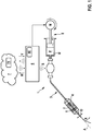
- Figure 1 shows an embodiment of a medical system 1 according to the invention for supporting the embedding of a stent 2 in a vessel of a patient in the area of a stenosis during an angioplasty, the system 1 having at least: a balloon catheter 3 for embedding the stent 2 in the vessel, such that the stenosis is expanded, the balloon catheter 3 having a balloon 4 extending in an axial direction and the stent 2 fixed thereon, the balloon 4 being expandable in the radial direction R for expanding the stent, and the system 1 having at least one sensor 5 , which is designed to measure an actual value of a balloon parameter and in particular to transmit it to the data processing unit, the balloon parameter here being a diameter D of the balloon 4 in the radial direction R of the balloon 4. Since the balloon 4 is expanded or unfolded for expanding the stent 2 by filling the balloon with a fluid medium M, this diameter D is also referred to herein as the expansion diameter D.
- the medical system also has a data processing unit 6 which is designed to acquire the following data: patient data, data on angioplasty, data on the current situation of the patient, data characterizing the stenosis, a balloon parameter.
- the data processing unit 6 can be formed, for example, by a computer with a microprocessor on which a program representing the functions of the data processing unit 6 is executed.
- the computer can be any imaginable computer, for example a desktop computer, a laptop, a tablet PC, a smartphone.
- the medical system 1 has a database unit 7, the data processing unit 6 being designed to transmit the data to the database unit 7, the database unit 7 being designed to store and / or analyze the data in a database 70.
- the database unit 7 can be formed, for example, by one or more networked computers and a program executed thereon.
- the database unit 7 is also designed to carry out a multivariate analysis of said data in order to estimate the likelihood of a complication occurring during the angioplasty and / or to generate a recommendation for a person performing the angioplasty and to display it to the person via a user interface 60 of the system 1.
- the data processing unit 6 can communicate with the database unit 7 via an internet connection or an internet protocol.
- the said recommended action corresponds to a proposal for a setpoint value for the balloon parameter or the diameter D of the balloon 4 in the radial direction R, which is to be set during the angioplasty.
- the setting can be made manually by the person or, after the proposal has been accepted (e.g. by a corresponding user input), it can be carried out automatically by the system 1, e.g. by means of an inflation pump 8, see below.
- the invention thus provides in particular an interface in the catheter laboratory that records all relevant information (in particular with regard to man, machine, method, tool, environment, material) of the angioplasty procedure and sends results or complications to the one preferably cross-location database 70 the database unit sends.
- a program executed by means of the database unit 7 preferably evaluates the collected data in a manner appropriate to the addressee.
- a procedural probability of success for the planned angioplasty can be determined using the database unit 7 prior to the angioplasty, for example on the basis of a success record of the person or hospital performing the angioplasty.
- the database unit 7 can be used, for example, to predict the clinical success of the angioplasty carried out based on an Database unit 7 transmitted case report and by feedback of the patient's well-being
- the person performing the angioplasty or the doctor can advantageously use the system 1 according to the invention for preoperative planning.
- a procedural probability of success for a proposal on how the angioplasty is to be carried out can be made on the basis of the case reports on previous angioplasties collected in the database 70.
- the data processing unit 6 is preferably configured to give instructions to the person performing the angioplasty or an inflation pump 8 during the angioplasty on how the balloon 4 is to be dilated.
- the instruction or handling instruction can relate, for example, to setting the target internal balloon pressure.
- the data processing unit 6 can be configured to specify a confidence interval for the achieved diameter of the balloon in vivo and z. B. also the current risks for the occurrence of a late stent thrombosis and / or a dissection (vascular rupture).
- the database unit 7 can be configured to propose suggestions for follow-up examinations with regard to postoperative planning, e.g. based on a comparison of the case report assigned to the patient with case reports or historical data already present in the database 70. Furthermore, the database unit 70 can be configured to notify the doctor of the personal current track record for angioplasties performed.
- the doctor or the person performing the angioplasty can use the database unit 7 to provide stored data at any point in time, wherein the database 70 can advantageously ensure that the stored or obtained data can be traced for scientific publications.
- the medical system according to the invention advantageously opens up the possibility of a simplified search for the cause for complaints regarding the functions of the balloon catheter 3, for example in that the database unit 7 is configured to record the inflation time and the deflation time of the balloon 4 in the event of inflation problems. Furthermore, for example, a maximum pressure in the event of the balloon bursting prematurely can be stored in the database 70.
- the storage of the catheter-related data advantageously enables a performance comparison of the individual catheter properties (eg from different manufacturers or between different models).
- the system 1 basically opens up the possibility of configuring the database unit 7 such that the track record of the relevant department can be extracted from the data compared to other hospitals.
- This allows in particular to optimize the time required for an angioplasty in the catheter laboratory and allows the necessity of the angioplasty to be easily demonstrated and documented if, for example, the catheter sensors by measuring the fractional flow reserve (FFR measurement) provide evidence of the necessity of angioplasty or placement create a stent.
- FFR measurement fractional flow reserve
- the system 1 opens up the possibility for the respective health insurance company responsible for the patient to prescribe a best treatment method ("best practice") with regard to the angioplasty, which can be obtained on the basis of the evaluation of the case reports for the individual angioplasties and the corresponding follow-up examinations. Finally, on the basis of the data collected in the database 70, compliance with the established best practice procedure can also be monitored.
- best practice a best treatment method with regard to the angioplasty
- the system 1 according to the invention is therefore preferably designed to provide the individual parties involved in the angioplasty, i.e. the person performing the angioplasty (doctor), the hospital, the manufacturer and the health insurance company, with specific information and evaluations of the data collected during the respective angioplasty put.
- the system 1 according to the invention enables, in particular, an exchange of data between the said persons / institutions and for all parties involved Optimization of the angioplasty procedure, advantageously adapted to the individual patient.
- the medical system 1 According to an embodiment of the medical system 1 according to the invention or the method made possible by this, it is provided, as already explained at the beginning, that prior to the dilation of the balloon 4 on the basis of the historical data (collected by means of the database unit) by means of a multivariate analysis or by means of a neural network the probability of complications occurring as a function of, for example, the expansion diameter D can be established.
- the doctor can then determine the optimal treatment strategy based on his assessment of the severity of the expected complications.
- the procedure is recorded and complications are added to database 70.
- the medical system 1 thus becomes more precise with each new data record.
- the relevant data can, for example, be collected directly on site, i.e. at the site of the angioplasty, by the data processing unit 6 (e.g. by user input in a user interface 60 of the system 1 or the data processing unit).
- the data relate, for example, to the patient on whom the angioplasty is to be performed, in particular his or her previous history, e.g. B: Age of the patient, illnesses and associated clinical picture, medication taken and, in particular, their dosage, any contraindications.
- the collected data can e.g. information about previous treatment steps (e.g. pre-dilatation, stent implantation, etc.) as well as a planned strategy for treatment (measuring, opening (balloon angioplasty POBA), stabilization with stents, especially drug-releasing stents (drug eluting stents or DES ), Correcting the stent expansion using post-dilatation (POBA)).
- previous treatment steps e.g. pre-dilatation, stent implantation, etc.
- a planned strategy for treatment measuring, opening (balloon angioplasty POBA), stabilization with stents, especially drug-releasing stents (drug eluting stents or DES ), Correcting the stent expansion using post-dilatation (POBA)).
- POBA post-dilatation
- the data recorded or to be recorded by the database device on the current situation of the patient can include the following information: patient's blood pressure, patient's pulse, supply of medication (heparin, antihypertensive agents, etc.)
- the database unit preferably records data such as: the exact location of the stenosis, healthy vessel diameter, length and minimum diameter of the stenosis, classification of the stenosis as scarred stenosis, calcified stenosis, or restenosis), the type of stenosis classification and, if known, the confidence interval the measurand.
- Well-known analysis methods are e.g. impedance spectroscopy, which can differentiate between calcified and scarred with regard to stenosis, for example.
- the stenosis is preferably analyzed by the attending physician and described as precisely as possible.
- the data can be recorded, for example, by user input via the user interface 60.
- the measurement of stenosis compliance can be used.
- the affected vessel of the patient can be closed distal from the stenosis with a balloon for the compliance measurement.
- the pulse rate in the vessel is then used to change the pressure and the change in diameter is observed or the volume flow is measured.
- a second balloon can also seal off the vessel proximally and hydraulic pressure is applied to the sealed area and the change in shape is observed.
- the vessel is closed distal and proximal to the stenosis and a volume is pumped into the sealed area and the increase in pressure is then measured.
- a multivariate analysis or artificial intelligence is preferably used according to a fundamental concept of the present invention used to estimate the likelihood of complications such as perforation, dissection or (very) late stent thrombosis from the current situation and historical data and to generate a situational recommendation for action for the doctor.
- the prediction model is ideally also available offline and preferably always in a state that has been checked and approved by experts.
- the start of the procedure or angioplasty it is preferably checked whether the chances of success are in a desired range, so that procedural success is guaranteed as far as possible.
- the function of the system 1 is only analytical and advisory. The doctor continues to carry out all treatment steps himself.
- the data processing unit 6 controls or regulates a digital inflation pump 8 on site, which performs the angioplasty after positioning the balloon 4 in the correct place and continuously monitors the main influencing variable, the said expansion diameter D as a balloon parameter, directly or indirectly.
- the said expansion diameter D is the most important parameter during stenting or post-dilating.
- the procedure can be monitored with somewhat less precision through the course of the hydraulic pressure plus the expansion volume. If the theoretical expansion volume of the balloon (measurement in vitro and documented in the Compliance Data Chart) and the in-vivo measured expansion volume are largely identical at a given pressure, the precision of the expansion is high and the result of the angioplasty is sufficient.
- the hydraulic pressure correlates independently of the expansion volume with the maximum possible balloon diameter and thus the risk of dissection (vascular rupture) if the stenosis suddenly ruptures.
- the course of the angioplasty is preferably monitored by X-rays.
- the data processing unit 6 records in particular acute complications that arise in the procedure, such as perforation of the vessel wall due to rupture and the severity of the corresponding bleeding.
- the quality of the prediction or the suggestions given to the doctor by the system increases with the quality of the data, so good sensors that describe and monitor the course of the angioplasty procedure are particularly preferred.
- the data processing unit in particular records whether complications arise later.
- the feedback of later problems such as late stent thrombosis is particularly important for the long-term improvement of clinical results.
- a particular advantage of the system 1 according to the invention lies in the continuous improvement of the angioplasty through the evaluation of a constantly growing database 70.
- the treating person can be shown the statistical consequences with a confidence interval for the maximum expansion diameter D.
- the balloon catheter 3 used to expand the stent thus helps the user to control the recommended overdistension of the stent (by 10-20%) compared to the healthy vessel diameter.
- the expansion of the balloon 4 from the moment in which the balloon 4 and a healthy vascular segment adjoining the stenosis continue steadily is set to "0%" and then the progressive overexpansion of the vessel or balloon 4 is displayed.
- the optimum diameter D soll target value for diameter D
- the operator receives a signal.
- the balloon catheter 3 has at least one strain sensor 5 in the cylindrical area of the balloon 4. Both components are designed in particular for a compliance of up to at least 15%, ideally> 30% and a resolution of +/- 5% (better +/- 1%) without delaminating or risking a line break.
- the sensor 5 is supplied with energy e.g. via a power line.
- the energy can come from a power source outside the patient's body, but can just as easily be provided by a suitable battery or antenna in the patient's body.
- the sensor data S are transmitted to the outside of the data processing unit 6, e.g. via a data line or wirelessly (e.g. by radio or ultrasound).
- the sensor data are preferably processed in the data processing device 6 by an algorithm.
- the processor for this can be located in or outside the body.
- the algorithm continuously calculates from the measuring or sensor data, the overstretching of the healthy vessel and the balloon 4, and continuously checks the reaching of the target area of the over-stretching (desired value D to the diameter D) and announces this preferably acoustically or visually at. If the recommended overstretching range is exceeded, there is preferably another signal which, through its frequency, conveys the increased risk of rupture to the user.
- the data W of the procedure are recorded with the classification of the stenosis and the procedural success is documented and transmitted to the central database 70 of the database unit 7.
- the balloon 4 used in angioplasty in the context of the present invention can in principle be produced from all thermoplastic balloon materials which offer a sufficiently high compressive strength with a small wall thickness. These are preferably polymers from the polyamide family or thermoplastic polyurethanes.
- the balloon forming process is preferably designed in such a way that a semi-compliant balloon behavior results that offers sufficient flexibility in the target diameter in the application.
- FIG Fig. 2 it can be provided that only the hydraulic pressure p is measured, for example, at the proximal catheter inlet (prox) and an expected range for the expansion diameter D is specified.
- the balloon 4 is designed so strong that its nominal diameter D nom is only exceeded after the stenosis has ruptured at pressure p B. This opening pressure for coronary arteries is approx. 14 bar. Before the stenosis breaks, the actual diameter in the area of the stenosis will be smaller than the one indicated on the compliance data. Fig.
- FIG. 2 shows in detail the pressure p present in the stenosis (central), the initial diameter of the stenosis D Ste increasing steadily as the balloon 4 is stretched.
- the nominal diameter D nom of the balloon 4 is above the healthy vessel diameter D Gef .
- the one you want The diameter D soll of the balloon 4 corresponds to the desired overdistension of the vessel or balloon 4 by 10% to 20% of the healthy vessel diameter D Gef .
- An alternative variant of the medical system 1 according to the invention compares the in-situ measured and thus expected balloon volume with the real in-vivo measured volume flow for each internal balloon pressure. If these volumes are identical, the in-vivo diameter will also match the in-vitro diameter D over the entire cylindrical length of the balloon 4.
- the most precise diameter measurement is preferably carried out by at least one sensor 5 (cf. Fig. 1 ) in the area of the balloon 4. This measurement can be determined in various ways using the physical principles described above.
- the advantages of the solution according to the invention lie in particular in the optimization of the stent implantation and avoidance of stent detachment (so-called wall apposition, where the stent at least partially loses contact with the vessel wall) through optimal overstretching of the stent and vessel as well as in the better endothelialization of the stent
- the pre-tensioned stent no longer loses contact with the vessel wall even with high blood pressure.
- the warning aid removes the fear of an impending dissection when the balloon is overstretched.
- the configuration of the system 1 according to the invention is furthermore advantageously designed in such a way that the system 1 learns from errors in the angioplasty. In this way, procedural and clinical success are significantly improved by learning the optimal stent overdistension
- reading errors from the previously common compliance data card can be avoided, and complicated conversion can be omitted.
- the system 1 provides, in particular, a closed feedback circuit for practitioners and the manufacturer of catheters for angioplasty.
- the change in balloon diameter is an essential indicator of the effect of the procedure more appropriate than changing the internal pressure of the balloon, which can give a deceptive feeling of effectiveness.
Landscapes
- Health & Medical Sciences (AREA)
- Life Sciences & Earth Sciences (AREA)
- Heart & Thoracic Surgery (AREA)
- Engineering & Computer Science (AREA)
- Biomedical Technology (AREA)
- Public Health (AREA)
- General Health & Medical Sciences (AREA)
- Veterinary Medicine (AREA)
- Animal Behavior & Ethology (AREA)
- Surgery (AREA)
- Biophysics (AREA)
- Medical Informatics (AREA)
- Pulmonology (AREA)
- Hematology (AREA)
- Molecular Biology (AREA)
- Oral & Maxillofacial Surgery (AREA)
- Child & Adolescent Psychology (AREA)
- Vascular Medicine (AREA)
- Anesthesiology (AREA)
- Nuclear Medicine, Radiotherapy & Molecular Imaging (AREA)
- Reproductive Health (AREA)
- Transplantation (AREA)
- Cardiology (AREA)
- Dentistry (AREA)
- Physics & Mathematics (AREA)
- Pathology (AREA)
- Media Introduction/Drainage Providing Device (AREA)
Priority Applications (1)
| Application Number | Priority Date | Filing Date | Title |
|---|---|---|---|
| EP19191683.2A EP3777782A1 (fr) | 2019-08-14 | 2019-08-14 | Système médical d'apprentissage pour une intégration optimale de stent dans l'angioplastie |
Applications Claiming Priority (1)
| Application Number | Priority Date | Filing Date | Title |
|---|---|---|---|
| EP19191683.2A EP3777782A1 (fr) | 2019-08-14 | 2019-08-14 | Système médical d'apprentissage pour une intégration optimale de stent dans l'angioplastie |
Publications (1)
| Publication Number | Publication Date |
|---|---|
| EP3777782A1 true EP3777782A1 (fr) | 2021-02-17 |
Family
ID=67658785
Family Applications (1)
| Application Number | Title | Priority Date | Filing Date |
|---|---|---|---|
| EP19191683.2A Withdrawn EP3777782A1 (fr) | 2019-08-14 | 2019-08-14 | Système médical d'apprentissage pour une intégration optimale de stent dans l'angioplastie |
Country Status (1)
| Country | Link |
|---|---|
| EP (1) | EP3777782A1 (fr) |
Cited By (2)
| Publication number | Priority date | Publication date | Assignee | Title |
|---|---|---|---|---|
| CN114678105A (zh) * | 2022-03-21 | 2022-06-28 | 南京圣德医疗科技有限公司 | 一种结合人工智能技术自动计算球囊参数的方法 |
| DE102023203919A1 (de) * | 2023-04-27 | 2024-10-31 | Siemens Healthineers Ag | Verfahren zur Abschätzung eines Risikos für eine Ruptur eines Hohlorgans, Datenverarbeitungsvorrichtung, Bildgebungssystem und Computerprogrammprodukt |
Citations (4)
| Publication number | Priority date | Publication date | Assignee | Title |
|---|---|---|---|---|
| EP2465423A1 (fr) * | 2010-05-25 | 2012-06-20 | MIRACOR Medical Systems GmbH | Système pour traiter le tissu du muscle cardiaque |
| US20160183807A1 (en) * | 2013-06-28 | 2016-06-30 | Cardiovascular Systems, Inc. | Methods, devices and systems for sensing, measuring and/or characterizing vessel and/or lesion compliance and/or elastance changes during vascular procedures |
| US20160287260A1 (en) * | 2010-02-16 | 2016-10-06 | Miracor Medical Systems Gmbh | Operating A Vessel Occlusion Catheter |
| US20180360545A1 (en) * | 2015-06-24 | 2018-12-20 | Koninklijke Philips N.V. | System and method for tracking and determining characteristics of inflatable medical instruments using fiber-optical realshape data |
-
2019
- 2019-08-14 EP EP19191683.2A patent/EP3777782A1/fr not_active Withdrawn
Patent Citations (4)
| Publication number | Priority date | Publication date | Assignee | Title |
|---|---|---|---|---|
| US20160287260A1 (en) * | 2010-02-16 | 2016-10-06 | Miracor Medical Systems Gmbh | Operating A Vessel Occlusion Catheter |
| EP2465423A1 (fr) * | 2010-05-25 | 2012-06-20 | MIRACOR Medical Systems GmbH | Système pour traiter le tissu du muscle cardiaque |
| US20160183807A1 (en) * | 2013-06-28 | 2016-06-30 | Cardiovascular Systems, Inc. | Methods, devices and systems for sensing, measuring and/or characterizing vessel and/or lesion compliance and/or elastance changes during vascular procedures |
| US20180360545A1 (en) * | 2015-06-24 | 2018-12-20 | Koninklijke Philips N.V. | System and method for tracking and determining characteristics of inflatable medical instruments using fiber-optical realshape data |
Non-Patent Citations (1)
| Title |
|---|
| "CIRSE 2016", CARDIOVASCULAR AND INTERVENTIONAL RADIOLOGY, SPRINGER VERLAG, INC., NEW YORK, NY, US, vol. 39, no. 3, 16 August 2016 (2016-08-16), pages 35 - 386, XP036036878, ISSN: 0174-1551, [retrieved on 20160816], DOI: 10.1007/S00270-016-1405-3 * |
Cited By (3)
| Publication number | Priority date | Publication date | Assignee | Title |
|---|---|---|---|---|
| CN114678105A (zh) * | 2022-03-21 | 2022-06-28 | 南京圣德医疗科技有限公司 | 一种结合人工智能技术自动计算球囊参数的方法 |
| CN114678105B (zh) * | 2022-03-21 | 2023-10-17 | 南京圣德医疗科技有限公司 | 一种结合人工智能技术自动计算球囊参数的方法 |
| DE102023203919A1 (de) * | 2023-04-27 | 2024-10-31 | Siemens Healthineers Ag | Verfahren zur Abschätzung eines Risikos für eine Ruptur eines Hohlorgans, Datenverarbeitungsvorrichtung, Bildgebungssystem und Computerprogrammprodukt |
Similar Documents
| Publication | Publication Date | Title |
|---|---|---|
| DE69029141T2 (de) | Angioplastie mit niedrigem physiologischen stress | |
| DE69913277T2 (de) | Druckempfindlicher ballonkatheter | |
| AT510252B1 (de) | Implantierbare vorrichtung zur intermittierenden okklusion eines blutgefässes | |
| DE102009025766A1 (de) | Personalisierte Fluidbewertung | |
| EP2433564A1 (fr) | Positionnement de cathéters à l'aide d'une mesure d'impédance | |
| EP3567598A1 (fr) | Contrôle d'une image de super position interventionnelle | |
| DE102009025913A1 (de) | Historische Patientendaten nutzende Vitalzeichenüberwachungsvorrichtung | |
| EP3025638A1 (fr) | Procédé de détermination de la longueur finale d'endoprothèses avant leur mise en place | |
| US20220288359A1 (en) | Learning electronic balloon catheter system for optimal stent embedding | |
| DE112020006050T5 (de) | Konfigurieren einer medizinischen Vorrichtung und Patientenbehandlung | |
| CN115813605A (zh) | 用于将假体植入物联接到开窗体的系统、装置和方法 | |
| DE102010016118A1 (de) | Patientenüberwachungssystem und -verfahren | |
| DE102016222102A1 (de) | Behandlungsplanung für eine Stenose in einem Gefäßsegment anhand einer virtuellen hämodynamischen Analyse | |
| WO2005028014A1 (fr) | Dispositif destine a etre introduit dans des organes corporels et dote d'un element de marquage servant a controler sa position | |
| DE112008000456T5 (de) | Aktivierung chirurgischer Implantate mit codierten Sequenzen | |
| EP3777782A1 (fr) | Système médical d'apprentissage pour une intégration optimale de stent dans l'angioplastie | |
| CN214966146U (zh) | 输送装置及栓塞弹簧圈系统 | |
| DE60123807T2 (de) | Verfahren zur herstellung von intravaskulären vorrichtungen nach mass | |
| EP3666210B1 (fr) | Aide à la planification pour une intervention de la médecine interventionnelle et systèmes et logiciels correspondants | |
| CN119497894A (zh) | 与基于导管的手术相关的系统和方法 | |
| CN115624393A (zh) | 一种介入手术机器人系统、提供操纵提示的方法 | |
| US8588489B2 (en) | Method for displaying a vessel of a particular biological subject | |
| DE102018129435A1 (de) | Multifunktionale Verwendung einer Elektrodenstruktur eines Ballons eines Ballonkathetersystems | |
| DE102006058908B4 (de) | Verfahren zur medizinischen Bilddarstellung | |
| EP4389186A1 (fr) | Dérivation dynamique du comportement d'inflation d'un cathéter de dilatation |
Legal Events
| Date | Code | Title | Description |
|---|---|---|---|
| PUAI | Public reference made under article 153(3) epc to a published international application that has entered the european phase |
Free format text: ORIGINAL CODE: 0009012 |
|
| STAA | Information on the status of an ep patent application or granted ep patent |
Free format text: STATUS: THE APPLICATION HAS BEEN PUBLISHED |
|
| AK | Designated contracting states |
Kind code of ref document: A1 Designated state(s): AL AT BE BG CH CY CZ DE DK EE ES FI FR GB GR HR HU IE IS IT LI LT LU LV MC MK MT NL NO PL PT RO RS SE SI SK SM TR |
|
| AX | Request for extension of the european patent |
Extension state: BA ME |
|
| STAA | Information on the status of an ep patent application or granted ep patent |
Free format text: STATUS: THE APPLICATION IS DEEMED TO BE WITHDRAWN |
|
| 18D | Application deemed to be withdrawn |
Effective date: 20210818 |