EP3833741B1 - Modèle de simulation in vitro du comportement de vaisseaux dysfonctionnels - Google Patents
Modèle de simulation in vitro du comportement de vaisseaux dysfonctionnels Download PDFInfo
- Publication number
- EP3833741B1 EP3833741B1 EP19769576.0A EP19769576A EP3833741B1 EP 3833741 B1 EP3833741 B1 EP 3833741B1 EP 19769576 A EP19769576 A EP 19769576A EP 3833741 B1 EP3833741 B1 EP 3833741B1
- Authority
- EP
- European Patent Office
- Prior art keywords
- scaffold
- bioreactor
- endothelial cells
- lumen
- growth medium
- Prior art date
- Legal status (The legal status is an assumption and is not a legal conclusion. Google has not performed a legal analysis and makes no representation as to the accuracy of the status listed.)
- Active
Links
Images














Classifications
-
- C—CHEMISTRY; METALLURGY
- C12—BIOCHEMISTRY; BEER; SPIRITS; WINE; VINEGAR; MICROBIOLOGY; ENZYMOLOGY; MUTATION OR GENETIC ENGINEERING
- C12N—MICROORGANISMS OR ENZYMES; COMPOSITIONS THEREOF; PROPAGATING, PRESERVING, OR MAINTAINING MICROORGANISMS; MUTATION OR GENETIC ENGINEERING; CULTURE MEDIA
- C12N5/00—Undifferentiated human, animal or plant cells, e.g. cell lines; Tissues; Cultivation or maintenance thereof; Culture media therefor
- C12N5/06—Animal cells or tissues; Human cells or tissues
- C12N5/0602—Vertebrate cells
- C12N5/069—Vascular Endothelial cells
- C12N5/0691—Vascular smooth muscle cells; 3D culture thereof, e.g. models of blood vessels
-
- G—PHYSICS
- G01—MEASURING; TESTING
- G01N—INVESTIGATING OR ANALYSING MATERIALS BY DETERMINING THEIR CHEMICAL OR PHYSICAL PROPERTIES
- G01N33/00—Investigating or analysing materials by specific methods not covered by groups G01N1/00 - G01N31/00
- G01N33/48—Biological material, e.g. blood, urine; Haemocytometers
- G01N33/50—Chemical analysis of biological material, e.g. blood, urine; Testing involving biospecific ligand binding methods; Immunological testing
- G01N33/5005—Chemical analysis of biological material, e.g. blood, urine; Testing involving biospecific ligand binding methods; Immunological testing involving human or animal cells
- G01N33/5008—Chemical analysis of biological material, e.g. blood, urine; Testing involving biospecific ligand binding methods; Immunological testing involving human or animal cells for testing or evaluating the effect of chemical or biological compounds, e.g. drugs, cosmetics
- G01N33/5082—Supracellular entities, e.g. tissue, organisms
-
- A—HUMAN NECESSITIES
- A61—MEDICAL OR VETERINARY SCIENCE; HYGIENE
- A61L—METHODS OR APPARATUS FOR STERILISING MATERIALS OR OBJECTS IN GENERAL; DISINFECTION, STERILISATION OR DEODORISATION OF AIR; CHEMICAL ASPECTS OF BANDAGES, DRESSINGS, ABSORBENT PADS OR SURGICAL ARTICLES; MATERIALS FOR BANDAGES, DRESSINGS, ABSORBENT PADS OR SURGICAL ARTICLES
- A61L27/00—Materials for grafts or prostheses or for coating grafts or prostheses
- A61L27/36—Materials for grafts or prostheses or for coating grafts or prostheses containing ingredients of undetermined constitution or reaction products thereof, e.g. transplant tissue, natural bone, extracellular matrix
- A61L27/38—Materials for grafts or prostheses or for coating grafts or prostheses containing ingredients of undetermined constitution or reaction products thereof, e.g. transplant tissue, natural bone, extracellular matrix containing added animal cells
- A61L27/3804—Materials for grafts or prostheses or for coating grafts or prostheses containing ingredients of undetermined constitution or reaction products thereof, e.g. transplant tissue, natural bone, extracellular matrix containing added animal cells characterised by specific cells or progenitors thereof, e.g. fibroblasts, connective tissue cells, kidney cells
- A61L27/3808—Endothelial cells
-
- A—HUMAN NECESSITIES
- A61—MEDICAL OR VETERINARY SCIENCE; HYGIENE
- A61L—METHODS OR APPARATUS FOR STERILISING MATERIALS OR OBJECTS IN GENERAL; DISINFECTION, STERILISATION OR DEODORISATION OF AIR; CHEMICAL ASPECTS OF BANDAGES, DRESSINGS, ABSORBENT PADS OR SURGICAL ARTICLES; MATERIALS FOR BANDAGES, DRESSINGS, ABSORBENT PADS OR SURGICAL ARTICLES
- A61L27/00—Materials for grafts or prostheses or for coating grafts or prostheses
- A61L27/36—Materials for grafts or prostheses or for coating grafts or prostheses containing ingredients of undetermined constitution or reaction products thereof, e.g. transplant tissue, natural bone, extracellular matrix
- A61L27/38—Materials for grafts or prostheses or for coating grafts or prostheses containing ingredients of undetermined constitution or reaction products thereof, e.g. transplant tissue, natural bone, extracellular matrix containing added animal cells
- A61L27/3804—Materials for grafts or prostheses or for coating grafts or prostheses containing ingredients of undetermined constitution or reaction products thereof, e.g. transplant tissue, natural bone, extracellular matrix containing added animal cells characterised by specific cells or progenitors thereof, e.g. fibroblasts, connective tissue cells, kidney cells
- A61L27/3826—Muscle cells, e.g. smooth muscle cells
-
- A—HUMAN NECESSITIES
- A61—MEDICAL OR VETERINARY SCIENCE; HYGIENE
- A61L—METHODS OR APPARATUS FOR STERILISING MATERIALS OR OBJECTS IN GENERAL; DISINFECTION, STERILISATION OR DEODORISATION OF AIR; CHEMICAL ASPECTS OF BANDAGES, DRESSINGS, ABSORBENT PADS OR SURGICAL ARTICLES; MATERIALS FOR BANDAGES, DRESSINGS, ABSORBENT PADS OR SURGICAL ARTICLES
- A61L27/00—Materials for grafts or prostheses or for coating grafts or prostheses
- A61L27/50—Materials characterised by their function or physical properties, e.g. injectable or lubricating compositions, shape-memory materials, surface modified materials
- A61L27/507—Materials characterised by their function or physical properties, e.g. injectable or lubricating compositions, shape-memory materials, surface modified materials for artificial blood vessels
-
- C—CHEMISTRY; METALLURGY
- C12—BIOCHEMISTRY; BEER; SPIRITS; WINE; VINEGAR; MICROBIOLOGY; ENZYMOLOGY; MUTATION OR GENETIC ENGINEERING
- C12N—MICROORGANISMS OR ENZYMES; COMPOSITIONS THEREOF; PROPAGATING, PRESERVING, OR MAINTAINING MICROORGANISMS; MUTATION OR GENETIC ENGINEERING; CULTURE MEDIA
- C12N5/00—Undifferentiated human, animal or plant cells, e.g. cell lines; Tissues; Cultivation or maintenance thereof; Culture media therefor
- C12N5/0062—General methods for three-dimensional culture
-
- C—CHEMISTRY; METALLURGY
- C12—BIOCHEMISTRY; BEER; SPIRITS; WINE; VINEGAR; MICROBIOLOGY; ENZYMOLOGY; MUTATION OR GENETIC ENGINEERING
- C12N—MICROORGANISMS OR ENZYMES; COMPOSITIONS THEREOF; PROPAGATING, PRESERVING, OR MAINTAINING MICROORGANISMS; MUTATION OR GENETIC ENGINEERING; CULTURE MEDIA
- C12N5/00—Undifferentiated human, animal or plant cells, e.g. cell lines; Tissues; Cultivation or maintenance thereof; Culture media therefor
- C12N5/06—Animal cells or tissues; Human cells or tissues
- C12N5/0602—Vertebrate cells
- C12N5/0652—Cells of skeletal and connective tissues; Mesenchyme
- C12N5/0661—Smooth muscle cells
-
- C—CHEMISTRY; METALLURGY
- C12—BIOCHEMISTRY; BEER; SPIRITS; WINE; VINEGAR; MICROBIOLOGY; ENZYMOLOGY; MUTATION OR GENETIC ENGINEERING
- C12N—MICROORGANISMS OR ENZYMES; COMPOSITIONS THEREOF; PROPAGATING, PRESERVING, OR MAINTAINING MICROORGANISMS; MUTATION OR GENETIC ENGINEERING; CULTURE MEDIA
- C12N5/00—Undifferentiated human, animal or plant cells, e.g. cell lines; Tissues; Cultivation or maintenance thereof; Culture media therefor
- C12N5/06—Animal cells or tissues; Human cells or tissues
- C12N5/0697—Artificial constructs associating cells of different lineages, e.g. tissue equivalents
-
- A—HUMAN NECESSITIES
- A61—MEDICAL OR VETERINARY SCIENCE; HYGIENE
- A61L—METHODS OR APPARATUS FOR STERILISING MATERIALS OR OBJECTS IN GENERAL; DISINFECTION, STERILISATION OR DEODORISATION OF AIR; CHEMICAL ASPECTS OF BANDAGES, DRESSINGS, ABSORBENT PADS OR SURGICAL ARTICLES; MATERIALS FOR BANDAGES, DRESSINGS, ABSORBENT PADS OR SURGICAL ARTICLES
- A61L2430/00—Materials or treatment for tissue regeneration
- A61L2430/20—Materials or treatment for tissue regeneration for reconstruction of the heart, e.g. heart valves
-
- C—CHEMISTRY; METALLURGY
- C12—BIOCHEMISTRY; BEER; SPIRITS; WINE; VINEGAR; MICROBIOLOGY; ENZYMOLOGY; MUTATION OR GENETIC ENGINEERING
- C12N—MICROORGANISMS OR ENZYMES; COMPOSITIONS THEREOF; PROPAGATING, PRESERVING, OR MAINTAINING MICROORGANISMS; MUTATION OR GENETIC ENGINEERING; CULTURE MEDIA
- C12N2500/00—Specific components of cell culture medium
- C12N2500/05—Inorganic components
- C12N2500/10—Metals; Metal chelators
- C12N2500/12—Light metals, i.e. alkali, alkaline earth, Be, Al, Mg
- C12N2500/14—Calcium; Ca chelators; Calcitonin
-
- C—CHEMISTRY; METALLURGY
- C12—BIOCHEMISTRY; BEER; SPIRITS; WINE; VINEGAR; MICROBIOLOGY; ENZYMOLOGY; MUTATION OR GENETIC ENGINEERING
- C12N—MICROORGANISMS OR ENZYMES; COMPOSITIONS THEREOF; PROPAGATING, PRESERVING, OR MAINTAINING MICROORGANISMS; MUTATION OR GENETIC ENGINEERING; CULTURE MEDIA
- C12N2500/00—Specific components of cell culture medium
- C12N2500/05—Inorganic components
- C12N2500/10—Metals; Metal chelators
- C12N2500/20—Transition metals
-
- C—CHEMISTRY; METALLURGY
- C12—BIOCHEMISTRY; BEER; SPIRITS; WINE; VINEGAR; MICROBIOLOGY; ENZYMOLOGY; MUTATION OR GENETIC ENGINEERING
- C12N—MICROORGANISMS OR ENZYMES; COMPOSITIONS THEREOF; PROPAGATING, PRESERVING, OR MAINTAINING MICROORGANISMS; MUTATION OR GENETIC ENGINEERING; CULTURE MEDIA
- C12N2500/00—Specific components of cell culture medium
- C12N2500/30—Organic components
- C12N2500/38—Vitamins
-
- C—CHEMISTRY; METALLURGY
- C12—BIOCHEMISTRY; BEER; SPIRITS; WINE; VINEGAR; MICROBIOLOGY; ENZYMOLOGY; MUTATION OR GENETIC ENGINEERING
- C12N—MICROORGANISMS OR ENZYMES; COMPOSITIONS THEREOF; PROPAGATING, PRESERVING, OR MAINTAINING MICROORGANISMS; MUTATION OR GENETIC ENGINEERING; CULTURE MEDIA
- C12N2500/00—Specific components of cell culture medium
- C12N2500/30—Organic components
- C12N2500/40—Nucleotides, nucleosides or bases
-
- C—CHEMISTRY; METALLURGY
- C12—BIOCHEMISTRY; BEER; SPIRITS; WINE; VINEGAR; MICROBIOLOGY; ENZYMOLOGY; MUTATION OR GENETIC ENGINEERING
- C12N—MICROORGANISMS OR ENZYMES; COMPOSITIONS THEREOF; PROPAGATING, PRESERVING, OR MAINTAINING MICROORGANISMS; MUTATION OR GENETIC ENGINEERING; CULTURE MEDIA
- C12N2500/00—Specific components of cell culture medium
- C12N2500/70—Undefined extracts
- C12N2500/80—Undefined extracts from animals
- C12N2500/84—Undefined extracts from animals from mammals
-
- C—CHEMISTRY; METALLURGY
- C12—BIOCHEMISTRY; BEER; SPIRITS; WINE; VINEGAR; MICROBIOLOGY; ENZYMOLOGY; MUTATION OR GENETIC ENGINEERING
- C12N—MICROORGANISMS OR ENZYMES; COMPOSITIONS THEREOF; PROPAGATING, PRESERVING, OR MAINTAINING MICROORGANISMS; MUTATION OR GENETIC ENGINEERING; CULTURE MEDIA
- C12N2513/00—3D culture
-
- C—CHEMISTRY; METALLURGY
- C12—BIOCHEMISTRY; BEER; SPIRITS; WINE; VINEGAR; MICROBIOLOGY; ENZYMOLOGY; MUTATION OR GENETIC ENGINEERING
- C12N—MICROORGANISMS OR ENZYMES; COMPOSITIONS THEREOF; PROPAGATING, PRESERVING, OR MAINTAINING MICROORGANISMS; MUTATION OR GENETIC ENGINEERING; CULTURE MEDIA
- C12N2533/00—Supports or coatings for cell culture, characterised by material
- C12N2533/50—Proteins
-
- G—PHYSICS
- G01—MEASURING; TESTING
- G01N—INVESTIGATING OR ANALYSING MATERIALS BY DETERMINING THEIR CHEMICAL OR PHYSICAL PROPERTIES
- G01N33/00—Investigating or analysing materials by specific methods not covered by groups G01N1/00 - G01N31/00
- G01N33/48—Biological material, e.g. blood, urine; Haemocytometers
- G01N33/50—Chemical analysis of biological material, e.g. blood, urine; Testing involving biospecific ligand binding methods; Immunological testing
- G01N33/5005—Chemical analysis of biological material, e.g. blood, urine; Testing involving biospecific ligand binding methods; Immunological testing involving human or animal cells
- G01N33/5008—Chemical analysis of biological material, e.g. blood, urine; Testing involving biospecific ligand binding methods; Immunological testing involving human or animal cells for testing or evaluating the effect of chemical or biological compounds, e.g. drugs, cosmetics
- G01N33/5044—Chemical analysis of biological material, e.g. blood, urine; Testing involving biospecific ligand binding methods; Immunological testing involving human or animal cells for testing or evaluating the effect of chemical or biological compounds, e.g. drugs, cosmetics involving specific cell types
- G01N33/5061—Muscle cells
-
- G—PHYSICS
- G01—MEASURING; TESTING
- G01N—INVESTIGATING OR ANALYSING MATERIALS BY DETERMINING THEIR CHEMICAL OR PHYSICAL PROPERTIES
- G01N33/00—Investigating or analysing materials by specific methods not covered by groups G01N1/00 - G01N31/00
- G01N33/48—Biological material, e.g. blood, urine; Haemocytometers
- G01N33/50—Chemical analysis of biological material, e.g. blood, urine; Testing involving biospecific ligand binding methods; Immunological testing
- G01N33/5005—Chemical analysis of biological material, e.g. blood, urine; Testing involving biospecific ligand binding methods; Immunological testing involving human or animal cells
- G01N33/5008—Chemical analysis of biological material, e.g. blood, urine; Testing involving biospecific ligand binding methods; Immunological testing involving human or animal cells for testing or evaluating the effect of chemical or biological compounds, e.g. drugs, cosmetics
- G01N33/5044—Chemical analysis of biological material, e.g. blood, urine; Testing involving biospecific ligand binding methods; Immunological testing involving human or animal cells for testing or evaluating the effect of chemical or biological compounds, e.g. drugs, cosmetics involving specific cell types
- G01N33/5064—Endothelial cells
Definitions
- the present invention refers to a model for in-vitro simulation of the behaviour of dysfunctional human vessels, such as for example vessels affected by aneurysm, stenosis or sclerosis plaques, as an instrument for testing medical devices and drugs with the aim of verifying the effectiveness and safety thereof prior to use thereof on humans.
- dysfunctional human vessels such as for example vessels affected by aneurysm, stenosis or sclerosis plaques
- the present invention refers to an in vitro model of a vascular structure, preferably a substantially tubular-shaped synthetic vascular structure having dysfunctional anatomical and physiological characteristics, simulating the vascular structure of a healthy subject whose vascular structure has been damaged or deformed or deteriorated due to a damage selected from among the group comprising or, alternatively, consisting of aneurysm, stenosis, sclerosis plaques, forms of tumours or cardiomyopathies having the characteristics as claimed in the attached claims.
- the present invention also refers to a reliable and reproducible industrialisation process for eliminating air bubbles for producing an engineered vascular tissue for the in vitro test of medicinal products for human use and veterinarian products for animal use.
- the evaluation of the biological safety and efficiency of the medicinal product is extensive and complex while the evaluation of the interaction with the tissue/s of only one constituent material cannot be considered as isolated from the overall planning of the medicinal or veterinarian product which must be evaluated as a whole and in a context that can reproduce the conditions of use as faithfully as possible.
- the 3R principle refers to three key concepts: replacement, reduction and refinement.
- the 3R principle argues that research in the biomedical industry should aim at, with utmost effort possible, replacing or substituting the animal model with an alternative model; reducing the number of animals used in a given experimental protocol as much as possible; refining, i.e. improving the experimental conditions to which the animals are subjected.
- vascular tissue engineering industry shows that it is crucial to be able to produce a continuous (i.e. having a monolayer of confluent cells) and functional endothelium mainly consisting of endothelial cells (ECs).
- ECs endothelial cells
- thermoresponsive polymers which have temperature-induced reversible phase transition characteristics
- Fabrication Factory for Tubular Vascular Tissue Mimics based on automated Rolling Manipulation and Thermo-Responsive Polymers Takehisa Matsuda et al
- a tissue-engineered construct that mimicked the structure of blood vessels using tubular electrospun silk fibroin scaffolds with suitable mechanical properties.
- Human coronary artery smooth muscle cells and human aortic endothelial cells (HAECs) were sequentially seeded onto the luminal surface of the tubular scaffolds and cultivated under physiological pulsatile flow (" Dynamic culture conditions to generate silk-based tissue-engineered vascular grafts", Zhang X et al).
- a three-layered bifurcated vascular scaffold is presented with a curved structure using a technique which combines 3D printed molds and casting hydrogel and ink to create vessel-mimicking constructs with customizable structural parameters, providing, therefore, a feasible approach for vascular scaffold construction.
- Enzymatically-crosslinked gelatin was used as the scaffold material.
- human umbilical cord derived endothelial cells (HUVECs) were seeded on the scaffolds and cultured (“ Fabrication of triple-layered bifurcated vascular scaffold with a certain degree of three-dimensional structure" Liu Y et al).
- WO 03/082145 describes materials and molding technique for making complex, living, vascularized tissues for organ and tissue replacement with polymer scaffolds and semipermeable membranes.
- WO 2018/063099 describes a device for patterning extracellular matrix (ECM) hydrogel with a first layer surface characterized by microchannel, and a second layer comprising a loading channel in fluid communication with loading ports to receive an ECM hydrogel wherein the ECM hydrogel is confined to form a perfusable channel and therefore to provide a improved and efficient method for patterning microchannel of different vascular geometries in ECM hydrogel.
- ECM extracellular matrix
- Static seeding and dynamic seeding currently represent the main endothelial cell seeding methods.
- the simplest static method consists in pipetting a cell suspension directly on the luminal surface of the scaffold followed by a short incubation step on a Petri dish. This method strongly depends on the operator and it is complicated by the difficulty to obtain a uniform endothelial layer.
- the dynamic methods on the contrary increase cell seeding effectiveness, uniformity and adhesion.
- Some of these dynamic principles could be directly transferred and paired with perfusion bioreactors, allowing a reduction in the handling of the scaffold. In this case, the scaffolds can be immediately seeded once housed in the bioreactor chamber.
- a completely different technique with respect to the ones described previously is based on dripping the cell suspension in the lumen of a scaffold.
- the main drawback lies in a low initial adhesion of the cells and a low reproducibility of the method given that it strongly depends on the operator.
- the process for developing and approving medicinal products normally requires conducting numerous preclinical tests to be conducted in- vitro, ex-vivo and in-vivo on animals prior to conducting clinical testing on humans.
- animal testing offering an in vivo model for the test of medicinal products, the anatomical structure, the physiology and the blood composition considerably differ from those of humans and the results achieved on the animal model may considerably differ from those achieved in human testing.
- Forming an object of the present invention is an in vitro model of a substantially tubular-shaped vascular structure having dysfunctional anatomical and physiological characteristics, simulating the vascular structure of a healthy subject whose vascular structure has been damaged or deformed or deteriorated due to a damage selected from among the group comprising or, alternatively, consisting of aneurysm, stenosis, sclerosis plaques, forms of tumours or cardiomyopathies having the characteristics as claimed in the attached claims.
- an object of the present invention is an in vitro model of a substantially tubular shaped vascular structure having dysfunctional anatomical and physiological characteristics simulating the same vascular structure of a healthy subject whose vascular structure has been damaged or deformed or deteriorated due to a damage selected from among the group comprising or, alternatively, consisting of aneurysm, stenosis, sclerosis plaques, forms of tumours or cardiomyopathies, wherein said model comprises or, alternatively, consists of one or more biocompatible porous polymeric supports (" scaffolds ") capable of promoting a cell adhesion and growth, wherein said scaffold is seeded with endothelial cells that cover a lumen of the scaffold and constitute an endothelium having a monolayer of confluent cells, said scaffold being provided with deformities or defects on a tubular structure thereof, having the characteristics as claimed in the attached claims.
- scaffolds biocompatible porous polymeric supports
- said in vitro model has a vascular structure that was selected from among the blood vessels or valves of the central or peripheral circulatory system; preferably arteries, veins, capillaries, aortic or mitral valve.
- said in vitro model is a dysfunctional vascular model.
- said vascular structure is synthetic vascular structure.
- said scaffold consists of electrospun silk fibroin, copolymers of polyglycolic acid/polylactic acid (PGA/PLA) or copolymers of polyglycolic acid/polycaprolactone (PGA/PCL).
- said in vitro model comprises or, alternatively, consists of one or more scaffolds seeded with endothelial cells, and optionally muscle cells.
- said deformities or said defects of the tubular structure comprise bifurcations, curvatures, elbows, constrictions, dilatations or combinations thereof.
- Forming an object of the present invention is a method for testing a medical device or a drug with the aim of verifying the effectiveness and safety thereof prior to the in vivo use thereof on humans or animals, said method comprising the following steps:
- Forming an object of the present invention is a model for in-vitro simulation of the behaviour of dysfunctional human vessels comprising or, alternatively, consisting of vessels affected by aneurysm, stenosis or sclerosis plaques, as an instrument for testing medical devices and drugs with the aim of verifying the effectiveness and safety thereof prior to use thereof on humans, having the characteristics as claimed in the attached claims.
- Also forming an object of the present invention is providing in vitro vascular structure models, mainly substantially tubular-shaped, having, depending on the need, dysfunctional anatomical and physiological characteristics with respect to the healthy human vascular structure, such as, by way of non-limiting example, aneurysms, stenosis, sclerosis plaques, having the characteristics as claimed in the attached claims.
- Forming another object of the present invention are dysfunctional vascular models comprising or, alternatively, consisting of one or more scaffolds seeded with suitable selected cells, having the characteristics as claimed in the attached claims.
- Forming another object of the present disclosure is a method for providing said dysfunctional vascular models using scaffolds seeded with suitable selected cells, having the characteristics as claimed in the attached claims.
- Such dysfunctional vascular structures include: (i) a scaffold consisting of biocompatible material suitable to be electrospun, such as for example silk fibroin, or molten in moulds or printed using bioprinters such as for example polylactic acid, polycaprolactone, etc.
- the structure of the scaffold is suitable to allow the seeding of endothelial vascular cells in the scaffold and to allow the through-flow of fluids such as growth medium for sustaining the cells, blood (artificial or whole human).
- a different embodiment of the structure of the scaffold described in the previous point provides for the possibility of seeding, on the outer surface of the scaffold, as a function of the characteristics to be tested in the medicinal product subject of the test, muscle cells or nervous cells, providing for a weft that promotes the orientation of said cells so as to be able to simulate the vasoconstriction or vasodilation depending on the mechanical, pharmacological or chemical stimulus to which the model is subjected.
- the scaffold for example in order to simulate clots, sclerosis plaques, thrombosis or stenosis, blood components (such as for example platelets, cholesterol, erythrocytes, etc.).
- blood components such as for example platelets, cholesterol, erythrocytes, etc.
- the scaffolds can be made having deformities or defects on the tubular structure thereof, such as for example, bifurcations, curvatures, elbows, constrictions, dilatations.
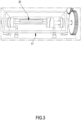
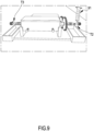
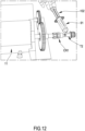
- the hydraulic circuit and the bioreactor into which the scaffold is inserted is outlined hereinafter in the present description to which reference shall be made with the help of the attached drawings.
- the circuit already as provided, allows the perfusion of the seeded cells on the scaffold with growth medium so as to allow the survival and growth thereof, with the possibility of passing to a pulsatile regime upon replacing the growth medium with blood (whole or diluted with growth medium or with eluate of the product to be tested), or with artificial blood, or with growth medium diluted with eluate of the product to be tested.
- the circuit allows - by means of suitable valve upstream of the bioreactor - to introduce the medicinal product (drug, medical device) into the scaffold.
- the dysfunctional scaffolds subject of the present invention and obtained by means of the method described herein were tested using: Blood analysis (blood alterations caused by the dysfunction of the vessel and/or by the product used for treating the dysfunction of the vessel) or hemocompatibility of the medicinal product.
- Biocompatibility of the medicinal product used for treating vascular dysfunction such as for example stents, devices for the embolization of the aneurysms, catheters, heart valves, drugs, etc.
- the Applicant created a process comprising a seeding method and a method for connecting the perfusion circuit to the bioreactor-scaffold system, having the characteristics as claimed in the attached claims.
- the expression continuous and functional endothelium is used to indicate an endothelium, with physiological-like behaviour, wherein the endothelial cells are adjacent to each other, adhered to the scaffold and expressing markers typical of the endothelial cells, such as for example Von Willebrand factor (VWF), cluster of differentiation 31 (CD31), vascular cell adhesion molecule 1 (VCAM-1).
- VWF Von Willebrand factor
- CD31 cluster of differentiation 31
- VCAM-1 vascular cell adhesion molecule 1
- the expression continuous endothelium is used to indicate an endothelium having a monolayer of confluent cells.
- the expression scaffold is used to indicate a biocompatible porous polymeric medium capable of promoting the cell adhesion and growth, endothelial cells in this case.
- the polymeric scaffold can be of synthetic or natural origin and consist of only one polymer or copolymers (entirety of polymers), chosen from electrospun silk fibroin or copolymers of PGA/PLA (polyglycolic acid/polylactic acid) or PGA/PCL (polyglycolic acid/polycaprolactone).
- the expression “confluence” refers to a surface of the scaffold (in particular an inner surface or lumen of such scaffold) which is covered by adherent cells.
- so-called “confluent cells” have a confluence equivalent to or greater than 90%, preferably comprised between 90% and 100%, even more preferably comprised between 95% and 100%, hence substantially the entire surface of the scaffold is covered by adherent cells and there is no more surface left available on the scaffold so that the cells can grow as a monolayer.
- endothelial cells constituting an endothelium of a vascular tissue are defined as endothelial cells.
- endothelial cells are the FIAOECs (human aortic endothelial cells), FICAECs (human coronary artery endothelial cells), FIMEVECs (human dermal microvascular endothelial cells), or FIUVECs (human umbilical vein endothelial cells).
- a scaffold having the lumen mainly covered by functional endothelial cells following the in vitro endothelisation process is defined as engineered vascular construct.
- Said endothelial cells covering the lumen of the scaffold constitute a continuous endothelium, i.e. an endothelium having a monolayer of confluent cells.
- the cell growth and maintenance fluid specific for each type of cell, is defined as growth medium.
- growth medium is Endothelial Growth Medium (EGM, Sigma Aldrich, 211-500).
- EGM contains fetal bovine serum (2%), adenine (0.2 pg/ml), ammonium metavanadate (0.0006 pg/ml), amphotericin B (0.3 pg/ml), calcium chloride 2H20(300 pg/ml), choline chloride (20 pg/ml), copper sulphate 5H2O (0.002 pg/ml), trioptic acid DL-6,8(0.003 pg/ml), folinic acid (calcium) (0.6 pg/ml), heparin (4 pg/ml), hydrocortisone (2 pg/ml), L-aspartic acid (15 pg/ml), L-cysteine (30 pg/ml), L-tyrosine (20 pg/ml), manganese sulphate monohydrate (0.0002 pg/ml), ammonium molybdate 4H2O (0.004 pg/ml), nicotinamide
- the process subject of the present invention comprises a seeding method and a method for connection between a bioreactor and a scaffold perfusion circuit (perfusion method), preferably tubular scaffolds, for engineering a vascular tissue with ensuing production of vascular grafts engineered (vascular constructs/tissues) for testing medicinal products.
- a scaffold perfusion circuit preferably tubular scaffolds
- Said process comprising the seeding method and the method for connecting a perfusion circuit for a bioreactor-scaffold system (perfusion method)
- an object of the present invention is a process for producing an engineered vascular tissue or construct, preferably a scaffold ( Fig. 2 , 21 ) having a lumen covered with a functional and continuous endothelium having a confluent cell monolayer, preferably usable for testing medicinal or veterinarian products wherein said process comprises applying:
- the proposed technical solution represented by the process subject of the present invention is, for ease of comprehension, divided into the two methods which are described in detail separately hereinafter: (1) method for seeding a cell culture in the lumen of a scaffold, preferably a tubular scaffold; (2) method for connecting a perfusion circuit to a bioreactor-scaffold system (perfusion method).
- the method for uniform seeding of endothelial cells for a bioreactor-scaffold system comprises a plurality of steps carried out sequentially and under sterile conditions:
- the seeding method created by the Applicant allows to operate under sterility conditions. Furthermore, the present seeding method subject of the present invention is rapid, reproducible, and advantageously allows to prepare a scaffold having the lumen surface (internal) with endothelial cells homogeneously and uniformly adhered along the entire length of the scaffold (from the proximal part to the medial part up to the distal part).
- the present seeding method subject of the present invention allows to seed the cells eliminating both the air bubbles present in the bioreactor-scaffold system and the air bubbles that are formed, hence avoiding to damage the cells.
- each step of the present method is standardised and reproducible and it optimises the cost and the operating time.
- the viability of the cells adhered to the scaffold was assessed using an assay which uses resazurin (trade name Alamar Blue, name IUPAC 7-hydroxy-10-oxidophenoxazin-10-ium-3-one, CAS 550-82-3 ) as reagent.
- resazurin trade name Alamar Blue, name IUPAC 7-hydroxy-10-oxidophenoxazin-10-ium-3-one, CAS 550-82-3
- Such assay consists in a metabolic reaction that allows to quantify cell viability due to the oxidation-reduction of the indicator (resazurin) which is reduced to resofluorine, a pink fluorescent compound in the presence of reducing atmosphere of a vital cell. After 24 hours of adhesion, the seeded scaffold is removed from the grips.
- the scaffold is sectioned (cutting it) into three areas measuring about 2cm each depending on the distance from the site of injection of the cell suspension: proximal, medial and distal. Subsequently, each section is divided into 4 parts measuring about 1cm ⁇ 2>.
- 3 samples each representing each region (proximal, medial, distal) of the scaffold with adhered endothelial cells were selected for the assay with resazurin. Each sample is positioned in a well of a 24-well dish and incubated with 1 ml of a 0.02 mg/ml resazurin sodium salt (Sigma Aldrich, R7017) solution with fresh growth medium preferably for 3 hours at 37°C with 5% of CO2.
- reaction that is developed between the 0.02 mg/ml resazurin sodium salt solution with fresh growth medium and the scaffold sample (with the adhered endothelial cells) is analysed using the A.U. (arbitrary unit of fluorescence) detection at 590 nm by using a spectrofluorometer.
- a further analysis conducted is the assessment of the amount of genomic DNA present in the cells adhered on the samples of the scaffold previously used for the assay with resazurin.
- the genomic DNA is extracted from the adhered cells by a scaffold through lysis and it is subsequently quantified using Quant- iT TM PicoGreen TM dsDNA Assay (P7589, Invitrogen, Molecular Probes) where the fluorescent stain of the nucleic acids (PicoGreen) allows - through a standard reference curve - to determine the concentration of genomic DNA in solution.
- Figures 15A and 15B represented in the chart are the values obtained using the assay with resazurin on samples representing each region (proximal, medial, distal) of a scaffold seeded with endothelial cells and incubated for 24 hours in three different experiments (named DYN1 , DYN2 and DYN3).
- the charts in Figures 15A and 15B show a good adhesion and viability of the endothelial cells.
- DAPI 4,6-Diamidino-2- Phenylindole, Dihydrochloride, D1306, ThermoFisher scientific
- Rhodamine-Phalloidin R415, ThermoFisher scientific
- the two DAPI and Rhodamine-Phalloidin reagents are specific respectively for the nuclear detection and for actin filaments (F-actin), morphological components of a live cell, visible after the staining using a fluorescence microscope or a confocal microscopy.
- RNA is extracted from cells (endothelial in this case) and after reverse transcription at cDNA is quantified using a specific Taqman Gene Expression Assay (ThermoFisher Scientific) using the real-time PCR technique.
- Functional levels for gene expression of the markers listed previously are indicators of good functionality and viability of the cells adhered on the lumen of the scaffold.
- H&E Haematoxylin and Eeosin staining analysis is conducted on samples of a scaffold seeded with endothelial cells according to the present invention with the aim of evaluating the distribution of the cells and the morphology thereof, and an immunofluorescence assay for specific endothelial functionality markers.
- the present seeding method subject of the present invention revealed to be efficient in that it guarantees a homogeneous, uniform and reproducible seeding of vital endothelial cells along the entire lumen.
- connection method is based on the following sequential steps, subsequent to seeding (method for seeding a cell culture in the lumen of a scaffold according to the embodiment described above under point (1)) and at 24 hours from adhesion of the endothelial cells:
- connection method is based on the following seguential steps, subseguent to seeding (method for seeding a cell culture in the lumen of a scaffold according to the embodiment described above under point (1)) and at 24 hours from adhesion of the endothelial cells:
- connection method allows to connect a perfusion circuit of a scaffold, preferably tubular, to the bioreactor-seeded scaffold system.
- This procedure allows to prevent the formation of air bubbles and prevents the bubbles, should they be formed, from reaching the scaffold seeded with endothelial cells of the bioreactor-scaffold system.
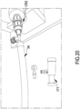
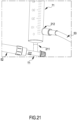
- the air bubbles possibly already present in the perfusion circuit do not reach the scaffold due to the presence of the bubble trap ( Fig. 21 , 71; Fig. 27 , BT) in the circuit.
- the method created by the Applicant is capable of ensuring complete absence of air bubbles in the lumen of the scaffold.
- This system meets the requirements set forth by the configuration of the bioreactor. All details indicated and described are required to make the method independent from the operator and thus for ensuring the reproducibility of the results during the in vitro generation of a construct with functional endothelium at industrial level.
- this method allows to be able to operate under sterility conditions, being based on simple actions.
- the quick and traceable procedure allows to reduce the risk that the air bubbles come into contact with the cells, thus avoiding damaging the endothelial cells adhered on the lumen of the scaffold, preferably tubular.
- Said perfusion method allows to obtain engineered vascular constructs/tissues having a scaffold having a lumen covered with a continuous (i.e. having a monolayer of confluent cells) and functional endothelium.
- the experimental analysis regarding the evaluation of the method for connecting the perfusion circuit to the bioreactor-scaffold system are the same ones applied for the evaluation of the seeding method of a cell culture in a scaffold preferably tubular.
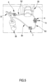
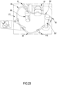
- the perfusion circuit ( Fig. 5 and Fig. 22 ) is defined as an assembly of: tubes ( Fig. 5 ; 51- 54 or Fig. 22 ; 51-55), a reservoir ( Fig. 5 or Fig. 22 ; 56), a peristaltic pump ( Fig. 5 ; 55 or Fig. 22 ; 57) and an element for removing the air bubbles ( Fig. 21 , 71 or Fig. 22 , BT).
- Said element for removing the air bubbles can be present in the perfusion circuit prior to the connection of the perfusion circuit to the bioreactor-seeded scaffold system (second embodiment of the perfusion method) or, alternatively, it may be inserted into the perfusion circuit after the connection of the perfusion circuit to the bioreactor-seeded scaffold system (first embodiment of the perfusion method).
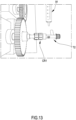
- Said tubes are made of biocompatible material and are connected to each other so as to allow the perfusion of the scaffold ( Fig. 21 and Fig. 27 ), preferably tubular, housed in the bioreactor 11 , by means of the peristaltic pump ( Fig. 5 ; 55, Fig.
- the perfusion circuit mainly consists of five tubes with an inner diameter of 3/16": a first tube 51 for suctioning from the reservoir, a second under-pump tube 52, a third tube 53 which connects the circuit to the bubble trap BT, a fourth tube 54 which connects the BT to the T-shaped connector T2 upstream of the bioreactor-scaffold system, a fifth tube 55 for return to the reservoir 56 connected to the T-shaped connector T3 downstream of the bioreactor-scaffold system.
- the tube 51 is connected to the under-pump tube 52, the under-pump tube 52 to the BT, the BT to the tube 53, the tube 53 to the T-shaped connector T2 upstream of the bioreactor-scaffold system, the tube 54 to the T-shaped connector T3 downstream of the bioreactor-scaffold system and to the reservoir 56.
- the reservoir ( Fig. 5 or Fig. 22 , 56) is the element containing the hot fresh growth medium (for example Endothelial Growth Medium EGM, Sigma Aldrich) from which the tube 51 ( Fig. 5 or Fig. 22 ) suctions and to which the tube 54 ( Fig. 5 ) or 55 ( Fig.
- EGM Endothelial Growth Medium
- the reservoir ( Fig. 5 or Fig. 22 , 56) returns, keeping the entire bioreactor-seeded scaffold system in a closed system.
- the reservoir ( Fig. 5 or Fig. 22 , 56) is under atmospheric pressure, due to a 0.22 m filter present in the cap of the reservoir, which guarantees the sterility of the air.
- the first phase to be carried out and optimised is the cell seeding phase, preferably endothelial cells, followed by a second critical phase of connecting the perfusion circuit to the system comprising the bioreactor and the scaffold, so as to ensure reliability, effectiveness and reproducibility to the industrialisation process for the in vitro generation of a continuous and functional endothelium.
- the success of the industrialisation process subject of the present invention for the production of the engineered vascular tissue/construct mainly consists in the success relating to the phase of seeding and connecting the perfusion circuit to the bioreactor-scaffold system, so as to globally guarantee the elimination of air bubbles in a reliable and reproducible manner.
- the seeding method depends on the cell source and on the density thereof, on the chemical and porosity properties and on the full removal of the air bubbles from the lumen of the scaffold during the injection of the cell suspension.
- the method for connection between the perfusion circuit and the bioreactor-scaffold system is based on maintaining the sterility of the assembled system and on the guarantee of absence of air bubbles that can come into contact with the seeded scaffold. It should be observed that the formation of air bubbles must be avoided given that the air bubbles can damage the cells, jeopardising the viability thereof with ensuing lack of endothelisation of the scaffolds.
- the description of the present invention shows that the choice of the method for connecting the perfusion circuit to the bioreactor-scaffold system depends on the method for seeding the previously optimised endothelial cells due to the fact that said connection method must be suitable to the experimental setup and to the perfusion needs and the position chosen for this system in the incubator.
- the process for seeding a scaffold, preferably a tubular scaffold is one of the factors crucial towards in vitro generation of functional engineered vascular constructs, using confluent endothelium, as shown in the description of the present invention. This process is responsible for a uniform and homogeneous distribution of endothelial cells in the lumen same case applying to the adhesion of the cells to the surface.
- the description of the present invention shows that the choice of the most appropriate seeding method, adapting it to each bioreactor-scaffold system, defines the reproducibility of the process, showing the advantages thereof in the reproducibility of the results.
- the seeding method of the present invention guarantees a highly uniform distribution in terms of adhesion of endothelial cells and good reproducibility of the results, required for a laboratory whose activity focuses on the production of in vitro vascular constructs as test models of the preclinical field as well as other fields.
- the cells adhered in the lumen of the scaffold mainly tubular, must maintain their morphology and viability so as to obtain a homogeneous vascular endothelium.
- the known methods for connection between the bioreactor and the perfusion circuit cause cell suffering with ensuing detachment - even partial - of the endothelial cells from the luminal surface, so that the formation of a functional endothelial layer is slowed or hindered.
- the known methods for connection between the bioreactor and the perfusion circuit do not guarantee the absence of air bubbles, that may be formed due to possible torsions or compressions (full or partial) of the connection tubes during the perfusion. It is important to absolutely prevent the contact between said air bubbles and the seeded scaffold which is avoided by introducing an element, for example a bubble-trap capable of eliminating the air bubbles before they reach the seeded scaffold. This drawback was successfully overcome thanks to the process subject of the present invention which allows to obtain an inner surface of the scaffold covered with a uniform and functional layer of endothelial cells, in particular a confluent cell layer).
Landscapes
- Health & Medical Sciences (AREA)
- Life Sciences & Earth Sciences (AREA)
- Engineering & Computer Science (AREA)
- Biomedical Technology (AREA)
- Chemical & Material Sciences (AREA)
- Cell Biology (AREA)
- Biotechnology (AREA)
- Bioinformatics & Cheminformatics (AREA)
- General Health & Medical Sciences (AREA)
- Immunology (AREA)
- Zoology (AREA)
- Urology & Nephrology (AREA)
- Wood Science & Technology (AREA)
- Genetics & Genomics (AREA)
- Organic Chemistry (AREA)
- Microbiology (AREA)
- Biochemistry (AREA)
- Molecular Biology (AREA)
- Medicinal Chemistry (AREA)
- Hematology (AREA)
- General Engineering & Computer Science (AREA)
- Animal Behavior & Ethology (AREA)
- General Physics & Mathematics (AREA)
- Public Health (AREA)
- Veterinary Medicine (AREA)
- Toxicology (AREA)
- Transplantation (AREA)
- Tropical Medicine & Parasitology (AREA)
- Oral & Maxillofacial Surgery (AREA)
- Dermatology (AREA)
- Pathology (AREA)
- Food Science & Technology (AREA)
- Physics & Mathematics (AREA)
- Analytical Chemistry (AREA)
- Epidemiology (AREA)
- Vascular Medicine (AREA)
- Botany (AREA)
- Chemical Kinetics & Catalysis (AREA)
- Rheumatology (AREA)
- Materials For Medical Uses (AREA)
Claims (14)
- Modèle in vitro d'une structure vasculaire humaine de forme tubulaire comportant des caractéristiques anatomiques et physiologiques dysfonctionnelles simulant la même structure vasculaire d'un sujet sain dont la structure vasculaire a été endommagée, ou déformée ou détériorée en raison d'une lésion choisie dans le groupe comprenant ou, alternativement, constitué par un anévrisme, une sténose, des plaques de sclérose, des formes de tumeurs ou des cardiomyopathies ;
dans lequel ledit modèle comprend ou, alternativement, est constitué d'un ou plusieurs échafaudages de supports polymères poreux biocompatibles (21) capables de favoriser l'adhésion et la croissance des cellules, dans lequel ledit échafaudage a été ensemencé avec des cellules endothéliales qui couvrent une lumière de l'échafaudage et constituent un endothélium comportant une seule couche de cellules confluentes, ledit échafaudage (21) a été fabriqué avec des déformations ou des défauts sur une structure tubulaire de celui-ci, lesdites déformations ou défauts comprenant des courbures, des coudes, des constrictions, des dilatations ; ledit échafaudage (21) est constitué de fibroïne de soie électrofilée, de copolymères d'acide polyglycolique/d'acide polylactique (PGA/PLA) ou de copolymères d'acide polyglycolique/polycaprolactone (PGA/PCL). - Modèle in vitro selon la revendication 1, dans lequel ladite structure vasculaire est choisie parmi les vaisseaux sanguins ou les canaux sanguins.
- Modèle in vitro selon la revendication 1 ou 2, dans lequel ladite structure vasculaire est choisie parmi les artères et les veines.
- Modèle in vitro selon l'une quelconque des revendications 1-3, dans lequel lesdites déformations ou lesdits défauts de la structure tubulaire comprennent des combinaisons de courbures, de coudes, de constrictions, de dilatations.
- Modèle in vitro selon l'une quelconque des revendications 1-4, dans lequel les cellules endothéliales sont choisies parmi les cellules qui forment un endothélium d'un tissu vasculaire.
- Modèle in vitro selon la revendication 5, dans lequel les cellules endothéliales sont choisies parmi les HAOEC (cellules endothéliales aortiques humaines), les HCAEC (cellules endothéliales d'artères coronaires humaines), les HMEVEC (cellules endothéliales microvasculaires de derme humain) ou les HUVEC (cellules endothéliales de la veine ombilicale humaine).
- Procédé pour tester un dispositif médical ou un médicament afin d'en vérifier l'efficacité et la sécurité avant de l'utiliser in vivo sur l'homme, ledit procédé comprenant les étapes suivantes :préparer un échafaudage (21) de forme tubulaire comportant des caractéristiques anatomiques et physiologiques dysfonctionnelles adaptées pour simuler une lésion ou une déformation ou une détérioration due à un anévrisme, une sténose, des plaques de sclérose, des formes de tumeurs ou des cardiomyopathies, ledit échafaudage (21) comportant des déformations ou des défauts sur sa structure tubulaire étant des courbures, des coudes, des constrictions, des dilatations ; ledit échafaudage (21) est constitué de fibroïne de soie électrofilée, de copolymères d'acide polyglycolique/d'acide polylactique (PGA/PLA) ou de copolymères d'acide polyglycolique/ polycaprolactone (PGA/PCL) ;ensemencer au moins une partie de la lumière intérieure dudit échafaudage (21) avec des lignées cellulaires endothéliales de manière à obtenir une couche continue et homogène de cellules endothéliales ensemencées (procédé d'ensemencement), éventuellement ensemencer au moins une partie de la surface extérieure dudit échafaudage (21) avec des lignées cellulaires musculaires ;favoriser la croissance desdites cellules endothéliales, et éventuellement desdites cellules musculaires, jusqu'à obtenir une seule couche continue et homogène de cellules endothéliales, pour obtenir ledit modèle in vitro ;introduire dans ledit modèle in vitro un dispositif médical ou un médicament à tester, etpermettre la circulation (modèle de perfusion) dans ledit modèle in vitro comprenant ledit dispositif médical ou médicament d'un échantillon de sang complet humain, de sang artificiel ou de ses dérivés afin d'évaluer le comportement et l'interaction dudit dispositif médical ou médicament avec ledit échantillon de sang complet humain, de sang artificiel ou de ses dérivés.
- Méthode ou procédé pour la production d'un tissu ou d'une construction vasculaire de synthèse qui est l'échafaudage (21) dans le modèle in vitro selon l'une quelconque des revendications 1-6, pour tester des produits médicaux, ledit procédé comprenant l'application :- d'une méthode d'ensemencement d'une culture de cellules endothéliales dans la lumière dudit échafaudage (21) pour obtenir un échafaudage ensemencé (21) ; ledit échafaudage ensemencé (21) étant présent dans un bioréacteur (11), pour obtenir un système bioréacteur (11)-échafaudage ensemencé (21) ; dans lequel ladite méthode d'ensemencement comprend les étapes de :et, ensuite,- libérer ladite culture de cellules endothéliales sous forme d'une suspension cellulaire comprenant un milieu de culture frais et des cellules endothéliales dans un récipient (91) monté sur un connecteur en « T » (T2) disposé en amont du bioréacteur (11) au moyen d'un connecteur rotatif (CR1) ; puis- libérer ladite culture de cellules endothéliales dans la lumière de l'échafaudage (21) présent dans la chambre du bioréacteur (11) avec un débit continu tel que la vitesse d'écoulement permette à ladite suspension cellulaire de s'égoutter dans le connecteur en « T » (T2) sans générer de bulles d'air et en poussant les bulles d'air présentes dans la lumière de l'échafaudage (21) vers une ouverture d'un connecteur en « T » (T3) disposé en aval du bioréacteur (11) permettant la sortie de ces dernières ;- une méthode de perfusion, avec un milieu de culture frais ayant une température comprise entre 30 et 45 °C, de préférence à 37 °C, des cellules endothéliales présentes dans la lumière dudit échafaudage ensemencé (21) ; ladite méthode de perfusion est obtenue en raccordant un circuit de perfusion (51-56) ou (51-57 et BT) audit système bioréacteur (11)-échafaudage ensemencé (21) ;dans lequel ladite méthode de perfusion comprend une étape de- remplir partiellement un élément pour retirer les bulles d'air (71) ou (BT) présentes dans le circuit de perfusion avec ledit milieu de culture frais, dans lequel ledit élément de retrait des bulles d'air (71) ou (BT) comprend une chambre, un capuchon qui ferme ladite chambre, un accès avec fonction d'entrée (211) et un accès avec fonction de sortie (212), dans lequel ladite chambre de l'élément destiné à retirer les bulles d'air (71 ou BT) a un certain volume et dans lequel une première partie dudit volume est remplie avec ledit milieu de culture frais et dans lequel une deuxième partie dudit volume est remplie d'air, ladite deuxième partie dudit volume ayant la fonction de piéger les bulles d'air présentes dans ledit milieu de culture frais qui s'écoule à travers ledit accès à fonction d'entrée (211) et ledit accès à fonction de sortie (212).
- Procédé selon la revendication 7 ou 8, dans lequel ladite méthode d'ensemencement de ladite culture de cellules endothéliales dans la lumière dudit échafaudage (21) comprend :- monter l'échafaudage (21) sur les accrochages d'un porte-échafaudage (13, 13a, 13b) et loger ledit porte-échafaudage (13, 13a, 13b) avec l'échafaudage (21) dans la chambre du bioréacteur (11), pour obtenir un système bioréacteur (11)-échafaudage (21) ;puis- injecter le milieu de culture frais dans la lumière dudit échafaudage (21) fixé sur ledit porte-échafaudage (13) disposé à l'intérieur de la chambre du bioréacteur (11) ; puis- ajouter ledit milieu de culture frais dans la chambre du bioréacteur (11) où se trouve ledit porte-échafaudage (13, 13a, 13b) avec l'échafaudage (21) injecté avec ledit milieu de culture ; puis- laisser pendant un intervalle de temps compris entre 1 heure et 18 heures à une température comprise entre 20 C° et 30 °C, de préférence 25 °C, ledit milieu de culture dans la lumière de l'échafaudage (21) et dans la chambre du bioréacteur (11) où se trouve ledit porte-échafaudage (13) avec l'échafaudage (21) injecté avec ledit milieu de culture ; puis- purifier l'intérieur de la lumière de l'échafaudage (21) et de la chambre du bioréacteur (11) du milieu de culture ; puis- libérer ladite culture de cellules endothéliales dans ledit récipient (91), de préférence ledit récipient (91) étant une seringue ; puis- libérer ladite suspension cellulaire dans la lumière de l'échafaudage (21) selon la revendication 1 ; puis- ajouter ledit milieu de culture frais dans la chambre du bioréacteur (11) où se trouve ledit porte-échafaudage (13) avec l'échafaudage (21) ensemencé contenant ladite suspension cellulaire dans la lumière ; et puis- incuber, de préférence pendant 24 heures à 37 °C en présence de 5 % de C02, l'échafaudage (21) logé dans la chambre du bioréacteur (11).
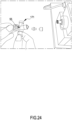
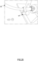
- Procédé selon l'une quelconque des revendications 7-9, dans lequel ladite méthode de perfusion des cellules endothéliales présentes dans la lumière dudit échafaudage ensemencé (21) comprend :préparer ledit circuit de perfusion fermé comprenant les tubes (51), (52), (53), (54) et, éventuellement, (55) ;- obstruer le tube (54) ou (55) du circuit de perfusion à l'aide d'un élément de fermeture (171) dans une position proche d'un connecteur (C), de préférence ledit élément de fermeture est un collier ou similaire ; puis- dévisser le connecteur (C) disposé entre le tube (53) ou (54) et le tube (54) ou (55) respectivement dans le circuit de perfusion ;- visser le tube (53) ou (54) du circuit de perfusion à une extrémité latérale ouverte du connecteur en « T » (T2) en amont du bioréacteur (11) en correspondance d'un accès latéral de celui-ci ; puis- ouvrir le connecteur en « T » (T3) en aval du bioréacteur (11) et dévisser un capuchon d'une ouverture latérale du connecteur en « T » (T3) ; puis- raccorder le tube (54) ou (55) du circuit de perfusion à l'ouverture latérale du connecteur en « T » (T3) disposé en aval du bioréacteur (11) et retirer l'élément de fermeture (171) ;puis, le cas échéant- insérer, entre le tube (53) et le tube de sous-pompe (52) du circuit de perfusion, l'élément destiné à retirer les bulles d'air (71).
- Procédé selon l'une quelconque des revendications précédentes 7-10, dans lequel l'élément permettant de retirer les bulles d'air (71) ou (BT) est un piège à bulles ou similaire.
- Procédé selon l'une quelconque des revendications précédentes 8-11, dans lequel le milieu de culture utilisé est le milieu de culture endothélial comprenant du sérum bovin fœtal (2 %), de l'adénine (0,2 pg/ml), du métavanadate d'ammonium (0,0006 pg/ml), de l'amphotéricine B (0,3 pg/ml), du chlorure de calcium 2H2O (300 pg/ml), du chlorure de choline (20 pg/ml), du sulfate de cuivre 5H2O (0,002 pg/ml), de l'acide trioptique DL-6,8(0,003 pg/ml), de l'acide folinique (calcium) (0,6 pg/ml), de l'héparine (4 pg/ml), de l'hydrocortisone (2 pg/ml), de l'acide L-aspartique (15 pg/ml), de l'L-cystéine (30 pg/ml), de l'L-tyrosine (20 pg/ml), du sulfate de manganèse monohydraté (0,0002 pg/ml), du molybdate d'ammonium 4H2O (0,004 pg/ml), du nicotinamide (8 pg/ml), du chlorure de nickel 6H2O (0,0001 pg/ml), de la pénicilline (60 pg/ml), du sel de sodium de rouge de phénol (15 pg/ml), du chlorure de potassium (300 pg/ml), du dihydrochlorure de putrescine (0,0002 pg/ml), du chlorhydrate de pyridoxine (3 pg/ml), du métasilicate de sodium 9H2O (3 pg/ml), du sulfate de sodium 7H2O (200 pg/ml), du sélénite de sodium (0,01 pg/ml), du sulfate de streptomycine (100 pg/ml), du chlorhydrate de thiamine (4 pg/ml) et du sulfate de zinc 7H2O (0,0003 pg/ml), de préférence chauffés à 37 °C.
- Échafaudage (21) à utiliser dans une préparation d'un modèle in vitro selon les revendications 1-6, comportant une lumière recouverte d'un endothélium fonctionnel et continu (21) comportant une monocouche cellulaire confluente, obtenu au moyen d'un procédé comprenant les étapes suivantes :- préparer un échafaudage de forme tubulaire comportant les caractéristiques anatomiques et physiologiques dysfonctionnelles adaptées pour simuler une lésion, une déformation ou une détérioration due à un anévrisme, une sténose, des plaques de sclérose, des formes de tumeurs ou des cardiomyopathies, dans lequel l'échafaudage est fabriqué avec des déformations ou des défauts sur sa structure tubulaire, lesdites déformations ou défauts comprenant des courbures, des coudes, des constrictions, des dilatations, et est constitué de fibroïne de soie électrofilée, de copolymères d'acide polyglycolique/d'acide polylactique (PGA/PLA) ou de copolymères d'acide polyglycolique/polycaprolactone (PGA/PCL) ;- ensemencer au moins une partie de la lumière intérieure dudit échafaudage avec des lignées cellulaires endothéliales de manière à obtenir une couche continue et homogène de cellules endothéliales ensemencées (procédé d'ensemencement), éventuellement ensemencer au moins une partie de la surface extérieure dudit échafaudage avec des lignées cellulaires musculaires ;- favoriser la croissance desdites cellules endothéliales, et éventuellement desdites cellules musculaires, jusqu'à obtenir une couche continue et homogène de cellules endothéliales, pour obtenir ledit modèle in vitro.
- Utilisation de l'échafaudage (21) selon l'une quelconque des revendications 1-6 ou 13, pour conduire des essais précliniques ou cliniques in vitro d'un médicament à usage humain destiné à être utilisé dans la région cardiovasculaire et vasculaire périphérique.
Applications Claiming Priority (2)
| Application Number | Priority Date | Filing Date | Title |
|---|---|---|---|
| IT102018000007946A IT201800007946A1 (it) | 2018-08-07 | 2018-08-07 | Modello per simulare in-vitro il comportamento di vasi disfunzionali |
| PCT/IB2019/056652 WO2020031067A1 (fr) | 2018-08-07 | 2019-08-05 | Modèle de simulation in vitro du comportement de vaisseaux dysfonctionnels |
Publications (3)
| Publication Number | Publication Date |
|---|---|
| EP3833741A1 EP3833741A1 (fr) | 2021-06-16 |
| EP3833741B1 true EP3833741B1 (fr) | 2023-11-15 |
| EP3833741B8 EP3833741B8 (fr) | 2024-03-06 |
Family
ID=64316663
Family Applications (1)
| Application Number | Title | Priority Date | Filing Date |
|---|---|---|---|
| EP19769576.0A Active EP3833741B8 (fr) | 2018-08-07 | 2019-08-05 | Modèle de simulation in vitro du comportement de vaisseaux dysfonctionnels |
Country Status (8)
| Country | Link |
|---|---|
| US (1) | US20210222130A1 (fr) |
| EP (1) | EP3833741B8 (fr) |
| JP (1) | JP2021533738A (fr) |
| CA (1) | CA3107380A1 (fr) |
| ES (1) | ES2970111T3 (fr) |
| IL (1) | IL280470A (fr) |
| IT (1) | IT201800007946A1 (fr) |
| WO (1) | WO2020031067A1 (fr) |
Families Citing this family (4)
| Publication number | Priority date | Publication date | Assignee | Title |
|---|---|---|---|---|
| IT201900002193A1 (it) * | 2019-02-14 | 2020-08-14 | 1Lab Sa | Metodo di caratterizzazione di un costrutto vascolare ingegnerizzato |
| CN111575183B (zh) * | 2020-04-30 | 2021-06-29 | 北京理工大学 | 一种气泡驱动的环状微单元阵列化组装系统及方法 |
| CN115747065A (zh) * | 2022-11-15 | 2023-03-07 | 南京中医药大学 | 一种体外循环给药模拟对血管内皮作用的实验装置 |
| CN116769598A (zh) * | 2023-07-24 | 2023-09-19 | 柔脉医疗(深圳)有限公司 | 一种人工组织的体外培养装置 |
Family Cites Families (11)
| Publication number | Priority date | Publication date | Assignee | Title |
|---|---|---|---|---|
| US7615373B2 (en) * | 1999-02-25 | 2009-11-10 | Virginia Commonwealth University Intellectual Property Foundation | Electroprocessed collagen and tissue engineering |
| US7759113B2 (en) * | 1999-04-30 | 2010-07-20 | The General Hospital Corporation | Fabrication of tissue lamina using microfabricated two-dimensional molds |
| US20070243574A1 (en) * | 2006-01-27 | 2007-10-18 | University Of Arizona | Vascular mimic for drug and device evaluation |
| WO2011093268A1 (fr) * | 2010-01-29 | 2011-08-04 | 株式会社オーガンテクノロジーズ | Procédé de culture à perfusion et dispositif de culture à perfusion pour un organe ou un tissu |
| FI20115670A0 (fi) * | 2011-06-23 | 2011-06-23 | Tampereen Yliopisto | Kardiovaskulaarinen in vitro-malli |
| WO2015005349A1 (fr) * | 2013-07-09 | 2015-01-15 | 国立大学法人東京大学 | Support de culture cellulaire, dispositif de culture cellulaire, kit de culture cellulaire et feuille cellulaire |
| KR102343744B1 (ko) * | 2013-10-30 | 2021-12-24 | 밀리차 라디식 | 3차원 조직 배양을 위한 장치 및 방법 |
| WO2017100782A1 (fr) * | 2015-12-11 | 2017-06-15 | Research Institute At Nationwide Children's Hospital | Systèmes et procédés pour greffons vasculaires de génie tissulaire optimisés spécifiques à un patient |
| WO2018053265A1 (fr) * | 2016-09-16 | 2018-03-22 | University Of Kansas | Vaisseaux sanguins obtenus par ingénierie |
| US11794184B2 (en) * | 2016-09-29 | 2023-10-24 | Nanyang Technological University | Three-dimensional (3D) hydrogel patterning in microfluidic vascular models |
| IT201700093925A1 (it) * | 2017-08-16 | 2019-02-16 | 1Lab Sa | Un processo di industrializzazione affidabile e riproducibile di eliminazione di bolle d'aria per la produzione di un tessuto vascolare ingegnerizzato. |
-
2018
- 2018-08-07 IT IT102018000007946A patent/IT201800007946A1/it unknown
-
2019
- 2019-08-05 ES ES19769576T patent/ES2970111T3/es active Active
- 2019-08-05 WO PCT/IB2019/056652 patent/WO2020031067A1/fr not_active Ceased
- 2019-08-05 US US17/259,898 patent/US20210222130A1/en not_active Abandoned
- 2019-08-05 CA CA3107380A patent/CA3107380A1/fr active Pending
- 2019-08-05 JP JP2021505662A patent/JP2021533738A/ja active Pending
- 2019-08-05 EP EP19769576.0A patent/EP3833741B8/fr active Active
-
2021
- 2021-01-28 IL IL280470A patent/IL280470A/en unknown
Also Published As
| Publication number | Publication date |
|---|---|
| WO2020031067A1 (fr) | 2020-02-13 |
| US20210222130A1 (en) | 2021-07-22 |
| JP2021533738A (ja) | 2021-12-09 |
| ES2970111T3 (es) | 2024-05-27 |
| WO2020031067A4 (fr) | 2020-05-14 |
| IL280470A (en) | 2021-03-01 |
| IT201800007946A1 (it) | 2020-02-07 |
| CA3107380A1 (fr) | 2020-02-13 |
| EP3833741A1 (fr) | 2021-06-16 |
| EP3833741B8 (fr) | 2024-03-06 |
Similar Documents
| Publication | Publication Date | Title |
|---|---|---|
| EP3833741B1 (fr) | Modèle de simulation in vitro du comportement de vaisseaux dysfonctionnels | |
| JP5356215B2 (ja) | 灌流可能な微小血管システムを作製する方法 | |
| EP2617811B1 (fr) | Procédé de fabrication de feuillet cellulaire multicouche, feuillet cellulaire multicouche ayant un réseau vasculaire obtenu par le procédé, procédé d'utilisation de celui-ci | |
| KR20150125926A (ko) | 인간 및 대형-포유류 폐 생물반응기 | |
| US12448607B2 (en) | Systems and methods for multilane vasculature | |
| JP2023014146A (ja) | 工学的操作された血管組織の作製において気泡を除去するための信頼性が高く再現可能な工業化プロセス | |
| US20210054319A1 (en) | Flow bioreactor device for monitoring cellular dynamics | |
| US11473054B2 (en) | Method of producing three-dimensional tissue having vascular system structure, and three-dimensional tissue including gel having vascular system structure | |
| Visconti et al. | Cardiovascular tissue engineering I. Perfusion bioreactors: a review | |
| BABA et al. | Combined automated culture system for tubular structure assembly and maturation for vascular tissue engineering | |
| Wacker et al. | Bacterial nanocellulose-based grafts for cell colonization studies: An in vitro bioreactor perfusion model | |
| US20220098549A1 (en) | Method for characterising a tissue-engineered construct | |
| Dew | Development of angiogenic models to investigate neovascularisation for tissue engineering applications | |
| Ji et al. | Development of a Novel In Vitro Co-Culture System for Discrete Subaortic Stenosis | |
| Delft et al. | Towards efficient perfusion recellularization of porcine liver grafts with liver-derived organoids | |
| Nguyen et al. | In vivo techniques and strategies for enhanced vascularization of engineered bone |
Legal Events
| Date | Code | Title | Description |
|---|---|---|---|
| STAA | Information on the status of an ep patent application or granted ep patent |
Free format text: STATUS: UNKNOWN |
|
| STAA | Information on the status of an ep patent application or granted ep patent |
Free format text: STATUS: THE INTERNATIONAL PUBLICATION HAS BEEN MADE |
|
| PUAI | Public reference made under article 153(3) epc to a published international application that has entered the european phase |
Free format text: ORIGINAL CODE: 0009012 |
|
| STAA | Information on the status of an ep patent application or granted ep patent |
Free format text: STATUS: REQUEST FOR EXAMINATION WAS MADE |
|
| 17P | Request for examination filed |
Effective date: 20210209 |
|
| AK | Designated contracting states |
Kind code of ref document: A1 Designated state(s): AL AT BE BG CH CY CZ DE DK EE ES FI FR GB GR HR HU IE IS IT LI LT LU LV MC MK MT NL NO PL PT RO RS SE SI SK SM TR |
|
| DAV | Request for validation of the european patent (deleted) | ||
| DAX | Request for extension of the european patent (deleted) | ||
| STAA | Information on the status of an ep patent application or granted ep patent |
Free format text: STATUS: EXAMINATION IS IN PROGRESS |
|
| 17Q | First examination report despatched |
Effective date: 20220126 |
|
| REG | Reference to a national code |
Ref country code: DE Ref legal event code: R079 Ipc: G01N0033500000 Ref country code: DE Ref legal event code: R079 Ref document number: 602019041539 Country of ref document: DE Free format text: PREVIOUS MAIN CLASS: C12N0005071000 Ipc: G01N0033500000 |
|
| GRAP | Despatch of communication of intention to grant a patent |
Free format text: ORIGINAL CODE: EPIDOSNIGR1 |
|
| STAA | Information on the status of an ep patent application or granted ep patent |
Free format text: STATUS: GRANT OF PATENT IS INTENDED |
|
| RIC1 | Information provided on ipc code assigned before grant |
Ipc: C12N 5/071 20100101ALI20230512BHEP Ipc: A61L 27/50 20060101ALI20230512BHEP Ipc: A61L 27/38 20060101ALI20230512BHEP Ipc: G01N 33/50 20060101AFI20230512BHEP |
|
| INTG | Intention to grant announced |
Effective date: 20230601 |
|
| GRAS | Grant fee paid |
Free format text: ORIGINAL CODE: EPIDOSNIGR3 |
|
| GRAA | (expected) grant |
Free format text: ORIGINAL CODE: 0009210 |
|
| STAA | Information on the status of an ep patent application or granted ep patent |
Free format text: STATUS: THE PATENT HAS BEEN GRANTED |
|
| AK | Designated contracting states |
Kind code of ref document: B1 Designated state(s): AL AT BE BG CH CY CZ DE DK EE ES FI FR GB GR HR HU IE IS IT LI LT LU LV MC MK MT NL NO PL PT RO RS SE SI SK SM TR |
|
| REG | Reference to a national code |
Ref country code: CH Ref legal event code: EP Ref country code: GB Ref legal event code: FG4D |
|
| REG | Reference to a national code |
Ref country code: DE Ref legal event code: R096 Ref document number: 602019041539 Country of ref document: DE |
|
| REG | Reference to a national code |
Ref country code: IE Ref legal event code: FG4D |
|
| REG | Reference to a national code |
Ref country code: CH Ref legal event code: PK Free format text: BERICHTIGUNG B8 |
|
| RAP2 | Party data changed (patent owner data changed or rights of a patent transferred) |
Owner name: 1MED SA |
|
| REG | Reference to a national code |
Ref country code: LT Ref legal event code: MG9D |
|
| REG | Reference to a national code |
Ref country code: NL Ref legal event code: MP Effective date: 20231115 |
|
| PG25 | Lapsed in a contracting state [announced via postgrant information from national office to epo] |
Ref country code: GR Free format text: LAPSE BECAUSE OF FAILURE TO SUBMIT A TRANSLATION OF THE DESCRIPTION OR TO PAY THE FEE WITHIN THE PRESCRIBED TIME-LIMIT Effective date: 20240216 |
|
| PG25 | Lapsed in a contracting state [announced via postgrant information from national office to epo] |
Ref country code: IS Free format text: LAPSE BECAUSE OF FAILURE TO SUBMIT A TRANSLATION OF THE DESCRIPTION OR TO PAY THE FEE WITHIN THE PRESCRIBED TIME-LIMIT Effective date: 20240315 |
|
| PG25 | Lapsed in a contracting state [announced via postgrant information from national office to epo] |
Ref country code: LT Free format text: LAPSE BECAUSE OF FAILURE TO SUBMIT A TRANSLATION OF THE DESCRIPTION OR TO PAY THE FEE WITHIN THE PRESCRIBED TIME-LIMIT Effective date: 20231115 |
|
| REG | Reference to a national code |
Ref country code: AT Ref legal event code: MK05 Ref document number: 1632220 Country of ref document: AT Kind code of ref document: T Effective date: 20231115 |
|
| PG25 | Lapsed in a contracting state [announced via postgrant information from national office to epo] |
Ref country code: NL Free format text: LAPSE BECAUSE OF FAILURE TO SUBMIT A TRANSLATION OF THE DESCRIPTION OR TO PAY THE FEE WITHIN THE PRESCRIBED TIME-LIMIT Effective date: 20231115 |
|
| PG25 | Lapsed in a contracting state [announced via postgrant information from national office to epo] |
Ref country code: AT Free format text: LAPSE BECAUSE OF FAILURE TO SUBMIT A TRANSLATION OF THE DESCRIPTION OR TO PAY THE FEE WITHIN THE PRESCRIBED TIME-LIMIT Effective date: 20231115 |
|
| PG25 | Lapsed in a contracting state [announced via postgrant information from national office to epo] |
Ref country code: NL Free format text: LAPSE BECAUSE OF FAILURE TO SUBMIT A TRANSLATION OF THE DESCRIPTION OR TO PAY THE FEE WITHIN THE PRESCRIBED TIME-LIMIT Effective date: 20231115 Ref country code: LT Free format text: LAPSE BECAUSE OF FAILURE TO SUBMIT A TRANSLATION OF THE DESCRIPTION OR TO PAY THE FEE WITHIN THE PRESCRIBED TIME-LIMIT Effective date: 20231115 Ref country code: IS Free format text: LAPSE BECAUSE OF FAILURE TO SUBMIT A TRANSLATION OF THE DESCRIPTION OR TO PAY THE FEE WITHIN THE PRESCRIBED TIME-LIMIT Effective date: 20240315 Ref country code: GR Free format text: LAPSE BECAUSE OF FAILURE TO SUBMIT A TRANSLATION OF THE DESCRIPTION OR TO PAY THE FEE WITHIN THE PRESCRIBED TIME-LIMIT Effective date: 20240216 Ref country code: BG Free format text: LAPSE BECAUSE OF FAILURE TO SUBMIT A TRANSLATION OF THE DESCRIPTION OR TO PAY THE FEE WITHIN THE PRESCRIBED TIME-LIMIT Effective date: 20240215 Ref country code: AT Free format text: LAPSE BECAUSE OF FAILURE TO SUBMIT A TRANSLATION OF THE DESCRIPTION OR TO PAY THE FEE WITHIN THE PRESCRIBED TIME-LIMIT Effective date: 20231115 Ref country code: PT Free format text: LAPSE BECAUSE OF FAILURE TO SUBMIT A TRANSLATION OF THE DESCRIPTION OR TO PAY THE FEE WITHIN THE PRESCRIBED TIME-LIMIT Effective date: 20240315 |
|
| REG | Reference to a national code |
Ref country code: ES Ref legal event code: FG2A Ref document number: 2970111 Country of ref document: ES Kind code of ref document: T3 Effective date: 20240527 |
|
| PG25 | Lapsed in a contracting state [announced via postgrant information from national office to epo] |
Ref country code: SE Free format text: LAPSE BECAUSE OF FAILURE TO SUBMIT A TRANSLATION OF THE DESCRIPTION OR TO PAY THE FEE WITHIN THE PRESCRIBED TIME-LIMIT Effective date: 20231115 Ref country code: RS Free format text: LAPSE BECAUSE OF FAILURE TO SUBMIT A TRANSLATION OF THE DESCRIPTION OR TO PAY THE FEE WITHIN THE PRESCRIBED TIME-LIMIT Effective date: 20231115 Ref country code: PL Free format text: LAPSE BECAUSE OF FAILURE TO SUBMIT A TRANSLATION OF THE DESCRIPTION OR TO PAY THE FEE WITHIN THE PRESCRIBED TIME-LIMIT Effective date: 20231115 Ref country code: NO Free format text: LAPSE BECAUSE OF FAILURE TO SUBMIT A TRANSLATION OF THE DESCRIPTION OR TO PAY THE FEE WITHIN THE PRESCRIBED TIME-LIMIT Effective date: 20240215 Ref country code: LV Free format text: LAPSE BECAUSE OF FAILURE TO SUBMIT A TRANSLATION OF THE DESCRIPTION OR TO PAY THE FEE WITHIN THE PRESCRIBED TIME-LIMIT Effective date: 20231115 Ref country code: HR Free format text: LAPSE BECAUSE OF FAILURE TO SUBMIT A TRANSLATION OF THE DESCRIPTION OR TO PAY THE FEE WITHIN THE PRESCRIBED TIME-LIMIT Effective date: 20231115 |
|
| PG25 | Lapsed in a contracting state [announced via postgrant information from national office to epo] |
Ref country code: DK Free format text: LAPSE BECAUSE OF FAILURE TO SUBMIT A TRANSLATION OF THE DESCRIPTION OR TO PAY THE FEE WITHIN THE PRESCRIBED TIME-LIMIT Effective date: 20231115 |
|
| PG25 | Lapsed in a contracting state [announced via postgrant information from national office to epo] |
Ref country code: CZ Free format text: LAPSE BECAUSE OF FAILURE TO SUBMIT A TRANSLATION OF THE DESCRIPTION OR TO PAY THE FEE WITHIN THE PRESCRIBED TIME-LIMIT Effective date: 20231115 |
|
| PG25 | Lapsed in a contracting state [announced via postgrant information from national office to epo] |
Ref country code: SK Free format text: LAPSE BECAUSE OF FAILURE TO SUBMIT A TRANSLATION OF THE DESCRIPTION OR TO PAY THE FEE WITHIN THE PRESCRIBED TIME-LIMIT Effective date: 20231115 |
|
| PG25 | Lapsed in a contracting state [announced via postgrant information from national office to epo] |
Ref country code: SM Free format text: LAPSE BECAUSE OF FAILURE TO SUBMIT A TRANSLATION OF THE DESCRIPTION OR TO PAY THE FEE WITHIN THE PRESCRIBED TIME-LIMIT Effective date: 20231115 Ref country code: SK Free format text: LAPSE BECAUSE OF FAILURE TO SUBMIT A TRANSLATION OF THE DESCRIPTION OR TO PAY THE FEE WITHIN THE PRESCRIBED TIME-LIMIT Effective date: 20231115 Ref country code: RO Free format text: LAPSE BECAUSE OF FAILURE TO SUBMIT A TRANSLATION OF THE DESCRIPTION OR TO PAY THE FEE WITHIN THE PRESCRIBED TIME-LIMIT Effective date: 20231115 Ref country code: IT Free format text: LAPSE BECAUSE OF FAILURE TO SUBMIT A TRANSLATION OF THE DESCRIPTION OR TO PAY THE FEE WITHIN THE PRESCRIBED TIME-LIMIT Effective date: 20231115 Ref country code: EE Free format text: LAPSE BECAUSE OF FAILURE TO SUBMIT A TRANSLATION OF THE DESCRIPTION OR TO PAY THE FEE WITHIN THE PRESCRIBED TIME-LIMIT Effective date: 20231115 Ref country code: DK Free format text: LAPSE BECAUSE OF FAILURE TO SUBMIT A TRANSLATION OF THE DESCRIPTION OR TO PAY THE FEE WITHIN THE PRESCRIBED TIME-LIMIT Effective date: 20231115 Ref country code: CZ Free format text: LAPSE BECAUSE OF FAILURE TO SUBMIT A TRANSLATION OF THE DESCRIPTION OR TO PAY THE FEE WITHIN THE PRESCRIBED TIME-LIMIT Effective date: 20231115 |
|
| REG | Reference to a national code |
Ref country code: DE Ref legal event code: R097 Ref document number: 602019041539 Country of ref document: DE |
|
| PLBE | No opposition filed within time limit |
Free format text: ORIGINAL CODE: 0009261 |
|
| STAA | Information on the status of an ep patent application or granted ep patent |
Free format text: STATUS: NO OPPOSITION FILED WITHIN TIME LIMIT |
|
| 26N | No opposition filed |
Effective date: 20240819 |
|
| PG25 | Lapsed in a contracting state [announced via postgrant information from national office to epo] |
Ref country code: SI Free format text: LAPSE BECAUSE OF FAILURE TO SUBMIT A TRANSLATION OF THE DESCRIPTION OR TO PAY THE FEE WITHIN THE PRESCRIBED TIME-LIMIT Effective date: 20231115 |
|
| PG25 | Lapsed in a contracting state [announced via postgrant information from national office to epo] |
Ref country code: SI Free format text: LAPSE BECAUSE OF FAILURE TO SUBMIT A TRANSLATION OF THE DESCRIPTION OR TO PAY THE FEE WITHIN THE PRESCRIBED TIME-LIMIT Effective date: 20231115 |
|
| REG | Reference to a national code |
Ref country code: DE Ref legal event code: R119 Ref document number: 602019041539 Country of ref document: DE |
|
| REG | Reference to a national code |
Ref country code: CH Ref legal event code: PL |
|
| PG25 | Lapsed in a contracting state [announced via postgrant information from national office to epo] |
Ref country code: LU Free format text: LAPSE BECAUSE OF NON-PAYMENT OF DUE FEES Effective date: 20240805 |
|
| GBPC | Gb: european patent ceased through non-payment of renewal fee |
Effective date: 20240805 |
|
| PG25 | Lapsed in a contracting state [announced via postgrant information from national office to epo] |
Ref country code: MC Free format text: LAPSE BECAUSE OF FAILURE TO SUBMIT A TRANSLATION OF THE DESCRIPTION OR TO PAY THE FEE WITHIN THE PRESCRIBED TIME-LIMIT Effective date: 20231115 Ref country code: CH Free format text: LAPSE BECAUSE OF NON-PAYMENT OF DUE FEES Effective date: 20240831 |
|
| REG | Reference to a national code |
Ref country code: BE Ref legal event code: MM Effective date: 20240831 |
|
| PG25 | Lapsed in a contracting state [announced via postgrant information from national office to epo] |
Ref country code: DE Free format text: LAPSE BECAUSE OF NON-PAYMENT OF DUE FEES Effective date: 20250301 |
|
| PG25 | Lapsed in a contracting state [announced via postgrant information from national office to epo] |
Ref country code: GB Free format text: LAPSE BECAUSE OF NON-PAYMENT OF DUE FEES Effective date: 20240805 |
|
| PG25 | Lapsed in a contracting state [announced via postgrant information from national office to epo] |
Ref country code: BE Free format text: LAPSE BECAUSE OF NON-PAYMENT OF DUE FEES Effective date: 20240831 |
|
| PG25 | Lapsed in a contracting state [announced via postgrant information from national office to epo] |
Ref country code: FR Free format text: LAPSE BECAUSE OF NON-PAYMENT OF DUE FEES Effective date: 20240831 |
|
| PG25 | Lapsed in a contracting state [announced via postgrant information from national office to epo] |
Ref country code: IE Free format text: LAPSE BECAUSE OF NON-PAYMENT OF DUE FEES Effective date: 20240805 |
|
| REG | Reference to a national code |
Ref country code: ES Ref legal event code: FD2A Effective date: 20250926 |
|
| PG25 | Lapsed in a contracting state [announced via postgrant information from national office to epo] |
Ref country code: FI Free format text: LAPSE BECAUSE OF FAILURE TO SUBMIT A TRANSLATION OF THE DESCRIPTION OR TO PAY THE FEE WITHIN THE PRESCRIBED TIME-LIMIT Effective date: 20231115 |
|
| PG25 | Lapsed in a contracting state [announced via postgrant information from national office to epo] |
Ref country code: ES Free format text: LAPSE BECAUSE OF NON-PAYMENT OF DUE FEES Effective date: 20240806 |
|
| PG25 | Lapsed in a contracting state [announced via postgrant information from national office to epo] |
Ref country code: CY Free format text: LAPSE BECAUSE OF FAILURE TO SUBMIT A TRANSLATION OF THE DESCRIPTION OR TO PAY THE FEE WITHIN THE PRESCRIBED TIME-LIMIT; INVALID AB INITIO Effective date: 20190805 |
|
| PG25 | Lapsed in a contracting state [announced via postgrant information from national office to epo] |
Ref country code: HU Free format text: LAPSE BECAUSE OF FAILURE TO SUBMIT A TRANSLATION OF THE DESCRIPTION OR TO PAY THE FEE WITHIN THE PRESCRIBED TIME-LIMIT; INVALID AB INITIO Effective date: 20190805 |