EP3848695A1 - Administration sélective de matériau à des cellules - Google Patents
Administration sélective de matériau à des cellules Download PDFInfo
- Publication number
- EP3848695A1 EP3848695A1 EP21158382.8A EP21158382A EP3848695A1 EP 3848695 A1 EP3848695 A1 EP 3848695A1 EP 21158382 A EP21158382 A EP 21158382A EP 3848695 A1 EP3848695 A1 EP 3848695A1
- Authority
- EP
- European Patent Office
- Prior art keywords
- cells
- group
- cell
- constriction
- cell suspension
- Prior art date
- Legal status (The legal status is an assumption and is not a legal conclusion. Google has not performed a legal analysis and makes no representation as to the accuracy of the status listed.)
- Withdrawn
Links
Images


Classifications
-
- B—PERFORMING OPERATIONS; TRANSPORTING
- B01—PHYSICAL OR CHEMICAL PROCESSES OR APPARATUS IN GENERAL
- B01L—CHEMICAL OR PHYSICAL LABORATORY APPARATUS FOR GENERAL USE
- B01L3/00—Containers or dishes for laboratory use, e.g. laboratory glassware; Droppers
- B01L3/50—Containers for the purpose of retaining a material to be analysed, e.g. test tubes
- B01L3/502—Containers for the purpose of retaining a material to be analysed, e.g. test tubes with fluid transport, e.g. in multi-compartment structures
- B01L3/5027—Containers for the purpose of retaining a material to be analysed, e.g. test tubes with fluid transport, e.g. in multi-compartment structures by integrated microfluidic structures, i.e. dimensions of channels and chambers are such that surface tension forces are important, e.g. lab-on-a-chip
- B01L3/502761—Containers for the purpose of retaining a material to be analysed, e.g. test tubes with fluid transport, e.g. in multi-compartment structures by integrated microfluidic structures, i.e. dimensions of channels and chambers are such that surface tension forces are important, e.g. lab-on-a-chip specially adapted for handling suspended solids or molecules independently from the bulk fluid flow, e.g. for trapping or sorting beads or physically stretching molecules
-
- B—PERFORMING OPERATIONS; TRANSPORTING
- B82—NANOTECHNOLOGY
- B82Y—SPECIFIC USES OR APPLICATIONS OF NANOSTRUCTURES; MEASUREMENT OR ANALYSIS OF NANOSTRUCTURES; MANUFACTURE OR TREATMENT OF NANOSTRUCTURES
- B82Y30/00—Nanotechnology for materials or surface science, e.g. nanocomposites
-
- C—CHEMISTRY; METALLURGY
- C12—BIOCHEMISTRY; BEER; SPIRITS; WINE; VINEGAR; MICROBIOLOGY; ENZYMOLOGY; MUTATION OR GENETIC ENGINEERING
- C12N—MICROORGANISMS OR ENZYMES; COMPOSITIONS THEREOF; PROPAGATING, PRESERVING, OR MAINTAINING MICROORGANISMS; MUTATION OR GENETIC ENGINEERING; CULTURE MEDIA
- C12N15/00—Mutation or genetic engineering; DNA or RNA concerning genetic engineering, vectors, e.g. plasmids, or their isolation, preparation or purification; Use of hosts therefor
- C12N15/09—Recombinant DNA-technology
- C12N15/87—Introduction of foreign genetic material using processes not otherwise provided for, e.g. co-transformation
-
- C—CHEMISTRY; METALLURGY
- C12—BIOCHEMISTRY; BEER; SPIRITS; WINE; VINEGAR; MICROBIOLOGY; ENZYMOLOGY; MUTATION OR GENETIC ENGINEERING
- C12N—MICROORGANISMS OR ENZYMES; COMPOSITIONS THEREOF; PROPAGATING, PRESERVING, OR MAINTAINING MICROORGANISMS; MUTATION OR GENETIC ENGINEERING; CULTURE MEDIA
- C12N5/00—Undifferentiated human, animal or plant cells, e.g. cell lines; Tissues; Cultivation or maintenance thereof; Culture media therefor
- C12N5/06—Animal cells or tissues; Human cells or tissues
- C12N5/0602—Vertebrate cells
- C12N5/0634—Cells from the blood or the immune system
-
- C—CHEMISTRY; METALLURGY
- C12—BIOCHEMISTRY; BEER; SPIRITS; WINE; VINEGAR; MICROBIOLOGY; ENZYMOLOGY; MUTATION OR GENETIC ENGINEERING
- C12N—MICROORGANISMS OR ENZYMES; COMPOSITIONS THEREOF; PROPAGATING, PRESERVING, OR MAINTAINING MICROORGANISMS; MUTATION OR GENETIC ENGINEERING; CULTURE MEDIA
- C12N5/00—Undifferentiated human, animal or plant cells, e.g. cell lines; Tissues; Cultivation or maintenance thereof; Culture media therefor
- C12N5/06—Animal cells or tissues; Human cells or tissues
- C12N5/0602—Vertebrate cells
- C12N5/0693—Tumour cells; Cancer cells
-
- C—CHEMISTRY; METALLURGY
- C12—BIOCHEMISTRY; BEER; SPIRITS; WINE; VINEGAR; MICROBIOLOGY; ENZYMOLOGY; MUTATION OR GENETIC ENGINEERING
- C12Q—MEASURING OR TESTING PROCESSES INVOLVING ENZYMES, NUCLEIC ACIDS OR MICROORGANISMS; COMPOSITIONS OR TEST PAPERS THEREFOR; PROCESSES OF PREPARING SUCH COMPOSITIONS; CONDITION-RESPONSIVE CONTROL IN MICROBIOLOGICAL OR ENZYMOLOGICAL PROCESSES
- C12Q1/00—Measuring or testing processes involving enzymes, nucleic acids or microorganisms; Compositions therefor; Processes of preparing such compositions
- C12Q1/02—Measuring or testing processes involving enzymes, nucleic acids or microorganisms; Compositions therefor; Processes of preparing such compositions involving viable microorganisms
- C12Q1/04—Determining presence or kind of microorganism; Use of selective media for testing antibiotics or bacteriocides; Compositions containing a chemical indicator therefor
-
- G—PHYSICS
- G01—MEASURING; TESTING
- G01N—INVESTIGATING OR ANALYSING MATERIALS BY DETERMINING THEIR CHEMICAL OR PHYSICAL PROPERTIES
- G01N1/00—Sampling; Preparing specimens for investigation
- G01N1/28—Preparing specimens for investigation including physical details of (bio-)chemical methods covered elsewhere, e.g. G01N33/50, C12Q
- G01N1/30—Staining; Impregnating ; Fixation; Dehydration; Multistep processes for preparing samples of tissue, cell or nucleic acid material and the like for analysis
-
- G—PHYSICS
- G01—MEASURING; TESTING
- G01N—INVESTIGATING OR ANALYSING MATERIALS BY DETERMINING THEIR CHEMICAL OR PHYSICAL PROPERTIES
- G01N15/00—Investigating characteristics of particles; Investigating permeability, pore-volume or surface-area of porous materials
- G01N15/10—Investigating individual particles
- G01N15/14—Optical investigation techniques, e.g. flow cytometry
- G01N15/1456—Optical investigation techniques, e.g. flow cytometry without spatial resolution of the texture or inner structure of the particle, e.g. processing of pulse signals
- G01N15/1459—Optical investigation techniques, e.g. flow cytometry without spatial resolution of the texture or inner structure of the particle, e.g. processing of pulse signals the analysis being performed on a sample stream
-
- G—PHYSICS
- G01—MEASURING; TESTING
- G01N—INVESTIGATING OR ANALYSING MATERIALS BY DETERMINING THEIR CHEMICAL OR PHYSICAL PROPERTIES
- G01N15/00—Investigating characteristics of particles; Investigating permeability, pore-volume or surface-area of porous materials
- G01N15/10—Investigating individual particles
- G01N15/14—Optical investigation techniques, e.g. flow cytometry
- G01N15/1484—Optical investigation techniques, e.g. flow cytometry microstructural devices
-
- G—PHYSICS
- G01—MEASURING; TESTING
- G01N—INVESTIGATING OR ANALYSING MATERIALS BY DETERMINING THEIR CHEMICAL OR PHYSICAL PROPERTIES
- G01N33/00—Investigating or analysing materials by specific methods not covered by groups G01N1/00 - G01N31/00
- G01N33/48—Biological material, e.g. blood, urine; Haemocytometers
- G01N33/50—Chemical analysis of biological material, e.g. blood, urine; Testing involving biospecific ligand binding methods; Immunological testing
- G01N33/53—Immunoassay; Biospecific binding assay; Materials therefor
- G01N33/575—Immunoassay; Biospecific binding assay; Materials therefor for cancer
-
- B—PERFORMING OPERATIONS; TRANSPORTING
- B01—PHYSICAL OR CHEMICAL PROCESSES OR APPARATUS IN GENERAL
- B01L—CHEMICAL OR PHYSICAL LABORATORY APPARATUS FOR GENERAL USE
- B01L2200/00—Solutions for specific problems relating to chemical or physical laboratory apparatus
- B01L2200/06—Fluid handling related problems
- B01L2200/0647—Handling flowable solids, e.g. microscopic beads, cells, particles
- B01L2200/0652—Sorting or classification of particles or molecules
-
- B—PERFORMING OPERATIONS; TRANSPORTING
- B01—PHYSICAL OR CHEMICAL PROCESSES OR APPARATUS IN GENERAL
- B01L—CHEMICAL OR PHYSICAL LABORATORY APPARATUS FOR GENERAL USE
- B01L2300/00—Additional constructional details
- B01L2300/08—Geometry, shape and general structure
-
- B—PERFORMING OPERATIONS; TRANSPORTING
- B01—PHYSICAL OR CHEMICAL PROCESSES OR APPARATUS IN GENERAL
- B01L—CHEMICAL OR PHYSICAL LABORATORY APPARATUS FOR GENERAL USE
- B01L2400/00—Moving or stopping fluids
- B01L2400/04—Moving fluids with specific forces or mechanical means
- B01L2400/0475—Moving fluids with specific forces or mechanical means specific mechanical means and fluid pressure
- B01L2400/0487—Moving fluids with specific forces or mechanical means specific mechanical means and fluid pressure fluid pressure, pneumatics
-
- C—CHEMISTRY; METALLURGY
- C12—BIOCHEMISTRY; BEER; SPIRITS; WINE; VINEGAR; MICROBIOLOGY; ENZYMOLOGY; MUTATION OR GENETIC ENGINEERING
- C12M—APPARATUS FOR ENZYMOLOGY OR MICROBIOLOGY; APPARATUS FOR CULTURING MICROORGANISMS FOR PRODUCING BIOMASS, FOR GROWING CELLS OR FOR OBTAINING FERMENTATION OR METABOLIC PRODUCTS, i.e. BIOREACTORS OR FERMENTERS
- C12M23/00—Constructional details, e.g. recesses, hinges
- C12M23/02—Form or structure of the vessel
- C12M23/16—Microfluidic devices; Capillary tubes
-
- G—PHYSICS
- G01—MEASURING; TESTING
- G01N—INVESTIGATING OR ANALYSING MATERIALS BY DETERMINING THEIR CHEMICAL OR PHYSICAL PROPERTIES
- G01N15/00—Investigating characteristics of particles; Investigating permeability, pore-volume or surface-area of porous materials
- G01N15/01—Investigating characteristics of particles; Investigating permeability, pore-volume or surface-area of porous materials specially adapted for biological cells, e.g. blood cells
-
- G—PHYSICS
- G01—MEASURING; TESTING
- G01N—INVESTIGATING OR ANALYSING MATERIALS BY DETERMINING THEIR CHEMICAL OR PHYSICAL PROPERTIES
- G01N15/00—Investigating characteristics of particles; Investigating permeability, pore-volume or surface-area of porous materials
- G01N15/10—Investigating individual particles
- G01N2015/1006—Investigating individual particles for cytology
-
- G—PHYSICS
- G01—MEASURING; TESTING
- G01N—INVESTIGATING OR ANALYSING MATERIALS BY DETERMINING THEIR CHEMICAL OR PHYSICAL PROPERTIES
- G01N15/00—Investigating characteristics of particles; Investigating permeability, pore-volume or surface-area of porous materials
- G01N15/10—Investigating individual particles
- G01N2015/1028—Sorting particles
Definitions
- the field of the invention relates to size-selective delivery of material to cells.
- CTCs Circulating tumor cells
- the current subject matter provides devices, systems, and methods for selectively delivering material to one or more cells based on their physical properties, such as size, volume, diameter, cytosol viscosity, or membrane stiffness.
- materials can be delivered in a cell size dependent manner.
- a cell suspension containing differentially sized cells can be run through a device in the presence of the target delivery material (e.g., a dye, a protein, nucleic acid, and the like) and these materials can be selectively delivered to the larger cells within the population.
- the target delivery material e.g., a dye, a protein, nucleic acid, and the like
- the mechanism of delivery in the data being through selective disruption of the cell membrane of larger cells as they are deformed in a channel constriction while smaller cells are not deformed enough to cause membrane disruption.
- labelling tumor cells relative to non-tumor cells can be achieved.
- Cells are run through a device for size selective tagging using fluorescent dyes or other detectable markers.
- the cells are optionally stained with an antibody, e.g., a tumor cell selective antibody, e.g., antibodies against CD45 to provide further contrast between cancer cells and blood cells (most blood cells are CD45+).
- the samples are run through a cell sorter, e.g. a standard fluorescence-activated cell sorter (FACS).
- FACS fluorescence-activated cell sorter
- labeling of cells based on their cell cycle can be achieved because cells within a population that are closer to division are larger than those that have just undergone division. Delivery of a dye to the bigger cells within a population can be used to identify the individual cells that are in a later stage of their cell cycle.
- therapeutics for blood cancers can be achieved because lymphoma cells are often bigger than the surrounding blood cells thus an intracellular toxin can be delivered to lymphoma cells but not the healthy surrounding blood cells. This can induce selective death of diseased cells.
- Tagged cells can be isolated by fluorescence or magnetic purification techniques. Flow cytometry or microarrays with robotic manipulators can be used to select cells based on fluorescence, while magnetic columns, microfluidic magnetic separation systems, or magnetic sweepers can be used to isolate magnetically tagged particles.
- Cells can be identified based on relative size or diameter.
- relatively larger cells selectively or preferentially take up markers, because the extent of cell membrane disruption is relatively greater in larger cells, i.e., larger cells are deformed to a greater extent compared to smaller cells.
- Due to the greater degree of membrane disruption of larger cells at least 10%, 25%, 50%, 2-fold, 5-fold, 10-fold, 100-fold or more of a payload molecule gains access to the inside (cytoplasm) of a larger cell compared to a smaller cell.
- the uptake of detectable markers in this manner and subsequent sorting based on uptake of the marker the purity of tumor cells is enhanced by 100 times; 1,000 times, and up to 10,000 times or more compared to the level of purity in peripheral blood.
- Purity is assessed by an antibody that targets/binds to a known marker that is expressed/overexpressed by tumor cells.
- antibodies against markers that are not expressed by tumor cells but are expressed/overexpressed by blood cells CD45 is an example. Either approach helps provide increased contrast to sort out the cells of interest.
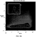
- Samples with high size-tag fluorescence and low CD45 fluorescence are captured as candidate/potential CTCs.
- FACS outputs are inherently relative.
- a "high” signal is minimum one decade (ten times higher level) of fluorescence intensity above the baseline control signal, and a “low” is one decade below the positive control population.
- a method for isolating or identifying a circulating tumor cell comprises the steps of providing a cell suspension; passing the solution through a microfluidic channel that includes a constriction, the constriction being sized to preferentially deform a circulating tumor cell compared to a leukocyte; passing the cell suspension through the constriction; and contacting the cell suspension solution with a detectable marker.
- the suspension can be passed through a microfluidic channel that includes a constriction, the constriction being sized to preferentially deliver a compound to a group of cells having a relatively different physical property than another group of cells.
- the physical property can include cell size, diameter, cytosol viscosity, and/or membrane stiffness (e.g., as measured by transit time assays, stiffer cells pass through specialized microchannels more slowly than less stiff cells, e.g., as described in Sharei et al., 2012, Anal. Chem. 84(15):6438-6443 ; Cross et al., 2007, Nature Nanotechnology 2:780-783 ).
- the contact can happen after deformation treatment. Or the material can be premixed with the cells before deformation treatment. Both CTCs and leukocytes are deformed; however larger cells are deformed to a greater degree and therefore, molecules are selectively delivered to such cells, thereby treating or tagging them.
- the marker comprises a detectably labeled, e.g., fluorescently or magnetically labeled material, such as a dye or particle.
- the dyes or particles need not be tumor specific.
- they differentially bind to tumor cells (e.g., at least 20%, 50%, 2 times, 5 times, or more compared to non-tumor cells).
- the specificity of the method is based on the discovery that tumor cells are slightly larger than leukocytes and the device is highly size selective. This size difference depends on the tumor type. For example, tumor cells are generally from 50%-400% larger than the leukocytes. Therefore, the delivery material preferentially enters into cells that are large enough to be tagged via size-specific deformation of cells.. The delivered tag is then in turn detected to identify the CTC.
- the suspension comprises whole blood.
- the cell suspension is a mixture of cells in a physiological saline solution other than blood.
- the cell suspension comprises whole blood of a subject at risk of or diagnosed as comprising a tumor.
- the patient is suspected of having, has been diagnosed as having, or is suspected or diagnosed as having metastatic disease of melanoma, colon, prostate, breast, liver, lung, pancreatic, brain, or blood.
- CTCs can be present before the patient has developed metastatic disease. Therefore, early detection of CTCs is clinically important, because such detection represents an early identification of patients likely to progress to develop metastatic disease.
- erythrocyte lysis is carried out as a pretreatment step prior to flowing cells through the device.
- the device is characterized by physical parameters that distinguish tumor cells from non-tumor cells, e.g., normal erythrocytes or leukocytes.
- the constriction comprises a width from 4 ⁇ m-10 ⁇ m, length of 1 ⁇ m-100 ⁇ m, and 1-10 constrictions in series.
- the estimated speed of the cells can range from 10mm/s to lOm/s.
- the method further comprises applying a pressure to cells. Pressure is used to drive the cell suspension through the device, and the transit through the constriction point is what deforms the cells and leads to membrane disruption, and therefore delivery.
- the method involves introducing into the tumor cell a detectable compound.
- the cell suspension comprises a payload or the method further comprises a step of incubating said cell suspension in the solution containing a payload for a predetermined time after it passes through the constriction.
- the payload comprises a magnetic particle such as a nanoparticle, a fluorescent particle, such as a quantum dot or carbon nanotube, or a fluorescent dye or protein, or genetic material (DNA or RNA) that codes for a fluorescent protein or other compound that enables detection (e.g., luciferase).
- a fluorescent particle to enable sorting and co-deliver DNA, RNA or a protein to facilitate subsequent tumor cell survival and encourage its growth and proliferation post-sorting to enable further studies of cultured metastatic cells.
- a microfluidic system for distinguishing tumor cells from non-tumor cells comprising a microfluidic channel defining a lumen and being configured such that a tumor cell suspended in a buffer can pass therethrough and is constricted compared to a non-tumor cell.
- Non tumor cells may be deformed to some extent; however, the key is that the tumor cells are deformed enough to cause a cell membrane disruption whereas the non-tumor cells are not deformed enough to result in membrane disruption due to their smaller relative size.
- the membranes of smaller cells are not disrupted or disrupted less than larger cells, e.g., in some cases, both smaller and larger cells are disrupted but smaller cells receive less material than the larger cells.
- the microfluidic channel includes a cell-deforming constriction, wherein a diameter of the constriction is a function of the diameter of the cell.
- the constriction is sized to preferentially deform a tumor cell compared to a non-tumor cell. This preferential deformation is designed to selectively facilitate the delivery of the target material to tumor cells vs. non tumor cells. Selective delivery enables one to enrich the desired tumor population through sorting/enrichment methods such as flow cytometery (FACS), micromanipulation, magnetic separation, cell culture.
- FACS flow cytometery
- the method is carried out at physiological temperature, e.g., 37 °C, room temperature, e.g., 20 °C, or alternatively, at 0-4 °C. In some cases, the latter is preferred, because it can yield better delivery performance due to delayed membrane repair and minimize background from endocytosis by reducing the endocytotic activity of cells.
- the cell suspension is whole blood or any mammalian cell suspension in a physiological buffer solution such as phosphate buffers saline (PBS) or tissue culture media as a delivery buffer.
- PBS phosphate buffers saline
- tissue culture media as a delivery buffer.
- PBS phosphate buffers saline
- isolating or identifying a cell based on a physical property of the cell can include providing a cell suspension; passing the suspension through a microfluidic channel that includes a constriction; passing the cell suspension through the constriction; and, contacting the cell suspension solution with a compound.
- the constriction can be sized to preferentially deform a relatively larger cell compared to a relatively smaller cell.
- a microfluidic system for distinguishing tumor cells from non-tumor cells can include a microfluidic channel defining a lumen and being configured such that a tumor cell suspended in a buffer can pass therethrough and is constricted compared to a non-tumor cell.
- the microfluidic channel can include a cell-deforming constriction. A diameter of the constriction can be a function of the diameter of the cell.
- the physical property can be one or more of size and diameter.
- the cell suspension can include one or more of: peripheral blood cells; and at least two different cell types having different physical properties.
- the cell suspension can include an erythrocyte-depleted population of peripheral blood cells.
- the larger cell can include a circulating tumor cell and the smaller cell can include a leukocyte.
- the compound can include a molecular mass of 0.5 kDa to 5 MDa.
- the compound can include a molecular mass of 3 kDa to 10 kDa.
- the compound can include a detectable marker (e.g., quantum dots, cyanine, fluorescein, rhodamine, and derivatives thereof such as fluorescein isothiocyanate (FITC) or Tetramethylrhodamine isothiocyanate (TRITC) or NHS-Rhodamine, maleimide activated fluorophores such as fluorescein-5-maleimide, as well as Alexa Fluors), an active biomolecule, and/or a toxin, (e.g., Pseudomonas exotoxin, Diphtheria toxin, and ricin, caspase proteins, antibodies that interfere with essential cell functions (e.g. antibodies against tubulin)) for selectively killing target cells.
- a detectable marker e.g., quantum dots, cyanine, fluorescein, rhodamine, and derivatives thereof such as fluorescein isothiocyanate (FITC) or Tetramethylrhodamine isothi
- the compound can influence cell function (e.g. transcription factors, siRNA, DNA, mRNA, antibodies, small molecule drugs) and/or can induce cell death.
- the compound can enter the cell after the cell has passed through the constriction.
- the suspension can include whole blood.
- the suspension can include whole blood of a subject at risk of or diagnosed as comprising a tumor.
- the tumor can include melanoma, colon, prostate, lung, pancreatic, breast, liver, brain, or blood cancer.
- the constriction can include a width from 4 ⁇ m-10 ⁇ m, length of 1 ⁇ m-100 ⁇ m, and 1-10 constrictions in series.
- a speed of the cells traversing a constriction can range from 10mm/s to lOm/s.
- a pressure can be applied to the cell suspension to drive cells through the constriction of a microfluidic channel.
- the cell suspension can include a payload or the cell suspension can be incubated in the solution containing a payload for a predetermined time after it passes through the constriction.
- the payload can include a magnetic particle a fluorescent particle, such as a quantum dot or carbon nanotube, or a fluorescent dye or protein, or genetic material (DNA or RNA) that codes for a fluorescent protein or other compound that enables detection (e.g. luciferase).
- the constriction can be sized to preferentially deform a tumor cell more than a non-tumor cell.
- CTCs are tumor cells that are found in the bloodstream, and are believed to be responsible for the dissemination of cancer to distant organs.
- CTCs are regarded as minimally-invasive, "liquid biopsies” for cancer patients and are useful as prognostic indicators for patient outcome and treatment efficacy. Comprehensive characterizations of these single cells provide a better understanding of metastatic dissemination, treatment resistance, and tumor biology.
- a typical human erythrocyte has a disk diameter of approximately 6.2-8.2 ⁇ m and a thickness at the thickest point of 2-2.5 ⁇ m and a minimum thickness in the center of 0.8-1 ⁇ m, being much smaller than most other human cells.
- Leukocytes white blood cells
- neutrophils (12-14 ⁇ m diameter)
- eosinophils (12-17 ⁇ m diameter
- basophils 14-16 ⁇ m diameter
- lymphocytes average 6-9 ⁇ m in diameter for resting, and 10-14 ⁇ m diameter for activated
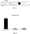
- monocytes the largest type of white blood cells that can be up to 20 ⁇ m in diameter. As shown in Fig.
- the size difference between CTCs and hematologic cells generally permits distinguishing CTCs from other cells in circulating blood (CTCs -9-20 ⁇ m; RBC ⁇ 8 ⁇ m discoid; leukocytes -7-12 ⁇ m). See Fig. 1 .
- Subsequent tumor cell specific labeling using antibodies (or cell-specific fragments thereof) or other tumor cell specific ligands increase the selectivity of the method.
- CTCs are present as one in 10 6 -10 7 mononuclear cells in the bloodstream, high-sensitivity enrichment techniques are used that rely on immunological or morphological differences in CTCs from the blood cells.
- Immunological approaches often target epithelial cell surface markers (such as EpCAM) and tumor-specific proteins (such as Her2-neu, MUC I/MUC2, carcinoembryonic antigen (CEA), mammaglobulin, and alpha-fetoprotein) or aim to deplete CD45+ cells.
- EAA carcinoembryonic antigen
- mammaglobulin and alpha-fetoprotein
- microfilters, density-gradient separations, and microfluidics platforms are examples of morphology-based methods.
- a combined enrichment method integrates both immunological and morphologic-based approaches to tag and isolate pure CTCs with less bias and based on tunable parameters.
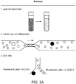
- the method combines microfluidic intracellular delivery ( Fig. 1 ) and antibody staining to yield robust, high sensitivity purification of circulating tumor cells from whole blood ( FIG. 2 ) comprises a width from 4 ⁇ -10 ⁇ m, length of 1 ⁇ m-100 ⁇ m, and 1-10 constrictions in series.
- the estimated speed of the cells can range from 10mm/s to lOm/s.
- the specific device parameters chosen are dictated by the target tumor cell type, e.g., a different device design is used to select CTCs for a melanoma patient vs. a colon cancer patient. Examples of tumor cell sizes/diameters include; melanoma ⁇ 15um, colon cancer ⁇ 11um, and pancreatic cancer ⁇ 15um.
- a rapid mechanical deformation delivery system exploits the inherent size difference between many CTCs and the surrounding blood cells to selectively deliver fluorescent, magnetic and/or other distinguishing materials to the tumor cells.
- antibody-based fluorescent and/or magnetic tagging is used to enhance the contrast between the candidate CTCs and the surrounding blood cells.
- a 6um width constriction can be used. Such a constriction would deform both cell types but would very preferentially disrupt the membrane of the 15 ⁇ m tumor cells not the 8 ⁇ m blood cells.
- CTCs are being explored as surrogates for tumor biopsies for understanding mechanisms of resistance and guiding the selection of targeted therapies. Measures of the number and composition of CTCs before and after treatment indicate treatment efficacy and prognosis.
- the approach utilizes a robust, high-throughput, disposable device for the tagging of CTCs based on cell size and surface antigens.
- the ability to deliver a diversity of macromolecules also enables one to deliver molecular probes (such as antibodies, quantum dots, carbon nanotubes, and molecular beacons) that respond to the intracellular environment and thus provide further information on the intracellular properties of the target cell.
- molecular probes such as antibodies, quantum dots, carbon nanotubes, and molecular beacons
- This combinatorial approach provides a robust platform capable of enriching CTC populations that would have been missed by alternative methods that rely solely on immunological or morphological separation. The technique is useful to isolate patients' CTCs.
- Whole blood or other cell suspensions are processed using both unlabeled and/or antibody-coated magnetic beads. These cells are then isolated using a high-fidelity, magnetic enrichment system for rare cells.
- a nanowell technology may also be used to achieve high purity isolations by imaging and robotically-retrieving single cells of interest from an elastomeric array of 84,672 subnanoliter wells.
- Magnetic nanoparticles are delivered to tumor cell lines & PBMCs.
- Nanoparticle delivery to EpCAM-expressing, epithelial cancer cell lines, e.g., HT-29, LNCaP, and SK-BR-3, is compared to bulk peripheral blood mononuclear cell (PBMC) suspensions derived from human blood.
- PBMC peripheral blood mononuclear cell
- 10nm iron-oxide nanoparticles with a polyethylene glycol (PEG) surface coating are delivered to cancer cells mixed with whole blood, and the resulting mixture of tagged cells are processed using the cell separation system described above.
- the microfluidic delivery system was used to induce a rapid mechanical deformation of a cell to generate transient pores in the cell membrane ( Fig. 1 ).
- the approach has demonstrated an ability to deliver a range of materials, including proteins, RNA, DNA and nanoparticles to a variety of cell types and works with whole blood, a medium that often poses problems for microfluidic systems.
- Exemplary tagging molecules e.g., 3kDa and 70kDa, fluorescently-labeled, dextran polymers as model molecules, were used to discriminate between PBMCs and two different cancer cell lines based on size alone. The results also indicate the utility of the system for the selective delivery of magnetic particles to tumor cells in the blood.
- PEG coated iron-oxide particles are used to magnetically tag colon cancer (e.g., as exemplified by the cell line HT-29). Further enrichment is accomplished using conjugation of FITC to the iron-oxide nanoparticle surface to directly measure nanoparticle uptake.
- PEG coated 10nm iron-oxide nanoparticles are delivered to cell suspensions that are suspected of containing or are known to contain CTCs, e.g., a patient-derived blood sample, or cell lines HT-29, LNCaP, and SK-BR-3 cells, separately mixed with whole blood.
- CTCs e.g., a patient-derived blood sample, or cell lines HT-29, LNCaP, and SK-BR-3 cells
- the resulting mixture of tagged cells are then purified, e.g., using a high fidelity magnetic separator.
- the separator accurately discriminates between the model CTCs with high iron-oxide content and less-effectively labeled PBMCs.
- red blood cells are lysed prior to treatment, nanoparticle concentration increased, their size altered, or incorporating multiple treatment steps.
- a combined immunological and morphologic-based method is carried out as follows. After cell size-based processing by the device, cells are treated with an antibody or other tumor cell specific ligand such as fluorescently labeled anti-CD45 antibodies. The sensitivity and specificity of three different separation approaches were compared:: 1) device only 2) anti-CD45 antibody only 3) device+ anti-CD45 antibody. Morphologic tagging (device) + immunological tagging (e.g., anti-CD45 antibodies) was found to show superior sensitivity (and specificity) relative to either of the individual techniques ( Fig. 2 ). For example, a 2-5x increase in sensitivity and/or a 2-5x increase in specificity relative to anti-CD45 antibodies alone is observed. Enrichment factor of over an order of magnitude was observed ( Fig. 2 ).
- the devices are fabricated out of silicon and glass.
- the device is fabricated using a polymer such as silicone, PDMS, polycarbonate, acrylic, polypropylene, polystyrene.
- Either device is sterilized (heat or gamma radiation) and disposable.
- Performance of the devices is validated for various cell types using materials and parameters. For example, performance at a range of flow speeds (100mm/s-10,000mm/s) using PEG coated quantum dots (ranging from 10-50nm in size) is used to determine if the delivery efficiency of nanoparticles and cell viability.
- Exemplary device are described in PCT/US2012/060646 , hereby incorporated by reference.
- this method When compared to existing approaches this method has the following advantages. Relative to antibody-based methods, this approach provides a non-biased isolation procedure that is generalizable to most cancer types and is independent of any particular cell surface marker. The device and method accomplishes the identification of CTCs that could not be isolated by existing markers and thus, has significant diagnostic and prognostic implications.
- the device and methods described herein provide far higher throughput and are tunable by varying "W" ( Fig. 1 ) to capture specific CTC size ranges.
- W Fig. 1
- a 6 ⁇ m width constriction is suitable for the capture of colon cancer cells whereas a 7 ⁇ m, and 8 ⁇ m width are suitable for the capture of pancreatic cancer and melanoma cells respectively.
- this system is combined with antibody-based technologies to enhance isolation sensitivity and/or enable multiparametric isolation of subsets of CTCs (for example by isolating CTCs of a certain size + surface marker).
- this technology would be a valuable platform in the fight against cancer.
- the prognostic and diagnostic potential of this technology could enable the identification of new genes that are critical to cancer progression and thus enable the development of novel therapeutics. It may also provide a more accurate prediction of patient life-expectancy and treatment efficacy.
- the CTC isolation methods described herein combines immunological and size-based isolation to yield a high enrichment factor/recovery rate and adjustable bias (marker specific vs. size specific).
Landscapes
- Health & Medical Sciences (AREA)
- Chemical & Material Sciences (AREA)
- Life Sciences & Earth Sciences (AREA)
- Engineering & Computer Science (AREA)
- Biomedical Technology (AREA)
- General Health & Medical Sciences (AREA)
- Organic Chemistry (AREA)
- Immunology (AREA)
- Biochemistry (AREA)
- Zoology (AREA)
- Wood Science & Technology (AREA)
- Biotechnology (AREA)
- Physics & Mathematics (AREA)
- Genetics & Genomics (AREA)
- Bioinformatics & Cheminformatics (AREA)
- Analytical Chemistry (AREA)
- General Physics & Mathematics (AREA)
- Microbiology (AREA)
- Hematology (AREA)
- Pathology (AREA)
- General Engineering & Computer Science (AREA)
- Molecular Biology (AREA)
- Dispersion Chemistry (AREA)
- Cell Biology (AREA)
- Proteomics, Peptides & Aminoacids (AREA)
- Urology & Nephrology (AREA)
- Biophysics (AREA)
- Nanotechnology (AREA)
- Oncology (AREA)
- Chemical Kinetics & Catalysis (AREA)
- Toxicology (AREA)
- Clinical Laboratory Science (AREA)
- Fluid Mechanics (AREA)
- Medicinal Chemistry (AREA)
- Food Science & Technology (AREA)
- Composite Materials (AREA)
- Condensed Matter Physics & Semiconductors (AREA)
- Crystallography & Structural Chemistry (AREA)
- Materials Engineering (AREA)
- Plant Pathology (AREA)
Applications Claiming Priority (3)
| Application Number | Priority Date | Filing Date | Title |
|---|---|---|---|
| US201361866972P | 2013-08-16 | 2013-08-16 | |
| PCT/US2014/051343 WO2015023982A1 (fr) | 2013-08-16 | 2014-08-15 | Administration sélective de matériau à des cellules |
| EP14836593.5A EP3033184B1 (fr) | 2013-08-16 | 2014-08-15 | Administration sélective de matériau à des cellules |
Related Parent Applications (2)
| Application Number | Title | Priority Date | Filing Date |
|---|---|---|---|
| EP14836593.5A Division EP3033184B1 (fr) | 2013-08-16 | 2014-08-15 | Administration sélective de matériau à des cellules |
| EP14836593.5A Division-Into EP3033184B1 (fr) | 2013-08-16 | 2014-08-15 | Administration sélective de matériau à des cellules |
Publications (1)
| Publication Number | Publication Date |
|---|---|
| EP3848695A1 true EP3848695A1 (fr) | 2021-07-14 |
Family
ID=52468732
Family Applications (2)
| Application Number | Title | Priority Date | Filing Date |
|---|---|---|---|
| EP14836593.5A Active EP3033184B1 (fr) | 2013-08-16 | 2014-08-15 | Administration sélective de matériau à des cellules |
| EP21158382.8A Withdrawn EP3848695A1 (fr) | 2013-08-16 | 2014-08-15 | Administration sélective de matériau à des cellules |
Family Applications Before (1)
| Application Number | Title | Priority Date | Filing Date |
|---|---|---|---|
| EP14836593.5A Active EP3033184B1 (fr) | 2013-08-16 | 2014-08-15 | Administration sélective de matériau à des cellules |
Country Status (13)
| Country | Link |
|---|---|
| US (3) | US10124336B2 (fr) |
| EP (2) | EP3033184B1 (fr) |
| JP (1) | JP6502940B2 (fr) |
| KR (4) | KR102530914B1 (fr) |
| CN (1) | CN105848793B (fr) |
| AU (2) | AU2014306423B2 (fr) |
| CA (1) | CA2921579C (fr) |
| ES (1) | ES2865107T3 (fr) |
| IL (1) | IL244113B (fr) |
| MX (2) | MX382654B (fr) |
| PL (1) | PL3033184T3 (fr) |
| SG (2) | SG10201801188XA (fr) |
| WO (1) | WO2015023982A1 (fr) |
Families Citing this family (54)
| Publication number | Priority date | Publication date | Assignee | Title |
|---|---|---|---|---|
| CN107058101B (zh) * | 2011-10-17 | 2021-06-01 | 麻省理工学院 | 细胞内传递 |
| EP3033184B1 (fr) | 2013-08-16 | 2021-03-31 | Massachusetts Institute of Technology | Administration sélective de matériau à des cellules |
| JP7523203B2 (ja) * | 2014-10-31 | 2024-07-26 | マサチューセッツ インスティテュート オブ テクノロジー | 生体分子の免疫細胞への送達 |
| WO2016077761A1 (fr) * | 2014-11-14 | 2016-05-19 | Massachusetts Institute Of Technology | Acheminement de composés et de compositions dans des cellules activé par des perturbations et un champ |
| CN107250373A (zh) | 2015-01-12 | 2017-10-13 | 麻省理工学院 | 通过微流体递送实现的基因编辑 |
| SG10201912910PA (en) | 2015-07-09 | 2020-02-27 | Massachusetts Inst Technology | Delivery of materials to anucleate cells |
| EP3344575B1 (fr) | 2015-09-04 | 2020-04-15 | SQZ Biotechnologies Company | Administration intracellulaire de biomolécules à des cellules comprenant une paroi cellulaire |
| ES2930017T3 (es) | 2015-09-04 | 2022-12-05 | Sqz Biotechnologies Co | Suministro intracelular de biomoléculas mediado por una superficie con poros |
| US20180327706A1 (en) * | 2015-10-19 | 2018-11-15 | The Methodist Hospital | Crispr-cas9 delivery to hard-to-transfect cells via membrane deformation |
| KR102860636B1 (ko) | 2015-12-28 | 2025-09-17 | 노파르티스 아게 | 혈색소병증의 치료를 위한 조성물 및 방법 |
| CN108779475A (zh) | 2016-01-12 | 2018-11-09 | Sqz生物技术公司 | 复合物的细胞内递送 |
| US20190275520A1 (en) | 2016-03-31 | 2019-09-12 | Massachusetts Institute Of Technology | Flow-through microfluidic methods and devices featuring membrane-perturbing surface interactions for intracellular delivery |
| JP7602702B2 (ja) | 2016-05-03 | 2024-12-19 | ステムセル テクノロジーズ カナダ インコーポレーテッド | 寛容性を誘導するための生体分子の細胞内送達 |
| KR102490952B1 (ko) | 2016-05-03 | 2023-01-19 | 에스큐지 바이오테크놀로지스 컴퍼니 | 관용을 유도하는 생체분자의 세포내 전달 |
| CN106378213B (zh) * | 2016-05-05 | 2018-08-17 | 海南大学 | 基于介电泳的可变形微颗粒分离芯片 |
| WO2018039084A1 (fr) | 2016-08-20 | 2018-03-01 | The Regents Of The University Of California | Système et procédé à haut débit pour la perméabilisation temporaire de cellules |
| WO2018089497A1 (fr) * | 2016-11-08 | 2018-05-17 | Georgia Tech Research Corporation | Procédés d'administration intracellulaire par convection |
| TW201839136A (zh) | 2017-02-06 | 2018-11-01 | 瑞士商諾華公司 | 治療血色素異常症之組合物及方法 |
| US11383241B2 (en) * | 2017-10-11 | 2022-07-12 | The Regents Of The University Of California | Mechano-node pore sensing |
| WO2019089034A1 (fr) * | 2017-11-02 | 2019-05-09 | The Methodist Hospital | Distribution de crispr-cas9 à des cellules difficiles à transfecter par déformation de membrane |
| EP3720878A1 (fr) | 2017-12-05 | 2020-10-14 | SQZ Biotechnologies Company | Administration intracellulaire de biomolécules pour moduler la production d'anticorps |
| US11365390B2 (en) | 2017-12-19 | 2022-06-21 | Xcell Biosciences, Inc. | Methods of modulating cell phenotype by way of regulating the gaseous environment |
| JP2021506309A (ja) | 2017-12-20 | 2021-02-22 | スクイーズ バイオテクノロジーズ カンパニー | 細胞の中へのペイロードの送達のためのシステム |
| US12599656B2 (en) | 2018-03-12 | 2026-04-14 | Stemcell Technologies Canada Inc. | Methods for treating HPV-associated diseases |
| JP2019154314A (ja) * | 2018-03-13 | 2019-09-19 | 国立大学法人九州大学 | キャピラリーチップ及びこれを備える装置、並びに細胞への外来物質の導入方法 |
| EP3556845A1 (fr) | 2018-04-20 | 2019-10-23 | Cellix Limited | Procédé et dispositif de transfection de cellules |
| CN109351370B (zh) * | 2018-11-21 | 2020-05-05 | 晶准生物医学(深圳)有限公司 | 微流控芯片和细胞筛选方法 |
| WO2020154696A1 (fr) | 2019-01-25 | 2020-07-30 | Sqz Biotechnologies Company | Vaccins dérivés de cellules anucléées |
| CA3131701A1 (fr) | 2019-02-28 | 2020-09-03 | Sqz Biotechnologies Company | Administration de biomolecules a des pbmc pour modifier une reponse immunitaire |
| DE102019108155B3 (de) | 2019-03-29 | 2020-06-04 | Leibniz-Institut Für Photonische Technologien E.V. | Mikrotropfenrückhalteanordnung |
| US11679388B2 (en) | 2019-04-08 | 2023-06-20 | Sqz Biotechnologies Company | Cartridge for use in a system for delivery of a payload into a cell |
| US12465912B2 (en) | 2019-04-11 | 2025-11-11 | The Chinese University Of Hong Kong | Systems and methods for controlled membrane disruption |
| US20220213422A1 (en) * | 2019-05-15 | 2022-07-07 | Cellfe, Inc. | Methods and systems for intracellular delivery and products thereof |
| WO2020252215A1 (fr) | 2019-06-12 | 2020-12-17 | Cellfe, Inc. | Procédés et systèmes de marquage et d'imagerie de cellule |
| CN115176005A (zh) | 2019-12-18 | 2022-10-11 | 诺华股份有限公司 | 用于治疗血红蛋白病的组合物和方法 |
| CN111413257B (zh) * | 2020-01-21 | 2021-05-14 | 中国科学院电子学研究所 | 细胞核电学性能检测装置及方法 |
| AU2021272340A1 (en) | 2020-05-11 | 2022-12-08 | F. Hoffmann-La Roche Ag | Combination therapy with modified pbmcs and an immunoconjugate |
| CN116348583A (zh) * | 2020-05-26 | 2023-06-27 | 川赛托斯有限责任公司 | 用于转染的装置和方法 |
| EP4188425B1 (fr) | 2020-07-29 | 2025-05-14 | Stemcell Technologies Canada Inc. | Procédés pour stimuler des réponses immunitaires à un ras mutant à l'aide de cellules anucléées |
| WO2022026620A1 (fr) | 2020-07-29 | 2022-02-03 | Sqz Biotechnologies Company | Méthodes pour stimuler des réponses immunitaires à un ras mutant à l'aide de cellules nucléées |
| JP7642813B2 (ja) | 2020-11-18 | 2025-03-10 | セルフィー、インク. | 生体細胞へのメカノポレーションベースのペイロード送達のための方法及びシステム |
| US12227729B2 (en) | 2020-12-24 | 2025-02-18 | Cellfe, Inc. | Methods and systems for high-throughput cell processing |
| CA3202806A1 (fr) | 2020-12-29 | 2022-07-07 | Maisam DADGAR | Puce microfluidique a haut debit presentant des etranglements parallelises pour perturber des membranes cellulaires |
| JP2024503279A (ja) | 2020-12-29 | 2024-01-25 | スクイーズ バイオテクノロジーズ カンパニー | Pbmcの凍結保存のための製剤 |
| EP4271793A1 (fr) | 2020-12-29 | 2023-11-08 | SQZ Biotechnologies Company | Puce microfluidique ayant un débit accru pour une utilisation dans un système permettant l'administration d'une charge utile dans une cellule |
| US20220296691A1 (en) | 2020-12-29 | 2022-09-22 | Sqz Biotechnologies Company | Methods for treating cancer with activating antigen carriers |
| US20220233676A1 (en) | 2020-12-29 | 2022-07-28 | Sqz Biotechnologies Company | Formulations of activating antigen carriers |
| TW202241466A (zh) | 2020-12-29 | 2022-11-01 | 美商Sqz生物科技公司 | 以經修飾pbmc治療癌症之方法 |
| US20250127809A1 (en) | 2021-06-23 | 2025-04-24 | Novartis Ag | Compositions and methods for the treatment of hemoglobinopathies |
| WO2023010090A1 (fr) | 2021-07-29 | 2023-02-02 | Sqz Biotechnologies Company | Procédés pour générer des lymphocytes infiltrant les tumeurs améliorés par administration microfluidique |
| WO2023087009A1 (fr) | 2021-11-11 | 2023-05-19 | Sqz Biotechnologies Company | Procédés pour générer des lymphocytes infiltrant les tumeurs améliorés par administration microfluidique |
| WO2023230253A1 (fr) | 2022-05-27 | 2023-11-30 | Sqz Biotechnologies Company | Cartouches et dispositifs à utiliser dans un système de délivrance d'une charge utile dans une cellule |
| CA3262756A1 (fr) | 2022-07-28 | 2024-02-01 | Stemcell Technologies Canada Inc. | Méthodes de traitement du cancer avec des cellules présentatrices d'antigène améliorées |
| CA3262748A1 (fr) | 2022-07-28 | 2024-02-01 | Stemcell Technologies Canada Inc. | Formulations de cellules présentatrices d'antigène améliorées |
Citations (3)
| Publication number | Priority date | Publication date | Assignee | Title |
|---|---|---|---|---|
| US20080318324A1 (en) * | 2007-06-20 | 2008-12-25 | University Of Washington | Biochip for High-Throughput Screening of Circulating Tumor Cells |
| WO2012097450A1 (fr) * | 2011-01-19 | 2012-07-26 | The University Of British Columbia | Appareil et procédé de séparation de particules |
| WO2013059343A1 (fr) * | 2011-10-17 | 2013-04-25 | Massachusetts Institute Of Technology | Administration intracellulaire |
Family Cites Families (120)
| Publication number | Priority date | Publication date | Assignee | Title |
|---|---|---|---|---|
| DE2502621C3 (de) | 1975-01-23 | 1978-09-14 | Kernforschungsanlage Juelich Gmbh, 5170 Juelich | Messung elastischer und dielektrischer Eigenschaften der Membran lebender Zellen |
| US4376634A (en) | 1980-05-30 | 1983-03-15 | Mallinckrodt, Inc. | Assay kit having syringe, dilution device and reagents within sealed container |
| US4478824A (en) | 1983-08-08 | 1984-10-23 | Franco Robert S | Method for altering red blood cell function and survival |
| FR2569477B1 (fr) | 1984-08-24 | 1987-01-02 | Descartes Universite Rene | Appareil et procede pour la determination de la deformabilite des globules rouges du sang |
| JP2720161B2 (ja) | 1988-02-01 | 1998-02-25 | 株式会社アドバンス | 細胞変形能測定装置 |
| JP2685544B2 (ja) | 1988-11-11 | 1997-12-03 | 株式会社日立製作所 | 血液フィルタおよび血液検査方法並びに血液検査装置 |
| JP2532707B2 (ja) | 1990-03-08 | 1996-09-11 | 佑二 菊池 | 血液回路及びこれを用いた血液測定装置及び血液測定方法 |
| US5643577A (en) | 1990-04-24 | 1997-07-01 | The University Of Newcastle Research Associates Limited | Oral vaccine comprising antigen surface-associated with red blood cells |
| US5658892A (en) | 1993-01-15 | 1997-08-19 | The General Hospital Corporation | Compound delivery using high-pressure impulse transients |
| US6218166B1 (en) | 1994-12-09 | 2001-04-17 | John Wayne Cancer Institute | Adjuvant incorporation into antigen carrying cells: compositions and methods |
| JP3926842B2 (ja) | 1995-03-08 | 2007-06-06 | ザ・スクリプス・リサーチ・インステイチユート | 抗原提示系およびt−細胞の活性化方法 |
| US6051409A (en) | 1995-09-25 | 2000-04-18 | Novartis Finance Corporation | Method for achieving integration of exogenous DNA delivered by non-biological means to plant cells |
| US6133503A (en) | 1995-10-31 | 2000-10-17 | The Regents Of The University Of California | Mammalian artificial chromosomes and methods of using same |
| DE69626853T2 (de) | 1995-12-04 | 2004-02-19 | Puget Sound Blood Center And Programm, Seattle | Nicht-immunogene Blutplättchen und rote Blutzellen enthaltende Zubereitungen |
| US5951976A (en) | 1996-03-28 | 1999-09-14 | Whitenead Institute For Biomedical Research | Opsonin-enhanced cells, and methods of modulating an immune response to an antigen |
| US5885470A (en) | 1997-04-14 | 1999-03-23 | Caliper Technologies Corporation | Controlled fluid transport in microfabricated polymeric substrates |
| EP0882448B1 (fr) | 1997-05-05 | 2005-01-12 | DIDECO S.r.l. | Méthode d'encapsulation d'agents biologiquement actifs dans des erythrocytes et appareil |
| US5842787A (en) | 1997-10-09 | 1998-12-01 | Caliper Technologies Corporation | Microfluidic systems incorporating varied channel dimensions |
| WO2003046170A1 (fr) | 2001-11-27 | 2003-06-05 | Cellectricon Ab | Procede d'administration parallele combinee d'agents et d'electroporation pour structures cellulaires et utilisation associee |
| SE9704076D0 (sv) | 1997-11-06 | 1997-11-06 | Holdingbolaget Vid Goeteborgs | Method for permeabilisation of cell structures and use thereof |
| GB9816583D0 (en) | 1998-07-31 | 1998-09-30 | Univ Ulster | Nucleic acid carrier |
| WO2000078920A1 (fr) | 1999-06-21 | 2000-12-28 | The General Hospital Corporation | Procedes et dispositifs de culture cellulaire et systemes d'assitance d'organes |
| JP2002325572A (ja) | 2000-12-25 | 2002-11-12 | Univ Osaka | 外来物質の導入方法 |
| AU2002252073A1 (en) | 2001-02-22 | 2002-09-12 | The Scepens Eye Research Institute, Inc. | Tolerogenic antigen presenting cells and in treating immune-inflammatory conditions |
| WO2003020039A1 (fr) | 2001-08-28 | 2003-03-13 | Rush-Presbyterian-St. Luke's Medical Center | Tolerance immunitaire a des antigenes predetermines |
| US20030133922A1 (en) | 2002-01-15 | 2003-07-17 | Kasha John R. | Oral tolerance using allogeneic platelets in ITP |
| EP1411351A4 (fr) | 2002-06-05 | 2010-07-07 | Panasonic Corp | Dispositif de mesure d'un potentiel extracellulaire et son procede de fabrication |
| GB0214528D0 (en) | 2002-06-24 | 2002-08-07 | Univ Aberdeen | Materials and methods for induction of immune tolerance |
| US20040176282A1 (en) | 2003-01-09 | 2004-09-09 | Brian Dalby | Cellular delivery and activation of polypeptide-nucleic acid complexes |
| US20060134067A1 (en) | 2003-02-18 | 2006-06-22 | Maxcyte, Inc. | Loading of cells with antigens by electroporation |
| WO2005123905A1 (fr) | 2004-06-17 | 2005-12-29 | Ken Nakata | Procédé de mise en culture de cellules par contrainte de stimulus biomécanique et son dispositif |
| EP1771608B1 (fr) | 2004-07-27 | 2010-09-15 | DSM IP Assets B.V. | Procédé pour la production de fibres composites de nanotubes de carbone et polyéthylène à poids moléculaire ultra élevé |
| FR2873925B1 (fr) | 2004-08-05 | 2006-10-13 | Erytech Pharma Soc Par Actions | Procede et dispositif de lyse-rescellement pour l'incorporation de principe actif notamment asparaginase ou inositol hexaphosphate, dans des erythrocytes |
| CN100591761C (zh) | 2004-08-19 | 2010-02-24 | 加的夫大学学院咨询有限公司 | 呈递抗原的人γδT细胞的制备和在免疫治疗中的用途 |
| US20060134772A1 (en) | 2004-11-18 | 2006-06-22 | The Regents Of The University Of California | System for locating cells and for cellular analysis |
| WO2006095330A2 (fr) | 2005-03-10 | 2006-09-14 | Yeda Research And Development Co. Ltd. | Methodes et preparations de cellules immunogenes destinees a traiter des maladies associees a un antigene |
| US7704743B2 (en) | 2005-03-30 | 2010-04-27 | Georgia Tech Research Corporation | Electrosonic cell manipulation device and method of use thereof |
| KR100760309B1 (ko) | 2005-07-06 | 2007-10-05 | 한국과학기술원 | 미소필터를 이용한 미소입자 변형성 분석기 |
| US7993821B2 (en) | 2005-08-11 | 2011-08-09 | University Of Washington | Methods and apparatus for the isolation and enrichment of circulating tumor cells |
| WO2007067032A1 (fr) | 2005-12-09 | 2007-06-14 | Academisch Medisch Cemtrum Bij De Universiteit Van Amsterdam | Moyen et methodes permettant d'influencer la stabilite de cellules |
| WO2007097934A2 (fr) | 2006-02-17 | 2007-08-30 | Elusys Therapeutics, Inc. | Procédés et compositions permettant d'utiliser des érythrocytes comme véhicules de délivrance de médicaments |
| US8293524B2 (en) | 2006-03-31 | 2012-10-23 | Fluxion Biosciences Inc. | Methods and apparatus for the manipulation of particle suspensions and testing thereof |
| US20070249038A1 (en) | 2006-04-21 | 2007-10-25 | Andrea Adamo | Microfluidic device for single cell targeted operations |
| AU2007284454A1 (en) | 2006-08-17 | 2008-02-21 | Massachusetts Institute Of Technology | Method and apparatus for microfluidic injection |
| WO2008098094A1 (fr) | 2007-02-06 | 2008-08-14 | Network Biosystems, Inc. | Dispositifs et procédés pour effectuer des dosages in vitro miniaturisés |
| US20080311140A1 (en) | 2007-05-29 | 2008-12-18 | Baylor College Of Medicine | Antigen specific immunosuppression by dendritic cell therapy |
| EP2169070B1 (fr) | 2007-06-21 | 2015-06-10 | National University Corporation Nagoya University | Procédé d'introduction d'une substance étrangère dans une cellule ayant une paroi cellulaire |
| FR2919804B1 (fr) | 2007-08-08 | 2010-08-27 | Erytech Pharma | Composition et vaccin therapeutique anti-tumoral |
| GB0718160D0 (en) | 2007-09-18 | 2007-10-24 | Medical Res Council | Methods |
| US20110014616A1 (en) | 2009-06-30 | 2011-01-20 | Sangamo Biosciences, Inc. | Rapid screening of biologically active nucleases and isolation of nuclease-modified cells |
| EP2057998A1 (fr) | 2007-10-31 | 2009-05-13 | Universitätsklinikum Hamburg-Eppendorf | Utilisation de cellules modifiées dans le traitement de la sclérose en plaques |
| KR100891487B1 (ko) | 2007-11-01 | 2009-04-01 | 한국과학기술원 | 미세유체제어기술 기반으로 한 난모세포 또는 난자의 핵제거 시스템 |
| EP2216639A4 (fr) | 2007-11-28 | 2014-01-15 | Konica Minolta Opto Inc | Appareil et procédé de mesure de la fluidité sanguine |
| US20090280518A1 (en) | 2008-05-12 | 2009-11-12 | Massachusetts Institute Of Technology | System for high throughput measurement of mechanical properties of cells |
| JP5137129B2 (ja) | 2008-07-23 | 2013-02-06 | 国立大学法人山口大学 | 血液細胞の力学的特性測定装置 |
| EP2321405A4 (fr) | 2008-08-08 | 2012-09-12 | Agency Science Tech & Res | Dispositif d'écoulement continu microfluidique |
| US8211656B2 (en) | 2008-08-13 | 2012-07-03 | The Invention Science Fund I, Llc | Biological targeting compositions and methods of using the same |
| US20110091973A1 (en) | 2008-12-09 | 2011-04-21 | Glaser Larry F | Modified and fusion enhanced erythrocytes, cells and uses thereof |
| US9157550B2 (en) | 2009-01-05 | 2015-10-13 | The Board Of Trustees Of The University Of Illinois | Microfluidic systems and methods |
| EP2405959A4 (fr) | 2009-03-13 | 2013-10-16 | Univ Tufts | Procédés, appareils et trousses pour introduire du matériel génétique dans des cellules vivantes |
| US9017991B2 (en) | 2009-03-13 | 2015-04-28 | Tufts University | Methods tip assemblies and kits for introducing material into cells |
| SG174373A1 (en) | 2009-03-20 | 2011-10-28 | Agency Science Tech & Res | Devices for separating cells and methods of using them |
| JP4849278B2 (ja) * | 2009-03-27 | 2012-01-11 | セイコーエプソン株式会社 | 細胞処理デバイス、細胞処理カートリッジおよび体液処理システム |
| WO2010129671A2 (fr) | 2009-05-08 | 2010-11-11 | Chauncey Sayre | Procédé et appareil pour transformer un type de cellule en un autre type de cellule |
| GB0909754D0 (en) | 2009-06-05 | 2009-07-22 | Magnani Mauro | Drug delivery systems |
| US20110300205A1 (en) | 2009-07-06 | 2011-12-08 | Novartis Ag | Self replicating rna molecules and uses thereof |
| KR101125060B1 (ko) * | 2009-07-22 | 2012-03-21 | 한국과학기술원 | 입자를 포획하는 미세유체 소자 및 이를 이용한 입자 포획 방법 |
| US9364831B2 (en) | 2009-08-08 | 2016-06-14 | The Regents Of The University Of California | Pulsed laser triggered high speed microfluidic switch and applications in fluorescent activated cell sorting |
| HRP20161223T1 (hr) | 2009-10-27 | 2016-11-18 | Erytech Pharma | Pripravak za induciranje specifične imune tolerancije |
| CA2951341A1 (fr) | 2009-12-21 | 2011-06-30 | Keygene N.V. | Techniques ameliorees de transfection de protoplastes |
| EP2516320A4 (fr) | 2009-12-23 | 2015-03-04 | Cytovera Inc | Système et procédé pour filtration de particules |
| US9005579B2 (en) | 2010-01-05 | 2015-04-14 | Contrafect Corporation | Methods and compositions for enhanced immunological therapy and targeting of gram-positive bacteria |
| US8844570B2 (en) | 2010-02-01 | 2014-09-30 | California Institute Of Technology | Generating binary states using a microfluidic channel |
| JP5704590B2 (ja) | 2010-02-05 | 2015-04-22 | 国立大学法人東京農工大学 | サイズ選択マイクロキャビティアレイを用いた循環腫瘍細胞の検出 |
| US9458489B2 (en) | 2010-03-04 | 2016-10-04 | Massachusetts Institute Of Technology | Microfluidics sorter for cell detection and isolation |
| US20110293558A1 (en) | 2010-03-22 | 2011-12-01 | Massachusetts Institute Of Technology | Material properties of t cells and related methods and compositions |
| CN103221070B (zh) | 2010-08-30 | 2019-07-12 | 哈佛大学校长及研究员协会 | 用于狭窄病变和溶解血栓疗法的切变控制释放 |
| WO2015061458A1 (fr) | 2013-10-22 | 2015-04-30 | Cellanyx Diagnostics, Inc. | Systèmes, dispositifs et procédés pour la culture, la manipulation et l'analyse microfluidiques de tissus et de cellules |
| ES2924027T3 (es) | 2010-11-25 | 2022-10-04 | Imcyse Sa | Péptidos inmunogénicos para su uso en la prevención y/o tratamiento de enfermedades infecciosas, enfermedades autoinmunitarias, respuestas inmunitarias a alofactores, enfermedades alérgicas, tumores, rechazo de injerto y respuestas inmunitarias contra vectores virales usados para terapia génica o vacunación génica |
| BR112013021785A8 (pt) | 2011-02-25 | 2018-07-03 | Recombinetics Inc | animais geneticamente modificados e métodos para fazer os mesmos |
| KR102117921B1 (ko) | 2011-02-28 | 2020-06-03 | 프레지던트 앤드 펠로우즈 오브 하바드 칼리지 | 세포 배양 시스템 |
| WO2012162779A1 (fr) | 2011-05-27 | 2012-12-06 | The University Of British Columbia | Appareil microfluidique de piégeage et d'essai cellulaires pour analyse à haut rendement |
| HRP20141230T1 (hr) | 2011-07-05 | 2015-02-13 | SOTIO a.s. | Naäśini i postupci aktivne staniäśne imunoterapije raka upotrebom tumorskih stanica usmrä†enih visokim hidrostatskim tlakom, te dendritiäśkih stanica |
| US20130065314A1 (en) | 2011-08-25 | 2013-03-14 | Phytocentric Biotech Inc. | Algal transformation systems, compositions and methods |
| JP2015520194A (ja) | 2012-06-07 | 2015-07-16 | プレジデント・アンド・フェロウズ・オブ・ハーバード・カレッジ | 薬物標的指向化のためのナノ療法 |
| US9255245B2 (en) | 2012-07-03 | 2016-02-09 | Agilent Technologies, Inc. | Sample probes and methods for sampling intracellular material |
| EP4357457B1 (fr) | 2012-10-23 | 2024-10-16 | Toolgen Incorporated | Composition pour le clivage d'un adn cible comprenant un arn guide spécifique de l'adn cible et un acide nucléique codant pour la protéine cas ou la protéine cas, et leur utilisation |
| CN102925337B (zh) | 2012-11-08 | 2014-06-18 | 武汉友芝友生物制药有限公司 | 一种微流体细胞捕获芯片及其制备方法 |
| US8697359B1 (en) | 2012-12-12 | 2014-04-15 | The Broad Institute, Inc. | CRISPR-Cas systems and methods for altering expression of gene products |
| GB201300049D0 (en) | 2013-01-03 | 2013-02-20 | Transimmune Ag | Method for obtaining immuno-stimulatory dendritic cells |
| GB201300052D0 (en) | 2013-01-03 | 2013-02-20 | Transimmune Ag | Method for obtaining immuno-suppressive dendritic cells |
| US20140295490A1 (en) | 2013-01-30 | 2014-10-02 | Asta Fluidic Technologies, Inc. | Fetal red blood cell detection |
| US9725709B2 (en) | 2013-03-12 | 2017-08-08 | OpenCell Technologies, Inc. | Intracellular delivery and transfection methods and devices |
| US10982217B2 (en) | 2013-03-15 | 2021-04-20 | The Regents Of The University Of California | High-throughput cargo delivery into live cells using photothermal platforms |
| KR20140115560A (ko) | 2013-03-21 | 2014-10-01 | 인하대학교 산학협력단 | 기계적 스트레스를 이용한 단세포 생물체 개질 방법 및 이에 사용되는 장치 |
| AU2014248119B2 (en) | 2013-04-03 | 2019-06-20 | Memorial Sloan-Kettering Cancer Center | Effective generation of tumor-targeted T-cells derived from pluripotent stem cells |
| KR101508974B1 (ko) * | 2013-05-14 | 2015-04-07 | 한국과학기술연구원 | 종양 세포의 검출장치 및 검출방법 |
| EP3033184B1 (fr) | 2013-08-16 | 2021-03-31 | Massachusetts Institute of Technology | Administration sélective de matériau à des cellules |
| WO2015103331A1 (fr) | 2013-12-31 | 2015-07-09 | Canon U.S. Life Sciences, Inc. | Procédés et systèmes pour lyse cellulaire à écoulement continu dans un dispositif microfluidique |
| ES2865825T3 (es) | 2014-04-01 | 2021-10-18 | Rubius Therapeutics Inc | Procedimientos y composiciones para inmunomodulación |
| CA2945335A1 (fr) | 2014-04-18 | 2015-10-22 | Editas Medicine, Inc. | Methodes, compositions et constituants associes a crispr/cas pour l'immunotherapie du cancer |
| US10947526B2 (en) | 2014-07-03 | 2021-03-16 | Massachusetts Institute Of Technology | Microfluidic assay for rapid optimization of cell electroporation |
| JP7523203B2 (ja) | 2014-10-31 | 2024-07-26 | マサチューセッツ インスティテュート オブ テクノロジー | 生体分子の免疫細胞への送達 |
| WO2016077761A1 (fr) | 2014-11-14 | 2016-05-19 | Massachusetts Institute Of Technology | Acheminement de composés et de compositions dans des cellules activé par des perturbations et un champ |
| WO2016094715A2 (fr) | 2014-12-10 | 2016-06-16 | Berkeley Lights, Inc. | Déplacement et sélection de micro-objets dans un appareil micro-fluidique |
| WO2016109864A1 (fr) | 2015-01-07 | 2016-07-14 | Indee. Inc. | Procédé de transfection microfluidique mécanique et hydrodynamique et appareil correspondant |
| CN107250373A (zh) | 2015-01-12 | 2017-10-13 | 麻省理工学院 | 通过微流体递送实现的基因编辑 |
| US20180135012A1 (en) | 2015-05-13 | 2018-05-17 | Rubius Therapeutics, Inc. | Membrane-receiver complex therapeutics |
| GB201513278D0 (en) | 2015-07-03 | 2015-09-09 | Transimmune Ag And Yale University | Device and method for obtaining immunostimulatory antigen-presenting cells |
| SG10201912910PA (en) | 2015-07-09 | 2020-02-27 | Massachusetts Inst Technology | Delivery of materials to anucleate cells |
| ES2930017T3 (es) | 2015-09-04 | 2022-12-05 | Sqz Biotechnologies Co | Suministro intracelular de biomoléculas mediado por una superficie con poros |
| EP3344575B1 (fr) | 2015-09-04 | 2020-04-15 | SQZ Biotechnologies Company | Administration intracellulaire de biomolécules à des cellules comprenant une paroi cellulaire |
| CA3009369A1 (fr) | 2015-12-22 | 2017-06-29 | Sangui Bio Pty. Ltd | Methodes therapeutiques utilisant les erythrocytes |
| AU2017207736A1 (en) | 2016-01-11 | 2018-07-12 | Rubius Therapeutics, Inc. | Compositions and methods related to multimodal therapeutic cell systems for immune indications |
| CN108779475A (zh) | 2016-01-12 | 2018-11-09 | Sqz生物技术公司 | 复合物的细胞内递送 |
| JP7602702B2 (ja) | 2016-05-03 | 2024-12-19 | ステムセル テクノロジーズ カナダ インコーポレーテッド | 寛容性を誘導するための生体分子の細胞内送達 |
| KR102490952B1 (ko) | 2016-05-03 | 2023-01-19 | 에스큐지 바이오테크놀로지스 컴퍼니 | 관용을 유도하는 생체분자의 세포내 전달 |
| US20170326213A1 (en) | 2016-05-16 | 2017-11-16 | Augusta University Research Institute, Inc. | Protein-Coupled Red Blood Cell Compositions and Methods of Their Use |
| CN106244543A (zh) | 2016-07-29 | 2016-12-21 | 北京时合生物科技有限公司 | 一种pbmc体外诱导分化成为树突状细胞的方法 |
| WO2018089497A1 (fr) | 2016-11-08 | 2018-05-17 | Georgia Tech Research Corporation | Procédés d'administration intracellulaire par convection |
-
2014
- 2014-08-15 EP EP14836593.5A patent/EP3033184B1/fr active Active
- 2014-08-15 MX MX2016002058A patent/MX382654B/es unknown
- 2014-08-15 KR KR1020227011724A patent/KR102530914B1/ko active Active
- 2014-08-15 ES ES14836593T patent/ES2865107T3/es active Active
- 2014-08-15 US US14/912,001 patent/US10124336B2/en active Active
- 2014-08-15 EP EP21158382.8A patent/EP3848695A1/fr not_active Withdrawn
- 2014-08-15 WO PCT/US2014/051343 patent/WO2015023982A1/fr not_active Ceased
- 2014-08-15 KR KR1020217010816A patent/KR102304167B1/ko active Active
- 2014-08-15 AU AU2014306423A patent/AU2014306423B2/en active Active
- 2014-08-15 KR KR1020167006938A patent/KR102243597B1/ko active Active
- 2014-08-15 PL PL14836593T patent/PL3033184T3/pl unknown
- 2014-08-15 KR KR1020217028921A patent/KR102386122B1/ko active Active
- 2014-08-15 CN CN201480056295.2A patent/CN105848793B/zh active Active
- 2014-08-15 CA CA2921579A patent/CA2921579C/fr active Active
- 2014-08-15 SG SG10201801188XA patent/SG10201801188XA/en unknown
- 2014-08-15 SG SG11201601927SA patent/SG11201601927SA/en unknown
- 2014-08-15 JP JP2016534877A patent/JP6502940B2/ja active Active
-
2016
- 2016-02-14 IL IL244113A patent/IL244113B/en unknown
- 2016-02-16 MX MX2021005777A patent/MX2021005777A/es unknown
-
2018
- 2018-09-28 US US16/145,865 patent/US10870112B2/en active Active
-
2019
- 2019-06-25 AU AU2019204480A patent/AU2019204480A1/en not_active Withdrawn
-
2020
- 2020-10-20 US US17/075,116 patent/US11806714B2/en active Active
Patent Citations (3)
| Publication number | Priority date | Publication date | Assignee | Title |
|---|---|---|---|---|
| US20080318324A1 (en) * | 2007-06-20 | 2008-12-25 | University Of Washington | Biochip for High-Throughput Screening of Circulating Tumor Cells |
| WO2012097450A1 (fr) * | 2011-01-19 | 2012-07-26 | The University Of British Columbia | Appareil et procédé de séparation de particules |
| WO2013059343A1 (fr) * | 2011-10-17 | 2013-04-25 | Massachusetts Institute Of Technology | Administration intracellulaire |
Non-Patent Citations (4)
| Title |
|---|
| BILL K LIN ET AL: "Highly selective biomechanical separation of cancer cells from leukocytes using microfluidic ratchets and hydrodynamic concentrator", BIOMICROFLUIDICS, vol. 7, no. 3, 1 May 2013 (2013-05-01), US, pages 34114 - 1, XP055279417, ISSN: 1932-1058, DOI: 10.1063/1.4812688 * |
| CROSS ET AL., NATURE NANOTECHNOLOGY, vol. 2, 2007, pages 780 - 783 |
| PER AUGUSTSSON ET AL: "Microfluidic, Label-Free Enrichment of Prostate Cancer Cells in Blood Based on Acoustophoresis", ANALYTICAL CHEMISTRY, vol. 84, no. 18, 18 September 2012 (2012-09-18), pages 7954 - 7962, XP055193403, ISSN: 0003-2700, DOI: 10.1021/ac301723s * |
| SHAREI ET AL., ANAL. CHEM., vol. 84, no. 15, 2012, pages 6438 - 6443 |
Also Published As
Similar Documents
| Publication | Publication Date | Title |
|---|---|---|
| US11806714B2 (en) | Selective delivery of material to cells | |
| Liu et al. | λ-DNA-and aptamer-mediated sorting and analysis of extracellular vesicles | |
| Shen et al. | Current detection technologies for circulating tumor cells | |
| Millner et al. | Circulating tumor cells: a review of present methods and the need to identify heterogeneous phenotypes | |
| Dickey et al. | Oligonucleotide aptamers: A next-generation technology for the capture and detection of circulating tumor cells | |
| Austin et al. | Clinical utility of non-EpCAM based circulating tumor cell assays | |
| Tadimety et al. | Liquid biopsy on chip: a paradigm shift towards the understanding of cancer metastasis | |
| CN109863398B (zh) | 用于检测和/或表征肿瘤细胞的方法及相关装置 | |
| Suzuki et al. | Mechanical low-pass filtering of cells for detection of circulating tumor cells in whole blood | |
| Guo et al. | Isolation of circulating tumor cells: recent progress and future perspectives | |
| Baird et al. | Tumor cell detection by mass spectrometry using signal ion emission reactive release amplification | |
| Khoo et al. | Advancing techniques and insights in circulating tumor cell (CTC) research | |
| Ho et al. | Quantification techniques for circulating tumor cells | |
| HK40056517A (en) | Selective delivery of material to cells | |
| Bai et al. | A Microfluidic Droplet Array Promotes Trastuzumab Sensitivity Exploration of Single Breast Cancer Cells | |
| Shakeri et al. | Comprehensive review of methods for cancer stem cell identification and characterization | |
| EP3027226A2 (fr) | Cellules témoins et procédé associé | |
| Russo et al. | Challenging the Current Paradigm of Liquid Biopsy Through Dielectrophoresis (DEP) In Prostate Cancer | |
| Kumari et al. | Microfluidic Platforms for Single Cell Analysis: Applications in Cellular Manipulation Optical Biosensing, Chemosensors 2023, Vol. 11, No. 2, 2023 | |
| Pittie et al. | Microfluidic Isolation and Molecular Characterization of Circulating Tumor Cells in Prostate Cancer | |
| Bremer et al. | Imaging flow cytometry: a potent method to identify distinct subpopulations of small extracellular vesicles | |
| Jones et al. | Microfluidics for High-Throughput Cellular Isolation and Analysis in Biomedicine |
Legal Events
| Date | Code | Title | Description |
|---|---|---|---|
| PUAI | Public reference made under article 153(3) epc to a published international application that has entered the european phase |
Free format text: ORIGINAL CODE: 0009012 |
|
| STAA | Information on the status of an ep patent application or granted ep patent |
Free format text: STATUS: THE APPLICATION HAS BEEN PUBLISHED |
|
| AC | Divisional application: reference to earlier application |
Ref document number: 3033184 Country of ref document: EP Kind code of ref document: P |
|
| AK | Designated contracting states |
Kind code of ref document: A1 Designated state(s): AL AT BE BG CH CY CZ DE DK EE ES FI FR GB GR HR HU IE IS IT LI LT LU LV MC MK MT NL NO PL PT RO RS SE SI SK SM TR |
|
| STAA | Information on the status of an ep patent application or granted ep patent |
Free format text: STATUS: REQUEST FOR EXAMINATION WAS MADE |
|
| 17P | Request for examination filed |
Effective date: 20220110 |
|
| RBV | Designated contracting states (corrected) |
Designated state(s): AL AT BE BG CH CY CZ DE DK EE ES FI FR GB GR HR HU IE IS IT LI LT LU LV MC MK MT NL NO PL PT RO RS SE SI SK SM TR |
|
| REG | Reference to a national code |
Ref country code: HK Ref legal event code: DE Ref document number: 40056517 Country of ref document: HK |
|
| STAA | Information on the status of an ep patent application or granted ep patent |
Free format text: STATUS: EXAMINATION IS IN PROGRESS |
|
| 17Q | First examination report despatched |
Effective date: 20230330 |
|
| STAA | Information on the status of an ep patent application or granted ep patent |
Free format text: STATUS: THE APPLICATION HAS BEEN WITHDRAWN |
|
| 18W | Application withdrawn |
Effective date: 20240920 |