EP3850367B1 - Histochemische und cytochemische methoden zum nachweis von ntrk-fusionsproteinen - Google Patents
Histochemische und cytochemische methoden zum nachweis von ntrk-fusionsproteinen Download PDFInfo
- Publication number
- EP3850367B1 EP3850367B1 EP19769757.6A EP19769757A EP3850367B1 EP 3850367 B1 EP3850367 B1 EP 3850367B1 EP 19769757 A EP19769757 A EP 19769757A EP 3850367 B1 EP3850367 B1 EP 3850367B1
- Authority
- EP
- European Patent Office
- Prior art keywords
- specific
- conjugate
- sample
- biomarker
- staining
- Prior art date
- Legal status (The legal status is an assumption and is not a legal conclusion. Google has not performed a legal analysis and makes no representation as to the accuracy of the status listed.)
- Active
Links
Images



Classifications
-
- G—PHYSICS
- G01—MEASURING; TESTING
- G01N—INVESTIGATING OR ANALYSING MATERIALS BY DETERMINING THEIR CHEMICAL OR PHYSICAL PROPERTIES
- G01N1/00—Sampling; Preparing specimens for investigation
- G01N1/28—Preparing specimens for investigation including physical details of (bio-)chemical methods covered elsewhere, e.g. G01N33/50, C12Q
- G01N1/30—Staining; Impregnating ; Fixation; Dehydration; Multistep processes for preparing samples of tissue, cell or nucleic acid material and the like for analysis
-
- G—PHYSICS
- G01—MEASURING; TESTING
- G01N—INVESTIGATING OR ANALYSING MATERIALS BY DETERMINING THEIR CHEMICAL OR PHYSICAL PROPERTIES
- G01N33/00—Investigating or analysing materials by specific methods not covered by groups G01N1/00 - G01N31/00
- G01N33/48—Biological material, e.g. blood, urine; Haemocytometers
- G01N33/50—Chemical analysis of biological material, e.g. blood, urine; Testing involving biospecific ligand binding methods; Immunological testing
- G01N33/53—Immunoassay; Biospecific binding assay; Materials therefor
- G01N33/566—Immunoassay; Biospecific binding assay; Materials therefor using specific carrier or receptor proteins as ligand binding reagents where possible specific carrier or receptor proteins are classified with their target compounds
-
- G—PHYSICS
- G01—MEASURING; TESTING
- G01N—INVESTIGATING OR ANALYSING MATERIALS BY DETERMINING THEIR CHEMICAL OR PHYSICAL PROPERTIES
- G01N33/00—Investigating or analysing materials by specific methods not covered by groups G01N1/00 - G01N31/00
- G01N33/48—Biological material, e.g. blood, urine; Haemocytometers
- G01N33/50—Chemical analysis of biological material, e.g. blood, urine; Testing involving biospecific ligand binding methods; Immunological testing
- G01N33/53—Immunoassay; Biospecific binding assay; Materials therefor
- G01N33/575—Immunoassay; Biospecific binding assay; Materials therefor for cancer
- G01N33/5758—Immunoassay; Biospecific binding assay; Materials therefor for cancer involving compounds serving as markers for tumours, cancers or neoplasias, e.g. cellular determinants, receptors, heat shock/stress proteins, A-protein, oligosaccharides or metabolites
-
- G—PHYSICS
- G01—MEASURING; TESTING
- G01N—INVESTIGATING OR ANALYSING MATERIALS BY DETERMINING THEIR CHEMICAL OR PHYSICAL PROPERTIES
- G01N1/00—Sampling; Preparing specimens for investigation
- G01N1/28—Preparing specimens for investigation including physical details of (bio-)chemical methods covered elsewhere, e.g. G01N33/50, C12Q
- G01N1/30—Staining; Impregnating ; Fixation; Dehydration; Multistep processes for preparing samples of tissue, cell or nucleic acid material and the like for analysis
- G01N2001/302—Stain compositions
Definitions
- the present disclosure relates, among other things, to methods for the histochemical and cytochemical detection of fusion proteins involving NTRK gene products; materials, kits, and systems useful in such methods; and products resulting from performance of such methods.
- NTRK1, NTRK2, and NTRK3 are a gene family encoding receptor tyrosine kinase proteins (TRKA, TRKB and TRKC) which play a role in the development and maturation of the central and peripheral nervous systems (Barbacid I, Barbacid II, Lemmon & Schlessinger, and Klein).
- TRKA receptor tyrosine kinase proteins
- TRKB receptor tyrosine kinase proteins
- TRKA receptor tyrosine kinase proteins
- TRKA receptor tyrosine kinase proteins
- TRKB and TRKC receptor tyrosine kinase proteins
- NTRK gene fusions are pathognomonic in certain rare tumors such as infantile fibrosarcoma, congenital mesoblastic nephroma, secretory breast cancer and secretory carcinoma (MASC) of the salivary gland (Argani, Bishop, Bourgeois, Rubin, & Tognon).
- NTRK gene fusions occur rarely in a variety of adult and pediatric solid tumors, including but not limited to: appendiceal cancer, breast cancer, cholangiocarcinoma, colorectal cancer (CRC), GIST, lung cancer, melanoma, pancreatic cancer, thyroid cancer, and diverse sarcomas (DeBraud, Brzezianska, Fernandez-Cuesta, Leeman-Neill, & Ross). Identifying NTRK gene fusions is clinically important because of the availability of small molecule inhibitors currently in development for the treatment of solid tumors with NTRK gene fusions (Vaishnavi & De Braud).
- NTRK Next Generation Sequencing
- IHC immunohistochemistry
- TRK wild-type and fusion protein expression in solid tumors characterization by immunohistochemistry and in situ hybridisation
- pan-TRK IHC is hampered in identifying potential fusions in neuroendocrine/neural tumors and GIST.
- Pan-Trk IHC can help determine whether translation occurs for novel NTRK rearrangements.
- Murphy et al., 2017, Applied Immunohistochemsitry & Molecular Morphology 25(7): 513-523 describe a two-step assay as an efficient method for detection of gene rearrangements in both clinical testing and studies of archival formalin-fixed paraffin-embedded specimens.
- WO 2016/089853 relates to methods and compositions for identifying and/or treating cancer patients harboring one or more molecular alterations in clinically important biomarkers, preferably in multiplexed assays, such that multiple biomarkers can be assayed simultaneously.
- WO 2015/017533 relates to NTRK ("Neurotrophic Tyrosine Receptor Kinase) gene fusions, NTRK fusion proteins, and fragments of those genes and polypeptides. It discloses methods of diagnosing and treating diseases or disorders associated with NTRK fusions, such as conditions mediated by aberrant NTRK expression or activity, or overexpression of NTRK.
- NTRK Neurotrophic Tyrosine Receptor Kinase
- WO 2018/055014 describes methods and systems for scoring hyaluronan-stained tissue samples by assessing the area of tumor-associated extracellular matrix (ECM) with hyaluronan staining compared to the entire surface area of the relevant portion of the sample.
- ECM extracellular matrix
- the methods and systems of the present disclosure may be used to, for example, to select patients for receipt of specific treatments.
- Hung et al. 2018, Histopathology 73(4): 634-644 describes diffuse pan-TRK immunoreactivity as a highly sensitive but not entirely specific diagnostic marker for infantile fibrosarcoma, and may be helpful in selecting patients for TRK-targeted therapy.
- Lipofibromatosis-like neural tumours which harbour NTRK1 fusions, also show diffuse pan-TRK immunoreactivity.
- the present disclosure relates to methods for identifying non-neuroendocrine tumors that are driven by TRK fusion proteins.
- the invention relates to methods detecting a fusion protein of TrkA, TrkB, or TrkC in a non-neuroendocrine tumor sample, methods of detecting an NTRK rearrangement in a non-neuroendocrine tumor sample and methods of selecting a patient to receive a Trk-directed therapy as defined by the claims.
- a method of detecting a fusion protein of TrkA, TrkB, or TrkC in a non-neuroendocrine tumor sample comprising: affinity histochemically staining the sample with a biomarker-specific reagent that specifically binds to one or more of: an amino acid sequence comprising or consisting of residues 363-760 of SEQ ID NO: 1; an amino acid sequence comprising or consisting of residues 646-838 of SEQ ID NO: 2; and an amino acid sequence comprising or consisting of residues 718-839 of SEQ ID NO: 3; detecting staining pattern in the sample; and scoring the sample as positive for a fusion protein involving TrkA, TrkB, or TrkC if the sample has a staining pattern in which greater than or equal to a threshold percentage of tumor cells stain above a threshold specific staining intensity, wherein
- a method of detecting a fusion protein of TrkA, TrkB, or TrkC in a non-neuroendocrine tumor sample comprising: affinity histochemically staining the sample with a biomarker-specific reagent that specifically binds to: an amino acid sequence comprising, consisting essentially of, or consisting of residues 363-760 of SEQ ID NO: 1; an amino acid sequence comprising, consisting essentially of, or consisting of residues 646-838 of SEQ ID NO: 2; and an amino acid sequence comprising, consisting essentially of, or consisting of residues 718-839 of SEQ ID NO: 3; detecting staining pattern in the sample; and scoring the sample as positive for a fusion protein involving TrkA, TrkB, or TrkC if: the sample has a cytoplasmic and/or membranous staining pattern and has a greater than or equal to a first threshold percentage of tumor cells staining above a pre-determined specific sta
- a method of detecting a fusion protein of TrkA, TrkB, or TrkC in a non-neuroendocrine tumor sample comprising: affinity histochemically staining the sample with a biomarker-specific reagent that specifically binds to an amino acid sequence consisting of amino acids 816-838 of SEQ ID NO: 2; detecting staining pattern in the sample; and scoring the sample as positive for a fusion protein involving TrkA, TrkB, or TrkC if: the sample has a cytoplasmic and/or membranous staining pattern and has a greater than or equal to a first threshold percentage of tumor cells staining above a pre-determined specific staining intensity; or the sample has a nuclear staining pattern and greater than or equal to a second threshold percentage of cells within a threshold tumor cell area that are specifically stained at any intensity.
- a method of detecting an NTRK rearrangement in a non-neuroendocrine tumor sample comprising detecting the presence of a Trk fusion protein in the sample according to the first aspect of the invention, and confirming the presence of an NTRK rearrangement by screening the sample by sequencing, reverse transcriptase polymerase chain reaction, or in situ hybridization if the sample is scored as positive for a fusion protein involving TrkA, TrkB, or TrkC.
- a method of selecting a patient to receive a Trk-directed therapy comprising detecting the presence of a Trk fusion protein according to the first aspect of the invention, and selecting the patient to receive the therapy if the sample is scored as positive for a fusion protein involving TrkA, TrkB, or TrkC or the NTRK rearrangement is detected.
- Also described are methods of staining samples for the presence or absence of TrkA, TrkB, or TrkC, and fusion proteins involving the kinase domain thereof, are also provided, the methods comprising (a) subjecting the sample to a heat-induced epitope retrieval process; and (b) contacting the sample with the biomarker-specific reagent and a set of detection reagents to deposit a brightfield dye in proximity to any biomarker-specific reagent bound to the sample, wherein the biomarker-specific reagent specifically binds to one or more of: an amino acid sequence comprising, consisting essentially of, or consisting of residues 363-760 of SEQ ID NO: 1; an amino acid sequence comprising, consisting essentially of, or consisting of residues 646-838 of SEQ ID NO: 2; or an amino acid sequence comprising, consisting essentially of, or consisting of residues 718-839 of SEQ ID NO: 3.
- the methods comprising (a) subjecting the sample to a heat-induced epitope retrieval process; and (b) contacting the sample with the biomarker-specific reagent and a set of detection reagents to deposit a brightfield dye in proximity to any biomarker-specific reagent bound to the sample, wherein the biomarker-specific reagent is a primary antibody that specifically binds to one or more of: an amino acid sequence comprising, consisting essentially of, or consisting of residues 363-760 of SEQ ID NO: 1; an amino acid sequence comprising, consisting essentially of, or consisting of residues 646-838 of SEQ ID NO: 2; or an amino acid sequence comprising, consisting essentially of, or consisting of residues 718-839 of SEQ ID NO: 3; and wherein the set of detection reagents comprises
- the methods comprising (a) subjecting the sample to a heat-induced epitope retrieval process; and (b) contacting the sample with the biomarker-specific reagent and a set of detection reagents to deposit a brightfield dye in proximity to any biomarker-specific reagent bound to the sample, wherein the biomarker-specific reagent is a primary antibody that specifically binds to each of: an amino acid sequence comprising, consisting essentially of, or consisting of residues 363-760 of SEQ ID NO: 1; an amino acid sequence comprising, consisting essentially of, or consisting of residues 646-838 of SEQ ID NO: 2; or an amino acid sequence comprising, consisting essentially of, or consisting of residues 718-839 of SEQ ID NO: 3; and wherein the set of detection reagents comprises a
- Antibody The term “antibody” herein is used in the broadest sense and encompasses various antibody structures, including but not limited to monoclonal antibodies, polyclonal antibodies, multispecific antibodies (e.g., bispecific antibodies), and antibody fragments so long as they exhibit the desired antigen-binding activity.
- Antibody fragment refers to a molecule other than an intact antibody that comprises a portion of an intact antibody that binds the antigen to which the intact antibody binds.
- antibody fragments include but are not limited to Fv, Fab, Fab', Fab'-SH, F(ab')2; diabodies; linear antibodies; single-chain antibody molecules (e.g. scFv); and multispecific antibodies formed from antibody fragments.
- Biomarker shall refer to any molecule or group of molecules found in a biological sample that can be used to characterize the biological sample or a subject from which the biological sample is obtained.
- a biomarker may be a molecule or group of molecules whose presence, absence, or relative abundance is:
- the biomarker may be a cell type or a microorganism (such as a bacteria, mycobacteria, fungi, viruses, and the like), or a substituent molecule or group of molecules thereof.
- Biomarker-specific reagent A specific detection reagent that is capable of specifically binding directly to one or more biomarkers in the cellular sample, such as a primary antibody.
- Cellular sample refers to any sample containing intact cells, such as cell cultures, bodily fluid samples or surgical specimens taken for pathological, histological, or cytological interpretation.
- Cytochemical detection A process involving labelling biomarkers or other structures in a cytological sample with biomarker-specific reagents and detection reagents in a manner that permits microscopic detection of the biomarker or other structures in the context of intact cells.
- Cytological sample shall refer to a cellular sample that either have no cross-sectional spatial relationship in vivo (such as cellular samples derived from blood samples, urine samples, sputum, etc.) or in which the cross-sectional spatial relationship has been at least partially disrupted (such as tissue smears, liquid-based cytology samples, fine needle aspirates, etc.).
- a "detection reagent” is any reagent that is used to deposit a stain in proximity to a biomarker-specific reagent in a cellular sample.
- biomarker-specific reagents such as primary antibodies
- secondary detection reagents such as secondary antibodies capable of binding to a primary antibody
- tertiary detection reagents such as tertiary antibodies capable of binding to secondary antibodies
- enzymes directly or indirectly associated with the biomarker specific reagent chemicals reactive with such enzymes to effect deposition of a fluorescent or chromogenic stain, wash reagents used between staining steps, and the like.
- Detectable moiety A molecule or material that can produce a detectable signal (such as visually, electronically or otherwise) that indicates the presence (i.e. qualitative analysis) and/or concentration (i.e. quantitative analysis) of the detectable moiety deposited on a sample.
- a detectable signal can be generated by any known or yet to be discovered mechanism including absorption, emission and/or scattering of a photon (including radio frequency, microwave frequency, infrared frequency, visible frequency and ultra-violet frequency photons).
- detectable moiety includes chromogenic, fluorescent, phosphorescent, and luminescent molecules and materials, catalysts (such as enzymes) that convert one substance into another substance to provide a detectable difference (such as by converting a colorless substance into a colored substance or vice versa, or by producing a precipitate or increasing sample turbidity).
- the detectable moiety is a fluorophore, which belongs to several common chemical classes including coumarins, fluoresceins (or fluorescein derivatives and analogs), rhodamines, resorufins, luminophores and cyanines.
- the detectable moiety is a molecule detectable via brightfield microscopy, such as dyes including diaminobenzidine (DAB), 4-(dimethylamino) azobenzene-4'-sulfonamide (DABSYL), tetramethylrhodamine (DISCOVERY Purple), N,N'-biscarboxypentyl-5,5'-disulfonato-indo-dicarbocyanine (Cy5), and Rhodamine 110 (Rhodamine).
- DAB diaminobenzidine
- DBSYL 4-(dimethylamino) azobenzene-4'-sulfonamide
- DISCOVERY Purple tetramethylrhodamine
- Cy5 N,N'-biscarboxypentyl-5,5'-disulfonato-indo-dicarbocyanine
- Rhodamine 110 Rhodamine
- Histochemical detection A process involving labelling biomarkers or other structures in a tissue sample with biomarker-specific reagents and detection reagents in a manner that permits microscopic detection of the biomarker or other structures in the context of the cross-sectional relationship between the structures of the tissue sample.
- Monoclonal antibody An antibody obtained from a population of substantially homogeneous antibodies, i.e., the individual antibodies comprising the population are identical and/or bind the same epitope, except for possible variant antibodies, e.g., containing naturally occurring mutations or arising during production of a monoclonal antibody preparation, such variants generally being present in minor amounts.
- polyclonal antibody preparations typically include different antibodies directed against different determinants (epitopes)
- each monoclonal antibody of a monoclonal antibody preparation is directed against a single determinant on an antigen.
- the modifier "monoclonal” indicates the character of the antibody as being obtained from a substantially homogeneous population of antibodies, and is not to be construed as requiring production of the antibody by any particular method.
- the monoclonal antibodies to be used in accordance with the present invention may be made by a variety of techniques, including but not limited to the hybridoma method, recombinant DNA methods, phage-display methods, and methods utilizing transgenic animals containing all or part of the human immunoglobulin loci, or a combination thereof.
- Retained portion shall mean any portion of a wild-type counterpart of an oncogenic fusion protein that is preserved in the oncogenic fusion protein.
- Specific binding refers to measurable and reproducible interactions between a target and a specific detection reagent, which is determinative of the presence of the target in the presence of a heterogeneous population of molecules including biological molecules.
- an antibody that specifically binds to a target is an antibody that binds this target with greater affinity, avidity, more readily, and/or with greater duration than it binds to other targets.
- the extent of binding of a specific detection reagent to an unrelated target is less than about 10% of the binding of the antibody to the target as measured, e.g., by a radioimmunoassay (RIA).
- a biomarker-specific reagent that specifically binds to a target has a dissociation constant (Kd) of ⁇ 1 ⁇ M, ⁇ 100 nM, ⁇ 10 nM, ⁇ 1 nM, or ⁇ 0.1 nM.
- Kd dissociation constant
- specific binding can include, but does not require exclusive binding.
- Specific detection reagent Any composition of matter that is capable of specifically binding to a target chemical structure in the context of a cellular sample.
- Stain When used as a noun, the term “stain” shall refer to any substance that can be used to visualize specific molecules or structures in a cellular sample for microscopic analysis, including brightfield microscopy, fluorescent microscopy, electron microscopy, and the like. When used as a verb, the term “stain” shall refer to any process that results in deposition of a stain on a cellular sample.
- Subject As used herein, the term “subject” or “individual” is a mammal. Mammals include, but are not limited to, domesticated animals (e.g., cows, sheep, cats, dogs, and horses), primates (e.g., humans and non-human primates such as monkeys), rabbits, and rodents (e.g., mice and rats). In certain embodiments, the individual or subject is a human.
- domesticated animals e.g., cows, sheep, cats, dogs, and horses
- primates e.g., humans and non-human primates such as monkeys
- rabbits e.g., mice and rats
- rodents e.g., mice and rats
- Test sample A tumor sample obtained from a subject having an unknown outcome at the time the sample is obtained.
- tissue sample shall refer to a cellular sample that preserves the cross-sectional spatial relationship between the cells as they existed within the subject from which the sample was obtained.
- Tumor sample A tissue sample obtained from a tumor.
- the disclosure is based on affinity histochemically or cytochemically-stained samples of non-neuroendocrine NTRK fusion proteins using a first biomarker specific reagent that targets a retained portion of one or more of TrkA, TrkB, and TrkC.
- the biomarker-specific reagent is selected by identifying the breakpoint of the wild-type protein that results in the fusion protein, and targeting a portion of the wild-type protein on the side of the breakpoint retained in the fusion protein.
- Many resources are available for identifying fusion protein breakpoints including, for example, the COSMIC database, which includes notations of breakpoints by genomic location and by the first exon expressed in the resulting fusion protein.
- Exemplary wild-type protein sequences are set forth in Table 1: Table 1 Wild-type protein Exemplary amino acid sequence Background References TrkA Uniprot P04629-3 (SEQ ID NO: 1) Amatu et al. ; Stransky et al. TrkB Uniprot Q16620-4 (SEQ ID NO: 2) Amatu et al. ; Stransky et al. TrkC Uniprot Q16288-1 (SEQ ID NO: 3) Amatu et al. ; Stransky et al.
- TrkA Exemplary breakpoints and consensus retained portions for fusion proteins retaining the C-terminus of TrkA, TrkB, or TrkC are illustrated in Table 2: Table 2 Wild-type protein Fusion Partner First Observed Exon Consensus Retained Portion TrkA LMNA 10 ⁇ Exon 10 through C-terminus (Residues 363-760 of SEQ ID NO: 1) TFG 6 ⁇ TP53 9 ⁇ TPM3 10 ⁇ TrkB AFAP 1 12* Exon 17 through C-terminus (Residues 646-838 of SEQ ID NO: 2) NACC2 13 ⁇ QKI 16 ⁇ PAN3 17* SQSTM 16 (terminates at exon 20)* TRIM24 15* TrkC ETV6 15 ⁇ Exon 15 through C-terminus (Residues 718-839 of SEQ ID NO: 3) 14 ⁇ TECR 10*
- the biomarker-specific reagent specifically binds to a retained portion of one or more of TrkA, TrkB, and TrkC, optionally to a retained portion of each of TrkA, TrkB, and TrkC.
- An alignment between exemplary retained portions of TrkA, TrkB, and TrkC is illustrated at Fig. 1 .
- the biomarker-specific reagent may specifically bind to one or more of: (a) an amino acid sequence comprising, consisting essentially of, or consisting of residues 363-760 of SEQ ID NO: 1; (b) an amino acid sequence comprising, consisting essentially of, or consisting of residues 646-838 of SEQ ID NO: 2; and (c) an amino acid sequence comprising, consisting essentially of, or consisting of residues 718-839 of SEQ ID NO: 3.
- the biomarker-specific reagent specifically binds to: (a) an amino acid sequence comprising, consisting essentially of, or consisting of residues 363-760 of SEQ ID NO: 1; (b) an amino acid sequence comprising, consisting essentially of, or consisting of residues 646-838 of SEQ ID NO: 2; and (c) an amino acid sequence comprising, consisting essentially of, or consisting of residues 718-839 of SEQ ID NO: 3.
- the biomarker-specific reagent may be any type of entity that is useful for in situ detection of proteins expressed by a cellular sample (such as by in histological or cytological staining methods).
- exemplary biomarker-specific reagents include antibodies and antigen binding fragments thereof and engineered specific binding compositions, such as ADNECTINs (scaffold based on 10th FN3 fibronectin; Bristol-Myers-Squibb Co.), AFFIBODYs (scaffold based on Z domain of protein A from S.
- the biomarker-specific reagent is an antibody.
- the antibody is a monoclonal antibody (such as mouse monoclonal or rabbit monoclonal antibodies).
- the antibody specifically binds to: (a) an amino acid sequence comprising, consisting essentially of, or consisting of residues 363-760 of SEQ ID NO: 1; (b) an amino acid sequence comprising, consisting essentially of, or consisting of residues 646-838 of SEQ ID NO: 2; and (c) an amino acid sequence comprising, consisting essentially of, or consisting of residues 718-839 of SEQ ID NO: 3.
- the antibody is a commercially available antibody according to Table 3: Table 3 EPR17341 Abcam (ab181560) Rabbit monoclonal IgG 816-838 of SEQ ID NO: 2 B-3 Santa Cruz Biotechnology Inc. (sc-7268) Mouse monoclonal IgG2a 747-760 of SEQ ID NO: 1
- the antibody is EPR17341.
- the biomarker-specific reagent is used to affinity histochemically or affinity cytochemically stain samples suspected of harboring a NTRK fusion protein.
- Affinity histochemical and cytochemical staining techniques typically involve contacting a sample deposited on a slide or other solid support with a biomarker-specific reagent under conditions sufficient to permit specific binding between the biomarker-specific reagent and the biomarker of interest. Binding of the biomarker-specific reagent to the biomarker facilitates deposition of a detectable moiety on the sample in proximity to locations containing the biomarker.
- the detectable moiety can be used to locate and/or quantify the biomarker to which the biomarker-specific reagent is directed. Thereby, the presence and/or relative amount of the target in a sample can be detected by detecting the signal produced by the detectable moiety.
- the staining process may be manual, automated, or a combination of manual and automated steps.
- the staining process may be carried out on an automated advanced staining platform.
- Automated advanced staining platforms typically include at least: reservoirs of the various reagents used in the staining protocols, a reagent dispense unit in fluid communication with the reservoirs for dispensing reagent to onto a slide, a waste removal system for removing used reagents and other waste from the slide, and a control system that coordinates the actions of the reagent dispense unit and waste removal system.
- steps ancillary to staining include: slide baking (for adhering the sample to the slide), dewaxing (also referred to as deparaffinization), epitope retrieval, counterstaining, dehydration and clearing, and coverslipping.
- IHC/ISH slide stainers and their various features, including the intelliPATH (Biocare Medical), WAVE (Celerus Diagnostics), DAKO OMNIS and DAKO AUTOSTAINER LINK 48 (Agilent Technologies), BENCHMARK (Ventana Medical Systems, Inc.), Leica BOND, and Lab Vision Autostainer (Thermo Scientific) automated slide stainers. Additionally, Ventana Medical Systems, Inc. is the assignee of a number of United States patents disclosing systems and methods for performing automated analyses, including U.S. Pat. Nos.
- staining units typically operate on one of the following principles: (1) open individual slide staining, in which slides are positioned horizontally and reagents are dispensed as a puddle on the surface of the slide containing a tissue sample (such as implemented on the DAKO AUTOSTAINER Link 48 (Agilent Technologies) and intelliPATH (Biocare Medical) stainers); (2) liquid overlay technology, in which reagents are either covered with or dispensed through an inert fluid layer deposited over the sample (such as implemented on VENTANA BenchMark and DISCOVERY stainers); (3) capillary gap staining, in which the slide surface is placed in proximity to another surface (which may be another slide or a coverplate) to create a narrow gap, through which capillary forces draw up and keep liquid reagents in contact with the samples (such as the staining principles used by DAKO TECHMATE, Leica BOND, and DAKO OMNIS stainers).
- capillary gap staining do not mix the fluids in the gap (such as on the DAKO TECHMATE and the Leica BOND).
- dynamic gap staining capillary forces are used to apply sample to the slide, and then the parallel surfaces are translated relative to one another to agitate the reagents during incubation to effect reagent mixing (such as the staining principles implemented on DAKO OMNIS slide stainers (Agilent)).
- a translatable head is positioned over the slide. A lower surface of the head is spaced apart from the slide by a first gap sufficiently small to allow a meniscus of liquid to form from liquid on the slide during translation of the slide.
- a mixing extension having a lateral dimension less than the width of a slide extends from the lower surface of the translatable head to define a second gap smaller than the first gap between the mixing extension and the slide.
- the lateral dimension of the mixing extension is sufficient to generate lateral movement in the liquid on the slide in a direction generally extending from the second gap to the first gap.
- the staining methods are practiced on cellular samples of the suspected tissue, including tissue samples and cytological samples.
- the cellular sample is obtained from a subject having or suspected of having a tumor.
- the sample is obtained directly from a tumor.
- the tumor is a solid tumor, such as a carcinoma, lymphoma, or sarcoma.
- the tumor is a non-neuroendocrine tumor.
- the non-neuroendocrine tumor is a solid tumor of the salivary gland, thyroid, skin, breast, head and/or neck, lung, upper gastrointestinal tract (including the esophagus and stomach), female reproductive system (including uterine, fallopian, and ovarian tumors), lower gastrointestinal tract (including the colon, rectal, and anal tumors), urogenital tract, exocrine, endocrine, renal, or of lymphocytic origin.
- subject has a melanoma, salivary cancer, thyroid cancer, breast cancer, ovarian cancer, pancreatic cancer, head and neck cancer, lung cancer, esophageal cancer, gastric cancer (excluding gastrointestinal stromal tumors (GIST)), colorectal cancer (including cancer of the colon, rectum, and anus), prostate cancer, urothelial cancer, or lymphoma.
- melanoma salivary cancer, thyroid cancer, breast cancer, ovarian cancer, pancreatic cancer, head and neck cancer, lung cancer, esophageal cancer, gastric cancer (excluding gastrointestinal stromal tumors (GIST)), colorectal cancer (including cancer of the colon, rectum, and anus), prostate cancer, urothelial cancer, or lymphoma.
- GIST gastrointestinal stromal tumors
- tissue samples are used, the tissue sample is processed in a manner compatible with histochemical staining, including, for example, fixation, embedding in a wax matrix (such as paraffin), and sectioning (such as with a microtome).
- a wax matrix such as paraffin
- sectioning such as with a microtome
- No specific processing step is required by the present disclosure, so long as the sample obtained is compatible with histochemical staining of the sample with the set of biomarker-specific reagents.
- microtome sections of formalin-fixed, paraffin-embedded (FFPE) samples are used in the staining process. Where cytological samples are used, the sample is fixed in formalin.
- the sample may be subjected to an epitope retrieval process (also referred to as antigen retrieval) prior to application of the biomarker-specific reagent.
- epitope retrieval processes include: heat-induced epitope retrieval (HIER), which involves heating the sample in various buffers at different pH levels; protease-based epitope retrieval (PBER), in which samples are digested by proteolytic enzymes prior to staining; and combinations of HIER and PBER.
- HIER heat-induced epitope retrieval
- PBER protease-based epitope retrieval
- Various specific epitope retrieval processes are reviewed by Shi et al. , D'Amico et al. , Yamashita et al. , Vinod et al.
- epitope retrieval and the particular form of epitope retrieval to use depends on the specific biomarker-specific reagent selected, and may need to be empirically determined for each biomarker-specific reagent used. Depending on the reagents and samples used, it may also be desirable to block activity of endogenous proteins prior to addition of biomarker-specific reagents and/or detection reagents. For example, where the detection reagents depend on biotin and biotin-binding proteins, it may be necessary to block endogenous biotin using, for example, free, unlabeled biotin-binding proteins.
- kits are available for such blocking processes, e.g., Endogenous Biotin Blocking Kit (Cat. No. E21390, ThermoFisher Scientific), Endogenous Avidin/Biotin Blocking Kit (Cat. No. ab64212, Abcam, plc.), Endogenous Biotin Blocking Kit Cat. No. 760-050, Ventana Medical Systems, Inc.), Hydrogen Peroxide Blocking Reagent (Cat. No. ab64218, Abcam plc.), Peroxidase and Alkaline Phosphatase Blocking Reagent, (Code S2003, Agilent Technologies), among others.
- Endogenous Biotin Blocking Kit Cat. No. E21390, ThermoFisher Scientific
- Endogenous Avidin/Biotin Blocking Kit Cat. No. ab64212, Abcam, plc.
- Endogenous Biotin Blocking Kit Cat. No. 760-050, Ventana Medical Systems, Inc.
- Hydrogen Peroxide Blocking Reagent Cat
- blocking agents include buffered solutions of normal serum, non-fat dry milk, BSA (bovine serum albumin), and gelatin, as well as commercially available blocking agents such as eBioscience TM IHC/ICC Blocking Buffer - High Protein (Cat. No. 00-4952-54, ThermoFisher Scientific), eBioscience TM IHC/ICC Blocking Buffer - Low Protein (Cat. No. 00-4953-54, ThermoFisher Scientific), DISCOVERY antibody Block (Cat. No. 760-4204, Ventana Medical Systems, Inc.), among others.
- BSA bovine serum albumin
- Washing steps may be performed after each of these pre-processing steps by applying one or more passes of a wash buffer.
- Wash buffers typically are neutrally-buffered saline solutions, which may also contain small amounts of detergent.
- Exemplary wash buffers include, for example, Phosphate Buffered Saline (PBS), PBS-Tween20, Tris Buffered Saline (TBS), TBS-Tween20 (polysorbate 20), Tris-HCl, Tris-HC-Tween20, Phosphate Buffer (PB), AP Buffer, and the like.
- the biomarker-specific reagent is applied to the sample and incubated for a sufficient period of time and under conditions to promote specific binding between the biomarker and the biomarker-specific reagent.
- a washing step may be performed after the sample is incubated with the biomarker-specific reagent by applying one or more passes of a wash buffer. This removes unbound or non-specifically bound biomarker-specific reagent from the sample to mitigate off-target and/or background staining.
- Detection of the biomarker in the sample is achieved by depositing a detectable moiety in close proximity to the biomarker-specific reagent bound to the sample.
- the detectable moiety is directly conjugated to the biomarker-specific reagent, and thus is deposited on the sample upon binding of the biomarker-specific reagent to its target (generally referred to as a direct labeling method).
- deposition of the detectable moiety is effected by the applying a set of detection reagents to the sample after the application of the biomarker-specific reagent, wherein the detection reagents bind to or otherwise react with the biomarker-specific reagent in a manner the effects deposition of the detectable moiety (generally referred to as an indirect labeling method).
- the detectable moiety is deposited via an enzymatic reaction localized to the biomarker-specific reagent.
- Suitable enzymes for such reactions are well-known and include, but are not limited to, oxidoreductases, hydrolases, and peroxidases. Specific enzymes explicitly included are horseradish peroxidase (HRP), alkaline phosphatase (AP), acid phosphatase, glucose oxidase, ⁇ -galactosidase, ⁇ -glucuronidase, and ⁇ -lactamase.
- HRP horseradish peroxidase
- AP alkaline phosphatase
- acid phosphatase glucose oxidase
- ⁇ -galactosidase ⁇ -glucuronidase
- ⁇ -lactamase The enzyme may be directly conjugated to the biomarker-specific reagent, or may be indirectly associated with the biomarker-specific reagent via a labeling conjugate.
- the specific detection reagent of the labeling conjugate may be a secondary detection reagent (such as a species-specific secondary antibody bound to a primary antibody, an anti-hapten antibody bound to a hapten-conjugated primary antibody, or a biotin-binding protein bound to a biotinylated primary antibody), a tertiary detection reagent (such as a species-specific tertiary antibody bound to a secondary antibody, an anti-hapten antibody bound to a hapten-conjugated secondary antibody, or a biotin-binding protein bound to a biotinylated secondary antibody),or other such arrangements.
- An enzyme thus localized to the sample-bound biomarker-specific reagent can then be used in a number of schemes to deposit a detectable moiety.
- the enzyme reacts with a chromogenic compound/substrate.
- chromogenic compounds/substrates include 4-nitrophenylphospate (pNPP), fast red, bromochloroindolyl phosphate (BCIP), nitro blue tetrazolium (NBT), BCIP/NBT, fast red, AP Orange, AP blue, tetramethylbenzidine (TMB), 2,2'-azino-di-[3-ethylbenzothiazoline sulphonate] (ABTS), o-dianisidine, 4-chloronaphthol (4-CN), nitrophenyl- ⁇ -D-galactopyranoside (ONPG), o-phenylenediamine (OPD), 5-bromo-4-chloro-3-indolyl- ⁇ -galactopyranoside (X-Gal), methylumbelliferyl- ⁇ -D-galactopyranoside (MU-Gal
- the enzyme can be used in a metallographic detection scheme.
- Metallographic detection methods include using an enzyme such as alkaline phosphatase in combination with a water-soluble metal ion and a redox-inactive substrate of the enzyme.
- the substrate is converted to a redox-active agent by the enzyme, and the redox-active agent reduces the metal ion, causing it to form a detectable precipitate.
- Metallographic detection methods include using an oxido-reductase enzyme (such as horseradish peroxidase) along with a water soluble metal ion, an oxidizing agent and a reducing agent, again to for form a detectable precipitate.
- an oxido-reductase enzyme such as horseradish peroxidase
- a water soluble metal ion such as horseradish peroxidase
- an oxidizing agent such as horseradish peroxidase
- the enzymatic action occurs between the enzyme and the dye itself, wherein the reaction converts the dye from a non-binding species to a species deposited on the sample.
- reaction of DAB with a peroxidase oxidizes the DAB, causing it to precipitate.
- the detectable moiety is deposited via a signaling conjugate comprising a latent reactive moiety configured to react with the enzyme to form a reactive species that can bind to the sample or to other detection components.
- a signaling conjugate comprising a latent reactive moiety configured to react with the enzyme to form a reactive species that can bind to the sample or to other detection components.
- These reactive species are capable of reacting with the sample proximal to their generation, i.e. near the enzyme, but rapidly convert to a non-reactive species so that the signaling conjugate is not deposited at sites distal from the site at which the enzyme is deposited.
- latent reactive moieties include: quinone methide (QM) analogs, such as those described at WO2015124703A1 , and tyramide conjugates, such as those described at, WO2012003476A2 .
- the latent reactive moiety is directly conjugated to a dye, such as N,N'-biscarboxypentyl-5,5'-disulfonato-indo-dicarbocyanine (Cy5), 4-(dimethylamino) azobenzene-4'-sulfonamide (DABSYL), tetramethylrhodamine (DISCO Purple), and Rhodamine 110 (Rhodamine).
- a dye such as N,N'-biscarboxypentyl-5,5'-disulfonato-indo-dicarbocyanine (Cy5), 4-(dimethylamino) azobenzene-4'-sulfonamide (DABSYL), tetramethylrhodamine (DISCO Purple), and Rhodamine 110 (Rhodamine).
- the latent reactive moiety is conjugated to one member of a specific binding pair, and the dye is linked to the other member of the specific binding pair.
- the latent reactive moiety is linked to one member of a specific binding pair, and an enzyme is linked to the other member of the specific binding pair, wherein the enzyme is (a) reactive with a chromogenic substrate to effect generation of the dye, or (b) reactive with a dye to effect deposition of the dye (such as DAB).
- specific binding pairs include:
- Biomarker-specific reagent and detection reagent combinations are set forth in Table 4 are specifically included.
- Table 4 A Biomarker-specific reagent linked directly to detectable moiety Biomarker-specific reagent-Dye conjugate
- B Biomarker-specific reagent linked to enzyme reacting with detectable moiety Biomarker-specific reagent-Enzyme conjugate + DAB Biomarker-specific reagent-Enzyme conjugate + Chromogen Biomarker-specific reagent-Enzyme conjugate + Fluorophore
- C Biomarker-specific reagent linked to Enzyme reacting with detectable moiety C1.
- Signaling conjugate comprises detectable moiety Biomarker-specific reagent-Enzyme conjugate + QM-Dye conjugate Biomarker-specific reagent-Enzyme conjugate + Tyramide-Dye conjugate C2.
- Signaling conjugate comprises enzyme that reacts directly with detectable moiety Biomarker-specific reagent-Enzyme conjugate + QM-Enzyme conjugate + DAB Biomarker-specific reagent-Enzyme conjugate + QM-Enzyme conjugate + Chromogen Biomarker-specific reagent-Enzyme conjugate + QM-Enzyme conjugate + Fluorophore Biomarker-specific reagent-Enzyme conjugate + Tyramide-Enzyme conjugate + DAB Biomarker-specific reagent-Enzyme conjugate + Tyramide-Enzyme conjugate + Chromogen Biomarker-specific reagent-Enzyme conjugate + Tyramide-Enzyme conjugate + Fluorophore C3.
- Signaling conjugate comprises enzyme that reacts with second signaling conjugate comprising detectable moiety Biomarker-specific reagent-Enzyme conjugate + QM-Enzyme conjugate + QM-Dye conjugate Biomarker-specific reagent-Enzyme conjugate + QM-Enzyme conjugate + Tyramide-Dye conjugate Biomarker-specific reagent-Enzyme conjugate + Tyramide-Enzyme conjugate + QM-Dye conjugate Biomarker-specific reagent-Enzyme conjugate + Tyramide-Enzyme conjugate + Tyramide-Enzyme conjugate + Tyramide-Dye conjugate C4.
- Signaling conjugate comprises member of a specific binding pair and other member of binding pair is linked to detectable moiety Biomarker-specific reagent-Enzyme conjugate + Tyramide-(biotin or hapten) conjugate + Dye-(avidin or anti-hapten specific detection reagent) conjugate Biomarker-specific reagent-Enzyme conjugate + QM-(biotin or hapten) conjugate + Dye-(avidin or anti-hapten specific detection reagent) conjugate C5.
- Signaling conjugate comprises member of a specific binding pair and other member of binding pair is linked to enzyme reactive with detectable moiety Biomarker-specific reagent-Enzyme conjugate + QM-(biotin or hapten) conjugate + Enzyme-(avidin or anti-hapten specific detection reagent) conjugate + DAB Biomarker-specific reagent-Enzyme conjugate + QM-(biotin or hapten) conjugate + Enzyme-(avidin or anti-hapten specific detection reagent) conjugate + Chromogen Biomarker-specific reagent-Enzyme conjugate + QM-(biotin or hapten) conjugate + Enzyme-(avidin or anti-hapten specific detection reagent) conjugate + Fluorophore Biomarker-specific reagent-Enzyme conjugate + Tyramide-(biotin or hapten) conjugate + Enzyme-(avidin or anti-hapten specific detection reagent) conjugate + DAB Biomarker-specific reagent-En
- Signaling conjugate comprises member of a specific binding pair and other member of binding pair is linked to enzyme reactive with second detectable moiety linked to a detectable moiety
- Enzyme linked to other member of specific binding pair wherein the enzyme is reactive with detectable moiety Biomarker-specific reagent-(biotin or hapten) conjugate + Enzyme-(avidin or anti-hapten specific detection reagent) conjugate + DAB Biomarker-specific reagent-(biotin or hapten) conjugate + Enzyme-(avidin or anti-hapten specific detection reagent) conjugate + Chromogen Biomarker-specific reagent-(biotin or hapten) conjugate + Enzyme-(avidin or anti-hapten specific detection reagent) conjugate + fluorophore Biomarker-specific reagent-(biotin or hapten) conjugate + Enzyme-(avidin or anti-hapten specific detection reagent) conjugate + QM-Dye conjugate Biomarker-specific reagent-(biotin or hapten) conjugate + Enzyme-(avidin or anti-hapten specific detection reagent) conjugate + Tyramide-D
- Signaling conjugate comprises detectable moiety Biomarker-specific reagent + 2° specific detection reagent-Enzyme conjugate + QM-Dye conjugate Biomarker-specific reagent + 2° specific detection reagent-Enzyme conjugate + Tyramide-Dye conjugate G2.
- Signaling conjugate comprises enzyme that reacts directly with detectable moiety Biomarker-specific reagent + 2° specific detection reagent-Enzyme conjugate + QM-Enzyme conjugate + DAB Biomarker-specific reagent + 2° specific detection reagent-Enzyme conjugate + QM-Enzyme conjugate + Chromogen Biomarker-specific reagent + 2° specific detection reagent-Enzyme conjugate + QM-Enzyme conjugate + Fluorophore Biomarker-specific reagent + 2° specific detection reagent-Enzyme conjugate + Tyramide-Enzyme conjugate + DAB Biomarker-specific reagent + 2° specific detection reagent-Enzyme conjugate + Tyramide-Enzyme conjugate + Chromogen Biomarker-specific reagent + 2° specific detection reagent-Enzyme conjugate + Tyramide-Enzyme conjugate + Fluorophore G3.
- Signaling conjugate comprises enzyme that reacts with second signaling conjugate comprising detectable moiety Biomarker-specific reagent + 2° specific detection reagent-Enzyme conjugate + QM-Enzyme conjugate + QM-Dye conjugate Biomarker-specific reagent + 2° specific detection reagent-Enzyme conjugate + QM-Enzyme conjugate + Tyramide-Dye conjugate Biomarker-specific reagent + 2° specific detection reagent-Enzyme conjugate + Tyramide-Enzyme conjugate + QM-Dye conjugate Biomarker-specific reagent + 2° specific detection reagent-Enzyme conjugate + Tyramide-Enzyme conjugate + Tyramide-Enzyme conjugate + Tyramide-Dye conjugate G4.
- Signaling conjugate comprises member of a specific binding pair and other member of binding pair is linked to detectable moiety Biomarker-specific reagent + 2° specific detection reagent-Enzyme conjugate + Tyramide-(biotin or hapten) conjugate + Dye-(avidin or anti-hapten specific detection reagent) conjugate Biomarker-specific reagent + 2° specific detection reagent-Enzyme conjugate + QM-(biotin or hapten) conjugate + Dye-(avidin or anti-hapten specific detection reagent) conjugate G5.
- Signaling conjugate comprises member of a specific binding pair and other member of binding pair is linked to enzyme reactive with detectable moiety Biomarker-specific reagent + 2° specific detection reagent-Enzyme conjugate + QM-(biotin or hapten) conjugate + Enzyme-(avidin or anti-hapten specific detection reagent) conjugate + DAB Biomarker-specific reagent + 2° specific detection reagent-Enzyme conjugate + QM-(biotin or hapten) conjugate + Enzyme-(avidin or anti-hapten specific detection reagent) conjugate + Chromogen Biomarker-specific reagent + 2° specific detection reagent-Enzyme conjugate + QM-(biotin or hapten) conjugate + Enzyme-(avidin or anti-hapten specific detection reagent) conjugate + Fluorophore Biomarker-specific reagent + 2° specific detection reagent-Enzyme conjugate + Tyramide-(biotin or hapten) conjugate + En
- Signalingconjugate comprises member of a specific binding pair and other member of binding pair is linked to enzyme reactive Biomarker-specific reagent + 2° specific detection reagent-Enzyme conjugate + QM-(biotin or hapten) conjugate + Enzyme-(avidin or anti-hapten specific detection reagent) conjugate + Tyramide-Dye conjugate with second detectable moiety linked to a detectable moiety Biomarker-specific reagent + 2° specific detection reagent-Enzyme conjugate + QM-(biotin or hapten) conjugate + Enzyme-(avidin or anti-hapten specific detection reagent) conjugate + QM-Dye conjugate Biomarker-specific reagent + 2° specific detection reagent-Enzyme conjugate + Tyramide-(biotin or hapten) conjugate + Enzyme-(avidin or anti-hapten specific detection reagent) conjugate + Tyramide-Dye conjugate Biomarker-specific reagent + 2° specific detection rea
- Enzyme linked to other member of specific binding pair wherein the enzyme is reactive with detectable moiety Biomarker-specific reagent + 2° specific detection reagent-(biotin or hapten) conjugate + Enzyme-(avidin or anti-hapten specific detection reagent) conjugate + DAB Biomarker-specific reagent + 2° specific detection reagent-(biotin or hapten) conjugate + Enzyme-(avidin or anti-hapten specific detection reagent) conjugate + Chromogen Biomarker-specific reagent + 2° specific detection reagent-(biotin or hapten) conjugate + Enzyme-(avidin or anti-hapten specific detection reagent) conjugate + Fluorophore Biomarker-specific reagent + 2° specific detection reagent-(biotin or hapten) conjugate + Enzyme-(avidin or anti-hapten specific detection reagent) conjugate + QM-Dye conjugate Biomarker-specific reagent + 2° specific detection
- Signaling conjugate comprises detectable moiety Biomarker-specific reagent + 2° specific detection reagent + 3° specific detection reagent-Enzyme conjugate + QM-Dye conjugate Biomarker-specific reagent + 2° specific detection reagent + 3° specific detection reagent-Enzyme conjugate + Tyramide-Dye conjugate K2.
- Signaling conjugate comprises enzyme that reacts directly with detectable moiety Biomarker-specific reagent + 2° specific detection reagent + 3° specific detection reagent-Enzyme conjugate + QM-Enzyme conjugate + DAB Biomarker-specific reagent + 2° specific detection reagent + 3° specific detection reagent-Enzyme conjugate + QM-Enzyme conjugate + Chromogen Biomarker-specific reagent + 2° specific detection reagent + 3° specific detection reagent-Enzyme conjugate + QM-Enzyme conjugate + Fluorophore Biomarker-specific reagent + 2° specific detection reagent + 3° specific detection reagent-Enzyme conjugate + Tyramide-Enzyme conjugate + DAB Biomarker-specific reagent + 2° specific detection reagent + 3° specific detection reagent-Enzyme conjugate + Tyramide-Enzyme conjugate + Chromogen Biomarker-specific reagent + 2° specific detection reagent + 3° specific detection reagent
- Signaling conjugate comprises enzyme that reacts with second signaling conjugate comprising detectable moiety Biomarker-specific reagent + 2° specific detection reagent + 3° specific detection reagent-Enzyme conjugate + QM-Enzyme conjugate + QM-Dye conjugate Biomarker-specific reagent + 2° specific detection reagent + 3° specific detection reagent-Enzyme conjugate + QM-Enzyme conjugate + Tyramide-Dye conjugate Biomarker-specific reagent + 2° specific detection reagent + 3° specific detection reagent-Enzyme conjugate + Tyramide-Enzyme conjugate + QM-Dye conjugate Biomarker-specific reagent + 2° specific detection reagent + 3° specific detection reagent-Enzyme conjugate + Tyramide-Enzyme conjugate + QM-Dye conjugate Biomarker-specific reagent + 2° specific detection reagent + 3° specific detection reagent-Enzyme conjugate + Tyramide-Enzyme conjugate + Tyr
- Signaling conjugate comprises member of a specific binding pair and other member of binding pair is linked to detectable moiety Biomarker-specific reagent + 2° specific detection reagent + 3° specific detection reagent-Enzyme conjugate + Tyramide-(biotin or hapten) conjugate + Dye-(avidin or anti-hapten specific detection reagent) conjugate Biomarker-specific reagent + 2° specific detection reagent + 3° specific detection reagent-Enzyme conjugate + QM-(biotin or hapten) conjugate + Dye-(avidin or anti-hapten specific detection reagent) conjugate K5.
- Signaling conjugate comprises member of a specific binding pair and other member of binding pair is linked to enzyme reactive with detectable moiety Biomarker-specific reagent + 2° specific detection reagent + 3° specific detection reagent-Enzyme conjugate + QM-(biotin or hapten) conjugate + Enzyme-(avidin or anti-hapten specific detection reagent) conjugate + DAB Biomarker-specific reagent + 2° specific detection reagent + 3° specific detection reagent-Enzyme conjugate + QM-(biotin or hapten) conjugate + Enzyme-(avidin or anti-hapten specific detection reagent) conjugate + Chromogen Biomarker-specific reagent + 2° specific detection reagent + 3° specific detection reagent-Enzyme conjugate + QM-(biotin or hapten) conjugate + Enzyme-(avidin or anti-hapten specific detection reagent) conjugate + Fluorophore Biomarker-specific reagent + 2° specific detection rea
- Signaling conjugate comprises member of a specific binding pair and Biomarker-specific reagent + 2° specific detection reagent + 3° specific detection reagent-Enzyme conjugate + QM-(biotin or hapten) conjugate + Enzyme-(avidin other member of binding pair is linked to enzyme reactive with second detectable moiety linked to a detectable moiety or anti-hapten specific detection reagent) conjugate + Tyramide-Dye conjugate Biomarker-specific reagent + 2° specific detection reagent + 3° specific detection reagent-Enzyme conjugate + QM-(biotin or hapten) conjugate + Enzyme-(avidin or anti-hapten specific detection reagent) conjugate + QM-Dye conjugate Biomarker-specific reagent + 2° specific detection reagent + 3° specific detection reagent-Enzyme conjugate + Tyramide-(biotin or hapten) conjugate + Enzyme-(avidin or anti-hapten specific detection reagent) conjugate +
- Enzyme linked to other member of specific binding pair wherein the enzyme is reactive with detectable moiety Biomarker-specific reagent + 2° specific detection reagent + 3° specific detection reagent-(biotin or hapten) conjugate + Enzyme-(avidin or anti-hapten specific detection reagent) conjugate + DAB Biomarker-specific reagent + 2° specific detection reagent + 3° specific detection reagent-(biotin or hapten) conjugate + Enzyme-(avidin or anti-hapten specific detection reagent) conjugate + Chromogen Biomarker-specific reagent + 2° specific detection reagent + 3° specific detection reagent-(biotin or hapten) conjugate + Enzyme-(avidin or anti-hapten specific detection reagent) conjugate + Fluorophore Biomarker-specific reagent + 2° specific detection reagent + 3° specific detection reagent-(biotin or hapten) conjugate + Enzyme-(avidin or anti-hap
- the biomarker-specific reagents and the specific detection reagents set forth in Table 4 are antibodies.
- the detection scheme for each of the biomarker-specific reagent may be the same, or it may be different.
- Non-limiting examples of commercially available detection reagents or kits comprising detection reagents suitable for use with present methods include: VENTANA ultra View detection systems (secondary antibodies conjugated to enzymes, including HRP and AP); VENTANA iVIEW detection systems (biotinylated anti-species secondary antibodies and streptavidin-conjugated enzymes); VENTANA OptiView detection systems (OptiView) (anti-species secondary antibody conjugated to a hapten and an anti-hapten tertiary antibody conjugated to an enzyme multimer); VENTANA Amplification kit (unconjugated secondary antibodies, which can be used with any of the foregoing VENTANA detection systems to amplify the number of enzymes deposited at the site of primary antibody binding); VENTANA OptiView Amplification system (Anti-species secondary antibody conjugated to a hapten, an anti-hapten tertiary antibody conjugated to an enzyme multimer, and a tyramide conjugated to the same hapten.
- the secondary antibody is contacted with the sample to effect binding to the primary antibody. Then the sample is incubated with the anti-hapten antibody to effect association of the enzyme to the secondary antibody. The sample is then incubated with the tyramide to effect deposition of additional hapten molecules. The sample is then incubated again with the anti-hapten antibody to effect deposition of additional enzyme molecules. The sample is then incubated with the detectable moiety to effect dye deposition); VENTANA DISCOVERY, DISCOVERY OmniMap, DISCOVERY UltraMap anti-hapten antibody, secondary antibody, chromogen, fluorophore, and dye kits, each of which are available from Ventana Medical Systems, Inc.
- the biomarker-stained slides may be counterstained to assist in identifying morphologically relevant areas.
- counterstains include chromogenic nuclear counterstains, such as hematoxylin (stains from blue to violet), Methylene blue (stains blue), toluidine blue (stains nuclei deep blue and polysaccharides pink to red), nuclear fast red (also called Kemechtrot dye, stains red), and methyl green (stains green); non-nuclear chromogenic stains, such as eosin (stains pink); fluorescent nuclear stains, including 4', 6-diamino- 2-pheylindole (DAPI, stains blue), propidium iodide (stains red), Hoechst stain (stains blue), nuclear green DCS1 (stains green), nuclear yellow (Hoechst S769121, stains yellow under neutral pH and stains blue under acidic pH), DRAQ5 (stains red), DRAQ7 (stains red); fluorescent non-nuclear stains, such as fluorophore-
- a set of stained samples generated by the presently disclosed methods are used to determine the presence or absence of a fusion protein in a patient sample.
- a sample obtained from a patient and prepared for analysis as set forth above.
- One portion of the sample for example, a first tissue section of a biopsy of tumor resection sample, or a first slide prepared from a cytological sample of tumor cells (such as cellular smears (such as cervical smears), fine needle aspirates, isolated circulating tumor cells and the like) are prepared and stained with the biomarker-specific reagent.
- the stained samples are then scored for the percentage of cells staining and/or the percentage of cells staining at or above a pre-determined threshold level.
- the samples are fixed in a formalin solution.
- the threshold staining intensity is at least 1.5+ and the threshold level of tumor cells having the threshold staining intensity is in the range of 25% to 75%. In an embodiment, the threshold staining intensity is at least 1.5+ and the threshold level of tumor cells having the threshold staining intensity is 50%. In an embodiment, the threshold staining intensity is at least 1.5+ and the threshold level of tumor cells having the threshold staining intensity is 60%. In an embodiment, the threshold staining intensity is at least 1.5+ and the threshold level of tumor cells having the threshold staining intensity is 75%. In an embodiment, the threshold staining intensity is at least 2+ and the threshold level of tumor cells having the threshold staining intensity is in the range of 25% to 75%.
- the threshold staining intensity is at least 2+ and the threshold level of tumor cells having the threshold staining intensity is 50%. In an embodiment, the threshold staining intensity is at least 2+ and the threshold level of tumor cells having the threshold staining intensity is 60%. In an embodiment, the threshold staining intensity is at least 2+ and the threshold level of tumor cells having the threshold staining intensity is 75%.
- the stained samples are stratified on the basis of expression pattern and then scored with a scoring methodology specific to the staining pattern. For example, where fixation gradients are observed, a first scoring methodology is applied to samples having a cytoplasmic and/or membranous expression pattern, and a second scoring methodology is applied to samples having a nuclear cellular localization.
- samples having a cytoplasmic and/or membranous staining pattern may be scored by determining whether the sample meets or exceeds a threshold level of tumor cells at or above a threshold staining intensity, which are considered to be TRK fusion protein positive.
- the threshold staining intensity is at least 1.5+ and the threshold level of tumor cells having the threshold staining intensity is in the range of 25% to 75%.
- the threshold staining intensity is at least 1.5+ and the threshold level of tumor cells having the threshold staining intensity is 50%.
- the threshold staining intensity is at least 1.5+ and the threshold level of tumor cells having the threshold staining intensity is 60%.
- the threshold staining intensity is at least 1.5+ and the threshold level of tumor cells having the threshold staining intensity is 75%. In an embodiment, the threshold staining intensity is at least 2+ and the threshold level of tumor cells having the threshold staining intensity is in the range of 25% to 75%. In an embodiment, the threshold staining intensity is at least 2+ and the threshold level of tumor cells having the threshold staining intensity is 50%. In an embodiment, the threshold staining intensity is at least 2+ and the threshold level of tumor cells having the threshold staining intensity is 60%. In an embodiment, the threshold staining intensity is at least 2+ and the threshold level of tumor cells having the threshold staining intensity is 75%.
- samples having a nuclear staining pattern may be scored by determining whether a minimum contiguous tumor cell area of the sample has a concentration of tumor cells staining at or above a threshold expression level.
- the threshold contiguous tumor cell area is at least 20 cells
- the threshold stain intensity is any specific staining over background
- the threshold percentage of cells is in the range of 25% to 75% or 25% to 80%.
- the threshold contiguous tumor cell area is at least 20 cells
- the threshold stain intensity is ⁇ 0.5+
- the threshold percentage of cells is in the range of 25% to 75% or 25% to 80%.
- the threshold contiguous tumor cell area is at least 50 cells, the threshold stain intensity is any specific staining over background, and the threshold percentage of cells is in the range of 25% to 75% or 25% to 80%. In an embodiment, the threshold contiguous tumor cell area is at least 50 cells, the threshold stain intensity is ⁇ 0.5+, and the threshold percentage of cells is in the range of 25% to 75%.
- the assay as described herein is used to characterize a tumor sample from a patient. For example, a biopsy section or a resection sample is obtained, fixed, embedded in paraffin, sectioned, and stained. Stained sections are scored as described above.
- a tumor having a score indicative of the presence of a fusion protein is characterized as "fusion positive,” while a tumor having a score that is not indicative of the presence of a fusion protein is characterized as "fusion negative.”
- an intermediate category of cells between "fusion positive” and "fusion negative” is characterized as "equivocal.”
- the scoring methodology is based on a threshold percentage of cells staining at or above a threshold level
- a range of percentages below the threshold percentage may be defined as “equivocal”
- all other samples having a percentage of cells falling below the equivocal range are deemed “fusion negative.”
- a range of percentages below the threshold tumor cell area may be defined as “equivocal,” and all other samples having a percentage of cells falling below the equivocal range are deemed “fusion negative.”
- the assay is used as a screening test to identify patients eligible for a nucleic acid-based assay to confirm the presence of the fusion protein.
- samples may be screened for the presence or absence of a fusion protein using the assay, and only those samples that are characterized as fusion positive are subjected to a sequencing-based or PCR-based assay to confirm the presence and/or identity of the fusion that is was detected in the assay.
- the assay is a reflex test to confirm the presence and expression of a fusion protein identified by a nucleic acid-based assay.
- samples may be screened for the presence or absence of a NTRK gene rearrangement using a sequencing-based or PCR-based assay, and only those samples that are characterized as rearrangement-positive by the sequencing-based or PCR-based assay are screened by the assay described herein to confirm the presence and/or expression of the fusion detected by the nucleic acid assay.
- characterization of the presence or absence of a fusion protein is made solely on the basis of the assay.
- samples characterized as "fusion negative” or "equivocal” may be screened for the presence of an NTRK gene rearrangement by a sequencing-based or PCR-based assay, and cells that are characterized as "fusion positive" are not.
- the assay is used to select a therapy for the patient.
- a patient having a tumor or sample characterized as "fusion positive” receives a targeted therapy directed against the wild-type counterpart, optionally in combination with a standard treatment course for the tumor.
- exemplary targeted therapies include those recited in Table 5: Table 5
- Drug type Exemplary active ingredients or compound class Small molecule tyrosine kinase inhibitor aliratinib; belizatinib; cabozantinib; dovitinib; DS-6052B; entrectinib; F17752; LOXO-101 (larotrectinib); milciclib; PLX7486; sitrivatinib
- a patient having a tumor or sample characterized as fusion negative receives a standard therapy, without inclusion of a targeted therapy for the wild-type counterpart.
- FFPE paraffin-embedded
- the NTRK1, 2, and 3 Break-Apart Oligo Probes each consist of two pools of oligonucleotides (oligos) targeting the genomic regions across the 5' and 3' ends of the NTRK1, 2, and 3 genes.
- DNP and Fluorescein haptens were attached to oligos in a basic insertion configuration during synthesis using DNP-TEG phosphoramidite (Link Technologies Ltd, Bellshill, Lanarkshire) and 6-fluorescein phosphoramidite (Link Technologies Ltd, Bellshill, Lanarkshire). Synthesized oligos were purified using reverse-phase cartridges and mass spectrometry was performed to verify removal of truncated oligos.
- Criteria used to define break-apart status for the ISH assays were determined based on evaluation of average break-apart rates in normal tissues and in tumors without pan-TRK IHC expression. Tumors in which break-apart rates were greater than 5 standard deviations above the average were considered to represent true NTRK gene fusions.
- pan-TRK staining across different tumor types with various genotypes are shown in Figs 2A-2B .
- 9% (n 324) of tumors demonstrated specific pan-TRK IHC staining when defined as >0% of tumor cell staining (range: 0%-54% in gastric cancers and salivary gland cancers, respectively).
- 164 cases demonstrating any staining by IHC were available for ISH testing.
- Figs. 3A & 3B show the distribution of ISH-confirmed samples by percentage of tumor cells staining (0-100%) and by intensity of the staining (0-3+). Circles depict fusion-negative cases while triangles represent fusion-positive cases. Colors represent various tumor histologies.
- Fig. 3A shows the staining distribution of tumor types with ISH-confirmed fusion-positive cases in the dataset and FIG. 3B shows the staining distribution of neuroendocrine tumors where no fusion-positive cases were detected.
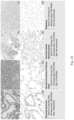
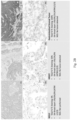
- staining patterns examples include aspect of membranous staining (A), cytoplasmic staining (A-C) and nuclear staining (C).
- Figure 7 shows that some samples experienced fixation gradients, which could affect staining levels of some cells. It was noted that samples with nuclear localization were more sensitive to fixation gradients than samples having a cytoplasmic/membrane localization.
- pan-TRK IHC staining varies in both intensity and in percentage of tumor cells staining across tumor types. Solid tumors harboring fusions based on ISH showed IHC staining with higher intensity and in a greater percentage of tumor cells than did tumors with wild-type TRK protein expression, with the exception of neuroendocrine and spindle cell tumors. In tumor types with low prevalence of wild-type TRK protein expression (e.g. CRC and papillary thyroid cancer) pan-TRK IHC staining may be useful in helping to identifying tumors harboring fusions. By contrast, in tumors with high prevalence of wild-type TRK protein expression (e.g.
Landscapes
- Health & Medical Sciences (AREA)
- Life Sciences & Earth Sciences (AREA)
- Immunology (AREA)
- Engineering & Computer Science (AREA)
- Molecular Biology (AREA)
- Biomedical Technology (AREA)
- Chemical & Material Sciences (AREA)
- Hematology (AREA)
- Urology & Nephrology (AREA)
- Analytical Chemistry (AREA)
- Pathology (AREA)
- General Physics & Mathematics (AREA)
- General Health & Medical Sciences (AREA)
- Biochemistry (AREA)
- Physics & Mathematics (AREA)
- Cell Biology (AREA)
- Biotechnology (AREA)
- Medicinal Chemistry (AREA)
- Food Science & Technology (AREA)
- Microbiology (AREA)
- Investigating Or Analysing Biological Materials (AREA)
- Measuring Or Testing Involving Enzymes Or Micro-Organisms (AREA)
- Peptides Or Proteins (AREA)
Claims (11)
- Verfahren zum Nachweisen eines Fusionsproteins von TrkA, TrkB oder TrkC in einer nicht-neuroendokrinen Tumorprobe, wobei das Verfahren Folgendes umfasst:- affinitätshistochemisches Färben der Probe mit einem biomarkerspezifischen Reagens, das spezifisch an eine Aminosäuresequenz, die die Reste 363-760 von SEQ ID NO: 1 umfasst oder aus diesen besteht; eine Aminosäuresequenz, die die Reste 646-838 von SEQ ID NO:2 umfasst oder aus diesen besteht; und eine Aminosäuresequenz, die die Reste 718-839 von SEQ ID NO:3 umfasst oder aus diesen besteht, bindet;- Nachweisen eines Färbungsmusters in der Probe und- Bewerten der Probe als positiv für ein Fusionsprotein, das TrkA, TrkB oder TrkC einschließt, wenn die Probe ein Färbungsmuster aufweist, in dem ein Schwellenprozentsatz an Tumorzellen oder mehr über einer schwellenspezifischen Färbungsintensität hinaus gefärbt sind, wobei- das Färbungsmuster ein membranöses und/oder zytoplasmatisches Färbungsmuster ist, das einen ersten Schwellenprozentsatz an Tumorzellen oder mehr aufweist, die über einer ersten Schwellenfärbungsintensität hinaus gefärbt sind, und wobei der erste Schwellenprozentsatz an Tumorzellen im Bereich von 25 % bis 75 % liegt und die erste schwellenspezifische Färbungsintensität mindestens 1,5+ beträgt; oder- das Färbungsmuster ein Kernfärbungsmuster ist, das einen zweiten Schwellenprozentsatz an Zellen oder mehr innerhalb einer Schwellentumorzellenfläche aufweist, die spezifisch mit einer zweiten Färbungsintensität gefärbt sind, wobei die zusammenhängende Schwellentumorzellenfläche mindestens 20 Zellen beträgt, die Schwellenfärbungsintensität eine spezifische Färbung gegenüber einem Hintergrund ist und der Schwellenprozentsatz an Zellen im Bereich von 25 % bis 75 % oder 25 % bis 80 % liegt.
- Verfahren nach Anspruch 1, wobei der erste Schwellenprozentsatz an Tumorzellen im Bereich von 50 % bis 75 % liegt und die erste Schwellenfärbungsintensität mindestens 1,5 beträgt.
- Verfahren nach Anspruch 1 oder Anspruch 2, wobei die Schwellentumorzellenfläche mindestens 50 zusammenhängende Zellen beträgt, die Schwellenfärbungsintensität ≥ 0,5+ beträgt und der Schwellenprozentsatz an Zellen im Bereich von 25 % bis 75 % liegt.
- Verfahren nach einem der Ansprüche 1 bis 3, wobei das biomarkerspezifische Reagens ein Antikörper ist und wobei der Antikörper immunspezifisch für ein Epitop ist, das in einer aus den Aminosäuren 816-838 von SEQ ID NO:2 bestehenden Aminosäuresequenz angeordnet ist.
- Verfahren nach Anspruch 4, wobei der Antikörper der monoklonale Antikörperklon EPR17341 ist.
- Verfahren nach Anspruch 1 oder 2, wobei das affinitätshistochemische Färben Folgendes umfasst:(a) Unterziehen der Probe einem wärmeinduzierten Epitoprückgewinnungsprozess und(b) Inkontaktbringen der Probe mit dem biomarkerspezifischen Reagens und einem Satz von Nachweisreagenzien, um einen Hellfeldfarbstoff in der Nähe eines biomarkerspezifischen Reagens, das an die Probe gebunden ist, abzulagern.
- Verfahren zum Nachweisen einer NTRK-Umordnung in einer nicht-neuroendokrinen Tumorprobe, wobei das Verfahren das Nachweisen der Gegenwart eines Trk-Fusionsproteins in der Probe gemäß dem Verfahren nach einem der Ansprüche 1 bis 6 und das Prüfen der Probe auf NTRK-Umordnung durch Sequenzieren, Reverse-Transkriptase-Polymerasekettenreaktion oder In-situ-Hybridisierung umfasst, wenn die Probe als positiv für ein Fusionsprotein, das TrkA, TrkB oder TrkC einschließt, bewertet wird.
- Verfahren nach Anspruch 7, ferner umfassend den Schritt des Identifizierens von Proben als entweder Trk-Fusion-positiv oder Trk-Fusion-negativ und den Prüfschritt, umfassend das Prüfen von Proben, die Trk-Fusion-negativ sind, auf die NTRK-Umordnung durch Sequenzieren, Reverse-Transkriptase-Polymerasekettenreaktion oder In-situ-Hybridisierung.
- Verfahren nach Anspruch 7, ferner umfassend den Schritt des Identifizierens von Proben als entweder Trk-Fusion-positiv, Trk-Fusion-unbestimmt oder Trk-Fusion-negativ und den Prüfschritt, umfassend das Prüfen von Proben, die Trk-Fusion-unbestimmt sind, auf die NTRK-Umordnung durch Sequenzieren, Reverse-Transkriptase-Polymerasekettenreaktion oder In-situ-Hybridisierung.
- Verfahren nach den Ansprüchen 8 oder 9, ferner umfassend das Auswählen eines Patienten, der eine Trk-gerichtete Therapie erhalten soll, wenn die nicht-neuroendokrine Tumorprobe als positiv für ein TRK-Fusionsprotein, das TrkA, TrkB oder TrkC einschließt, bewertet oder die NTRK-Umordnung nachgewiesen wird.
- Verfahren zum Auswählen eines Patienten, der eine Trk-gerichtete Therapie erhalten soll, umfassend das Nachweisen der Gegenwart eines Trk-Fusionsproteins in einer nicht-neuroendokrinen Tumorprobe gemäß einem Verfahren nach einem der Ansprüche 1 bis 6 und das Auswählen des Patienten, der die Therapie erhalten soll, wenn die Probe als positiv für ein Fusionsprotein, das TrkA, TrkB oder TrkC einschließt, bewertet oder die NTRK-Umordnung nachgewiesen wird.
Applications Claiming Priority (2)
| Application Number | Priority Date | Filing Date | Title |
|---|---|---|---|
| US201862731032P | 2018-09-13 | 2018-09-13 | |
| PCT/EP2019/074463 WO2020053376A1 (en) | 2018-09-13 | 2019-09-13 | Histochemical and cytochemical methods for detecting ntrk fusion proteins |
Publications (2)
| Publication Number | Publication Date |
|---|---|
| EP3850367A1 EP3850367A1 (de) | 2021-07-21 |
| EP3850367B1 true EP3850367B1 (de) | 2024-11-06 |
Family
ID=67988978
Family Applications (1)
| Application Number | Title | Priority Date | Filing Date |
|---|---|---|---|
| EP19769757.6A Active EP3850367B1 (de) | 2018-09-13 | 2019-09-13 | Histochemische und cytochemische methoden zum nachweis von ntrk-fusionsproteinen |
Country Status (5)
| Country | Link |
|---|---|
| US (2) | US20210199655A1 (de) |
| EP (1) | EP3850367B1 (de) |
| JP (2) | JP7576024B2 (de) |
| CN (1) | CN112673258B (de) |
| WO (1) | WO2020053376A1 (de) |
Family Cites Families (18)
| Publication number | Priority date | Publication date | Assignee | Title |
|---|---|---|---|---|
| US1564604A (en) | 1925-06-12 | 1925-12-08 | Chas S Hodges | Water wheel |
| US5595707A (en) | 1990-03-02 | 1997-01-21 | Ventana Medical Systems, Inc. | Automated biological reaction apparatus |
| US20030211630A1 (en) | 1998-02-27 | 2003-11-13 | Ventana Medical Systems, Inc. | Automated molecular pathology apparatus having independent slide heaters |
| ES2354598T3 (es) | 1998-02-27 | 2011-03-16 | Ventana Medical Systems, Inc. | Aparato de patología molecular automatizado que tiene calefactores de portaobjetos independientes. |
| US6582962B1 (en) | 1998-02-27 | 2003-06-24 | Ventana Medical Systems, Inc. | Automated molecular pathology apparatus having independent slide heaters |
| US6670113B2 (en) | 2001-03-30 | 2003-12-30 | Nanoprobes | Enzymatic deposition and alteration of metals |
| DK1636586T3 (da) | 2003-06-24 | 2009-11-23 | Ventana Med Syst Inc | Enzymkatalyseret metaldeponering til foröget in situ-detektion af immunhistokemisker epitoper og nucleinsyresekvenser |
| WO2011139978A1 (en) | 2010-05-04 | 2011-11-10 | Ventana Medical Systems, Inc. | Moving meniscus rinsing and mixing in cell staining |
| WO2012003476A2 (en) | 2010-07-02 | 2012-01-05 | Ventana Medical Systems, Inc. | Hapten conjugates for target detection |
| AU2013337277B2 (en) | 2012-11-05 | 2018-03-08 | Foundation Medicine, Inc. | Novel NTRK1 fusion molecules and uses thereof |
| EP3628749A1 (de) * | 2013-07-30 | 2020-04-01 | Blueprint Medicines Corporation | Ntrk2-fusionen |
| US9689875B2 (en) | 2013-08-28 | 2017-06-27 | Ventana Medical Systems, Inc. | Immunohistochemical assay for detection of CD3 and CD16 |
| WO2015124777A1 (en) | 2014-02-21 | 2015-08-27 | Ventana Medical Systems, Inc. | Medical image analysis for identifying biomarker-positive tumor cells |
| WO2015124703A1 (en) | 2014-02-24 | 2015-08-27 | Ventana Medical Systems, Inc. | Quinone methide analog signal amplification |
| WO2016089853A1 (en) * | 2014-12-03 | 2016-06-09 | Ignyta, Inc. | Multiplexed immunohistochemistry assays for diagnosis and treatment of cancer |
| WO2016170008A1 (en) | 2015-04-20 | 2016-10-27 | Ventana Medical Systems, Inc. | Inkjet deposition of reagents for histological samples |
| WO2018055014A1 (en) * | 2016-09-23 | 2018-03-29 | Ventana Medical Systems, Inc. | Methods and systems for scoring extracellular matrix biomarkers in tumor samples |
| EP4220164B1 (de) * | 2016-12-19 | 2024-08-21 | Ventana Medical Systems, Inc. | Verfahren und systeme zur quantitativen immunhistochemie |
-
2019
- 2019-09-13 EP EP19769757.6A patent/EP3850367B1/de active Active
- 2019-09-13 WO PCT/EP2019/074463 patent/WO2020053376A1/en not_active Ceased
- 2019-09-13 JP JP2021513301A patent/JP7576024B2/ja active Active
- 2019-09-13 CN CN201980060054.8A patent/CN112673258B/zh active Active
-
2021
- 2021-03-11 US US17/249,762 patent/US20210199655A1/en active Pending
-
2023
- 2023-12-18 JP JP2023212951A patent/JP7757382B2/ja active Active
-
2025
- 2025-07-07 US US19/261,521 patent/US20250389718A1/en active Pending
Non-Patent Citations (1)
| Title |
|---|
| RECONDO G. ET AL: "Preliminary results on mechanisms of resistance to ALK inhibitors: A prospective cohort from the MATCH-R study", ANNALS OF ONCOLOGY, vol. 29, 1 September 2018 (2018-09-01), pages vi21, XP093080141, ISSN: 0923-7534, DOI: 10.1093/annonc/mdy314 * |
Also Published As
| Publication number | Publication date |
|---|---|
| JP7576024B2 (ja) | 2024-10-30 |
| CN112673258B (zh) | 2025-07-25 |
| JP7757382B2 (ja) | 2025-10-21 |
| US20250389718A1 (en) | 2025-12-25 |
| EP3850367A1 (de) | 2021-07-21 |
| US20210199655A1 (en) | 2021-07-01 |
| WO2020053376A1 (en) | 2020-03-19 |
| JP2024037938A (ja) | 2024-03-19 |
| CN112673258A (zh) | 2021-04-16 |
| JP2022500633A (ja) | 2022-01-04 |
Similar Documents
| Publication | Publication Date | Title |
|---|---|---|
| EP3408410B1 (de) | Prädiktiver diagnostischer arbeitsfluss für tumore mithilfe automatisierter dissektion, sequenzierung der nächsten generation und automatisierte objektträgerfärber | |
| JP2024069211A (ja) | 定量的免疫組織化学のための方法及びシステム | |
| US12487242B2 (en) | DCIS recurrence and invasive breast cancer | |
| US12540359B2 (en) | HER2 heterogeneity as a biomarker in cancer | |
| US11821900B2 (en) | Method of selection for treatment of subjects at risk of invasive breast cancer | |
| US20190219579A1 (en) | Methods and systems for scoring extracellular matrix biomarkers in tumor samples | |
| US20240361213A1 (en) | Materials and methods for bleaching melanin-pigmented tissues | |
| EP3283881B1 (de) | Verfahren und systeme zur auf thermochemie basierten antikörperdeaktivierung | |
| EP3850367B1 (de) | Histochemische und cytochemische methoden zum nachweis von ntrk-fusionsproteinen | |
| US20210132075A1 (en) | Materials and methods for detecting fusion proteins | |
| JP2009506303A (ja) | 癌の化学療法のための予測方法 | |
| WO2025014787A1 (en) | Brightfield triplex immunohistochemistry assay for evaluating the colocalization of the er, pr, and ki-67 biomarkers in cells |
Legal Events
| Date | Code | Title | Description |
|---|---|---|---|
| STAA | Information on the status of an ep patent application or granted ep patent |
Free format text: STATUS: UNKNOWN |
|
| STAA | Information on the status of an ep patent application or granted ep patent |
Free format text: STATUS: THE INTERNATIONAL PUBLICATION HAS BEEN MADE |
|
| PUAI | Public reference made under article 153(3) epc to a published international application that has entered the european phase |
Free format text: ORIGINAL CODE: 0009012 |
|
| STAA | Information on the status of an ep patent application or granted ep patent |
Free format text: STATUS: REQUEST FOR EXAMINATION WAS MADE |
|
| 17P | Request for examination filed |
Effective date: 20210413 |
|
| AK | Designated contracting states |
Kind code of ref document: A1 Designated state(s): AL AT BE BG CH CY CZ DE DK EE ES FI FR GB GR HR HU IE IS IT LI LT LU LV MC MK MT NL NO PL PT RO RS SE SI SK SM TR |
|
| DAV | Request for validation of the european patent (deleted) | ||
| DAX | Request for extension of the european patent (deleted) | ||
| STAA | Information on the status of an ep patent application or granted ep patent |
Free format text: STATUS: EXAMINATION IS IN PROGRESS |
|
| 17Q | First examination report despatched |
Effective date: 20220311 |
|
| GRAP | Despatch of communication of intention to grant a patent |
Free format text: ORIGINAL CODE: EPIDOSNIGR1 |
|
| STAA | Information on the status of an ep patent application or granted ep patent |
Free format text: STATUS: GRANT OF PATENT IS INTENDED |
|
| INTG | Intention to grant announced |
Effective date: 20240604 |
|
| GRAS | Grant fee paid |
Free format text: ORIGINAL CODE: EPIDOSNIGR3 |
|
| GRAA | (expected) grant |
Free format text: ORIGINAL CODE: 0009210 |
|
| STAA | Information on the status of an ep patent application or granted ep patent |
Free format text: STATUS: THE PATENT HAS BEEN GRANTED |
|
| AK | Designated contracting states |
Kind code of ref document: B1 Designated state(s): AL AT BE BG CH CY CZ DE DK EE ES FI FR GB GR HR HU IE IS IT LI LT LU LV MC MK MT NL NO PL PT RO RS SE SI SK SM TR |
|
| REG | Reference to a national code |
Ref country code: GB Ref legal event code: FG4D |
|
| REG | Reference to a national code |
Ref country code: CH Ref legal event code: EP |
|
| REG | Reference to a national code |
Ref country code: DE Ref legal event code: R096 Ref document number: 602019061538 Country of ref document: DE |
|
| REG | Reference to a national code |
Ref country code: IE Ref legal event code: FG4D |
|
| REG | Reference to a national code |
Ref country code: LT Ref legal event code: MG9D |
|
| REG | Reference to a national code |
Ref country code: NL Ref legal event code: MP Effective date: 20241106 |
|
| PG25 | Lapsed in a contracting state [announced via postgrant information from national office to epo] |
Ref country code: PT Free format text: LAPSE BECAUSE OF FAILURE TO SUBMIT A TRANSLATION OF THE DESCRIPTION OR TO PAY THE FEE WITHIN THE PRESCRIBED TIME-LIMIT Effective date: 20250306 Ref country code: HR Free format text: LAPSE BECAUSE OF FAILURE TO SUBMIT A TRANSLATION OF THE DESCRIPTION OR TO PAY THE FEE WITHIN THE PRESCRIBED TIME-LIMIT Effective date: 20241106 Ref country code: IS Free format text: LAPSE BECAUSE OF FAILURE TO SUBMIT A TRANSLATION OF THE DESCRIPTION OR TO PAY THE FEE WITHIN THE PRESCRIBED TIME-LIMIT Effective date: 20250306 |
|
| PG25 | Lapsed in a contracting state [announced via postgrant information from national office to epo] |
Ref country code: FI Free format text: LAPSE BECAUSE OF FAILURE TO SUBMIT A TRANSLATION OF THE DESCRIPTION OR TO PAY THE FEE WITHIN THE PRESCRIBED TIME-LIMIT Effective date: 20241106 Ref country code: NL Free format text: LAPSE BECAUSE OF FAILURE TO SUBMIT A TRANSLATION OF THE DESCRIPTION OR TO PAY THE FEE WITHIN THE PRESCRIBED TIME-LIMIT Effective date: 20241106 |
|
| REG | Reference to a national code |
Ref country code: AT Ref legal event code: MK05 Ref document number: 1739886 Country of ref document: AT Kind code of ref document: T Effective date: 20241106 |
|
| PG25 | Lapsed in a contracting state [announced via postgrant information from national office to epo] |
Ref country code: BG Free format text: LAPSE BECAUSE OF FAILURE TO SUBMIT A TRANSLATION OF THE DESCRIPTION OR TO PAY THE FEE WITHIN THE PRESCRIBED TIME-LIMIT Effective date: 20241106 |
|
| PG25 | Lapsed in a contracting state [announced via postgrant information from national office to epo] |
Ref country code: ES Free format text: LAPSE BECAUSE OF FAILURE TO SUBMIT A TRANSLATION OF THE DESCRIPTION OR TO PAY THE FEE WITHIN THE PRESCRIBED TIME-LIMIT Effective date: 20241106 |
|
| PG25 | Lapsed in a contracting state [announced via postgrant information from national office to epo] |
Ref country code: NO Free format text: LAPSE BECAUSE OF FAILURE TO SUBMIT A TRANSLATION OF THE DESCRIPTION OR TO PAY THE FEE WITHIN THE PRESCRIBED TIME-LIMIT Effective date: 20250206 |
|
| PG25 | Lapsed in a contracting state [announced via postgrant information from national office to epo] |
Ref country code: LV Free format text: LAPSE BECAUSE OF FAILURE TO SUBMIT A TRANSLATION OF THE DESCRIPTION OR TO PAY THE FEE WITHIN THE PRESCRIBED TIME-LIMIT Effective date: 20241106 Ref country code: GR Free format text: LAPSE BECAUSE OF FAILURE TO SUBMIT A TRANSLATION OF THE DESCRIPTION OR TO PAY THE FEE WITHIN THE PRESCRIBED TIME-LIMIT Effective date: 20250207 Ref country code: AT Free format text: LAPSE BECAUSE OF FAILURE TO SUBMIT A TRANSLATION OF THE DESCRIPTION OR TO PAY THE FEE WITHIN THE PRESCRIBED TIME-LIMIT Effective date: 20241106 |
|
| PG25 | Lapsed in a contracting state [announced via postgrant information from national office to epo] |
Ref country code: PL Free format text: LAPSE BECAUSE OF FAILURE TO SUBMIT A TRANSLATION OF THE DESCRIPTION OR TO PAY THE FEE WITHIN THE PRESCRIBED TIME-LIMIT Effective date: 20241106 |
|
| PG25 | Lapsed in a contracting state [announced via postgrant information from national office to epo] |
Ref country code: RS Free format text: LAPSE BECAUSE OF FAILURE TO SUBMIT A TRANSLATION OF THE DESCRIPTION OR TO PAY THE FEE WITHIN THE PRESCRIBED TIME-LIMIT Effective date: 20250206 |
|
| PG25 | Lapsed in a contracting state [announced via postgrant information from national office to epo] |
Ref country code: SM Free format text: LAPSE BECAUSE OF FAILURE TO SUBMIT A TRANSLATION OF THE DESCRIPTION OR TO PAY THE FEE WITHIN THE PRESCRIBED TIME-LIMIT Effective date: 20241106 |
|
| PG25 | Lapsed in a contracting state [announced via postgrant information from national office to epo] |
Ref country code: DK Free format text: LAPSE BECAUSE OF FAILURE TO SUBMIT A TRANSLATION OF THE DESCRIPTION OR TO PAY THE FEE WITHIN THE PRESCRIBED TIME-LIMIT Effective date: 20241106 |
|
| PG25 | Lapsed in a contracting state [announced via postgrant information from national office to epo] |
Ref country code: EE Free format text: LAPSE BECAUSE OF FAILURE TO SUBMIT A TRANSLATION OF THE DESCRIPTION OR TO PAY THE FEE WITHIN THE PRESCRIBED TIME-LIMIT Effective date: 20241106 |
|
| PG25 | Lapsed in a contracting state [announced via postgrant information from national office to epo] |
Ref country code: RO Free format text: LAPSE BECAUSE OF FAILURE TO SUBMIT A TRANSLATION OF THE DESCRIPTION OR TO PAY THE FEE WITHIN THE PRESCRIBED TIME-LIMIT Effective date: 20241106 |
|
| PG25 | Lapsed in a contracting state [announced via postgrant information from national office to epo] |
Ref country code: SK Free format text: LAPSE BECAUSE OF FAILURE TO SUBMIT A TRANSLATION OF THE DESCRIPTION OR TO PAY THE FEE WITHIN THE PRESCRIBED TIME-LIMIT Effective date: 20241106 |
|
| PG25 | Lapsed in a contracting state [announced via postgrant information from national office to epo] |
Ref country code: CZ Free format text: LAPSE BECAUSE OF FAILURE TO SUBMIT A TRANSLATION OF THE DESCRIPTION OR TO PAY THE FEE WITHIN THE PRESCRIBED TIME-LIMIT Effective date: 20241106 |
|
| PG25 | Lapsed in a contracting state [announced via postgrant information from national office to epo] |
Ref country code: IT Free format text: LAPSE BECAUSE OF FAILURE TO SUBMIT A TRANSLATION OF THE DESCRIPTION OR TO PAY THE FEE WITHIN THE PRESCRIBED TIME-LIMIT Effective date: 20241106 |
|
| REG | Reference to a national code |
Ref country code: DE Ref legal event code: R097 Ref document number: 602019061538 Country of ref document: DE |
|
| PG25 | Lapsed in a contracting state [announced via postgrant information from national office to epo] |
Ref country code: SE Free format text: LAPSE BECAUSE OF FAILURE TO SUBMIT A TRANSLATION OF THE DESCRIPTION OR TO PAY THE FEE WITHIN THE PRESCRIBED TIME-LIMIT Effective date: 20241106 |
|
| PLBE | No opposition filed within time limit |
Free format text: ORIGINAL CODE: 0009261 |
|
| STAA | Information on the status of an ep patent application or granted ep patent |
Free format text: STATUS: NO OPPOSITION FILED WITHIN TIME LIMIT |
|
| REG | Reference to a national code |
Ref country code: CH Ref legal event code: U11 Free format text: ST27 STATUS EVENT CODE: U-0-0-U10-U11 (AS PROVIDED BY THE NATIONAL OFFICE) Effective date: 20251001 |
|
| PGFP | Annual fee paid to national office [announced via postgrant information from national office to epo] |
Ref country code: DE Payment date: 20250820 Year of fee payment: 7 |
|
| 26N | No opposition filed |
Effective date: 20250807 |
|
| PGFP | Annual fee paid to national office [announced via postgrant information from national office to epo] |
Ref country code: GB Payment date: 20250822 Year of fee payment: 7 |
|
| PGFP | Annual fee paid to national office [announced via postgrant information from national office to epo] |
Ref country code: FR Payment date: 20250821 Year of fee payment: 7 |
|
| REG | Reference to a national code |
Ref country code: DE Ref legal event code: R079 Ref document number: 602019061538 Country of ref document: DE Free format text: PREVIOUS MAIN CLASS: G01N0033574000 Ipc: G01N0033575000 |
|
| PGFP | Annual fee paid to national office [announced via postgrant information from national office to epo] |
Ref country code: CH Payment date: 20251001 Year of fee payment: 7 |