EP3901964B1 - Recommandation intelligente de scan pour l'imagerie par résonance magnétique - Google Patents
Recommandation intelligente de scan pour l'imagerie par résonance magnétique Download PDFInfo
- Publication number
- EP3901964B1 EP3901964B1 EP21165484.3A EP21165484A EP3901964B1 EP 3901964 B1 EP3901964 B1 EP 3901964B1 EP 21165484 A EP21165484 A EP 21165484A EP 3901964 B1 EP3901964 B1 EP 3901964B1
- Authority
- EP
- European Patent Office
- Prior art keywords
- scan
- data
- ihri
- uii
- recommendation
- Prior art date
- Legal status (The legal status is an assumption and is not a legal conclusion. Google has not performed a legal analysis and makes no representation as to the accuracy of the status listed.)
- Active
Links
Images



Classifications
-
- G—PHYSICS
- G16—INFORMATION AND COMMUNICATION TECHNOLOGY [ICT] SPECIALLY ADAPTED FOR SPECIFIC APPLICATION FIELDS
- G16H—HEALTHCARE INFORMATICS, i.e. INFORMATION AND COMMUNICATION TECHNOLOGY [ICT] SPECIALLY ADAPTED FOR THE HANDLING OR PROCESSING OF MEDICAL OR HEALTHCARE DATA
- G16H50/00—ICT specially adapted for medical diagnosis, medical simulation or medical data mining; ICT specially adapted for detecting, monitoring or modelling epidemics or pandemics
- G16H50/20—ICT specially adapted for medical diagnosis, medical simulation or medical data mining; ICT specially adapted for detecting, monitoring or modelling epidemics or pandemics for computer-aided diagnosis, e.g. based on medical expert systems
-
- G—PHYSICS
- G16—INFORMATION AND COMMUNICATION TECHNOLOGY [ICT] SPECIALLY ADAPTED FOR SPECIFIC APPLICATION FIELDS
- G16H—HEALTHCARE INFORMATICS, i.e. INFORMATION AND COMMUNICATION TECHNOLOGY [ICT] SPECIALLY ADAPTED FOR THE HANDLING OR PROCESSING OF MEDICAL OR HEALTHCARE DATA
- G16H40/00—ICT specially adapted for the management or administration of healthcare resources or facilities; ICT specially adapted for the management or operation of medical equipment or devices
- G16H40/60—ICT specially adapted for the management or administration of healthcare resources or facilities; ICT specially adapted for the management or operation of medical equipment or devices for the operation of medical equipment or devices
-
- A—HUMAN NECESSITIES
- A61—MEDICAL OR VETERINARY SCIENCE; HYGIENE
- A61B—DIAGNOSIS; SURGERY; IDENTIFICATION
- A61B5/00—Measuring for diagnostic purposes; Identification of persons
- A61B5/0033—Features or image-related aspects of imaging apparatus, e.g. for MRI, optical tomography or impedance tomography apparatus; Arrangements of imaging apparatus in a room
-
- A—HUMAN NECESSITIES
- A61—MEDICAL OR VETERINARY SCIENCE; HYGIENE
- A61B—DIAGNOSIS; SURGERY; IDENTIFICATION
- A61B5/00—Measuring for diagnostic purposes; Identification of persons
- A61B5/05—Detecting, measuring or recording for diagnosis by means of electric currents or magnetic fields; Measuring using microwaves or radio waves
- A61B5/055—Detecting, measuring or recording for diagnosis by means of electric currents or magnetic fields; Measuring using microwaves or radio waves involving electronic [EMR] or nuclear [NMR] magnetic resonance, e.g. magnetic resonance imaging
-
- G—PHYSICS
- G06—COMPUTING OR CALCULATING; COUNTING
- G06N—COMPUTING ARRANGEMENTS BASED ON SPECIFIC COMPUTATIONAL MODELS
- G06N20/00—Machine learning
- G06N20/10—Machine learning using kernel methods, e.g. support vector machines [SVM]
-
- G—PHYSICS
- G06—COMPUTING OR CALCULATING; COUNTING
- G06N—COMPUTING ARRANGEMENTS BASED ON SPECIFIC COMPUTATIONAL MODELS
- G06N3/00—Computing arrangements based on biological models
- G06N3/02—Neural networks
- G06N3/04—Architecture, e.g. interconnection topology
- G06N3/045—Combinations of networks
-
- G—PHYSICS
- G06—COMPUTING OR CALCULATING; COUNTING
- G06N—COMPUTING ARRANGEMENTS BASED ON SPECIFIC COMPUTATIONAL MODELS
- G06N3/00—Computing arrangements based on biological models
- G06N3/02—Neural networks
- G06N3/08—Learning methods
-
- G—PHYSICS
- G06—COMPUTING OR CALCULATING; COUNTING
- G06T—IMAGE DATA PROCESSING OR GENERATION, IN GENERAL
- G06T7/00—Image analysis
- G06T7/0002—Inspection of images, e.g. flaw detection
- G06T7/0012—Biomedical image inspection
-
- G—PHYSICS
- G16—INFORMATION AND COMMUNICATION TECHNOLOGY [ICT] SPECIALLY ADAPTED FOR SPECIFIC APPLICATION FIELDS
- G16H—HEALTHCARE INFORMATICS, i.e. INFORMATION AND COMMUNICATION TECHNOLOGY [ICT] SPECIALLY ADAPTED FOR THE HANDLING OR PROCESSING OF MEDICAL OR HEALTHCARE DATA
- G16H30/00—ICT specially adapted for the handling or processing of medical images
- G16H30/20—ICT specially adapted for the handling or processing of medical images for handling medical images, e.g. DICOM, HL7 or PACS
-
- G—PHYSICS
- G16—INFORMATION AND COMMUNICATION TECHNOLOGY [ICT] SPECIALLY ADAPTED FOR SPECIFIC APPLICATION FIELDS
- G16H—HEALTHCARE INFORMATICS, i.e. INFORMATION AND COMMUNICATION TECHNOLOGY [ICT] SPECIALLY ADAPTED FOR THE HANDLING OR PROCESSING OF MEDICAL OR HEALTHCARE DATA
- G16H30/00—ICT specially adapted for the handling or processing of medical images
- G16H30/40—ICT specially adapted for the handling or processing of medical images for processing medical images, e.g. editing
-
- G—PHYSICS
- G16—INFORMATION AND COMMUNICATION TECHNOLOGY [ICT] SPECIALLY ADAPTED FOR SPECIFIC APPLICATION FIELDS
- G16H—HEALTHCARE INFORMATICS, i.e. INFORMATION AND COMMUNICATION TECHNOLOGY [ICT] SPECIALLY ADAPTED FOR THE HANDLING OR PROCESSING OF MEDICAL OR HEALTHCARE DATA
- G16H40/00—ICT specially adapted for the management or administration of healthcare resources or facilities; ICT specially adapted for the management or operation of medical equipment or devices
- G16H40/60—ICT specially adapted for the management or administration of healthcare resources or facilities; ICT specially adapted for the management or operation of medical equipment or devices for the operation of medical equipment or devices
- G16H40/63—ICT specially adapted for the management or administration of healthcare resources or facilities; ICT specially adapted for the management or operation of medical equipment or devices for the operation of medical equipment or devices for local operation
-
- G—PHYSICS
- G06—COMPUTING OR CALCULATING; COUNTING
- G06T—IMAGE DATA PROCESSING OR GENERATION, IN GENERAL
- G06T2207/00—Indexing scheme for image analysis or image enhancement
- G06T2207/10—Image acquisition modality
- G06T2207/10072—Tomographic images
- G06T2207/10088—Magnetic resonance imaging [MRI]
-
- G—PHYSICS
- G06—COMPUTING OR CALCULATING; COUNTING
- G06T—IMAGE DATA PROCESSING OR GENERATION, IN GENERAL
- G06T2207/00—Indexing scheme for image analysis or image enhancement
- G06T2207/20—Special algorithmic details
- G06T2207/20081—Training; Learning
-
- G—PHYSICS
- G06—COMPUTING OR CALCULATING; COUNTING
- G06T—IMAGE DATA PROCESSING OR GENERATION, IN GENERAL
- G06T2207/00—Indexing scheme for image analysis or image enhancement
- G06T2207/20—Special algorithmic details
- G06T2207/20084—Artificial neural networks [ANN]
Definitions
- the present disclosure relates to techniques for developing a workflow for magnetic resonance imaging (MRI), and in particular to techniques for recommending MR protocols (MR is an abbreviation of magnetic resonance using various data sources, which may be part of an automated process.
- MRI magnetic resonance imaging
- MR is an abbreviation of magnetic resonance using various data sources, which may be part of an automated process.
- Magnetic resonance imaging is a medical imaging technique that provides diagnostically relevant information of biological tissue. Its diagnostic relevance originates from the informative power and versatility of this imaging modality, which offers differentiated and precise structure information in a non-invasive fashion. Magnetic resonance imaging is based on the controlled manipulation of nuclear spins inside a patient's body and subsequent detection of the nuclear spin response. The space-dependent encoding of the spin response allows for a reconstruction of the patient's structural composition and functional constitution for the radiologist to perform diagnostic reading.
- the MRI technician conducting the MR scan selects a sequential arrangement of MR protocols from an MR scanner library of imaging protocols, which may be based, e.g., on a relevant body region, a suspected disease (e.g., through the clinical indication of the referring physician or the radiologist), patient-specific requirements (e.g., implants, motion-related incompliance), and preferences of the clinical institution and radiologists who will later perform reading of the MR images for diagnosis.
- the technician typically performs checks to ensure that the image quality of the recorded scans allows for diagnosis. The technician may then make protocol modifications, e.g. adapt the field-of-view to the patient's body and others and will perform re-scans, if required.
- the apparatus comprises a disease selector configured to select a suspected disease of a patient based on patient information; and an image selector configured to determine a set of imaging conditions of a multi-parametric magnetic resonance image corresponding to the suspected disease based on a multi-parametric magnetic resonance imaging model.
- a disease selector configured to select a suspected disease of a patient based on patient information
- an image selector configured to determine a set of imaging conditions of a multi-parametric magnetic resonance image corresponding to the suspected disease based on a multi-parametric magnetic resonance imaging model.
- a method for the provision of confidence information about a diagnosis of a diagnosis system based on medical image data is described.
- the diagnosis system is designed as an algorithm that uses as its input the medical image data and further patient specific data.
- the measure of confidence is determined by execution of a determination algorithm that has as its input parameter a characteristic value for the diagnosis system, for example a sensitivity or a specificity of the diagnosis system, and/or an image content from medical image data.
- a further acquisition of additional image data can be proposed based on the confidence information.
- the additional medical image data can be acquired such that the data exhibit a changed image contrast with respect to the already existing medical image data.
- an initial scout sequence is performed of a patient.
- the initial scout sequence is validated and an abbreviated acquisition protocol is performed.
- the abbreviated acquisition protocol is validated and additional sequences are performed.
- Subsequent sequences are configured based on the analysis of the previous scans using deep learning-based reasoning to select the next appropriate settings and procedures.
- the identification of abnormal regions in the initial scout sequence is used to determine one or more additional acquisition sequences.
- a scan recommendation method according to claim 1
- a personal scan preparation method according to claim 5
- a medical imaging method according to claim 8
- a scan workflow performing method according to claim 9
- a scan recommendation system according to claim 10
- a personal scan preparation system according to claim 14
- a medical imaging system according to claim 17
- a scan workflow performing system according to claim 18.
- the scan recommendation method comprises the step of receiving evaluation data comprising classification data.
- the classification data classify anomalies of image derived metrics, which were detected based on input data concerning the medical condition of a patient.
- the anomalies are detected in medical image data of the patient.
- a classification comprises a type of identification of a detected anomaly, assigning probability values for different possible types of anomalies to the detected anomaly.
- the anomalies can be anomalies detected in the input data, for example medical image data, or they can comprise information about anomalies based on medical data about the patient of a different type.
- Anomalies detected in image data comprise detected image abnormalities or distinctive image features such as hyperintense areas, hypointense areas, abnormal tissues, midline shift, vascular stenosis, plaques, demyelination or mass effects. As explained further below in the text, anomalies may also be detected image metric abnormalities or distinctive features.
- the next recommended MR protocol which is intended to be executed with an MR scan of the patient, is automatically determined based on the evaluation data.
- the evaluation is not limited to the evaluation of image data. Hence, a plurality of different type of input data about a patient's condition can be analysed to determine the evaluation data. Also, a plurality of data sets of a single special type of input data concerning a patient's condition can be evaluated.
- the scan recommendation can be based on receiving a plurality of evaluation data of different types.
- the detection of the anomalies can be realized based on machine learning methods, for example based on neural networks such as a convolutional neural network taking the input data, for example image data, as input and providing as output the probability of the input data to be normal versus anomalous.
- the classification of the detected anomalies can be realized using machine learning methods, for example neural network-based techniques, or more specifically using a convolutional neural network. If the probability value of a detected anomaly for the received input data, for example image data, is higher than a certain customizable threshold value ⁇ d , then the input data, for example image data, assigned to the detected anomaly are taken as input for the subsequent classification step, as mentioned above.
- the scan recommendation can also be realized based on machine learning based techniques, especially neural network techniques as below explained.
- the evaluation data simply include the classification data.
- the evaluation data may additionally comprise also different types of data.
- the method according to the invention allows the trained or untrained technician to perform a highly personalized MRI scan workflow on each patient, generating an optimal set of MR images that are required for diagnosis of that patient's condition.
- the method may provide a basis for a fully autonomously optimized MR imaging workflow yet allowing for manual or electronic customization thereof as desired by the operator.
- the MR scanner operator may choose to run the MR scan workflow either in fully autonomous, fully manual, or in a hybrid mode.
- the personal scan preparation method comprises the steps of receiving input data, for example medical image data, concerning the medical condition of a patient, wherein the input data comprise image-derived metrics based on image data.
- Evaluation data based on the input data are determined by detecting anomalies of image derived metrics in the input data, determining classification data based on the detected anomalies as evaluation data and performing the scan recommendation method according to the invention.
- the classification of the anomalies can be used to adapt an MR protocol to a special kind of anomaly. For example, a special contrast is chosen for the recommended MR protocol.
- the personal scan preparation method shares the advantages of the scan recommendation method.
- the medical imaging method according to the invention comprises the steps of determining a next recommended protocol based on the scan recommendation method according to the invention and transmitting the next recommended protocol to an MR scan system and performing an MR scan by the MR scan system based on the received protocol.
- the medical imaging method shares the advantages of the scan recommendation method according to the invention.
- the scan workflow performing method according to the invention comprises the steps of performing the medical imaging method according to the invention. After the MR scan of the medical imaging method, anomalies of image derived metrics are detected in the reconstructed MR-image data and classification data are determined based on the detected anomalies. Then the aforementioned three steps are iteratively carried out taking into account the determined classification data, until an abortion criterion is attained. In other words, first a next recommended protocol is determined based on the scan recommendation method of the invention or the personal scan preparation method according to the invention. Then, the next recommended protocol is transmitted to an MR scan system and the MR scan is performed by the MR scan system based on the recommended protocol.
- an iterative protocol determination and measurement comprising the steps of controlling the MR scan, evaluating the reconstructed MR-image data and iteratively improving input data for the next recommended protocol based on classification of anomalies of the reconstructed MR-image data.
- the operator can first record a standard set of MR protocol and employ the scan recommendation method based on the recorded first images for additional MR scans that might be additionally indicated for the current patient based on the imaging information.
- a complete automatic workflow for an optimized MR scan of a patient is realized, wherein the protocol for the MR imaging and hence the MR imaging itself is optimized with each iteration loop. It has to be mentioned that all the methods described above, i.e.
- the scan recommendation method, the personal scan preparation method, the medical imaging method and the scan workflow performing method can also be carried out in the case that the operator does not sit in front of the scanner console.
- all of the described methods can be performed without even a console room next to the MR scan system, i.e. fully automated, or the monitoring operator is sitting somewhere else and controls remotely.
- a skilled operator can perform an investigation of a patient and a surveillance of a medical imaging process remotely and therefore professional skills can be accessed where an MR system is available.
- the scan recommendation system comprises at least one interface, preferably a plurality of input interfaces, for receiving evaluation data.
- the evaluation data comprise classification data, wherein the classification data classify anomalies of image derived metrics detected based on input data, wherein the input data concern the medical condition of a patient.
- the scan recommendation system according to the invention includes a recommendation unit for automatic determination of a next recommended MR protocol to be executed in accordance with an MR scan for determining a next recommended MR protocol to be executed in accordance with an MR scan of the patient based on the received evaluation data.
- the scan recommendation system can be configured to fully and automatically recommend the next, diagnostically most insightful MR protocol for measurement.
- the evaluation data for the recommendation system may be based on either a full set or subsets of inputs including image data (historical or current), image derived metrics, for example geometric properties of tissue and cavities like vascular wall thickness, cross section, and ventricular volume etc., electronic health record information, and/or user interface input.
- image data historical or current
- image derived metrics for example geometric properties of tissue and cavities like vascular wall thickness, cross section, and ventricular volume etc.
- electronic health record information electronic health record information
- user interface input user interface input.
- the personal scan preparation system comprises an input interface for receiving input data concerning the medical condition of a patient, wherein the input data comprise image-derived metrics based on image data. Further, the personal scan preparation system includes an evaluation unit, which comprises a detection unit for detecting anomalies in the image-derived metrics in the input data, a classification unit for determining classification data based on the detected anomalies as evaluation data, and the scan recommendation system according to the invention.
- the personal scan preparation system shares the advantages of the personal scan preparation method according to the invention.
- the medical imaging system according to the invention comprises an MR scan system and a scan recommendation system or a personal scan preparation system according to the invention, which is coupled to the MR scan system to determine a next recommended protocol and to send the next recommended protocol to the MR scan system for performing an MR scan.
- the medical imaging system according to the invention shares the advantages of the medical imaging method according to the invention.
- the scan workflow performing system comprises the medical imaging system according to the invention. Further, the scan workflow performing system includes an anomaly detection unit for detecting anomalies of image-derived metrics in the reconstructed MR-image data. Furthermore, the scan workflow performing system comprises a classification unit for determining classification data based on the detected anomalies. The scan workflow performing system also comprises an iteration unit for determining, based on the determined classification data, if iteratively performing the three steps of medical imaging, anomaly detection and anomaly classification, until an abortion criterion is attained. The scan workflow performing system shares the advantages of the scan workflow performing method according to the invention.
- the essential components of the scan recommendation system according to the invention, the personal scan preparation system according to the invention, the medical imaging system according to the invention and the scan workflow performing system according to the invention can for the most part be designed in the form of software components.
- some of these components can also be implemented in the form of software-supported hardware, for example FPGAs or the like, especially when it comes to particularly fast calculations.
- the required interfaces for example if it is only a matter of transferring data from other software components, can be designed as software interfaces. However, they can also be designed as hardware-based interfaces that are controlled by a suitable software. Furthermore, some parts of the above-mentioned components may be distributed and stored in a local or regional or global electronic network or a combination of a network and software, in particular a cloud system.
- a largely software-based implementation has the advantage that medical imaging systems including a magnetic resonance imaging system that have already been used can easily be retrofitted by a software update in order to work in the manner according to the invention.
- the object is also achieved by a corresponding computer program product with a computer program that can be loaded directly into a memory device, for example a control device of a medical imaging system, with program sections, in order to carry out all steps of the method according to the invention, if the program is executed in the medical imaging system, in particular the control device.
- a computer program product may contain additional components such as a documentation and/ or additional components, including hardware components such as Hardware keys (dongles etc.) for using the software.
- a computer-readable medium for example a memory stick, a hard disk or some other transportable or permanently installed data carrier is used on which the program sections of the computer program that can be read in and executed by a computer unit of the medical imaging system are stored.
- the computer unit can comprise for example, one or more cooperating microprocessors or the like used for this purpose.
- the computer program can also be acquired by download from a central webshop of the applicant or another internet based source or a cloud based source.
- the automatic scan recommendation is based on using at least one of the following software types or types of a combination of software and hardware:
- the scan recommendation method may be carried out using a trained model (for example a fully connected neural network or a random forest).
- the trained model receives the above-mentioned evaluation data, which for example comprise classified anomalies.
- the trained model is used to determine a next recommended MR protocol to be executed in accordance with an MR scan of a patient.
- the mentioned input data for example image data of the patient, can be information of all kind about the patient, which may influence an imaging process to be planned.
- the mentioned input data can either be directly used as input for the trained model or can undergo one or several preprocessing step, for example by a machine learning or deep learning model, before they are fed into the trained model.
- the evaluation data comprise a first possible scan workflow direction determined by mapping topics, based on extracted keywords in the input data concerning a medical condition of the patient, onto probabilities of a next recommended protocol.
- the input data can be for example written information, i.e. text data about a suspected diagnosis, symptoms, an indication referring to the symptoms or a body region, where the symptoms are localized and to which the indication or suspected diagnosis refers.
- the input data can comprise electronic health record information comprising at least one of a suspected diagnosis, a suspected disease, relevant observations of the technician, an indication from referring clinician, a body region, patient data, or an MR protocol.
- electronic health record information can add additional information about the health condition of a patient and can be used for modifying an MR imaging protocol. Further, the protocol can be modified by direct input by the user. Therefore, the input data can also include data input by the user.
- the keyword extraction can be performed based on an expert system or a machine learning approach or a deep learning approach or combinations thereof.
- the extracted keywords can be determined based on data preprocessing, for example lemmatization or stemming and bag-of words generation.
- the topics are determined by a topic detection algorithm or a generated topic model.
- the topic detection can be realized by a trained unsupervised topic detection with latent Dirichlet allocation.
- Latent Dirichlet allocation can be carried out using classical machine learning methods, for example a hierarchical Bayesian model trained with variational inference as decribed in D. Blei et al. JMLR 3, 2003 , or using neural variational inference as decribed in Y. Miao et al. Proc. ICML, 2017 .
- the mapping of the topics onto a possible scan workflow direction i.e. a set of possible recommended next scan protocols can be based on expert knowledge, customer preference or an additional designated machine learning algorithm or deep learning algorithm. To each of the topics a set of recommended next scan protocols with values of probabilities is assigned.
- a second possible scan workflow direction can be determined by the scan recommendation method according to the invention, wherein the classification data, which are included in the evaluation data, are mapped onto probability values of a next recommended protocol.
- a mapping can be realized using a mapping model, for example a random forest or a fully connected neural network.
- the final recommendation step is performed based on the second possible scan workflow direction and optionally additionally based on the first possible scan workflow direction.
- the final recommending step can be realized using a weighted averaging of the first and the second possible scan workflow direction or using a fully connected neural network.
- the input data comprise image data and the step of detecting anomalies comprises detecting anomalies in the image data.
- the detection and classification of anomalies in image data can be realized using a neural network, for example a convolutional neural network.
- the anomalies can be direct anomalies of image data or anomalies of metrics related to image data. Therefore, the input data comprise image derived metrics based on image data and the step of detecting anomalies comprises detecting anomalies in the image-derived metrics.
- Image derived metrics include geometric properties of tissues and cavities, potentially compared to normative data from a cohort. Such derived metrics encompass vascular wall thickness, cross sections of blood vessels and ventricular volume, grey mass volume in the brain and white mass volume in the brain.
- the input data can also comprise electronic health record information and/or user interface input data and the step of determining evaluation data can comprise the steps of extracting keywords from the electronic health record information and/or the user interface input, determining topics based on the extracted keywords and determining the first possible scan workflow direction by mapping the topics onto probabilities of a next recommended protocol.
- Keywords can comprise for example information regarding at least one of a suspected diagnosis, a suspected disease, relevant observations of the technician, an indication from referring clinician, a body region, patient data, or an MR protocol.
- the keyword extraction can be performed based on an expert system or a machine learning approach or a deep learning approach or combinations thereof.
- the scan recommendation can be realized by one or several classical and or machine learning algorithms.
- electronic health record information can add additional information about the health condition of a patient and can be used for modifying an MR imaging protocol.
- the MR imaging protocol can be modified by direct input by the user.
- Using a keyword extraction enables an automatic analysis of electronic health record information and/or the user interface input concerning MR protocol related content.
- information concerning former medical examination can be integrated into the scan recommendation based on written text or direct protocol data input by a user. In this way, the data base for the automatic determination of a next recommended MR protocol is being broadened.
- the aforementioned listed input data may include patient information, user information and hospital information, which may either be directly used as input to the scan recommendation system or may undergo one or several preprocessing steps (e.g., by a machine learning or deep learning model) before being fed into the scan recommendation system.
- the recommendation unit comprises at least one of:
- the scan recommendation system can also comprise an input interface for receiving evaluation data comprising a first possible scan workflow direction determined by mapping topics based on extracted keywords concerning a medical condition of the patient, onto probability values of a next recommended protocol.
- evaluation data comprising a first possible scan workflow direction determined by mapping topics based on extracted keywords concerning a medical condition of the patient, onto probability values of a next recommended protocol.
- the recommendation unit comprises a mapping unit for determining a second possible scan workflow direction by automated mapping of the classification data onto probability values of a next recommended protocol and a final protocol recommendation unit for final recommending the next recommended protocol based on the second possible scan workflow direction and optionally additionally based on the first possible scan workflow direction.
- the input interface is configured to receive input data comprising the medical image data and the detection unit is configured to detect anomalies in the image data.
- the input interface can also be configured to receive input data comprising image derived metrics based on image data and the detection unit can be configured to detect anomalies in the image-derived metrics.
- the input interface is configured to receive input data comprising electronic health record information and/or user interface input data.
- the evaluation unit comprises an extraction unit for extracting keywords from the electronic health record information and/or the user interface input, a topic finding unit for determining topics based on the extracted keywords and a mapping unit for determining the first possible scan workflow direction by mapping the topics onto probability values of a next recommended protocol as evaluation data.
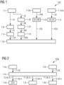
- FIG. 1 illustrates a flow chart 100 of a scan preparation method according to an embodiment of the invention for recommending a workflow for medical imaging of a patient.
- image data ID from the patient to be examined are acquired.
- the image data ID may be acquired by an initial scan for creating a T1-map and a T2-map. Further, the image data ID may be received from historical images acquired from the patient in former times.
- the image data ID are directly used as first input data for determination of evaluation data.
- the image data are analysed regarding image anomalies IA.
- the detection of anomalies IA in the image data ID can be realized using a convolutional neural network.
- step 1.III the image data ID are first subjected to pre-diagnostic analysis steps, wherein some metrics IM of the image data ID are derived.
- some pre-diagnostic analysis data such as, for instance, geometric properties of tissue and cavities like vascular wall thickness, cross section, and ventricular volume etc. are detected and measured and are compared to normative data from a cohort.
- these image metrics IM are used as second input data and are subjected to an anomaly detection using a fully connected neural network, wherein some anomalies MA of metrics IM can be detected.
- step 1.V the image anomalies IA are further classified using the above-mentioned convolutional neural network.
- step 1.VI the detected anomalies MA of metrics IM are classified using for example a fully connected neural network, a decision tree or a random forest.
- evaluation data comprising classification data CIA, CMA for image anomalies IA and metrics anomalies MA are provided for later step 1.XI, which is the actual scan recommendation step.
- the classification data CIA concerning image anomalies IA comprise sets of probability values P(i) for the probability of the existence of different image anomalies IA in the image data ID input to the scan preparation method in step 1.I.
- the classification data CMA concerning metrics anomalies MA comprise sets of probability values P(j) for the probability of the existence of different metrics anomalies IM, which can also be understand as input data provided to the scan preparation method in step 1.IV.
- the initial image data ID are recorded to provide a first measurement step benchmark and subsequently input these image data ID for an anomaly detection and anomaly classification.
- Quality assurance steps following each measurement can be employed to ensure sufficient image quality and trigger rescans, if required (e.g., due to patient motion).
- the anomaly detection and anomaly classification may be implemented, e.g., by a convolutional neural net, possibly as independent implementations. If anomaly detection/classification is applied, embodiments include advantageously using quantitative MR imaging protocols, for example for T1-maps and T2-maps for the measurement of the initial input images to provide a standardized image input for the anomaly detection and classification.
- step 1.VII some electronic health record information IHRI is used as third input data, which comprises data from various sources such as objects, for example suspected diagnoses, referring indications, etc., and/or other data sources such as RIS, patient data, etc.
- step 1.VIII some additional information UII, also named as user interface input UII that comprises data indicating, for example, suspected diagnoses, referring indications, a body region(s), a desired MR protocol, etc., are acquired as fourth input data for the personal scan preparation method.
- step 1.IX the input electronic health record information IHRI is processed using, for example, natural language processing (NLP) to perform keyword extraction. By this way, extracted keywords EK-IHRI of input electronic health record information IHRI are acquired.
- NLP natural language processing
- the user interface input UII is processed using, for example, NLP to additionally or alternatively extract keywords EK-UII in step 1.X.
- the extracted keywords EK-IHRI, EK-UII are then assigned to different topics and the topics are then mapped to sets of probability values P(n), P(o), which represent a possible scan workflow direction PDI, PDU.
- P(n), P(o) represent a possible scan workflow direction PDI, PDU.
- the possible scan workflow directions PDI, PDU are provided as third and fourth evaluation data to the automatic scan recommendation method of step 1.XI.
- an automatic scan recommendation is performed based on a combination of various evaluation values CIA, CMA, PDI, PDU to identify the next recommended protocol, as further discussed herein.
- the automatic scan recommendation can be realized based on a classical or machine learning system, such as a fully connected neural network (FCNN).
- FCNN fully connected neural network
- the scan recommendation is not limited to this implementation and may be implemented in accordance with any suitable type of classical algorithm, expert system, or machine learning design that may process inputs, e.g. in accordance with classical, training, or learning processes to recommend protocols based upon specific type of inputs.
- step 1.Ia a T1-map measurement is carried out based on an initial MR scan of the patient and a T1-map T1-M is acquired.
- the initial scan of the patient can be done with low resolution and does not take much time compared to the main imaging process of the examination.
- step 1.Ib a T2-map measurement is carried out based on the initial MR scan of the patient and a T2-map T2-M is acquired.
- step 1.Ic a quality analysis of the acquired data T1-M, T2-M is performed and some quality proved image data T1-M-A, T2-M-A are provided to further process and analysis.
- Step 1.Id the acquired data T1-M-A, T2-M-A, which are comprised by the image data ID mentioned in context with step 1.I, are used for generation of synthetic contrast images SCA. Further, the acquired data T1-M-A, T2-M-A are also used as image data ID in steps 1.II and 1.III as above-discussed in detail in context with Figure 1 .
- a flow chart 300 is depicted, which illustrates a scan workflow performing method comprising the above-mentioned personal scan preparation method including a scan recommendation of a workflow for a scan and an additional iterative protocol determination and measurement process that may run until a relevant scan abortion criterion combination is fulfilled.
- the flow chart 300 begins with step 3.I, which includes the complete personal scan preparation method as explained in detail in context with Figure 1 comprising step 1.I to step 1.XI.
- step 3.I the method as described in Figure 1 is performed and a next recommended protocol NRP is created based on the input data ID, IM, IHRI, UII, which are analysed and processed as described in context with Figure 1 .
- the next recommended protocol NRP is put into a scan queue.
- step 3.II some additional manually or electronically selected protocols EP to be scanned are added to the scan queue due to for example clinic standard, health insurance guidelines, radiologist or technician preferences.
- step 3.III the next scan protocol NSP is transmitted from the scan queue to an MR scan unit.
- step 3.IV the actual scan process for the examination of the patient and acquisition of raw data as a basis for the reconstruction of image data MR-ID with high resolution and quality from a patient is performed.
- step 3.V the reconstructed image data MR-ID are then analysed concerning potential anomalies MR-IA in the image data MR-ID. In case some anomalies MR-IA are detected, which is symbolized in Figure 3 with "j", the process continues with step 3.VII.
- step 3.VI it is determined, if any additional item RI remains in the queue, which means that a protocol remains in the queue, which has to be worked off. If this is the case, which is symbolized in Figure 3 with "j”, the process continues with step 3.III. In case no additional item RI remains in the queue, which is symbolized in Figure 3 with "n”, the process continues with step 3.IX, i.e. the scan is completed and the reconstructed image data MR-ID are transmitted to the medical personal. If the process continues with step 3.VII, a classification of the detected anomalies MR-IA is performed, for example using a fully connected neural network.
- step 3.VIII it is determined, if the predicted probabilities P(i) of the classes i of the classified anomaly data MR-CIA are all limited by a tunable probability threshold value ⁇ c . If this is the case, which is symbolized in Figure 3 with "j", the process continues with step 3.IX. In case at least one of the probabilities P(i) of the classes i exceeds the tunable probability threshold value ⁇ c , which is symbolized in Figure 3 with "n”, the process continues with step 3.X, wherein it is checked if any scan abortion flags were raised requiring to complete the scan workflow.
- the abortion flag can be raised based on an abortion criterion ACR, which can be a time limit that is reached, a maximum number of scan protocols reached or no scan protocol is left in the scan queue. If no abortion criterion ACR is satisfied, which is symbolized with "n” in Figure 3 , the process continues with step 3.I, wherein the detected anomalies MR-CIA are taken into account for the scan recommendation of the next MR scan. In case at least one of the abortion criteria ACR is satisfied, which is symbolized with "j" in Figure 3 , the process ends with step 3.IX.
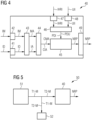
- FIG. 4 a block diagram is shown, which illustrates a personal scan preparation system 40 according to an embodiment of the invention.
- the various blocks or modules as shown and described with reference to Figure 4 may be implemented as any suitable number of hardware circuitry, processors, and/or software (e.g. executed algorithms).
- the personal scan preparation system 40 comprises an image data input interface 41, which receives the above-mentioned image data ID, for example coming from an initial scan.
- the scan recommendation system 40 comprises a metrics data input interface 42, which receives image metrics IM.
- the personal scan preparation system 40 comprises an anomaly detection unit 43, which is configured to detect some anomalies IA in the image data ID and some anomalies MA in the image metrics IM.
- the image anomalies IA and metrics anomalies MA are transmitted to an anomaly classification unit 44, which is configured to classify the detected anomalies IA, MA and which transmits the detected classification CIA, CMA to a scan recommendation unit 45.
- the anomaly detection unit 43 may perform the anomaly detection using as a convolutional neural network configured to process the initial images ID as an input, and to provide as output the probability of the input images ID to be normal (i.e., healthy) vs. anomalous (i.e., requiring further imaging). If the probability of anomaly IA for the provided input image ID is higher than a certain, customizable threshold value ⁇ d , the initial input image ID with marked anomalies IA may then be inserted into the subsequent anomaly classification unit 44, which classifies the anomalies IA into categories i with probability values P(i) for different classes that serve a meaningful input to the scan recommendation unit 45.
- a convolutional neural network configured to process the initial images ID as an input, and to provide as output the probability of the input images ID to be normal (i.e., healthy) vs. anomalous (i.e., requiring further imaging). If the probability of anomaly IA for the provided input image ID is higher than a certain, customizable threshold value ⁇ d , the initial input image
- Suitable classification categories i may include, for instance, one or several of the following exemplary class groups: disease classes (tumor, hemorrhage, MS, etc.), anomalous anatomical regions (white matter, dark matter, brain stem, CSF, etc.), anomalous metrics (white matter volume, vascular wall thickness, carotid cross section, etc.).
- the classes i with high predicted classification probability P(i) are then forwarded as one of the inputs to the scan recommendation unit 45.
- a selection of which classes are being forwarded to the scan recommendation unit 45 may be performed, e.g., by selecting a custom threshold probability value ⁇ c , such that all classes i are forwarded the probability P(i) of which satisfy P(i) > ⁇ c .
- the forwarding criterion for a class i may alternatively be set to satisfy max i P i and P(i) > ⁇ c .
- the personal scan preparation system 40 also comprises a third input interface 47 for receiving electronic health record information IHRI.
- These electronic health record information IHRI of a patient can include for example known diseases, symptoms, clinical indications for the scan, contraindications, like contrast intolerances and can be pulled for example from the RIS or another electronic system containing patient data.
- the personal scan preparation system 40 includes a manual input interface 48 as a fourth input interface for receiving some user interface input UII, for example containing suspected disease or manually selected protocols to be included in the scan workflow.
- the additionally received data IHRI, UII are transmitted to a keyword extraction unit 46, which extracts some relevant keywords EK-IHRI, EK-UII from the received data IHRI, UII.
- the keywords are assigned to topics, which are mapped to possible scan workflow directions PDI based on the electronic health record information IHRI and possible scan workflow directions PDU based on the user interface input UII as evaluation values. These evaluation values are then also transmitted to the scan recommendation system 45.
- the scan recommendation system 45 performs an automatic scan recommendation based on a combination of various inputs CIA, CMA, PDI, PDU to identify a next recommended protocol NRP.
- the identified next recommended protocol NRP is output by an output interface 49, which is also part of the personal scan preparation system 40.
- the personal scan preparation system 40 may be implemented as any suitable number of hardware circuitry, processors, and/or software (e.g. executed algorithms).
- the scan recommendation system 40 may be implemented as any suitable type of classical or machine learning system, such as a fully connected neural network (FCNN).
- the personal scan preparation system 40 may work in conjunction with other classical or machine learning processes, processors, hardware components and/or interfaces, and/or additional executed algorithms to receive data via any suitable number of inputs.
- the personal scan preparation system 40 comprises four input interfaces 41, 42, 43, 44 and works based on four inputs ID, IM, IHRI, UII.
- Embodiments include the personal scan preparation system 40 adapted to any suitable number of inputs that may be greater than or less than four inputs and may include alternate inputs than the examples shown in Figure 4 .
- each of the input interfaces of the scan recommendation system 45 may be implemented using any suitable number of hardware circuitry, processors, and/or software (e.g. executed algorithms).
- each of the input interfaces of the scan recommendation system 45 may be implemented as a system (e.g. a classical or machine learning system) that is best or preferably configured in accordance with the particular type of data that is received at each respective input interface.
- the scan recommendation system 45 may be implemented based upon, e.g., an expert system, a machine learning or deep learning approach, or combinations thereof.
- the scan recommendation algorithm of the scan recommendation system 45 could be, e.g., realized by one or several classical or machine learning algorithms that are able to accept the evaluation data (which were predicted by the anomaly classification units and keyword extraction units upstream) and then predict the most appropriate scan protocol to be added to a scan queue (as can be seen in Figure 6 ).
- an approach based on natural language processing with suitable word embeddings could be appropriate for this task.
- the scan recommendation system 45 may be designed/trained, e.g., based on clinical expert knowledge and medical society guidelines.
- a medical imaging system 50 is schematically illustrated, comprising the personal scan preparation system 40, explained in detail in context with Figure 4 .
- the medical imaging system 50 also comprises an MR scan system 51, which provides the personal scan preparation system 40 with T1-map data T1-M and T2-map data T2-M.
- the medical imaging system 50 includes a contrast image generation unit 52, which generates based on the T1-map data T1-M and T2-map data T2-M, some synthetic contrast images for the use of a radiologist who is used to a certain type of image with a certain contrast.
- the scan recommendation system of the personal scan preparation system 40 proposes a next recommend protocol NRP for an MR imaging process.
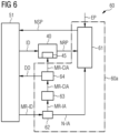
- FIG. 6 a block diagram is depicted, showing a scan workflow performing system 60 with a personal scan preparation system 40 according to an embodiment of the invention and an additional iterative protocol determination and measurement system 60a that may run until a relevant scan abortion criterion ACR combination is met.
- the iterative protocol determination and measurement system 60a comprises a scan queue managing unit 61, which receives a next recommended protocol NRP from the scan recommendation system 45 of the personal scan preparation system 40 and which further receives some manually or electronically selected protocols EP to be scanned and creates a scan queue, which comprises the received protocols NRP, EP.
- embodiments include adding protocols to the scan queue manually or electronically.
- scan protocols NRP, EP from both channels are fed into a scan queue managing unit 61.
- the scan queue managing unit 61 orders and/or curates the scan protocol jobs NRP, EP according to, e.g., scheduling time, relevance, redundancies, or the input channel.
- the scan queue managing unit 61 may (dependent on operator and user preferences) accept both protocol input channels or, in two extreme cases, reject one or the other input channels to enforce manual or automatic mode on a high-level.
- the scan queue managing unit 61 may be configured to add only certain protocols, protocols with certain parameters, or protocols satisfying specific requirements, to the scan queue.
- the protocols in the scan queue may be executed during the next MR scanner measurement, resulting in a reconstructed MR image.
- a quality assurance step and an optional rescan (if indicated) may be inserted at this point or before image reconstruction, when applicable.
- the iterative protocol determination and measurement system 60a further comprises an anomaly detection unit 62, which is configured to detect anomalies MR-IA in the MR-image data MR-ID received from an MR scan system 51.
- the anomaly data MR-IA are transmitted to an anomaly classification unit 63, which is also part of the iterative protocol determination and measurement system 60a.
- the readily reconstructed image MR-ID is inserted into the anomaly detection unit 62 (e.g., similar in function as the anomaly detection unit 43 above) suited for the respective MR scan protocol.
- the anomaly MR-IA may be classified by the above-mentioned subsequent anomaly classification unit 63, e.g., similar in function as the anomaly classification unit 44 above.
- the anomaly classification unit 63 arranges the anomaly data MR-IA in anomaly classes MR-CIA.
- the also called classification data MR-CIA are transmitted to an iteration unit 64, which determines based on the classification data MR-CIA and on a predetermined criterion ACR, whether the scan is completed or an additional MR scan has to be planned based on the classified anomalies MR-CIA.
- the scan is completed. If the probability P(i) of the anomaly classes i of the classification data MR-CIA is smaller than a threshold value ⁇ c , the scan is completed. If the probability P(i) of at least one of the anomaly classes i is higher than the threshold ⁇ c , it is determined, whether an abortion criterion ACR, which can be a time limit that is reached, a maximum number of scan protocols reached or no scan protocol is left in queue, is satisfied. In case one of these abortion criteria ACR is satisfied, the scan is completed. In case the abortion criteria ACR are all not satisfied, a new recommended protocol NRP is created based on the detected anomalies MR-CA. I.e. the classification data MR-CIA can be used as evaluation data for the scan recommendation system 45 of the scan preparation system 40 and a further MR scan is carried out using a newly recommended scan protocol.
- an abortion criterion ACR which can be a time limit that is reached, a maximum number of scan protocols reached or no scan protocol is left in queue.
- a step might be recommended that checks if any scan abortion flags were raised requiring a completion of the scan workflow, e.g., due to a time limit being reached, a maximum number of scan protocols being reached, no scan protocols being left in the queue, etc. If no scan workflow abortion criterion ACR is positive (i.e. identified), then the scan recommendation system 45 can be queried again with the classification data MR-CIA of the new scan image data MR-ID for one or several additional scan recommendation loops, until one or a relevant combination of the following criteria ACR are met and lead to a scan workflow completion:
- an order DO is transmitted to the MR scan unit 51 for transmitting the image data MR-ID to the medical personal.
- the anomaly detection unit 62 does not detect an anomaly MR-IA in the MR image data MR-ID from the MR scan system 51, an information N-IA is sent to the scan queue managing unit 61, which determines, if there is an additional item remaining in the queue. Then the scan queue status may decide, if another pending scan will be executed next or if the scan workflow is completed. In the first case, the scan queue managing unit 61 continues with the next protocol in the queue.
- an anomaly detection unit with an anomaly classification unit connected in series is described herein, this is not the only practical configuration, and is provided by way of example and not limitation.
- the embodiments described herein may alternatively feed an image, image metrics, or other suitable data into the anomaly detection unit and the anomaly classification unit simultaneously for later combination of the results of both modules.
- the embodiments also include the modification of the thresholds for anomaly detection and anomaly classification (e.g., ⁇ d , ⁇ c , ⁇ d , ⁇ c ) to different optimal values depending, e.g., on the position of the unit respective inside the scan workflow performing system 60, the scan protocol NSP employed for image generation, as well as other factors, a particular application, etc.
- the thresholds for anomaly detection and anomaly classification e.g., ⁇ d , ⁇ c , ⁇ d , ⁇ c
- the methods and systems described herein allow for a trained or an untrained technician to perform a highly personalized MRI scan workflow on each patient, and to generate an optimal set of MRI images that are required for diagnosis of that patient's condition(s) considering potential anomalies detected on the recorded image data, electronic health record information, as well as manually- or electronically-set requirements from the side of the operator.
- MR scanner operator may choose to run the MR scan workflow either in fully autonomous, fully manual, or in a hybrid mode.
- the MR scan workflow may be configured to, e.g., minimize the overall scan time per patient and/or to minimize the probability of missing a relevant MR protocol required for image reading and diagnosis.
- the operator may first record a standard set of MR protocols (by means of setting workflow step 3.II, 3.III in Figure 3 appropriately) and employ the scan recommendation system 45 on demand to determine and record any additional MR scans that might be additionally indicated for the current patient.
- FIG. 7 an example for an image data anomaly classification is illustrated for a more detailed understanding.
- a training process of an image data anomaly classification is depicted.
- labelled image data L-ID as training data are input to a combined anomaly detection and classification unit 43, 44.
- the combined anomaly detection and classification unit 43, 44 comprises a convolutional neural network, which creates classification data CIA as an answer to the labelled image data L-ID.
- the classification data CIA are compared with the labelled classification data L-CIA and the convolutional neural network is adapted such that the classification data CIA are assimilated to the labelled classification data L-CIA.
- tissue specific abnormalities or anomalies IA can also be used for anomaly classification.
- the anomalies IA are categorized by the tissue type and the anomaly type.
- a tissue type for the brain can be white matter, grey matter, Hippocampus, Cerebellum or Corpus Callosum.
- the anomaly type can be hypertrophic, atrophic, or abnormal morphology.
- image data ID are input to the anomaly detection and classification unit 43, 44.
- the trained convolutional neural network of the anomaly detection and classification unit 43, 44 now outputs classification data CIA comprising a 1 ⁇ i-vector including probability values P(i) for the above-mentioned abnormalities IA.
- FIG 8 an example for a metrics anomaly classification is illustrated for a more detailed understanding.
- a training process of a metrics anomaly classification is depicted.
- labelled metrics data L-IM are input to an anomaly detection and classification unit 43, 44 as training data.
- the anomaly detection and classification unit 43, 44 comprises a convolutional neural network, which creates classification data CMA as an answer to the labelled metrics data L-IM.
- the classification data CMA are compared with the labelled classification data L-CMA and the convolutional neural network is adapted such that the classification data CMA are assimilated to the labelled classification data L-CMA.
- the metrics data IM comprise values concerning WMv volume, GMv volume, arterial cross section acs, venous cross section vcs, ventricular volume vv and additional information about patient age pa and sex male sm of a patient.
- a classification process is described. Firstly, metrics data IM are input to the anomaly detection and classification unit 43, 44.
- the trained convolutional neural network of the anomaly detection and classification unit 43, 44 now outputs classification data CMA comprising a 1 ⁇ j-vector including probability values P(j) for the above-mentioned abnormalities MA.
- the electronic health record input data IHRI can comprise information about suspected diagnosis SD, symptoms SP, referring indication RIN, the assigned body region BR and the MR protocol requested PRR.
- the keyword extraction starts with a data preprocessing step 9.I, for example a lemmatization and stemming. Then, in a step 9.II a bag-of-words generation is carried out.
- Lemmatization is an important NLP data preparation step, reducing words from different inflectional forms to their base form (better -> good). Bag-of-words generation aims at generating an unsorted catalogue of preprocessed, cleaned words (see above) from a text document, in which the frequency of the words is resolved.
- step 9.III a topic detection based on a topic model is performed, wherein different topics T1, T2, T3, .., TN are detected.
- each identified topic which is a set of keywords, is mapped to a possible scan workflow direction PDI, which is a set of recommended next scan protocols.
- the set PDI of recommended next scan protocols is represented by a 1 ⁇ n -vector.
- the set PDI of recommended next scan protocols for example comprises a T1 weighted protocol T1w, a T2 weighted protocol T2w, a T2 FLAIR protocol T2 FL, a diffusion weighted imaging protocol DWI and a susceptibility weighted imaging protocol SWI.
- Another possibility, if no human mapping from topics to protocols is desired or possible, is to skip topic detection 9.III and instead train a model, e.g., a convolutional neural network, that predicts protocols directly from step 9.II. This is more direct, but offers less explainability to the user.
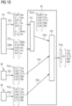
- FIG 10 the function of a scan recommendation 45 is illustrated in detail.
- an anomaly classification unit 44 for classification of image anomalies IA and metrics anomalies MA is depicted.
- keyword extraction units 46 for extracting keywords from electronic health redord information data IHRI and user input data UII are depicted.
- different classification data CIA, CMA are output by the classification unit 44 and possible scan workflow direction data PDI, PDU are output by the keyword extraction units 46. These data are input as evaluation data into the scan recommendation system 45.
- the anomaly classification data CIA, CMA are then processed by a mapping unit 101, which functions based on a protocol mapping model, for example a random forest model or a fully connected neural network.
Landscapes
- Health & Medical Sciences (AREA)
- Engineering & Computer Science (AREA)
- Life Sciences & Earth Sciences (AREA)
- Medical Informatics (AREA)
- General Health & Medical Sciences (AREA)
- Physics & Mathematics (AREA)
- Biomedical Technology (AREA)
- Public Health (AREA)
- Nuclear Medicine, Radiotherapy & Molecular Imaging (AREA)
- Radiology & Medical Imaging (AREA)
- Theoretical Computer Science (AREA)
- Epidemiology (AREA)
- Primary Health Care (AREA)
- Biophysics (AREA)
- Molecular Biology (AREA)
- Pathology (AREA)
- Veterinary Medicine (AREA)
- Animal Behavior & Ethology (AREA)
- Surgery (AREA)
- Data Mining & Analysis (AREA)
- Heart & Thoracic Surgery (AREA)
- General Physics & Mathematics (AREA)
- Software Systems (AREA)
- High Energy & Nuclear Physics (AREA)
- General Engineering & Computer Science (AREA)
- Mathematical Physics (AREA)
- Computing Systems (AREA)
- Evolutionary Computation (AREA)
- Artificial Intelligence (AREA)
- Computer Vision & Pattern Recognition (AREA)
- Computational Linguistics (AREA)
- Business, Economics & Management (AREA)
- General Business, Economics & Management (AREA)
- Databases & Information Systems (AREA)
- Quality & Reliability (AREA)
- Medical Treatment And Welfare Office Work (AREA)
- Measuring And Recording Apparatus For Diagnosis (AREA)
Claims (20)
- Procédé de recommandation de scan, comprenant les stades de :- réception de données d'évaluation comprenant des données (CIA, CMA) de classification, dans lequel les données (CIA, CMA) de classification classent des anomalies (MA) de métriques (IM) déduites d'une image détectées dans des données (ID, IM, IHRI, UII) d'entrée concernant l'état médical d'un patient, et dans lequel les métriques déduites d'images comprennent des propriétés géométriques de tissus et de cavités,- détermination automatique d'un protocole (NRP) de résonnance magnétique suivant recommandé à exécuter en accord avec un scan de résonnance magnétique du patient sur la base des données d'évaluation.
- Procédé de recommandation de scan suivant la revendication 1, dans lequel la détermination automatique d'un protocole (NRP) de résonnance magnétique suivant repose sur l'utilisation d'au moins l'un de :- un modèle d'apprentissage automatique, comprenant au moins l'un de :- un réseau neuronal convolutif,- un réseau neuronal entièrement connecté,- un modèle de réseau neuronal superficiel,- un modèle de réseau neuronal profond,- un arbre de décision,- une forêt d'arbres décisionnels,- une machine à vecteurs de support,- un système expert,- un algorithme de modèle classique.
- Procédé de recommandation de scan suivant la revendication 1 ou 2, dans lequel les données d'évaluation comprennent un premier sens (PDI, PDU) possible de plan de circulation de scan déterminé par des sujets (T1, T2, T3, ..., TN) de topographie, reposant sur des mots-clés (EK-IHRI, EK-UII) extraits dans la donnée (IHRI, UII) d'entrée concernant un état médical du patient, sur des valeurs (P(n), P(o)) de probabilité pour la probabilité d'un protocole (NRP) suivant recommandé.
- Procédé de recommandation de scan suivant l'une quelconque des revendications 1 à 3, dans lequel la détermination automatique d'un protocole (NRP) suivant de résonnance magnétique recommandé comprend les stades de :- détermination d'un deuxième sens (PDA) de plan de circulation de scan par topographie automatique des données (CIA, CMA) de classification sur des probabilités (P(m)) d'un protocole (NRP) suivant recommandé,- recommandation finale automatique du protocole (NRP) suivant recommandé sur la base du deuxième sens possible (PDA) de plan de circulation de scan et sur la base en outre éventuellement du premier sens (PDI, PDU) possible de plan de circulation de scan.
- Procédé de préparation d'un scan personnel, comprenant les stades de :- réception des données (ID, IM, IHRI, UII) d'entrée concernant l'état médical d'un patient, dans lequel les données (ID, IM, IHRI, UII) d'entrée comprennent des métriques (IM) déduites d'une image sur la base des données (ID) d'image,- détermination de données d'évaluation sur la base des données (ID, IM, IHRI, UII) d'entrée comprenant les stades de :- détection d'anomalies (MA) dans les métriques (IM) déduites d'une image dans les données (ID, IM, IHRI, UII) d'entrée,- détermination de données (CMA) de classification sur la base des anomalies (MA) détectées comme données d'évaluation,- effectuer le procédé de recommandation de scan suivant l'une quelconque des revendications 1 à 4 sur la base des données d'évaluation.
- Procédé de préparation d'un scan personnel suivant la revendication 5, dans lequel- les données (ID, IM, IHRI, UII) d'entrée comprennent des données (ID) d'image, et- le stade de détection d'anomalies (IA, MA) comprend détecter des anomalies (IA) dans les données (ID) d'image.
- Procédé de préparation d'un scan personnel suivant la revendication 5 ou 6, dans lequel- les données (ID, IM, IHRI, UII) d'entrée comprennent en outre une information (IHRI) électronique d'enregistrement de santé et/ou des données d'entrée (UII) d'interface d'utilisateur , et le stade de détermination des données d'évaluation comprend les stades de :- extraction des mots-clés (EK-IHRI, EK-UII) de l'information (IHRI) électronique d'enregistrement de santé et/ou de l'entrée (UII) d'interface d'utilisateur,- déterminer des sujets (T1, T2, T3, ... TN) sur la base des mots-clés (EK-IHRI, EK-UII) extraits,- déterminer le premier sens (PDI, PDU) possible de plan de circulation de scan en topographiant les sujets (T1, T2, T3, ..., TN) sur des valeurs (P (n), P (o)) de probabilité pour une probabilité d'un protocole (NRP) suivant recommandé.
- Procédé d'imagerie médicale, comprenant les stades :- déterminer un protocole (NRP) recommandé suivant sur la base du procédé suivant l'une des revendications 1 à 7,- transmettre du protocole (NRP) recommandé suivant à un système (51) de scan à résonnance magnétique,- effectuer un scan à résonnance magnétique par le système (51) de scan à résonnance magnétique sur la base du protocole (NRP) reçu, dans lequel des données (MR-ID) d'image de résonnance magnétique sont enregistrées du patient.
- Procédé pour effectuer un plan de circulation de scan, comprenant les stades :- effectuer le procédé d'imagerie médicale suivant la revendication 8,- détecter des anomalies (MR-IA) de métriques (IM) déduites d'une image dans les données (MR-ID) de résonnance magnétique reconstruites,- déterminer des données (MR-CIA) de classification sur la base des anomalies (MR-IA) détectées,- effectuer itérativement les trois stades mentionnés ci-dessus, sur la base des données (MR-CIA) de classification déterminées jusqu'à atteindre un critère (ACR) de fin.
- Système (45) de recommandation de scan, comprenant :- une interface (41, 42) d'entrée pour recevoir des données d'évaluation comprenant des données (CMA) de classification, dans lequel les données (CMA) de classification classe des anomalies (MA) de métriques (IM) déduites d'une image détectées sur la base des données (ID, IM, IHRI, UII) d'entrée concernant l'état médical d'un patient, et dans lequel les métriques déduites d'une image comprennent des propriétés géométriques de tissus et de cavités,- une unité (101, 102) de recommandation pour la détermination automatique d'un protocole (NRP) suivant de résonnance magnétique recommandé à exécuter en accord avec un scan de résonnance magnétique du patient sur la base des données d'évaluation reçues.
- Système de recommandation de scan suivant la revendication 10,
dans lequel l'unité (101, 102) comprend au moins l'un de :- un modèle d'apprentissage automatique, comprenant au moins l'un de :- un réseau neuronal convolutif (CNN),- un réseau neuronal entièrement connecté (FCNN),- un modèle de réseau neuronal superficiel,- un modèle de réseau neuronal profond,- un arbre de décision,- une forêt d'arbres décisionnels,- une machine à vecteurs de support,- un système expert,- un algorithme de modèle classique. - Système de recommandation de scan suivant la revendication 10 ou 11, comprenant une interface d'entrée pour recevoir des données d'évaluation comprenant un premier sens (PDI, PDU) possible de plan de circulation de scan déterminé par des objets (T1, T2, T3, ..., TN) de topographie, reposant sur des mots-clés (EK-IHRI, EK-UII) extraits concernant un état médical du patient, sur des valeurs (P(n), P(o)) de probabilité pour la probabilité d'un protocole (NRP) suivant recommandé.
- Système de recommandation de scan suivant l'une quelconque des revendications 10 à 12,
dans lequel l'unité (101, 102) de recommandation comprend :- une unité (101) de topographie pour déterminer un deuxième sens (PDA) de plan de circulation de scan possible par topographie automatique des données (CIA, CMA) de classification sur des valeurs (P(m)) de probabilité pour une probabilité d'un protocole (NRP) suivant recommandé,- une unité (102) finale de recommandation de protocole pour recommander finalement le protocole (NRP) suivant recommandé sur la base du deuxième sens (PDA) possible de plan de circulation de scan et éventuellement en outre sur la base du premier sens (PDI, PDU) possible de plan de circulation de scan. - Système (40) de préparation de scan personnel, comprenant :- une interface (41, 42, 47, 48) d'entrée pour recevoir des données (ID, IM, IHRI, UII) d'entrée concernant l'état médical d'un patient, dans lequel les données (ID, IM, IHRI, UII) d'entrée comprennent des métriques (IM) déduites d'une image sur la base des données (ID) d'image et dans lequel les métriques déduites d'une image comprennent des propriétés géométriques de tissus et de cavités,- une unité d'évaluation comprenant :- une unité (43) de détection pour détecter des anomalies (MA) dans les métriques (IM) déduites d'une image dans les données (ID, IM, IHRI, UII) d'entrée,- une unité (44) de classification pour déterminer des données (CMA) de classification sur la base des anomalies (MA) détectées comme données d'évaluation,- le système (45) d'évaluation de scan suivant l'une quelconque des revendications 9 à 13.
- Système de préparation de scan personnel suivant la revendication 14, dans lequel- l'interface (41) d'entrée est configurée pour recevoir des données (ID) d'entrée comprenant les données (ID) d'image médicale, et- l'unité (43) de détection est configurée pour détecter des anomalies (IA) dans les données (ID) d'image médicale.
- Système de préparation de scan personnel suivant la revendication 14 ou 15, dans lequel- l'interface (47, 48) d'entrée est configurée pour recevoir des données (IHRI, UII) d'entrée comprenant de l'information d'enregistrement (IHRI) électronique de santé et/ou des données (UII) d'interface d'utilisateur,- l'unité d'évaluation comprenant :- une unité d'extraction pour extraire des mots-clés (EK-IHRI, EK-UII) de d'information (IHRI) d'enregistrement électronique de santé et/ou de l'entrée (UII) d'interface d'utilisateur,- une unité pour trouver des sujets pour déterminer des sujets (T1, T2, T3, ..., TN) sur la base des mots-clés (EK-IHRI, EK-UII) extraits,- une unité de topographie pour déterminer le premier sens possible de plan de circulation de scan en topographiant les sujets (T1, T2, T3, ..., TN) sur des valeurs (P(m), P(o)) de probabilité pour une probabilité d'un protocole (NRP) suivant recommandé comme données d'évaluation.
- Système (50) d'imagerie médicale, comprenant :- un système (51) de scan à résonnance magnétique,- un système (40) suivant l'une quelconque des revendications 10 à la revendication 16, qui est reliée au système (51) de scan à résonnance magnétique pour déterminer un protocole (NRP) suivant recommandé et pour envoyer le protocole (NRP) suivant recommandé au système (51) de scan à résonnance magnétique pour effectuer un scan de résonnance magnétique, dans lequel les données (MR-ID) d'image de résonnance magnétique sont enregistrées chez un patient.
- Système (60) pour effectuer un plan de circulation de scan, comprenant :- un système (50) d'imagerie médicale suivant la revendication 17,- une unité (62) de détection d'anomalies pour détecter des anomalies (MR-IA) de métriques (IM) déduites d'une image dans les données (MR-ID) de résonnance magnétique reconstruites,- une unité (63) de classification pour déterminer des données (MR-CIA) de classification sur la base des anomalies (MR-IA) détectées,- une unité (64) d'itération pour déterminer, si l'on effectue itérativement les trois stades d'imagerie médicale, une détection d'anomalies et une classification d'anomalies sur la base des données (MR-CIA) de classification déterminées jusqu'à ce qu'une critère (ACR) de fin soit atteint.
- Produit de programme d'ordinateur ayant un programme d'ordinateur, qui peut être chargé directement dans un dispositif de mémoire d'un dispositif de commande d'un système (50) d'imagerie médicale, ayant des parties de programme pour effectuer tous les stades du procédé suivant l'une quelconque des revendications 1 à 9, lorsque le programme d'ordinateur est exécuté dans le dispositif de commande.
- Support, déchiffrable par ordinateur, sur lequel des parties de programme, qui peuvent être entrées et exécutées par une unité informatique d'un système (50) d'imagerie médicale sont mises en mémoire afin d'effectuer tous les stades du procédé suivant l'une quelconque des revendications 1 à 9, lorsque les parties du programme sont exécutées par l'unité informatique.
Applications Claiming Priority (1)
| Application Number | Priority Date | Filing Date | Title |
|---|---|---|---|
| US202063013825P | 2020-04-22 | 2020-04-22 |
Publications (3)
| Publication Number | Publication Date |
|---|---|
| EP3901964A1 EP3901964A1 (fr) | 2021-10-27 |
| EP3901964C0 EP3901964C0 (fr) | 2024-11-06 |
| EP3901964B1 true EP3901964B1 (fr) | 2024-11-06 |
Family
ID=75539047
Family Applications (1)
| Application Number | Title | Priority Date | Filing Date |
|---|---|---|---|
| EP21165484.3A Active EP3901964B1 (fr) | 2020-04-22 | 2021-03-29 | Recommandation intelligente de scan pour l'imagerie par résonance magnétique |
Country Status (3)
| Country | Link |
|---|---|
| US (1) | US20210335490A1 (fr) |
| EP (1) | EP3901964B1 (fr) |
| CN (1) | CN113539464B (fr) |
Families Citing this family (3)
| Publication number | Priority date | Publication date | Assignee | Title |
|---|---|---|---|---|
| US12333065B1 (en) | 2018-10-08 | 2025-06-17 | Floreo, Inc. | Customizing virtual and augmented reality experiences for neurodevelopmental therapies and education |
| CN116570264A (zh) * | 2022-02-10 | 2023-08-11 | 西门子医疗有限公司 | 给定医学测试的自主磁共振扫描 |
| CN119626524A (zh) * | 2025-02-17 | 2025-03-14 | 安徽中医药大学第一附属医院 | 基于ehr的肝豆状核变性临床表型筛选确定方法及系统 |
Family Cites Families (24)
| Publication number | Priority date | Publication date | Assignee | Title |
|---|---|---|---|---|
| JP4675633B2 (ja) * | 2004-03-09 | 2011-04-27 | 株式会社東芝 | 放射線レポートシステム |
| US10600136B2 (en) * | 2011-02-04 | 2020-03-24 | Koninklijke Philips N.V. | Identification of medical concepts for imaging protocol selection |
| US10366783B2 (en) * | 2011-12-30 | 2019-07-30 | Koninklijke Philips N.V. | Imaging examination protocol update recommender |
| KR20140088434A (ko) | 2013-01-02 | 2014-07-10 | 삼성전자주식회사 | 환자별 특성을 기반으로 한 mri 다변수 영상 획득 지원 장치 및 방법 |
| WO2014193552A1 (fr) * | 2013-04-12 | 2014-12-04 | The Research Foundation For The State University Of New York | Procédé d'imagerie à résonance magnétique |
| US11127495B2 (en) * | 2015-12-08 | 2021-09-21 | Koninklijke Philips N.V. | Quality management of imaging workflows |
| US20170156630A1 (en) * | 2015-12-08 | 2017-06-08 | Board Of Regents Of The University Of Texas System | System and method for adaptive and patient-specific magnetic resonance imaging |
| DE102016209032B3 (de) * | 2016-05-24 | 2017-09-14 | Siemens Healthcare Gmbh | Bildbegebendes Verfahren zum Durchführen einer medizinischen Untersuchung nebst zugehörigem Bildgebenden System und zugehörigem Computerprogrammprodukt |
| US11246551B2 (en) * | 2016-09-20 | 2022-02-15 | KUB Technologies, Inc. | System and method for computer aided detection (CAD) in a breast specimen radiograph |
| DE102016219488A1 (de) | 2016-10-07 | 2018-04-12 | Siemens Healthcare Gmbh | Verfahren zum Bereitstellen einer Konfidenzinformation |
| US11360942B2 (en) * | 2017-03-13 | 2022-06-14 | Wandisco Inc. | Methods, devices and systems for maintaining consistency of metadata and data across data centers |
| EP3401918A1 (fr) * | 2017-05-12 | 2018-11-14 | Samsung Electronics Co., Ltd. | Procédé de fourniture d'informations de protocole de balayage pour des dispositifs médicaux et dispositifs électroniques |
| DE102017213222A1 (de) * | 2017-08-01 | 2019-02-07 | Siemens Healthcare Gmbh | Automatisches Erkennen und Einstellen von MR-Protokollen anhand von eingelesenen Bilddaten |
| US20190320934A1 (en) * | 2018-04-18 | 2019-10-24 | Siemens Healthcare Gmbh | Medical image acquisition with sequence prediction using deep learning |
| EP3557274A1 (fr) * | 2018-04-19 | 2019-10-23 | Koninklijke Philips N.V. | Détection automatique d'artéfacts et modification d'une séquence d'impulsions dans une imagerie par résonance magnétique |
| EP3564903A1 (fr) * | 2018-05-01 | 2019-11-06 | Koninklijke Philips N.V. | Fusion d'images de résolution inférieure à supérieure |
| US10878561B2 (en) * | 2018-05-31 | 2020-12-29 | General Electric Company | Automated scanning workflow |
| WO2019236579A1 (fr) * | 2018-06-05 | 2019-12-12 | Fresenius Medical Care Holdings, Inc. | Systèmes et méthodes d'identification de comorbidités |
| US20200043579A1 (en) * | 2018-08-06 | 2020-02-06 | David McEwing | Diagnositic and treatmetnt tool and method for electronic recording and indexing patient encounters for allowing instant search of patient history |
| CN112754458A (zh) * | 2019-11-01 | 2021-05-07 | 上海联影医疗科技股份有限公司 | 磁共振成像方法、系统及存储介质 |
| EP4078407A4 (fr) * | 2019-12-16 | 2024-01-24 | Trialmatch.Me, Inc. D/B/A/ Trialjectory | Extraction de taxinomie non supervisée pour essais cliniques médicaux |
| US11734333B2 (en) * | 2019-12-17 | 2023-08-22 | Shanghai United Imaging Intelligence Co., Ltd. | Systems and methods for managing medical data using relationship building |
| EP3865066A1 (fr) * | 2020-02-17 | 2021-08-18 | Koninklijke Philips N.V. | Examen de balayage de repérage |
| US20210287783A1 (en) * | 2020-03-11 | 2021-09-16 | GE Precision Healthcare LLC | Methods and systems for a workflow tracker |
-
2021
- 2021-03-29 EP EP21165484.3A patent/EP3901964B1/fr active Active
- 2021-04-19 US US17/233,943 patent/US20210335490A1/en not_active Abandoned
- 2021-04-20 CN CN202110424338.XA patent/CN113539464B/zh active Active
Also Published As
| Publication number | Publication date |
|---|---|
| US20210335490A1 (en) | 2021-10-28 |
| EP3901964A1 (fr) | 2021-10-27 |
| EP3901964C0 (fr) | 2024-11-06 |
| CN113539464B (zh) | 2024-12-20 |
| CN113539464A (zh) | 2021-10-22 |
Similar Documents
| Publication | Publication Date | Title |
|---|---|---|
| CN108784655B (zh) | 针对医疗患者的快速评估和后果分析 | |
| US11096674B2 (en) | Method and means of CAD system personalization to provide a confidence level indicator for CAD system recommendations | |
| RU2616985C2 (ru) | Система и способ для поддержки принятия клинических решений для планирования терапии с помощью логического рассуждения на основе прецедентов | |
| JP5553972B2 (ja) | 電子医療記録影響データ取得、処理および表示システムおよび方法 | |
| CN103460213B (zh) | 图像采集和/或图像相关参数推荐器 | |
| JP6542664B2 (ja) | 患者情報を臨床基準にマッチングするシステム及び方法 | |
| US10929973B2 (en) | Medical image pre-processing at the scanner for facilitating joint interpretation by radiologists and artificial intelligence algorithms | |
| EP3614390B1 (fr) | Combinaison d'établissement de rapports et d'imagerie en imagerie médicale | |
| EP3901964B1 (fr) | Recommandation intelligente de scan pour l'imagerie par résonance magnétique | |
| US20180107798A1 (en) | Method for aiding a diagnosis, program and apparatus | |
| AU2017308120B2 (en) | Method and means of CAD system personalization to provide a confidence level indicator for CAD system recommendations | |
| JP2007524461A (ja) | 乳房撮像の自動診断及び決定支援システム及び方法 | |
| US20060200010A1 (en) | Guiding differential diagnosis through information maximization | |
| EP2812828B1 (fr) | Optimisation interactive de bases de données de balayage pour test statistique | |
| JP2018536936A (ja) | 診断情報項目のソート方法、システム及び記憶媒体 | |
| CN109906487A (zh) | 用于针对临床护理连续性进行工作流程敏感的结构化发现对象(sfo)推荐的系统和方法 | |
| Rizkallah et al. | Impact of automated transfer of semi-automated segmentation and structured report rule requirements on cardiac MRI report quality, standardization, and efficiency | |
| CN112567471A (zh) | 处理医学图像 | |
| JP6543308B2 (ja) | 情報処理装置、情報処理装置の作動方法および情報処理システム | |
| WO2023219836A1 (fr) | Procédé d'automatisation de flux de travail de radiologie | |
| US20220101963A1 (en) | Medical data processing system and method | |
| JP7383391B2 (ja) | 医用検査管理システム、オーダリングシステム及びプログラム | |
| Bryan et al. | Artificial Intelligence in Radiology: Not If, But How and When | |
| US20230047826A1 (en) | Context based performance benchmarking | |
| Denck | Machine Learning-based Workflow Enhancements in Magnetic Resonance Imaging |
Legal Events
| Date | Code | Title | Description |
|---|---|---|---|
| PUAI | Public reference made under article 153(3) epc to a published international application that has entered the european phase |
Free format text: ORIGINAL CODE: 0009012 |
|
| STAA | Information on the status of an ep patent application or granted ep patent |
Free format text: STATUS: THE APPLICATION HAS BEEN PUBLISHED |
|
| AK | Designated contracting states |
Kind code of ref document: A1 Designated state(s): AL AT BE BG CH CY CZ DE DK EE ES FI FR GB GR HR HU IE IS IT LI LT LU LV MC MK MT NL NO PL PT RO RS SE SI SK SM TR |
|
| B565 | Issuance of search results under rule 164(2) epc |
Effective date: 20210805 |
|
| STAA | Information on the status of an ep patent application or granted ep patent |
Free format text: STATUS: REQUEST FOR EXAMINATION WAS MADE |
|
| 17P | Request for examination filed |
Effective date: 20220318 |
|
| RBV | Designated contracting states (corrected) |
Designated state(s): AL AT BE BG CH CY CZ DE DK EE ES FI FR GB GR HR HU IE IS IT LI LT LU LV MC MK MT NL NO PL PT RO RS SE SI SK SM TR |
|
| RAP1 | Party data changed (applicant data changed or rights of an application transferred) |
Owner name: SIEMENS HEALTHINEERS AG |
|
| GRAP | Despatch of communication of intention to grant a patent |
Free format text: ORIGINAL CODE: EPIDOSNIGR1 |
|
| STAA | Information on the status of an ep patent application or granted ep patent |
Free format text: STATUS: GRANT OF PATENT IS INTENDED |
|
| INTG | Intention to grant announced |
Effective date: 20240701 |
|
| GRAS | Grant fee paid |
Free format text: ORIGINAL CODE: EPIDOSNIGR3 |
|
| GRAA | (expected) grant |
Free format text: ORIGINAL CODE: 0009210 |
|
| STAA | Information on the status of an ep patent application or granted ep patent |
Free format text: STATUS: THE PATENT HAS BEEN GRANTED |
|
| AK | Designated contracting states |
Kind code of ref document: B1 Designated state(s): AL AT BE BG CH CY CZ DE DK EE ES FI FR GB GR HR HU IE IS IT LI LT LU LV MC MK MT NL NO PL PT RO RS SE SI SK SM TR |
|
| REG | Reference to a national code |
Ref country code: GB Ref legal event code: FG4D |
|
| REG | Reference to a national code |
Ref country code: CH Ref legal event code: EP |
|
| REG | Reference to a national code |
Ref country code: DE Ref legal event code: R096 Ref document number: 602021021253 Country of ref document: DE |
|
| REG | Reference to a national code |
Ref country code: IE Ref legal event code: FG4D |
|
| U01 | Request for unitary effect filed |
Effective date: 20241106 |
|
| U07 | Unitary effect registered |
Designated state(s): AT BE BG DE DK EE FI FR IT LT LU LV MT NL PT RO SE SI Effective date: 20241113 |
|
| PG25 | Lapsed in a contracting state [announced via postgrant information from national office to epo] |
Ref country code: IS Free format text: LAPSE BECAUSE OF FAILURE TO SUBMIT A TRANSLATION OF THE DESCRIPTION OR TO PAY THE FEE WITHIN THE PRESCRIBED TIME-LIMIT Effective date: 20250306 Ref country code: HR Free format text: LAPSE BECAUSE OF FAILURE TO SUBMIT A TRANSLATION OF THE DESCRIPTION OR TO PAY THE FEE WITHIN THE PRESCRIBED TIME-LIMIT Effective date: 20241106 |
|
| PG25 | Lapsed in a contracting state [announced via postgrant information from national office to epo] |
Ref country code: ES Free format text: LAPSE BECAUSE OF FAILURE TO SUBMIT A TRANSLATION OF THE DESCRIPTION OR TO PAY THE FEE WITHIN THE PRESCRIBED TIME-LIMIT Effective date: 20241106 |
|
| PG25 | Lapsed in a contracting state [announced via postgrant information from national office to epo] |
Ref country code: NO Free format text: LAPSE BECAUSE OF FAILURE TO SUBMIT A TRANSLATION OF THE DESCRIPTION OR TO PAY THE FEE WITHIN THE PRESCRIBED TIME-LIMIT Effective date: 20250206 |
|
| U20 | Renewal fee for the european patent with unitary effect paid |
Year of fee payment: 5 Effective date: 20250320 |
|
| PG25 | Lapsed in a contracting state [announced via postgrant information from national office to epo] |
Ref country code: GR Free format text: LAPSE BECAUSE OF FAILURE TO SUBMIT A TRANSLATION OF THE DESCRIPTION OR TO PAY THE FEE WITHIN THE PRESCRIBED TIME-LIMIT Effective date: 20250207 |
|
| PG25 | Lapsed in a contracting state [announced via postgrant information from national office to epo] |
Ref country code: PL Free format text: LAPSE BECAUSE OF FAILURE TO SUBMIT A TRANSLATION OF THE DESCRIPTION OR TO PAY THE FEE WITHIN THE PRESCRIBED TIME-LIMIT Effective date: 20241106 |
|
| PG25 | Lapsed in a contracting state [announced via postgrant information from national office to epo] |
Ref country code: RS Free format text: LAPSE BECAUSE OF FAILURE TO SUBMIT A TRANSLATION OF THE DESCRIPTION OR TO PAY THE FEE WITHIN THE PRESCRIBED TIME-LIMIT Effective date: 20250206 |
|
| PG25 | Lapsed in a contracting state [announced via postgrant information from national office to epo] |
Ref country code: SM Free format text: LAPSE BECAUSE OF FAILURE TO SUBMIT A TRANSLATION OF THE DESCRIPTION OR TO PAY THE FEE WITHIN THE PRESCRIBED TIME-LIMIT Effective date: 20241106 |
|
| PGFP | Annual fee paid to national office [announced via postgrant information from national office to epo] |
Ref country code: GB Payment date: 20250403 Year of fee payment: 5 |
|
| PG25 | Lapsed in a contracting state [announced via postgrant information from national office to epo] |
Ref country code: SK Free format text: LAPSE BECAUSE OF FAILURE TO SUBMIT A TRANSLATION OF THE DESCRIPTION OR TO PAY THE FEE WITHIN THE PRESCRIBED TIME-LIMIT Effective date: 20241106 |
|
| PG25 | Lapsed in a contracting state [announced via postgrant information from national office to epo] |
Ref country code: CZ Free format text: LAPSE BECAUSE OF FAILURE TO SUBMIT A TRANSLATION OF THE DESCRIPTION OR TO PAY THE FEE WITHIN THE PRESCRIBED TIME-LIMIT Effective date: 20241106 |
|
| PLBE | No opposition filed within time limit |
Free format text: ORIGINAL CODE: 0009261 |
|
| STAA | Information on the status of an ep patent application or granted ep patent |
Free format text: STATUS: NO OPPOSITION FILED WITHIN TIME LIMIT |
|
| PG25 | Lapsed in a contracting state [announced via postgrant information from national office to epo] |
Ref country code: MC Free format text: LAPSE BECAUSE OF FAILURE TO SUBMIT A TRANSLATION OF THE DESCRIPTION OR TO PAY THE FEE WITHIN THE PRESCRIBED TIME-LIMIT Effective date: 20241106 |
|
| 26N | No opposition filed |
Effective date: 20250807 |
|
| REG | Reference to a national code |
Ref country code: CH Ref legal event code: H13 Free format text: ST27 STATUS EVENT CODE: U-0-0-H10-H13 (AS PROVIDED BY THE NATIONAL OFFICE) Effective date: 20251023 |
|
| PG25 | Lapsed in a contracting state [announced via postgrant information from national office to epo] |
Ref country code: CH Free format text: LAPSE BECAUSE OF NON-PAYMENT OF DUE FEES Effective date: 20250331 |
|
| PG25 | Lapsed in a contracting state [announced via postgrant information from national office to epo] |
Ref country code: IE Free format text: LAPSE BECAUSE OF NON-PAYMENT OF DUE FEES Effective date: 20250329 |
|
| U20 | Renewal fee for the european patent with unitary effect paid |
Year of fee payment: 6 Effective date: 20260320 |