EP3906962A1 - Dispositif de fermeture d'une voie veineuse pour le traitement des varices - Google Patents
Dispositif de fermeture d'une voie veineuse pour le traitement des varices Download PDFInfo
- Publication number
- EP3906962A1 EP3906962A1 EP20173475.3A EP20173475A EP3906962A1 EP 3906962 A1 EP3906962 A1 EP 3906962A1 EP 20173475 A EP20173475 A EP 20173475A EP 3906962 A1 EP3906962 A1 EP 3906962A1
- Authority
- EP
- European Patent Office
- Prior art keywords
- catheter
- sheath
- balloon
- vein
- adhesive
- Prior art date
- Legal status (The legal status is an assumption and is not a legal conclusion. Google has not performed a legal analysis and makes no representation as to the accuracy of the status listed.)
- Pending
Links
- 210000003462 vein Anatomy 0.000 title claims abstract description 88
- 239000000853 adhesive Substances 0.000 claims abstract description 23
- 230000001070 adhesive effect Effects 0.000 claims abstract description 22
- 206010046996 Varicose vein Diseases 0.000 claims abstract description 6
- 208000027185 varicose disease Diseases 0.000 claims abstract description 6
- 239000003229 sclerosing agent Substances 0.000 claims description 11
- 241000283690 Bos taurus Species 0.000 claims description 2
- 210000003516 pericardium Anatomy 0.000 claims description 2
- 229920002994 synthetic fiber Polymers 0.000 claims description 2
- 210000003752 saphenous vein Anatomy 0.000 description 12
- 210000003191 femoral vein Anatomy 0.000 description 8
- 239000000463 material Substances 0.000 description 6
- WYUYEJNGHIOFOC-NWBUNABESA-N triprolidine hydrochloride (anh.) Chemical compound Cl.C1=CC(C)=CC=C1C(\C=1N=CC=CC=1)=C/CN1CCCC1 WYUYEJNGHIOFOC-NWBUNABESA-N 0.000 description 6
- 210000002414 leg Anatomy 0.000 description 5
- 239000012530 fluid Substances 0.000 description 4
- 210000004379 membrane Anatomy 0.000 description 4
- 239000012528 membrane Substances 0.000 description 4
- 238000000034 method Methods 0.000 description 4
- 210000003513 popliteal vein Anatomy 0.000 description 3
- FAPWRFPIFSIZLT-UHFFFAOYSA-M Sodium chloride Chemical compound [Na+].[Cl-] FAPWRFPIFSIZLT-UHFFFAOYSA-M 0.000 description 2
- 206010002091 Anaesthesia Diseases 0.000 description 1
- 102000008186 Collagen Human genes 0.000 description 1
- 108010035532 Collagen Proteins 0.000 description 1
- 229920001651 Cyanoacrylate Polymers 0.000 description 1
- 206010014523 Embolism and thrombosis Diseases 0.000 description 1
- MWCLLHOVUTZFKS-UHFFFAOYSA-N Methyl cyanoacrylate Chemical compound COC(=O)C(=C)C#N MWCLLHOVUTZFKS-UHFFFAOYSA-N 0.000 description 1
- 208000007536 Thrombosis Diseases 0.000 description 1
- 206010047163 Vasospasm Diseases 0.000 description 1
- 238000004026 adhesive bonding Methods 0.000 description 1
- 230000037005 anaesthesia Effects 0.000 description 1
- 210000004369 blood Anatomy 0.000 description 1
- 239000008280 blood Substances 0.000 description 1
- 229920001436 collagen Polymers 0.000 description 1
- 239000013256 coordination polymer Substances 0.000 description 1
- 230000006378 damage Effects 0.000 description 1
- 230000007423 decrease Effects 0.000 description 1
- SFNALCNOMXIBKG-UHFFFAOYSA-N ethylene glycol monododecyl ether Chemical compound CCCCCCCCCCCCOCCO SFNALCNOMXIBKG-UHFFFAOYSA-N 0.000 description 1
- 239000012634 fragment Substances 0.000 description 1
- 239000003292 glue Substances 0.000 description 1
- 210000004394 hip joint Anatomy 0.000 description 1
- 210000003127 knee Anatomy 0.000 description 1
- 239000007788 liquid Substances 0.000 description 1
- 238000002690 local anesthesia Methods 0.000 description 1
- 239000003589 local anesthetic agent Substances 0.000 description 1
- 229910052751 metal Inorganic materials 0.000 description 1
- 239000002184 metal Substances 0.000 description 1
- 238000012986 modification Methods 0.000 description 1
- 230000004048 modification Effects 0.000 description 1
- 230000036407 pain Effects 0.000 description 1
- 210000002381 plasma Anatomy 0.000 description 1
- 238000010992 reflux Methods 0.000 description 1
- 230000035945 sensitivity Effects 0.000 description 1
- 239000011780 sodium chloride Substances 0.000 description 1
- 239000003106 tissue adhesive Substances 0.000 description 1
- 238000002604 ultrasonography Methods 0.000 description 1
Images


Classifications
-
- A—HUMAN NECESSITIES
- A61—MEDICAL OR VETERINARY SCIENCE; HYGIENE
- A61B—DIAGNOSIS; SURGERY; IDENTIFICATION
- A61B17/00—Surgical instruments, devices or methods
- A61B17/12—Surgical instruments, devices or methods for ligaturing or otherwise compressing tubular parts of the body, e.g. blood vessels or umbilical cord
- A61B17/12022—Occluding by internal devices, e.g. balloons or releasable wires
- A61B17/12131—Occluding by internal devices, e.g. balloons or releasable wires characterised by the type of occluding device
- A61B17/12168—Occluding by internal devices, e.g. balloons or releasable wires characterised by the type of occluding device having a mesh structure
- A61B17/12177—Occluding by internal devices, e.g. balloons or releasable wires characterised by the type of occluding device having a mesh structure comprising additional materials, e.g. thrombogenic, having filaments, having fibers or being coated
-
- A—HUMAN NECESSITIES
- A61—MEDICAL OR VETERINARY SCIENCE; HYGIENE
- A61B—DIAGNOSIS; SURGERY; IDENTIFICATION
- A61B17/00—Surgical instruments, devices or methods
- A61B17/00491—Surgical glue applicators
-
- A—HUMAN NECESSITIES
- A61—MEDICAL OR VETERINARY SCIENCE; HYGIENE
- A61B—DIAGNOSIS; SURGERY; IDENTIFICATION
- A61B17/00—Surgical instruments, devices or methods
- A61B17/12—Surgical instruments, devices or methods for ligaturing or otherwise compressing tubular parts of the body, e.g. blood vessels or umbilical cord
- A61B17/12022—Occluding by internal devices, e.g. balloons or releasable wires
- A61B17/12027—Type of occlusion
- A61B17/12031—Type of occlusion complete occlusion
-
- A—HUMAN NECESSITIES
- A61—MEDICAL OR VETERINARY SCIENCE; HYGIENE
- A61B—DIAGNOSIS; SURGERY; IDENTIFICATION
- A61B17/00—Surgical instruments, devices or methods
- A61B17/12—Surgical instruments, devices or methods for ligaturing or otherwise compressing tubular parts of the body, e.g. blood vessels or umbilical cord
- A61B17/12022—Occluding by internal devices, e.g. balloons or releasable wires
- A61B17/12099—Occluding by internal devices, e.g. balloons or releasable wires characterised by the location of the occluder
- A61B17/12109—Occluding by internal devices, e.g. balloons or releasable wires characterised by the location of the occluder in a blood vessel
-
- A—HUMAN NECESSITIES
- A61—MEDICAL OR VETERINARY SCIENCE; HYGIENE
- A61B—DIAGNOSIS; SURGERY; IDENTIFICATION
- A61B17/00—Surgical instruments, devices or methods
- A61B17/12—Surgical instruments, devices or methods for ligaturing or otherwise compressing tubular parts of the body, e.g. blood vessels or umbilical cord
- A61B17/12022—Occluding by internal devices, e.g. balloons or releasable wires
- A61B17/12131—Occluding by internal devices, e.g. balloons or releasable wires characterised by the type of occluding device
- A61B17/12136—Balloons
-
- A—HUMAN NECESSITIES
- A61—MEDICAL OR VETERINARY SCIENCE; HYGIENE
- A61B—DIAGNOSIS; SURGERY; IDENTIFICATION
- A61B17/00—Surgical instruments, devices or methods
- A61B17/32—Surgical cutting instruments
- A61B17/3205—Excision instruments
- A61B17/3207—Atherectomy devices working by cutting or abrading; Similar devices specially adapted for non-vascular obstructions
- A61B17/320725—Atherectomy devices working by cutting or abrading; Similar devices specially adapted for non-vascular obstructions with radially expandable cutting or abrading elements
-
- A—HUMAN NECESSITIES
- A61—MEDICAL OR VETERINARY SCIENCE; HYGIENE
- A61F—FILTERS IMPLANTABLE INTO BLOOD VESSELS; PROSTHESES; DEVICES PROVIDING PATENCY TO, OR PREVENTING COLLAPSING OF, TUBULAR STRUCTURES OF THE BODY, e.g. STENTS; ORTHOPAEDIC, NURSING OR CONTRACEPTIVE DEVICES; FOMENTATION; TREATMENT OR PROTECTION OF EYES OR EARS; BANDAGES, DRESSINGS OR ABSORBENT PADS; FIRST-AID KITS
- A61F2/00—Filters implantable into blood vessels; Prostheses, i.e. artificial substitutes or replacements for parts of the body; Appliances for connecting them with the body; Devices providing patency to, or preventing collapsing of, tubular structures of the body, e.g. stents
- A61F2/82—Devices providing patency to, or preventing collapsing of, tubular structures of the body, e.g. stents
- A61F2/86—Stents in a form characterised by the wire-like elements; Stents in the form characterised by a net-like or mesh-like structure
-
- A—HUMAN NECESSITIES
- A61—MEDICAL OR VETERINARY SCIENCE; HYGIENE
- A61F—FILTERS IMPLANTABLE INTO BLOOD VESSELS; PROSTHESES; DEVICES PROVIDING PATENCY TO, OR PREVENTING COLLAPSING OF, TUBULAR STRUCTURES OF THE BODY, e.g. STENTS; ORTHOPAEDIC, NURSING OR CONTRACEPTIVE DEVICES; FOMENTATION; TREATMENT OR PROTECTION OF EYES OR EARS; BANDAGES, DRESSINGS OR ABSORBENT PADS; FIRST-AID KITS
- A61F2/00—Filters implantable into blood vessels; Prostheses, i.e. artificial substitutes or replacements for parts of the body; Appliances for connecting them with the body; Devices providing patency to, or preventing collapsing of, tubular structures of the body, e.g. stents
- A61F2/95—Instruments specially adapted for placement or removal of stents or stent-grafts
- A61F2/958—Inflatable balloons for placing stents or stent-grafts
-
- A—HUMAN NECESSITIES
- A61—MEDICAL OR VETERINARY SCIENCE; HYGIENE
- A61L—METHODS OR APPARATUS FOR STERILISING MATERIALS OR OBJECTS IN GENERAL; DISINFECTION, STERILISATION OR DEODORISATION OF AIR; CHEMICAL ASPECTS OF BANDAGES, DRESSINGS, ABSORBENT PADS OR SURGICAL ARTICLES; MATERIALS FOR BANDAGES, DRESSINGS, ABSORBENT PADS OR SURGICAL ARTICLES
- A61L31/00—Materials for other surgical articles, e.g. stents, stent-grafts, shunts, surgical drapes, guide wires, materials for adhesion prevention, occluding devices, surgical gloves, tissue fixation devices
- A61L31/005—Ingredients of undetermined constitution or reaction products thereof
-
- A—HUMAN NECESSITIES
- A61—MEDICAL OR VETERINARY SCIENCE; HYGIENE
- A61B—DIAGNOSIS; SURGERY; IDENTIFICATION
- A61B17/00—Surgical instruments, devices or methods
- A61B2017/00004—(bio)absorbable, (bio)resorbable or resorptive
-
- A—HUMAN NECESSITIES
- A61—MEDICAL OR VETERINARY SCIENCE; HYGIENE
- A61B—DIAGNOSIS; SURGERY; IDENTIFICATION
- A61B17/00—Surgical instruments, devices or methods
- A61B2017/00743—Type of operation; Specification of treatment sites
- A61B2017/00778—Operations on blood vessels
-
- A—HUMAN NECESSITIES
- A61—MEDICAL OR VETERINARY SCIENCE; HYGIENE
- A61B—DIAGNOSIS; SURGERY; IDENTIFICATION
- A61B17/00—Surgical instruments, devices or methods
- A61B2017/00831—Material properties
- A61B2017/00951—Material properties adhesive
-
- A—HUMAN NECESSITIES
- A61—MEDICAL OR VETERINARY SCIENCE; HYGIENE
- A61B—DIAGNOSIS; SURGERY; IDENTIFICATION
- A61B17/00—Surgical instruments, devices or methods
- A61B17/12—Surgical instruments, devices or methods for ligaturing or otherwise compressing tubular parts of the body, e.g. blood vessels or umbilical cord
- A61B17/12022—Occluding by internal devices, e.g. balloons or releasable wires
- A61B2017/1205—Introduction devices
- A61B2017/12054—Details concerning the detachment of the occluding device from the introduction device
- A61B2017/12081—Details concerning the detachment of the occluding device from the introduction device detachable by inflation
-
- A—HUMAN NECESSITIES
- A61—MEDICAL OR VETERINARY SCIENCE; HYGIENE
- A61M—DEVICES FOR INTRODUCING MEDIA INTO, OR ONTO, THE BODY; DEVICES FOR TRANSDUCING BODY MEDIA OR FOR TAKING MEDIA FROM THE BODY; DEVICES FOR PRODUCING OR ENDING SLEEP OR STUPOR
- A61M25/00—Catheters; Hollow probes
- A61M25/10—Balloon catheters
- A61M2025/1043—Balloon catheters with special features or adapted for special applications
- A61M2025/1056—Balloon catheters with special features or adapted for special applications having guide wire lumens outside the main shaft, i.e. the guide wire lumen is within or on the surface of the balloon
Definitions
- the invention relates to a device for closing a vein opening in the treatment of varicose veins.
- the perforating veins can also be affected, in which the physiological direction of flow is directed from the superficial veins into the deep veins.
- the perforating veins there is a reversal of flow in the perforating veins, with congestion of the superficial veins.
- the endovenous procedures mentioned at the beginning do not offer an optimal solution for treating the problem of insufficient perforating veins.
- the device according to claim 1 comprises a sheath which can be expanded by means of a balloon in order to close a venous opening.
- the device can be designed for different applications.
- the cross of the great saphenous / parva vein can be sealed with millimeter precision so that no thrombosis can penetrate the deep leg vein system and there is no risk of early variceal recurrence.
- a perforating vein in another application, can be securely sealed so that the reflux in its area can be eliminated without endangering the deep leg vein system as a result of this intervention.
- the device can be configured in different ways.
- the casing of the device for example, can have a tubular central part which is designed to be open either only at one casing end or at both casing ends.
- the sheath is, for example, sack-like so that, in the expanded state, it can be used to delimit a free space in the vein that is closed at one end and open at the other end.
- the sheath is continuously tubular, so that in the expanded state it can be used to define a free space in the vein which is open at both ends.
- proximal and distal are used from the perspective of the user of the device.
- the proximal end is thus the end facing the user and the distal end is the end facing away from the user.
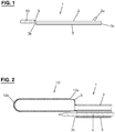
- Fig. 1 shows a catheter 1 which can be inserted into a vein and which has an expandable balloon 5 at the distal end.
- An expandable sheath 10 is arranged on this.
- the catheter 1 has a first channel 2, which fluidly connects a connection 2a to the balloon 5, and a second channel 3, which connects an inlet opening 3a to an outlet opening 3b.
- a syringe or the like can be connected to connection 2a, for example, in order to pump a fluid into the balloon 5 via the first channel 2 or to let it out again and thus enable the balloon 5 to inflate and deflate.
- air is not used as the fluid for inflating, but a liquid, for example a sodium chloride solution.
- a guide wire 4 can be received in the second channel 3.
- the balloon 5 and the envelope 10 are arranged in relation to the exit opening 3b in such a way that there is sufficient space to the side for the guide wire 4 to be guided past.
- the sheath 10 is shaped like a sack and is therefore open at the proximal sheath end 10a and closed at the distal sheath end 10b.
- the length of the sleeve 10 (distance between the ends 10a and 10b) is typically less than 10 cm in the non-expanded state.
- the envelope 10 is designed as a thin layer like a membrane and is impermeable to the blood, in particular the blood plasma.
- the sheath 10 is made of a body-compatible material, e.g., bovine pericardium. Another biological or synthetic material that is tolerated by the body and not rejected can also be used.
- the material of the cover 10, which is intended to remain in the body at least for a certain time, is soft so that it does not hurt and / or chafe when moving, especially when moving the hip joint when the cover 10 is used nearby.
- the material of the cover 10 is chosen so that it is not broken down by the body or that it dissolves after a certain time. In the latter case, the period of time until resorption is at least long enough that, when the device is used to close the cross, no neocross is formed.
- the device described here can be used in many ways to close a vein opening.
- Fig. 3 shows, for example, the confluence CM of the great saphenous vein VSM into the common femoral vein VFC ("Crosse").
- the superficial epigastric vein VES is also shown, which opens into the great saphenous vein VSM.
- the guide wire 4 is pushed into the vein VSM up to the cross CM and then the device according to FIG Fig. 1 Introduced into the VSM vein by sliding the second canal 3 over the wire 4. If the balloon 5 is together with the sheath 10 at the desired location, the guide wire 4 is removed. Then the balloon 5 is inflated. The shell 10 expands accordingly, cf. Fig. 3 . The sleeve 10 finally comes into contact with the vein wall and remains adhered there. The balloon 10 is deflated and removed from the VSM vein. The sheath 10 is now in place, as in FIG Fig. 4 shown.
- the Crosse CM is now closed and the VSM can be treated further, for example as described below in connection with the Fig. 17-19 explained.
- Fig. 5 shows another example in which the sheath 10 is analogous to that in the example according to FIG Fig. 4 has been set in such a way that the cross CP of the small saphenous vein VSP, which opens into the popliteal vein VP, is closed.
- the sheath 10 can adhere to the vein wall by means of at least one adhesive. This is designed in such a way that, together with the cover 10, a non-rigid structure is created which enables painless movements.
- the sheath 10 can, for example, have an outer layer which adheres there when it comes into contact with the vein wall.
- the outer layer is preferably expandable and can contain collagen, for example.
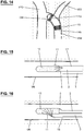
- Fig. 6 shows an example in which the casing 10 has a plurality of reservoirs all around on its surface, which contain a body-compatible adhesive 11 and are covered by a membrane 12.
- a body-compatible adhesive 11 When the balloon 5 is inflated, the membranes 12 burst and the adhesive 11 is released. If the balloon 5 is sufficiently expanded, as in Fig. 7 shown, the adhesive 11 comes into contact with the vein wall so that the sheath 10 is adhered thereto.
- a tissue adhesive for example one based on cyanoacrylate, is suitable as the adhesive 11.
- Fig. 8 shows an example in which the sleeve 10 is provided on the outside and circumferentially with hook elements 13 which are set up to engage the vein wall.
- the hook elements 13 When expanding the balloon 9, cf. Fig. 9 , the hook elements 13 come into contact with the vein wall VW and are finally anchored therein, cf. Fig. 10 .
- Fig. 11 shows an example in which the shell 10 is provided with a tubular lattice framework 14.
- This can, for example, be designed similar to a stent, but does not necessarily extend over the entire length of the sheath 10.
- the lattice framework 14 expands when the balloon 5 is expanded and remains in the expanded state when the balloon 5 is removed.
- the lattice framework 14 is arranged between the balloon 5 and the envelope 10. It can also be arranged on the outside of the casing 10.
- the lattice framework 14 can be attached to the shell 14.
- the lattice frame 14 is made of a body-compatible material, for example metal. Depending on the intended use, the material is absorbable.
- the balloon 5 together with the elements 10 and 14 arranged thereon is brought to the desired location in the vein and then inflated, cf. Fig. 12 until the sheath 10 rests against the vein wall VW.
- the balloon 5 is deflated and taken out.
- the sheath 10 is now fixed on the vein wall VW by means of the lattice framework 14, cf. Fig. 13 .
- Fig. 14 shows an example of a shell 10 with two lattice frames 14a, 14b, which has been placed in the cross CM.
- the respective lattice framework 14a, 14b is arranged on the shell 10, ie on or in the shell 10, and is preferably attached thereto.
- Figures 15 and 16 show variants in which an adhesive 11 can be applied to the surface of the envelope 10 within the vein after the balloon 5 has been placed but not yet expanded. The adhesive 11 is released and is distributed over the envelope 10. The balloon 5 is then inflated and the envelope 10 is thus glued to the vein wall VW.
- the further channel 18 is formed separately from the catheter 2, 3.
- the further channel 18 is introduced, for example, by means of a second venipuncture.
- Fig. 17-19 show a supplemented embodiment in which the device, in addition to the closure of a vein opening, enables the vein to be treated by applying a sclerosant and cutting it up.
- This treatment is in the patent EP3135206B1 by the same applicant.
- the catheter 1 ' is an external catheter and has the balloon 5 with the expandable sheath 10 at the distal end. As Fig. 19 shows, the balloon 5 is fluidically connected to the connection 2a at the proximal end via a first channel 2.
- the catheter 1 ' has the second channel 3, which connects the inlet opening 3a with the outlet opening 3b and in which a guide wire can be received.
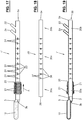
- the catheter 1 ' is fenestrated.
- it is provided with side openings 24 (“catheter window”) between the balloon 20 and the connections 2a, 22. These are arranged distributed around the circumference of the catheter 1 'and are fluidically connected to the inlet opening 3 a via the channel 3.
- a sclerosant can be injected into the vein to be treated via the side openings 24.
- Two adjacent side openings 24 are arranged axially and radially offset from one another. The offset allows the most homogeneous possible distribution of sclerosant in the vein.
- Markings 23 are provided on the catheter shaft between the balloon 20 and the connections 2a, 22, which markings are attached, for example, at regular intervals and, among other things, provide information about how far the catheter 1 'has been inserted into a vein.
- markings 23 in the form of lines and numbers 0, 10, 20, 30, ..., 80 can be seen. Of course, other types of markings are also possible.
- the side openings 24 are arranged in groups, so that the part of the catheter 1 ′ provided with the side openings 24 is divided into sections which each have the same arrangement of side openings 24.
- a group with three side openings can be seen between sections 0 to 10.
- the same arrangement of side openings is repeated in the respective subsequent section 10 to 20, 20 to 30, etc.
- the side openings 24 in the respective group are arranged radially offset by an angle. In the example with three side openings, this angle can be 120 degrees. However, an uneven radial distribution is also conceivable.
- the number of side openings 24 per group or section can differ from that in FIG Fig. 17 be shown and be one, two or more. Since the side openings 24 extend through the outer wall of the catheter 1 ', the number and arrangement are selected so that enough space remains to be able to provide the channels from the connections 2a and 22 to the balloons 5 and 20.
- a second catheter 30 (hereinafter also “inner catheter”) can be introduced via the inlet opening 3a of the catheter 1 ′, as shown in FIG Fig. 18 is shown.
- the catheter 30 has a distal end section 30a and a proximal end section 30b with a connector 34.
- the distal end section 30a is formed by a closed wall.
- the end 36 of the end section 30a is free of an end opening.
- the closed end 36 of the inner catheter 30 allows the lumen at the tip of the outer catheter 1 'to be closed, which prevents it from flowing away through the outlet opening 3b when sclerosant is used.
- the catheter 30 has an intermediate part 30c which has an inner channel (“lumen”) and which is provided with side openings 37. These are fluidically connected to the connection 34 via the inner channel. Subsequent to the distal closed end section 30a, the catheter 30 is only provided with side openings 37 on a partial section, while the rest of the catheter shaft has no openings. In contrast to the external catheter 1 ′, only a single group of side openings 37 is therefore provided.
- the number and / or arrangement of the side openings 37 preferably corresponds to the number or arrangement of the first group of side openings 24 in the external catheter 1 '. In the example according to Fig.
- three side openings 37 can be seen, which are arranged radially and axially offset to one another, similar to the side openings 24 in a group in the outer catheter 1 'according to FIG Fig. 17 .
- the number of side openings 37 can be one, two or more.
- Markings 38 are provided along the intermediate part 30c of the catheter 30, which are attached, for example, at regular intervals and, among other things, provide information about how far the catheter 30 is inserted into the outer catheter 1 '. Lines, numbers, etc. serve as markings 38.
- the outer shape of the inner catheter 30 is designed in such a way that space can be provided in the outer catheter 1 ′ for the channel 2 to the balloon 5 and for the channel to the balloon 20.
- the inner catheter 30 is received in the outer catheter 1 'and then withdrawn in sections.
- the side openings 38 are located here first near the side openings 24 of the first group, then near the side openings 24 of the second group, etc.
- the closed end section 30a of the inner catheter 30 seals the channel 3 of the outer catheter 1 'between the ends 3b and 36.
- a sclerosant can thus be introduced into the vein to be treated in sections via the connection 34 and the side openings 37 and 24.
- the balloon 20 is in the Figures 17 and 19 is shown in a slightly inflated condition.
- the direction in which the axis A runs, along which the outer catheter 1 ′ extends from the outlet opening 3b to the inlet opening 3a, is referred to as "axial", while “radial” is transverse to the axis A.
- the balloon 20 is a “cutting balloon” and has one or more cutting elements 21 (“blades”) for this purpose.
- a respective cutting element 32 does not run straight when viewed in the axial direction A.
- the cutting edge of a cutting element can be curved so that it winds around axis A, e.g., helically. It is also conceivable that, viewed in the direction of the axis A, the cutting edge has a section with a straight axial course which merges again into a straight section via a curved intermediate section. It is also conceivable to provide only a single cutting element 3 which runs around the axis A.
- the uneven course of a cutting element 3 means that, viewed in the axial direction A, the ends of a cutting element 3 are arranged radially offset by an angle which is greater than 0 degrees.
- the angle is preferably at least 10 degrees and particularly preferably at least 20 degrees.
- the course can be such that said angle is less than 360 degrees. It is preferably at most 180 degrees and particularly preferably at most 90 degrees.
- the maximum extension of a cutting element 21 transversely to the axis A can also be variable, in that the cutting edge runs at a height which decreases in the direction of the connection 3a.
- the cutting element 21 is wedge-shaped.
- the maximum height H of a cutting element 21 is typically in the range of 0.5-1.5 mm.
- the balloon 21 extends axially over a length which is typically in the range of 5-30 mm.
- the axially or radially variable shape of a cutting element 21 allows a comprehensive mechanical action on an inner wall of the vein when the catheter 1 'inserted into the vein is withdrawn again.
- the tapering cutting edges of the cutting elements 21 gradually dig into the inner wall of the veins, similar to a plow. An abrupt mechanical action is therefore avoided, so that a more painless treatment is possible, which under certain circumstances can also be carried out without local anesthesia in the form of a tumescent anesthesia. This is the case, for example, when Aethoxysklerol® is used as a sclerosant, which is also a local anesthetic.
- the device according to Fig. 17-19 can be used, for example, as follows: The catheter 1 'is inserted into the trunk vein VSM, if necessary by means of a guide wire, and the sheath 10 is placed by inflating and deflating the balloon 5 so that the cross CM is closed, cf. Fig. 4 .
- the balloon 20 is then inflated.
- the inner catheter 20 is pushed into the channel 3 of the outer catheter 1 ', unless it has already been introduced together with the catheter 1' in the event that no guide wire has been used.
- the sclerosant is supplied via the inner catheter 20 by means of a syringe attached to the connection 34.
- the inner catheter 30 is withdrawn in e.g. 10 cm steps. Sclerosant is applied every 10 cm and a certain time, e.g. approx. 1 minute, is waited before the next section is treated. The procedure is continued until the entire inner catheter 30 is removed.
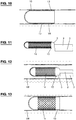
- the device comprises a catheter 41 which has a first channel which connects an inlet opening 43a to the outlet opening 43b and which is used to receive a guide wire 4 (cf. Fig. 2 ) is designed.
- a balloon 45 is arranged at the distal end of the catheter 41. This runs around the outer wall of the catheter 41 so that the first channel can extend through the balloon 45 and the exit opening 43b is axially offset from the balloon 45.
- the balloon 45 is fluidically connected to a connector 42a at the proximal end of the catheter 41 via a second channel. A fluid can be conveyed via the second channel in order to be able to inflate and deflate the balloon 45.
- An expandable envelope 50 is arranged on the balloon 45. Like the sheath 10 in the first exemplary embodiment, this is designed as a membrane and can consist of the same material.
- the length of the sleeve 50 (distance between the ends 50a and 50b) is typically less than 10 cm in the non-expanded state.
- the device according to Fig. 20 can be used, for example, to close a vein opening into a perforating vein.
- Fig. 21 shows schematically a perforating vein PV, which connects a superficial vein OV with a deep vein TV.
- the perforating vein PV is insufficient and should be shut down. To do this, proceed as follows: After the guide wire 4 has been pushed under ultrasound control via the vein OV into the area of the perforating vein PV to be treated, the catheter becomes 41 is advanced along the guide wire 4 until the balloon 45, together with the sheath 50, covers the perforating vein PV. The guide wire 4 is removed and the balloon 45 is inflated until the sheath 50 contacts the vein wall VW, cf. Fig. 21 . Because of the adhesive, the sheath 50 remains adhered to the vein wall VW. The balloon 45 is deflated and removed by pulling out the catheter 41. The vein opening into the perforating vein PV is now closed by means of the sheath 50, cf. Fig. 22 .
- Fig. 20 can also be used for the treatment of the Magnacrosse CM.
- Fig. 23 shows the situation in which the sheath 50, which is open on both sides, has been placed in such a way that the open end 50b extends into the superficial epigastric vein VES, whereby the connection to the common femoral vein VFC is sealed.
- the sheath 50 in such a way that it closes the great saphenous vein VSM or its cross CM from the common femoral vein VFC, cf. Fig. 25 .
- Fig. 24 shows some leg veins, namely popliteal vein VP, femoral vein VF, common femoral vein VFC, deep femoral vein VPF and great saphenous vein VSM.
- the catheter 41 with the balloon 45 and the sheath 50 is introduced into the popliteal vein VP and thus into the deep leg vein system via a puncture in the hollow of the knee.
- the balloon 45 and envelope 50 are pushed up over the femoral vein VF into the common femoral vein VPF and the envelope is placed at the magnacross CM.
- the devices described herein can be designed for single use and are provided in sterile form in packages.
- the cutting device 20, 21 and / or the side openings 24 in the variant according to FIG Fig. 17 can also be formed on a separately formed catheter which can be inserted into the vein after the sheath 10, 50 has been placed and the device according to FIG Fig. 1 or 20 has been removed.
Landscapes
- Health & Medical Sciences (AREA)
- Life Sciences & Earth Sciences (AREA)
- Surgery (AREA)
- Biomedical Technology (AREA)
- Engineering & Computer Science (AREA)
- Public Health (AREA)
- Heart & Thoracic Surgery (AREA)
- Veterinary Medicine (AREA)
- Animal Behavior & Ethology (AREA)
- General Health & Medical Sciences (AREA)
- Vascular Medicine (AREA)
- Nuclear Medicine, Radiotherapy & Molecular Imaging (AREA)
- Medical Informatics (AREA)
- Molecular Biology (AREA)
- Reproductive Health (AREA)
- Cardiology (AREA)
- Oral & Maxillofacial Surgery (AREA)
- Transplantation (AREA)
- Epidemiology (AREA)
- Chemical & Material Sciences (AREA)
- Chemical Kinetics & Catalysis (AREA)
- Media Introduction/Drainage Providing Device (AREA)
Priority Applications (2)
| Application Number | Priority Date | Filing Date | Title |
|---|---|---|---|
| EP20173475.3A EP3906962A1 (fr) | 2020-05-07 | 2020-05-07 | Dispositif de fermeture d'une voie veineuse pour le traitement des varices |
| US17/308,770 US12295586B2 (en) | 2020-05-07 | 2021-05-05 | Device for closing a vein juncture in the treatment of varicose veins |
Applications Claiming Priority (1)
| Application Number | Priority Date | Filing Date | Title |
|---|---|---|---|
| EP20173475.3A EP3906962A1 (fr) | 2020-05-07 | 2020-05-07 | Dispositif de fermeture d'une voie veineuse pour le traitement des varices |
Publications (1)
| Publication Number | Publication Date |
|---|---|
| EP3906962A1 true EP3906962A1 (fr) | 2021-11-10 |
Family
ID=70616976
Family Applications (1)
| Application Number | Title | Priority Date | Filing Date |
|---|---|---|---|
| EP20173475.3A Pending EP3906962A1 (fr) | 2020-05-07 | 2020-05-07 | Dispositif de fermeture d'une voie veineuse pour le traitement des varices |
Country Status (2)
| Country | Link |
|---|---|
| US (1) | US12295586B2 (fr) |
| EP (1) | EP3906962A1 (fr) |
Families Citing this family (1)
| Publication number | Priority date | Publication date | Assignee | Title |
|---|---|---|---|---|
| EP4716505A1 (fr) * | 2023-05-22 | 2026-04-01 | Clearstream Technologies Limited | Implant médical pour fermer un vaisseau |
Citations (10)
| Publication number | Priority date | Publication date | Assignee | Title |
|---|---|---|---|---|
| WO1997027893A1 (fr) * | 1996-02-02 | 1997-08-07 | Transvascular, Inc. | Methodes et appareil d'arret de l'ecoulement du sang dans les vaisseaux |
| WO2002003893A2 (fr) * | 2000-06-26 | 2002-01-17 | Rex Medical, L.P. | Dispositif vasculaire pour apposition de valvule |
| WO2003032815A2 (fr) * | 2001-10-15 | 2003-04-24 | Scimed Life Systems, Inc. | Dispositif medical servant a mettre en place des pieces |
| WO2004045393A2 (fr) * | 2002-11-20 | 2004-06-03 | Fogarty, Thomas, J. | Dispositifs et methodes de traitement d'anevrismes vasculaires |
| WO2005048884A1 (fr) * | 2003-11-17 | 2005-06-02 | Vnus Medical Technologies, Inc. | Dispositif d'occlusion veineuse a absorption temporaire et procede de traitement veineux superficiel |
| US20120232581A1 (en) * | 2011-03-10 | 2012-09-13 | Western New England University | Tamponade for Biopsy Surgery and Method of Operation |
| WO2014201434A2 (fr) * | 2013-06-14 | 2014-12-18 | Artventive Medical Group, Inc. | Dispositifs luminaux implantables |
| WO2015052703A2 (fr) * | 2013-10-13 | 2015-04-16 | V.V.T. Med Ltd. | Dispositif et méthode de résection d'une veine |
| WO2019075354A1 (fr) * | 2017-10-12 | 2019-04-18 | Vimal Nanavati | Cathéter à lumières multiples pour déploiement de dispositif cardiaque |
| EP3135206B1 (fr) | 2015-08-31 | 2020-01-15 | Gefässpraxis Dr. Erpen AG | Dispositif de traitement des varices |
Family Cites Families (10)
| Publication number | Priority date | Publication date | Assignee | Title |
|---|---|---|---|---|
| US5669936A (en) * | 1983-12-09 | 1997-09-23 | Endovascular Technologies, Inc. | Endovascular grafting system and method for use therewith |
| US5634901A (en) * | 1992-11-02 | 1997-06-03 | Localmed, Inc. | Method of using a catheter sleeve |
| CA2202363C (fr) * | 1994-10-19 | 2004-01-20 | Bradly Jendersee | Fixation d'un extenseur a la surface d'un vaisseau sanguin |
| US6379329B1 (en) * | 1999-06-02 | 2002-04-30 | Cordis Neurovascular, Inc. | Detachable balloon embolization device and method |
| US20060206140A1 (en) * | 2005-02-24 | 2006-09-14 | Samuel Shaolian | Adjustable embolic aneurysm coil |
| US20090112239A1 (en) * | 2007-10-31 | 2009-04-30 | Specialized Vascular Technologies, Inc. | Sticky dilatation balloon and methods of using |
| US8974519B2 (en) * | 2010-02-19 | 2015-03-10 | Cardiovascular Systems, Inc. | Therapeutic agent delivery system, device and method for localized application of therapeutic substances to a biological conduit |
| US8764793B2 (en) * | 2011-06-17 | 2014-07-01 | Northwestern University | Left atrial appendage occluder |
| US20140180069A1 (en) * | 2012-12-21 | 2014-06-26 | Volcano Corporation | Intraluminal imaging system |
| CN113274099B (zh) * | 2020-10-14 | 2022-10-11 | 普利瑞医疗科技(苏州)有限公司 | 一种用于治疗动脉粥样硬化闭塞症的切割装置 |
-
2020
- 2020-05-07 EP EP20173475.3A patent/EP3906962A1/fr active Pending
-
2021
- 2021-05-05 US US17/308,770 patent/US12295586B2/en active Active
Patent Citations (10)
| Publication number | Priority date | Publication date | Assignee | Title |
|---|---|---|---|---|
| WO1997027893A1 (fr) * | 1996-02-02 | 1997-08-07 | Transvascular, Inc. | Methodes et appareil d'arret de l'ecoulement du sang dans les vaisseaux |
| WO2002003893A2 (fr) * | 2000-06-26 | 2002-01-17 | Rex Medical, L.P. | Dispositif vasculaire pour apposition de valvule |
| WO2003032815A2 (fr) * | 2001-10-15 | 2003-04-24 | Scimed Life Systems, Inc. | Dispositif medical servant a mettre en place des pieces |
| WO2004045393A2 (fr) * | 2002-11-20 | 2004-06-03 | Fogarty, Thomas, J. | Dispositifs et methodes de traitement d'anevrismes vasculaires |
| WO2005048884A1 (fr) * | 2003-11-17 | 2005-06-02 | Vnus Medical Technologies, Inc. | Dispositif d'occlusion veineuse a absorption temporaire et procede de traitement veineux superficiel |
| US20120232581A1 (en) * | 2011-03-10 | 2012-09-13 | Western New England University | Tamponade for Biopsy Surgery and Method of Operation |
| WO2014201434A2 (fr) * | 2013-06-14 | 2014-12-18 | Artventive Medical Group, Inc. | Dispositifs luminaux implantables |
| WO2015052703A2 (fr) * | 2013-10-13 | 2015-04-16 | V.V.T. Med Ltd. | Dispositif et méthode de résection d'une veine |
| EP3135206B1 (fr) | 2015-08-31 | 2020-01-15 | Gefässpraxis Dr. Erpen AG | Dispositif de traitement des varices |
| WO2019075354A1 (fr) * | 2017-10-12 | 2019-04-18 | Vimal Nanavati | Cathéter à lumières multiples pour déploiement de dispositif cardiaque |
Also Published As
| Publication number | Publication date |
|---|---|
| US12295586B2 (en) | 2025-05-13 |
| US20210346034A1 (en) | 2021-11-11 |
Similar Documents
| Publication | Publication Date | Title |
|---|---|---|
| EP3135206B1 (fr) | Dispositif de traitement des varices | |
| DE69219878T2 (de) | Vorrichtung zur Aufweiten einer Stenose | |
| DE69936223T2 (de) | Katheter für endovaskuläres behandeln von stenosen der kopfschlagader | |
| DE69631531T2 (de) | Katheter zur Verabreichung von Arzneien | |
| DE69023362T2 (de) | Katheter und Methode zur lokal angewandten Medikation der Wand eines Blutgefässes oder eines anderen Körperlumens. | |
| EP0080436B1 (fr) | Dispositif pour la suppression ou dilatation d'occlusions dans les vaisseaux porteurs de fluides corporels | |
| DE60306502T2 (de) | Vaskuläre kupplungsvorrichtung | |
| DE69825162T2 (de) | Intravaskulärer stent mit einer ablenkvorrichtung | |
| DE69825200T2 (de) | Kathetersystem zum Anbringen eines Stent | |
| DE69719648T2 (de) | Mit einem Ballon aktivierte Kraftkonzentrationsvorrichtung zum Einschneiden von stenotischen Segmenten | |
| DE69703681T2 (de) | Katheter zur Gefässneubildung | |
| DE60204258T2 (de) | Feder zum Falten eines Katheterballons | |
| EP0604761B1 (fr) | Dispositif pour obstruer une ouverture dans un vaisseau | |
| DE60031270T2 (de) | System zur einbringung einer expandierbaren medizinischen vorrichtung | |
| DE60020881T2 (de) | Gerät zur kontrolle der extra-vascular-blutungen | |
| EP2785265B1 (fr) | Dispositif pour retirer des thrombus pariétaux d'un vaisseau du corps | |
| DE69532267T2 (de) | Katheter für Stentimplantation | |
| EP2510972B1 (fr) | Dispositif de cathéter | |
| CH684307A5 (de) | Verlängerung von linearem und rohrförmigem Gewebe. | |
| WO1995003081A1 (fr) | Catheter a manchon | |
| EP2446918A1 (fr) | Cathéter à ballonnet avec une section extensible asymétrique radiale | |
| DE3821544A1 (de) | Dilatationskatheter | |
| EP2730309A1 (fr) | Cathéter à ballonnet pour récipients courbés | |
| WO2006047977A1 (fr) | Stent destine a etre implante a l'interieur ou autour d'un organe creux, comportant des elements marqueurs constitues d'un materiau opaque aux rayons x | |
| WO1999035975A1 (fr) | Catheter a expansion pour chirurgie de pontage |
Legal Events
| Date | Code | Title | Description |
|---|---|---|---|
| PUAI | Public reference made under article 153(3) epc to a published international application that has entered the european phase |
Free format text: ORIGINAL CODE: 0009012 |
|
| STAA | Information on the status of an ep patent application or granted ep patent |
Free format text: STATUS: THE APPLICATION HAS BEEN PUBLISHED |
|
| AK | Designated contracting states |
Kind code of ref document: A1 Designated state(s): AL AT BE BG CH CY CZ DE DK EE ES FI FR GB GR HR HU IE IS IT LI LT LU LV MC MK MT NL NO PL PT RO RS SE SI SK SM TR |
|
| B565 | Issuance of search results under rule 164(2) epc |
Effective date: 20201109 |
|
| STAA | Information on the status of an ep patent application or granted ep patent |
Free format text: STATUS: REQUEST FOR EXAMINATION WAS MADE |
|
| 17P | Request for examination filed |
Effective date: 20220509 |
|
| RBV | Designated contracting states (corrected) |
Designated state(s): AL AT BE BG CH CY CZ DE DK EE ES FI FR GB GR HR HU IE IS IT LI LT LU LV MC MK MT NL NO PL PT RO RS SE SI SK SM TR |
|
| STAA | Information on the status of an ep patent application or granted ep patent |
Free format text: STATUS: EXAMINATION IS IN PROGRESS |
|
| 17Q | First examination report despatched |
Effective date: 20241202 |