EP3919625A1 - Verfahren zur diagnose von hirnmetastasen - Google Patents
Verfahren zur diagnose von hirnmetastasen Download PDFInfo
- Publication number
- EP3919625A1 EP3919625A1 EP21184435.2A EP21184435A EP3919625A1 EP 3919625 A1 EP3919625 A1 EP 3919625A1 EP 21184435 A EP21184435 A EP 21184435A EP 3919625 A1 EP3919625 A1 EP 3919625A1
- Authority
- EP
- European Patent Office
- Prior art keywords
- mir
- pdpk1
- brain
- cells
- metastasis
- Prior art date
- Legal status (The legal status is an assumption and is not a legal conclusion. Google has not performed a legal analysis and makes no representation as to the accuracy of the status listed.)
- Withdrawn
Links
- 210000004556 brain Anatomy 0.000 title claims abstract description 359
- 238000000034 method Methods 0.000 title claims abstract description 184
- 206010027476 Metastases Diseases 0.000 title abstract description 333
- 230000009401 metastasis Effects 0.000 title abstract description 316
- 108091064825 miR-181c stem-loop Proteins 0.000 claims abstract description 366
- 108091044400 miR-181c-1 stem-loop Proteins 0.000 claims abstract description 366
- 108091048818 miR-181c-2 stem-loop Proteins 0.000 claims abstract description 366
- 108091032779 miR-181c-3 stem-loop Proteins 0.000 claims abstract description 366
- 206010028980 Neoplasm Diseases 0.000 claims abstract description 244
- 230000008499 blood brain barrier function Effects 0.000 claims abstract description 168
- 210000001218 blood-brain barrier Anatomy 0.000 claims abstract description 167
- 102100037263 3-phosphoinositide-dependent protein kinase 1 Human genes 0.000 claims abstract description 157
- 230000014509 gene expression Effects 0.000 claims abstract description 149
- 230000000694 effects Effects 0.000 claims abstract description 99
- 239000000203 mixture Substances 0.000 claims abstract description 60
- 230000035699 permeability Effects 0.000 claims abstract description 52
- 239000004480 active ingredient Substances 0.000 claims abstract description 33
- 101000600756 Homo sapiens 3-phosphoinositide-dependent protein kinase 1 Proteins 0.000 claims abstract 19
- 239000003112 inhibitor Substances 0.000 claims description 124
- 239000002679 microRNA Substances 0.000 claims description 106
- 108020004459 Small interfering RNA Proteins 0.000 claims description 58
- 239000003795 chemical substances by application Substances 0.000 claims description 47
- 239000008194 pharmaceutical composition Substances 0.000 claims description 32
- 101150036454 Pdpk1 gene Proteins 0.000 claims description 31
- 108091023037 Aptamer Proteins 0.000 claims description 30
- 239000012634 fragment Substances 0.000 claims description 13
- 108091034117 Oligonucleotide Proteins 0.000 claims description 12
- 239000000074 antisense oligonucleotide Substances 0.000 claims description 12
- 238000012230 antisense oligonucleotides Methods 0.000 claims description 12
- 239000005557 antagonist Substances 0.000 claims description 10
- 230000001225 therapeutic effect Effects 0.000 claims description 10
- 108091070501 miRNA Proteins 0.000 claims description 9
- 108090000765 processed proteins & peptides Proteins 0.000 claims description 9
- 238000009472 formulation Methods 0.000 claims description 6
- 208000003174 Brain Neoplasms Diseases 0.000 claims description 5
- 208000026106 cerebrovascular disease Diseases 0.000 claims description 5
- 208000014644 Brain disease Diseases 0.000 claims description 3
- DLGOEMSEDOSKAD-UHFFFAOYSA-N Carmustine Chemical compound ClCCNC(=O)N(N=O)CCCl DLGOEMSEDOSKAD-UHFFFAOYSA-N 0.000 claims description 3
- 208000012902 Nervous system disease Diseases 0.000 claims description 3
- 208000025966 Neurological disease Diseases 0.000 claims description 3
- 208000029033 Spinal Cord disease Diseases 0.000 claims description 3
- 208000020016 psychiatric disease Diseases 0.000 claims description 3
- 208000024827 Alzheimer disease Diseases 0.000 claims description 2
- 206010008025 Cerebellar ataxia Diseases 0.000 claims description 2
- 206010008111 Cerebral haemorrhage Diseases 0.000 claims description 2
- 206010012289 Dementia Diseases 0.000 claims description 2
- 208000032612 Glial tumor Diseases 0.000 claims description 2
- 206010018338 Glioma Diseases 0.000 claims description 2
- 208000003618 Intervertebral Disc Displacement Diseases 0.000 claims description 2
- 201000008450 Intracranial aneurysm Diseases 0.000 claims description 2
- 201000009906 Meningitis Diseases 0.000 claims description 2
- 208000018737 Parkinson disease Diseases 0.000 claims description 2
- 208000007913 Pituitary Neoplasms Diseases 0.000 claims description 2
- 201000005746 Pituitary adenoma Diseases 0.000 claims description 2
- 206010061538 Pituitary tumour benign Diseases 0.000 claims description 2
- 208000013738 Sleep Initiation and Maintenance disease Diseases 0.000 claims description 2
- 208000010112 Spinocerebellar Degenerations Diseases 0.000 claims description 2
- 229940120638 avastin Drugs 0.000 claims description 2
- 206010008118 cerebral infarction Diseases 0.000 claims description 2
- 230000003412 degenerative effect Effects 0.000 claims description 2
- 206010015037 epilepsy Diseases 0.000 claims description 2
- 229940084910 gliadel Drugs 0.000 claims description 2
- 206010022437 insomnia Diseases 0.000 claims description 2
- 206010027191 meningioma Diseases 0.000 claims description 2
- 208000021310 pituitary gland adenoma Diseases 0.000 claims description 2
- 208000005198 spinal stenosis Diseases 0.000 claims description 2
- 206010044652 trigeminal neuralgia Diseases 0.000 claims description 2
- 201000011510 cancer Diseases 0.000 abstract description 246
- 238000003745 diagnosis Methods 0.000 abstract description 34
- 230000002401 inhibitory effect Effects 0.000 abstract description 15
- 230000007246 mechanism Effects 0.000 abstract description 12
- 230000002265 prevention Effects 0.000 abstract description 6
- 239000003814 drug Substances 0.000 abstract description 4
- 238000011156 evaluation Methods 0.000 abstract description 3
- 238000012377 drug delivery Methods 0.000 abstract 1
- 210000004027 cell Anatomy 0.000 description 320
- 239000000523 sample Substances 0.000 description 148
- 108010082078 3-Phosphoinositide-Dependent Protein Kinases Proteins 0.000 description 138
- 108090000623 proteins and genes Proteins 0.000 description 85
- 108700011259 MicroRNAs Proteins 0.000 description 82
- 239000013642 negative control Substances 0.000 description 74
- 210000002889 endothelial cell Anatomy 0.000 description 66
- 238000000338 in vitro Methods 0.000 description 64
- 238000004590 computer program Methods 0.000 description 58
- 102000004169 proteins and genes Human genes 0.000 description 57
- 239000004055 small Interfering RNA Substances 0.000 description 56
- 108091032973 (ribonucleotides)n+m Proteins 0.000 description 47
- 239000002773 nucleotide Substances 0.000 description 43
- 125000003729 nucleotide group Chemical group 0.000 description 43
- 102000039446 nucleic acids Human genes 0.000 description 42
- 108020004707 nucleic acids Proteins 0.000 description 42
- 150000007523 nucleic acids Chemical class 0.000 description 42
- 239000000126 substance Substances 0.000 description 40
- 102000000591 Tight Junction Proteins Human genes 0.000 description 38
- 108010002321 Tight Junction Proteins Proteins 0.000 description 38
- 238000012502 risk assessment Methods 0.000 description 36
- 238000005259 measurement Methods 0.000 description 34
- 108020004999 messenger RNA Proteins 0.000 description 29
- 102000000905 Cadherin Human genes 0.000 description 28
- 108050007957 Cadherin Proteins 0.000 description 28
- 108050000637 N-cadherin Proteins 0.000 description 27
- 238000007792 addition Methods 0.000 description 26
- 230000027455 binding Effects 0.000 description 25
- 210000003556 vascular endothelial cell Anatomy 0.000 description 25
- 210000004369 blood Anatomy 0.000 description 24
- 239000008280 blood Substances 0.000 description 24
- 102000040430 polynucleotide Human genes 0.000 description 23
- 108091033319 polynucleotide Proteins 0.000 description 23
- 239000002157 polynucleotide Substances 0.000 description 23
- 210000001808 exosome Anatomy 0.000 description 22
- 230000028327 secretion Effects 0.000 description 22
- 206010006187 Breast cancer Diseases 0.000 description 21
- 208000026310 Breast neoplasm Diseases 0.000 description 20
- 239000012528 membrane Substances 0.000 description 20
- 210000004204 blood vessel Anatomy 0.000 description 19
- LOKCTEFSRHRXRJ-UHFFFAOYSA-I dipotassium trisodium dihydrogen phosphate hydrogen phosphate dichloride Chemical compound P(=O)(O)(O)[O-].[K+].P(=O)(O)([O-])[O-].[Na+].[Na+].[Cl-].[K+].[Cl-].[Na+] LOKCTEFSRHRXRJ-UHFFFAOYSA-I 0.000 description 19
- 239000002953 phosphate buffered saline Substances 0.000 description 19
- 238000013208 measuring procedure Methods 0.000 description 18
- 238000003556 assay Methods 0.000 description 17
- 230000004807 localization Effects 0.000 description 17
- 102000007469 Actins Human genes 0.000 description 16
- 108010085238 Actins Proteins 0.000 description 16
- 241000699670 Mus sp. Species 0.000 description 16
- 241000699666 Mus <mouse, genus> Species 0.000 description 15
- 230000008859 change Effects 0.000 description 15
- 102000015693 Actin Depolymerizing Factors Human genes 0.000 description 14
- 108010038798 Actin Depolymerizing Factors Proteins 0.000 description 14
- 108020004635 Complementary DNA Proteins 0.000 description 14
- 238000010804 cDNA synthesis Methods 0.000 description 14
- 239000002299 complementary DNA Substances 0.000 description 14
- 208000037819 metastatic cancer Diseases 0.000 description 14
- 208000011575 metastatic malignant neoplasm Diseases 0.000 description 14
- 102000040650 (ribonucleotides)n+m Human genes 0.000 description 13
- 238000001262 western blot Methods 0.000 description 13
- 108020005345 3' Untranslated Regions Proteins 0.000 description 12
- 239000003153 chemical reaction reagent Substances 0.000 description 12
- 210000000056 organ Anatomy 0.000 description 12
- 239000012099 Alexa Fluor family Substances 0.000 description 11
- 102100031181 Glyceraldehyde-3-phosphate dehydrogenase Human genes 0.000 description 11
- 102000003940 Occludin Human genes 0.000 description 11
- 108090000304 Occludin Proteins 0.000 description 11
- 230000000295 complement effect Effects 0.000 description 11
- 230000006378 damage Effects 0.000 description 11
- 230000006870 function Effects 0.000 description 11
- 108020004445 glyceraldehyde-3-phosphate dehydrogenase Proteins 0.000 description 11
- 230000010365 information processing Effects 0.000 description 11
- 108010082117 matrigel Proteins 0.000 description 11
- 238000002360 preparation method Methods 0.000 description 11
- 108091007428 primary miRNA Proteins 0.000 description 11
- 238000012360 testing method Methods 0.000 description 11
- 238000013518 transcription Methods 0.000 description 11
- 230000035897 transcription Effects 0.000 description 11
- 150000001413 amino acids Chemical class 0.000 description 10
- 210000001130 astrocyte Anatomy 0.000 description 10
- 238000001727 in vivo Methods 0.000 description 10
- 238000002372 labelling Methods 0.000 description 10
- 239000002609 medium Substances 0.000 description 10
- 206010061289 metastatic neoplasm Diseases 0.000 description 10
- 238000012545 processing Methods 0.000 description 10
- 239000000047 product Substances 0.000 description 10
- 102000004057 Claudin-5 Human genes 0.000 description 9
- 108090000582 Claudin-5 Proteins 0.000 description 9
- 230000000692 anti-sense effect Effects 0.000 description 9
- 238000002474 experimental method Methods 0.000 description 9
- 210000001165 lymph node Anatomy 0.000 description 9
- 210000003632 microfilament Anatomy 0.000 description 9
- 210000003668 pericyte Anatomy 0.000 description 9
- 230000003449 preventive effect Effects 0.000 description 9
- 238000001890 transfection Methods 0.000 description 9
- 210000004781 brain capillary Anatomy 0.000 description 8
- 230000003247 decreasing effect Effects 0.000 description 8
- 230000008595 infiltration Effects 0.000 description 8
- 238000001764 infiltration Methods 0.000 description 8
- 230000005764 inhibitory process Effects 0.000 description 8
- 238000002347 injection Methods 0.000 description 8
- 239000007924 injection Substances 0.000 description 8
- -1 patches Substances 0.000 description 8
- 210000001578 tight junction Anatomy 0.000 description 8
- 241001465754 Metazoa Species 0.000 description 7
- 230000003321 amplification Effects 0.000 description 7
- 238000005415 bioluminescence Methods 0.000 description 7
- 230000029918 bioluminescence Effects 0.000 description 7
- 239000003550 marker Substances 0.000 description 7
- 230000001394 metastastic effect Effects 0.000 description 7
- 238000002493 microarray Methods 0.000 description 7
- 238000003199 nucleic acid amplification method Methods 0.000 description 7
- 210000002966 serum Anatomy 0.000 description 7
- 238000010186 staining Methods 0.000 description 7
- 108020004414 DNA Proteins 0.000 description 6
- WZUVPPKBWHMQCE-UHFFFAOYSA-N Haematoxylin Chemical compound C12=CC(O)=C(O)C=C2CC2(O)C1C1=CC=C(O)C(O)=C1OC2 WZUVPPKBWHMQCE-UHFFFAOYSA-N 0.000 description 6
- 108010073929 Vascular Endothelial Growth Factor A Proteins 0.000 description 6
- 102000005789 Vascular Endothelial Growth Factors Human genes 0.000 description 6
- 108010019530 Vascular Endothelial Growth Factors Proteins 0.000 description 6
- 230000037396 body weight Effects 0.000 description 6
- 239000003184 complementary RNA Substances 0.000 description 6
- 201000010099 disease Diseases 0.000 description 6
- 208000037265 diseases, disorders, signs and symptoms Diseases 0.000 description 6
- 239000002552 dosage form Substances 0.000 description 6
- 238000009396 hybridization Methods 0.000 description 6
- 238000004020 luminiscence type Methods 0.000 description 6
- 210000004925 microvascular endothelial cell Anatomy 0.000 description 6
- 102000005962 receptors Human genes 0.000 description 6
- 108020003175 receptors Proteins 0.000 description 6
- 208000024891 symptom Diseases 0.000 description 6
- 102100025222 CD63 antigen Human genes 0.000 description 5
- 108020004394 Complementary RNA Proteins 0.000 description 5
- 101000934368 Homo sapiens CD63 antigen Proteins 0.000 description 5
- 102100033810 RAC-alpha serine/threonine-protein kinase Human genes 0.000 description 5
- 108091027967 Small hairpin RNA Proteins 0.000 description 5
- 238000004458 analytical method Methods 0.000 description 5
- 239000000470 constituent Substances 0.000 description 5
- 238000001514 detection method Methods 0.000 description 5
- 238000005516 engineering process Methods 0.000 description 5
- 230000001747 exhibiting effect Effects 0.000 description 5
- 239000007850 fluorescent dye Substances 0.000 description 5
- 230000009545 invasion Effects 0.000 description 5
- 238000012544 monitoring process Methods 0.000 description 5
- 230000026731 phosphorylation Effects 0.000 description 5
- 238000006366 phosphorylation reaction Methods 0.000 description 5
- 238000010257 thawing Methods 0.000 description 5
- PRDFBSVERLRRMY-UHFFFAOYSA-N 2'-(4-ethoxyphenyl)-5-(4-methylpiperazin-1-yl)-2,5'-bibenzimidazole Chemical compound C1=CC(OCC)=CC=C1C1=NC2=CC=C(C=3NC4=CC(=CC=C4N=3)N3CCN(C)CC3)C=C2N1 PRDFBSVERLRRMY-UHFFFAOYSA-N 0.000 description 4
- 108091026890 Coding region Proteins 0.000 description 4
- 102000018832 Cytochromes Human genes 0.000 description 4
- 108010052832 Cytochromes Proteins 0.000 description 4
- 108060001084 Luciferase Proteins 0.000 description 4
- 239000005089 Luciferase Substances 0.000 description 4
- 239000002033 PVDF binder Substances 0.000 description 4
- 239000012980 RPMI-1640 medium Substances 0.000 description 4
- 230000000903 blocking effect Effects 0.000 description 4
- 210000000170 cell membrane Anatomy 0.000 description 4
- 238000012790 confirmation Methods 0.000 description 4
- 238000010790 dilution Methods 0.000 description 4
- 239000012895 dilution Substances 0.000 description 4
- 239000012530 fluid Substances 0.000 description 4
- 238000010166 immunofluorescence Methods 0.000 description 4
- 239000002245 particle Substances 0.000 description 4
- 229920002981 polyvinylidene fluoride Polymers 0.000 description 4
- 238000012340 reverse transcriptase PCR Methods 0.000 description 4
- ANRHNWWPFJCPAZ-UHFFFAOYSA-M thionine Chemical compound [Cl-].C1=CC(N)=CC2=[S+]C3=CC(N)=CC=C3N=C21 ANRHNWWPFJCPAZ-UHFFFAOYSA-M 0.000 description 4
- 210000001519 tissue Anatomy 0.000 description 4
- 238000011144 upstream manufacturing Methods 0.000 description 4
- IAZDPXIOMUYVGZ-UHFFFAOYSA-N Dimethylsulphoxide Chemical compound CS(C)=O IAZDPXIOMUYVGZ-UHFFFAOYSA-N 0.000 description 3
- AOJJSUZBOXZQNB-TZSSRYMLSA-N Doxorubicin Chemical compound O([C@H]1C[C@@](O)(CC=2C(O)=C3C(=O)C=4C=CC=C(C=4C(=O)C3=C(O)C=21)OC)C(=O)CO)[C@H]1C[C@H](N)[C@H](O)[C@H](C)O1 AOJJSUZBOXZQNB-TZSSRYMLSA-N 0.000 description 3
- 206010015866 Extravasation Diseases 0.000 description 3
- 241000282553 Macaca Species 0.000 description 3
- DNIAPMSPPWPWGF-UHFFFAOYSA-N Propylene glycol Chemical compound CC(O)CO DNIAPMSPPWPWGF-UHFFFAOYSA-N 0.000 description 3
- 101710113459 RAC-alpha serine/threonine-protein kinase Proteins 0.000 description 3
- 208000024313 Testicular Neoplasms Diseases 0.000 description 3
- 239000003708 ampul Substances 0.000 description 3
- 239000003429 antifungal agent Substances 0.000 description 3
- 239000002246 antineoplastic agent Substances 0.000 description 3
- 230000006399 behavior Effects 0.000 description 3
- 230000015572 biosynthetic process Effects 0.000 description 3
- 230000005754 cellular signaling Effects 0.000 description 3
- 210000003710 cerebral cortex Anatomy 0.000 description 3
- 238000006243 chemical reaction Methods 0.000 description 3
- 238000004891 communication Methods 0.000 description 3
- 238000009833 condensation Methods 0.000 description 3
- 230000005494 condensation Effects 0.000 description 3
- 210000000805 cytoplasm Anatomy 0.000 description 3
- 230000030609 dephosphorylation Effects 0.000 description 3
- 238000006209 dephosphorylation reaction Methods 0.000 description 3
- 238000010586 diagram Methods 0.000 description 3
- YQGOJNYOYNNSMM-UHFFFAOYSA-N eosin Chemical compound [Na+].OC(=O)C1=CC=CC=C1C1=C2C=C(Br)C(=O)C(Br)=C2OC2=C(Br)C(O)=C(Br)C=C21 YQGOJNYOYNNSMM-UHFFFAOYSA-N 0.000 description 3
- 230000005284 excitation Effects 0.000 description 3
- 230000036251 extravasation Effects 0.000 description 3
- 238000001215 fluorescent labelling Methods 0.000 description 3
- 238000003125 immunofluorescent labeling Methods 0.000 description 3
- 238000001802 infusion Methods 0.000 description 3
- 238000007912 intraperitoneal administration Methods 0.000 description 3
- 210000005240 left ventricle Anatomy 0.000 description 3
- 230000003902 lesion Effects 0.000 description 3
- 208000014018 liver neoplasm Diseases 0.000 description 3
- 210000005244 lower chamber Anatomy 0.000 description 3
- 238000010208 microarray analysis Methods 0.000 description 3
- 238000010606 normalization Methods 0.000 description 3
- 239000000546 pharmaceutical excipient Substances 0.000 description 3
- 238000003753 real-time PCR Methods 0.000 description 3
- 230000002829 reductive effect Effects 0.000 description 3
- 238000003860 storage Methods 0.000 description 3
- 230000001629 suppression Effects 0.000 description 3
- 201000003120 testicular cancer Diseases 0.000 description 3
- 239000012096 transfection reagent Substances 0.000 description 3
- 238000005406 washing Methods 0.000 description 3
- YBJHBAHKTGYVGT-ZKWXMUAHSA-N (+)-Biotin Chemical compound N1C(=O)N[C@@H]2[C@H](CCCCC(=O)O)SC[C@@H]21 YBJHBAHKTGYVGT-ZKWXMUAHSA-N 0.000 description 2
- IJGRMHOSHXDMSA-UHFFFAOYSA-N Atomic nitrogen Chemical compound N#N IJGRMHOSHXDMSA-UHFFFAOYSA-N 0.000 description 2
- 208000005623 Carcinogenesis Diseases 0.000 description 2
- 206010009944 Colon cancer Diseases 0.000 description 2
- 229920000742 Cotton Polymers 0.000 description 2
- 241001337994 Cryptococcus <scale insect> Species 0.000 description 2
- PCDQPRRSZKQHHS-UHFFFAOYSA-N Cytidine 5'-triphosphate Natural products O=C1N=C(N)C=CN1C1C(O)C(O)C(COP(O)(=O)OP(O)(=O)OP(O)(O)=O)O1 PCDQPRRSZKQHHS-UHFFFAOYSA-N 0.000 description 2
- IGXWBGJHJZYPQS-SSDOTTSWSA-N D-Luciferin Chemical compound OC(=O)[C@H]1CSC(C=2SC3=CC=C(O)C=C3N=2)=N1 IGXWBGJHJZYPQS-SSDOTTSWSA-N 0.000 description 2
- 238000000018 DNA microarray Methods 0.000 description 2
- CYCGRDQQIOGCKX-UHFFFAOYSA-N Dehydro-luciferin Natural products OC(=O)C1=CSC(C=2SC3=CC(O)=CC=C3N=2)=N1 CYCGRDQQIOGCKX-UHFFFAOYSA-N 0.000 description 2
- 102000001301 EGF receptor Human genes 0.000 description 2
- 108060006698 EGF receptor Proteins 0.000 description 2
- 206010014733 Endometrial cancer Diseases 0.000 description 2
- 206010014759 Endometrial neoplasm Diseases 0.000 description 2
- BJGNCJDXODQBOB-UHFFFAOYSA-N Fivefly Luciferin Natural products OC(=O)C1CSC(C=2SC3=CC(O)=CC=C3N=2)=N1 BJGNCJDXODQBOB-UHFFFAOYSA-N 0.000 description 2
- GHASVSINZRGABV-UHFFFAOYSA-N Fluorouracil Chemical compound FC1=CNC(=O)NC1=O GHASVSINZRGABV-UHFFFAOYSA-N 0.000 description 2
- 201000003741 Gastrointestinal carcinoma Diseases 0.000 description 2
- WQZGKKKJIJFFOK-GASJEMHNSA-N Glucose Natural products OC[C@H]1OC(O)[C@H](O)[C@@H](O)[C@@H]1O WQZGKKKJIJFFOK-GASJEMHNSA-N 0.000 description 2
- PEDCQBHIVMGVHV-UHFFFAOYSA-N Glycerine Chemical compound OCC(O)CO PEDCQBHIVMGVHV-UHFFFAOYSA-N 0.000 description 2
- 101001129076 Homo sapiens Serine/threonine-protein kinase N1 Proteins 0.000 description 2
- 101000864831 Homo sapiens Serine/threonine-protein kinase Sgk3 Proteins 0.000 description 2
- 108060003951 Immunoglobulin Proteins 0.000 description 2
- 108010040135 Junctional Adhesion Molecule C Proteins 0.000 description 2
- 102100023429 Junctional adhesion molecule C Human genes 0.000 description 2
- 208000008839 Kidney Neoplasms Diseases 0.000 description 2
- DDWFXDSYGUXRAY-UHFFFAOYSA-N Luciferin Natural products CCc1c(C)c(CC2NC(=O)C(=C2C=C)C)[nH]c1Cc3[nH]c4C(=C5/NC(CC(=O)O)C(C)C5CC(=O)O)CC(=O)c4c3C DDWFXDSYGUXRAY-UHFFFAOYSA-N 0.000 description 2
- 206010058467 Lung neoplasm malignant Diseases 0.000 description 2
- 241000282567 Macaca fascicularis Species 0.000 description 2
- 238000000585 Mann–Whitney U test Methods 0.000 description 2
- NWIBSHFKIJFRCO-WUDYKRTCSA-N Mytomycin Chemical compound C1N2C(C(C(C)=C(N)C3=O)=O)=C3[C@@H](COC(N)=O)[C@@]2(OC)[C@@H]2[C@H]1N2 NWIBSHFKIJFRCO-WUDYKRTCSA-N 0.000 description 2
- 206010033128 Ovarian cancer Diseases 0.000 description 2
- 206010061535 Ovarian neoplasm Diseases 0.000 description 2
- ISWSIDIOOBJBQZ-UHFFFAOYSA-N Phenol Chemical compound OC1=CC=CC=C1 ISWSIDIOOBJBQZ-UHFFFAOYSA-N 0.000 description 2
- 206010060862 Prostate cancer Diseases 0.000 description 2
- 208000000236 Prostatic Neoplasms Diseases 0.000 description 2
- 238000012228 RNA interference-mediated gene silencing Methods 0.000 description 2
- 238000011530 RNeasy Mini Kit Methods 0.000 description 2
- 238000011529 RT qPCR Methods 0.000 description 2
- 101150040481 Rab27b gene Proteins 0.000 description 2
- 206010038389 Renal cancer Diseases 0.000 description 2
- 208000006265 Renal cell carcinoma Diseases 0.000 description 2
- 108010034782 Ribosomal Protein S6 Kinases Proteins 0.000 description 2
- 102000009738 Ribosomal Protein S6 Kinases Human genes 0.000 description 2
- 102100031206 Serine/threonine-protein kinase N1 Human genes 0.000 description 2
- 102100027910 Serine/threonine-protein kinase PAK 1 Human genes 0.000 description 2
- 102100030071 Serine/threonine-protein kinase Sgk3 Human genes 0.000 description 2
- FAPWRFPIFSIZLT-UHFFFAOYSA-M Sodium chloride Chemical compound [Na+].[Cl-] FAPWRFPIFSIZLT-UHFFFAOYSA-M 0.000 description 2
- 238000000692 Student's t-test Methods 0.000 description 2
- NKANXQFJJICGDU-QPLCGJKRSA-N Tamoxifen Chemical compound C=1C=CC=CC=1C(/CC)=C(C=1C=CC(OCCN(C)C)=CC=1)/C1=CC=CC=C1 NKANXQFJJICGDU-QPLCGJKRSA-N 0.000 description 2
- 206010057644 Testis cancer Diseases 0.000 description 2
- RJURFGZVJUQBHK-UHFFFAOYSA-N actinomycin D Natural products CC1OC(=O)C(C(C)C)N(C)C(=O)CN(C)C(=O)C2CCCN2C(=O)C(C(C)C)NC(=O)C1NC(=O)C1=C(N)C(=O)C(C)=C2OC(C(C)=CC=C3C(=O)NC4C(=O)NC(C(N5CCCC5C(=O)N(C)CC(=O)N(C)C(C(C)C)C(=O)OC4C)=O)C(C)C)=C3N=C21 RJURFGZVJUQBHK-UHFFFAOYSA-N 0.000 description 2
- 210000002867 adherens junction Anatomy 0.000 description 2
- 229940121375 antifungal agent Drugs 0.000 description 2
- 239000002257 antimetastatic agent Substances 0.000 description 2
- 230000031018 biological processes and functions Effects 0.000 description 2
- 210000001185 bone marrow Anatomy 0.000 description 2
- 239000000872 buffer Substances 0.000 description 2
- 238000010805 cDNA synthesis kit Methods 0.000 description 2
- 230000036952 cancer formation Effects 0.000 description 2
- 231100000504 carcinogenesis Toxicity 0.000 description 2
- 238000004113 cell culture Methods 0.000 description 2
- 210000003169 central nervous system Anatomy 0.000 description 2
- 208000029742 colonic neoplasm Diseases 0.000 description 2
- 239000002872 contrast media Substances 0.000 description 2
- 239000012228 culture supernatant Substances 0.000 description 2
- PCDQPRRSZKQHHS-ZAKLUEHWSA-N cytidine-5'-triphosphate Chemical compound O=C1N=C(N)C=CN1[C@H]1[C@H](O)[C@@H](O)[C@H](CO[P@](O)(=O)O[P@@](O)(=O)OP(O)(O)=O)O1 PCDQPRRSZKQHHS-ZAKLUEHWSA-N 0.000 description 2
- 238000012217 deletion Methods 0.000 description 2
- 230000037430 deletion Effects 0.000 description 2
- 238000013461 design Methods 0.000 description 2
- 238000009826 distribution Methods 0.000 description 2
- 229960004679 doxorubicin Drugs 0.000 description 2
- 229940079593 drug Drugs 0.000 description 2
- 239000000975 dye Substances 0.000 description 2
- 230000002255 enzymatic effect Effects 0.000 description 2
- 239000012091 fetal bovine serum Substances 0.000 description 2
- 229960002949 fluorouracil Drugs 0.000 description 2
- MKXKFYHWDHIYRV-UHFFFAOYSA-N flutamide Chemical compound CC(C)C(=O)NC1=CC=C([N+]([O-])=O)C(C(F)(F)F)=C1 MKXKFYHWDHIYRV-UHFFFAOYSA-N 0.000 description 2
- 230000009368 gene silencing by RNA Effects 0.000 description 2
- 238000010231 histologic analysis Methods 0.000 description 2
- 238000003384 imaging method Methods 0.000 description 2
- 102000018358 immunoglobulin Human genes 0.000 description 2
- 238000011503 in vivo imaging Methods 0.000 description 2
- 201000002313 intestinal cancer Diseases 0.000 description 2
- 238000001361 intraarterial administration Methods 0.000 description 2
- 238000001990 intravenous administration Methods 0.000 description 2
- 238000010253 intravenous injection Methods 0.000 description 2
- 230000001678 irradiating effect Effects 0.000 description 2
- 201000010982 kidney cancer Diseases 0.000 description 2
- 239000003446 ligand Substances 0.000 description 2
- 201000007270 liver cancer Diseases 0.000 description 2
- 201000005202 lung cancer Diseases 0.000 description 2
- 208000020816 lung neoplasm Diseases 0.000 description 2
- 210000002751 lymph Anatomy 0.000 description 2
- 210000004880 lymph fluid Anatomy 0.000 description 2
- 210000001259 mesencephalon Anatomy 0.000 description 2
- 108091027943 miR-16 stem-loop Proteins 0.000 description 2
- 238000013508 migration Methods 0.000 description 2
- 230000005012 migration Effects 0.000 description 2
- 230000004048 modification Effects 0.000 description 2
- 238000012986 modification Methods 0.000 description 2
- 230000024717 negative regulation of secretion Effects 0.000 description 2
- 108010058266 p21-Activated Kinases Proteins 0.000 description 2
- 210000004738 parenchymal cell Anatomy 0.000 description 2
- 238000007911 parenteral administration Methods 0.000 description 2
- 230000036961 partial effect Effects 0.000 description 2
- 230000037361 pathway Effects 0.000 description 2
- 229940124531 pharmaceutical excipient Drugs 0.000 description 2
- 210000002381 plasma Anatomy 0.000 description 2
- BASFCYQUMIYNBI-UHFFFAOYSA-N platinum Chemical compound [Pt] BASFCYQUMIYNBI-UHFFFAOYSA-N 0.000 description 2
- 229940071643 prefilled syringe Drugs 0.000 description 2
- 239000000700 radioactive tracer Substances 0.000 description 2
- 208000015347 renal cell adenocarcinoma Diseases 0.000 description 2
- 229920006395 saturated elastomer Polymers 0.000 description 2
- 238000013207 serial dilution Methods 0.000 description 2
- 239000002904 solvent Substances 0.000 description 2
- 238000001228 spectrum Methods 0.000 description 2
- 210000000130 stem cell Anatomy 0.000 description 2
- 238000007920 subcutaneous administration Methods 0.000 description 2
- 238000006467 substitution reaction Methods 0.000 description 2
- 238000003786 synthesis reaction Methods 0.000 description 2
- 230000009885 systemic effect Effects 0.000 description 2
- 230000009466 transformation Effects 0.000 description 2
- 238000013519 translation Methods 0.000 description 2
- 210000003462 vein Anatomy 0.000 description 2
- XLYOFNOQVPJJNP-UHFFFAOYSA-N water Substances O XLYOFNOQVPJJNP-UHFFFAOYSA-N 0.000 description 2
- FPVKHBSQESCIEP-UHFFFAOYSA-N (8S)-3-(2-deoxy-beta-D-erythro-pentofuranosyl)-3,6,7,8-tetrahydroimidazo[4,5-d][1,3]diazepin-8-ol Natural products C1C(O)C(CO)OC1N1C(NC=NCC2O)=C2N=C1 FPVKHBSQESCIEP-UHFFFAOYSA-N 0.000 description 1
- FDKXTQMXEQVLRF-ZHACJKMWSA-N (E)-dacarbazine Chemical compound CN(C)\N=N\c1[nH]cnc1C(N)=O FDKXTQMXEQVLRF-ZHACJKMWSA-N 0.000 description 1
- LKJPYSCBVHEWIU-KRWDZBQOSA-N (R)-bicalutamide Chemical compound C([C@@](O)(C)C(=O)NC=1C=C(C(C#N)=CC=1)C(F)(F)F)S(=O)(=O)C1=CC=C(F)C=C1 LKJPYSCBVHEWIU-KRWDZBQOSA-N 0.000 description 1
- BFPYWIDHMRZLRN-UHFFFAOYSA-N 17alpha-ethynyl estradiol Natural products OC1=CC=C2C3CCC(C)(C(CC4)(O)C#C)C4C3CCC2=C1 BFPYWIDHMRZLRN-UHFFFAOYSA-N 0.000 description 1
- GCKMFJBGXUYNAG-UHFFFAOYSA-N 17alpha-methyltestosterone Natural products C1CC2=CC(=O)CCC2(C)C2C1C1CCC(C)(O)C1(C)CC2 GCKMFJBGXUYNAG-UHFFFAOYSA-N 0.000 description 1
- VGIRNWJSIRVFRT-UHFFFAOYSA-N 2',7'-difluorofluorescein Chemical compound OC(=O)C1=CC=CC=C1C1=C2C=C(F)C(=O)C=C2OC2=CC(O)=C(F)C=C21 VGIRNWJSIRVFRT-UHFFFAOYSA-N 0.000 description 1
- JKMHFZQWWAIEOD-UHFFFAOYSA-N 2-[4-(2-hydroxyethyl)piperazin-1-yl]ethanesulfonic acid Chemical compound OCC[NH+]1CCN(CCS([O-])(=O)=O)CC1 JKMHFZQWWAIEOD-UHFFFAOYSA-N 0.000 description 1
- RTQWWZBSTRGEAV-PKHIMPSTSA-N 2-[[(2s)-2-[bis(carboxymethyl)amino]-3-[4-(methylcarbamoylamino)phenyl]propyl]-[2-[bis(carboxymethyl)amino]propyl]amino]acetic acid Chemical compound CNC(=O)NC1=CC=C(C[C@@H](CN(CC(C)N(CC(O)=O)CC(O)=O)CC(O)=O)N(CC(O)=O)CC(O)=O)C=C1 RTQWWZBSTRGEAV-PKHIMPSTSA-N 0.000 description 1
- NDMPLJNOPCLANR-UHFFFAOYSA-N 3,4-dihydroxy-15-(4-hydroxy-18-methoxycarbonyl-5,18-seco-ibogamin-18-yl)-16-methoxy-1-methyl-6,7-didehydro-aspidospermidine-3-carboxylic acid methyl ester Natural products C1C(CC)(O)CC(CC2(C(=O)OC)C=3C(=CC4=C(C56C(C(C(O)C7(CC)C=CCN(C67)CC5)(O)C(=O)OC)N4C)C=3)OC)CN1CCC1=C2NC2=CC=CC=C12 NDMPLJNOPCLANR-UHFFFAOYSA-N 0.000 description 1
- GOLORTLGFDVFDW-UHFFFAOYSA-N 3-(1h-benzimidazol-2-yl)-7-(diethylamino)chromen-2-one Chemical compound C1=CC=C2NC(C3=CC4=CC=C(C=C4OC3=O)N(CC)CC)=NC2=C1 GOLORTLGFDVFDW-UHFFFAOYSA-N 0.000 description 1
- FWBHETKCLVMNFS-UHFFFAOYSA-N 4',6-Diamino-2-phenylindol Chemical compound C1=CC(C(=N)N)=CC=C1C1=CC2=CC=C(C(N)=N)C=C2N1 FWBHETKCLVMNFS-UHFFFAOYSA-N 0.000 description 1
- AOJJSUZBOXZQNB-VTZDEGQISA-N 4'-epidoxorubicin Chemical compound O([C@H]1C[C@@](O)(CC=2C(O)=C3C(=O)C=4C=CC=C(C=4C(=O)C3=C(O)C=21)OC)C(=O)CO)[C@H]1C[C@H](N)[C@@H](O)[C@H](C)O1 AOJJSUZBOXZQNB-VTZDEGQISA-N 0.000 description 1
- FWMNVWWHGCHHJJ-SKKKGAJSSA-N 4-amino-1-[(2r)-6-amino-2-[[(2r)-2-[[(2r)-2-[[(2r)-2-amino-3-phenylpropanoyl]amino]-3-phenylpropanoyl]amino]-4-methylpentanoyl]amino]hexanoyl]piperidine-4-carboxylic acid Chemical compound C([C@H](C(=O)N[C@H](CC(C)C)C(=O)N[C@H](CCCCN)C(=O)N1CCC(N)(CC1)C(O)=O)NC(=O)[C@H](N)CC=1C=CC=CC=1)C1=CC=CC=C1 FWMNVWWHGCHHJJ-SKKKGAJSSA-N 0.000 description 1
- DHMYGZIEILLVNR-UHFFFAOYSA-N 5-fluoro-1-(oxolan-2-yl)pyrimidine-2,4-dione;1h-pyrimidine-2,4-dione Chemical compound O=C1C=CNC(=O)N1.O=C1NC(=O)C(F)=CN1C1OCCC1 DHMYGZIEILLVNR-UHFFFAOYSA-N 0.000 description 1
- STQGQHZAVUOBTE-UHFFFAOYSA-N 7-Cyan-hept-2t-en-4,6-diinsaeure Natural products C1=2C(O)=C3C(=O)C=4C(OC)=CC=CC=4C(=O)C3=C(O)C=2CC(O)(C(C)=O)CC1OC1CC(N)C(O)C(C)O1 STQGQHZAVUOBTE-UHFFFAOYSA-N 0.000 description 1
- 208000031261 Acute myeloid leukaemia Diseases 0.000 description 1
- 208000010507 Adenocarcinoma of Lung Diseases 0.000 description 1
- 102400001318 Adrenomedullin Human genes 0.000 description 1
- 101800004616 Adrenomedullin Proteins 0.000 description 1
- 102000002260 Alkaline Phosphatase Human genes 0.000 description 1
- 108020004774 Alkaline Phosphatase Proteins 0.000 description 1
- 102100029229 Alpha-N-acetylgalactosaminide alpha-2,6-sialyltransferase 5 Human genes 0.000 description 1
- 206010061424 Anal cancer Diseases 0.000 description 1
- 108020004491 Antisense DNA Proteins 0.000 description 1
- 108020005544 Antisense RNA Proteins 0.000 description 1
- 208000007860 Anus Neoplasms Diseases 0.000 description 1
- 241001550224 Apha Species 0.000 description 1
- BFYIZQONLCFLEV-DAELLWKTSA-N Aromasine Chemical compound O=C1C=C[C@]2(C)[C@H]3CC[C@](C)(C(CC4)=O)[C@@H]4[C@@H]3CC(=C)C2=C1 BFYIZQONLCFLEV-DAELLWKTSA-N 0.000 description 1
- MLDQJTXFUGDVEO-UHFFFAOYSA-N BAY-43-9006 Chemical compound C1=NC(C(=O)NC)=CC(OC=2C=CC(NC(=O)NC=3C=C(C(Cl)=CC=3)C(F)(F)F)=CC=2)=C1 MLDQJTXFUGDVEO-UHFFFAOYSA-N 0.000 description 1
- 238000009020 BCA Protein Assay Kit Methods 0.000 description 1
- 208000032791 BCR-ABL1 positive chronic myelogenous leukemia Diseases 0.000 description 1
- VGGGPCQERPFHOB-MCIONIFRSA-N Bestatin Chemical compound CC(C)C[C@H](C(O)=O)NC(=O)[C@@H](O)[C@H](N)CC1=CC=CC=C1 VGGGPCQERPFHOB-MCIONIFRSA-N 0.000 description 1
- 206010004593 Bile duct cancer Diseases 0.000 description 1
- 206010005003 Bladder cancer Diseases 0.000 description 1
- 108010006654 Bleomycin Proteins 0.000 description 1
- 108091003079 Bovine Serum Albumin Proteins 0.000 description 1
- 206010055113 Breast cancer metastatic Diseases 0.000 description 1
- COVZYZSDYWQREU-UHFFFAOYSA-N Busulfan Chemical compound CS(=O)(=O)OCCCCOS(C)(=O)=O COVZYZSDYWQREU-UHFFFAOYSA-N 0.000 description 1
- 101150071146 COX2 gene Proteins 0.000 description 1
- 101100114534 Caenorhabditis elegans ctc-2 gene Proteins 0.000 description 1
- GAGWJHPBXLXJQN-UHFFFAOYSA-N Capecitabine Natural products C1=C(F)C(NC(=O)OCCCCC)=NC(=O)N1C1C(O)C(O)C(C)O1 GAGWJHPBXLXJQN-UHFFFAOYSA-N 0.000 description 1
- GAGWJHPBXLXJQN-UORFTKCHSA-N Capecitabine Chemical compound C1=C(F)C(NC(=O)OCCCCC)=NC(=O)N1[C@H]1[C@H](O)[C@H](O)[C@@H](C)O1 GAGWJHPBXLXJQN-UORFTKCHSA-N 0.000 description 1
- 101800001318 Capsid protein VP4 Proteins 0.000 description 1
- AOCCBINRVIKJHY-UHFFFAOYSA-N Carmofur Chemical compound CCCCCCNC(=O)N1C=C(F)C(=O)NC1=O AOCCBINRVIKJHY-UHFFFAOYSA-N 0.000 description 1
- 241000282693 Cercopithecidae Species 0.000 description 1
- 206010008342 Cervix carcinoma Diseases 0.000 description 1
- JWBOIMRXGHLCPP-UHFFFAOYSA-N Chloditan Chemical compound C=1C=CC=C(Cl)C=1C(C(Cl)Cl)C1=CC=C(Cl)C=C1 JWBOIMRXGHLCPP-UHFFFAOYSA-N 0.000 description 1
- 208000010833 Chronic myeloid leukaemia Diseases 0.000 description 1
- PTOAARAWEBMLNO-KVQBGUIXSA-N Cladribine Chemical compound C1=NC=2C(N)=NC(Cl)=NC=2N1[C@H]1C[C@H](O)[C@@H](CO)O1 PTOAARAWEBMLNO-KVQBGUIXSA-N 0.000 description 1
- 102000002029 Claudin Human genes 0.000 description 1
- 108050009302 Claudin Proteins 0.000 description 1
- 208000035473 Communicable disease Diseases 0.000 description 1
- 201000007336 Cryptococcosis Diseases 0.000 description 1
- 241000221204 Cryptococcus neoformans Species 0.000 description 1
- 108010049894 Cyclic AMP-Dependent Protein Kinases Proteins 0.000 description 1
- 102000008130 Cyclic AMP-Dependent Protein Kinases Human genes 0.000 description 1
- 108010037462 Cyclooxygenase 2 Proteins 0.000 description 1
- CMSMOCZEIVJLDB-UHFFFAOYSA-N Cyclophosphamide Chemical compound ClCCN(CCCl)P1(=O)NCCCO1 CMSMOCZEIVJLDB-UHFFFAOYSA-N 0.000 description 1
- UHDGCWIWMRVCDJ-CCXZUQQUSA-N Cytarabine Chemical compound O=C1N=C(N)C=CN1[C@H]1[C@@H](O)[C@H](O)[C@@H](CO)O1 UHDGCWIWMRVCDJ-CCXZUQQUSA-N 0.000 description 1
- JOJYUFGTMHSFEE-YONYXQDTSA-M Cytarabine ocfosphate Chemical compound [Na+].O[C@H]1[C@H](O)[C@@H](COP([O-])(=O)OCCCCCCCCCCCCCCCCCC)O[C@H]1N1C(=O)N=C(N)C=C1 JOJYUFGTMHSFEE-YONYXQDTSA-M 0.000 description 1
- 230000004544 DNA amplification Effects 0.000 description 1
- 108010092160 Dactinomycin Proteins 0.000 description 1
- ZBNZXTGUTAYRHI-UHFFFAOYSA-N Dasatinib Chemical compound C=1C(N2CCN(CCO)CC2)=NC(C)=NC=1NC(S1)=NC=C1C(=O)NC1=C(C)C=CC=C1Cl ZBNZXTGUTAYRHI-UHFFFAOYSA-N 0.000 description 1
- AHCYMLUZIRLXAA-SHYZEUOFSA-N Deoxyuridine 5'-triphosphate Chemical compound O1[C@H](COP(O)(=O)OP(O)(=O)OP(O)(O)=O)[C@@H](O)C[C@@H]1N1C(=O)NC(=O)C=C1 AHCYMLUZIRLXAA-SHYZEUOFSA-N 0.000 description 1
- SHIBSTMRCDJXLN-UHFFFAOYSA-N Digoxigenin Natural products C1CC(C2C(C3(C)CCC(O)CC3CC2)CC2O)(O)C2(C)C1C1=CC(=O)OC1 SHIBSTMRCDJXLN-UHFFFAOYSA-N 0.000 description 1
- BWGNESOTFCXPMA-UHFFFAOYSA-N Dihydrogen disulfide Chemical compound SS BWGNESOTFCXPMA-UHFFFAOYSA-N 0.000 description 1
- SAMRUMKYXPVKPA-VFKOLLTISA-N Enocitabine Chemical compound O=C1N=C(NC(=O)CCCCCCCCCCCCCCCCCCCCC)C=CN1[C@H]1[C@@H](O)[C@H](O)[C@@H](CO)O1 SAMRUMKYXPVKPA-VFKOLLTISA-N 0.000 description 1
- 102000004190 Enzymes Human genes 0.000 description 1
- 108090000790 Enzymes Proteins 0.000 description 1
- 206010014967 Ependymoma Diseases 0.000 description 1
- HTIJFSOGRVMCQR-UHFFFAOYSA-N Epirubicin Natural products COc1cccc2C(=O)c3c(O)c4CC(O)(CC(OC5CC(N)C(=O)C(C)O5)c4c(O)c3C(=O)c12)C(=O)CO HTIJFSOGRVMCQR-UHFFFAOYSA-N 0.000 description 1
- 208000000461 Esophageal Neoplasms Diseases 0.000 description 1
- BFPYWIDHMRZLRN-SLHNCBLASA-N Ethinyl estradiol Chemical compound OC1=CC=C2[C@H]3CC[C@](C)([C@](CC4)(O)C#C)[C@@H]4[C@@H]3CCC2=C1 BFPYWIDHMRZLRN-SLHNCBLASA-N 0.000 description 1
- PIICEJLVQHRZGT-UHFFFAOYSA-N Ethylenediamine Chemical compound NCCN PIICEJLVQHRZGT-UHFFFAOYSA-N 0.000 description 1
- HKVAMNSJSFKALM-GKUWKFKPSA-N Everolimus Chemical compound C1C[C@@H](OCCO)[C@H](OC)C[C@@H]1C[C@@H](C)[C@H]1OC(=O)[C@@H]2CCCCN2C(=O)C(=O)[C@](O)(O2)[C@H](C)CC[C@H]2C[C@H](OC)/C(C)=C/C=C/C=C/[C@@H](C)C[C@@H](C)C(=O)[C@H](OC)[C@H](O)/C(C)=C/[C@@H](C)C(=O)C1 HKVAMNSJSFKALM-GKUWKFKPSA-N 0.000 description 1
- WSFSSNUMVMOOMR-UHFFFAOYSA-N Formaldehyde Chemical compound O=C WSFSSNUMVMOOMR-UHFFFAOYSA-N 0.000 description 1
- 229910052688 Gadolinium Inorganic materials 0.000 description 1
- 208000022072 Gallbladder Neoplasms Diseases 0.000 description 1
- 206010051066 Gastrointestinal stromal tumour Diseases 0.000 description 1
- 206010067807 Gingival cancer Diseases 0.000 description 1
- 102000003886 Glycoproteins Human genes 0.000 description 1
- 108090000288 Glycoproteins Proteins 0.000 description 1
- 108010069236 Goserelin Proteins 0.000 description 1
- BLCLNMBMMGCOAS-URPVMXJPSA-N Goserelin Chemical compound C([C@@H](C(=O)N[C@H](COC(C)(C)C)C(=O)N[C@@H](CC(C)C)C(=O)N[C@@H](CCCN=C(N)N)C(=O)N1[C@@H](CCC1)C(=O)NNC(N)=O)NC(=O)[C@H](CO)NC(=O)[C@H](CC=1C2=CC=CC=C2NC=1)NC(=O)[C@H](CC=1NC=NC=1)NC(=O)[C@H]1NC(=O)CC1)C1=CC=C(O)C=C1 BLCLNMBMMGCOAS-URPVMXJPSA-N 0.000 description 1
- 108010043121 Green Fluorescent Proteins Proteins 0.000 description 1
- 239000007995 HEPES buffer Substances 0.000 description 1
- 102400001369 Heparin-binding EGF-like growth factor Human genes 0.000 description 1
- 101800001649 Heparin-binding EGF-like growth factor Proteins 0.000 description 1
- 208000017604 Hodgkin disease Diseases 0.000 description 1
- 208000010747 Hodgkins lymphoma Diseases 0.000 description 1
- 101001050038 Homo sapiens Kalirin Proteins 0.000 description 1
- 101001026854 Homo sapiens Protein kinase C delta type Proteins 0.000 description 1
- 101000971468 Homo sapiens Protein kinase C zeta type Proteins 0.000 description 1
- 101000779418 Homo sapiens RAC-alpha serine/threonine-protein kinase Proteins 0.000 description 1
- 101000798015 Homo sapiens RAC-beta serine/threonine-protein kinase Proteins 0.000 description 1
- 101000798007 Homo sapiens RAC-gamma serine/threonine-protein kinase Proteins 0.000 description 1
- 101000944909 Homo sapiens Ribosomal protein S6 kinase alpha-1 Proteins 0.000 description 1
- 101000945090 Homo sapiens Ribosomal protein S6 kinase alpha-3 Proteins 0.000 description 1
- 101001051706 Homo sapiens Ribosomal protein S6 kinase beta-1 Proteins 0.000 description 1
- 101001059454 Homo sapiens Serine/threonine-protein kinase MARK2 Proteins 0.000 description 1
- 101000691459 Homo sapiens Serine/threonine-protein kinase N2 Proteins 0.000 description 1
- 101000864800 Homo sapiens Serine/threonine-protein kinase Sgk1 Proteins 0.000 description 1
- 101000864806 Homo sapiens Serine/threonine-protein kinase Sgk2 Proteins 0.000 description 1
- 101000803403 Homo sapiens Vimentin Proteins 0.000 description 1
- 101000994496 Homo sapiens cAMP-dependent protein kinase catalytic subunit alpha Proteins 0.000 description 1
- VSNHCAURESNICA-UHFFFAOYSA-N Hydroxyurea Chemical compound NC(=O)NO VSNHCAURESNICA-UHFFFAOYSA-N 0.000 description 1
- XDXDZDZNSLXDNA-TZNDIEGXSA-N Idarubicin Chemical compound C1[C@H](N)[C@H](O)[C@H](C)O[C@H]1O[C@@H]1C2=C(O)C(C(=O)C3=CC=CC=C3C3=O)=C3C(O)=C2C[C@@](O)(C(C)=O)C1 XDXDZDZNSLXDNA-TZNDIEGXSA-N 0.000 description 1
- XDXDZDZNSLXDNA-UHFFFAOYSA-N Idarubicin Natural products C1C(N)C(O)C(C)OC1OC1C2=C(O)C(C(=O)C3=CC=CC=C3C3=O)=C3C(O)=C2CC(O)(C(C)=O)C1 XDXDZDZNSLXDNA-UHFFFAOYSA-N 0.000 description 1
- 102000006992 Interferon-alpha Human genes 0.000 description 1
- 108010047761 Interferon-alpha Proteins 0.000 description 1
- 102000003996 Interferon-beta Human genes 0.000 description 1
- 108090000467 Interferon-beta Proteins 0.000 description 1
- 102000008070 Interferon-gamma Human genes 0.000 description 1
- 108010074328 Interferon-gamma Proteins 0.000 description 1
- 102000015696 Interleukins Human genes 0.000 description 1
- 108010063738 Interleukins Proteins 0.000 description 1
- 102100024319 Intestinal-type alkaline phosphatase Human genes 0.000 description 1
- 101710184243 Intestinal-type alkaline phosphatase Proteins 0.000 description 1
- 108010040082 Junctional Adhesion Molecule A Proteins 0.000 description 1
- 108010040149 Junctional Adhesion Molecule B Proteins 0.000 description 1
- 108010064064 Junctional Adhesion Molecules Proteins 0.000 description 1
- 102000014748 Junctional Adhesion Molecules Human genes 0.000 description 1
- 102100022304 Junctional adhesion molecule A Human genes 0.000 description 1
- 102100023093 Kalirin Human genes 0.000 description 1
- ZDXPYRJPNDTMRX-VKHMYHEASA-N L-glutamine Chemical compound OC(=O)[C@@H](N)CCC(N)=O ZDXPYRJPNDTMRX-VKHMYHEASA-N 0.000 description 1
- 229930182816 L-glutamine Natural products 0.000 description 1
- FBOZXECLQNJBKD-ZDUSSCGKSA-N L-methotrexate Chemical compound C=1N=C2N=C(N)N=C(N)C2=NC=1CN(C)C1=CC=C(C(=O)N[C@@H](CCC(O)=O)C(O)=O)C=C1 FBOZXECLQNJBKD-ZDUSSCGKSA-N 0.000 description 1
- 239000005517 L01XE01 - Imatinib Substances 0.000 description 1
- 239000005411 L01XE02 - Gefitinib Substances 0.000 description 1
- 239000005551 L01XE03 - Erlotinib Substances 0.000 description 1
- 239000002147 L01XE04 - Sunitinib Substances 0.000 description 1
- 239000005511 L01XE05 - Sorafenib Substances 0.000 description 1
- 239000002067 L01XE06 - Dasatinib Substances 0.000 description 1
- 239000002136 L01XE07 - Lapatinib Substances 0.000 description 1
- 206010023825 Laryngeal cancer Diseases 0.000 description 1
- 229920001491 Lentinan Polymers 0.000 description 1
- 108010000817 Leuprolide Proteins 0.000 description 1
- NNJVILVZKWQKPM-UHFFFAOYSA-N Lidocaine Chemical compound CCN(CC)CC(=O)NC1=C(C)C=CC=C1C NNJVILVZKWQKPM-UHFFFAOYSA-N 0.000 description 1
- 206010025323 Lymphomas Diseases 0.000 description 1
- 208000005450 Maxillary Sinus Neoplasms Diseases 0.000 description 1
- 102000018697 Membrane Proteins Human genes 0.000 description 1
- 108010052285 Membrane Proteins Proteins 0.000 description 1
- IVDYZAAPOLNZKG-KWHRADDSSA-N Mepitiostane Chemical compound O([C@@H]1[C@]2(CC[C@@H]3[C@@]4(C)C[C@H]5S[C@H]5C[C@@H]4CC[C@H]3[C@@H]2CC1)C)C1(OC)CCCC1 IVDYZAAPOLNZKG-KWHRADDSSA-N 0.000 description 1
- 206010027406 Mesothelioma Diseases 0.000 description 1
- GCKMFJBGXUYNAG-HLXURNFRSA-N Methyltestosterone Chemical compound C1CC2=CC(=O)CC[C@]2(C)[C@@H]2[C@@H]1[C@@H]1CC[C@](C)(O)[C@@]1(C)CC2 GCKMFJBGXUYNAG-HLXURNFRSA-N 0.000 description 1
- 102000002151 Microfilament Proteins Human genes 0.000 description 1
- 208000003445 Mouth Neoplasms Diseases 0.000 description 1
- 208000034578 Multiple myelomas Diseases 0.000 description 1
- 208000033761 Myelogenous Chronic BCR-ABL Positive Leukemia Diseases 0.000 description 1
- 208000033776 Myeloid Acute Leukemia Diseases 0.000 description 1
- ZDZOTLJHXYCWBA-VCVYQWHSSA-N N-debenzoyl-N-(tert-butoxycarbonyl)-10-deacetyltaxol Chemical compound O([C@H]1[C@H]2[C@@](C([C@H](O)C3=C(C)[C@@H](OC(=O)[C@H](O)[C@@H](NC(=O)OC(C)(C)C)C=4C=CC=CC=4)C[C@]1(O)C3(C)C)=O)(C)[C@@H](O)C[C@H]1OC[C@]12OC(=O)C)C(=O)C1=CC=CC=C1 ZDZOTLJHXYCWBA-VCVYQWHSSA-N 0.000 description 1
- 206010028767 Nasal sinus cancer Diseases 0.000 description 1
- 208000015914 Non-Hodgkin lymphomas Diseases 0.000 description 1
- 108091092724 Noncoding DNA Proteins 0.000 description 1
- 108091028043 Nucleic acid sequence Proteins 0.000 description 1
- 108091005461 Nucleic proteins Proteins 0.000 description 1
- 206010030155 Oesophageal carcinoma Diseases 0.000 description 1
- 108700020796 Oncogene Proteins 0.000 description 1
- 102000043276 Oncogene Human genes 0.000 description 1
- 241000283973 Oryctolagus cuniculus Species 0.000 description 1
- 208000010191 Osteitis Deformans Diseases 0.000 description 1
- 238000012408 PCR amplification Methods 0.000 description 1
- 229910019142 PO4 Inorganic materials 0.000 description 1
- 101150000187 PTGS2 gene Proteins 0.000 description 1
- 229930012538 Paclitaxel Natural products 0.000 description 1
- 208000027868 Paget disease Diseases 0.000 description 1
- 206010061902 Pancreatic neoplasm Diseases 0.000 description 1
- 229930040373 Paraformaldehyde Natural products 0.000 description 1
- 108010057150 Peplomycin Proteins 0.000 description 1
- NBIIXXVUZAFLBC-UHFFFAOYSA-L Phosphate ion(2-) Chemical compound OP([O-])([O-])=O NBIIXXVUZAFLBC-UHFFFAOYSA-L 0.000 description 1
- 108091000080 Phosphotransferase Proteins 0.000 description 1
- KMSKQZKKOZQFFG-HSUXVGOQSA-N Pirarubicin Chemical compound O([C@H]1[C@@H](N)C[C@@H](O[C@H]1C)O[C@H]1C[C@@](O)(CC=2C(O)=C3C(=O)C=4C=CC=C(C=4C(=O)C3=C(O)C=21)OC)C(=O)CO)[C@H]1CCCCO1 KMSKQZKKOZQFFG-HSUXVGOQSA-N 0.000 description 1
- 206010035226 Plasma cell myeloma Diseases 0.000 description 1
- 206010035603 Pleural mesothelioma Diseases 0.000 description 1
- 102100038280 Prostaglandin G/H synthase 2 Human genes 0.000 description 1
- 102000001253 Protein Kinase Human genes 0.000 description 1
- 108091008611 Protein Kinase B Proteins 0.000 description 1
- 108090000315 Protein Kinase C Proteins 0.000 description 1
- 102000003923 Protein Kinase C Human genes 0.000 description 1
- 102100024924 Protein kinase C alpha type Human genes 0.000 description 1
- 102100037340 Protein kinase C delta type Human genes 0.000 description 1
- 102100021538 Protein kinase C zeta type Human genes 0.000 description 1
- 102100032315 RAC-beta serine/threonine-protein kinase Human genes 0.000 description 1
- 102100032314 RAC-gamma serine/threonine-protein kinase Human genes 0.000 description 1
- 101710086015 RNA ligase Proteins 0.000 description 1
- 239000012979 RPMI medium Substances 0.000 description 1
- 101150088707 RPS6KA gene Proteins 0.000 description 1
- AHHFEZNOXOZZQA-ZEBDFXRSSA-N Ranimustine Chemical compound CO[C@H]1O[C@H](CNC(=O)N(CCCl)N=O)[C@@H](O)[C@H](O)[C@H]1O AHHFEZNOXOZZQA-ZEBDFXRSSA-N 0.000 description 1
- 208000015634 Rectal Neoplasms Diseases 0.000 description 1
- 108010052090 Renilla Luciferases Proteins 0.000 description 1
- 108700008625 Reporter Genes Proteins 0.000 description 1
- 102000006382 Ribonucleases Human genes 0.000 description 1
- 108010083644 Ribonucleases Proteins 0.000 description 1
- 102100033536 Ribosomal protein S6 kinase alpha-1 Human genes 0.000 description 1
- 102100033643 Ribosomal protein S6 kinase alpha-3 Human genes 0.000 description 1
- 102100024908 Ribosomal protein S6 kinase beta-1 Human genes 0.000 description 1
- 108010069296 ST6GalNAc V brain-specific GD1alpha synthase Proteins 0.000 description 1
- 208000004337 Salivary Gland Neoplasms Diseases 0.000 description 1
- 206010061934 Salivary gland cancer Diseases 0.000 description 1
- 206010039491 Sarcoma Diseases 0.000 description 1
- 102100028904 Serine/threonine-protein kinase MARK2 Human genes 0.000 description 1
- 102100026180 Serine/threonine-protein kinase N2 Human genes 0.000 description 1
- 102100030070 Serine/threonine-protein kinase Sgk1 Human genes 0.000 description 1
- 108010003723 Single-Domain Antibodies Proteins 0.000 description 1
- 208000000453 Skin Neoplasms Diseases 0.000 description 1
- OCOKWVBYZHBHLU-UHFFFAOYSA-N Sobuzoxane Chemical compound C1C(=O)N(COC(=O)OCC(C)C)C(=O)CN1CCN1CC(=O)N(COC(=O)OCC(C)C)C(=O)C1 OCOKWVBYZHBHLU-UHFFFAOYSA-N 0.000 description 1
- 238000002105 Southern blotting Methods 0.000 description 1
- 102100024550 Sphingomyelin phosphodiesterase 2 Human genes 0.000 description 1
- 208000005718 Stomach Neoplasms Diseases 0.000 description 1
- ZSJLQEPLLKMAKR-UHFFFAOYSA-N Streptozotocin Natural products O=NN(C)C(=O)NC1C(O)OC(CO)C(O)C1O ZSJLQEPLLKMAKR-UHFFFAOYSA-N 0.000 description 1
- LSNNMFCWUKXFEE-UHFFFAOYSA-N Sulfurous acid Chemical compound OS(O)=O LSNNMFCWUKXFEE-UHFFFAOYSA-N 0.000 description 1
- 101710137500 T7 RNA polymerase Proteins 0.000 description 1
- BPEGJWRSRHCHSN-UHFFFAOYSA-N Temozolomide Chemical compound O=C1N(C)N=NC2=C(C(N)=O)N=CN21 BPEGJWRSRHCHSN-UHFFFAOYSA-N 0.000 description 1
- 206010062122 Testicular choriocarcinoma Diseases 0.000 description 1
- FOCVUCIESVLUNU-UHFFFAOYSA-N Thiotepa Chemical compound C1CN1P(N1CC1)(=S)N1CC1 FOCVUCIESVLUNU-UHFFFAOYSA-N 0.000 description 1
- 108091036066 Three prime untranslated region Proteins 0.000 description 1
- 206010043515 Throat cancer Diseases 0.000 description 1
- 208000024770 Thyroid neoplasm Diseases 0.000 description 1
- 206010062129 Tongue neoplasm Diseases 0.000 description 1
- 229920004890 Triton X-100 Polymers 0.000 description 1
- 208000007097 Urinary Bladder Neoplasms Diseases 0.000 description 1
- 208000006105 Uterine Cervical Neoplasms Diseases 0.000 description 1
- 208000002495 Uterine Neoplasms Diseases 0.000 description 1
- 102000013127 Vimentin Human genes 0.000 description 1
- 108010065472 Vimentin Proteins 0.000 description 1
- JXLYSJRDGCGARV-WWYNWVTFSA-N Vinblastine Natural products O=C(O[C@H]1[C@](O)(C(=O)OC)[C@@H]2N(C)c3c(cc(c(OC)c3)[C@]3(C(=O)OC)c4[nH]c5c(c4CCN4C[C@](O)(CC)C[C@H](C3)C4)cccc5)[C@@]32[C@H]2[C@@]1(CC)C=CCN2CC3)C JXLYSJRDGCGARV-WWYNWVTFSA-N 0.000 description 1
- 108010084455 Zeocin Proteins 0.000 description 1
- AZRNEVJSOSKAOC-VPHBQDTQSA-N [[(2r,3s,5r)-5-[5-[(e)-3-[6-[5-[(3as,4s,6ar)-2-oxo-1,3,3a,4,6,6a-hexahydrothieno[3,4-d]imidazol-4-yl]pentanoylamino]hexanoylamino]prop-1-enyl]-2,4-dioxopyrimidin-1-yl]-3-hydroxyoxolan-2-yl]methoxy-hydroxyphosphoryl] phosphono hydrogen phosphate Chemical compound O1[C@H](COP(O)(=O)OP(O)(=O)OP(O)(O)=O)[C@@H](O)C[C@@H]1N1C(=O)NC(=O)C(\C=C\CNC(=O)CCCCCNC(=O)CCCC[C@H]2[C@H]3NC(=O)N[C@H]3CS2)=C1 AZRNEVJSOSKAOC-VPHBQDTQSA-N 0.000 description 1
- 230000002159 abnormal effect Effects 0.000 description 1
- USZYSDMBJDPRIF-SVEJIMAYSA-N aclacinomycin A Chemical compound O([C@H]1[C@@H](O)C[C@@H](O[C@H]1C)O[C@H]1[C@H](C[C@@H](O[C@H]1C)O[C@H]1C[C@]([C@@H](C2=CC=3C(=O)C4=CC=CC(O)=C4C(=O)C=3C(O)=C21)C(=O)OC)(O)CC)N(C)C)[C@H]1CCC(=O)[C@H](C)O1 USZYSDMBJDPRIF-SVEJIMAYSA-N 0.000 description 1
- 229960004176 aclarubicin Drugs 0.000 description 1
- 108091000387 actin binding proteins Proteins 0.000 description 1
- RJURFGZVJUQBHK-IIXSONLDSA-N actinomycin D Chemical compound C[C@H]1OC(=O)[C@H](C(C)C)N(C)C(=O)CN(C)C(=O)[C@@H]2CCCN2C(=O)[C@@H](C(C)C)NC(=O)[C@H]1NC(=O)C1=C(N)C(=O)C(C)=C2OC(C(C)=CC=C3C(=O)N[C@@H]4C(=O)N[C@@H](C(N5CCC[C@H]5C(=O)N(C)CC(=O)N(C)[C@@H](C(C)C)C(=O)O[C@@H]4C)=O)C(C)C)=C3N=C21 RJURFGZVJUQBHK-IIXSONLDSA-N 0.000 description 1
- 239000000654 additive Substances 0.000 description 1
- 201000005188 adrenal gland cancer Diseases 0.000 description 1
- 208000024447 adrenal gland neoplasm Diseases 0.000 description 1
- ULCUCJFASIJEOE-NPECTJMMSA-N adrenomedullin Chemical compound C([C@@H](C(=O)N[C@@H](CCC(N)=O)C(=O)NCC(=O)N[C@@H]([C@@H](C)CC)C(=O)N[C@@H](CCCNC(N)=N)C(=O)N[C@@H](CO)C(=O)N[C@@H](CC=1C=CC=CC=1)C(=O)NCC(=O)N[C@@H]1C(N[C@@H](CCCNC(N)=N)C(=O)N[C@@H](CC=2C=CC=CC=2)C(=O)NCC(=O)N[C@H](C(=O)N[C@@H](CSSC1)C(=O)N[C@@H]([C@@H](C)O)C(=O)N[C@@H](C(C)C)C(=O)N[C@@H](CCC(N)=O)C(=O)N[C@@H](CCCCN)C(=O)N[C@@H](CC(C)C)C(=O)N[C@@H](C)C(=O)N[C@@H](CC=1NC=NC=1)C(=O)N[C@@H](CCC(N)=O)C(=O)N[C@@H]([C@@H](C)CC)C(=O)N[C@@H](CC=1C=CC(O)=CC=1)C(=O)N[C@@H](CCC(N)=O)C(=O)N[C@@H](CC=1C=CC=CC=1)C(=O)N[C@@H]([C@@H](C)O)C(=O)N[C@@H](CC(O)=O)C(=O)N[C@@H](CCCCN)C(=O)N[C@@H](CC(O)=O)C(=O)N[C@@H](CCCCN)C(=O)N[C@@H](CC(O)=O)C(=O)N[C@@H](CC(N)=O)C(=O)N[C@@H](C(C)C)C(=O)N[C@@H](C)C(=O)N1[C@@H](CCC1)C(=O)N[C@@H](CCCNC(N)=N)C(=O)N[C@@H](CO)C(=O)N[C@@H](CCCCN)C(=O)N[C@@H]([C@@H](C)CC)C(=O)N[C@@H](CO)C(=O)N1[C@@H](CCC1)C(=O)N[C@@H](CCC(N)=O)C(=O)NCC(=O)N[C@@H](CC=1C=CC(O)=CC=1)C(N)=O)[C@@H](C)O)=O)NC(=O)[C@H](CC(N)=O)NC(=O)[C@H](CC(N)=O)NC(=O)[C@H](CCSC)NC(=O)[C@H](CO)NC(=O)[C@H](CCC(N)=O)NC(=O)[C@H](CCCNC(N)=N)NC(=O)[C@@H](N)CC=1C=CC(O)=CC=1)C1=CC=CC=C1 ULCUCJFASIJEOE-NPECTJMMSA-N 0.000 description 1
- 230000032683 aging Effects 0.000 description 1
- 229930013930 alkaloid Natural products 0.000 description 1
- 229940100198 alkylating agent Drugs 0.000 description 1
- 239000002168 alkylating agent Substances 0.000 description 1
- SHGAZHPCJJPHSC-YCNIQYBTSA-N all-trans-retinoic acid Chemical compound OC(=O)\C=C(/C)\C=C\C=C(/C)\C=C\C1=C(C)CCCC1(C)C SHGAZHPCJJPHSC-YCNIQYBTSA-N 0.000 description 1
- 229960002550 amrubicin Drugs 0.000 description 1
- VJZITPJGSQKZMX-XDPRQOKASA-N amrubicin Chemical compound O([C@H]1C[C@](CC2=C(O)C=3C(=O)C4=CC=CC=C4C(=O)C=3C(O)=C21)(N)C(=O)C)[C@H]1C[C@H](O)[C@H](O)CO1 VJZITPJGSQKZMX-XDPRQOKASA-N 0.000 description 1
- 229960002932 anastrozole Drugs 0.000 description 1
- YBBLVLTVTVSKRW-UHFFFAOYSA-N anastrozole Chemical compound N#CC(C)(C)C1=CC(C(C)(C#N)C)=CC(CN2N=CN=C2)=C1 YBBLVLTVTVSKRW-UHFFFAOYSA-N 0.000 description 1
- 239000003242 anti bacterial agent Substances 0.000 description 1
- 230000000340 anti-metabolite Effects 0.000 description 1
- 230000000259 anti-tumor effect Effects 0.000 description 1
- 229940088710 antibiotic agent Drugs 0.000 description 1
- 239000000427 antigen Substances 0.000 description 1
- 108091007433 antigens Proteins 0.000 description 1
- 102000036639 antigens Human genes 0.000 description 1
- 229940100197 antimetabolite Drugs 0.000 description 1
- 239000002256 antimetabolite Substances 0.000 description 1
- 229940045719 antineoplastic alkylating agent nitrosoureas Drugs 0.000 description 1
- 229940045985 antineoplastic platinum compound Drugs 0.000 description 1
- 239000003816 antisense DNA Substances 0.000 description 1
- 201000011165 anus cancer Diseases 0.000 description 1
- 208000021780 appendiceal neoplasm Diseases 0.000 description 1
- 210000001742 aqueous humor Anatomy 0.000 description 1
- GRHLMSBCOPRFNA-UHFFFAOYSA-M azanide 2-oxidoacetate platinum(4+) Chemical compound N[Pt]1(N)OCC(=O)O1 GRHLMSBCOPRFNA-UHFFFAOYSA-M 0.000 description 1
- 230000000721 bacterilogical effect Effects 0.000 description 1
- 230000004888 barrier function Effects 0.000 description 1
- YTKUWDBFDASYHO-UHFFFAOYSA-N bendamustine Chemical compound ClCCN(CCCl)C1=CC=C2N(C)C(CCCC(O)=O)=NC2=C1 YTKUWDBFDASYHO-UHFFFAOYSA-N 0.000 description 1
- 229960002707 bendamustine Drugs 0.000 description 1
- WQZGKKKJIJFFOK-VFUOTHLCSA-N beta-D-glucose Chemical compound OC[C@H]1O[C@@H](O)[C@H](O)[C@@H](O)[C@@H]1O WQZGKKKJIJFFOK-VFUOTHLCSA-N 0.000 description 1
- 229960000397 bevacizumab Drugs 0.000 description 1
- 229960000997 bicalutamide Drugs 0.000 description 1
- 208000026900 bile duct neoplasm Diseases 0.000 description 1
- 238000010876 biochemical test Methods 0.000 description 1
- 230000004071 biological effect Effects 0.000 description 1
- 238000001574 biopsy Methods 0.000 description 1
- 229960002685 biotin Drugs 0.000 description 1
- 235000020958 biotin Nutrition 0.000 description 1
- 239000011616 biotin Substances 0.000 description 1
- 229960001561 bleomycin Drugs 0.000 description 1
- OYVAGSVQBOHSSS-UAPAGMARSA-O bleomycin A2 Chemical compound N([C@H](C(=O)N[C@H](C)[C@@H](O)[C@H](C)C(=O)N[C@@H]([C@H](O)C)C(=O)NCCC=1SC=C(N=1)C=1SC=C(N=1)C(=O)NCCC[S+](C)C)[C@@H](O[C@H]1[C@H]([C@@H](O)[C@H](O)[C@H](CO)O1)O[C@@H]1[C@H]([C@@H](OC(N)=O)[C@H](O)[C@@H](CO)O1)O)C=1N=CNC=1)C(=O)C1=NC([C@H](CC(N)=O)NC[C@H](N)C(N)=O)=NC(N)=C1C OYVAGSVQBOHSSS-UAPAGMARSA-O 0.000 description 1
- 238000009534 blood test Methods 0.000 description 1
- 210000000988 bone and bone Anatomy 0.000 description 1
- 229960001467 bortezomib Drugs 0.000 description 1
- GXJABQQUPOEUTA-RDJZCZTQSA-N bortezomib Chemical compound C([C@@H](C(=O)N[C@@H](CC(C)C)B(O)O)NC(=O)C=1N=CC=NC=1)C1=CC=CC=C1 GXJABQQUPOEUTA-RDJZCZTQSA-N 0.000 description 1
- 210000005013 brain tissue Anatomy 0.000 description 1
- 210000000481 breast Anatomy 0.000 description 1
- 201000008274 breast adenocarcinoma Diseases 0.000 description 1
- 229960002092 busulfan Drugs 0.000 description 1
- 239000011575 calcium Substances 0.000 description 1
- 230000017484 calcium-dependent cell-cell adhesion Effects 0.000 description 1
- 244000309466 calf Species 0.000 description 1
- 229940022399 cancer vaccine Drugs 0.000 description 1
- 238000009566 cancer vaccine Methods 0.000 description 1
- 229960004117 capecitabine Drugs 0.000 description 1
- 229960004562 carboplatin Drugs 0.000 description 1
- YAYRGNWWLMLWJE-UHFFFAOYSA-L carboplatin Chemical compound O=C1O[Pt](N)(N)OC(=O)C11CCC1 YAYRGNWWLMLWJE-UHFFFAOYSA-L 0.000 description 1
- 229960003261 carmofur Drugs 0.000 description 1
- 229960005243 carmustine Drugs 0.000 description 1
- 230000008568 cell cell communication Effects 0.000 description 1
- 239000013592 cell lysate Substances 0.000 description 1
- 239000006285 cell suspension Substances 0.000 description 1
- 230000017455 cell-cell adhesion Effects 0.000 description 1
- 230000001413 cellular effect Effects 0.000 description 1
- 238000005119 centrifugation Methods 0.000 description 1
- 210000001175 cerebrospinal fluid Anatomy 0.000 description 1
- 201000010881 cervical cancer Diseases 0.000 description 1
- 229960005395 cetuximab Drugs 0.000 description 1
- 201000004948 cheek mucosa cancer Diseases 0.000 description 1
- 239000007795 chemical reaction product Substances 0.000 description 1
- 239000002975 chemoattractant Substances 0.000 description 1
- 229960003996 chlormadinone Drugs 0.000 description 1
- VUHJZBBCZGVNDZ-TTYLFXKOSA-N chlormadinone Chemical compound C1=C(Cl)C2=CC(=O)CC[C@]2(C)[C@@H]2[C@@H]1[C@@H]1CC[C@@](C(=O)C)(O)[C@@]1(C)CC2 VUHJZBBCZGVNDZ-TTYLFXKOSA-N 0.000 description 1
- 208000006990 cholangiocarcinoma Diseases 0.000 description 1
- 208000014911 choriocarcinoma of testis Diseases 0.000 description 1
- 201000000336 choriocarcinoma of the testis Diseases 0.000 description 1
- 230000001684 chronic effect Effects 0.000 description 1
- 229960004316 cisplatin Drugs 0.000 description 1
- DQLATGHUWYMOKM-UHFFFAOYSA-L cisplatin Chemical compound N[Pt](N)(Cl)Cl DQLATGHUWYMOKM-UHFFFAOYSA-L 0.000 description 1
- 229960002436 cladribine Drugs 0.000 description 1
- 230000000052 comparative effect Effects 0.000 description 1
- 230000001010 compromised effect Effects 0.000 description 1
- 239000003636 conditioned culture medium Substances 0.000 description 1
- 238000010276 construction Methods 0.000 description 1
- 239000013068 control sample Substances 0.000 description 1
- 229960004397 cyclophosphamide Drugs 0.000 description 1
- 229960000684 cytarabine Drugs 0.000 description 1
- 230000003436 cytoskeletal effect Effects 0.000 description 1
- 229960003901 dacarbazine Drugs 0.000 description 1
- 229960000640 dactinomycin Drugs 0.000 description 1
- 229960002448 dasatinib Drugs 0.000 description 1
- 229960000975 daunorubicin Drugs 0.000 description 1
- STQGQHZAVUOBTE-VGBVRHCVSA-N daunorubicin Chemical compound O([C@H]1C[C@@](O)(CC=2C(O)=C3C(=O)C=4C=CC=C(C=4C(=O)C3=C(O)C=21)OC)C(C)=O)[C@H]1C[C@H](N)[C@H](O)[C@H](C)O1 STQGQHZAVUOBTE-VGBVRHCVSA-N 0.000 description 1
- 230000007423 decrease Effects 0.000 description 1
- 230000001419 dependent effect Effects 0.000 description 1
- 238000011161 development Methods 0.000 description 1
- 230000018109 developmental process Effects 0.000 description 1
- 229960003957 dexamethasone Drugs 0.000 description 1
- UREBDLICKHMUKA-CXSFZGCWSA-N dexamethasone Chemical compound C1CC2=CC(=O)C=C[C@]2(C)[C@]2(F)[C@@H]1[C@@H]1C[C@@H](C)[C@@](C(=O)CO)(O)[C@@]1(C)C[C@@H]2O UREBDLICKHMUKA-CXSFZGCWSA-N 0.000 description 1
- 238000002059 diagnostic imaging Methods 0.000 description 1
- NLORYLAYLIXTID-ISLYRVAYSA-N diethylstilbestrol diphosphate Chemical compound C=1C=C(OP(O)(O)=O)C=CC=1C(/CC)=C(\CC)C1=CC=C(OP(O)(O)=O)C=C1 NLORYLAYLIXTID-ISLYRVAYSA-N 0.000 description 1
- QONQRTHLHBTMGP-UHFFFAOYSA-N digitoxigenin Natural products CC12CCC(C3(CCC(O)CC3CC3)C)C3C11OC1CC2C1=CC(=O)OC1 QONQRTHLHBTMGP-UHFFFAOYSA-N 0.000 description 1
- SHIBSTMRCDJXLN-KCZCNTNESA-N digoxigenin Chemical compound C1([C@@H]2[C@@]3([C@@](CC2)(O)[C@H]2[C@@H]([C@@]4(C)CC[C@H](O)C[C@H]4CC2)C[C@H]3O)C)=CC(=O)OC1 SHIBSTMRCDJXLN-KCZCNTNESA-N 0.000 description 1
- 238000002224 dissection Methods 0.000 description 1
- VHJLVAABSRFDPM-QWWZWVQMSA-N dithiothreitol Chemical compound SC[C@@H](O)[C@H](O)CS VHJLVAABSRFDPM-QWWZWVQMSA-N 0.000 description 1
- 229960003668 docetaxel Drugs 0.000 description 1
- ZWAOHEXOSAUJHY-ZIYNGMLESA-N doxifluridine Chemical compound O[C@@H]1[C@H](O)[C@@H](C)O[C@H]1N1C(=O)NC(=O)C(F)=C1 ZWAOHEXOSAUJHY-ZIYNGMLESA-N 0.000 description 1
- 229950005454 doxifluridine Drugs 0.000 description 1
- 230000009977 dual effect Effects 0.000 description 1
- 230000001490 effect on brain Effects 0.000 description 1
- 229950011487 enocitabine Drugs 0.000 description 1
- 229960001904 epirubicin Drugs 0.000 description 1
- 229960003649 eribulin Drugs 0.000 description 1
- UFNVPOGXISZXJD-XJPMSQCNSA-N eribulin Chemical compound C([C@H]1CC[C@@H]2O[C@@H]3[C@H]4O[C@H]5C[C@](O[C@H]4[C@H]2O1)(O[C@@H]53)CC[C@@H]1O[C@H](C(C1)=C)CC1)C(=O)C[C@@H]2[C@@H](OC)[C@@H](C[C@H](O)CN)O[C@H]2C[C@@H]2C(=C)[C@H](C)C[C@H]1O2 UFNVPOGXISZXJD-XJPMSQCNSA-N 0.000 description 1
- 229960001433 erlotinib Drugs 0.000 description 1
- AAKJLRGGTJKAMG-UHFFFAOYSA-N erlotinib Chemical compound C=12C=C(OCCOC)C(OCCOC)=CC2=NC=NC=1NC1=CC=CC(C#C)=C1 AAKJLRGGTJKAMG-UHFFFAOYSA-N 0.000 description 1
- 201000004101 esophageal cancer Diseases 0.000 description 1
- 229960001842 estramustine Drugs 0.000 description 1
- FRPJXPJMRWBBIH-RBRWEJTLSA-N estramustine Chemical compound ClCCN(CCCl)C(=O)OC1=CC=C2[C@H]3CC[C@](C)([C@H](CC4)O)[C@@H]4[C@@H]3CCC2=C1 FRPJXPJMRWBBIH-RBRWEJTLSA-N 0.000 description 1
- 229960002568 ethinylestradiol Drugs 0.000 description 1
- 229960005420 etoposide Drugs 0.000 description 1
- VJJPUSNTGOMMGY-MRVIYFEKSA-N etoposide Chemical compound COC1=C(O)C(OC)=CC([C@@H]2C3=CC=4OCOC=4C=C3[C@@H](O[C@H]3[C@@H]([C@@H](O)[C@@H]4O[C@H](C)OC[C@H]4O3)O)[C@@H]3[C@@H]2C(OC3)=O)=C1 VJJPUSNTGOMMGY-MRVIYFEKSA-N 0.000 description 1
- 229960005167 everolimus Drugs 0.000 description 1
- 229960000255 exemestane Drugs 0.000 description 1
- 238000010195 expression analysis Methods 0.000 description 1
- 229960000390 fludarabine Drugs 0.000 description 1
- GIUYCYHIANZCFB-FJFJXFQQSA-N fludarabine phosphate Chemical compound C1=NC=2C(N)=NC(F)=NC=2N1[C@@H]1O[C@H](COP(O)(O)=O)[C@@H](O)[C@@H]1O GIUYCYHIANZCFB-FJFJXFQQSA-N 0.000 description 1
- GNBHRKFJIUUOQI-UHFFFAOYSA-N fluorescein Chemical compound O1C(=O)C2=CC=CC=C2C21C1=CC=C(O)C=C1OC1=CC(O)=CC=C21 GNBHRKFJIUUOQI-UHFFFAOYSA-N 0.000 description 1
- MHMNJMPURVTYEJ-UHFFFAOYSA-N fluorescein-5-isothiocyanate Chemical compound O1C(=O)C2=CC(N=C=S)=CC=C2C21C1=CC=C(O)C=C1OC1=CC(O)=CC=C21 MHMNJMPURVTYEJ-UHFFFAOYSA-N 0.000 description 1
- 238000002073 fluorescence micrograph Methods 0.000 description 1
- 229960002074 flutamide Drugs 0.000 description 1
- 229960000297 fosfestrol Drugs 0.000 description 1
- 239000012737 fresh medium Substances 0.000 description 1
- 201000004457 frontal sinus cancer Diseases 0.000 description 1
- 238000011207 functional examination Methods 0.000 description 1
- 230000004927 fusion Effects 0.000 description 1
- UIWYJDYFSGRHKR-UHFFFAOYSA-N gadolinium atom Chemical compound [Gd] UIWYJDYFSGRHKR-UHFFFAOYSA-N 0.000 description 1
- 201000010175 gallbladder cancer Diseases 0.000 description 1
- 206010017758 gastric cancer Diseases 0.000 description 1
- 201000011243 gastrointestinal stromal tumor Diseases 0.000 description 1
- 229960002584 gefitinib Drugs 0.000 description 1
- XGALLCVXEZPNRQ-UHFFFAOYSA-N gefitinib Chemical compound C=12C=C(OCCCN3CCOCC3)C(OC)=CC2=NC=NC=1NC1=CC=C(F)C(Cl)=C1 XGALLCVXEZPNRQ-UHFFFAOYSA-N 0.000 description 1
- 229960005277 gemcitabine Drugs 0.000 description 1
- SDUQYLNIPVEERB-QPPQHZFASA-N gemcitabine Chemical compound O=C1N=C(N)C=CN1[C@H]1C(F)(F)[C@H](O)[C@@H](CO)O1 SDUQYLNIPVEERB-QPPQHZFASA-N 0.000 description 1
- 229960003297 gemtuzumab ozogamicin Drugs 0.000 description 1
- 238000011223 gene expression profiling Methods 0.000 description 1
- 239000008103 glucose Substances 0.000 description 1
- 235000011187 glycerol Nutrition 0.000 description 1
- 229960002913 goserelin Drugs 0.000 description 1
- 230000003394 haemopoietic effect Effects 0.000 description 1
- 201000010536 head and neck cancer Diseases 0.000 description 1
- 208000014829 head and neck neoplasm Diseases 0.000 description 1
- 210000001308 heart ventricle Anatomy 0.000 description 1
- 210000003958 hematopoietic stem cell Anatomy 0.000 description 1
- 238000007490 hematoxylin and eosin (H&E) staining Methods 0.000 description 1
- 206010073071 hepatocellular carcinoma Diseases 0.000 description 1
- 229940088597 hormone Drugs 0.000 description 1
- 239000005556 hormone Substances 0.000 description 1
- 102000057393 human VIM Human genes 0.000 description 1
- 229960001330 hydroxycarbamide Drugs 0.000 description 1
- 229960001001 ibritumomab tiuxetan Drugs 0.000 description 1
- 229960000908 idarubicin Drugs 0.000 description 1
- 229960001101 ifosfamide Drugs 0.000 description 1
- HOMGKSMUEGBAAB-UHFFFAOYSA-N ifosfamide Chemical compound ClCCNP1(=O)OCCCN1CCCl HOMGKSMUEGBAAB-UHFFFAOYSA-N 0.000 description 1
- 229960002411 imatinib Drugs 0.000 description 1
- KTUFNOKKBVMGRW-UHFFFAOYSA-N imatinib Chemical compound C1CN(C)CCN1CC1=CC=C(C(=O)NC=2C=C(NC=3N=C(C=CN=3)C=3C=NC=CC=3)C(C)=CC=2)C=C1 KTUFNOKKBVMGRW-UHFFFAOYSA-N 0.000 description 1
- 239000000367 immunologic factor Substances 0.000 description 1
- 238000012744 immunostaining Methods 0.000 description 1
- 238000007901 in situ hybridization Methods 0.000 description 1
- 238000010874 in vitro model Methods 0.000 description 1
- 238000010348 incorporation Methods 0.000 description 1
- 238000011534 incubation Methods 0.000 description 1
- 229960003130 interferon gamma Drugs 0.000 description 1
- 229960001388 interferon-beta Drugs 0.000 description 1
- 239000007925 intracardiac injection Substances 0.000 description 1
- 230000003834 intracellular effect Effects 0.000 description 1
- 239000007928 intraperitoneal injection Substances 0.000 description 1
- 229960004768 irinotecan Drugs 0.000 description 1
- UWKQSNNFCGGAFS-XIFFEERXSA-N irinotecan Chemical compound C1=C2C(CC)=C3CN(C(C4=C([C@@](C(=O)OC4)(O)CC)C=4)=O)C=4C3=NC2=CC=C1OC(=O)N(CC1)CCC1N1CCCCC1 UWKQSNNFCGGAFS-XIFFEERXSA-N 0.000 description 1
- 238000002955 isolation Methods 0.000 description 1
- 229960004891 lapatinib Drugs 0.000 description 1
- BCFGMOOMADDAQU-UHFFFAOYSA-N lapatinib Chemical compound O1C(CNCCS(=O)(=O)C)=CC=C1C1=CC=C(N=CN=C2NC=3C=C(Cl)C(OCC=4C=C(F)C=CC=4)=CC=3)C2=C1 BCFGMOOMADDAQU-UHFFFAOYSA-N 0.000 description 1
- 206010023841 laryngeal neoplasm Diseases 0.000 description 1
- 229940115286 lentinan Drugs 0.000 description 1
- 229960003881 letrozole Drugs 0.000 description 1
- HPJKCIUCZWXJDR-UHFFFAOYSA-N letrozole Chemical compound C1=CC(C#N)=CC=C1C(N1N=CN=C1)C1=CC=C(C#N)C=C1 HPJKCIUCZWXJDR-UHFFFAOYSA-N 0.000 description 1
- 208000032839 leukemia Diseases 0.000 description 1
- GFIJNRVAKGFPGQ-LIJARHBVSA-N leuprolide Chemical compound CCNC(=O)[C@@H]1CCCN1C(=O)[C@H](CCCNC(N)=N)NC(=O)[C@H](CC(C)C)NC(=O)[C@@H](CC(C)C)NC(=O)[C@@H](NC(=O)[C@H](CO)NC(=O)[C@H](CC=1C2=CC=CC=C2NC=1)NC(=O)[C@H](CC=1N=CNC=1)NC(=O)[C@H]1NC(=O)CC1)CC1=CC=C(O)C=C1 GFIJNRVAKGFPGQ-LIJARHBVSA-N 0.000 description 1
- 229960004338 leuprorelin Drugs 0.000 description 1
- 229960004194 lidocaine Drugs 0.000 description 1
- 239000007788 liquid Substances 0.000 description 1
- 238000003468 luciferase reporter gene assay Methods 0.000 description 1
- 210000004072 lung Anatomy 0.000 description 1
- 201000005249 lung adenocarcinoma Diseases 0.000 description 1
- 230000036210 malignancy Effects 0.000 description 1
- 208000015486 malignant pancreatic neoplasm Diseases 0.000 description 1
- 208000027202 mammary Paget disease Diseases 0.000 description 1
- 210000005075 mammary gland Anatomy 0.000 description 1
- 238000004519 manufacturing process Methods 0.000 description 1
- 239000000463 material Substances 0.000 description 1
- 239000011159 matrix material Substances 0.000 description 1
- 201000004488 maxillary sinus cancer Diseases 0.000 description 1
- 208000019303 maxillary sinus carcinoma Diseases 0.000 description 1
- 229960004616 medroxyprogesterone Drugs 0.000 description 1
- FRQMUZJSZHZSGN-HBNHAYAOSA-N medroxyprogesterone Chemical compound C([C@@]12C)CC(=O)C=C1[C@@H](C)C[C@@H]1[C@@H]2CC[C@]2(C)[C@@](O)(C(C)=O)CC[C@H]21 FRQMUZJSZHZSGN-HBNHAYAOSA-N 0.000 description 1
- 229960001924 melphalan Drugs 0.000 description 1
- SGDBTWWWUNNDEQ-LBPRGKRZSA-N melphalan Chemical compound OC(=O)[C@@H](N)CC1=CC=C(N(CCCl)CCCl)C=C1 SGDBTWWWUNNDEQ-LBPRGKRZSA-N 0.000 description 1
- 229950009246 mepitiostane Drugs 0.000 description 1
- GLVAUDGFNGKCSF-UHFFFAOYSA-N mercaptopurine Chemical compound S=C1NC=NC2=C1NC=N2 GLVAUDGFNGKCSF-UHFFFAOYSA-N 0.000 description 1
- 229960001428 mercaptopurine Drugs 0.000 description 1
- 229960000485 methotrexate Drugs 0.000 description 1
- 229960001566 methyltestosterone Drugs 0.000 description 1
- 238000003253 miRNA assay Methods 0.000 description 1
- 231100000782 microtubule inhibitor Toxicity 0.000 description 1
- 230000002438 mitochondrial effect Effects 0.000 description 1
- 229960004857 mitomycin Drugs 0.000 description 1
- 229960000350 mitotane Drugs 0.000 description 1
- 229960001156 mitoxantrone Drugs 0.000 description 1
- KKZJGLLVHKMTCM-UHFFFAOYSA-N mitoxantrone Chemical compound O=C1C2=C(O)C=CC(O)=C2C(=O)C2=C1C(NCCNCCO)=CC=C2NCCNCCO KKZJGLLVHKMTCM-UHFFFAOYSA-N 0.000 description 1
- 238000010369 molecular cloning Methods 0.000 description 1
- 230000009456 molecular mechanism Effects 0.000 description 1
- 239000003068 molecular probe Substances 0.000 description 1
- 238000004264 monolayer culture Methods 0.000 description 1
- 239000012120 mounting media Substances 0.000 description 1
- 208000030427 mucinous ovarian cancer Diseases 0.000 description 1
- 230000035772 mutation Effects 0.000 description 1
- 239000007923 nasal drop Substances 0.000 description 1
- 229940100662 nasal drops Drugs 0.000 description 1
- 210000000822 natural killer cell Anatomy 0.000 description 1
- 229930014626 natural product Natural products 0.000 description 1
- 229950007221 nedaplatin Drugs 0.000 description 1
- IXOXBSCIXZEQEQ-UHTZMRCNSA-N nelarabine Chemical compound C1=NC=2C(OC)=NC(N)=NC=2N1[C@@H]1O[C@H](CO)[C@@H](O)[C@@H]1O IXOXBSCIXZEQEQ-UHTZMRCNSA-N 0.000 description 1
- 229960000801 nelarabine Drugs 0.000 description 1
- 208000025189 neoplasm of testis Diseases 0.000 description 1
- 238000007857 nested PCR Methods 0.000 description 1
- 108040005466 neutral sphingomyelin phosphodiesterase activity proteins Proteins 0.000 description 1
- 229960001420 nimustine Drugs 0.000 description 1
- VFEDRRNHLBGPNN-UHFFFAOYSA-N nimustine Chemical compound CC1=NC=C(CNC(=O)N(CCCl)N=O)C(N)=N1 VFEDRRNHLBGPNN-UHFFFAOYSA-N 0.000 description 1
- 229910052757 nitrogen Inorganic materials 0.000 description 1
- 230000009871 nonspecific binding Effects 0.000 description 1
- 239000002674 ointment Substances 0.000 description 1
- 201000008968 osteosarcoma Diseases 0.000 description 1
- 229950000193 oteracil Drugs 0.000 description 1
- 229960001756 oxaliplatin Drugs 0.000 description 1
- DWAFYCQODLXJNR-BNTLRKBRSA-L oxaliplatin Chemical compound O1C(=O)C(=O)O[Pt]11N[C@@H]2CCCC[C@H]2N1 DWAFYCQODLXJNR-BNTLRKBRSA-L 0.000 description 1
- VYNDHICBIRRPFP-UHFFFAOYSA-N pacific blue Chemical compound FC1=C(O)C(F)=C2OC(=O)C(C(=O)O)=CC2=C1 VYNDHICBIRRPFP-UHFFFAOYSA-N 0.000 description 1
- 229960001592 paclitaxel Drugs 0.000 description 1
- 201000002528 pancreatic cancer Diseases 0.000 description 1
- 208000008443 pancreatic carcinoma Diseases 0.000 description 1
- 229960001972 panitumumab Drugs 0.000 description 1
- 230000001575 pathological effect Effects 0.000 description 1
- 239000013610 patient sample Substances 0.000 description 1
- 239000008188 pellet Substances 0.000 description 1
- 229960005079 pemetrexed Drugs 0.000 description 1
- QOFFJEBXNKRSPX-ZDUSSCGKSA-N pemetrexed Chemical compound C1=N[C]2NC(N)=NC(=O)C2=C1CCC1=CC=C(C(=O)N[C@@H](CCC(O)=O)C(O)=O)C=C1 QOFFJEBXNKRSPX-ZDUSSCGKSA-N 0.000 description 1
- 229960002340 pentostatin Drugs 0.000 description 1
- FPVKHBSQESCIEP-JQCXWYLXSA-N pentostatin Chemical compound C1[C@H](O)[C@@H](CO)O[C@H]1N1C(N=CNC[C@H]2O)=C2N=C1 FPVKHBSQESCIEP-JQCXWYLXSA-N 0.000 description 1
- 229950003180 peplomycin Drugs 0.000 description 1
- QIMGFXOHTOXMQP-GFAGFCTOSA-N peplomycin Chemical compound N([C@H](C(=O)N[C@H](C)[C@@H](O)[C@H](C)C(=O)N[C@@H]([C@H](O)C)C(=O)NCCC=1SC=C(N=1)C=1SC=C(N=1)C(=O)NCCCN[C@@H](C)C=1C=CC=CC=1)[C@@H](O[C@H]1[C@H]([C@@H](O)[C@H](O)[C@H](CO)O1)O[C@@H]1[C@H]([C@@H](OC(N)=O)[C@H](O)[C@@H](CO)O1)O)C=1NC=NC=1)C(=O)C1=NC([C@H](CC(N)=O)NC[C@H](N)C(N)=O)=NC(N)=C1C QIMGFXOHTOXMQP-GFAGFCTOSA-N 0.000 description 1
- 238000002135 phase contrast microscopy Methods 0.000 description 1
- CWCMIVBLVUHDHK-ZSNHEYEWSA-N phleomycin D1 Chemical compound N([C@H](C(=O)N[C@H](C)[C@@H](O)[C@H](C)C(=O)N[C@@H]([C@H](O)C)C(=O)NCCC=1SC[C@@H](N=1)C=1SC=C(N=1)C(=O)NCCCCNC(N)=N)[C@@H](O[C@H]1[C@H]([C@@H](O)[C@H](O)[C@H](CO)O1)O[C@@H]1[C@H]([C@@H](OC(N)=O)[C@H](O)[C@@H](CO)O1)O)C=1N=CNC=1)C(=O)C1=NC([C@H](CC(N)=O)NC[C@H](N)C(N)=O)=NC(N)=C1C CWCMIVBLVUHDHK-ZSNHEYEWSA-N 0.000 description 1
- NBIIXXVUZAFLBC-UHFFFAOYSA-K phosphate Chemical compound [O-]P([O-])([O-])=O NBIIXXVUZAFLBC-UHFFFAOYSA-K 0.000 description 1
- 239000010452 phosphate Substances 0.000 description 1
- 102000020233 phosphotransferase Human genes 0.000 description 1
- 230000035479 physiological effects, processes and functions Effects 0.000 description 1
- 229960001221 pirarubicin Drugs 0.000 description 1
- 239000013612 plasmid Substances 0.000 description 1
- 229920003023 plastic Polymers 0.000 description 1
- 239000004033 plastic Substances 0.000 description 1
- 229910052697 platinum Inorganic materials 0.000 description 1
- 150000003058 platinum compounds Chemical class 0.000 description 1
- 229920000642 polymer Polymers 0.000 description 1
- 239000011148 porous material Substances 0.000 description 1
- MREOOEFUTWFQOC-UHFFFAOYSA-M potassium;5-chloro-4-hydroxy-1h-pyridin-2-one;4,6-dioxo-1h-1,3,5-triazine-2-carboxylate;5-fluoro-1-(oxolan-2-yl)pyrimidine-2,4-dione Chemical compound [K+].OC1=CC(=O)NC=C1Cl.[O-]C(=O)C1=NC(=O)NC(=O)N1.O=C1NC(=O)C(F)=CN1C1OCCC1 MREOOEFUTWFQOC-UHFFFAOYSA-M 0.000 description 1
- 239000002243 precursor Substances 0.000 description 1
- 239000003755 preservative agent Substances 0.000 description 1
- 230000002335 preservative effect Effects 0.000 description 1
- 230000001480 pro-metastatic effect Effects 0.000 description 1
- CPTBDICYNRMXFX-UHFFFAOYSA-N procarbazine Chemical compound CNNCC1=CC=C(C(=O)NC(C)C)C=C1 CPTBDICYNRMXFX-UHFFFAOYSA-N 0.000 description 1
- 229960000624 procarbazine Drugs 0.000 description 1
- 230000008569 process Effects 0.000 description 1
- 102000004196 processed proteins & peptides Human genes 0.000 description 1
- XJMOSONTPMZWPB-UHFFFAOYSA-M propidium iodide Chemical compound [I-].[I-].C12=CC(N)=CC=C2C2=CC=C(N)C=C2[N+](CCC[N+](C)(CC)CC)=C1C1=CC=CC=C1 XJMOSONTPMZWPB-UHFFFAOYSA-M 0.000 description 1
- 108060006633 protein kinase Proteins 0.000 description 1
- 238000003908 quality control method Methods 0.000 description 1
- 229960002185 ranimustine Drugs 0.000 description 1
- 230000008707 rearrangement Effects 0.000 description 1
- 206010038038 rectal cancer Diseases 0.000 description 1
- 201000001275 rectum cancer Diseases 0.000 description 1
- 230000025053 regulation of cell proliferation Effects 0.000 description 1
- 238000011160 research Methods 0.000 description 1
- 230000002441 reversible effect Effects 0.000 description 1
- 201000009410 rhabdomyosarcoma Diseases 0.000 description 1
- 229960004641 rituximab Drugs 0.000 description 1
- 210000003296 saliva Anatomy 0.000 description 1
- 229960000714 sipuleucel-t Drugs 0.000 description 1
- 201000000849 skin cancer Diseases 0.000 description 1
- 150000003384 small molecules Chemical class 0.000 description 1
- 229950010372 sobuzoxane Drugs 0.000 description 1
- 239000011780 sodium chloride Substances 0.000 description 1
- 239000000243 solution Substances 0.000 description 1
- 229960003787 sorafenib Drugs 0.000 description 1
- 201000007937 sphenoidal sinus cancer Diseases 0.000 description 1
- 210000000278 spinal cord Anatomy 0.000 description 1
- 230000006641 stabilisation Effects 0.000 description 1
- 238000011105 stabilization Methods 0.000 description 1
- 239000003381 stabilizer Substances 0.000 description 1
- 238000007619 statistical method Methods 0.000 description 1
- 150000003431 steroids Chemical class 0.000 description 1
- 201000011549 stomach cancer Diseases 0.000 description 1
- 229960001052 streptozocin Drugs 0.000 description 1
- ZSJLQEPLLKMAKR-GKHCUFPYSA-N streptozocin Chemical compound O=NN(C)C(=O)N[C@H]1[C@@H](O)O[C@H](CO)[C@@H](O)[C@@H]1O ZSJLQEPLLKMAKR-GKHCUFPYSA-N 0.000 description 1
- 229960001796 sunitinib Drugs 0.000 description 1
- WINHZLLDWRZWRT-ATVHPVEESA-N sunitinib Chemical compound CCN(CC)CCNC(=O)C1=C(C)NC(\C=C/2C3=CC(F)=CC=C3NC\2=O)=C1C WINHZLLDWRZWRT-ATVHPVEESA-N 0.000 description 1
- 239000000829 suppository Substances 0.000 description 1
- 230000004083 survival effect Effects 0.000 description 1
- 210000001179 synovial fluid Anatomy 0.000 description 1
- 230000002194 synthesizing effect Effects 0.000 description 1
- 230000009897 systematic effect Effects 0.000 description 1
- 238000012353 t test Methods 0.000 description 1
- 229950010130 tamibarotene Drugs 0.000 description 1
- MUTNCGKQJGXKEM-UHFFFAOYSA-N tamibarotene Chemical compound C=1C=C2C(C)(C)CCC(C)(C)C2=CC=1NC(=O)C1=CC=C(C(O)=O)C=C1 MUTNCGKQJGXKEM-UHFFFAOYSA-N 0.000 description 1
- 229960001603 tamoxifen Drugs 0.000 description 1
- RCINICONZNJXQF-MZXODVADSA-N taxol Chemical compound O([C@@H]1[C@@]2(C[C@@H](C(C)=C(C2(C)C)[C@H](C([C@]2(C)[C@@H](O)C[C@H]3OC[C@]3([C@H]21)OC(C)=O)=O)OC(=O)C)OC(=O)[C@H](O)[C@@H](NC(=O)C=1C=CC=CC=1)C=1C=CC=CC=1)O)C(=O)C1=CC=CC=C1 RCINICONZNJXQF-MZXODVADSA-N 0.000 description 1
- 229960001674 tegafur Drugs 0.000 description 1
- WFWLQNSHRPWKFK-ZCFIWIBFSA-N tegafur Chemical compound O=C1NC(=O)C(F)=CN1[C@@H]1OCCC1 WFWLQNSHRPWKFK-ZCFIWIBFSA-N 0.000 description 1
- 229940061532 tegafur / uracil Drugs 0.000 description 1
- 229960004964 temozolomide Drugs 0.000 description 1
- WGTODYJZXSJIAG-UHFFFAOYSA-N tetramethylrhodamine chloride Chemical compound [Cl-].C=12C=CC(N(C)C)=CC2=[O+]C2=CC(N(C)C)=CC=C2C=1C1=CC=CC=C1C(O)=O WGTODYJZXSJIAG-UHFFFAOYSA-N 0.000 description 1
- JGVWCANSWKRBCS-UHFFFAOYSA-N tetramethylrhodamine thiocyanate Chemical compound [Cl-].C=12C=CC(N(C)C)=CC2=[O+]C2=CC(N(C)C)=CC=C2C=1C1=CC=C(SC#N)C=C1C(O)=O JGVWCANSWKRBCS-UHFFFAOYSA-N 0.000 description 1
- MPLHNVLQVRSVEE-UHFFFAOYSA-N texas red Chemical compound [O-]S(=O)(=O)C1=CC(S(Cl)(=O)=O)=CC=C1C(C1=CC=2CCCN3CCCC(C=23)=C1O1)=C2C1=C(CCC1)C3=[N+]1CCCC3=C2 MPLHNVLQVRSVEE-UHFFFAOYSA-N 0.000 description 1
- 229960001196 thiotepa Drugs 0.000 description 1
- 210000000779 thoracic wall Anatomy 0.000 description 1
- 201000002510 thyroid cancer Diseases 0.000 description 1
- 201000006134 tongue cancer Diseases 0.000 description 1
- UCFGDBYHRUNTLO-QHCPKHFHSA-N topotecan Chemical compound C1=C(O)C(CN(C)C)=C2C=C(CN3C4=CC5=C(C3=O)COC(=O)[C@]5(O)CC)C4=NC2=C1 UCFGDBYHRUNTLO-QHCPKHFHSA-N 0.000 description 1
- 229960000303 topotecan Drugs 0.000 description 1
- 229960005026 toremifene Drugs 0.000 description 1
- XFCLJVABOIYOMF-QPLCGJKRSA-N toremifene Chemical compound C1=CC(OCCN(C)C)=CC=C1C(\C=1C=CC=CC=1)=C(\CCCl)C1=CC=CC=C1 XFCLJVABOIYOMF-QPLCGJKRSA-N 0.000 description 1
- 229960000575 trastuzumab Drugs 0.000 description 1
- 229960001727 tretinoin Drugs 0.000 description 1
- 239000001226 triphosphate Substances 0.000 description 1
- 235000011178 triphosphate Nutrition 0.000 description 1
- UNXRWKVEANCORM-UHFFFAOYSA-N triphosphoric acid Chemical compound OP(O)(=O)OP(O)(=O)OP(O)(O)=O UNXRWKVEANCORM-UHFFFAOYSA-N 0.000 description 1
- 230000010415 tropism Effects 0.000 description 1
- 231100000588 tumorigenic Toxicity 0.000 description 1
- 230000000381 tumorigenic effect Effects 0.000 description 1
- 229950009811 ubenimex Drugs 0.000 description 1
- 201000005112 urinary bladder cancer Diseases 0.000 description 1
- 210000002700 urine Anatomy 0.000 description 1
- 206010046766 uterine cancer Diseases 0.000 description 1
- 108700026220 vif Genes Proteins 0.000 description 1
- 210000005048 vimentin Anatomy 0.000 description 1
- 229960003048 vinblastine Drugs 0.000 description 1
- JXLYSJRDGCGARV-XQKSVPLYSA-N vincaleukoblastine Chemical compound C([C@@H](C[C@]1(C(=O)OC)C=2C(=CC3=C([C@]45[C@H]([C@@]([C@H](OC(C)=O)[C@]6(CC)C=CCN([C@H]56)CC4)(O)C(=O)OC)N3C)C=2)OC)C[C@@](C2)(O)CC)N2CCC2=C1NC1=CC=CC=C21 JXLYSJRDGCGARV-XQKSVPLYSA-N 0.000 description 1
- 229960004528 vincristine Drugs 0.000 description 1
- OGWKCGZFUXNPDA-UHFFFAOYSA-N vincristine Natural products C1C(CC)(O)CC(CC2(C(=O)OC)C=3C(=CC4=C(C56C(C(C(OC(C)=O)C7(CC)C=CCN(C67)CC5)(O)C(=O)OC)N4C=O)C=3)OC)CN1CCC1=C2NC2=CC=CC=C12 OGWKCGZFUXNPDA-UHFFFAOYSA-N 0.000 description 1
- OGWKCGZFUXNPDA-XQKSVPLYSA-N vincristine Chemical compound C([N@]1C[C@@H](C[C@]2(C(=O)OC)C=3C(=CC4=C([C@]56[C@H]([C@@]([C@H](OC(C)=O)[C@]7(CC)C=CCN([C@H]67)CC5)(O)C(=O)OC)N4C=O)C=3)OC)C[C@@](C1)(O)CC)CC1=C2NC2=CC=CC=C12 OGWKCGZFUXNPDA-XQKSVPLYSA-N 0.000 description 1
- 229960004355 vindesine Drugs 0.000 description 1
- UGGWPQSBPIFKDZ-KOTLKJBCSA-N vindesine Chemical compound C([C@@H](C[C@]1(C(=O)OC)C=2C(=CC3=C([C@]45[C@H]([C@@]([C@H](O)[C@]6(CC)C=CCN([C@H]56)CC4)(O)C(N)=O)N3C)C=2)OC)C[C@@](C2)(O)CC)N2CCC2=C1N=C1[C]2C=CC=C1 UGGWPQSBPIFKDZ-KOTLKJBCSA-N 0.000 description 1
- 229960002066 vinorelbine Drugs 0.000 description 1
- GBABOYUKABKIAF-GHYRFKGUSA-N vinorelbine Chemical compound C1N(CC=2C3=CC=CC=C3NC=22)CC(CC)=C[C@H]1C[C@]2(C(=O)OC)C1=CC([C@]23[C@H]([C@]([C@H](OC(C)=O)[C@]4(CC)C=CCN([C@H]34)CC2)(O)C(=O)OC)N2C)=C2C=C1OC GBABOYUKABKIAF-GHYRFKGUSA-N 0.000 description 1
- FYQZGCBXYVWXSP-STTFAQHVSA-N zinostatin stimalamer Chemical compound O1[C@H](C)[C@H](O)[C@H](O)[C@@H](NC)[C@H]1OC1C/2=C/C#C[C@H]3O[C@@]3([C@H]3OC(=O)OC3)C#CC\2=C[C@H]1OC(=O)C1=C(C)C=CC2=C(C)C=C(OC)C=C12 FYQZGCBXYVWXSP-STTFAQHVSA-N 0.000 description 1
- 229950009233 zinostatin stimalamer Drugs 0.000 description 1
Images

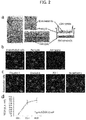


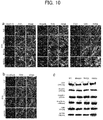
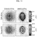


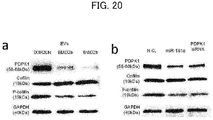


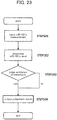


Classifications
-
- C—CHEMISTRY; METALLURGY
- C12—BIOCHEMISTRY; BEER; SPIRITS; WINE; VINEGAR; MICROBIOLOGY; ENZYMOLOGY; MUTATION OR GENETIC ENGINEERING
- C12Q—MEASURING OR TESTING PROCESSES INVOLVING ENZYMES, NUCLEIC ACIDS OR MICROORGANISMS; COMPOSITIONS OR TEST PAPERS THEREFOR; PROCESSES OF PREPARING SUCH COMPOSITIONS; CONDITION-RESPONSIVE CONTROL IN MICROBIOLOGICAL OR ENZYMOLOGICAL PROCESSES
- C12Q1/00—Measuring or testing processes involving enzymes, nucleic acids or microorganisms; Compositions therefor; Processes of preparing such compositions
- C12Q1/68—Measuring or testing processes involving enzymes, nucleic acids or microorganisms; Compositions therefor; Processes of preparing such compositions involving nucleic acids
- C12Q1/6876—Nucleic acid products used in the analysis of nucleic acids, e.g. primers or probes
- C12Q1/6883—Nucleic acid products used in the analysis of nucleic acids, e.g. primers or probes for diseases caused by alterations of genetic material
- C12Q1/6886—Nucleic acid products used in the analysis of nucleic acids, e.g. primers or probes for diseases caused by alterations of genetic material for cancer
-
- A—HUMAN NECESSITIES
- A61—MEDICAL OR VETERINARY SCIENCE; HYGIENE
- A61K—PREPARATIONS FOR MEDICAL, DENTAL OR TOILETRY PURPOSES
- A61K49/00—Preparations for testing in vivo
- A61K49/001—Preparation for luminescence or biological staining
- A61K49/0013—Luminescence
- A61K49/0017—Fluorescence in vivo
- A61K49/005—Fluorescence in vivo characterised by the carrier molecule carrying the fluorescent agent
- A61K49/0054—Macromolecular compounds, i.e. oligomers, polymers, dendrimers
-
- A—HUMAN NECESSITIES
- A61—MEDICAL OR VETERINARY SCIENCE; HYGIENE
- A61K—PREPARATIONS FOR MEDICAL, DENTAL OR TOILETRY PURPOSES
- A61K31/00—Medicinal preparations containing organic active ingredients
- A61K31/70—Carbohydrates; Sugars; Derivatives thereof
- A61K31/7088—Compounds having three or more nucleosides or nucleotides
-
- A—HUMAN NECESSITIES
- A61—MEDICAL OR VETERINARY SCIENCE; HYGIENE
- A61K—PREPARATIONS FOR MEDICAL, DENTAL OR TOILETRY PURPOSES
- A61K45/00—Medicinal preparations containing active ingredients not provided for in groups A61K31/00 - A61K41/00
-
- A—HUMAN NECESSITIES
- A61—MEDICAL OR VETERINARY SCIENCE; HYGIENE
- A61K—PREPARATIONS FOR MEDICAL, DENTAL OR TOILETRY PURPOSES
- A61K49/00—Preparations for testing in vivo
- A61K49/001—Preparation for luminescence or biological staining
- A61K49/0013—Luminescence
- A61K49/0017—Fluorescence in vivo
- A61K49/0019—Fluorescence in vivo characterised by the fluorescent group, e.g. oligomeric, polymeric or dendritic molecules
- A61K49/0021—Fluorescence in vivo characterised by the fluorescent group, e.g. oligomeric, polymeric or dendritic molecules the fluorescent group being a small organic molecule
- A61K49/0032—Methine dyes, e.g. cyanine dyes
-
- A—HUMAN NECESSITIES
- A61—MEDICAL OR VETERINARY SCIENCE; HYGIENE
- A61P—SPECIFIC THERAPEUTIC ACTIVITY OF CHEMICAL COMPOUNDS OR MEDICINAL PREPARATIONS
- A61P35/00—Antineoplastic agents
- A61P35/04—Antineoplastic agents specific for metastasis
-
- C—CHEMISTRY; METALLURGY
- C12—BIOCHEMISTRY; BEER; SPIRITS; WINE; VINEGAR; MICROBIOLOGY; ENZYMOLOGY; MUTATION OR GENETIC ENGINEERING
- C12M—APPARATUS FOR ENZYMOLOGY OR MICROBIOLOGY; APPARATUS FOR CULTURING MICROORGANISMS FOR PRODUCING BIOMASS, FOR GROWING CELLS OR FOR OBTAINING FERMENTATION OR METABOLIC PRODUCTS, i.e. BIOREACTORS OR FERMENTERS
- C12M1/00—Apparatus for enzymology or microbiology
- C12M1/34—Measuring or testing with condition measuring or sensing means, e.g. colony counters
-
- C—CHEMISTRY; METALLURGY
- C12—BIOCHEMISTRY; BEER; SPIRITS; WINE; VINEGAR; MICROBIOLOGY; ENZYMOLOGY; MUTATION OR GENETIC ENGINEERING
- C12N—MICROORGANISMS OR ENZYMES; COMPOSITIONS THEREOF; PROPAGATING, PRESERVING, OR MAINTAINING MICROORGANISMS; MUTATION OR GENETIC ENGINEERING; CULTURE MEDIA
- C12N15/00—Mutation or genetic engineering; DNA or RNA concerning genetic engineering, vectors, e.g. plasmids, or their isolation, preparation or purification; Use of hosts therefor
- C12N15/09—Recombinant DNA-technology
-
- C—CHEMISTRY; METALLURGY
- C12—BIOCHEMISTRY; BEER; SPIRITS; WINE; VINEGAR; MICROBIOLOGY; ENZYMOLOGY; MUTATION OR GENETIC ENGINEERING
- C12N—MICROORGANISMS OR ENZYMES; COMPOSITIONS THEREOF; PROPAGATING, PRESERVING, OR MAINTAINING MICROORGANISMS; MUTATION OR GENETIC ENGINEERING; CULTURE MEDIA
- C12N15/00—Mutation or genetic engineering; DNA or RNA concerning genetic engineering, vectors, e.g. plasmids, or their isolation, preparation or purification; Use of hosts therefor
- C12N15/09—Recombinant DNA-technology
- C12N15/11—DNA or RNA fragments; Modified forms thereof; Non-coding nucleic acids having a biological activity
- C12N15/113—Non-coding nucleic acids modulating the expression of genes, e.g. antisense oligonucleotides; Antisense DNA or RNA; Triplex- forming oligonucleotides; Catalytic nucleic acids, e.g. ribozymes; Nucleic acids used in co-suppression or gene silencing
-
- C—CHEMISTRY; METALLURGY
- C12—BIOCHEMISTRY; BEER; SPIRITS; WINE; VINEGAR; MICROBIOLOGY; ENZYMOLOGY; MUTATION OR GENETIC ENGINEERING
- C12Q—MEASURING OR TESTING PROCESSES INVOLVING ENZYMES, NUCLEIC ACIDS OR MICROORGANISMS; COMPOSITIONS OR TEST PAPERS THEREFOR; PROCESSES OF PREPARING SUCH COMPOSITIONS; CONDITION-RESPONSIVE CONTROL IN MICROBIOLOGICAL OR ENZYMOLOGICAL PROCESSES
- C12Q1/00—Measuring or testing processes involving enzymes, nucleic acids or microorganisms; Compositions therefor; Processes of preparing such compositions
- C12Q1/68—Measuring or testing processes involving enzymes, nucleic acids or microorganisms; Compositions therefor; Processes of preparing such compositions involving nucleic acids
-
- A—HUMAN NECESSITIES
- A61—MEDICAL OR VETERINARY SCIENCE; HYGIENE
- A61P—SPECIFIC THERAPEUTIC ACTIVITY OF CHEMICAL COMPOUNDS OR MEDICINAL PREPARATIONS
- A61P25/00—Drugs for disorders of the nervous system
-
- A—HUMAN NECESSITIES
- A61—MEDICAL OR VETERINARY SCIENCE; HYGIENE
- A61P—SPECIFIC THERAPEUTIC ACTIVITY OF CHEMICAL COMPOUNDS OR MEDICINAL PREPARATIONS
- A61P25/00—Drugs for disorders of the nervous system
- A61P25/08—Antiepileptics; Anticonvulsants
-
- A—HUMAN NECESSITIES
- A61—MEDICAL OR VETERINARY SCIENCE; HYGIENE
- A61P—SPECIFIC THERAPEUTIC ACTIVITY OF CHEMICAL COMPOUNDS OR MEDICINAL PREPARATIONS
- A61P25/00—Drugs for disorders of the nervous system
- A61P25/14—Drugs for disorders of the nervous system for treating abnormal movements, e.g. chorea, dyskinesia
- A61P25/16—Anti-Parkinson drugs
-
- A—HUMAN NECESSITIES
- A61—MEDICAL OR VETERINARY SCIENCE; HYGIENE
- A61P—SPECIFIC THERAPEUTIC ACTIVITY OF CHEMICAL COMPOUNDS OR MEDICINAL PREPARATIONS
- A61P25/00—Drugs for disorders of the nervous system
- A61P25/28—Drugs for disorders of the nervous system for treating neurodegenerative disorders of the central nervous system, e.g. nootropic agents, cognition enhancers, drugs for treating Alzheimer's disease or other forms of dementia
-
- C—CHEMISTRY; METALLURGY
- C12—BIOCHEMISTRY; BEER; SPIRITS; WINE; VINEGAR; MICROBIOLOGY; ENZYMOLOGY; MUTATION OR GENETIC ENGINEERING
- C12N—MICROORGANISMS OR ENZYMES; COMPOSITIONS THEREOF; PROPAGATING, PRESERVING, OR MAINTAINING MICROORGANISMS; MUTATION OR GENETIC ENGINEERING; CULTURE MEDIA
- C12N2310/00—Structure or type of the nucleic acid
- C12N2310/10—Type of nucleic acid
- C12N2310/14—Type of nucleic acid interfering nucleic acids [NA]
- C12N2310/141—MicroRNAs, miRNAs
-
- C—CHEMISTRY; METALLURGY
- C12—BIOCHEMISTRY; BEER; SPIRITS; WINE; VINEGAR; MICROBIOLOGY; ENZYMOLOGY; MUTATION OR GENETIC ENGINEERING
- C12Q—MEASURING OR TESTING PROCESSES INVOLVING ENZYMES, NUCLEIC ACIDS OR MICROORGANISMS; COMPOSITIONS OR TEST PAPERS THEREFOR; PROCESSES OF PREPARING SUCH COMPOSITIONS; CONDITION-RESPONSIVE CONTROL IN MICROBIOLOGICAL OR ENZYMOLOGICAL PROCESSES
- C12Q2600/00—Oligonucleotides characterized by their use
- C12Q2600/112—Disease subtyping, staging or classification
-
- C—CHEMISTRY; METALLURGY
- C12—BIOCHEMISTRY; BEER; SPIRITS; WINE; VINEGAR; MICROBIOLOGY; ENZYMOLOGY; MUTATION OR GENETIC ENGINEERING
- C12Q—MEASURING OR TESTING PROCESSES INVOLVING ENZYMES, NUCLEIC ACIDS OR MICROORGANISMS; COMPOSITIONS OR TEST PAPERS THEREFOR; PROCESSES OF PREPARING SUCH COMPOSITIONS; CONDITION-RESPONSIVE CONTROL IN MICROBIOLOGICAL OR ENZYMOLOGICAL PROCESSES
- C12Q2600/00—Oligonucleotides characterized by their use
- C12Q2600/158—Expression markers
-
- C—CHEMISTRY; METALLURGY
- C12—BIOCHEMISTRY; BEER; SPIRITS; WINE; VINEGAR; MICROBIOLOGY; ENZYMOLOGY; MUTATION OR GENETIC ENGINEERING
- C12Q—MEASURING OR TESTING PROCESSES INVOLVING ENZYMES, NUCLEIC ACIDS OR MICROORGANISMS; COMPOSITIONS OR TEST PAPERS THEREFOR; PROCESSES OF PREPARING SUCH COMPOSITIONS; CONDITION-RESPONSIVE CONTROL IN MICROBIOLOGICAL OR ENZYMOLOGICAL PROCESSES
- C12Q2600/00—Oligonucleotides characterized by their use
- C12Q2600/178—Oligonucleotides characterized by their use miRNA, siRNA or ncRNA
Definitions
- the present invention relates to the permeability control of the blood-brain barrier and particularly to a technique for increasing the permeability of the blood-brain barrier.
- the present invention further relates to the field of brain metastasis of cancer. More specifically, the present invention relates to the fields of diagnosis or risk assessment of brain metastasis and treatment or prevention of brain metastasis.
- Brain metastasis in cancer patient is known to relate to poor prognoses.
- BBB blood-brain barrier
- main constituent cells of the BBB are brain microvascular endothelial cells (BMECs), and that BMECs are connected each other by tight junctions between cells and thereby have very selective permeability.
- BMECs brain microvascular endothelial cells
- ZO-1 and ZO-2 proteins junctional adhesion molecules
- Non Patent Literature 1 cyclooxygenase -2 (COX -2), the epidermal growth factor receptor (EGFR) ligand HB-EGF, and ⁇ 2,6-sialyltransferase (ST6GALNAC5) have been identified as mediators involved in the BBB invasion of cancer cells.
- COX -2 the epidermal growth factor receptor (EGFR) ligand HB-EGF
- ST6GALNAC5 ⁇ 2,6-sialyltransferase
- VEGF vascular endothelial growth factor
- Extracellular vesicles (EVs) including exosomes are known to mediate communication between cells by transporting protein, mRNA, and microRNA (miRNA) contained therein (Non Patent Literature 5).
- EVs Extracellular vesicles
- Non Patent Literature 8 it has been reported that extracellular vesicles released from cancer cells are involved in the malignancy of cancer cells in various ways (Non Patent Literature 6) and increase the expression of the oncogene protein MET in bone marrow progenitor cells to change the bone marrow progenitor cells toward a pro-metastatic phenotype.
- the relation between EVs and cancer metastasis has been suggested. However, little was known about the relation between EVs (and substances contained therein) and the BBB.
- a further object of the present invention is to provide an agent, a composition, and a kit to be used in these methods.
- the present inventors transplanted a highly metastatic human breast cancer cell line to mice, and analyzed EVs obtained from cancer cells metastasized to the brain.
- cancer cell-derived EVs are taken in brain microvascular endothelial cells and destruct the blood-brain barrier (BBB).
- BBB blood-brain barrier
- the present inventors searched for a substance related to the destruction of the blood-brain barrier from the substances contained in the EVs, and found that miR-181c, a microRNA specifically contained in the EVs derived from the brain metastatic cancer cells, has an effect to destruct the blood-brain barrier.
- the present inventors further examined the effect of miR-181c in brain microvascular endothelial cells, and found that miR-181c binds to a noncoding region of 3-phosphoinositide dependent protein kinase-1 (hereinafter referred to as "PDPK1") and decreases expression of PDPK1.
- PDPK1 3-phosphoinositide dependent protein kinase-1
- the present inventors further examined the relation between the decreased expression of PDPK1 and the disruption of the blood-brain barrier (BBB), and found that the inhibition of PDPK1 expression changes the localization of tight junction proteins.
- BBB blood-brain barrier
- cancer cell-derived EVs release miR-181c contained therein in brain microvascular endothelial cells, which inhibits the expression of PDPK1, and thereby change the localization of tight junction proteins to destruct the blood-brain barrier.
- miR-181c has been reported to be involved in the aggravation of cells, but has not been known in relation with the blood-brain barrier.
- PDPK1 has been known to mainly target protein kinase B (PKB/AKT1, PKB/AKT2, PKB/AKT3), p70 ribosomal protein S6 kinase (RPS6KB1), p90 ribosomal protein S6 kinase (RPS6KA1, RPS6KA and RPS6KA3), cyclic AMP-dependent protein kinase (PRKACA), protein kinase C (PRKCD and PRKCZ), serum- and glucocorticoid-inducible kinase (SGK1, SGK2 and SGK3), P21 activated kinase-1 (PAK1), and protein kinase PKN (PKN1 and PKN2), and known to be involved in the regulation of cell proliferation and survival as well as intake and storage of glucose and amino acids.
- PPKACA protein kinase C
- PAK1 protein kinase PKN
- the present invention is based on the elucidation of the mechanism for destruction of the blood-brain barrier by brain metastatic cancer cells, and specifically relates to the following inventions:
- cancer includes epithelial malignant tumors, hematopoietic malignant tumors derived from spinal cord, and the like, and particularly includes ovarian cancer (non-mucinous ovarian cancers and the like), uterine cancer, endometrial cancer, breast cancer, breast adenocarcinoma, prostate cancer, testicular cancer (testicular choriocarcinoma and the like), brain cancer (ependymoma and the like), throat cancer, lung cancer, lung adenocarcinoma, kidney cancer (renal cell cancer and the like), liver cancer, large bowel cancer (colon cancer), pleural mesothelioma, sarcoma, chronic and acute myeloid leukemia, and metastatic cancers such as lung metastatic cancer.
- ovarian cancer non-mucinous ovarian cancers and the like
- uterine cancer endometrial cancer
- breast cancer breast adenocarcinoma
- prostate cancer testicular cancer (testicular chori
- a "cancer patient” means a patient having cancer (at a site except the brain) with or without metastasis and may be a patient with, for example, hematopoietic cell malignant tumors such as leukemia, lymphomas (Hodgkin's disease and non-Hodgkin's lymphom, and the like), multiple myeloma; breast cancer; endometrial cancer; cervical cancer; ovarian cancer; esophageal cancer; gastric cancer; appendiceal cancer; large bowel cancer (colon cancer, rectal cancer, and the like); liver cancer (hepatocellular cancer, and the like); gallbladder cancer; bile duct cancer; pancreatic cancer; adrenal cancer; gastrointestinal stromal tumor; mesothelioma; head and neck cancers such as laryngeal cancer, oral cancers (oral floor cancer, gingival cancer, tongue cancer, buccal mucosa cancer, and the like); salivary gland cancer; nasal sinus cancers (maxill)
- the stage of cancer is determined according to a stage classification (clinical advanced stage classification).
- stage classification clinical advanced stage classification
- the detailed criteria for the stage classification of each cancer (organ) has been established, based on, for example, the size of cancer (T), metastasis to nearby lymph nodes (N) and metastasis to a distant organ (M) (TNM classification), which has been widely known in the art (see the website " Cancer staging" of U.S. National Cancer Center, http://www.cancer.gov/cancertopics/factsheet/detection/st aging; the latest edition of AJCC Cancer Staging Manual (American Joint Commitee on Cancer) ).
- T tumor necrosis toma distant organ
- M distant organ
- the staging criteria for breast cancer is as follows.
- Stage 0 non-invasive cancer: the breast cancer stays in the mammary gland where it has developed (including the Paget's disease); Stage I: lump size of 2 cm or smaller without metastasis to the lymph nodes; Stage IIA: lump size of 2 cm or smaller with metastasis to the axillary lymph nodes, or lump size of 2.1-5 cm without metastasis to the lymph nodes; Stage IIB: lump size of 2.1-5 cm with metastasis to the axillary lymph, or lump size of 5.1 cm or larger without metastasis to the lymph nodes; Stage IIIA: lump size of 5.1 cm or larger with metastasis to the axillary lymph, any size of lump with heavy metastasis to the axillary lymph nodes, or any size of lump without metastasis to the axillary lymph nodes but with metastasis to parasternal lymph nodes; Stage IIIB: any size of lump with infiltration to the chest wall and/or skin; Stage IIIC: any size of lump with metastasis spread to
- Stage IV in the cancer staging usually have metastasis to distant organs. Accordingly, in such patients, cancer cells have already invaded into the blood vessels, and therefore brain metastasis is very highly possible if miR-181c level in blood (in EVs contained in the blood) is high and the blood-brain barrier is open.
- Stage I to III patients who do not have metastasis to distant organs since cancer cell is not likely to invade into blood vessels, brain metastasis does not occur immediately even if the miR-181c level in blood (in EVs contained in blood) is high and the blood-brain barrier is open.
- these Stage I to III patients have high possibility (risk) of brain metastasis in the future invasion of cancer cells into the blood.
- a method, agent, apparatus, computer program or the like according to the present invention is used for diagnosis of brain metastasis or for risk assessment of brain metastasis can be determined according to a state of the patient from which the measured sample is derived.
- cancer staging is employed as the state, risk assessment of brain metastasis can be conducted for Stage I to III patients, and diagnosis of brain metastasis can be conducted for Stage IV patients.
- the presence or absence of metastasis to a distant organ or the presence or absence of infiltration of cancer cell into the blood may be employed as the state to select either of diagnosis of brain metastasis or risk assessment of brain metastasis.
- the present invention when there is metastasis to a distant organ or when there is infiltration of cancer cell into the blood, the present invention can be used for a diagnosis of brain metastasis, while when there is no metastasis to a distant organ or when there is no infiltration of cancer cell into the blood, the present invention is used for risk assessment of brain metastasis.
- the "Stage IV patients” may be replaced with patients with metastasis to a distant organ or patients with infiltration of cancer cell into the blood.
- the “Stage I to III patients” may be replaced with patients with no metastasis to a distant organ or patients with no infiltration of cancer cell into the blood.
- the "brain metastasis” and “metastasis to the brain” mean that cancer cells leave the primary lesion developed in other sites than brain, infiltrate into the brain, and grow in the brain, or the state that such infiltrated cancer cells have grown in the brain.
- Whether there is brain metastasis or not in a cancer patient having a primary lesion other than brain can be determined by a method of diagnostic imaging well known in the art. For example, when tumorigenesis in the brain is confirmed in images such as CT or MRI image for a cancer patient having a primary lesion other than brain to which a gadolinium contrast agent or the like has been administered, it can be determined that there is brain metastasis.
- the "healthy subject” means a human having no cancer development.
- the healthy subject herein is not necessary to have no disease other than cancer, but is preferably a healthy human having no disease even which is not cancer.
- miR-181c serves to open the BBB and promote brain metastasis in cancer patients. Accordingly, information about brain metastasis in a cancer patient can be obtained by measuring the miR-181c levels in the cancer patient.
- the "miR-181c” is a human miRNA consisting of approximately 22 nucleotides (SEQ ID NO: 1) ( Lim LP, et al. (2003) Science, 299: 1540 ; Landgraf P, et al. (2007) Cell, 129:1401-1414 ).
- the miR-181c levels are measured by using a substance that specifically binds to miR-181c.
- the "substance that specifically binds to miR-181c” is not particularly limited as long as it is a substance that can specifically bind to miR-181c. Since a nucleic acid usually bind specifically to a nucleic acid having its complementary sequence, a preferable substance that specifically binds to miR-181c is a nucleic acid that specifically binds to miR-181c.
- nucleic acid that specifically binds to miR-181c means a nucleic acid molecule that can specifically bind to miR-181c. Such a nucleic acid molecule usually has a complementary sequence to miR-181c.
- the "nucleic acid” as used herein comprises a DNA, an RNA, or an artificially generated nucleic acid (including bridged nucleic acids such as locked nucleic acids (2',4'-BNA)), or a combination thereof.
- the nucleic acid that specifically binds to miR-181c includes, but is not limited to, a primer or a probe.
- the "primer” is usually a nucleic acid molecule of 10-30 mer (preferably, 17-25 mer, 15-20 mer, or the like) that is used for the amplification of nucleic acid and has, at least in a part (preferably, 7 mer or more, 8 mer or more, 9 mer or more, 10 mer or more, 11 mer or more, 12 mer or more, 13 mer or more, 14 mer or more, 15 mer or more, 20 mer or more, 25 mer or more, or 30 mer or more) thereof, a sequence complementary to a sequence located at an end of the sequence to be amplified.
- the "probe” is a nucleic acid molecule of 10-200 mer (preferably 10-100 mer, 10-50 mer, 10-30 mer, 10-20 mer, and the like) that has a sequence complementary to a target sequence, and can specifically bind to a target sequence, and has a complementary to a sequence, in at least a part (preferably, 7 mer or more, 8 mer or more, 9 mer or more, 10 mer or more, 11 mer or more, 12 mer or more, 13 mer or more, 14 mer or more, 15 mer or more, 20 mer or more, 25 mer or more, 30 mer or more, 50 mer or more, 100 mer or more) thereof.
- the nucleic acid that specifically binds to miR-181c comprises at least partially an artificially designed sequence (for example, sequences for labeling and tagging).
- a substance "specifically binds” means that the substance binds to the target sequence or structure with a substantially higher affinity than binding to other nucleotide sequences or amino acid sequences or structures thereof.
- the "substantially higher affinity” means a sufficiently high affinity to allow the detection of the target sequence or structure with distinguishing from other sequences or structures in using a desired measuring apparatus or method.
- the substantially higher affinity may mean that the molecules number of the (miR-181c specifically binding) substances that bind to miR-181c is 3 times or more, 4 times or more, 5 times or more, 6 times or more, 7 times or more, 8 times or more, 9 times or more, 10 times or more, 15 times or more, 20 times or more, 30 times or more, 50 times or more of the molecules number of the (miR-181c specifically binding) substances that bind to other sequences or structures.
- the binding constant (Ka) in the binding between the miR-181c specifically binding substance of the present invention and miR-181c may be, for example, at least 10 7 M -1 , at least 10 8 M -1 , at least 10 9 M -1 , at least 10 10 M -1 , at least 10 11 M -1 , at least 10 12 M -1 , or at least 10 13 M -1 .
- the "substance that specifically binds to miR-181c (miR-181c specifically binding substances)" may be labelled as needed.
- methods for labeling include the radioisotope (RI) labeling, fluorescent labeling, and enzymatic labeling.
- RI radioisotope
- fluorescent labeling fluorescent labeling
- enzymatic labeling examples of a radioisotope used in the RI labelling include 32P, 1311, 35S, 45Ca, 3H, and 14C.
- Examples of a fluorescent dye used in the fluorescent labeling include DAPI, SYTOX(R) Green, SYTO(R) 9, TO-PRO(R)-3, Propidium Iodide, Alexa Fluor(R) 350, Alexa Fluor(R) 647, Oregon Green(R), Alexa Fluor(R) 405, Alexa Fluor(R) 680, Fluorescein (FITC), Alexa Fluor(R) 488, Alexa Fluor(R) 750, Cy(R) 3, Alexa Fluor(R) 532, Pacific Blue(TM), Pacific Orange(TM), Alexa Fluor(R) 546, Coumarin, Tetramethylrhodamine (TRITC), Alexa Fluor(R) 555, BODIPY(R) FL, Texas Red(R), Alexa Fluor(R) 568, Pacific Green(TM), Cy(R) 5, and Alexa Fluor(R) 594.
- biotin-16-dUTP biotin-16-dUTP
- biotin-11-dUTP digoxigenin
- DIG digoxigenin
- alkaline phosphatase alkaline phosphatase
- PDPK1 is one of the substances playing major roles in maintaining the structure of the BBB. Therefore, the BBB can be opened by suppressing the expression or inhibiting activity of PDPK1. Accordingly, a PDPK1 expression inhibitor or a PDPK1 activity inhibitor can be used as a DDS agent for an agent intended to be delivered into the brain.
- PDPK1 is an abbreviation of 3-phosphoinositide dependent protein kinase-1.
- PDPK1 is a serine/threonine kinase that phosphorylates and activates the AGC family of protein kinases. It is known that there are plural variants of the PDPK1 mRNA and protein.
- the nucleotide sequence of the mRNA of PDPK1 vulariant1, and the amino acid sequence of the PDPK1 vulariant1 protein are set forth in SEQ ID NO: 2 and SEQ ID NO: 3, respectively.
- the nucleotide sequence of the mRNA of PDPK1 vulariant2, and the amino acid sequence of the PDPK1 vulariant2 protein are set forth in SEQ ID NO: 4 and SEQ ID NO: 5, respectively.
- the nucleotide sequence of the mRNA of PDPK1 vulariant3 protein is set forth in SEQ ID NO: 6 and SEQ ID NO: 7, respectively.
- the nucleotide sequence of the mRNA of PDPK1 vulariantX1, and the amino acid sequence of the PDPK1 vulariantX1 protein is set forth in SEQ ID NO: 8 and SEQ ID NO: 9, respectively.
- the PDPK1 herein may be any of these variants or one of their mutants that may occur in the body.
- the present inventors have also found that, in cancer patients, miR-181c is contained in exosomes during traveling through the blood, and when the exosomes have reached the BBB, the exosomes are incorporated into the BBB constituting cells and open the blood-brain barrier. Accordingly, the inhibition of secretion of exosomes in cancer patients can prevent the opening of the blood-brain barrier and thereby can suppress brain metastasis.
- the present invention relates to suppression of brain metastasis using an exosome secretion inhibitor.
- the "exosome secretion inhibitor” is not particularly limited as long as it is an agent that can inhibit the exosome secretion from cancer cells.
- nSMase2 neutral sphingomyelinase
- RAB27B the cDNA sequence is set forth in SEQ ID NO: 12 and the amino acid sequence is set forth in SEQ ID NO: 13
- the "expression inhibitor" of a gene, a protein, or an miRNA is not particularly limited, as long as it is an agent that suppresses the expression of the gene, protein, or miRNA. It is well-understood in the art that an expression inhibitor of a target gene can be obtained and used by designing and/or selecting, for example, an antisense to the gene sequence or to the mRNA sequence generated by transcription of the gene, dsRNA to the mRNA generated by the transcription of the gene, an aptamer to the mRNA generated by the transcription of the gene, or miRNA (including pri-miRNA and pre-miRNA) that can bind to a nucleotide sequence which is located upstream of a coding region and is essential for the expression of the gene.
- an expression inhibitor of a target gene can be obtained and used by designing and/or selecting, for example, an antisense to the gene sequence or to the mRNA sequence generated by transcription of the gene, dsRNA to the mRNA generated by the transcription of the gene
- an expression inhibitor of a target protein can be obtained or used by designing and/or selecting, for example, an antisense to a gene sequence encoding the protein or to an mRNA sequence generated by the transcription of the gene, dsRNA to an mRNA generated by the transcription of a gene encoding the protein, an aptamer to a gene sequence encoding the protein or to an mRNA generated by the transcription of the gene, or miRNA (including pri-miRNA and pre-miRNA) that can bind to a nucleotide sequence which is located upstream of a coding region and is essential for the expression of a gene encoding the protein.
- miRNA including pri-miRNA and pre-miRNA
- an expression inhibitor of miRNA can be obtained or used by designing and/or selecting, for example, an antisense to a gene encoding the miRNA, pri-miR of the miRNA, or pre-miR of the miRNA; dsRNA to pri-miR of the miRNA or pre-miR of the miRNA; an aptamer to the pri-miR of the miRNA or pre-miR of the miRNA; or another miRNA (including pri-miRNA and pre-miRNA) that can bind to a nucleotide sequence which is located upstream of a coding region and is essential for the expression of a gene encoding the miRNA.
- expression inhibitors of PDPK1 include pri-miR-181c, pre-miR-181c, miR-181c and derivatives thereof.
- the derivatives of pri-miRNA, pre-miRNA and miRNA are molecules that have at least a seed sequence (the 7 nucleotides at positions 2 to 8 from the 5' end: ACUUACA) in the miRNA and that act to suppress the expression of the gene (or protein) of interest (PDPK1 in the case of miR-181c).
- the function of such a derivative is not necessarily quantitatively same as that of the original miRNA, but the derivative may have stronger or weaker activity than the original miRNA as long as it achieves an object of the present invention.
- the derivatives have a nucleotide sequence having an identity of about 70% or more and less than 100%, about 75% or more and less than 100%, about 80% or more and less than 100%, about 85% or more and less than 100%, about 90% or more and less than 100%, about 95% or more and less than 100% with the pri-miRNA, pre-miRNA and miRNA.
- the higher sequence identity means the more similarity in the structures between the pri-miRNA, pre-miRNA, or miRNA and the derivative thereof.
- the derivatives of the pri-miRNA, pre-miRNA, and miRNA may have a sequence that the original pri-miRNA, pre-miRNA, and miRNA do not have.
- nucleotide lengths of the derivative of pri-miRNA, pre-miRNA, and miRNA may be different from the original nucleotide length as long as they can exhibit the function of the miRNA and may be, for example, 10-50 mer, 15-30 mer, or 20-25 mer.
- the derivatives of the pri-miRNA, pre-miRNA, and miRNA include a nucleic acid molecule which has a seed sequence of the pri-miRNA, pre-miRNA and miRNA, and has one or more of addition, substitution, deletion, and modification selected from the following (i) to (iv) (at other than the seed sequence):
- the "activity inhibitor" of a protein or miRNA is not particularly limited as long as it is an agent that makes the protein or miRNA incapable of exhibiting its activity.
- the activity inhibitor may be a substance that binds to the protein or miRNA, or a substance that binds to a substance to which the protein or miRNA binds.
- Known activity inhibitors of proteins are mimetics, antibodies or immunoreactive fragments thereof, aptamers, receptor derivatives, or antagonists.
- Known activity inhibitors of miRNA are mimetics, antisense DNA/RNA or dsRNA having a sequence complementary to at least a partial sequence of the miRNA, or aptamers to the miRNA.
- the "antisense” is a nucleic acid having a complementary sequence of the target sequence (for example, the sequence of pri-miR-181c, pre-miR-181c, or miR-181c or a DNA encoding pri-miR-181c) and may be DNA or RNA.
- the antisense is not required to be 100% complementary to the target sequence and may contain non-complementary nucleotides as long as it can specifically hybridize under stringent conditions ( Sambrook et al., 1989, Molecular Cloning, A Laboratory Manual, Cold Spring Harbor Press, N.Y ., and Ausubel et al. (eds.), 1995, Current Protocols in Molecular Biology, (John Wiley & Sons.
- the antisense binds to the target sequence and inhibits the transcription, the processing of RNA, the translation, or the stability thereof.
- the antisense includes antisense polynucleotides as well as polynucleotide mimetics and those that contain a modified back bone. Such an antisense can be designed and produced (for example, chemically synthesized) appropriately using a method well known in the art based on the target sequence information.
- the "dsRNA” is RNA containing double strand RNA structure that has at least partially a sequence complementary to the target sequence.
- the dsRNA binds to mRNA having the target sequence, and thereby degrades the mRNA to suppress the translation (expression) of the target sequence by RNA interference (RNAi).
- RNAi RNA interference
- the dsRNA includes siRNA (short interfering RNA) and shRNA (short hairpin RNA).
- shRNA short hairpin RNA
- the dsRNA is not required to have 100% identity with the target sequence as long as it can suppress the expression of the target gene. Moreover, a part of the dsRNA may be substituted with DNA for stabilization or another purpose.
- the siRNA is preferably double strand RNA having 21-23 nucleotide each.
- the siRNA can be obtained by a method well known in the art, for example, by chemical synthesis or as an analog of naturally occurring RNA.
- the shRNA is a RNA short chain having a hairpin turn structure.
- the shRNA can be obtained by a method well known in the art, for example, by chemical synthesis or introducing a gene encoding the shRNA into cells and expressing the gene.
- the "aptamer” are nucleic acids that bind to a substance such as protein or nucleic acid.
- the aptamers may be constituted of RNA or DNA.
- the form of the nucleic acids may be a double strand or a single strand.
- the length of the aptamer is not particularly limited as long as it can specifically bind to the target molecule, but is for example, 10-200 nucleotides, preferably 10-100 nucleotides, more preferably 15-80 nucleotides, or further preferably 15-50 nucleotides.
- the aptamers can be selected using a method well known in the art and, for example, the SELEX (Systematic Evolution of Ligands by Exponential Enrichment) method ( Tuerk, C. and Gold, L., 1990, Science, 249: 505-510 ).
- the “mimetics” are substances that have a structure similar to that of the protein or the miRNA.
- the mimetics can bind to the binding partners of the protein or the miRNA or can compete with the protein or miRNA, without exhibiting the activity of the protein or miRNA.
- the "antibody” as used herein may be polyclonal antibody or monoclonal antibody and includes a non-human animal antibody, antibody having both of an amino acid sequence of a non-human animal antibody and an amino acid sequence of a human antibody (chimeric antibody and humanized antibody), and a human antibody.
- the immune globulin class of the antibodies may be any immune globulin class (isotype), IgG, IgM, IgA, IgE, IgD, or IgY, and, in the case of IgG, may be any subclass (IgG1, IgG2, IgG3, or IgG4).
- the antibody may be monospecific, bispecific (bispecific antibody), or trispecific (trispecific antibody) (for example, WO1991/003493 ).
- the immunoreactive fragments of an antibody mean proteins or peptides containing a part (partial fragment) of the antibody with maintaining the antibody's activity (immunoreactivity, binding activity) to the antigen.
- immunoreactive fragment examples include F(ab') 2 , Fab', Fab, Fab 3 , single strand Fv (hereinafter, referred to as the "scFv"), (tandem) bispecific single strand Fv (sc(Fv) 2 ), single strand triple bodies, nanobodies, divalent V H HS, pentavalent V H HS, minibodies, (double strand) diabodies, tandem diabodies, bispecific tribodies, bispecific bibodies, dual affinity retargeting molecules (DART), triabodies (or tribodies), tetrabodies (or [sc(Fv) 2 ] 2 ) or (scFv-SA) 4 ), disulfide linked Fv (hereinafter, referred to as "dsFv”), compact IgGs, heavy chain antibodies or polymers thereof (see Nature Biotechnology, 29 (1): 5-6 (2011 ); Maneesh Jain et al., TRENDS in Biotechnology, 25
- the immunoreactive fragments may be any of monopecific, bispecific, trispecific, and multispecific antibodies.
- the aptamers are nucleic acid molecules capable of binding to a specific nucleic acid or protein.
- the receptor derivative means a substance having a binding region structure of a receptor to which the protein of interest binds.
- the receptor derivative includes a conjugate of the constant region of an antibody with a receptor and a membrane protein receptor solubilized by deleting the transmembrane region.
- the antagonist includes a wide range of substances that competes with the protein or miRNA of interest and inhibits the function thereof.
- the binding is preferably specific and the binding constant (Ka) thereof may be for example, at least 10 7 M -1 , at least 10 8 M -1 , at least 10 9 M -1 , at least 10 10 M -1 , at least 10 11 M -1 , at least 10 12 M -1 , or at least 10 13 M -1 .
- the "agent” is a substance mainly used as an active ingredient, and includes a small molecule, a nucleic acid molecule (DNA and/or RNA), a protein, and a fusion thereof.
- the “composition” comprises at least an agent that serves as an active ingredient and optionally comprises water, a solvent, and other additives as needed.
- the composition herein may comprise only one substance or 2 or more substances as an active ingredient(s).
- the "active ingredient” is not particularly limited, as long as it can exhibit a desired biological activity, and includes a substance capable of exhibiting a therapeutic effect or preventive effect as well as a substance exhibiting a function as DDS. Whether a substance is an active ingredient or not can be determined not only depending on whether the substance has a biological action or not, but also depending on whether the amount of the substance contained in the composition can allow the substance to exhibit the biological action.
- composition for diagnosis or risk assessment of brain metastasis is a composition to be used in a method for diagnosing brain metastasis or a method for assessing risk of brain metastasis as described below, and includes a diagnostic composition (including an extracorporeal diagnostic composition).
- the composition for diagnosis or risk assessment of brain metastasis may be used for in vivo, ex vivo, or in vitro diagnosis, but preferably is for ex vivo or in vitro use.
- Compositions for in vivo diagnosis or risk assessment may be formulated to be administered to a patient in accordance with the formulation of pharmaceutical compositions as described below.
- the composition for diagnosis or risk assessment herein may be provided as a kit for diagnosis or risk assessment comprising the composition as an active ingredient.
- the kit can be constituted by any materials as long as miRNA levels can be determined.
- the kit for diagnosis or risk assessment herein may be a kit comprising a primer that can bind to miR-181c (a kit for measuring miRNA levels by using gene amplification technique such as PCR) or a kit comprising a probe that can bind to miR-181c (a kit for measuring miRNA levels by using a nucleic acid binding detection technique such as hybridization).
- the kit for diagnosis or risk assessment herein may be a kit which determines the miRNA level utilizing an expression inhibitory effect of miRNA as indicator.
- the kit may comprise, for example, a nucleic acid encoding a label (luciferase, or the like) having an mRNA 3'UTR that can specifically bind to miR-181c (a kit for measuring miRNA levels by detecting the expression of the label in the reporter assay or the like).
- the "pharmaceutical composition” is a composition to be used for treatment or prevention and comprises a therapeutic or preventive agent.
- the pharmaceutical composition is preferably prepared into a unit dosage form comprising a suitable dose of the active ingredient.
- any oral or parenteral formulation may be employed as long as it can be administered to a patient.
- compositions for parenteral administration include injections, nasal drops, suppositories, patches, ointments, and the like, and are preferably injections.
- Examples of the dosage form of the pharmaceutical composition of the present invention include solutions or lyophilized formulations.
- an excipient (see “ Japanese Pharmaceutical Excipients Directory” Yakuji Nippo Limited .; “ Handbook of Pharmaceutical Excipients Fifth Edition” APhA Publications ) including a solubilizing agent such as propylene glycol or ethylenediamine, a buffer such as phosphate, an isotonizing agent such as sodium chloride or glycerin, a stabilizer such as sulfite, a preservative such as phenol, or a soothing agent such as lidocaine may be added as needed.
- a solubilizing agent such as propylene glycol or ethylenediamine
- a buffer such as phosphate
- an isotonizing agent such as sodium chloride or glycerin
- a stabilizer such as sulfite
- a preservative such as phenol
- a soothing agent such as lidocaine
- a storage container therefor may be an ampoule, a vial, a prefilled syringe, a cartridge for pen type syringes, a bag for infusion, and the like.
- the pharmaceutical composition may usually contain 5-500 mg, 5-100 mg, 10-250 mg of the active ingredient per unit dosage form.
- the pharmaceutical composition for suppressing brain metastasis may comprise one or more agents selected from an miR-181c expression inhibitor, an miR-181c activity inhibitor, and an exosome secretion inhibitor as an active ingredient.
- the pharmaceutical composition for suppressing brain metastasis may comprise one or more agents selected from an miR-181c expression inhibitor, an miR-181c activity inhibitor, and an exosome secretion inhibitor as active ingredient(s), or further comprise other agent(s) to be used in combination as active ingredient(s). Examples of such other agents include other cancer metastasis inhibitors and anticancer agents.
- Examples of the other cancer metastasis inhibitors or anticancer agents that can be included in the pharmaceutical composition for suppressing brain metastasis include alkylating agents including nitrogen mustards such as cyclophosphamide, ifosfamide, melphalan, busulfan, and thiotepa, and nitrosoureas such as nimustine, ranimustine, dacarbazine, procarbazine, temozolomide, carmustine, Streptozotocin, bendamustine; platinum compounds such as cisplatin, carboplatin, oxaliplatin, and nedaplatin; antimetabolites such as enocitabine, capecitabine, carmofur, cladribine, gemcitabine, cytarabine, cytarabine ocfosphate, tegafur, tegafur/uracil, tegafur/gimeracil/oteracil potassium, doxiflur
- the pharmaceutical composition for increasing permeability of the blood-brain barrier may comprise one or more PDPK1 expression inhibitors or PDPK1 activity inhibitors as an active ingredient(s).
- the pharmaceutical composition for increasing permeability of the blood-brain barrier may solely comprise the PDPK1 expression inhibitor and/or PDPK1 activity inhibitor as active ingredient(s), or may further comprise another agent to be used in combination as an active ingredient.
- another agent include drugs that act on and increase the permeability of the blood-brain barrier as well as agents intended to be delivered to the brain.
- the pharmaceutical composition for increasing permeability of the blood-brain barrier is used particularly for delivering a desired agent to the brain, such a pharmaceutical composition is referred to as the "agent-delivery composition".
- the agent intended to be delivered to the brain may be referred to as the "medicinal component" to be distinguished from the agent (PDPK1 expression inhibitor or PDPK1 activity inhibitor) intended to open the BBB.
- the "agent-delivery composition” comprises at least a PDPK1 expression inhibitor or activity inhibitor as an active ingredient.
- the agent-delivery composition is preferably prepared into a unit dosage form comprising a suitable dose of the medicinal component.
- An example of such a unit dosage form is an injection (ampoule, vial, prefilled syringe).
- 5-500 mg, 5-100 mg, or 10-250 mg of the PDPK1 expression inhibitor or activity inhibitor may be contained per unit dosage form.
- the agent-delivery composition may comprise, in addition to the PDPK1 expression inhibitor or activity inhibitor, an agent intended to be delivered to the brain, a medicinal component.
- the PDPK1 expression inhibitor or activity inhibitor and the medicinal component may be contained in one formulation or provided as separate formulations to be used in combination.
- the "medicinal component” is a medicine intended to be delivered into the brain, and includes, for example, a therapeutic or preventive agent for a target disease such as neurosurgical diseases such as cerebrovascular disorders (such as cerebral infarction/cerebral hemorrhage/cerebral aneurysm), brain tumor (such as meningioma, pituitary adenoma, including brain metastasis), infectious diseases (such as meningitis), functional brain diseases (such as trigeminal neuralgia), the spinal cord disease (such as disc herniation, spinal stenosis); neurological diseases such as Parkinson's disease, spinocerebellar degeneration, and epilepsy; and mental diseases such as Alzheimer's disease, non-Alzheimer type degenerative dementia, insomnia, and depression.
- a target disease such as neurosurgical diseases such as cerebrovascular disorders (such as cerebral infarction/cerebral hemorrhage/cerebral aneurysm), brain tumor (such as meningio
- the agent-delivery composition herein may be provided as a kit for treatment or prevention, comprising the composition as an active ingredient.
- the constitution of the kit is not limited as long as it comprises the PDPK1 expression inhibitor or activity inhibitor, and the kit may comprise, for example, the PDPK1 expression inhibitor or activity inhibitor and the medicinal component.
- the PDPK1 expression inhibitor or activity inhibitor and the medicinal component may be provided in a form to be administered simultaneously or separately.
- the "level” means a numerical value indicating the quantity of miR-181c, which includes, for example, the concentration, the amount, or any alternative numerically value indicating the quantity to be used instead thereof. Accordingly, the level may be a measurement itself of such as fluorescence or may be a value converted into the concentration or the amount.
- the level may be absolute numerical value (abundance, abundance per unit area) or a numerical value relative to a control provided as needed.
- the level may be the mean or median when the same sample is measured plural times (at the same time or at different times).
- the "kit” may comprise, in addition to the active ingredient, a package storing components of the kit, such as a paper box or a plastic case, an ampoule, a vial, a tube, a syringe, or the like storing each component, an instruction manual, and the like.
- a package storing components of the kit, such as a paper box or a plastic case, an ampoule, a vial, a tube, a syringe, or the like storing each component, an instruction manual, and the like.
- the present invention involves an apparatus for judging brain metastasis in a cancer patient, an apparatus for assessing risk of brain metastasis in a cancer patient, and a computer program used in these apparatuses ( Figure 23 ).
- These apparatuses provide information for diagnosis to a person (usually a doctor) who diagnoses whether there is brain metastasis or not.
- the apparatus for judging brain metastasis of the present invention comprises miR-181c-measuring means.
- the “miR-181c-measuring means” is an apparatus that conducts the measurement of a polynucleotide having the nucleotide sequence of or a part of miR-181c (SEQ ID NO: 1) in a sample derived from a subject, which processes the sample from the subject cancer patient as prescribed to convert information about the miR-181c levels in the sample into digits or electrical signals.
- the miR-181c-measuring means allows to contact the agent for diagnosis or risk assessment of brain metastasis described above and the sample from the subject cancer patient and to react them if necessary, and measures generated parameters such as a strength of a light (fluorescence, luminescence) which reflects the miR-181c levels.
- the contact between the agent for diagnosis or risk assessment of brain metastasis and the sample from the subject cancer patient usually takes place in a container (reaction vessel) such as a plate or a tube.
- the computer program of the present invention includes a computer program that can make an existing apparatus to be used for judging brain metastasis or for assessing risk of brain metastasis in a cancer patient by installing, even if the existing apparatus is widely used and can be used for other measurement or judgment.
- the computer program of the present invention is not required to be installed in the apparatus for judging brain metastasis and the apparatus for assessing risk of brain metastasis.
- the computer program of the present invention may be provided, for example, in such a form to be stored on a recording medium.
- the "recording medium” is a medium that can carry a program without occupying space, and includes a flexible disk, a hard disk, CD-R, CD-RW, MO (magneto-optical disk), DVD-R, DVD-RW, a flash memory, and the like.
- the computer program of the present invention can be transmitted from the computer program storing computer to other computers or other apparatuses through a communication line.
- the computer program of the present invention includes those stored on such a computer and being transmitted.
- miR-181c contributes to brain metastasis by destructing the BBB. Since expression of miR-181c is specifically increased in cancer patients with brain metastasis, miR-181c can be used as indicator for diagnosis or risk assessment of brain metastasis. A brain metastasis can be suppressed by inhibiting the expression or activity of miR-181c or inhibiting secretion of EVs (containing miR-181c).
- PDPK1 expression inhibitors and PDPK1 activity inhibitors including miR-181c increases the permeability of the BBB, and thus can be used as DDS of agents desired to be delivered into the brain.
- the present invention relates to a method for diagnosing brain metastasis or a method for assessing risk of brain metastasis, comprising measuring an miR-181c level in a sample from a subject cancer patient.
- the present invention relates to a method for diagnosing brain metastasis in a subject cancer patient (or a method for assessing risk of brain metastasis), comprising: determining an miR-181c level in a sample from the subject cancer patient, and judging the presence or absence of brain metastasis (or assessing risk of brain metastasis) in the subject cancer patient from the determined miR-181c level, wherein the subject cancer patient is judged to have metastasis to brain (or assessed to have a high risk of brain metastasis) when the miR-181c level in the sample from the subject cancer patient is higher than the miR-181c level in a sample from a negative control.
- the present invention relates to a method for providing information for diagnosing brain metastasis, comprising measuring an miR-181c level in a sample from a subject cancer patient.
- the present invention relates to a method for providing information for diagnosing brain metastasis (or for assessing risk of brain metastasis) in a subject cancer patient, comprising: a step of determining an miR-181c level in a sample from the subject cancer patient, and a step comprising providing information for judging the presence or absence of brain metastasis (or assessing a risk of brain metastasis) in the subject cancer patient from the determined miR-181c level, wherein information that the subject cancer patient has brain metastasis (or has a high risk of brain metastasis) is provided when the miR-181c level in the sample from the subject cancer patient is higher than the miR-181c level in a sample from a negative control.
- the step of determining an miR-181c level is conducted by measuring a polynucleotide having the nucleotide sequence or a part of miR-181c (SEQ ID NO: 1) in the sample from the subject.
- the polynucleotide may be measured by a method utilizing detection of nucleic acid binding such as hybridization; a method utilizing amplification of nucleic acids such as PCR; a method utilizing the expression inhibitory activity of miR-181c such as a reporter assay; or decoding the sequence of miRNA or cDNA transcribed from miRNA.
- Examples of the method utilizing hybridization include Southern hybridization, Northern hybridization, dot hybridization, fluorescence in situ hybridization (FISH), microarray, and the ASO method.
- step of determining an miR-181c level may comprise the following steps:
- the "binding between the probe and miR-181c" to be measured may be a bound amount, the number of binding, or a percentage of the bound substances.
- the step of determining the miR-181c level in the sample from the measured binding between the probe and miR-181c may be conducted, for example, by the following procedures. Binding between the probe and miR-181c in a standard sample which is a serial dilution series of miR-181c is measured to generate a standard curve.
- the miR-181c level in a sample from a subject cancer patient can be calculated by comparing a measurement of the binding between the probe and miR-181c measured in the sample from the subject cancer patient with the standard curve.
- the standard curve may be determined by measuring standard samples simultaneously with the sample from the subject cancer patient, or alternatively by measuring standard samples separately from (at different time from) the samples from the subject cancer patient. Accordingly, the standard curve may be pre-obtained from the measurements of standard samples which is measured before the measurement of a sample from a subject cancer patient.
- cDNA may be synthesized from miRNA in a sample from a patient, and the binding of the synthesized cDNA and probe may be measured.
- Examples of the method utilizing PCR include ARMS (amplification refractory mutation system), RT-PCR (reverse transcriptase-PCR), and nested PCR.
- the level of miR-181c can be determined by synthesizing cDNA from the RNA as needed, decoding the sequence of synthesized cDNA with a sequencer, and determining the level of miR-181c from the amount of the decoded miR-181c.
- the information processing device 3 comprises at least the level determination unit 31 and the metastasis judgment unit 32.
- the information processing device 3 is preferably a computer with a CPU, a ROM, a RAM, and an HDD. Processing in both of the level judgment unit 31 and the metastasis judgment unit 32 is mainly performed by the CPU reading a computer program in the HDD.
- the computer program may not be stored in the HDD, for example, but stored in the RAM or the ROM.
- the input device 2 and the information processing device 3 may comprise a common CPU, common ROM, common RAM, or a common HDD. In this case, the computer program may be stored in an HDD, a RAM, or a ROM that is common to the input device 2 and the information processing device 3.
- the apparatus for assessing risk of brain metastasis 1a comprises a risk assessment unit 33 instead of the metastasis judgment unit 32 in the apparatus for judging brain metastasis 1.
- the other configuration of the apparatus for assessing risk of brain metastasis 1a is common with the configuration of the apparatus for judging brain metastasis 1 except for the metastasis judgment unit 32. Therefore, the redundant descriptions are omitted.
- BMD1cells brain metastatic derivative 1a, hereinafter, referred to as the "BMD1cells”
- BMD2a cells brain metastatic derivative cell populations 2a and 2b
- BBB kit (TM) (# MTB-24H, PharmaCo-Cell Company Ltd., Nagasaki, Japan), which is a BBB model system that consists of primary cultures of brain capillary endothelial cells, brain pericytes and astrocytes ( Figure 2a ) was used.
- Brain capillary endothelial cells, brain pericytes, and astrocytes were confirmed by Hoechst33342 staining. As a result, the presence of brain capillary endothelial cells, brain pericytes, and astrocytes in the constructed in vitro blood-brain barrier model was confirmed as illustrated in Figure 2b .
- the endothelial cells were incubated at 37°C with a rabbit polyclonal antibody to Claudin-5 (Z43.JK, Invitrogen, CA, USA), Occludin (ZMD.467, Invitrogen), ZO-1 (ZMD.437, Invitrogen), or N-cadherin (3B9, Invitrogen) for 1 hour. After washing with PBS containing Mg 2+ and Ca 2+ , the cells were incubated at 37°C with 594 Alexa Fluor-fused antirabbit IgG (Invitrogen) for 1 hour. Actin was stained with ActinGreen (TM) 488 ReadyProbes (TN) Reagent (R37110, Molecular Probes).
- TM ActinGreen
- TN ReadyProbes
- Brain metastatic cancer cells are also considered to be highly invasive. Therefore, invasion chamber assays using Matrigel(TM) were used to confirm the pathological implications of the metastatic potential of established cell lines.
- BMD2a, BMD2b, D3H2LN, and D3H1 cells were trypsinized and labelled with PKH26 (Sigma Aldrich). 2 ⁇ 10 4 cells of each cancer cell line were suspend in serum-free RPMI1640 medium and plated in the top chamber with a Matrigel(R)-coated membrane (24-well insert, BD Biosciences, NJ, USA). RPMI1640 medium containing 10% serum was added to the lower chamber as a chemoattractant.
- BMD2a, BMD2b, D3H2LN and D3H1 cells were trypsinized and labelled with PKH26 (Sigma Aldrich).
- 2 ⁇ 10 4 cancer cells were suspended in serum-free Ham's F-12 medium (Gibco) and added to the upper chamber of the in vitro BBB model prepared in Example 2 and cultured.
- non-invading cells were removed with cotton swabs and the nuclei were stained with Hoechst 33342 (Dojindo Laboratories, Kumamoto, Japan).
- the PKH26 fluorescence of invading cells that had migrated to the lower surface side was counted with a fluorescence microscope ( Figure 4a ). All cells were assayed in triplicate.
- D3H1, D3H2LN, BMD2a, and BMD2b cells were washed three times with Advanced RPMI medium containing antibiotic-antimycotic agents and 2 mM L-glutamine (hereinafter, referred to as the "medium A").
- Fresh medium A was added to the washed cells and cultured for 2 days, and then the medium was collected and centrifuged at 2000*g for 10 min at 4°C. To thoroughly remove cellular debris, the culture supernatant was filtered with a 0.22 - ⁇ m Stericup(R) (Millipore, MA, USA). The resultant conditioned medium was ultracentrifuged at 110,000*g for 70 min at 4°C. The resultant pellets were washed with 11 mL of phosphate buffered saline (PBS) and resuspended in PBS.
- PBS phosphate buffered saline
- the fraction containing the EVs was measured for its protein content using the Micro BCA protein assay kit (Thermo Scientific, MA, USA). The obtained EVs was observed by phase contrast microscopy. Furthermore, the size and the number of the EVs were measured with NanoSight.
- the resultant membranes were blocked in Blocking One (Nacalai Tesque, Kyoto, Japan) and then incubated for 1 hour at room temperature with a primary antibody, anti-CD63 antibody (purified mouse anti-human CD63, H5C6, 1/200, BD), anti-CD9 antibody (ALB6, sc-59140, 1/200, BD), or anti-cytochrome C antibody (purified mouse anti-cytochrome C, 7H8.2C12, 1/200, BD).
- An HRP-linked anti-mouse IgG secondary antibody NA931, GE Healthcare
- HRP-linked anti-rabbit IgG secondary antibody NA934, GE Healthcare
- FIG. 5a illustrates photographs resulted from the phase contrast microscope observation and the result of the analysis of EV size.
- the average particle sizes of the EVs derived from D3H1 cells, D3H2LN cells, BMD2a cells, and BMD2b cells were 149 nm, 157 nm, 136 nm, and 148 nm, respectively. All these EVs expressed the standard exosomal markers CD63 and CD9 but not Cytochrome C ( Figure 5b ). Furthermore, the number of EV release did not differ among the cells ( Figure 5c ).
- RNU6 was used as an internal control according to a described method ( Kosaka, N. et al., Net.Med., 18:883-891 (2012 )).
- the expression level of each mRNA and RNU6 was measured with qRT-PCR performed in 96-well plates using Platinum Quantitative PCR SuperMix (Applied Biosystems) with the 7300 Real-Time PCR System (all Applied Biosystems, CA, USA). All reactions were performed in triplicate.
- the primers and probes were defined as follows: PAB27B (assay ID: Hs01072206_m1), nSMase2 (assay ID: Hs00920354_m1).
- the number of EV release from siRNA-introduced cells were measured with NanoSight.
- the Matrigel invasion capacity of generated transformation cells and migration across BBB were examined according to the methods in Example 3 and Example 4, respectively.
- EVs derived from brain metastatic cancer cells directly affects BBB disruption
- EVs were prepared according to the description in Example 5.
- purified EVs derived from D3H2LN, BMD2a, and BMD2b cells or PBS (negative control) was added.
- TEER transendothelial electrical resistance
- Example 2 (2) a permeability test using NaF was conducted according to the method described in Example 2 (2) to measure the permeability of BBB. 24 hours after the addition of purified EVs derived from D3H2LN, BMD2a, and BMD2b cells, NaF was added. NaF moved across BBB was measured with a fluorocytometer. The apparent permeability coefficient (Papp) (10 -6 cms -1 ) was calculated according to the calculating formula described in Example 2 (2). All assays were performed in triplicate.
- Papp apparent permeability coefficient
- EVs derived from D3H1, D3H2LN, BMD2a, and BMD2b cells prepared by the method in Example 4 were labelled with PKH67 green fluorescent labeling kit (Sigma Aldrich, MO, USA). EVs were reacted with 2 ⁇ M PKH67 for 5 minutes and washed using a 100 kDa filter (Microcon YM-100, Millipore) five times to remove excess dye.
- PKH67 green fluorescent labeling kit Sigma Aldrich, MO, USA
- the labelled EVs or PBS (negative control) was added to the upper surface side in the in vitro BBB model, which was cultured at 37°C, 5% CO 2 for 24 hours.
- Brain capillary endothelial cells, brain pericytes, and astrocytes were confirmed by Hoechst33342 staining.
- Purified EVs derived from D3H1, D3H2LN, BMD2a, and BMD2b cells prepared by the method in Example 4 or PBS (negative control) was added and, 24 hours later, Claudin-5, Occludin, ZO-1, or N-cadherin in the endothelial cells in the in vitro BBB model was fluorescently immunostained with actin filaments to examine whether the localization is changed using a method similar to that in Example 2 (2).
- Proteins were isolated from each cell line using M-PER (Thermo Scientific, MA, USA), separated in Mini-PROTEAN(R) TGX Gel (4-12%) (Bio-Rad), and electrotransferred onto PVDF membranes (Millipore). The resultant membranes were blocked in Blocking One (Nacalai Tesque, Kyoto, Japan) and then incubated for 1 hour at room temperature with a primary antibody, anti-Claudin-5 antibody (Z43.JK, 1/200, Invitrogen), anti-Occludin antibody (ZMD.481, 1/200, Invitrogen), anti-ZO-1 antibody (H-300, sc-10804, 1/100, Santa Cruz Biotechnology), anti-N-cadherin antibody(3B9, 1/500, Invitrogen), or anti-GAPDH antibody (6C5, 1/1000, Millipore).
- FIG. 10a and Figure 10b The results of localization of the tight junction proteins in the endothelial cells in the in vitro BBB model are illustrated in Figure 10a and Figure 10b .
- the tight junction proteins and the N-cadherin were localized on the cell membrane surface of the endothelial cells treated with PBS or the EVs derived from D3H2LN cells.
- the tight junction proteins and the N-cadherin were found in the cytoplasm in the cells treated with the EVs derived from BMD2a and BMD2b,.
- the treatment of cerebrovascular endothelial cells with the EVs derived from BMD2a and BMD2b had no effect on the expression of the tight junction proteins, N-cadherin, and actin ( Figure 10c ).
- cancer-derived EVs changes the localization of tight junction proteins, N-cadherin, and actin filament without affecting the expression thereof.
- the upper panels in Figure 11 illustrate the intake of EVs and the lower panels illustrate the permeability of brain blood vessels.
- BMD2a cell-derived EVs were more incorporated within the brains of mice than were D3H2LN cell-derived EVs.
- BMD2a cell-derived EV-treated mice exhibited greater permeability of brain blood vessels compared to D3H2LN cell-derived EV-treated mice.
- miRNA contained in EVs derived from D3H2LN, BMD2a, and BMD2b cells was analyzed.
- CIP Calf Intestinal Alkaline Phosphatase
- the microarray was scanned using an Agilent DNA microarray scanner. Intensity values of each scanned feature were quantified using Agilent Feature Extraction software version 10.7.3.1, which performs background subtractions. Only using features that were flagged as no errors (Detected flags), the expression analysis was performed with Agilent GeneSpring GX version 12.6.1. Features that were not positive, not significant, not uniform, not above background, saturated and population outliers (not detected flags) were excluded. In this experiment, the significant differences of gene expression were identified by applying more than 2-fold change in signal intensity.
- EVs isolated from BMD2a and BMD2b was added to endothelial cells and the change of miR-181c abundance was examined.
- EVs were prepared according to the description in Example 5.
- Four days after thawing the in vitro BBB model purified EVs derived from D3H2LN, BMD2a, and BMD2b cells or PBS (negative control) were added.
- 24 hours after the addition of EVs the expression of miR-181c in endothelial cells was measured. All assays were performed in triplicate.
- EVs were purified from sera from brain metastasis breast cancer patients and the expression of miR-181c was examined.
- Sera from 56 breast cancer patients (10 brain metastasis patients, 46 non-brain metastasis patients) were collected at National Cancer Center Japan (No. 2013-111). Informed consent was obtained from all the patients. Serum was aliquoted and stored at -80°C until used, and freeze-thawing was avoided as much as possible after that.
- EVs were isolated from the sera using Total Exosome Isolation (from sera) (Invitrogen), The total RNA was then isolated from the obtained EVs using the RNeasy Mini Kit (Qiagen).
- the expression of miRNA was measured by qRT-PCT using RNU6 as an internal control by an already reported method ( Kosaka, N. et al. Net. Med., 18:883-891 (2012 )). More specifically, the expression levels of miR-181c and RNU6 were measured in a 96-well plate by qRT-PCR using TaqMan(R) MicroRNA Assays and TaqMan(R) Universal PCR Master Mix in 7300 Real-Time PCR System (all from Applied Biosystems, CA, USA). All reactions were performed in triplicate.
- EVs derived from brain metastatic cancer cells induce the abnormal localization of tight junction proteins by transferring miR-181c into endothelial cells, which results in the destruction of the cell-cell contact.
- the total RNA from respective endothelial cells was isolated using the QIAzol reagent and the mirRNeasy Mini Kit (both Qiagen). The quality and quantity of the RNA were determined using NanoDrop ND-1000 spectrophotometer (Thermo Fisher Scientific Inc.) and Agilent Bioanalyzer (Agilent Technologies) according to the recommended methods. The obtained total RNA was labelled with cyanine 3 (Cy3) during amplification using Low Input Quick Amp Labeling Kit, one-color (Agilent Technologies) according to the manufacturer's instructions. More specifically, 100 ng of total RNA was reverse-transcripted into double strand complementary DNA (cDNA) using poly dT-T7 promoter primers.
- cDNA double strand complementary DNA
- the primers, the template RNA, and a quality control transcription product of known concentration and quality were first denatured at 65°C for 10 minutes and then incubated for 2 hours at 40°C with 5 x first strand buffer, 0.1 M dithiothreitol, 10 mM deoxynucleotide triphosphate mixture, and AffinityScript RNase Block Mix, which was then incubated with AffinityScript enzyme at 70°C for 15 minutes.
- fluorescent complementary RNA cRNA
- the cDNA product was mixed with transcription master mix in the presence of T7 RNA polymerase and Cy3-labeled CTP (cytidine 5'-triphosphate) and incubated at 40°C for 2 hours.
- the labelled cRNA was purified using RNeasy Mini Spin Columns (Qiagen) and eluted with 30 mL of nuclease-free water. After the amplification and labeling, the quantity of the cRNA and incorporation of cyanine were determined using NanoDrop ND-1000 spectrophotometer (Thermo Fisher Scientific Inc.) and Agilent Bioanalyzer (Agilent Technologies).
- transendothelial electrical resistance TEER
- tight junction proteins N-cadherin
- actin filaments actin filaments after treatment with PDPK1 siRNA were examined.
- PDPK1 siRNA 25 nM was introduced into brain blood vessel endothelial cells using DharmaFECT transfection reagent (Thermo Scientific) according to the manufacturer' protocol.
- the localization and expression of tight junction proteins, N-cadherin, and actin filaments in the transfected cells after 24 hours were measured according to the method described in Example 10.
- PDPK1 siRNA reduced the expression of PDPK1 protein (see Figure 20b ). While tight junction proteins and N-cadherin were located on the cell membrane in the cells treated with the control siRNA, the localization on the cell membrane had been lost in the PDPK1 siRNA-treated cells ( Figure 18a ). Tight junction proteins, N-cadherin, and actin were found in cytoplasm, which is consistent with the results of the observation of the cells treated with BMD2a cell- or BMD2b cell-derived EVs or the miR-181c-transfected cells. Actin condensation was observed in the PDPK1 siRNA-treated endothelial cells.
- miR-181c can directly suppress the expression of PDPK1 in endothelial cells or not is examined by 3'-untranslated region (3' UTR) luciferase reporter assay using the 3' UTR of the PDPK1 gene (Macaca: SEQ ID NO: 14; Human: SEQ ID NO: 15).
- the 3' UTR for PDPK1 was obtained by PCR amplification using total RNA extracted from brain blood vessel endothelial cells of the Macaca fascicularis as template.
- the PCR primers used for the 3'UTR amplification are as follows:
- the PCR products were cloned and incorporated into a pGEM-T Easy Vector (Promega, WI, USA).
- the amplified 3' UTR was cloned downstream of the Renilla luciferase coding region in the psiCHECK-2(TM)(Promega).
- An alignment analysis confirmed that there is a sequence complementary to miR-181c in the 3' UTR of PDPK1 ( Figure 19b ) .
- HEK293 cells were seeded at 5 ⁇ 10 4 cells/well in 96-well plates and cultured overnight. 100 ng of a reporter plasmid along with 100 nM of pre-miR-181c was co-transfected using DharmaFECT Duo transfection reagent (Thermo Scientific).
- PDPK1 is known to be one of the proteins positioning upstream of the pathway that controls Cofilin phosphorylation ( Lyle, A.N, et al., Physiology (Bethesda), 21:269-280 (2006 ); Higuchi, M., et al., Nat. Cell Biol., 10:1356-1364 (2008 )).
- Cofilin is a family of actin-binding proteins, which disassembles actin filaments, that is activated with dephosphorylation. Since actin condensation was observed in the PDPK1 siRNA-treated endothelial cells, whether Cofilin is involve in BBB destruction by miR-181c or not was examined.
- Brain blood vessel endothelial cells were treated with EVs derived from D3H2LN, BMD2a, or BMD2b cells. Moreover, miR-181c was introduced into brain blood vessel endothelial cells. Furthermore, brain blood vessel endothelial cells were treated with PDPK1 siRNA according to the method described in Example 17. The expression of phosphorylated Cofilin in each of these cells was analyzed by Western blot analysis.
- the resultant membranes were blocked in Blocking One (Nacalai Tesque, Kyoto, Japan) and then incubated for 1 hour at room temperature with a primary antibody, anti-PDPKl antibody (#3062, 1/500, Cell Signaling), anti-Cofilin antibody (D3F9, #5157, 1/1000, Cell Signaling), anti-Phospho-Cofilin antibody (Ser3) (#3311, 1/500, Cell Signaling), or anti-GAPDH antibody (6C5, 1/1000, Millipore).
- An HRP-linked anti-mouse IgG secondary antibody NA931, GE Healthcare
- HRP-linked anti-rabbit IgG secondary antibody NA934, GE Healthcare
Landscapes
- Health & Medical Sciences (AREA)
- Life Sciences & Earth Sciences (AREA)
- Chemical & Material Sciences (AREA)
- Engineering & Computer Science (AREA)
- Organic Chemistry (AREA)
- General Health & Medical Sciences (AREA)
- Genetics & Genomics (AREA)
- Zoology (AREA)
- Wood Science & Technology (AREA)
- Bioinformatics & Cheminformatics (AREA)
- Biotechnology (AREA)
- General Engineering & Computer Science (AREA)
- Molecular Biology (AREA)
- Biomedical Technology (AREA)
- Proteomics, Peptides & Aminoacids (AREA)
- Public Health (AREA)
- Veterinary Medicine (AREA)
- Animal Behavior & Ethology (AREA)
- Medicinal Chemistry (AREA)
- Microbiology (AREA)
- Biochemistry (AREA)
- Analytical Chemistry (AREA)
- Pharmacology & Pharmacy (AREA)
- Epidemiology (AREA)
- Biophysics (AREA)
- Physics & Mathematics (AREA)
- Immunology (AREA)
- Pathology (AREA)
- Oncology (AREA)
- Plant Pathology (AREA)
- Chemical Kinetics & Catalysis (AREA)
- General Chemical & Material Sciences (AREA)
- Nuclear Medicine, Radiotherapy & Molecular Imaging (AREA)
- Hospice & Palliative Care (AREA)
- Sustainable Development (AREA)
- Pharmaceuticals Containing Other Organic And Inorganic Compounds (AREA)
- Measuring Or Testing Involving Enzymes Or Micro-Organisms (AREA)
- Medicines That Contain Protein Lipid Enzymes And Other Medicines (AREA)
- Apparatus Associated With Microorganisms And Enzymes (AREA)
- Medicines Containing Antibodies Or Antigens For Use As Internal Diagnostic Agents (AREA)
Priority Applications (1)
| Application Number | Priority Date | Filing Date | Title |
|---|---|---|---|
| EP21184435.2A EP3919625A1 (de) | 2015-02-24 | 2015-02-24 | Verfahren zur diagnose von hirnmetastasen |
Applications Claiming Priority (3)
| Application Number | Priority Date | Filing Date | Title |
|---|---|---|---|
| PCT/JP2015/000906 WO2016135772A1 (ja) | 2015-02-24 | 2015-02-24 | がんの脳転移の診断、予防及び治療方法、並びに血液脳関門を通過させるための医薬送達システム |
| EP21184435.2A EP3919625A1 (de) | 2015-02-24 | 2015-02-24 | Verfahren zur diagnose von hirnmetastasen |
| EP15883072.9A EP3263714B1 (de) | 2015-02-24 | 2015-02-24 | Verfahren zur diagnose gehirnmetastasen |
Related Parent Applications (2)
| Application Number | Title | Priority Date | Filing Date |
|---|---|---|---|
| EP15883072.9A Division EP3263714B1 (de) | 2015-02-24 | 2015-02-24 | Verfahren zur diagnose gehirnmetastasen |
| EP15883072.9A Division-Into EP3263714B1 (de) | 2015-02-24 | 2015-02-24 | Verfahren zur diagnose gehirnmetastasen |
Publications (1)
| Publication Number | Publication Date |
|---|---|
| EP3919625A1 true EP3919625A1 (de) | 2021-12-08 |
Family
ID=56789258
Family Applications (2)
| Application Number | Title | Priority Date | Filing Date |
|---|---|---|---|
| EP21184435.2A Withdrawn EP3919625A1 (de) | 2015-02-24 | 2015-02-24 | Verfahren zur diagnose von hirnmetastasen |
| EP15883072.9A Active EP3263714B1 (de) | 2015-02-24 | 2015-02-24 | Verfahren zur diagnose gehirnmetastasen |
Family Applications After (1)
| Application Number | Title | Priority Date | Filing Date |
|---|---|---|---|
| EP15883072.9A Active EP3263714B1 (de) | 2015-02-24 | 2015-02-24 | Verfahren zur diagnose gehirnmetastasen |
Country Status (6)
| Country | Link |
|---|---|
| US (2) | US10544466B2 (de) |
| EP (2) | EP3919625A1 (de) |
| JP (1) | JP6612842B2 (de) |
| CN (1) | CN107250378B (de) |
| CA (2) | CA2979792C (de) |
| WO (1) | WO2016135772A1 (de) |
Families Citing this family (8)
| Publication number | Priority date | Publication date | Assignee | Title |
|---|---|---|---|---|
| WO2019178317A1 (en) | 2018-03-14 | 2019-09-19 | Children's Medical Center Corporation | Platform for assessment of the transcytosis of extracellular vesicles across endothelial barriers and uses thereof |
| EP3765032A4 (de) * | 2018-03-14 | 2022-06-22 | The Regents of The University of California | Suppressive exosomen bei krebs und zur immunsuppression |
| CN108588226A (zh) * | 2018-06-22 | 2018-09-28 | 南阳师范学院 | 检测乳腺癌脑转移的miRNA组合及含有该组合的试剂盒 |
| JP2020068673A (ja) * | 2018-10-29 | 2020-05-07 | 国立大学法人 鹿児島大学 | 口腔癌判定装置、口腔癌判定方法、プログラム及び口腔癌判定キット |
| JP2022053549A (ja) * | 2019-01-31 | 2022-04-06 | 国立大学法人 東京医科歯科大学 | 心房性不整脈検査方法及び心房性不整脈検査システム |
| CA3137525A1 (en) * | 2019-04-23 | 2020-10-29 | Children's Medical Center Corporation | Use of rab7 gtpase (rab7) inhibitors in enhancing permeability of the blood brain barrier (bbb) |
| WO2021048868A1 (en) * | 2019-09-12 | 2021-03-18 | Gundala Venkata Naveen Kumar | A drug delivery carrier composition for brain disorders |
| CN118067996B (zh) * | 2024-04-17 | 2024-10-01 | 中国人民解放军军事科学院军事医学研究院 | 一种PDK1 Nedd8修饰的结直肠癌标志物及应用 |
Citations (2)
| Publication number | Priority date | Publication date | Assignee | Title |
|---|---|---|---|---|
| WO1991003493A1 (en) | 1989-08-29 | 1991-03-21 | The University Of Southampton | Bi-or trispecific (fab)3 or (fab)4 conjugates |
| WO2009047362A2 (en) * | 2007-10-12 | 2009-04-16 | The Provost, Fellows And Scholars Of The College Of The Holy And Undivided Trinity Of Queen Elizabeth, Near Dublin | Method for opening tight junctions |
Family Cites Families (14)
| Publication number | Priority date | Publication date | Assignee | Title |
|---|---|---|---|---|
| EP2530157B1 (de) * | 2003-07-31 | 2016-09-28 | Regulus Therapeutics Inc. | Oligomerverbindungen und Zusammensetzungen zur Verwendung bei der Modulation von miRNAs |
| EP2484782B1 (de) * | 2006-01-05 | 2015-11-18 | The Ohio State University Research Foundation | Verfahren und Zusammensetzungen auf Mikro-RNA-Basis zur Diagnose und Behandlung von soliden Krebs |
| CN101384273B (zh) * | 2006-01-05 | 2013-07-10 | 俄亥俄州立大学研究基金会 | 胰腺内分泌和腺泡肿瘤中的微小rna表达异常 |
| WO2008079988A2 (en) * | 2006-12-22 | 2008-07-03 | Novartis Ag | Quinazolines for pdk1 inhibition |
| WO2009017670A2 (en) * | 2007-07-26 | 2009-02-05 | University Of Massachusetts | Ras-mediated epigenetic silencing effectors and uses thereof |
| EP2108701A1 (de) * | 2008-04-10 | 2009-10-14 | Ganymed Pharmaceuticals AG | Verfahren mit MS4A12 und MS4A12-Wirkstoffe für Therapie, Diagnose und Tests |
| US8258111B2 (en) * | 2008-05-08 | 2012-09-04 | The Johns Hopkins University | Compositions and methods related to miRNA modulation of neovascularization or angiogenesis |
| US20100003674A1 (en) * | 2008-07-03 | 2010-01-07 | Cope Frederick O | Adult stem cells, molecular signatures, and applications in the evaluation, diagnosis, and therapy of mammalian conditions |
| CN102725632A (zh) * | 2009-08-28 | 2012-10-10 | 奥斯瑞根公司 | 肺病的miRNA生物标志物 |
| US9128101B2 (en) * | 2010-03-01 | 2015-09-08 | Caris Life Sciences Switzerland Holdings Gmbh | Biomarkers for theranostics |
| US9816998B2 (en) * | 2011-04-01 | 2017-11-14 | Cornell University | Circulating exosomes as diagnostic/prognostic indicators and therapeutic targets of melanoma and other cancers |
| CN102908621A (zh) * | 2011-08-02 | 2013-02-06 | 中国科学院上海生命科学研究院 | miRNAs作为调节胰岛素敏感性的靶标的新用途 |
| US20150152503A1 (en) | 2012-01-16 | 2015-06-04 | Herlev Hospital | Micrornas for prediction of treatment efficacy and prognosis of cancer patients |
| WO2016115033A1 (en) * | 2015-01-12 | 2016-07-21 | Massachusetts Institute Of Technology | Microrna combinations for anti-cancer therapeutics |
-
2015
- 2015-02-24 EP EP21184435.2A patent/EP3919625A1/de not_active Withdrawn
- 2015-02-24 CN CN201580076769.4A patent/CN107250378B/zh active Active
- 2015-02-24 US US15/552,766 patent/US10544466B2/en active Active
- 2015-02-24 CA CA2979792A patent/CA2979792C/en active Active
- 2015-02-24 EP EP15883072.9A patent/EP3263714B1/de active Active
- 2015-02-24 WO PCT/JP2015/000906 patent/WO2016135772A1/ja not_active Ceased
- 2015-02-24 JP JP2017501541A patent/JP6612842B2/ja active Active
- 2015-02-24 CA CA3188733A patent/CA3188733A1/en active Pending
-
2020
- 2020-01-17 US US16/745,967 patent/US20200216912A1/en not_active Abandoned
Patent Citations (2)
| Publication number | Priority date | Publication date | Assignee | Title |
|---|---|---|---|---|
| WO1991003493A1 (en) | 1989-08-29 | 1991-03-21 | The University Of Southampton | Bi-or trispecific (fab)3 or (fab)4 conjugates |
| WO2009047362A2 (en) * | 2007-10-12 | 2009-04-16 | The Provost, Fellows And Scholars Of The College Of The Holy And Undivided Trinity Of Queen Elizabeth, Near Dublin | Method for opening tight junctions |
Non-Patent Citations (30)
| Title |
|---|
| "Current Protocols in Molecular Biology", 1995, JOHN WILEY & SONS |
| ARSHAD, F. ET AL., PATHOLOG RES INT 2011, 2010, pages 920509 |
| BOS, P.D. ET AL., NATURE, vol. 459, 2009, pages 1005 - 1009 |
| CHEN, H.M.STEVEN ET AL., JOURNAL OF MEDICAL MICROBIOLOGY, vol. 52, 2003, pages 961 - 970 |
| CHRISTOPH STEIN ET AL., ANTIBODIES, vol. 1, pages 88 - 123 |
| GAILLARD, P.J. ET AL., EUR. J. PHARM. SCI., vol. 12, 2001, pages 215 - 222 |
| H. GEEKIYANAGE ET AL: "MicroRNA-137/181c Regulates Serine Palmitoyltransferase and In Turn Amyloid , Novel Targets in Sporadic Alzheimer's Disease", THE JOURNAL OF NEUROSCIENCE, vol. 31, no. 41, 12 October 2011 (2011-10-12), US, pages 14820 - 14830, XP055666742, ISSN: 0270-6474, DOI: 10.1523/JNEUROSCI.3883-11.2011 * |
| HIGUCHI, M. ET AL., NAT. CELL BIOL., vol. 10, 2008, pages 1356 - 1364 |
| JANG HYUN CHOI ET AL: "Potential Inhibition of PDK1/Akt Signaling by Phenothiazines Suppresses Cancer Cell Proliferation and Survival", ANNALS OF THE NEW YORK ACADEMY OF SCIENCES, vol. 1138, no. 1, 1 September 2008 (2008-09-01), pages 393 - 403, XP055192148, ISSN: 0077-8923, DOI: 10.1196/annals.1414.041 * |
| KOSAKA, N. ET AL., J. BIOL. CHEM., vol. 288, 2013, pages 10849 - 10859 |
| KOSAKA, N. ET AL., NET. MED., vol. 18, 2012, pages 883 - 891 |
| KOSAKA, N. ET AL., NET.MED., vol. 18, 2012, pages 883 - 891 |
| LANDGRAF P ET AL., CELL, vol. 129, 2007, pages 1401 - 1414 |
| LEE, T.H. ET AL., J BIOL CHEM, vol. 278, 2003, pages 5277 - 5284 |
| LIM LP ET AL., SCIENCE, vol. 299, 2003, pages 1540 |
| LIU, CUNREN ET AL., THE JOURNAL OF IMMUNOLOGY, vol. 176, 2006, pages 1375 - 1385 |
| LYLE, A.N ET AL., PHYSIOLOGY (BETHESDA, vol. 21, 2006, pages 269 - 280 |
| MANEESH JAIN ET AL., TRENDS IN BIOTECHNOLOGY, vol. 25, no. 7, 2007, pages 307 - 316 |
| NAKAGAWA, S ET AL., NEUROCHEM. INT., vol. 54, 2009, pages 253 - 263 |
| NATURE BIOTECHNOLOGY, vol. 29, no. 1, 2011, pages 5 - 6 |
| NEUROREPORT, vol. 12, 2001, pages 4139 - 4142 |
| OSTROWSKI, M ET AL., NAT. CELL BIOL., vol. 12, 2010, pages 19 - 30 |
| PEINADO, H. ET AL., NAT MED, vol. 18, 2012, pages 883 - 891 |
| SAMBROOK ET AL.: "Molecular Cloning, A Laboratory Manual", 1989, COLD SPRING HARBOR PRESS |
| TUERK, C.GOLD, L., SCIENCE, vol. 249, 1990, pages 505 - 510 |
| VALADI, H. ET AL., NAT CELL BIOL, vol. 9, 2007, pages 654 - 659 |
| WILHELM, I. ET AL., ACTA. NEUROBIOL. EXP. (WARS, vol. 71, 2011, pages 113 - 128 |
| Y. LI ET AL: "MiR-181c modulates the proliferation, migration, and invasion of neuroblastoma cells by targeting Smad7", ACTA BIOCHIMICA ET BIOPHYSICA SINICA, vol. 46, no. 1, 16 December 2013 (2013-12-16), US, pages 48 - 55, XP055492435, ISSN: 1672-9145, DOI: 10.1093/abbs/gmt124 * |
| YANG, C. ET AL., CLIN DEV IMMUNOL 2011, 2011, pages 842 - 849 |
| YANO, S. ET AL., CANCER RES, vol. 60, 2000, pages 4959 - 4967 |
Also Published As
| Publication number | Publication date |
|---|---|
| WO2016135772A1 (ja) | 2016-09-01 |
| EP3263714A1 (de) | 2018-01-03 |
| CA2979792A1 (en) | 2016-09-01 |
| CN107250378A (zh) | 2017-10-13 |
| EP3263714A4 (de) | 2018-12-12 |
| CA3188733A1 (en) | 2016-09-01 |
| CA2979792C (en) | 2023-04-04 |
| US20200216912A1 (en) | 2020-07-09 |
| CN107250378B (zh) | 2021-09-03 |
| US10544466B2 (en) | 2020-01-28 |
| JP6612842B2 (ja) | 2019-11-27 |
| JPWO2016135772A1 (ja) | 2017-12-07 |
| US20180274035A1 (en) | 2018-09-27 |
| EP3263714B1 (de) | 2021-08-25 |
Similar Documents
| Publication | Publication Date | Title |
|---|---|---|
| US20200216912A1 (en) | Method for suppressing brain metastasis | |
| Wu et al. | CircIRAK3 sponges miR-3607 to facilitate breast cancer metastasis | |
| Ghaemmaghami et al. | Role of exosomes in malignant glioma: microRNAs and proteins in pathogenesis and diagnosis | |
| US12133860B2 (en) | MicroRNA-based therapy targeted against LCP-1 positive cancers | |
| ES2911505T3 (es) | Método para el diagnóstico, pronóstico y tratamiento de la metástasis de cáncer de mama | |
| US9322016B2 (en) | MicroRNA-based methods and assays for osteosarcoma | |
| JP2019512489A (ja) | マイクロrnaおよびその使用方法 | |
| US10301684B2 (en) | Treatment of angiogenesis disorders | |
| Mäki-Jouppila et al. | MicroRNA let-7b regulates genomic balance by targeting Aurora B kinase | |
| JP5931897B2 (ja) | マイクロrna−21、ミスマッチ修復および結腸直腸癌に関連する物質および方法 | |
| JP2017006137A (ja) | 肺がんの腫瘍発生および化学療法耐性を低減するために有用なmiRNAならびに関連する組成物および方法 | |
| WO2014126233A1 (ja) | マイクロrnaの測定方法、並びに、がん治療剤及びこれを含有するがん治療のための医薬組成物 | |
| US10793913B2 (en) | Methods and compositions for treatment of breast cancer | |
| Hua et al. | Senescence-related circRNA circHIF-1α is associated with pancreatic cancer progression | |
| JP6924506B2 (ja) | がんの脳転移の診断、予防及び治療方法、並びに血液脳関門を通過させるための医薬送達システム | |
| ES2379918B1 (es) | Método para el diagnóstico, pronóstico y tratamiento de la metástasis de cáncer de mama. | |
| US10905708B2 (en) | MicroRNA-based methods and assays for osteocarcinoma | |
| US11260072B2 (en) | MicroRNA-based methods and assays for osteocarcinoma | |
| KR101150410B1 (ko) | 간암 진단용 마커로서의 s100p의 용도 | |
| Zhao et al. | Hsa_circ_0053943 drives uveal melanoma progression via regulating N6-methyladenosine modification of EGFR with IGF2BP3 | |
| Leonard | Overcoming Breast Cancer Metastasis with Novel RNA Aptamers | |
| JP2018061481A (ja) | 癌の予後マーカー及びその使用 | |
| HK1193429A (en) | Treatment of angiogenesis disorders | |
| HK1193429B (en) | Treatment of angiogenesis disorders |
Legal Events
| Date | Code | Title | Description |
|---|---|---|---|
| PUAI | Public reference made under article 153(3) epc to a published international application that has entered the european phase |
Free format text: ORIGINAL CODE: 0009012 |
|
| STAA | Information on the status of an ep patent application or granted ep patent |
Free format text: STATUS: THE APPLICATION HAS BEEN PUBLISHED |
|
| AC | Divisional application: reference to earlier application |
Ref document number: 3263714 Country of ref document: EP Kind code of ref document: P |
|
| AK | Designated contracting states |
Kind code of ref document: A1 Designated state(s): AL AT BE BG CH CY CZ DE DK EE ES FI FR GB GR HR HU IE IS IT LI LT LU LV MC MK MT NL NO PL PT RO RS SE SI SK SM TR |
|
| B565 | Issuance of search results under rule 164(2) epc |
Effective date: 20211108 |
|
| STAA | Information on the status of an ep patent application or granted ep patent |
Free format text: STATUS: THE APPLICATION IS DEEMED TO BE WITHDRAWN |
|
| 18D | Application deemed to be withdrawn |
Effective date: 20220609 |