EP3924733B1 - Procédés à base de ccr5 pour prédire la survie globale et sans progression chez des sujets atteints d'un cancer - Google Patents
Procédés à base de ccr5 pour prédire la survie globale et sans progression chez des sujets atteints d'un cancer Download PDFInfo
- Publication number
- EP3924733B1 EP3924733B1 EP20710020.7A EP20710020A EP3924733B1 EP 3924733 B1 EP3924733 B1 EP 3924733B1 EP 20710020 A EP20710020 A EP 20710020A EP 3924733 B1 EP3924733 B1 EP 3924733B1
- Authority
- EP
- European Patent Office
- Prior art keywords
- cancer
- ccr5
- subject
- clusters
- cells
- Prior art date
- Legal status (The legal status is an assumption and is not a legal conclusion. Google has not performed a legal analysis and makes no representation as to the accuracy of the status listed.)
- Active
Links
Images






Classifications
-
- G—PHYSICS
- G01—MEASURING; TESTING
- G01N—INVESTIGATING OR ANALYSING MATERIALS BY DETERMINING THEIR CHEMICAL OR PHYSICAL PROPERTIES
- G01N33/00—Investigating or analysing materials by specific methods not covered by groups G01N1/00 - G01N31/00
- G01N33/48—Biological material, e.g. blood, urine; Haemocytometers
- G01N33/50—Chemical analysis of biological material, e.g. blood, urine; Testing involving biospecific ligand binding methods; Immunological testing
- G01N33/5005—Chemical analysis of biological material, e.g. blood, urine; Testing involving biospecific ligand binding methods; Immunological testing involving human or animal cells
- G01N33/5091—Chemical analysis of biological material, e.g. blood, urine; Testing involving biospecific ligand binding methods; Immunological testing involving human or animal cells for testing the pathological state of an organism
-
- G—PHYSICS
- G01—MEASURING; TESTING
- G01N—INVESTIGATING OR ANALYSING MATERIALS BY DETERMINING THEIR CHEMICAL OR PHYSICAL PROPERTIES
- G01N33/00—Investigating or analysing materials by specific methods not covered by groups G01N1/00 - G01N31/00
- G01N33/48—Biological material, e.g. blood, urine; Haemocytometers
- G01N33/50—Chemical analysis of biological material, e.g. blood, urine; Testing involving biospecific ligand binding methods; Immunological testing
- G01N33/5005—Chemical analysis of biological material, e.g. blood, urine; Testing involving biospecific ligand binding methods; Immunological testing involving human or animal cells
- G01N33/5094—Chemical analysis of biological material, e.g. blood, urine; Testing involving biospecific ligand binding methods; Immunological testing involving human or animal cells for blood cell populations
-
- G—PHYSICS
- G01—MEASURING; TESTING
- G01N—INVESTIGATING OR ANALYSING MATERIALS BY DETERMINING THEIR CHEMICAL OR PHYSICAL PROPERTIES
- G01N33/00—Investigating or analysing materials by specific methods not covered by groups G01N1/00 - G01N31/00
- G01N33/48—Biological material, e.g. blood, urine; Haemocytometers
- G01N33/50—Chemical analysis of biological material, e.g. blood, urine; Testing involving biospecific ligand binding methods; Immunological testing
- G01N33/53—Immunoassay; Biospecific binding assay; Materials therefor
- G01N33/575—Immunoassay; Biospecific binding assay; Materials therefor for cancer
-
- G—PHYSICS
- G01—MEASURING; TESTING
- G01N—INVESTIGATING OR ANALYSING MATERIALS BY DETERMINING THEIR CHEMICAL OR PHYSICAL PROPERTIES
- G01N2800/00—Detection or diagnosis of diseases
- G01N2800/52—Predicting or monitoring the response to treatment, e.g. for selection of therapy based on assay results in personalised medicine; Prognosis
Definitions
- the present invention generally relates to the use of biomarkers in the blood and other bodily fluids to provide companion and complementary diagnostics and to make predictions regarding overall survival and progression free survival in subjects having cancer, such as solid tumors.
- CTCs circulating tumor cells
- CTCs are not consistently associated with the development and/or presence of cancer in a subject, even in stage IV cancer patients. While CTCs are found most often in stage IV of breast, prostate and colorectal cancers, they are rare in early stages of the same cancer. CTCs are also rare in other cancers.
- Circulating cancer associated macrophage-like cells are another cancer-related cell type that is found in the blood of subjects having cancer.
- CAMLs are associated with all solid tumors tested and all stages of cancer.
- CAMLs are polyploid and very large in size, ⁇ 25 ⁇ m to ⁇ 300 ⁇ m in size. These polyploid cells can be either CD45(-) or CD45(+) and express CD11c, CD 14 and CD31, which confirms their origin as a myeloid lineage. They are often found in the process of engulfing CTCs and cell debris [14,22] .
- the examination of protein expression levels in CAMLs can also assist clinicians in making informed treatment decisions.
- Assays associated with the identification and characterization of biomarkers in CTCs and CAMLs, in blood and other body fluids, can be used to provide important prognostic and treatment information for a subject having cancer.
- the present invention is directed to providing such tools to clinicians and other important goals.
- WO 2016/209926 discloses methods for treating cancer using CCR5 inhibitors and detecting CCR5 on circulating tumor cells.
- WO 2018/213732 discloses methods of treatment for cancer(s) through combination therapy with an agent that inhibits poly [ADP-ribose] polymerase (PARP) signaling and an agent that regulates activity within the tumor microenvironment.
- PARP poly [ADP-ribose] polymerase
- the present invention is related to methods for predicting overall survival (OS) and progression free survival (PFS) of a subject having cancer, based on the relative amount of the protein receptor CCR5 found in circulating cells of the subject.
- the circulating cells can be one or more of several different cell types (defined below), at a minimum CCR5 expression is determined in a type of cell with unique characteristics that is found in the blood of subjects having solid tumors, including carcinoma, sarcoma, neuroblastoma and melanoma.
- These circulating cells termed a "circulating Cancer Associated Macrophage-like cell” (CAML)
- CAMLs Five morphologies associated with CAMLs have been characterized and described [14,21] . CAMLs have been found consistently in the peripheral blood of subjects having stage I to stage IV solid tumors by microfiltration using precision microfilters.
- CAMLs Medical applications associated with CAMLs include, but are not limited to, use of the cells themselves as a biomarker to provide early detection of cancer and diagnosis of cancer, in particular, in the early detection and diagnosis of cancer relapse or recurrence, and in the determination of cancer mutation.
- CAMLs are also shown through the data presented herein as expressing biomarkers having clinical utility in making predictions regarding disease progression and patient survival, such as CCR5, which can aid in making informed treatment decisions.
- Biomarker expression by CAMLs may be used independently as a cancer marker, or in combination with biomarker expression by other circulating cells, such as circulating tumor cells (CTCs), CTC subtypes undergoing epithelial mesenchymal transition cells (EMTCTCs), circulating cancer associated vascular endothelial cells (CAVEs), as well as the presence of these additional cell types themselves and also cell-free DNA (cfDNA), circulating tumor DNA (ctDNA), methylated DNA, proteomic, metabolomic, lipidomic and other biomarkers to provide a more complete understanding of a patient's disease.
- CTCs circulating tumor cells
- EMTCTCs epithelial mesenchymal transition cells
- CAVEs circulating cancer associated vascular endothelial cells
- the present invention is directed to a method for predicting overall survival (OS) and progression free survival (PFS) of a subject having cancer, said method comprising determining thenumber of CCR5 clusters in circulating cells in a biological sample from a subject having cancer, wherein the circulating cells comprise cancer associated macrophage-like cells (CAMLs), and wherein the CAMLs have the following characteristics: (a) large atypical polyploid nucleus of about 14-64 ⁇ m in size, or multiple nuclei in a single cell; (b) cell size of about 20-300 ⁇ m in size; and (c) morphological shape selected from the group consisting of spindle, tadpole, round, oblong, two legs, more than two legs, thin legs, and amorphous; optionally wherein the CAMLs have one or more of the following additional characteristics: (d) CD14 positive phenotype; (e) CD45 expression; (
- the method comprises determining the number of CCR5 pools/clusters in circulating cells in a biological sample from a subject having cancer, wherein when at least one cell in said sample has about 15 or more CCR5 pools/clusters, the OS and PFS of the subject is predicted to be shortened; or
- the method comprises determining the number of CCR5 pools/clusters in circulating cells in a biological sample from a subject having cancer, wherein when at least one cell in said sample has about 15 or more CCR5 pools/clusters, the OS and PFS of the subject is predicted to be shorter than a subject having the same type of cancer where none of the cells in a corresponding biological sample has about 15 or more CCR5 pools/clusters; or
- the present invention is directed to a method for predicting OS and PFS of a subject having cancer, said method comprising (i) determining the number of CCR5 clusters in circulating cells in a biological sample from a first subject having cancer, and comparing the results to those determined from a second subject having the same type of cancer, wherein the OS and PFS of the subject having a greater number of CCR5 clusters is predicted to be shorter than the OS and PFS of the subject having a fewer CCR5 clusters; (ii) determining the mean number of CCR5 clusters in a selected number of circulating cells in a biological sample from a first subject having cancer, and comparing the results with those determined from a second subject having the same type of cancer, wherein the OS and PFS of the subject with a greater mean number of CCR5 clusters is predicted to be shorter than the OS and PFS of the subject having a lesser mean number of CCR5 clusters; or (iii) determining the median number of CCR5 clusters in a
- the CAMLs can be further identified as having one or more of the following additional characteristics:
- the CCR5 clusters/pools may be localized to the nucleus of the cell, or localized to the cytoplasm of the cell, or localized to the surface of the cell, or some combination thereof.
- the CCR5 of the circulating cells may be in an activated state, or an inactivated state, or the cells may have both activated and inactivated receptors.
- OS or PFS, or both is over a period of at least 12 months. In other aspects of the embodiments of the invention, OS or PFS, or both, is over a period of at least 24 months.
- the size of the biological sample is between 5 and 15 mL.
- the circulating cells further comprise one or more of CTCs, PDCTCs, apoptotic CTCs, EMTCTCs and CAVEs.
- the methods of the invention can be performed by assaying CCR5 expression in CAMLs alone, or the methods of the invention can be performed by assaying CCR5 expression in CAMLs and one or more of CTCs, PDCTCs, apoptotic CTCs, EMTCTCs and CAVEs.
- the biological sample when the biological sample is blood, the blood may be antecubital-vein blood, inferior-vena-cava blood, femoral vein blood, portal vein blood, or jugular-vein blood.
- the sample may be a fresh sample or a properly prepared cryo-preserved sample that is thawed.
- the cancer is one where the cells thereof express CCR5.
- the CCR5-expressing cancers may be a solid tumor, Stage I cancer, Stage II cancer, Stage III cancer, Stage IV cancer, carcinoma, sarcoma, neuroblastoma, melanoma, epithelial cell cancer, breast cancer, prostate cancer, lung cancer, pancreatic cancer, colorectal cancer, liver cancer, head and neck cancer, kidney cancer, ovarian cancer, esophageal cancer or other solid tumor cancer.
- the circulating cells are isolated from the biological samples for the determining steps using one or more means selected from size exclusion methodology, immunocapture, red blood cell lysis, white blood cell depletion, FICOLL, electrophoresis, dielectrophoresis, flow cytometry, magnetic levitation, and various microfluidic chips, or a combination thereof.
- circulating cells are isolated from the biological samples using size exclusion methodology that comprises using a microfilter.
- the microfilter may have a pore size ranging from about 5 microns to about 20 microns.
- the pores of the microfilter may have any shape, with acceptable shapes including round, race-track shape, oval, slit, square, rectangular and/or other shapes.
- the microfilter may have precision pore geometry, uniform pore distribution, more than one pore geometry, and/or non-uniform pore distribution.
- the microfilter may be single layer, or multi-layers with different shapes on different layers.
- circulating cells are isolated from the biological samples using a microfluidic chip via physical size-based sorting, slits, channels, hydrodynamic size-based sorting, grouping, trapping, immunocapture, concentrating large cells, eliminating small cells based on size, or a combination thereof.
- circulating cells are isolated from the biological samples using a low-pressure microfiltration assay.
- Liquid biopsies provide real-time, sequential tracking of diagnostically-important circulating cells isolated from a subject having cancer.
- Cells such as circulating tumor cells (CTCs) are found in the peripheral blood of cancer patients and previous work has shown that CTC-based assays can be used as a substitute to tissue biopsies [1-4] .
- Different subgroups of CTCs upregulate and/or down regulate phenotypes and marker expression in relation to tumor progression, tumor spread, and in response to tumor treatments. Therefore, assessing CTCs and the markers expressed by such cells in the peripheral blood can provide important information regarding the status of the cancer in the subject.
- CTCs that can be used as cancer diagnostics, as well as means for screening and monitoring treatment and determining the susceptibility of a tumor in a particular subject to a particular treatment, can be divided into three subgroups.
- the first subgroup is pathologically definable CTCs (PDCTCs) [6-10] .
- PDCTCs pathologically definable CTCs
- the cytokeratin pattern is filamentous.
- the second subgroup is apoptotic CTCs.
- the cytokeratin pattern degrades into dots. Therefore, early apoptotic CTC have some cytokeratin dots and later apoptotic CTCs have all of the cellular cytokeratin degraded into dots.
- the cells are undergoing epithelial to mesenchymal transition (EMTCTCs) [2, 3, 5-9] .
- EMT is a gradual morphogenetic process, and EMTCTCs encompass cells in various stages of transition [6] .
- EMTCTCs can be generally described by the down regulation of epithelial proteins, e.g. EpCAM and CK, and the upregulation of mesenchymal stem cell proteins, e.g. vimentin and CD34 [13] .
- EMTCTC subtyping is typically performed using non-proteomic methods, i.e. mRNA expression or DNA analysis [13] .
- CAVEs are a subtype of circulating endothelial cells. Tumors require blood supply provided by tumor endothelial cells. CAVES are tumor endothelial cells that have broken off from the tumor site into the blood stream. CAVEs are often found in clusters. CAVEs express cytokeratin and various subtypes endothelial cell markers such as CD31, CD146, CD144, CD105, but do not express CD14 or CD45 [20] .
- CAML cancer associated macrophage-like cell
- CAMLs have been identified in the blood using a non-affinity microfiltration based method which captures both CTCs and CAMLs, and allows for singular or parallel analysis of these cancer specific circulating cell subtypes [1,6-16] .
- CAMLs are a recently defined circulating myeloid derived stromal cell, found in all the stages of invasive malignancy and in various solid malignancies (e.g. breast, prostate, non-small cell lung carcinoma (NSCLC), and pancreatic) [11, 13, 14, 17] .
- NSCLC non-small cell lung carcinoma
- CAMLs are specialized myeloid polyploid cells in the blood in all stages of solid tumors. They are easy to identify by their large size (greater than 25 ⁇ m), polyploid nucleus and morphologies: round, rod shaped, with one tail or two tails 180 degrees apart.
- CAMLs typically express CD31, CD14, CD45 and cytokeratin, and can also express EpCAM, CD146, CD11c and tie2 [11, 13, 14, 17] .
- the level of CCR5 expression in circulating cells such as CAMLs, CTCs, PDCTCs, apoptotic CTCs, EMTCTCs and CAVEs is associated with overall survival (OS) and progression free survival (PFS) of a subject having cancer.
- OS overall survival
- PDCTCs apoptotic CTCs
- EMTCTCs apoptotic CTCs
- CAVEs CAVEs
- OS overall survival
- PFS progression free survival
- high levels of expression of the CCR5 protein receptor correlates with shorter overall survival (OS) and progression free survival (PFS) in a subject having cancer.
- CCR5 is a protein receptor expressed on the surface of T cells, macrophages, dendritic cells, eosinophils, microglia and subpopulations of some cancer cells, such as breast cancer and prostate cancer cells.
- CCR5 plays a vital role in the inflammatory response by directing cells to sites of inflammation.
- CCR5 has been a key player in HIV-1 entry into target cells.
- CCR5 has been implicated in other infectious diseases and non-infectious diseases such as cancer, atherosclerosis, and inflammatory bowel disease.
- CCR5 has been implicated in the development of various types of cancer, including breast cancer, ovarian and cervical cancer, prostate cancer, colorectal cancer, melanoma, pancreatic cancer, Hodgkin lymphoma, multiple myeloma and others [24, 25] .
- Approximately 50% of the human breast cancers express CCR5, especially in triple negative breast cancer and most basal breast cancers. Higher cytoplasmic CCR5 staining correlated with poor prognosis [23] .
- CCRS's cognate ligands include CCL3, CCL4, CCL5 and CCL3L1.
- CCR5 inhibitors and drugs that activate CCR5 antagonists have been shown to reduce metastasis [26] .
- activated CCR5 moves into the nucleus.
- activation of CCR5 and subsequent endocytosis of the receptor is indicative of highly aggressive cancer cells that promote metastasis and cause tumor spread.
- the MDA-MB-231 breast cell line has been used as a model system to visualize activation and internalization of CCR5 via binding of the receptor by its ligand CCL5.
- CCL5-dependent CCR5 activation and endocytosis is visualized by the formation of endocytic CCR5 endosomes, which have been previously defined as "pools" by Signoret et al. [29] .
- ligand binding activates cell surface-localized CCR5, leading to its phosphorylation and subsequent endocytosis and translocation from the cell surface to the nucleus from the cell surface [27,29] .
- This receptor-mediated endocytosis is the internalization of the activated receptor-ligand complex from the cell surface to within the cell [28] .
- the expression level of CCR5 is thus regulated by endocytosis and recycling [29,30] .
- receptors are shown to aggregate together into CCR5 clusters or pools generally ranging in size from nanometer scale to micrometer scale [30-32] . Such pools can be seen in the Figures as dots that are clear and distinct, or more diffuse in nature.
- ⁇ -arrestin binds CCR5 at sites of phosphorylation on the receptor and desensitizes the receptor to further activation [29] .
- ⁇ -arrestin then enables endocytosis by assisting the binding of the phosphorylated receptor to the clathrin-coated pits [29-30] .
- These clathrin-coated pits are then fused to early endosomes which are translocated to the perinuclear space [29] .
- Endosomes are vesicle organelles within the cells that carry out the sorting of endocytosed material [28] .
- CCR5 is sensitized to its dephosphorylated form and returned to the cell surface [29,30] .
- CCR5 pools means distinct clusters of CCR5 receptors, whether on the surface of a cell, in the cytoplasm, in the perinuclear space, or in the nucleus.
- CTCs associated with carcinomas express a number of cytokeratins (CKs). CK 8, 18, & 19 are the cytokeratins most commonly expressed and used in diagnostics, but surveying need not be limited to these markers alone.
- the surface of solid tumor CTCs usually express epithelial cell adhesion molecule (EpCAM). However, this expression is not uniform or consistent.
- CTCs do not express any CD45 because it is a white blood cell marker.
- Assays to identify tumor-associated cells, such as CTCs and CAMLs it is sufficient to use antibodies against markers associated with the solid tumor such as CK 8, 18, & 19, or antibodies against CD45 or DAPI. Combining staining techniques with morphology, pathologically-definable CTCs (PDCTC), apoptotic CTCs and CAMLs can be identified [6] .
- PDCTC pathologically-definable CTCs
- CAMLs can be identified [6] .
- PDCTCs associated with solid tumors express CK 8, 18, & 19, and can be identified and defined by the following characteristics:
- An apoptotic CTCs associated with cancer express CK 8, 18, & 19 and can be identified and defined by the following characteristics:
- CAMLs circulating cells used in the methods of the invention
- circulating cells are synonymous with CAMLs
- CAMLs circulating cells.
- CAMLs or circulating cells are characterized by having one or more of the following features:
- the CAMLs of the present invention express 1, 2, 3, 4, 5, 6, 7, 8, 9, 10, 11, 12, 13, 14, 15, 16, 17, 18, 19, 20 or all 21 of the markers shown in Figure 12 .
- the markers were screened against 1118 CAMLs from 93 different patients with different cancers.
- CAMLs were initially isolated and identified with DAPI, cytokeratin, and CD45; then sequentially restained with total of 27 markers including myeloid/macrophage, white blood cell, megakaryocyte, epithelial, endothelial, progenitor/stem, and motility markers.
- marker expression ranges from 0% to 96%. Almost all CAMLs were found to express levels of CD31, and commonly co-expressed cytokeratin, CD14, CXCR4, vimentin and other markers. However, while CAMLs contained clear myeloid lineage marker (CD14), CD31 marker was expressed more often at 96%.
- CAMLs also present with numerous phenotypes which do not appear to match the understanding of classical cellular differentiation (i.e. co-expression of CD45 [leukocyte] and cytokeratin [epithelial], CD11c/CD14 [macrophage] and CD41 [macrophage/megakaryocyte], CD146 [endothelial] and CD61 [macrophage/endothelial/megakaryocyte], CD31 [white blood cell/macrophage/endothelial/megakaryocyte/stem cell] and CD68/CD163 [macrophage]). Many of the markers appear on multiple cell types. Combined, these data show CAMLs are myeloid-derived cells early in their differentiation process that possess many phenotypic attributes associated with stem cell and proangiogenic capabilities.
- CAMLs can be visualized by colorimetric stains, such as H&E, or fluorescent staining of specific markers as shown in Figure 12 .
- CD31 is the most positive phenotype. CD31 alone, or in combination with other positive markers in Figure 12 , or cancer markers associated with the tumor are recommended.
- the circulating cells can be defined as having each of the following characteristics:
- the circulating cells can be further identified as having one or more of the following additional characteristics:
- CAMLs and CTCs described herein make them well-suited for use in clinical methodology including methods of screening and diagnosis diseases such as cancer, monitoring treatment, monitoring of disease progression and recurrence.
- the present invention is directed to a method for predicting overall survival (OS) and progression free survival (PFS) of a subject having cancer, said method comprising: determining the number of CCR5 pools/clusters in circulating cells in a biological sample from a first subject having cancer, and comparing the results to those determined from a second subject having the same type of cancer, wherein the OS and PFS of the subject having a greater number of CCR5 pools/clusters is predicted to be shorter than the OS and PFS of the subject having a fewer number of CCR5 pools/clusters; or determining the mean number of CCR5 pools/clusters in a selected number of circulating cells in a biological sample from a first subject having cancer, and comparing the results with those determined from a second subject having the same type of cancer, wherein the OS and PFS of the subject with a greater mean number of CCR5 pools/clusters is predicted to be shorter than the OS and PFS of the subject having a lesser mean number of CCR5 pools
- the subjects When comparing the number of CCR5 pools/clusters in the circulating cells (e.g., CAMLs) between two subjects having cancer, it is preferable for the subjects to have the same type of cancer. However, it may be difficult to fully match two subjects in terms of the type of cancer, the stage of the cancer, the rate of progression of the cancer, history of treatment, and history of remission and/or reoccurrence of the cancer, among other factors. Therefore, it should be understood that there may be some variations in the characteristics in the cancers of two subject that are being compared in this method.
- the data to which the determinations are compared may be data from a single subject, or the combined results of data from two or more subjects.
- the predictive value of the methods of the invention will be increased over time as baselines are established on data from groups of subjects having the same or similar cancers. Therefore, as used herein, "second subject having the same type of cancer" means a single subject or data from a group of two or more subjects having the same type of cancer.
- the number of circulating cells in which CCR5 pools/clusters are counted can range from 1 to 100 cells, and includes 1 cell, and the ranges of 1-2 cells, 1-3 cells, 1-4 cells, 1-5 cells, 1-6 cells, 1-7 cells, 1-8 cells, 1-9 cells, 1-10 cells, 1-20 cells, 1-30 cells, 1-40 cells, and 1-50 cells.
- the circulating cells are CAMLs.
- the methods can be performed using CAMLs and as well as one or more of CTCs, PDCTCs, apoptotic CTCs, EMTCTCs and CAVEs.
- the present invention is also directed to a method for predicting OS and PFS of a subject having cancer, said method comprising determining the number of CCR5 pools/clusters in circulating cells in a biological sample from a subject having cancer, wherein the circulating cells comprise cancer associated macrophage-like cells (CAMLs) and wherein the CAMLs have the following characteristics: (a) large atypical polyploid nucleus of about 14-64 ⁇ m in size, or multiple nuclei in a single cell; (b) cell size of about 20-300 ⁇ m in size; and (c) morphological shape selected from the group consisting of spindle, tadpole, round, oblong, two legs, more than two legs, thin legs, and amorphous; optionally wherein the CAMLs have one or more of the following additional characteristics: (d) CD14 positive phenotype; (e) CD45 expression; (f) EpCAM expression; (g) vimentin expression; (h) PD-L1 expression; (i) CD
- the volume of the blood sample is about 7.5 ml.
- the method also comprises determining the number of CCR5 pools/clusters in circulating cells in a biological sample from a subject having cancer, wherein when at least one cell in said sample has about 15 or more CCR5 pools/clusters, the OS and PFS of the subject is predicted to be shortened; or
- the method also comprises determining the number of CCR5 pools/clusters in circulating cells in a biological sample from a subject having cancer, wherein when at least one cell in said sample has about 15 or more CCR5 pools/clusters, the OS and PFS of the subject is predicted to be shorter than a subject having the same type of cancer where none of the cells in a corresponding biological sample has about 15 or more CCR5 pools/clusters; or
- the method also comprises determining the number of CCR5 pools/clusters in circulating cells in a biological sample from a subject having cancer, wherein when at least one cell in said sample has about 21 or more CCR5 pools/clusters, the OS and PFS of the subject is predicted to be shortened, and wherein the biological sample is 7.5 ml of blood.
- the method also comprises determining the number of CCR5 pools/clusters in circulating cells in a biological sample from a subject having cancer, wherein when at least one cell in said sample has about 21 or more CCR5 pools/clusters, the OS and PFS of the subject is predicted to be shorter than a subject having the same type of cancer where none of the cells in a corresponding biological sample has about 21 or more CCR5 pools/clusters, and wherein the biological sample is 7.5 ml of blood.
- the phrase "none of the cells in a corresponding biological sample has about 21 or more CCR5 pools/clusters" should be understood to mean that each of the cells in the biological sample has 20 or fewer CCR5 pools/clusters.
- the "21 or more" CCR5 pools/clusters value can be considered a cut-off value for this method.
- the cut-off value may be any one of 15 or more, 16 or more, 17 or more, 18 or more, 19 or more, 20 or more, 21 or more, 22 or more, 23 or more, 24 or more, or 25 CCR5 pools/clusters or more.
- the CCR5 pools/clusters may be localized to the nucleus of the cell, or localized to the cytoplasm of the cell, or localized to the surface of the cell, or some combination thereof.
- the CCR5 of the circulating cells may be in an activated state, or an inactivated state, or the cells may have both activated and inactivated receptors.
- the circulating cells are CAMLs.
- the methods can be performed using CAMLs and one or more of CTCs, PDCTCs, apoptotic CTCs, EMTCTCs and CAVEs.
- OS overall survival
- progression free survival means the length of time survived by a subject having cancer from a selected date, such as the date on which treatment began or the date on which blood is drawn to assess cancer progression, and where the cancer has not worsened or progressed.
- OS or PFS, or both is over a period of at least about 1, 2, 3, 4, 5, 6, 7, 8, 9, 10, 11, 12, 13, 14, 15, 16, 17, 18, 19, 20, 21, 22, 23, 24, 25, 26, 27, 28, 29 or 30 months, or more. In one aspect of the invention, OS or PFS, or both, is over a period of at least about 24 months.
- OS and/or PFS when OS and/or PFS is predicted to be “shortened”, OS and/or PFS is shorter in duration that would be the case for a subject having cancer in which the recited number of CCR5 pools/clusters had not been found.
- the amount of the biological sample in which the circulating cells can vary.
- the biological sample should generally be at least about 2.5 mL.
- the amount of biological sample may also be at least about 3, 4, 5, 6, 7, 7.5, 8, 9, 10, 11, 12, 12.5, 13, 14, 15, 16, 17, 17.5, 18, 19, 20, 21, 22, 22.5, 23, 24, 25, 26, 27, 27.5, 28, 29, or 30 mL, or more.
- the amount of biological sample may also be between about 2.5 and 20 mL, between about 5 and 15 mL, or between about 5 and 10 mL. In one aspect of the invention, the biological sample is about 7.5 mL.
- the source of the biological sample is one or more of peripheral blood, blood, lymph node, bone marrow, cerebral spinal fluid, tissue, and urine.
- the biological sample is blood
- the blood may be antecubital-vein blood, inferior-vena-cava blood femoral vein blood, portal vein blood, or jugular-vein blood, for example.
- the sample may be a fresh sample or a cryo-preserved sample that is thawed [14] .
- the subjects When comparing the number of CCR5 pools/clusters in the circulating cells (e.g., CAMLs) between two subjects having cancer, it is preferable for the subjects to have the same type of cancer. However, it may be difficult to fully match two subjects in terms of the type of cancer, the stage of the cancer, the rate of progression of the cancer, history of treatment, and history of remission and/or reoccurrence of the cancer, among other factors. Therefore, it should be understood that there may be some variations in the characteristics in the cancers of two subject that are being compared in this method.
- the data to which the determinations are compared may be data from a single subject, or the combined results of data from two or more subjects.
- the predictive value of the methods of the invention will be increased over time as baselines are established on data from groups of subjects having the same or similar cancers. Therefore, as used herein, "a subject having the same type of cancer" means a single subject or data from a group of two or more subjects having the same type of cancer.
- the cancer is cancer where the cells thereof express CCR5.
- the CCR5-expressing cancers may be a solid tumor, Stage I cancer, Stage II cancer, Stage III cancer, Stage IV cancer, carcinoma, sarcoma, neuroblastoma, melanoma, epithelial cell cancer, lung cancer, breast cancer, prostate cancer, pancreatic cancer, melanoma, bladder cancer, kidney cancer, head and neck cancer, colorectal cancer, liver cancer, ovarian cancer, neuroblastoma, sarcoma, osteosarcoma, esophageal, brain & ONS, larynx, bronchus, oral cavity and pharynx, stomach, testis, thyroid, uterine cervix, uterine corpus cancer or other solid tumor cancer.
- the skilled artisan will appreciate that the methods of the invention are not limited to particular forms or types of cancer and that they may be practiced in association with a wide variety of cancers
- the circulating cells are isolated from the biological samples for the determining steps using one or more means selected from size exclusion methodology, immunocapture, red blood cell lysis, white blood cell depletion, FICOLL, electrophoresis, dielectrophoresis, flow cytometry, magnetic levitation, and various microfluidic chips, slits, channels, hydrodynamic size-based sorting, grouping, trapping, concentrating large cells, eliminating small cells, or a combination thereof.
- the size exclusion methodology comprises use of a microfilter.
- circulating cells are isolated from the biological samples using size exclusion methodology that comprises using a microfilter.
- Suitable microfilters can have a variety of pore sizes and shapes.
- the microfilter may have a pore size ranging from about 5 microns to about 20 microns. In certain aspects of the invention, the pore size is between about 5 and 10 microns; in other aspects, the pore size is between about 7 and 8 microns. The larger pore sizes will eliminate most of the WBC contamination on the filter.
- the pores of the microfilter may have any shape, with acceptable shapes including round, race-track shape, oval, slit, square, rectangular and/or other shapes.
- the microfilter may have precision pore geometry, uniform pore distribution, more than one pore geometry, and/or non-uniform distribution.
- the microfilter may be single layer, or multi-layers with different shapes on different layers.
- circulating cells are isolated from the biological samples using a microfluidic chip via physical size-based sorting, slits, channels, hydrodynamic size-based sorting, grouping, trapping, immunocapture, concentrating large cells, or eliminating small cells based on size.
- the circulating cell capture efficiency can vary depending on the collection method.
- the size of a circulating cell that can be captured on different platforms can also vary depending, for example, on the identity of the cell. Collection of circulating cells using CellSieve TM microfilters provides 100% capture efficiency and high quality cells.
- the sample when the biological sample is peripheral blood or blood, the sample may be collected from a subject using a blood collection tube.
- CellSave blood collection tubes (Menarini Silicon Biosystems Inc., San Diego, CA), for example, provide stable cell morphology and size.
- circulating cells can be captured and analyzed without specifically identifying the cells as CAML cells per se. Instead, one may simply identify the cells based on size of the cytoplasm and nucleus. Examples for doing so are techniques using color metric stains, such as H&E stains, or just looking at CK (+) cells.
- circulating cells are isolated from the biological samples using a low-pressure microfiltration assay.
- the number of CCR5 pools/clusters in a cell can be determined by means that include contacting the cells with a labeled marker that has binding specificity for CCR5, and then detecting the marker.
- markers include anti-CCR5 antibodies conjugated to a detectable label.
- Suitable antibodies include, but are not limited to, R&D Systems MAB183 clones 45549, and 45531, 45523.
- Suitable labels that may be conjugated to the antibodies include, but are not limited to, FITC, PE, Cyanine, Alexa Fluor, and DyLight.
- Suitable means for detecting the labeled markers include, but are not limited to, a fluorescent microscope and a standard light microscope for IHC colorimetric staining.
- the CCR5 pools/clusters are generally imaged in a fluorescent microscope at 40x magnification.
- the subjects mentioned in the methods of the present invention will be a human, a non-human primate, bird, horse, cow, goat, sheep, a companion animal, such as a dog, cat or rodent, or other mammal.
- the subject having cancer may be undergoing treatment for the cancer. Such treatments include, but are not limited to targeted agents, chemotherapy, and radiation therapy.
- the information obtained using the methods of the invention can be used as a companion or complementary diagnostic.
- a companion or complementary diagnostic is a diagnostic test that can be used in combination with a therapeutic drug for a selected disease or condition.
- the companion diagnostic determines the suitability of the drug for treatment of the disease or condition in a particular patient, i.e. the companion diagnostic can help to predict whether the subject will be a responder or non-responder to the effects of the drug on the disease or condition in the subject.
- the companion diagnostic provides information that is used in treatment decisions and that is essential for the safe and effective use of a corresponding drug or biological product.
- the levels of CCR5 expression in circulating cells can be used as an indicator of a patient's potential to respond to drugs targeting CCR5, such as Leronlimab.
- drugs targeting CCR5 such as Leronlimab.
- the expression level of CCR5 on circulating cells can be used as a companion or complementary diagnostic for drugs targeting CCR5 in cancer therapy, and determining levels of CCR5 in circulating cells per the methods of the present invention can be used in making treatment decisions for the subject to which the methods of the present invention are applied.
- the determination of level of expression also involve the determination of the intensity of the fluorescence of the dye conjugated to the CCR5 antibody on the CCR5 pools/clusters.
- Companion or complementary diagnostics often require tissue. Chemotherapeutic therapy and radiation therapy can potentially alter the levels of CCR5 expression on tumor cells.
- the circulating cells in blood provide the ability to analyze CCR5 expression and to monitor expression over time.
- Blood samples (7.5 mL) collected in CellSave preservative tubes TM were processed with a CellSieve TM Microfiltration Assay using a low-pressure vacuum system [1, 12] or syringe pump [12] .
- the CellSieve TM Microfiltration Assay isolates circulating cells based on size exclusion, >7 micron.
- a trained cytologist identified prognostically relevant pathologically definable CTCs (PDCTCs), EMTCTCs and CAMLs based on morphological features and the phenotypic expression of CD45, EpCAM, Cytokeratins 8, 18, 19 and DAPI [1, 6, 12] using preestablished cytological features described [6, 11, 14] .
- PDCTCs are CD45 negative, with filamentous cytokeratin positivity and DAPI positive nuclei with malignant pathological criteria, classified as the CellSearch ® subtype of CTC [1, 6, 10-14] .
- EMTCTCs are CD45 negative with a diffuse and often low cytokeratin signal and a DAPI positive nucleus with abnormal criteria, as previously defined [1, 6-9, 12, 13] .
- CAMLs are described as enlarged (>25 ⁇ m), multinuclear cells with diffuse cytoplasmic cytokeratin staining, and/or one or more of CD45+, CD14+, and CD31+ [6, 11, 14, 17-19] . All 3 cell types were identified and imaged by a trained cytologist and confirmed by a pathologist. Apoptotic CTCs and CTCs that could not be cytologically classified as previously described were not included. After identification, cells were imaged and x-y axis of each cell was marked for future analysis. Samples were archived at 4°C for 1-3 years.
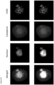
- Figure 1 shows a CAML cell initially stained with DAPI, Cytokeratin 8, 18 & 19, EpCAM and CD45 (top row).
- the second row of Figure 1 shows the cell was restained for DAPI, PD-L1, CCR5 and PD-1.
- both PD-L1 and CCR5 expressions are high indicating that a combination of immunotherapies that target these two proteins might be a suitable treatment for this patient.
- CCR5 is in the cytoplasm; it was not translocated into the nucleus, indicating it was in an active form.
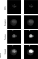
- Figure 2 shows a CAML cell initially stained with DAPI, Cytokeratin 8, 18 & 19, EpCAM and CD45 (top row).
- the second row of Figure 2 shows the cell was restained for DAPI, PD-L1, CCR5 and PD-1.
- CCR5 expression but no PD-L1 expression, indicating that an immunotherapies alone will not be effective, while therapy targeting CCR5 might be a suitable treatment for this patient.
- CCR5 pools/clusters are in the cytoplasm; it was not translocated into the nucleus, indicating it was in an active form.
- Figures 3-10 focus on CCR5 expression. Black & white and color versions of these figures are provided. Color images allow for the visualization of translocation into the nucleus. For the color images, the nuclei are red, cytoplasm is blue, and CCR5 staining is yellow.
- the image sizes for Figures 3 , 4 , and 6 are 60 ⁇ m x 60 ⁇ m.
- the image size for Figure 5A is 60 ⁇ m x 60 ⁇ m;
- Figure 5B is 90 ⁇ m x 90 ⁇ m;
- Figure 5C is 60 ⁇ m x 60 ⁇ m;
- Figure 6A is 60 ⁇ m x 60 ⁇ m;
- Figure 6B is 75 ⁇ m x 75 ⁇ m.
- the image size for Figures 7-10 are 30 ⁇ m x 30 ⁇ m.
- Figures 3A-C are CAMLs with high/medium CCR5 expression in the form of pools/clusters found on the surface of the cell and in the cytoplasm, indicating the protein is in an activated form.
- Figures 4A-C are CAMLs with low/negative CCR5 expression in the form of pools/clusters found on the surface of the cell and in the cytoplasm, indicating the protein is in an activated form.
- Figures 5A-C are CAMLs with high CCR5 expression in the form of pools/clusters, with most pools/clusters translocated into the nucleus, indicating the protein is in an inactivated form.
- Figure 5A shows high expression of CCR5 in the cytoplasm.
- Figure 5B shows CCR5 has mostly translocated into the nucleus.
- Figure 5C shows CCR5 has completely translocated into the nucleus.
- Figures 6A-B are CAMLs with medium/low CCR5 expression in the form of pools/clusters, with most pools/clusters translocated into the nucleus in Figure 6B , indicating the protein is in an inactivated form.
- CCR5 pools/clusters are found on other cancer associated cells such as EMTCTCs and CTCs.
- Figure 7 shows EMTCTCs with localized high CCR5 expression aggregated to one spot on the cell surface.
- Figure 8 shows EMTCTCs without CCR5 expression.
- Figures 9A-C show CTCs with moderate or high CCR5 expression.
- the amount of CCR5 in Figure 9C is significantly higher than usual.
- Figures 10A-B are CTCs with low expressions of CCR5 with only a few small pools/clusters.
- Figure 11 shows the importance of the pools/clusters or pools/clusters based on 21 breast cancer patients in Stages III and IV. If the patient has a CAML where the number of CCR5 pools/clusters is greater than or equal to 21, the patient had very poor prognosis.
- OS overall survival
- CCR5 can be activated or inactivated, i.e. localized to either the nucleus or the cytoplasm.
- the cutoff of 20 CCR5 pools/clusters or pools/clusters used in Figure 11 is an example providing the best p-value for the 21 breast cancer patients.
- Kaplan-Meier analyses were done in MATLAB R2013A using the counts from all subtypes and the known patient populations.
- the time to progression was defined as the interval between when T0 blood sample was obtained to date of progression, all patients remained on study through 24 month end point, i.e. no patients were censored.
Landscapes
- Health & Medical Sciences (AREA)
- Life Sciences & Earth Sciences (AREA)
- Immunology (AREA)
- Engineering & Computer Science (AREA)
- Urology & Nephrology (AREA)
- Biomedical Technology (AREA)
- Hematology (AREA)
- Chemical & Material Sciences (AREA)
- Molecular Biology (AREA)
- Cell Biology (AREA)
- Food Science & Technology (AREA)
- Pathology (AREA)
- Biotechnology (AREA)
- Medicinal Chemistry (AREA)
- Physics & Mathematics (AREA)
- Analytical Chemistry (AREA)
- Biochemistry (AREA)
- General Health & Medical Sciences (AREA)
- General Physics & Mathematics (AREA)
- Microbiology (AREA)
- Tropical Medicine & Parasitology (AREA)
- Physiology (AREA)
- Ecology (AREA)
- Measuring Or Testing Involving Enzymes Or Micro-Organisms (AREA)
- Investigating Or Analysing Biological Materials (AREA)
- Hospice & Palliative Care (AREA)
- Oncology (AREA)
Claims (15)
- Procédé d'estimation de la survie globale (SG) et de la survie sans progression (SSP) d'un sujet atteint d'un cancer, ledit procédé comprenant la détermination du nombre de groupes CCR5 dans des cellules circulantes dans un échantillon biologique provenant d'un sujet atteint d'un cancer,dans lequel les cellules circulantes comprennent des cellules de type macrophage associées au cancer (CAML) et dans lequel les CAML présentent les caractéristiques suivantes :(a) grand noyau polyploïde atypique d'une taille d'environ 14-64 µm, ou plusieurs noyaux dans une seule cellule ;(b) taille de cellule d'environ 20-300 µm ; et(c) forme morphologique sélectionnée dans le groupe consistant en fuseau, têtard, ronde, oblongue, à deux pattes, à plus de deux pattes, à pattes fines et amorphes ; éventuellement, dans lequel les CAML présentent une ou plusieurs des caractéristiques supplémentaires suivantes :(d) phénotype CD14 positif ;(e) expression de CD45 ;(f) expression d'EpCAM ;(g) expression de vimentine ;(h) expression de PD-L1 ;(i) expression du marqueur CD11C ;(j) expression du marqueur CD146 ;(k) expression du marqueur CD202b ;(l) expression du marqueur CD31 ; et(m) phénotype épithélial CK8, 18, 19,dans lequel la source de l'échantillon biologique est un ou plusieurs éléments parmi le sang périphérique, le sang, les ganglions lymphatiques, la moelle osseuse, le liquide céphalo-rachidien, les tissus et l'urine, etdans lequel, quand au moins une cellule CAML dans ledit échantillon présente environ 10 groupes CCR5 ou plus(i) il est estimé que la SG et la SSP du sujet sont raccourcies, ou(ii) il est estimé que la SG et la SSP du sujet sont plus courtes que celles d'un sujet atteint du même type de cancer pour lequel aucune des cellules d'un échantillon biologique correspondant ne présente environ 10 groupes CCR5 ou plus.
- Procédé d'estimation de la SG et la SSP d'un sujet atteint d'un cancer, ledit procédé comprenant :(i) la détermination du nombre de groupes CCR5 dans les cellules circulantes dans un échantillon biologique provenant d'un premier sujet atteint d'un cancer, et la comparaison des résultats à ceux déterminés à partir d'un second sujet atteint du même type de cancer, dans lequel il est estimé que la SG et la SSP du sujet présentant un plus grand nombre de groupes CCR5 sont plus courtes que la SG et la SSP du sujet présentant moins de groupes CCR5 ;(ii) la détermination d'un nombre moyen de groupes CCR5 dans un nombre sélectionné de cellules circulantes dans un échantillon biologique provenant d'un premier sujet atteint d'un cancer, et la comparaison des résultats à ceux déterminés à partir d'un second sujet atteint du même type de cancer, dans lequel il est estimé que la SG et la SSP du sujet avec un nombre moyen plus élevé de groupes CCR5 sont plus courtes que la SG et la SSP du sujet présentant un nombre moyen de groupes CCR5 inférieur ; ou(iii) la détermination d'un nombre médian de groupes CCR5 dans un nombre sélectionné de cellules circulantes dans un échantillon biologique provenant d'un premier sujet atteint d'un cancer, et la comparaison des résultats à ceux déterminés à partir d'un second sujet atteint du même type de cancer, dans lequel il est estimé que la SG et la SSP du sujet présentant un nombre médian plus élevé de groupes CCR5 sont plus courtes que la SG et la SSP du sujet présentant un nombre médian de groupes CCR5 inférieur,dans lequel les cellules circulantes comprennent des cellules de type macrophage associées au cancer (CAML) et dans lequel les CAML présentent les caractéristiques suivantes :(a) grand noyau polyploïde atypique d'une taille d'environ 14-64 µm, ou plusieurs noyaux dans une seule cellule ;(b) taille de cellule d'environ 20-300 µm ; et(c) forme morphologique sélectionnée dans le groupe consistant en fuseau, têtard, ronde, oblongue, à deux pattes, à plus de deux pattes, à pattes fines et amorphes ; éventuellement, dans lequel les CAML présentent une ou plusieurs des caractéristiques supplémentaires suivantes :(d) phénotype CD14 positif ;(e) expression de CD45 ;(f) expression d'EpCAM ;(g) expression de vimentine ;(h) expression de PD-L1 ;(i) expression du marqueur CD11C ;(j) expression du marqueur CD146 ;(k) expression du marqueur CD202b ;(l) expression du marqueur CD31 ; et(m) phénotype épithélial CK8, 18, 19,et dans lequel la source de l'échantillon biologique est un ou plusieurs éléments parmi le sang périphérique, le sang, les ganglions lymphatiques, la moelle osseuse, le liquide céphalo-rachidien, les tissus et l'urine.
- Procédé selon la revendication 1, dans lequel le nombre de groupes CCR5 est de 15 ou plus, 16 ou plus, 17 ou plus, 18 ou plus, 19 ou plus, 20 ou plus, 21 ou plus, 22 ou plus, 23 ou plus, 24 ou plus, ou 25 ou plus.
- Procédé selon l'une quelconque des revendications 1-3, dans lequel ladite SG et/ou ladite SSP s'étend(ent) sur une période d'au moins 12 mois, ou dans lequel ladite SG et/ou ladite SSP s'étend(ent) sur une période d'au moins 24 mois.
- Procédé selon l'une quelconque des revendications 1-4, dans lequel la taille de l'échantillon biologique est comprise entre 5 et 15 ml.
- Procédé selon l'une quelconque des revendications 1 à 5, dans lequel les cellules circulantes comprennent en outre un ou plusieurs parmi les CTC, PDCTC, CTC apoptotiques, EMTCTC et CAVE.
- Procédé selon l'une quelconque des revendications 1 à 6, dans lequel l'échantillon biologique est du sang de la veine antécubitale, du sang de la veine cave inférieure, du sang de la veine fémorale, du sang de la veine porte ou du sang de la veine jugulaire.
- Procédé selon la revendication 7, dans lequel l'échantillon biologique est constitué de 7,5 ml de sang de la veine antécubitale, de sang de la veine cave inférieure, de sang de la veine fémorale, de sang de la veine porte ou de sang de veine jugulaire.
- Procédé selon l'une quelconque des revendications 1-8, dans lequel le cancer est un cancer exprimant CCRS sélectionné parmi une tumeur solide, un cancer de stade I, un cancer de stade II, un cancer de stade III, un cancer de stade IV, un carcinome, un sarcome, un neuroblastome, un mélanome, un cancer de cellule épithéliale, un cancer du sein, un cancer de la prostate, un cancer du poumon, un cancer du pancréas, un cancer colorectal, un cancer du foie, un cancer de la tête et du cou, un cancer du rein, un cancer des ovaires, un cancer de l'œsophage ou un autre cancer à tumeur solide.
- Procédé selon l'une quelconque des revendications 1-9, dans lequel les cellules circulantes sont isolées des échantillons biologiques pour les étapes de détermination en utilisant un ou plusieurs moyens sélectionnés dans le groupe consistant en la méthodologie d'exclusion de taille, l'immunocapture, la lyse des globules rouges, la déplétion des globules blancs, le FICOLL, l'électrophorèse, la diélectrophorèse, la cytométrie en flux, la lévitation magnétique et diverses puces microfluidiques, ou une combinaison de celles-ci.
- Procédé selon la revendication 10, dans lequel les cellules circulantes sont isolées des échantillons biologiques en utilisant une méthodologie d'exclusion de taille qui comprend l'utilisation d'un microfiltre, de préférence dans lequel le microfiltre présente une taille de pores allant d'environ 5 micromètres à environ 20 micromètres, éventuellement dans lequel les pores du microfiltre présentent une forme ronde, une forme de circuit, une forme de pores ovale, carrée et rectangulaire, ou dans lequel le microfiltre présente une géométrie de pores précise et une distribution uniforme des pores.
- Procédé selon la revendication 10, dans lequel les cellules circulantes sont isolées en utilisant une puce microfluidique via un tri basé sur la taille physique, des fentes, des canaux, un tri hydrodynamique basé sur la taille, un regroupement, un piégeage, une immunocapture, une concentration de grandes cellules ou une élimination de petites cellules sur la base de la taille.
- Procédé selon l'une quelconque des revendications 1-9, dans lequel les cellules circulantes sont isolées des échantillons biologiques pour les étapes de détermination en utilisant un test de microfiltration à basse pression.
- Procédé selon une quelconque revendication précédente, dans lequel les groupes CCR5 sont localisés dans le noyau de la cellule, ou les groupes CCR5 sont localisés dans le cytoplasme de la cellule, ou localisés à la surface de la cellule, ou une combinaison de ceux-ci ; et/ou dans lequel le CCR5 des cellules circulantes est dans un état activé ou dans un état inactivé, ou dans lequel les cellules présentent à la fois des récepteurs activés et inactivés.
- Procédé selon l'une quelconque des revendications précédentes, comprenant en outre l'utilisation de la détermination du nombre de groupes CCRS dans les cellules circulantes dans un échantillon biologique provenant d'un sujet comme diagnostic compagnon ou complémentaire lors des décisions de traitement pour ledit sujet.
Applications Claiming Priority (2)
| Application Number | Priority Date | Filing Date | Title |
|---|---|---|---|
| US201962805031P | 2019-02-13 | 2019-02-13 | |
| PCT/US2020/018074 WO2020168052A1 (fr) | 2019-02-13 | 2020-02-13 | Procédés à base de ccr5 pour prédire la survie globale et sans progression chez des sujets atteints d'un cancer |
Publications (3)
| Publication Number | Publication Date |
|---|---|
| EP3924733A1 EP3924733A1 (fr) | 2021-12-22 |
| EP3924733C0 EP3924733C0 (fr) | 2024-08-28 |
| EP3924733B1 true EP3924733B1 (fr) | 2024-08-28 |
Family
ID=69771254
Family Applications (1)
| Application Number | Title | Priority Date | Filing Date |
|---|---|---|---|
| EP20710020.7A Active EP3924733B1 (fr) | 2019-02-13 | 2020-02-13 | Procédés à base de ccr5 pour prédire la survie globale et sans progression chez des sujets atteints d'un cancer |
Country Status (4)
| Country | Link |
|---|---|
| US (1) | US20220128544A1 (fr) |
| EP (1) | EP3924733B1 (fr) |
| ES (1) | ES2986628T3 (fr) |
| WO (1) | WO2020168052A1 (fr) |
Families Citing this family (1)
| Publication number | Priority date | Publication date | Assignee | Title |
|---|---|---|---|---|
| WO2023283264A1 (fr) * | 2021-07-06 | 2023-01-12 | Creatv Microtech Inc. | Méthodes pour prédire une maladie métastasique multi-organe et une survie globale et sans progression chez des sujets ayant des cellules de type macrophages géants circulants associées au cancer (caml) |
Family Cites Families (4)
| Publication number | Priority date | Publication date | Assignee | Title |
|---|---|---|---|---|
| AU2013267253B2 (en) | 2012-06-01 | 2018-03-29 | Creatv Microtech, Inc. | Capture, identification and use of a new biomarker of solid tumors in body fluids |
| WO2016033103A1 (fr) | 2014-08-25 | 2016-03-03 | Creatv Microtech, Inc. | Utilisation de biomarqueurs de cellules circulant dans le sang pour la détection et le diagnostic de maladies, et leurs procédés d'isolement |
| WO2016209926A1 (fr) * | 2015-06-22 | 2016-12-29 | Thomas Jefferson University | Cancers exprimant le ccr5 et méthodes de traitement associées |
| KR20200005662A (ko) * | 2017-05-18 | 2020-01-15 | 테사로, 인코포레이티드 | 암을 치료하기 위한 조합 요법 |
-
2020
- 2020-02-13 ES ES20710020T patent/ES2986628T3/es active Active
- 2020-02-13 US US17/430,457 patent/US20220128544A1/en active Pending
- 2020-02-13 WO PCT/US2020/018074 patent/WO2020168052A1/fr not_active Ceased
- 2020-02-13 EP EP20710020.7A patent/EP3924733B1/fr active Active
Non-Patent Citations (2)
| Title |
|---|
| RAGHAVAKAIMAL ASHVATHI ET AL: "CCR5 activation and endocytosis in circulating tumor-derived cells isolated from the blood of breast cancer patients provide information about clinical outcome.", BREAST CANCER RESEARCH, vol. 24, 35, 23 May 2022 (2022-05-23), pages 1 - 12, DOI: 10.1186/s13058-022-01528-w * |
| SINGER IRWIN I ET AL: "CCR5, CXCR4, and CD4 are clustered and closely apposed on microvilli of human macrophages and T cells", JOURNAL OF VIROLOGY, vol. 75, no. 8, 15 April 2001 (2001-04-15), pages 3779 - 3790, DOI: doi.org/10.1128/jvi.75.8.3779-3790.2001 * |
Also Published As
| Publication number | Publication date |
|---|---|
| EP3924733C0 (fr) | 2024-08-28 |
| EP3924733A1 (fr) | 2021-12-22 |
| ES2986628T3 (es) | 2024-11-12 |
| US20220128544A1 (en) | 2022-04-28 |
| WO2020168052A1 (fr) | 2020-08-20 |
Similar Documents
| Publication | Publication Date | Title |
|---|---|---|
| US20230085158A1 (en) | Method of using non-rare cells to detect rare cells | |
| US10613089B2 (en) | Method of using non-rare cells to detect rare cells | |
| KR20150008842A (ko) | 순환 종양 세포를 확인하기 위한 장치, 시스템 및 방법 | |
| CN107850587B (zh) | 循环肿瘤细胞有丝分裂指数在癌症分层和诊断中的应用 | |
| EP3583426B1 (fr) | Procédés de prédiction de la survie globale et sans progression chez des sujets atteints d'un cancer, à l'aide de cellules circulantes de type macrophage associées au cancer (ctmac) | |
| EP3924733B1 (fr) | Procédés à base de ccr5 pour prédire la survie globale et sans progression chez des sujets atteints d'un cancer | |
| US20260072033A1 (en) | Use of extracellular vesicles and micronuclei of circulating stromal cells as pan-cancer biomarkers for predicting clinical outcomes | |
| WO2023004038A1 (fr) | Méthodes pour prédire et/ou surveiller une réponse de traitement du cancer à l'aide de changements dans des cellules de type macrophage associées au cancer circulant (caml) | |
| WO2024250002A1 (fr) | Méthodes de prédiction de survie sans progression et de survie globale chez des sujets atteints d'un cancer à l'aide de cellules hybrides de macrophages tumoraux | |
| Adams et al. | Biopsies in the Twenty-First Century: Have Circulating Stromal Cells Come of Age? |
Legal Events
| Date | Code | Title | Description |
|---|---|---|---|
| STAA | Information on the status of an ep patent application or granted ep patent |
Free format text: STATUS: UNKNOWN |
|
| STAA | Information on the status of an ep patent application or granted ep patent |
Free format text: STATUS: THE INTERNATIONAL PUBLICATION HAS BEEN MADE |
|
| PUAI | Public reference made under article 153(3) epc to a published international application that has entered the european phase |
Free format text: ORIGINAL CODE: 0009012 |
|
| STAA | Information on the status of an ep patent application or granted ep patent |
Free format text: STATUS: REQUEST FOR EXAMINATION WAS MADE |
|
| 17P | Request for examination filed |
Effective date: 20210910 |
|
| AK | Designated contracting states |
Kind code of ref document: A1 Designated state(s): AL AT BE BG CH CY CZ DE DK EE ES FI FR GB GR HR HU IE IS IT LI LT LU LV MC MK MT NL NO PL PT RO RS SE SI SK SM TR |
|
| DAV | Request for validation of the european patent (deleted) | ||
| DAX | Request for extension of the european patent (deleted) | ||
| STAA | Information on the status of an ep patent application or granted ep patent |
Free format text: STATUS: EXAMINATION IS IN PROGRESS |
|
| 17Q | First examination report despatched |
Effective date: 20220905 |
|
| GRAP | Despatch of communication of intention to grant a patent |
Free format text: ORIGINAL CODE: EPIDOSNIGR1 |
|
| STAA | Information on the status of an ep patent application or granted ep patent |
Free format text: STATUS: GRANT OF PATENT IS INTENDED |
|
| INTG | Intention to grant announced |
Effective date: 20240417 |
|
| GRAS | Grant fee paid |
Free format text: ORIGINAL CODE: EPIDOSNIGR3 |
|
| GRAA | (expected) grant |
Free format text: ORIGINAL CODE: 0009210 |
|
| STAA | Information on the status of an ep patent application or granted ep patent |
Free format text: STATUS: THE PATENT HAS BEEN GRANTED |
|
| AK | Designated contracting states |
Kind code of ref document: B1 Designated state(s): AL AT BE BG CH CY CZ DE DK EE ES FI FR GB GR HR HU IE IS IT LI LT LU LV MC MK MT NL NO PL PT RO RS SE SI SK SM TR |
|
| REG | Reference to a national code |
Ref country code: CH Ref legal event code: EP |
|
| REG | Reference to a national code |
Ref country code: DE Ref legal event code: R096 Ref document number: 602020036604 Country of ref document: DE |
|
| REG | Reference to a national code |
Ref country code: IE Ref legal event code: FG4D |
|
| U01 | Request for unitary effect filed |
Effective date: 20240904 |
|
| U07 | Unitary effect registered |
Designated state(s): AT BE BG DE DK EE FI FR IT LT LU LV MT NL PT RO SE SI Effective date: 20240923 |
|
| REG | Reference to a national code |
Ref country code: ES Ref legal event code: FG2A Ref document number: 2986628 Country of ref document: ES Kind code of ref document: T3 Effective date: 20241112 |
|
| PG25 | Lapsed in a contracting state [announced via postgrant information from national office to epo] |
Ref country code: NO Free format text: LAPSE BECAUSE OF FAILURE TO SUBMIT A TRANSLATION OF THE DESCRIPTION OR TO PAY THE FEE WITHIN THE PRESCRIBED TIME-LIMIT Effective date: 20241128 |
|
| PG25 | Lapsed in a contracting state [announced via postgrant information from national office to epo] |
Ref country code: GR Free format text: LAPSE BECAUSE OF FAILURE TO SUBMIT A TRANSLATION OF THE DESCRIPTION OR TO PAY THE FEE WITHIN THE PRESCRIBED TIME-LIMIT Effective date: 20241129 Ref country code: PL Free format text: LAPSE BECAUSE OF FAILURE TO SUBMIT A TRANSLATION OF THE DESCRIPTION OR TO PAY THE FEE WITHIN THE PRESCRIBED TIME-LIMIT Effective date: 20240828 |
|
| PG25 | Lapsed in a contracting state [announced via postgrant information from national office to epo] |
Ref country code: IS Free format text: LAPSE BECAUSE OF FAILURE TO SUBMIT A TRANSLATION OF THE DESCRIPTION OR TO PAY THE FEE WITHIN THE PRESCRIBED TIME-LIMIT Effective date: 20241228 |
|
| PG25 | Lapsed in a contracting state [announced via postgrant information from national office to epo] |
Ref country code: HR Free format text: LAPSE BECAUSE OF FAILURE TO SUBMIT A TRANSLATION OF THE DESCRIPTION OR TO PAY THE FEE WITHIN THE PRESCRIBED TIME-LIMIT Effective date: 20240828 |
|
| PG25 | Lapsed in a contracting state [announced via postgrant information from national office to epo] |
Ref country code: RS Free format text: LAPSE BECAUSE OF FAILURE TO SUBMIT A TRANSLATION OF THE DESCRIPTION OR TO PAY THE FEE WITHIN THE PRESCRIBED TIME-LIMIT Effective date: 20241128 |
|
| PG25 | Lapsed in a contracting state [announced via postgrant information from national office to epo] |
Ref country code: RS Free format text: LAPSE BECAUSE OF FAILURE TO SUBMIT A TRANSLATION OF THE DESCRIPTION OR TO PAY THE FEE WITHIN THE PRESCRIBED TIME-LIMIT Effective date: 20241128 Ref country code: PL Free format text: LAPSE BECAUSE OF FAILURE TO SUBMIT A TRANSLATION OF THE DESCRIPTION OR TO PAY THE FEE WITHIN THE PRESCRIBED TIME-LIMIT Effective date: 20240828 Ref country code: NO Free format text: LAPSE BECAUSE OF FAILURE TO SUBMIT A TRANSLATION OF THE DESCRIPTION OR TO PAY THE FEE WITHIN THE PRESCRIBED TIME-LIMIT Effective date: 20241128 Ref country code: IS Free format text: LAPSE BECAUSE OF FAILURE TO SUBMIT A TRANSLATION OF THE DESCRIPTION OR TO PAY THE FEE WITHIN THE PRESCRIBED TIME-LIMIT Effective date: 20241228 Ref country code: HR Free format text: LAPSE BECAUSE OF FAILURE TO SUBMIT A TRANSLATION OF THE DESCRIPTION OR TO PAY THE FEE WITHIN THE PRESCRIBED TIME-LIMIT Effective date: 20240828 Ref country code: GR Free format text: LAPSE BECAUSE OF FAILURE TO SUBMIT A TRANSLATION OF THE DESCRIPTION OR TO PAY THE FEE WITHIN THE PRESCRIBED TIME-LIMIT Effective date: 20241129 |
|
| U20 | Renewal fee for the european patent with unitary effect paid |
Year of fee payment: 6 Effective date: 20250218 |
|
| PG25 | Lapsed in a contracting state [announced via postgrant information from national office to epo] |
Ref country code: SM Free format text: LAPSE BECAUSE OF FAILURE TO SUBMIT A TRANSLATION OF THE DESCRIPTION OR TO PAY THE FEE WITHIN THE PRESCRIBED TIME-LIMIT Effective date: 20240828 |
|
| PG25 | Lapsed in a contracting state [announced via postgrant information from national office to epo] |
Ref country code: CZ Free format text: LAPSE BECAUSE OF FAILURE TO SUBMIT A TRANSLATION OF THE DESCRIPTION OR TO PAY THE FEE WITHIN THE PRESCRIBED TIME-LIMIT Effective date: 20240828 |
|
| PG25 | Lapsed in a contracting state [announced via postgrant information from national office to epo] |
Ref country code: SK Free format text: LAPSE BECAUSE OF FAILURE TO SUBMIT A TRANSLATION OF THE DESCRIPTION OR TO PAY THE FEE WITHIN THE PRESCRIBED TIME-LIMIT Effective date: 20240828 |
|
| PLBE | No opposition filed within time limit |
Free format text: ORIGINAL CODE: 0009261 |
|
| STAA | Information on the status of an ep patent application or granted ep patent |
Free format text: STATUS: NO OPPOSITION FILED WITHIN TIME LIMIT |
|
| 26N | No opposition filed |
Effective date: 20250530 |
|
| PG25 | Lapsed in a contracting state [announced via postgrant information from national office to epo] |
Ref country code: MC Free format text: LAPSE BECAUSE OF FAILURE TO SUBMIT A TRANSLATION OF THE DESCRIPTION OR TO PAY THE FEE WITHIN THE PRESCRIBED TIME-LIMIT Effective date: 20240828 |
|
| REG | Reference to a national code |
Ref country code: CH Ref legal event code: U11 Free format text: ST27 STATUS EVENT CODE: U-0-0-U10-U11 (AS PROVIDED BY THE NATIONAL OFFICE) Effective date: 20260301 |
|
| U20 | Renewal fee for the european patent with unitary effect paid |
Year of fee payment: 7 Effective date: 20260227 |
|
| PGFP | Annual fee paid to national office [announced via postgrant information from national office to epo] |
Ref country code: GB Payment date: 20260218 Year of fee payment: 7 |
|
| PGFP | Annual fee paid to national office [announced via postgrant information from national office to epo] |
Ref country code: ES Payment date: 20260303 Year of fee payment: 7 |
|
| PGFP | Annual fee paid to national office [announced via postgrant information from national office to epo] |
Ref country code: IE Payment date: 20260218 Year of fee payment: 7 |
|
| PGFP | Annual fee paid to national office [announced via postgrant information from national office to epo] |
Ref country code: CH Payment date: 20260301 Year of fee payment: 7 |