EP3984504A1 - Dispositif scléral pour le traitement d'un glaucome à angle ouvert - Google Patents
Dispositif scléral pour le traitement d'un glaucome à angle ouvert Download PDFInfo
- Publication number
- EP3984504A1 EP3984504A1 EP20201616.8A EP20201616A EP3984504A1 EP 3984504 A1 EP3984504 A1 EP 3984504A1 EP 20201616 A EP20201616 A EP 20201616A EP 3984504 A1 EP3984504 A1 EP 3984504A1
- Authority
- EP
- European Patent Office
- Prior art keywords
- scleral
- sector
- sclera
- tissue
- curved
- Prior art date
- Legal status (The legal status is an assumption and is not a legal conclusion. Google has not performed a legal analysis and makes no representation as to the accuracy of the status listed.)
- Withdrawn
Links
- 206010030348 Open-Angle Glaucoma Diseases 0.000 title claims abstract description 9
- 210000003786 sclera Anatomy 0.000 claims abstract description 36
- 230000001886 ciliary effect Effects 0.000 claims abstract description 23
- 210000003205 muscle Anatomy 0.000 claims abstract description 22
- 210000001519 tissue Anatomy 0.000 claims abstract description 19
- 210000000795 conjunctiva Anatomy 0.000 claims description 13
- 201000004569 Blindness Diseases 0.000 abstract description 2
- 230000004438 eyesight Effects 0.000 abstract description 2
- 230000004410 intraocular pressure Effects 0.000 description 7
- 210000001585 trabecular meshwork Anatomy 0.000 description 6
- 210000001742 aqueous humor Anatomy 0.000 description 5
- 210000001760 tenon capsule Anatomy 0.000 description 5
- 238000003780 insertion Methods 0.000 description 3
- 230000037431 insertion Effects 0.000 description 3
- 238000004519 manufacturing process Methods 0.000 description 3
- QCHFTSOMWOSFHM-WPRPVWTQSA-N (+)-Pilocarpine Chemical compound C1OC(=O)[C@@H](CC)[C@H]1CC1=CN=CN1C QCHFTSOMWOSFHM-WPRPVWTQSA-N 0.000 description 2
- 208000010412 Glaucoma Diseases 0.000 description 2
- QCHFTSOMWOSFHM-UHFFFAOYSA-N SJ000285536 Natural products C1OC(=O)C(CC)C1CC1=CN=CN1C QCHFTSOMWOSFHM-UHFFFAOYSA-N 0.000 description 2
- 230000006835 compression Effects 0.000 description 2
- 238000007906 compression Methods 0.000 description 2
- 230000007423 decrease Effects 0.000 description 2
- 238000010586 diagram Methods 0.000 description 2
- 239000012530 fluid Substances 0.000 description 2
- 238000000034 method Methods 0.000 description 2
- 229960001416 pilocarpine Drugs 0.000 description 2
- 238000010521 absorption reaction Methods 0.000 description 1
- 210000002159 anterior chamber Anatomy 0.000 description 1
- 230000017531 blood circulation Effects 0.000 description 1
- 239000002775 capsule Substances 0.000 description 1
- 210000004240 ciliary body Anatomy 0.000 description 1
- 230000000295 complement effect Effects 0.000 description 1
- 230000008602 contraction Effects 0.000 description 1
- 210000004087 cornea Anatomy 0.000 description 1
- 230000007850 degeneration Effects 0.000 description 1
- 239000003814 drug Substances 0.000 description 1
- 229940079593 drug Drugs 0.000 description 1
- 230000001939 inductive effect Effects 0.000 description 1
- 239000007788 liquid Substances 0.000 description 1
- 239000003550 marker Substances 0.000 description 1
- 239000000463 material Substances 0.000 description 1
- 230000001404 mediated effect Effects 0.000 description 1
- 239000002184 metal Substances 0.000 description 1
- 230000000149 penetrating effect Effects 0.000 description 1
- 239000011148 porous material Substances 0.000 description 1
- 210000001747 pupil Anatomy 0.000 description 1
- 230000003014 reinforcing effect Effects 0.000 description 1
- 208000029257 vision disease Diseases 0.000 description 1
Images



Classifications
-
- A—HUMAN NECESSITIES
- A61—MEDICAL OR VETERINARY SCIENCE; HYGIENE
- A61F—FILTERS IMPLANTABLE INTO BLOOD VESSELS; PROSTHESES; DEVICES PROVIDING PATENCY TO, OR PREVENTING COLLAPSING OF, TUBULAR STRUCTURES OF THE BODY, e.g. STENTS; ORTHOPAEDIC, NURSING OR CONTRACEPTIVE DEVICES; FOMENTATION; TREATMENT OR PROTECTION OF EYES OR EARS; BANDAGES, DRESSINGS OR ABSORBENT PADS; FIRST-AID KITS
- A61F9/00—Methods or devices for treatment of the eyes; Devices for putting in contact-lenses; Devices to correct squinting; Apparatus to guide the blind; Protective devices for the eyes, carried on the body or in the hand
- A61F9/007—Methods or devices for eye surgery
- A61F9/00781—Apparatus for modifying intraocular pressure, e.g. for glaucoma treatment
Definitions
- the present invention is directed to a scleral device for treatment of open angle glaucoma, configured to be attached to an eye and to prevent loss of eyesight and blindness. Thanks to the scleral device the sclera is supported and reinforced, while the special relationship between the ciliary muscle and the lens remain substantially unchanged. For this purpose, incisions in the conjunctiva have to be made to gain access to the sclera overlying the ciliary muscle.
- Glaucoma is a vision disorder associated with excess intraocular pressure in the eye.
- Intraocular pressure is a function of production of liquid aqueous humor by the ciliary processes of the eye, and its drainage through the trabecular meshwork.
- Aqueous humor flows from the ciliary processes into the posterior chamber, and it flows through the pupil of the iris into the anterior chamber, bounded posteriorly by the iris and anteriorly by the cornea. From here, the trabecular meshwork drains aqueous humor via the scleral venous sinus into scleral plexuses and general blood circulation.
- a scleral device for treatment of open angle glaucoma configured to be attached to an eye having an iris, a ciliary muscle, a scleral tissue and a conjunctiva covering the scleral tissue and comprising at least one tunnelled incision adjacent to the iris that overlies the ciliary muscle
- the scleral device is symmetrical with respect to a longitudinal axis and is configured to be partially housed in the tunnelled incision and to be linked to the scleral tissue and comprises a curved central portion comprising two ends and an inner face having a concave curvature adapted to the curvature of the eye and configured to contact the scleral tissue and two arms which extends from the ends relatively movable with respect to each other from an open position to a closed position.
- Each arm comprises a L-shaped longitudinal section formed by a first sector which extends outwardly from the end in opposite direction to the inner face, and a second sector extending from the first sector, which is longer than the first sector, which comprises a hook in a free end configured to grasp the sclera tissue in a closed position.
- the scleral device supports and reinforces the sclera, while substantially maintaining the special relationship between the ciliary muscle and the lens.
- incision in the conjunctiva has to be done, to gain access to the sclera overlying the ciliary muscle.
- the tenon's capsule are moved laterally to expose the sclera, and the sclera is extended outwardly.
- a scleral device, or various scleral devices, are provided having two arms for engaging the outwardly extended sclera there between. The arms of the scleral device are extended in the sclera so as to excerpt pressure on the sclera, and then the tenon's capsules are slid over the scleral device and the conjunctiva is closed.
- Pilocarpine reduces intraocular pressure (IOP) by inducing contraction of the ciliary muscle.
- IOP intraocular pressure
- the mechanism of IOP reduction is mediated by increased tension exerted by the longitudinal ciliary muscles at its circumferential insertion on the scleral spur.
- the scleral device assists the ciliary muscles to facilitate the flow of aqueous fluid through the trabecular meshwork.
- the scleral device treats glaucoma by supporting and reinforcing the sclera with a removable device.
- the scleral device is attached to the sclera by first making an incision, tunnelled incisions, in the conjunctiva to gain access to the sclera's overlying the ciliary muscle.
- the Tenon's capsule is moved laterally to expose the sclera and the sclera is incised appropriately.
- the arms of the device engage a portion of the sclera resulting in compression over the ciliary body.
- the Tenon's capsule covers the device and the conjunctiva is closed.
- the scleral device restores the level of force which the ciliary muscle exerts on the trabecular meshwork, thus opening the drainage pores and relieving the intraocular pressure (IOP).
- the scleral device decreases the production and increases the outflow of aqueous fluids through the trabecular meshwork. The decrease in production results from compression of the ciliary muscle.
- the increase in outflow occurs due to the anatomical changes induced by the scleral device to treat Open Angle Glaucoma.
- the scleral device strengthens the sclera and assist in maintaining the spatial relationship between the sclera and the ciliary system.
- the material of the scleral device can be metallic machined or Metal Injected (MIM) or non-metallic machined or moulded.
- the central portion and the first sector may be joined by a first curved junction and the first sector and the second sector may be joined by a second curved junction.
- the distance between the first junction and the curved configuration of an external face, opposite to the inner face, in this area, once inserted in the sclera give an outwardly pressure to the scleral device pushing the arms into tunnelled incisions of the sclera resulting in downward constant pressure on the eye from the inner face.
- an incision in sclera are made and then the surgical pocket is made.
- the arms are inserted in the sclera incision and twisted in surgical pocket.
- the device in order to insert the second arm in sclera, the device has to be bended 90° and pushed in sclera incision and then the arm will follow in surgical pocket.
- the surgical pockets are cavities in the sclera.
- the thickness of the curved junctions is wider than the rest of the scleral device, wherein the rest of the scleral device may have a constant thickness.
- the hook preferably comprises a narrow sector limited by two cut-outs defined on each side of the longitudinal axis and a wide sector, extending from the narrow sector which comprises two flaps on each side of the longitudinal axis wherein said flaps are limited by a curved outer edge.
- the flaps secure the device and prevent slippage of the scleral device, thereto, and the Tenon's capsules are slid over the scleral device and the conjunctiva is closed.
- Preferably, up to four such scleral device is applied to the sclera substantially equally spaced about the lens between the medial, inferior, lateral and superior rectus muscle.
- the scleral device serves to prevent the sclera from buckling under tension applied by the ciliary muscle.
- the narrow sector give flexibility to the wide sector and the outer edge ease the insertion in the surgical pocket.
- the first sector may form a first angle with the central portion bigger than 90° and the second sector forms a second angle with the first sector bigger than 90°.
- the scleral device has a length of no more than 6.0mm and no less than 2.5mm so as to fit between adjacent rectus muscles.
- the arms contain but not limited to the flaps but may also be provided with means, such as barbs, teeth or spurs, for grasping but not penetrating the sclera.
- the arms of the scleral device remain in the closed position in the absence of an external force applied therebetween.
- the incisions for are positioned centred between the ciliary muscles at a depth no more than 260 microns and pocket length between 0.3mm and 0.5mm. The distance from the limbus to the incision may vary depending on the diameter of the eye and the limbus.
- the parallel incision accommodates the variable size of the scleral device have to be parallel incision between 2.5mm and 6.0mm with a tunnelling incision between 0.3mm and 0.5mm
- Figure 1 is a front view of an eye (2) with four scleral devices (1), according to the present invention, wherein the scleral devices (1) for treatment of open angle glaucoma, are attached to the eye (2) having a iris (3), a ciliary muscle (4) and a scleral tissue (5) and a conjunctiva (6) covering the scleral tissue (5) wherein the conjunctiva (6) comprises four tunnelled incisions (7) adjacent to the iris (3) that overlie the ciliary muscle (4).
- the scleral device (1) comprises two arms (11) and is configured to be partially housed in the tunnelled incision (7) and to be linked to the scleral tissue (5).
- first of all two surgical pockets (31) are made, followed by tunnelled incisions (7) that connect the surgical pockets (31), for the placement of the surgical devices in the eye (2).
- one of the arms (11) is inserted in the tunnelled incision (7) and twisted to be introduced in one surgical pocket (31).
- the other arm (11) has to be bended and pushed into the tunnelled incision (7) and placed in the other surgical pocket (31).
- Figure 2 is a side view of the scleral device (1), wherein the scleral device (1) is symmetrical with respect to a longitudinal and transversal axis and comprises a curved central portion (8) comprising two ends (9) and an inner face (10) having a concave curvature adapted to the curvature of the eye (2) and the inner face (10) is configured to contact the scleral tissue (5) once the tunnelled incision (7) has been made in the conjunctiva (6) to gain access to the scleral tissue (5).
- the scleral device (1) is symmetrical with respect to a longitudinal and transversal axis and comprises a curved central portion (8) comprising two ends (9) and an inner face (10) having a concave curvature adapted to the curvature of the eye (2) and the inner face (10) is configured to contact the scleral tissue (5) once the tunnelled incision (7) has been made in the conjunctiva (6) to gain access to the scleral tissue (5).
- the scleral device (1) comprises two arms (11) which extends from the ends (9) relatively movable with respect to each other from an open position to a closed position.
- Each arm (11) comprises a L-shaped longitudinal section formed by a first sector (12) which extends outwardly from the end (9) in opposite direction to the inner face (10), and a second sector (13) extending from the first sector (12), which is longer than the first sector (12) and which comprises a hook (14) in a free end configured to grasp the sclera tissue (5) in a closed position.
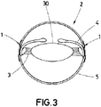
- Figure 3 is a simplified diagram showing two scleral devices (1) attached to an eye (2), according to the present invention, wherein the devices (1) grasp the sclera tissue (5) overlying the ciliary muscle (4) which are placed adjacent to the iris (3) and connected to a lens capsule (30).
- the incisions (7), not shown, are positioned at a depth less than 260 microns and with a pocket length between 0.3mm and 0.5mm.
- the device is (1) is partially inserted in sclera tissue (5) and covered by conjunctiva (6), not shown.
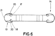
- FIG 4A is a top view of the scleral device (1), in an open situation, according to the present invention, wherein each hook (14) comprises two cut-outs (17) on each side according to the longitudinal axis of the scleral device (1) that defined a narrow sector (18).
- the two cut-outs (17) are curved.
- the device (1) also comprises a wide sector (19), attached to the narrow sector (18), comprising two flaps (20) on each side according to the longitudinal axis of the scleral device (1) and it is defined by a curved outer edge (21).
- the distance between the ends (9) varies from 2.5 mm to 6.0 mm.
- the scleral device (1) is in an open position and the hook are fold out, after being inserted.
- Figure 4B is a section view of the scleral device, according to the present invention, wherein the central portion (8) and the first sector (12) are joined by a first curved junction (15) and the first sector (12) and the second sector (13) are joined by a second curved junction (16).
- the thickness of the curved junctions (15,16) is wider than the rest of the scleral device (1) and the rest of the scleral device (1) has a constant thickness.
- the first sector (12) forms a first angle ( ⁇ ) with the central portion (8) which is obtuse and the second sector (13) forms a second angle ( ⁇ ) with the first sector (12) which in a preferred embodiment is obtuse.
- Figure 5 is a perspective view of a positioning ring for marking the position of the incisions (7), wherein the positioning ring comprises a cone shaped main body (22) with a through cavity (23) intended to partially house the iris (3) of the eye (2), where the main body (22) is defined by a perimeter surface (24) comprising a lower edge (25) intended to contact conjunctiva (6), an upper edge (26) and 4 through slots (27) near the upper edge (26) arranged equidistant from each other with possibility of housing a marking element to mark the position of the tunnelled incisions (7).
- the positioning ring comprises a chamfered area (28) on the upper edge with a hole (29) designed to accommodate a vacuum port from a suction device. A marker fits in the slots (27) and marks the sclera.
- Figure 6 is a top view of the scleral device (1) in a close situation or locked situation, where the hooks are closed and facilitate the insertion of the device (1).
Landscapes
- Health & Medical Sciences (AREA)
- Ophthalmology & Optometry (AREA)
- Heart & Thoracic Surgery (AREA)
- Surgery (AREA)
- Engineering & Computer Science (AREA)
- Biomedical Technology (AREA)
- Nuclear Medicine, Radiotherapy & Molecular Imaging (AREA)
- Vascular Medicine (AREA)
- Life Sciences & Earth Sciences (AREA)
- Animal Behavior & Ethology (AREA)
- General Health & Medical Sciences (AREA)
- Public Health (AREA)
- Veterinary Medicine (AREA)
- Prostheses (AREA)
Priority Applications (1)
| Application Number | Priority Date | Filing Date | Title |
|---|---|---|---|
| EP20201616.8A EP3984504A1 (fr) | 2020-10-13 | 2020-10-13 | Dispositif scléral pour le traitement d'un glaucome à angle ouvert |
Applications Claiming Priority (1)
| Application Number | Priority Date | Filing Date | Title |
|---|---|---|---|
| EP20201616.8A EP3984504A1 (fr) | 2020-10-13 | 2020-10-13 | Dispositif scléral pour le traitement d'un glaucome à angle ouvert |
Publications (1)
| Publication Number | Publication Date |
|---|---|
| EP3984504A1 true EP3984504A1 (fr) | 2022-04-20 |
Family
ID=72852511
Family Applications (1)
| Application Number | Title | Priority Date | Filing Date |
|---|---|---|---|
| EP20201616.8A Withdrawn EP3984504A1 (fr) | 2020-10-13 | 2020-10-13 | Dispositif scléral pour le traitement d'un glaucome à angle ouvert |
Country Status (1)
| Country | Link |
|---|---|
| EP (1) | EP3984504A1 (fr) |
Cited By (1)
| Publication number | Priority date | Publication date | Assignee | Title |
|---|---|---|---|---|
| CN115399942A (zh) * | 2022-08-16 | 2022-11-29 | 北京大学第三医院(北京大学第三临床医学院) | 视网膜镇压器 |
Citations (3)
| Publication number | Priority date | Publication date | Assignee | Title |
|---|---|---|---|---|
| US6217594B1 (en) * | 1999-10-21 | 2001-04-17 | Retinalabs.Com, Inc. | Apparatus, system and method for securing scleral tissue |
| WO2002019950A1 (fr) * | 2000-09-05 | 2002-03-14 | Clear Sight, Inc. | Pince sclerale et procedes d'utilisation de cette derniere |
| WO2007076533A2 (fr) * | 2005-12-29 | 2007-07-05 | Caro Nicholas C | Attache opthalmique et procédé chirurgical associé |
-
2020
- 2020-10-13 EP EP20201616.8A patent/EP3984504A1/fr not_active Withdrawn
Patent Citations (3)
| Publication number | Priority date | Publication date | Assignee | Title |
|---|---|---|---|---|
| US6217594B1 (en) * | 1999-10-21 | 2001-04-17 | Retinalabs.Com, Inc. | Apparatus, system and method for securing scleral tissue |
| WO2002019950A1 (fr) * | 2000-09-05 | 2002-03-14 | Clear Sight, Inc. | Pince sclerale et procedes d'utilisation de cette derniere |
| WO2007076533A2 (fr) * | 2005-12-29 | 2007-07-05 | Caro Nicholas C | Attache opthalmique et procédé chirurgical associé |
Cited By (1)
| Publication number | Priority date | Publication date | Assignee | Title |
|---|---|---|---|---|
| CN115399942A (zh) * | 2022-08-16 | 2022-11-29 | 北京大学第三医院(北京大学第三临床医学院) | 视网膜镇压器 |
Similar Documents
| Publication | Publication Date | Title |
|---|---|---|
| EP0102747B1 (fr) | Dispositif pour réduire la pression occulaire | |
| US4729761A (en) | Tissue-implantable, fluid-dissipating device | |
| JP6074011B2 (ja) | 流体の排出、供給システム | |
| EP0532654B1 (fr) | Implant oculaire pour le traitement des glaucomes | |
| US7357778B2 (en) | Aqueous drainage and flow regulating implant | |
| US4886488A (en) | Glaucoma drainage the lacrimal system and method | |
| EP2286773B1 (fr) | Système de drainage uvéoscléral | |
| US4604087A (en) | Aqueous humor drainage device | |
| US8353856B2 (en) | Glaucoma drainage shunts and methods of use | |
| CA1295907C (fr) | Drainage du glaucome dans le systeme lacrimal | |
| CN109890334B (zh) | 用于对眼球的房水进行引流的引流装置和方法 | |
| US9655778B2 (en) | Position responsive flow adjusting implantable device and method | |
| US12042431B2 (en) | Glaucoma drainage implant venting assembly | |
| US20190046356A1 (en) | Methods Materials Assemblies Apparatuses and Implants for Surgical Reduction of Intraocular Pressure to Suprachoidal Space Ab Externo and Subconjunctival Space | |
| CN112689488A (zh) | 具有弹性膜的用于治疗过量眼内流体的设备 | |
| US20160067093A1 (en) | Devices, systems and methods for posterior segment drainage | |
| IL109499A (en) | Implant device for draining excess intraocular fluid | |
| KR19990014778A (ko) | 눈내부 이식체, 전달장치 및 이식방법 | |
| WO2008030958A4 (fr) | Appareil, procédés et dispositifs pour le traitement de troubles oculaires | |
| EP0515489A1 (fr) | Valve anti-glaucome | |
| US20180078416A1 (en) | Inlet tube protector for glaucoma shunts | |
| EP3984504A1 (fr) | Dispositif scléral pour le traitement d'un glaucome à angle ouvert | |
| US12496221B2 (en) | Shunt and method for treating glaucoma | |
| AU2022407710B2 (en) | An ocular implant for the treatment of glaucoma | |
| ES2834679T3 (es) | Dispositivos y sistemas de extensión de malla trabecular |
Legal Events
| Date | Code | Title | Description |
|---|---|---|---|
| PUAI | Public reference made under article 153(3) epc to a published international application that has entered the european phase |
Free format text: ORIGINAL CODE: 0009012 |
|
| STAA | Information on the status of an ep patent application or granted ep patent |
Free format text: STATUS: THE APPLICATION HAS BEEN PUBLISHED |
|
| AK | Designated contracting states |
Kind code of ref document: A1 Designated state(s): AL AT BE BG CH CY CZ DE DK EE ES FI FR GB GR HR HU IE IS IT LI LT LU LV MC MK MT NL NO PL PT RO RS SE SI SK SM TR |
|
| STAA | Information on the status of an ep patent application or granted ep patent |
Free format text: STATUS: THE APPLICATION IS DEEMED TO BE WITHDRAWN |
|
| 18D | Application deemed to be withdrawn |
Effective date: 20221021 |