EP4007580B1 - Composition comprenant des dérivés de stérols pour son utilisation dans le traitement d'une pathologie neuronale liée à une hypoxie, à une hypoglycémie et/ou une hyperglycémie - Google Patents
Composition comprenant des dérivés de stérols pour son utilisation dans le traitement d'une pathologie neuronale liée à une hypoxie, à une hypoglycémie et/ou une hyperglycémie Download PDFInfo
- Publication number
- EP4007580B1 EP4007580B1 EP20746623.6A EP20746623A EP4007580B1 EP 4007580 B1 EP4007580 B1 EP 4007580B1 EP 20746623 A EP20746623 A EP 20746623A EP 4007580 B1 EP4007580 B1 EP 4007580B1
- Authority
- EP
- European Patent Office
- Prior art keywords
- nhr
- formula
- compound
- composition
- neuronal
- Prior art date
- Legal status (The legal status is an assumption and is not a legal conclusion. Google has not performed a legal analysis and makes no representation as to the accuracy of the status listed.)
- Active
Links
Images




Classifications
-
- C—CHEMISTRY; METALLURGY
- C07—ORGANIC CHEMISTRY
- C07J—STEROIDS
- C07J43/00—Normal steroids having a nitrogen-containing hetero ring spiro-condensed or not condensed with the cyclopenta(a)hydrophenanthrene skeleton
- C07J43/003—Normal steroids having a nitrogen-containing hetero ring spiro-condensed or not condensed with the cyclopenta(a)hydrophenanthrene skeleton not condensed
-
- A—HUMAN NECESSITIES
- A61—MEDICAL OR VETERINARY SCIENCE; HYGIENE
- A61K—PREPARATIONS FOR MEDICAL, DENTAL OR TOILETRY PURPOSES
- A61K31/00—Medicinal preparations containing organic active ingredients
- A61K31/56—Compounds containing cyclopenta[a]hydrophenanthrene ring systems; Derivatives thereof, e.g. steroids
- A61K31/575—Compounds containing cyclopenta[a]hydrophenanthrene ring systems; Derivatives thereof, e.g. steroids substituted in position 17 beta by a chain of three or more carbon atoms, e.g. cholane, cholestane, ergosterol, sitosterol
-
- A—HUMAN NECESSITIES
- A61—MEDICAL OR VETERINARY SCIENCE; HYGIENE
- A61K—PREPARATIONS FOR MEDICAL, DENTAL OR TOILETRY PURPOSES
- A61K31/00—Medicinal preparations containing organic active ingredients
- A61K31/56—Compounds containing cyclopenta[a]hydrophenanthrene ring systems; Derivatives thereof, e.g. steroids
- A61K31/58—Compounds containing cyclopenta[a]hydrophenanthrene ring systems; Derivatives thereof, e.g. steroids containing heterocyclic rings, e.g. danazol, stanozolol, pancuronium or digitogenin
-
- A—HUMAN NECESSITIES
- A61—MEDICAL OR VETERINARY SCIENCE; HYGIENE
- A61P—SPECIFIC THERAPEUTIC ACTIVITY OF CHEMICAL COMPOUNDS OR MEDICINAL PREPARATIONS
- A61P11/00—Drugs for disorders of the respiratory system
-
- A—HUMAN NECESSITIES
- A61—MEDICAL OR VETERINARY SCIENCE; HYGIENE
- A61P—SPECIFIC THERAPEUTIC ACTIVITY OF CHEMICAL COMPOUNDS OR MEDICINAL PREPARATIONS
- A61P25/00—Drugs for disorders of the nervous system
-
- A—HUMAN NECESSITIES
- A61—MEDICAL OR VETERINARY SCIENCE; HYGIENE
- A61P—SPECIFIC THERAPEUTIC ACTIVITY OF CHEMICAL COMPOUNDS OR MEDICINAL PREPARATIONS
- A61P25/00—Drugs for disorders of the nervous system
- A61P25/28—Drugs for disorders of the nervous system for treating neurodegenerative disorders of the central nervous system, e.g. nootropic agents, cognition enhancers, drugs for treating Alzheimer's disease or other forms of dementia
-
- A—HUMAN NECESSITIES
- A61—MEDICAL OR VETERINARY SCIENCE; HYGIENE
- A61P—SPECIFIC THERAPEUTIC ACTIVITY OF CHEMICAL COMPOUNDS OR MEDICINAL PREPARATIONS
- A61P3/00—Drugs for disorders of the metabolism
- A61P3/08—Drugs for disorders of the metabolism for glucose homeostasis
-
- A—HUMAN NECESSITIES
- A61—MEDICAL OR VETERINARY SCIENCE; HYGIENE
- A61P—SPECIFIC THERAPEUTIC ACTIVITY OF CHEMICAL COMPOUNDS OR MEDICINAL PREPARATIONS
- A61P3/00—Drugs for disorders of the metabolism
- A61P3/08—Drugs for disorders of the metabolism for glucose homeostasis
- A61P3/10—Drugs for disorders of the metabolism for glucose homeostasis for hyperglycaemia, e.g. antidiabetics
-
- A—HUMAN NECESSITIES
- A61—MEDICAL OR VETERINARY SCIENCE; HYGIENE
- A61P—SPECIFIC THERAPEUTIC ACTIVITY OF CHEMICAL COMPOUNDS OR MEDICINAL PREPARATIONS
- A61P43/00—Drugs for specific purposes, not provided for in groups A61P1/00-A61P41/00
-
- A—HUMAN NECESSITIES
- A61—MEDICAL OR VETERINARY SCIENCE; HYGIENE
- A61P—SPECIFIC THERAPEUTIC ACTIVITY OF CHEMICAL COMPOUNDS OR MEDICINAL PREPARATIONS
- A61P9/00—Drugs for disorders of the cardiovascular system
- A61P9/10—Drugs for disorders of the cardiovascular system for treating ischaemic or atherosclerotic diseases, e.g. antianginal drugs, coronary vasodilators, drugs for myocardial infarction, retinopathy, cerebrovascula insufficiency, renal arteriosclerosis
-
- C—CHEMISTRY; METALLURGY
- C07—ORGANIC CHEMISTRY
- C07J—STEROIDS
- C07J41/00—Normal steroids containing one or more nitrogen atoms not belonging to a hetero ring
- C07J41/0005—Normal steroids containing one or more nitrogen atoms not belonging to a hetero ring the nitrogen atom being directly linked to the cyclopenta(a)hydro phenanthrene skeleton
Definitions
- the invention relates to the field of neuronal pathologies linked to the deprivation of glucose and/or oxygen. More specifically, the invention relates to a composition comprising compounds derived from sterols of formula (I) or a pharmaceutically acceptable salt of such a compound, for its use in the treatment and/or prevention of cerebral ischemia due to a stroke, head trauma, brain injury, hypoglycaemia linked to diabetes, hyperglycaemia or a respiratory problem due in particular to a bacterial or viral infection.
- Ischemia is a decrease in blood supply to an organ, cerebral ischemia is therefore the decrease in blood supply to at least part of the brain. Ischemia therefore causes the interruption of both the supply of oxygen, the supply of nutrients, such as glucose, and the elimination of toxic molecules resulting from anaerobic metabolism.
- Cerebral ischemia is implicated in particular in cerebral vascular accidents (AVC) commonly called “cerebral attacks”, and cranial traumatisms.
- AVC cerebral vascular accidents
- Strokes are the third leading cause of death behind cardiovascular disease and cancer. But they are the leading causes of severe disability and the second leading causes of dementia in the Western world.
- AVC Alzheimer's disease
- ischemic in nature ie due to the obstruction of a blood vessel by a clot, reducing the blood supply in a cerebral area.
- the clot can form locally in a small artery or come from the heart or from a lesion in the wall of one of the large cervical arteries.
- This obstruction therefore prevents a sufficient supply of oxygen and metabolites, such as glucose, to meet the metabolic and energy demands of the central nervous system, and causes cerebral ischemia.
- the consequences are all the more important since the brain is not able to switch from a metabolism aerobic to anaerobic metabolism to produce the energy it needs. So some brain cells can die.
- an ischemic stroke can occur when inflammation of blood vessels (vasculitis) or infection (herpes simplex virus, for example) narrows the blood vessels that supply the brain. More and more evidence also tends to demonstrate that there is, for example, a negative impact of Covid-19 on the nervous system.
- Covid-19 receptors which are found massively in the lungs, are also found at the level of the blood-brain barrier and nerve endings. Once in the nervous system, it is possible that the virus destroys the neurons responsible for the proper functioning of respiration. The destruction of neurons of the autonomic nervous system present in the medulla oblongata leads to and/or aggravates respiratory distress in patients. On the other hand, it is also possible that a low level of oxygen in the blood due to the acute respiratory symptoms of patients with Covid-19 is itself responsible for the neurological disorders. The neurons being very sensitive to the lack of oxygen, a prolonged and pronounced decrease in the level of blood oxygen would alter the neurons which would eventually die.
- head trauma Another cause of cerebral ischemia is also head trauma which can be mild or severe, with all possible intermediate states. Its severity depends on the existence of intra-cerebral lesions or the existence of an extra-cerebral hematoma, bleeding located between the skull and the brain.
- a head injury can accompanied by bruising, neuronal damage, edema, intracerebral hemorrhage and/or ischemia.
- Cerebral ischemia is the major threat to the functional and anatomical future of the traumatized brain. This is a global, diffuse, or multicentric ischemia, which affects the cerebral cortex as anoxia or cardiac arrest would have done. Ischemia affects all of the gray matter, part of the tissues of the central nervous system with the highest metabolic demand for oxygen and glucose. All brain functions are thus threatened. The cell loss can then be massive and marked by visible cerebral atrophy.
- Ischemia can take another more local form.
- the microcirculation of the tissues located around a focus of contusion or hemorrhage is threatened by vasoconstriction, the effect of tissue compression, capillary micro-thromboses, or even cellular metabolism disorders.
- the cell's energy production is compromised because its oxidative metabolism is broken down. Toxic products like free radicals are released. The slightest respiratory or circulatory event can then precipitate the cells below the viability threshold.
- head trauma can be physical damage such as paraplegia, hemiplegia, visual disturbances... They can also be neuropsychological damage affecting memory, attention, the ability to communicate. Finally, they modify the behavior and personality of the injured and his daily life, as well as that of his relatives.
- hypoglycaemia coupled with hypoxia, the consequences of which are deleterious for the affected patients.
- hypoglycemia is the most common clinical complication in the daily management of insulin-treated diabetics, and continues to be the limiting factor in the glycemic management of diabetes. Severe hypoglycaemia affects 40% of insulin-treated diabetics and can lead to brain damage, particularly to vulnerable neurons in the cortex and hippocampus. For example, learning and memory deficits are a direct consequence of this neuronal damage to the hippocampus caused by severe hypoglycemia.
- hyperglycemia is one of the telltale symptoms of diabetes. Indeed, diabetes systematically leads to hyperglycemia. In addition, hyperglycaemia unrelated to diabetes also exists and the causative facts are varied, for example, the ingestion of foods or drinks containing sugars or the effects side effects of a drug. Hyperglycemia linked to diabetes or not induces deleterious effects on the nervous system.
- composition comprising, in a pharmaceutically acceptable vehicle, at least one compound of formula (I) for preventing hearing loss in a subject or for obtaining at least partial restoration of the hearing of a treated subject having, before treatment, a reduced auditory function.
- compositions comprising at least one compound of formula (I) for its use in the treatment a neuronal pathology of a subject, said neuronal pathology being linked to hypoxia and/or hypoglycemia affecting cells of the central nervous system.
- One idea underlying the invention is to provide preventive and/or curative compositions useful for the treatment of neurological diseases affecting in particular the neurons of the central nervous system involving hypoxia, hypoglycemia and/or hyperglycemia.
- the COOC(CH 3 ) 3 substituent is also called the tert-butoxycarbonyl or Boc functional group.
- the compound of formula (I) belongs to the group of steroids.
- the numbering of the carbon atoms of the compound of formula (I) therefore follows the nomenclature defined by the IUPAC in Pure & Appl. Chem., Vol.61, No.10, pp.1783-1822,1989 .
- the numbering of the carbon atoms of a compound belonging to the group of steroids according to IUPAC is shown below:
- composition may possess one or more of the following characteristics, considered alone or in combination.
- the compound is called DX243.
- the results obtained with this compound are particularly interesting. Indeed, a curative effect of this compound is observed against the pathophysiological phenomenon of hypoxia, hypoglycaemia and/or hyperglycaemia even at very low concentrations.
- the compound is called DX245.
- the results obtained with this compound are particularly interesting. Indeed, we observe a curative effect of this compound against the pathophysiological phenomenon of hypoxia, hypoglycemia and/or hyperglycemia greater than that of DX243.
- the compound is called DX242.
- the results obtained with this compound are particularly interesting. In fact, a curative effect of this compound against the pathophysiological phenomenon of hypoxia, hypoglycaemia and/or hyperglycaemia greater than that of DX243 is observed.
- the compound is called DX244.
- the results obtained with this compound are particularly interesting. In fact, a curative effect of this compound against the pathophysiological phenomenon of hypoxia, hypoglycaemia and/or hyperglycaemia greater than that of DX243 is observed.
- the neuronal pathology of the central nervous system is taken from the group consisting of cerebral trauma and cerebrovascular accidents.
- the neuronal pathology of the central nervous system is brain damage due to ischemia.
- the neuronal pathology of the central nervous system is brain damage due to respiratory failure.
- Respiratory failure means obstructive respiratory failure and restrictive respiratory failure.
- the latter can come, for example, from a pulmonary infection of bacterial or viral origin, for example following an infection by a coronavirus.
- the hypoglycemia is due to diabetes.
- the hyperglycemia is due to diabetes.
- the invention also provides a composition for its use in the treatment of a neuronal pathology of a subject, said neuronal pathology being linked to hypoxia, hypoglycemia and/or hyperglycemia affecting cells central nervous system, said composition being in the form of an aqueous solution and having a concentration of compound of formula (I) of between 1 pmol.L -1 and 1 mmol.L -1 , preferably between 10 pmol.L -1 and 0 .1 mmol.L -1 , more preferably between 0.1 nmol.L -1 and 1 ⁇ mol.L -1 .
- the concentration of compound of formula (I) is between 1 nmol.L -1 and 150 nmol.L -1 .
- stars indicate the statistical power of the results.
- a star indicates that it is 95% certain that the results are not due to chance.
- the presence of 2 stars means that it is 99% certain that the results are not due to chance, and the presence of 3 stars indicates that it is 99.9% certain that the results are not due to chance .
- concentrations or molarities of the compounds are expressed in moles per liter, the symbol of which is mol.L -1 or M.
- Example 1 obtaining cortical neurons
- the first step in the protocol for studying the neuroprotective effect of the compounds of formula (I), in particular those indicated in Table 1, consists in obtaining a primary culture of cortical neurons from cells taken from the brains of wild-type mouse embryos placed in suitable culture conditions (step 1). More specifically, the collected cells are grown in Neurobasal TM Medium (Ref. 21103049 ThermoFisher Scientific) to which L-Glutamine and B27 supplement 50X (Ref. 17504044 ThermoFisher Scientific) have been added (step 2). The neurons from the primary culture are then isolated and purified.
- Neurobasal TM Medium Ref. 21103049 ThermoFisher Scientific
- L-Glutamine and B27 supplement 50X Ref. 17504044 ThermoFisher Scientific
- step 3 it is the culture conditions in themselves which make it possible to obtain a purified culture in neurons from the dissociation of the embryonic cortices.
- a deprivation of oxygen and glucose (DOG) is then carried out (step 3) on these neurons in order to mimic as closely as possible what happens in vivo during a cerebrovascular accident, i.e. a decreased oxygen and glucose delivery due to decreased blood perfusion to the cells.
- DOG oxygen and glucose
- the neurons are placed for 4 hours in an incubator whose atmosphere has an oxygen content of approximately 1% and the culture medium is replaced by a medium without glucose (step 3).
- the neurons are then placed in a medium containing glucose under normoxic conditions for 24 h (reperfusion) (step 4) following the ischemia step.
- Group A of neurons is a group of neurons having been treated with a solution of compound of formula (I) during DOG and reperfusion, i.e. a molecule from Table 1 is added to the medium without glucose as soon as glucose and oxygen deprivation begins (step 3).
- the concentration of the compound of formula (I) named DX243 is between 1 nmol.L -1 and 1 pmol.L -1 in the medium without glucose.
- the concentration of the other molecules in Table 1 is 100 nmol.L -1 in the medium without glucose.
- Group B of neurons is a group of neurons that have been treated with DX243 solution during reperfusion only (step 4), to approximate what happens clinically, i.e. treatment possible only after several hours with conventional treatment .
- the DX243 solution is added to the medium without glucose as soon as the reperfusion begins (step 4).
- the concentration of DX243 is between 1 nmol.L -1 and 1 pmol.L -1 in the medium without glucose.
- the negative control consists of a group of neurons undergoing DOG (step 3) followed by reperfusion (step 4) with no DX243 present. This group of neurons is called “Ctrl” in the figures 2 to 4 .
- the normal control consists of a group of neurons from the primary culture placed in normoxic conditions for 28 hours. The normal control is referred to as “normox” in the figures 2 to 4 .
- the positive control consists of a group of neurons from the primary culture having undergone DOG for 4 hours (step 3) in the presence of roscovitine, a cell cycle inhibitor, resulting in neuronal protection, followed by reperfusion for 24 hours (step 4) .
- the positive control is referred to as “ROSCO” in the figures 2 to 4 .
- the survival of the neurons is evaluated using three different tests: an immunocytochemical test 5, an MTT test (bromide of 3-(4,5- dimethylthiazol-2-yl)-2,5-diphenyl tetrazolium) 6 and a test based on the integrity of cell membranes via the use of triptan blue 7. The results are then compared to the results of the three survival tests of the groups control.
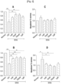
- FIG. 1 presents the results of the first neuronal survival test in the case of the use of the DX243.
- Graphs A and C are a representation of the results obtained for group A, the ordinate represents the proportion of living cells relative to the Normox group.
- Graphs B and D are a representation of the results obtained for group B, the ordinate represents the proportion of cells in apoptosis relative to the Normox group.
- This first test is an immunocytochemical test which makes it possible to highlight living cells and dead cells.
- a first series of images is produced with chemical labeling by fluorescence. This labeling is carried out with DAPI (4′,6-diamidino-2-phenylindole) which is capable of strongly binding to the adenine (A) and thymine (T) bases of DNA. It detects living cells.
- a second series of images is produced with fluorescence labeling using the CC3 antibody.
- the CC3 antibody makes it possible to detect activated caspase 3, and therefore cells in a state of apoptosis.
- TUJ1 To visualize the proportion of healthy neurons among the different culture conditions, the TUJ1 antibody is used. TUJ1 reacts with beta-tubulin III, a structural protein that makes up tubulin and is specific to neurons. Beta-tubulin III is widely used as a marker to distinguish neurons from other cell types.
- the proportion of living cells was calculated by determining the ratio of the number of living cells to the total number of cells.
- the proportion of cells in apoptosis was carried out using the ratio between the number of cells detected by the CC3 antibody over the total number of cells. The values obtained were related to those obtained by the normox group.
- DOG induces a decrease in the proportion of living cells on the Ctrl group on graphs A and B of the figure 2 , and an increase in the proportion of cells in apoptosis on the Ctrl group on graphs C and D of the picture 2 .
- both treatment with DX243 during DOG and reperfusion and treatment with DX243 only during reperfusion partially protect against neuronal death.
- an increase in the proportion of living cells and a decrease in the proportion of cells in apoptosis following the treatment with DX243 are observed in comparison with the Ctrl group.
- a DX243 concentration between 1 nmol.L -1 and 100 nmol.L -1 corresponds to an ideal concentration range to maximize the neuroprotective effects.
- Example 3 MTT neuronal survival evaluation test in the case of the use of the DX243
- a test based on the metabolic activity of neurons was carried out to assess neuronal survival in another way.
- This test is based on the use of the tetrazolium salt MTT (3-(4,5-dimethylthiazol-2-yl)-2,5-diphenyl tetrazolium bromide).
- Tetrazolium is reduced by active living cell mitochondrial succinate dehydrogenase to formazan, a violet-colored precipitate.
- the quantity of precipitate formed is proportional to the quantity of living cells but also to the metabolic activity of each cell.
- a simple assay of the optical density at 550 nm by spectroscopy makes it possible to know the relative quantity of living and metabolically active cells.
- DOG induces a significant decrease in the number of cells having preserved the integrity of their membrane, whether on graph A of the figure 4 or on graph B of the figure 4 .
- Treatment of neurons with DX243 restores the percentage of living cells to levels close to that observed in the normox group, mainly with concentrations between 1 nmol.L -1 and 100 nmol.L -1 .
- DX243 is therefore effective at concentrations of between 1 nmol.L -1 and 1 ⁇ mol.L -1 .
- concentrations between 10 nmol.L -1 and 100 nmol.L -1 , the results are more stable and the protective effect of DX243 is statistically greater. This can be explained by the fragility of primary cultures outside their natural environment.
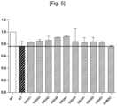
- the third cell survival test was carried out. The results obtained were reported on the graph of the figure 5 including the results obtained with the DX243.
- the abbreviation NT stands for untreated.
- the non-hatched bar NT corresponds to a group of neurons from the primary culture placed in normoxic conditions for 28 hours.
- the hatched bars represent groups of neurons from the primary culture that have undergone a DOG and reperfusion either without molecules from table 1 (hatched bar named NT) or in the presence of a molecule from table 1.
- the calculation of the percentage of living cells was carried out by counting the proportion of blue and non-blue cells, and relative to the percentage of living cells observed in the NT group under normoxia, that is to say the NT group not having undergone DOG (not shown).
- the survival rate of the groups of neurons is higher than that of the groups of NT neurons having undergone DOG and reperfusion (hatched bar NT), they therefore exhibit a neuroprotective effect.
- the DX245, DX244 and DX242 have a higher survival rate than the DX243.
- the survival rate of the group treated with DX243BOC is lower than that of the group treated with DX243 and that of the NT group having undergone DOG and reperfusion, thus demonstrating the importance of the R 3 group in the activity of the compound. of formula (I).
- MK801 is a non-competitive N-methyl-D-aspartate (NMDA) receptor antagonist and is known to have a neuroprotective effect. All compounds in Table 1 were tested. It is observed figure 6 significant neuronal protection with 100nM treatment of DX243 and DX245. A neuroprotective effect is observed with this test for DX101, DX242, DX244, DX301, DX302, DX401, DX249.
- NMDA N-methyl-D-aspartate
- the survival rate of the groups of neurons is higher than that of the groups of NT neurons, so they exhibit a neuroprotective effect.
- FIG. 7 A new cell survival test of the various compounds in Table 1 was carried out, the results are presented figure 7 .
- This test is similar to the one performed figure 4 . It is based on the integrity of cell membranes, which is broken in dead cells. This test uses trypan blue which will stain dead cells blue. It is carried out in a similar way to the test of the Figure 4A , that is to say based on the protocol for obtaining the cortical neurons of example 1 stated above.
- the Roscovitine positive control has been replaced by the Dizocilpine positive control (MK801).
- MK801 Dizocilpine positive control
- the purpose of this test is to evaluate the neuroprotective effect of the molecules after having undergone a treatment lasting 4 hours DOG then 24 hours of reperfusion, ie a total of 28 hours of experiments. All compounds in Table 1 were tested multiple times. Additionally, DX243 and DX245 have been tested in different quantities shown on the figure 7 .
- This in vivo model classically used to study the effect of molecules on the AVC consists of the installation of a catheter at the level of the anterior cingulate cortex (ACC), to insert a filament there at the level of the common carotid artery ( CCA-common carotid artery) and to push the filament through the common carotid artery, passing through the internal carotid artery (ICA) to the level of the middle cerebral artery (MCA) resulting in a significant decrease (>80% ) of the blood flow at the level of the cerebral territory 8 irrigated by this artery ( Estelle ROUSSELET et al, Murine model of intraluminal MCAO: Evaluation of cerebral infarction by cresyl violet staining, Journal of Visualized Experiments and Carl Zeiss ). Following a period of 1 hour, the filament is removed to allow reperfusion and the addition of treatment with DX243. The injected DX243 is diluted in an aqueous solution and is concentrated at 50mg/Kg.
- FIG. 8 (A) shows two TTC staining images showing the surface of dead tissue in white and representing the reduction in the infarcted surface in the mice treated with DX243.
- Chart B of the figure 9 represents the reduction in the percentage of infarcted area between treated or untreated mice (Ctrl), along the ordinate is indicated the percentage of the infarcted area with regard to the total size of the brain of the mouse.
- Graph C illustrates behavioral improvement (neuroscore) in treated mice.
- Graph D represents the decrease in the percentage of dead mice in mice treated with DX243.
- Graphs E and F represent the disappearance of the correlation between decrease in blood flow and histological and behavioral consequences following treatment with DX 243.
- hypoglycaemia is capable of inducing oxidative stress in neuronal cells, oxidative stress present in the context of oxygen and glucose deprivation and which is a major component of the induction of neuronal death.
- neuroprotective effect of the compounds of formulas (I) according to the invention extends to cases of hypoxia alone or hypoglycemia alone.
Landscapes
- Health & Medical Sciences (AREA)
- Chemical & Material Sciences (AREA)
- General Health & Medical Sciences (AREA)
- Medicinal Chemistry (AREA)
- Pharmacology & Pharmacy (AREA)
- Life Sciences & Earth Sciences (AREA)
- Animal Behavior & Ethology (AREA)
- Public Health (AREA)
- Veterinary Medicine (AREA)
- Organic Chemistry (AREA)
- Engineering & Computer Science (AREA)
- Bioinformatics & Cheminformatics (AREA)
- Chemical Kinetics & Catalysis (AREA)
- General Chemical & Material Sciences (AREA)
- Nuclear Medicine, Radiotherapy & Molecular Imaging (AREA)
- Diabetes (AREA)
- Biomedical Technology (AREA)
- Epidemiology (AREA)
- Neurology (AREA)
- Neurosurgery (AREA)
- Hematology (AREA)
- Emergency Medicine (AREA)
- Endocrinology (AREA)
- Obesity (AREA)
- Psychiatry (AREA)
- Hospice & Palliative Care (AREA)
- Pulmonology (AREA)
- Urology & Nephrology (AREA)
- Vascular Medicine (AREA)
- Cardiology (AREA)
- Heart & Thoracic Surgery (AREA)
- Pharmaceuticals Containing Other Organic And Inorganic Compounds (AREA)
- Steroid Compounds (AREA)
Description
- L'invention se rapporte au domaine des pathologies neuronales liées à la déprivation de glucose et/ou d'oxygène. Plus précisément, l'invention concerne une composition comprenant des composés dérivés de stérols de formule (I) ou un sel pharmaceutiquement acceptable d'un tel composé, pour son utilisation dans le traitement et/ou la prévention d'une ischémie cérébrale due à un accident vasculaire cérébral, un traumatisme crânien, une lésion cérébrale, une hypoglycémie liée à un diabète, une hyperglycémie ou un problème respiratoire dû notamment à une infection bactérienne ou virale.
- Une ischémie est une diminution de l'apport sanguin à un organe, l'ischémie cérébrale est donc la diminution de l'apport sanguin à au moins une partie du cerveau. L'ischémie provoque par conséquent l'interruption à la fois de l'apport d'oxygène, de l'apport en nutriments, comme le glucose, et de l'élimination des molécules toxiques résultant du métabolisme anaérobique.
- L'ischémie cérébrale est impliquée notamment dans les accidents vasculaires cérébraux (AVC) communément appelés "attaques cérébrales", et les traumatismes crâniens.
- Les AVC représentent la troisième cause de mortalité derrière les maladies cardiovasculaires et le cancer. Mais ce sont les premières causes d'invalidité lourde et les deuxièmes causes de démence dans le monde occidental. A l'origine des AVC, on trouve une perturbation de l'irrigation sanguine du cerveau. Près de 85 % des AVC sont de nature ischémique c'est à dire dus à l'obstruction d'un vaisseau sanguin par un caillot, réduisant l'irrigation sanguine dans une zone cérébrale. Le caillot peut se former localement dans une petite artère ou venir du coeur ou d'une lésion de la paroi d'une des grosses artères cervicales. Cette obstruction empêche donc un apport suffisant en oxygène et en métabolites, comme le glucose, pour répondre à la demande métabolique et énergétique du système nerveux central, et provoque une ischémie cérébrale. Les conséquences sont d'autant plus importantes que le cerveau n'est pas en mesure de passer d'un métabolisme aérobie à un métabolisme anaérobie pour produire l'énergie dont il a besoin. Ainsi certaines cellules du cerveau peuvent mourir.
- Les conséquences des AVC dépendent de nombreux facteurs : la vitesse de rétablissement de la circulation sanguine, la durée de la privation en oxygène et/ou en métabolites, comme le glucose, et/ou la localisation cérébrale de l'accident. Les manifestations cliniques se traduisent, selon le territoire atteint, par une paralysie plus ou moins étendue, une perte de la parole ou du langage, ou encore un coma, avec des risques séquellaires invalidants.
- En outre, une insuffisance respiratoire due notamment à une infection bactérienne ou virale peut également entrainer une ischémie cérébrale. Par exemple, un AVC ischémique peut se déclencher lorsqu'une inflammation de vaisseaux sanguins (vascularite) ou une infection (à virus herpès simplex, par exemple) rétrécit les vaisseaux sanguins qui approvisionnent le cerveau. De plus en plus d'éléments tendent à démontrer également qu'il existe par exemple un impact négatif du Covid-19 sur le système nerveux. Différentes études ont en effet montré qu'il existait une corrélation entre infection au Covid-19 et perte de gout et d'odorat, confusion, maux de tête, étourdissements voire même AVC, encéphalopathie ou encore myélite (Ling Mao et al., Neurogical Manifestations of Hospitalized Patients with COVID-19 in Wuhan, china: a rétrospective case series study et Neo Poyiadji et al COVID-19-associated Acute Hemorrhagic Necrotizing Encephalopathy: CT and MRI, <doi : https://doi.org/10.1101/2020.02.22.20026500>). Le virus serait en effet capable d'entrer dans le système nerveux via les fosses nasales et le bulbe olfactif. Des récepteurs au Covid-19, que l'on retrouve massivement dans les poumons, se retrouvent d'ailleurs également au niveau de la barrière hémato-encéphalique et des terminaisons nerveuses. Une fois dans le système nerveux, il est possible que le virus détruise les neurones responsables du bon fonctionnement de la respiration. La destruction des neurones du système nerveux autonome présents dans le bulbe rachidien, entraine et/ou aggrave les détresses respiratoires des patients. A contrario, il est par ailleurs possible qu'un niveau d'oxygène bas dans le sang dû aux symptômes respiratoires aigus des patients atteints du Covid-19 soit lui-même responsable des troubles neurologiques. Les neurones étant très sensibles au manque d'oxygène, une diminution prolongée et prononcée du taux d'oxygène sanguin altérerait les neurones qui finiraient par mourir.
- Une autre cause d'ischémie cérébrale est également le traumatisme crânien qui peut être bénin ou grave, avec tous les états intermédiaires possibles. Sa gravité dépend de l'existence de lésions intra-cérébrales ou de l'existence d'un hématome extra-cérébral, saignement situé entre le crâne et le cerveau. Un traumatisme crânien peut s'accompagner de contusions, de lésions des neurones, d'un oedème, d'hémorragies intra-cérébrales et/ou d'une ischémie.
- L'ischémie cérébrale est la menace majeure qui pèse sur le devenir fonctionnel et anatomique du cerveau traumatisé. Il s'agit là d'une ischémie globale, diffuse, ou multicentrique, qui touche le cortex cérébral comme l'aurait fait une anoxie ou un arrêt cardiaque. L'ischémie touche l'ensemble de la substance grise, partie des tissus du système nerveux central ayant la demande métabolique la plus forte en oxygène et en glucose. Toutes les fonctions cérébrales sont ainsi menacées. La perte cellulaire peut alors être massive et marquée par une atrophie cérébrale visible.
- L'ischémie peut prendre une autre forme plus locale. La microcirculation des tissus situés autour d'un foyer de contusion ou d'hémorragie est menacée par une vasoconstriction, l'effet d'une compression tissulaire, des micro-thromboses capillaires, ou encore des désordres du métabolisme cellulaire. La production d'énergie de la cellule est compromise car son métabolisme oxydatif est en panne. Des produits toxiques comme les radicaux libres sont libérés. Le moindre événement respiratoire ou circulatoire peut alors précipiter les cellules en dessous du seuil de viabilité.
- Les conséquences des traumatismes crâniens peuvent être des atteintes physiques de type paraplégie, hémiplégie, troubles de la vue... Elles peuvent être aussi des atteintes neuropsychologiques affectant la mémoire, l'attention, la capacité à communiquer. Enfin elles modifient le comportement et la personnalité du blessé et son quotidien, ainsi que celui de ses proches.
- Certains cas de diabète présentent une hypoglycémie couplée à une hypoxie dont les conséquences sont délétères pour les patients touchés.
- L'hypoglycémie est la complication clinique la plus répandue dans la gestion quotidienne des diabétiques traités à l'insuline, et continue d'être le facteur limitant dans la gestion glycémique du diabète. L'hypoglycémie sévère touche 40% des diabétiques traités à l'insuline et peut entrainer des lésions cérébrales, en particulier au niveau des neurones vulnérables du cortex et de l'hippocampe. Par exemple, les déficits d'apprentissage et de mémoire sont une conséquence directe de cette lésion neuronale de l'hippocampe provoquée par une hypoglycémie sévère.
- Il est connu que l'hyperglycémie est un des symptômes révélateurs du diabète. En effet, le diabète entraine systématiquement une hyperglycémie. En outre, une hyperglycémie non liée au diabète existe également et les fait en causes sont variés, par exemple, l'ingestion d'aliments ou de boissons contenant des sucres ou les effets secondaires d'un médicament. L'hyperglycémie liée à un diabète ou non induit des effets délétères au niveau du système nerveux.
- De plus, l'association d'un diabète et d'une hyperglycémie aggrave les dommages neuronaux consécutifs à d'autres formes d'atteinte du système nerveux central, comme les accidents vasculaires cérébraux. Plusieurs études démontrant la relation entre hyperglycémie, diabète et dommages neuronaux ont été publiées, notamment Yazi Li et al. (« Autophagy impairment mediated by S-nitrosation of ATG48 leads to neurotoxicity in response to hyperglycemia », <doi :https://doi.org/10.1080/15548627.2017.1320467>), Ruchi Sharma et al. (« Hyperglycemia Induces Oxidative Stress and Impairs Axonal Transport Rates in Mice », Published October 18, 2010 ) et Wenjuan Zhou et al. (« TIGAR Attenuates High Glucose-Induced Neuronal Apoptosis via an Autophagy Pathway », <doi : https://doi.org/10.3389/fnmol.2019.00193>).
- Actuellement, les techniques développées pour traiter un AVC sont d'intervenir très rapidement, dans les 4 heures à 5 heures suivant l'AVC et d'éliminer le caillot chimiquement via un activateur tissulaire du plasminogène recombinant, rtPA ou mécaniquement, par thrombectomie pour permettre la reperfusion. Cependant, ces deux façons d'intervenir face à un AVC ne présentent pas une efficacité optimale et sont soumis à des critères d'inclusion des patients assez restrictifs.
- On connaît du document
WO2016016518A2 une composition comprenant dans un véhicule pharmaceutiquement acceptable au moins un composé de formule (I) pour prévenir une perte d'audition chez un sujet ou pour obtenir une restauration au moins partielle de l'audition d'un sujet traité ayant, avant traitement, une fonction auditive réduite. - On connaît également du document de Zhao H-B et al. (« Ganoderma total sterol (GS) and GS1 protect rat cérébral cortical neurons from hypoxia/reoxygenation injury », <doi :https://doi.org/10.1016/j.Ifs.2004.08.013>), l'effet du Ganoderma total stérol (GS) et de ses principaux composants (GS1) sur les cultures de neurones corticaux de rats exposés à l'hypoxie / réoxygénation.
- On connaît encore du document Brigitte Malgrange et al. (« Targeting Cholestérol Homeostasis to Fight Hearing Loss: A New Perspective », <doi: 10.3389/fnagi.2015.00003>), un moyen de lutte contre la perte auditive en ciblant l'homéostasie du cholestérol. En outre, il est indiqué que le rôle du cholestérol et de ses métabolites n'est pas clair.
- Les trois documents précités ne divulguent pas ni ne suggèrent une composition comprenant au moins un composé de formule (I) pour son utilisation dans le traitement d'une pathologie neuronale d'un sujet, ladite pathologie neuronale étant liée à une hypoxie et/ou à une hypoglycémie affectant des cellules du système nerveux central.
- Il existe donc un besoin de développer des composés permettant de traiter des patients ayant subis une déprivation en oxygène et/ou en glucose, provoqué par exemple par une ischémie cérébrale, une hypoglycémie ou une hypoxie, afin d'améliorer leur récupération.
- Une idée à la base de l'invention est de fournir des compositions préventives et/ou curatives utiles pour le traitement de maladies neurologiques touchant notamment les neurones du système nerveux central impliquant une hypoxie, une hypoglycémie et/ou une hyperglycémie.
- Pour cela, l'invention fournit une composition comprenant au moins un composé de formule (I) :

- R1 = OH, F, OCnH2n+1, R-COO, R-OCOO, RHN-COO ou OPO(OR)2 avec R = H ou CnH2n+1 avec n ≤ 16 ;
- R2 = H ou OH ;
- R3 = -NR5R6,
- R5 étant H ou -(CH2)3NH2, et
- R6 étant pris dans le groupe formé par -(CH2)3NH(CH2)4NHR7 ; -(CH2)4NH(CH2)3NHR7 ; -(CH2)3NH(CH2)4NH(CH2)3NHR7 ; -(CH2)3NHR7 ; -(CH2)4NHR7 avec R7 = H ou COCH3 ; -(CH2)2-imidazol-4-yl ;-(CH2)2-indol-3-yl ; et
- R4 = H ou OH en position 20, 22, 24, 25, 26 ou 27, positionné de façon à créer un centre asymétrique de configuration R ou S ;
- Z1 et Z2 représentent chacun le nombre de double liaisons entre les atomes de carbone C7 et C8 et C22 et C23 respectivement (soit 0 soit 1); T1, T2 et T3 = H ou CH3 indépendamment les uns des autres ; T4 = H, CH3, C2H5 positionné de façon à obtenir un centre asymétrique de configuration R ou S en position 24 ;
- et/ou au moins un sel pharmaceutiquement acceptable d'au moins un composé de formule (I),
- pour son utilisation dans le traitement d'une pathologie neuronale d'un sujet, ladite pathologie neuronale étant liée à une hypoxie, à une hypoglycémie et/ou à une hyperglycémie affectant des cellules du système nerveux central.
- Le composé de formule (I) et défini par : Z1 = Z2 = 0 ; R1 = R2 = OH ; R4 = H ; R5 = H ; R6 = -(CH2)3-NCOOC(CH3)3-(CH2)4-NHCOOC(CH3)3 ; T1 = T2 = T3 = T4 = H est appelé DX243BOC, illustré tableau 1.
- Le substituant COOC(CH3)3 est aussi appelé groupe fonctionnel tert-butoxycarbonyle ou Boc.
- Le composé de formule (I) appartient au groupe des stéroïdes. La numérotation des atomes de carbone du composé de formule (I) suit donc la nomenclature définie par l'IUPAC dans Pure & Appl. Chem., Vol.61, No.10, pp.1783-1822,1989. La numérotation des atomes de carbone d'un composé appartenant au groupe des stéroïdes selon l'IUPAC est illustrée ci-dessous :

- Les méthodes de préparation du composé de formule (I) ont déjà été décrites auparavant, et notamment dans DE MEDINA, P. et al. Synthesis of New Alkylaminooxysterols with Potent Cell Differentiating Activities: Identification of Leads for the Treatment of Cancer and Neurodegenerative Diseases. Journal of Médicinal Chemistry, 52(23), 2009, pp. 7765-7777.
- En outre, la composition peut posséder une ou plusieurs caractéristiques suivantes, considérées isolément ou en combinaison.
- Selon un mode de réalisation, le composé de formule (I) est défini par Z2 = 0 ; R1 = R2 = OH ; R4 = H ; R5 = H ; et T1 = T2 = T3 = T4 = H.
- Selon un mode de réalisation, le composé de formule (I) est défini par Z1 =0 et R5 = H.
- Selon un mode de réalisation, le composé de formule (I) est défini par R6 = - (CH2)4NH(CH2)3NHR7 avec R7 = COCH3.
- Selon un mode de réalisation, le composé de formule (I) est défini par R6 = -(CH2)2-imidazol-4-yl.
- Selon un mode de réalisation, le composé de formule (I) est défini par R6 = - (CH2)3NH(CH2)4NHR7, -(CH2)4NH(CH2)3NHR7, -(CH2)3NH(CH2)4NH(CH2)3NHR7; ou - (CH2)4NHR7 ; et R7 = H.
- Selon un mode de réalisation, le composé de formule (I) est défini par Z1 = 1 et R5 = H.
- Selon un mode de réalisation, le composé de formule (I) est défini par :
- R1 = F, OCnH2n+1, R-COO, R-OCOO, RHN-COO ou OPO(OR)2 avec R = H ou CnH2n+1, avec n ≤ 16 ;
- R2= OH ;
- R5 = H ;
- R6 = -(CH2)4NH(CH2)3NHR7, -(CH2)3NH(CH2)4NHR7, -(CH2)3NH(CH2)4NH(CH2)3NHR7 ; - (CH2)4NHR7 ;
- Z1 = 0 ou Z1 = 1
- Z2 = 0.
- Selon un mode de réalisation, le composé de formule (I) est défini par R6 = - (CH2)3NH(CH2)4NHR7 ; -(CH2)4NH(CH2)3NHR7 ; ou -(CH2)3NH(CH2)4NH(CH2)3NHR7 ; et R7 = H.
- Selon un mode de réalisation, le au moins un composé de formule (I), est défini par Z1 = Z2 = 0 ; R1 = R2 = OH ; R4 = H ; R5 = H ; R6 = (CH2)3NH(CH2)4NH2 ; T1 = T2 = T3 = T4 = H. Dans ce mode de réalisation le composé est appelé DX243. Les résultats obtenus avec ce composé sont particulièrement intéressants. En effet, on observe un effet curatif de ce composé contre le phénomène pathophysiologique d'hypoxie, d'hypoglycémie et/ou d' hyperglycémie même à de très faibles concentrations.
- Selon un mode de réalisation, le au moins un composé de formule (I), est défini par Z1 = Z2 = 0 ; R1 = R4 = H ; R2 = OH ; R5 = H ; R6 = (CH2)4NH(CH2)3NH2 ; T1 = T2 = T3 = T4 = H. Dans ce mode de réalisation le composé est appelé DX245. Les résultats obtenus avec ce composé sont particulièrement intéressants. En effet, on observe un effet curatif de ce composé contre le phénomène pathophysiologique d'hypoxie, d'hypoglycémie et/ou d' hyperglycémie supérieur à celui du DX243.
- Selon un mode de réalisation, le au moins un composé de formule (I), est défini par Z1 = 1 ; Z2 = 0 ; R1 = R4 = H ; R2 = OH ; R5 = H ; R6 = (CH2)3NH(CH2)4NH2 ; T1 = T2 = T3 = T4 = H. Dans ce mode de réalisation le composé est appelé DX242. Les résultats obtenus avec ce composé sont particulièrement intéressants. En effet, on observe un effet curatif de ce composé contre le phénomène pathophysiologique d'hypoxie, d'hypoglycémie et/ou d' hyperglycémie supérieur à celui du DX243.
- Selon un mode de réalisation, le au moins un composé de formule (I), est défini par Z1 = 1 ; Z2 = 0 ; R1 = R4 = H ; R2 = OH ; R5 = H ; R6 = (CH2)4NH(CH2)3NH2 ; T1 = T2 = T3 = T4 = H. Dans ce mode de réalisation le composé est appelé DX244. Les résultats obtenus avec ce composé sont particulièrement intéressants. En effet, on observe un effet curatif de ce composé contre le phénomène pathophysiologique d'hypoxie, d'hypoglycémie et/ou d' hyperglycémie supérieur à celui du DX243.
- Selon un mode de réalisation, la pathologie neuronale du système nerveux central est prise dans le groupe constitué des traumatismes cérébraux et des accidents vasculaires cérébraux.
- Selon un mode de réalisation, la pathologie neuronale du système nerveux central est une lésion cérébrale due à une ischémie.
- Selon un mode de réalisation, la pathologie neuronale du système nerveux central est une lésion cérébrale due à une insuffisance respiratoire. On entend par insuffisance respiratoire l'insuffisance respiratoire obstructive et l'insuffisance respiratoire restrictive. Cette dernière pouvant provenir par exemple d'une infection pulmonaire d'origine bactérienne ou virale, par exemple suite à une infection par un coronavirus.
- Selon un mode de réalisation, l'hypoglycémie est due à un diabète.
- Selon un mode de réalisation, l'hyperglycémie est due à un diabète.
- Selon un mode de réalisation, l'invention fournit aussi une composition pour son utilisation dans le traitement d'une pathologie neuronale d'un sujet, ladite pathologie neuronale étant liée à une hypoxie, à une hypoglycémie et/ou à une hyperglycémie affectant des cellules du système nerveux central, ladite composition étant sous forme de solution aqueuse et présentant une concentration de composé de formule (I) comprise entre 1 pmol.L-1 et 1 mmol.L-1, préférentiellement entre 10 pmol.L-1 et 0,1 mmol.L-1, plus préférentiellement entre 0,1 nmol.L-1 et 1 µmol.L-1. Par exemple, la concentration de composé de formule (I) est comprise entre 1 nmol.L-1 et 150 nmol.L-1.
- L'invention sera mieux comprise, et d'autres buts, détails, caractéristiques et avantages de celle-ci apparaîtront plus clairement au cours de la description suivante de plusieurs modes de réalisation particuliers de l'invention, donnés uniquement à titre illustratif et non limitatif, en référence aux dessins annexés.
- [
fig. 1 ] Lafigure 1 représente schématiquement un protocole d'étude de l'efficacité de composés de formule (I), en particulier du DX243, sur un modèle in vitro d'ischémie. - [
fig. 2 ] Lafigure 2 illustre les résultats d'un test immunocytochimique afin d'évaluer la survie neuronale dans un modèle in vitro d'ischémie/reperfusion en présence de DX243. - [
fig. 3 ] Lafigure 3 illustre les résultats d'un test d'évaluation de survie neuronale MTT dans un modèle in vitro d'ischémie/reperfusion en présence de DX243. - [
fig. 4 ] Lafigure 4 illustre les résultats d'un test d'évaluation de survie neuronale via le bleu de trypan dans un modèle in vitro d'ischémie/reperfusion en présence de DX243. - [
fig. 5 ] Lafigure 5 illustre les résultats d'un test d'évaluation de survie neuronale via le bleu de trypan dans un modèle in vitro d'ischémie/reperfusion en présence de composés formule (I) du tableau 1, autre que le DX243. - [
fig. 6 ] Lafigure 6 illustre les résultats d'un test d'évaluation de survie neuronale MTT dans un modèle in vitro d'ischémie/reperfusion en présence des composés du tableau 1. - [
fig. 7 ] Lafigure 7 illustre les résultats d'un second test d'évaluation de survie neuronale via le bleu de trypan dans un modèle in vitro d'ischémie/reperfusion en présence des composés de formule (I) du tableau 1. - [
fig. 8 ] Lafigure 8 illustre les résultats du test in vivo d'AVC chez la souris afin d'évaluer la survie neuronale après avoir subi un AVC suivi d'une reperfusion avec traitement au DX243. - [
fig. 9 ] Lafigure 9 est un ensemble de graphique illustrant les résultats du test in vivo d'AVC chez la souris afin d'évaluer la survie neuronale après avoir subi un AVC suivi d'une reperfusion avec traitement au DX243. - Sur les
figures 2 à 4 ,6 et7 , les étoiles indiquent la puissance statistique des résultats. Une étoile indique qu'il est certain à 95% que les résultats ne sont pas dus au hasard. La présence de 2 étoiles signifie qu'il est certains à 99% que les résultats ne sont pas dus au hasard, et la présence de 3 étoiles indiquent qu'il est certain à 99,9% que les résultats ne sont pas dus au hasard. - On va décrire ci-dessous plusieurs protocoles expérimentaux qui mettent en évidence l'effet protecteur des composés de formule (I) indiqués dans le tableau 1, dont le composé DX243, contre l'hypoxie et l'hypoglycémie.
- Les concentrations ou molarités des composés sont exprimées en mole par litre dont le symbole est mol.L-1 ou M.
- En référence à la
figure 1 , la première étape du protocole d'étude de l'effet neuroprotecteur des composés de formule (I), en particulier ceux indiquées dans le tableau 1, consiste à obtenir une culture primaire de neurones corticaux à partir de cellules prélevées sur des cerveaux d'embryons de souris de type sauvage mises dans des conditions de culture adéquates (étape 1). Plus précisément, les cellules prélevées sont crues dans un milieu Neurobasal™ Medium (Ref. 21103049 ThermoFisher Scientific) auquel de la L-Glutamine et du B27 supplement 50X (Ref. 17504044 ThermoFisher Scientific) ont été ajoutés (étape 2). Les neurones issus de la culture primaire sont ensuite isolés et purifiés. Il est à noter que ce sont les conditions de culture en elles-mêmes qui permettent d'obtenir une culture purifiée en neurones à partir de la dissociation des cortex embryonnaires. Une déprivation en oxygène et en glucose (DOG) est alors réalisée (étape 3) sur ces neurones afin de mimer le plus fidèlement possible ce qu'il se passe in vivo lors d'un accident vasculaire cérébral c'est-à-dire une diminution de l'apport en oxygène et en glucose dû à une diminution de la perfusion sanguine pour les cellules. Pour réaliser cette déprivation en oxygène et en glucose mimant l'ischémie, les neurones sont placés pendant 4h dans un incubateur dont l'atmosphère a une teneur en oxygène d'environ 1% et en remplaçant le milieu de culture par un milieu sans glucose (étape 3). Comme le modèle d'ischémie est un modèle dit d'ischémie/reperfusion, les neurones sont ensuite placés dans un milieu contenant du glucose dans des conditions normoxiques pendant 24h (reperfusion) (étape 4) suite à l'étape d'ischémie. Le groupe A de neurones est un groupe de neurones ayant été traité avec une solution de composé de formule (I) pendant la DOG et la reperfusion, c'est-à-dire qu'une molécule du tableau 1 est ajoutée dans le milieu sans glucose dès que la déprivation en glucose et en oxygène commence (étape 3). La concentration du composé de formule (I) nommé DX243 est comprise entre 1 nmol.L-1 et 1 pmol.L-1 dans le milieu sans glucose. La concentration des autres molécules du tableau 1, est de 100 nmol.L-1 dans le milieu sans glucose. Le groupe B de neurones est un groupe de neurones ayant été traité avec une solution de DX243 pendant la reperfusion seulement (étape 4), pour se rapprocher de ce qui se passe cliniquement, à savoir le traitement possible uniquement après plusieurs heures avec un traitement conventionnel. La solution de DX243 est ajoutée au milieu sans glucose dès que la reperfusion commence (étape 4). La concentration de DX243 est comprise entre 1 nmol.L-1 et 1 pmol.L-1 dans le milieu sans glucose. - Le contrôle négatif consiste en un groupe de neurones subissant la DOG (étape 3) suivie de la reperfusion (étape 4) sans DX243 présent. Ce groupe de neurones est dénommé « Ctrl » dans les
figures 2 à 4 . Le contrôle normal consiste en un groupe de neurones issus de la culture primaire placé en conditions normoxiques pendant 28h. Le contrôle normal est dénommé « normox » dans lesfigures 2 à 4 . Le contrôle positif consiste en un groupe de neurones issus de la culture primaire ayant subi la DOG pendant 4h (étape 3) en présence de roscovitine, un inhibiteur du cycle cellulaire, entrainant une protection neuronale, suivie de la reperfusion pendant 24h (étape 4). Le contrôle positif est dénommé « ROSCO » dans lesfigures 2 à 4 . - A la fin de la reperfusion (étape 4), 24 h après la DOG, la survie des neurones est évaluée à l'aide de trois tests différents : un test immunocytochimique 5, un test MTT (bromure de 3-(4,5-dimethylthiazol-2-yl)-2,5-diphenyl tetrazolium) 6 et un test basé sur l'intégrité des membranes cellulaire via l'utilisation du bleu de triptan 7. Les résultats sont alors comparés aux résultats des trois tests de survie des groupes de contrôle.
- La
figure 2 présente les résultats du premier test de survie neuronale dans le cas de l'utilisation du DX243. Les graphes A et C sont une représentation des résultats obtenus pour le groupe A, l'ordonnée représente la proportion de cellules vivantes par rapport au groupe Normox. Les graphes B et D sont une représentation des résultats obtenus pour le groupe B, l'ordonnée représente la proportion de cellules en apoptoses par rapport au groupe Normox. Ce premier test est un test immunocytochimique qui permet de mettre en évidence les cellules vivantes et les cellules mortes. - Une première série d'images est réalisée avec un marquage chimique par fluorescence. Ce marquage est effectué avec du DAPI (4',6-diamidino-2-phénylindole) qui est capable de se lier fortement aux bases adénine (A) et thymine (T) de l'ADN. Il permet de détecter les cellules vivantes. Une deuxième série d'images est réalisée avec un marquage par fluorescence utilisant l'anticorps CC3. L'anticorps CC3 permet de détecter la caspase 3 activée, et donc les cellules en état d'apoptose.
- Pour visualiser la proportion de neurones sains parmi les différentes conditions de cultures, l'anticorps TUJ1 est utilisé. TUJ1 réagit avec la bêta-tubuline III, une protéine structurale composant la tubuline et spécifique aux neurones. La bêta-tubuline III est largement utilisée comme marqueur pour distinguer les neurones d'autres types de cellules.
- A partir de ces images, la proportion de cellules vivantes (cell survival) a été calculée en déterminant le rapport du nombre de cellules vivantes sur le nombre total de cellules. La proportion de cellules en apoptose a été réalisée à l'aide du rapport entre le nombre de cellules détectées par l'anticorps CC3 sur le nombre total de cellules. Les valeurs obtenues ont été rapportées à celles obtenues par le groupe normox.
- On observe que la DOG induit une diminution de la proportion de cellules vivantes sur le groupe Ctrl sur les graphes A et B de la
figure 2 , et une augmentation de la proportion de cellules en apoptose sur le groupe Ctrl sur les graphes C et D de lafigure 2 . En revanche, aussi bien un traitement avec du DX243 pendant la DOG et la reperfusion qu'un traitement avec du DX243 uniquement pendant la reperfusion, protège partiellement de la mort neuronale. En effet, on observe une augmentation de la proportion de cellules vivantes et une diminution de la proportion de cellules en apoptose suite au traitement au DX243 en comparaison avec le groupe Ctrl. Une concentration en DX243 située entre 1 nmol.L-1 et 100 nmol.L-1 correspond à une gamme de concentration idéale pour maximiser les effets neuroprotecteurs. - De plus, il apparaît qu'un traitement post-DOG pendant 24h au DX243 protège encore plus efficacement les neurones.
- Pour confirmer les résultats obtenus avec le DX243, un test basé sur l'activité métabolique des neurones a été réalisé pour évaluer d'une autre façon la survie neuronale. Ce test est basé sur l'utilisation du sel de tétrazolium MTT (bromure de 3-(4,5-dimethylthiazol-2-yl)-2,5-diphenyl tetrazolium). Le tétrazolium est réduit par la succinate déshydrogénase mitochondriale des cellules vivantes actives, en formazan, précipité de couleur violette. La quantité de précipité formée est proportionnelle à la quantité de cellules vivantes mais également à l'activité métabolique de chaque cellule. Ainsi, un simple dosage de la densité optique à 550 nm par spectroscopie permet de connaître la quantité relative de cellules vivantes et actives métaboliquement. Etant donné que plus le nombre de cellules vivante est important, plus l'intensité colorimétrique est importante, l'intensité colorimétrique a dès lors été quantifiée et rapportée à l'intensité observée en conditions normoxiques. Les résultats obtenus ont été rapportés dans le graphe A de la
figure 3 pour le groupe A et dans le graphe B de lafigure 3 pour le groupe B. - Sur les graphes A et B de la
figure 3 , on observe que la DOG entraine une diminution marquée de cellules métaboliquement actives dans le groupe Ctrl. En revanche, le traitement des neurones avec du DX243 protège partiellement de cette diminution de survie suite à la DOG. Cet effet est d'autant plus marqué pour le groupe de neurones ayant bénéficié du DX243 que pendant la reperfusion de 24h comme le montre le graphe B de lafigure 3 . Sur les deux graphes, un traitement comprenant une concentration de DX243 comprise entre 10 nmol.L-1 et 100 nmol.L-1 semble le plus efficace. - Un troisième test de survie cellulaire dans le cas de l'utilisation du DX243, basé sur l'intégrité des membranes cellulaire, laquelle est rompue chez les cellules mortes, a été réalisé. Ce dernier test utilise le bleu de trypan qui colorera les cellules mortes en bleu. Le calcul du pourcentage de cellules vivantes a dès lors été effectué en comptant la proportion de cellules bleues et non-bleues, et rapporté au pourcentage de cellules vivantes observé dans le groupe normox. Les résultats obtenus ont été rapportés dans le graphe A de la
figure 4 pour le groupe A et dans le graphe B de lafigure 4 pour le groupe B. - On observe que la DOG induit une diminution significative du nombre de cellules ayant conservé l'intégrité de leur membrane que ce soit sur le graphe A de la
figure 4 ou sur le graphe B de lafigure 4 . Le traitement des neurones avec du DX243 restore le pourcentage de cellules vivantes à des niveaux proches de celui observé dans le groupe normox, principalement avec des concentrations entre 1 nmol.L-1 et 100 nmol.L-1. - Le DX243 est donc efficace à des concentrations comprises entre 1 nmol.L-1 et 1 µmol.L-1. Pour les concentrations de DX243 comprises entre 10 nmol.L-1 et 100 nmol.L-1, les résultats sont plus stables et l'effet protecteur du DX243 est statistiquement plus importants. Cela peut s'expliquer par la fragilité des cultures primaires en dehors de leur environnement naturel.
- Pour toutes les molécules du tableau I autre que le DX243 utilisées sur le groupe A, le troisième test de survie cellulaire a été réalisé. Les résultats obtenus ont été rapportés sur le graphe de la
figure 5 en y incluant les résultats obtenus avec le DX243. Le sigle NT signifie non traité. La barre non hachurée NT correspond à un groupe de neurones issus de la culture primaire placé en conditions normoxiques pendant 28h. Les barres hachurées représentent des groupes de neurones issus de la culture primaire ayant subie une DOG et la reperfusion soit sans molécules du tableau 1 (barre hachurée nommée NT) soit en présence d'une molécule du tableau 1. Le calcul du pourcentage de cellules vivantes a été effectué en comptant la proportion de cellules bleues et non-bleues, et rapporté au pourcentage de cellules vivantes observé dans le groupe NT en condition de normoxie, c'est-à-dire le groupe NT n'ayant pas subi la DOG (non représenté). - Pour tous les composés testés à l'exception du DX243BOC, le taux de survie des groupes de neurones est supérieur à celui des groupes de neurones NT ayant subie la DOG et la reperfusion (barre hachurée NT), ils présentent donc un effet neuroprotecteur. En particulier, il est à noter que le DX245, le DX244 et le DX242 ont un taux de survie supérieur à celui du DX243.
- Le taux de survie du groupe traité avec DX243BOC est quant à lui inférieur à celui du groupe traité avec le DX243 et à celui du groupe NT ayant subi la DOG et la reperfusion démontrant ainsi l'importance du groupe R3 dans l'activité du composé de formule (I).
- Sur le graphe
figure 6 , un test similaire au tableau graphe A de lafigure 3 a été réalisé. C'est-à-dire un test MTT afin d'évaluer l'effet neuroprotecteur des molécules après avoir subi un traitement d'une durée de 4h DOG puis 24h de reperfusion. Le test est également basé sur le protocole d'obtention des neurones corticaux de l'exemple 1 énoncé ci-dessus. A la différence de l'exemplefigure 3 A , le contrôle positif Roscovitine a été remplacé par un contrôle positif Dizocilpine (MK801). Ce contrôle positif consiste également en un groupe de neurones issus de la culture primaire ayant subi la DOG pendant 4h en présence de Dizocilpine, suivie de la reperfusion pendant 24h. Le contrôle positif est dénommé « MK801 » dans lesfigures 6 et7 . En ordonnée desfigures 6 et7 , il est illustré en pourcentage le rapport des cellules vivantes traitées au regard des cellules vivantes en condition de normoxie, non représenté dans le graphique car égal à 100%. MK801 est un antagoniste non compétitif du récepteur N-méthyl-D-aspartate (NMDA) et est connu pour avoir un effet neuroprotecteur. Tous les composés du tableau 1 ont été testés. Il est observéfigure 6 une protection neuronale significative avec un traitement à 100nM de DX243 et DX245. Un effet neuroprotecteur est observé avec ce test pour DX101, DX242, DX244, DX301, DX302, DX401, DX249. - Pour tous les composés testés dans cet exemple, le taux de survie des groupes de neurones est supérieur à celui des groupes de neurones NT, ils présentent donc un effet neuroprotecteur.
- Un nouveau test de survie cellulaire des différents composés du tableau 1 a été réalisé, les résultats sont présentés
figure 7 . Ce test est similaire à celui réaliséfigure 4 . Il est basé sur l'intégrité des membranes cellulaire, laquelle est rompue chez les cellules mortes. Ce test utilise le bleu de trypan qui colorera les cellules mortes en bleu. Il est réalisé de manière similaire au test de lafigure 4 A , c'est-à-dire basé sur le protocole d'obtention des neurones corticaux de l'exemple 1 énoncé ci-dessus. A la différence de l'exemplefigure 4 A, le contrôle positif Roscovitine a été remplacé par le contrôle positif Dizocilpine (MK801). Ce test a pour but d'évaluer l'effet neuroprotecteur des molécules après avoir subi un traitement d'une durée de 4h DOG puis 24h de reperfusion, soit un total de 28 h d'expériences. Tous les composés du tableau 1 ont été testés plusieurs fois. En outre, DX243 et DX245 ont été testés dans différentes quantités indiquées sur lafigure 7 . - A l'issu de cette expérience, il est observé une protection neuronale significative avec un traitement à 100 nM de DX245 et pour les concentrations allant de 1 à 100 nM de DX243. Il est également observé un effet neuroprotecteur avec ce test pour le 10 nM de DX245, 1000 nM de DX245, 1000 nM de DX243, DX242 et 244.
- Il est donc attendu que dans une solution aqueuse pharmaceutiquement acceptable comprenant le composé de formule (I), une concentration en composé de formule (I) comprise entre 1 pmol.L-1 et 1 mmol.L-1, préférentiellement entre 10 pmol.L-1 et 0,1 mmol.L- 1, plus préférentiellement entre 0,1 nmol.L-1 et 1 pmol.L-1 soit efficace.
- Une étude a été réalisée in vivo afin de démontrer l'effet neuroprotecteur de la molécule DX243 dans un modèle murin mâle d'environ 20,5 grammes (g) à 22g d'AVC nommé Middle Cérébral Artery Occlusion (MCAO), représenté
figure 8 . Ce modèle in vivo classiquement utilisé pour étudier l'effet de molécules sur l'AVC consiste en l'installation d'un cathéter au niveau du cortex cingulaire antérieur (CCA), à y insérer un filament au niveau de l'artère carotide commune (CCA-common carotide artery) et de pousser le filament à travers l'artère carotide commune, passant par l'artère carotide interne (ICA) jusqu'au niveau de l'artère cérébrale moyenne (MCA) entrainant une diminution importante (>80%) du flux sanguin au niveau du territoire cérébral 8 irrigué par cette artère (Estelle ROUSSELET et al, Modèle murin d'MCAO intraluminale: Évaluation infarctus cérébral par coloration au violet de crésyl, Journal of Visualized Experiments and Carl Zeiss). A la suite d'une durée de 1h, le filament est retiré pour permettre la reperfusion et l'ajout du traitement au DX243. Le DX243 injecté est dilué dans une solution aqueuse et est concentré à 50mg/Kg. - Les conséquences sont observées 24h après l'insertion du filament jusqu'au niveau de l'artère cérébrale moyenne, qu'elles soient tissulaires (chlorure de triphényltétrazolium (TTC) marquant en rouge le tissu vivant) ou comportementales (neuroscore). Le neuroscore est une évaluation des déficits neurologiques de la souris permettant l'évaluation de la réussite de MCAO. L'échelle utilisée comporte 5 points :
- 0 : état normal
- 1 : un léger comportement d'encerclement avec ou sans rotation incohérente lorsqu'il est saisi par la queue, <50% de tentatives de rotation vers le côté controlatéral.
- 2 : cerclage léger et constant, >50% de tentatives de rotation vers le côté controlatéral.
- 3 : effectue des cercles réguliers, forts et immédiats, la souris maintient une position de rotation pendant plus de 1 à 2 secondes, son nez atteignant presque sa queue.
- 4 : rotation sévère progressant en tonneau, perte du réflexe de marche ou de redressement.
- 5 : comateux ou moribond.
- Les résultats in vivo de l'effet neuroprotecteur du DX243 sont présentés
figures 8 et9 . On observe figure 8 (A) deux images en coloration au TTC montrant en blanc la surface de tissu mort et représentant la diminution de la surface infarcie chez les souris traitées au DX243. Le graphique B de lafigure 9 représente la diminution du pourcentage de surface infarcie entre souris traitées ou non-traitées (Ctrl), en ordonnée est indiquée le pourcentage de la zone infarcie au regard de la taille totale du cerveau de la souris. Le graphique C illustre l'amélioration comportementale (neuroscore) chez les souris traitées. Le graphique D représente la diminution du pourcentage de souris morte chez les souris traitées au DX243. Les graphiques E et F représentent la disparition de corrélation entre diminution du flux sanguin et conséquence histologiques et comportementale suite au traitement au DX 243. La diminution du flux sanguin a été mesurée par Laser Doppler Flowmetry (LDF). Il a été mis en évidencefigure 9 qu'un traitement au DX243 permettait d'améliorer en 24h les conséquences d'un AVC chez la souris. En effet, chez le groupe de souris traitées au DX243, il est observé sur le graphe D une diminution du pourcentage de souris mortes. Il est également illustré sur le graphe B une diminution de la taille de la lésion (zone infarcie) et dans le graphe C une diminution des déficits comportementaux (neuroscore) chez les souris vivantes. En outre, il est observé via un laser doppler dont les résultats sont présentés graphes E et F que plus la diminution du flux sanguin était importante, plus les déficits étaient importants. Par contre, chez les souris traitées, il ne semble plus y avoir de corrélations entre la diminution du flux sanguin et les conséquences de l'AVC. Ces résultats in vivo démontrent l'effet neuroprotecteur observé des molécules selon l'invention. - L'ensemble de ces résultats indique que les composés DX101, DX243, DX242, DX245, DX244, DX249, DX301, DX302, DX401 du tableau 1 possèdent un effet neuroprotecteur contre l'ischémie cérébrale, et donc contre les pathologies neuronales liées à une hypoxie, à une hypoglycémie et/ou à une hyperglycémie telles que les traumatismes cérébraux et les accidents vasculaires cérébraux.
- Bien que les expériences aient été réalisées avec dix composés de formule (I) différents, il est bien évident que des résultats similaires sont attendus pour les composés de formule (I) autre que ceux du tableau 1.
- En outre, dans le cadre d'une hypoxie seule ou d'une hypoglycémie seule, une composante mort neuronale apoptotique est observée, comme décrit dans KATO, et al. (« Recurrent short-term hypoglycemia and hyperglycemia induce apoptosis and oxidative stress via the ER stress response in immortalized adult mouse Schwann (INMS32) cells ». Neuroscience Research, 13 novembre 2018, retrieved from < https://www.sciencedirect.com/science/article/pii/S0168010218304371?via%3Dihub> <doi : https://doi.org/10.1016/j.neures.2018.11.004>), dans Sendoel, A., et al. (« Apoptotic Cell Death Under Hypoxia ». Physiology, 29, pp.168-176, 2014), ou encore dans Xu, Y., et al. (« Protective effect of lithium chloride agains hypoglycemia-induced apoptosis in neuronalPC12 cell ». Neuroscience, 330, 25 août 2016, pp.100-108). Ainsi, les résultats observés ci-dessus dans le modèle d'ischémie/reperfusion montre une protection due aux composés de formule (I) face à cette composante apoptotique. De plus, une hypoglycémie est capable d'induire un stress oxydatif dans des cellules neuronales, stress oxydatif présent dans le cadre d'une déprivation en oxygène et en glucose et qui est une composante majeure de l'induction de la mort neuronale. En conséquence, des mécanismes communs induisant la mort neuronale existent dans le cadre d'une mort neuronale induite par hypoglycémie et hypoxie, par hypoglycémie seule et par hypoxie seule, tels que l'apoptose. En conclusion l'effet neuroprotecteur des composés de formules (I) selon l'invention s'étend aux cas d'hypoxie seule ou d'hypoglycémie seule.
- L'usage du verbe « comporter », « comprendre » ou « inclure » et de ses formes conjuguées n'exclut pas la présence d'autres éléments ou d'autres étapes que ceux énoncés dans une revendication.
- Dans les revendications, tout signe de référence entre parenthèses ne saurait être interprété comme une limitation de la revendication.
Tableau 1 : Nom Structure Nom Structure DX101 
DX249 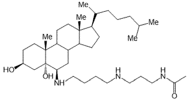
DX243 
DX301 
DX242 
DX302 
DX245 
DX401 
DX244 
DX243BOC 
Claims (14)
- Composition comprenant au moins un composé de formule (I) :
 R1 = OH;R2 = OH ;R3 = -NR5R6,R5 étant H, etR6 étant pris dans le groupe formé par -(CH2)3NH(CH2)4NHR7 ; - (CH2)4NH(CH2)3NHR7 ; -(CH2)3NH(CH2)4NH(CH2)3NHR7 ; -(CH2)3NHR7 ; - (CH2)4NHR7 avec R7 = H ou COCH3 ; -(CH2)2-imidazol-4-yl ;et -(CH2)2-indol-3-yl ; etR4 = H en position 20, 22, 24, 25, 26 ou 27, positionné de façon à créer un centre asymétrique de configuration R ou S ;Z1 et Z2 représentent chacun le nombre de double liaisons entre les atomes de carbone C7 et C8 et C22 et C23 respectivement ; Z1 est soit 0 soit 1 ; Z2 = 0 ; T1=T2=T3=T4=H, T4 étant positionné de façon à obtenir un centre asymétrique de configuration R ou S en position 24 ;et/ou au moins un sel pharmaceutiquement acceptable d'au moins un composé de formule (I),pour son utilisation dans le traitement d'une pathologie neuronale d'un sujet, ladite pathologie neuronale étant liée à une hypoxie, à une hypoglycémie et/ou à une hyperglycémie affectant des cellules du système nerveux central.
 - Composition pour utilisation selon la revendication 1, dans laquelle le composé de formule (I) est défini par Z1 =0.
- Composition pour utilisation selon la revendication 2, dans laquelle le composé de formule (I) est défini par R6 = -(CH2)4NH(CH2)3NHR7 avec R7 = COCH3.
- Composition pour utilisation selon la revendication 2, dans laquelle le composé de formule (I) est défini par R6 = -(CH2)2-imidazol-4-yl.
- Composition pour utilisation selon la revendication 2, dans laquelle le composé de formule (I) est défini par R6 = -(CH2)3NH(CH2)4NHR7, - (CH2)4NH(CH2)3NHR7, -(CH2)3NH(CH2)4NH(CH2)3NHR7 ; ou -(CH2)4NHR7 ; et R7 = H.
- Composition pour utilisation selon la revendication 1, dans laquelle le composé de formule (I) est défini par Z1 = 1.
- Composition pour utilisation selon la revendication 6, dans laquelle le composé de formule (I) est défini par R6 = -(CH2)3NH(CH2)4NHR7 ; - (CH2)4NH(CH2)3NHR7 ; ou -(CH2)3NH(CH2)4NH(CH2)3NHR7 ; et R7 = H.
- Composition comprenant au moins un composé de formule (I) :
 R1 = F, OCnH2n+1, R-COO, R-OCOO, RHN-COO ou OPO(OR)2 avec R = H ou CnH2n+1, avec n ≤ 16 ;R2 = OHR3 = -NR5R6,R4 = H ou OH en position 20, 22, 24, 25, 26 ou 27, positionné de façon à créer un centre asymétrique de configuration R ou S ;R5 = H ;R6 étant pris dans le groupe formé par -(CH2)4NH(CH2)3NHR7, - (CH2)3NH(CH2)4NHR7, -(CH2)3NH(CH2)4NH(CH2)3NHR7 ; -(CH2)4NHR7 ; avec R7 = H ou COCH3 ;-(CH2)2-imidazol-4-yl ; et -(CH2)2-indol-3-yl ;Z1 et Z2 représentent chacun le nombre de double liaisons entre les atomes de carbone C7 et C8 et C22 et C23 respectivement,Z1 = 0 ou Z1 = 1Z2 = 0 ;T1, T2, T3 et T4 = H, T4 étant positionné de façon à obtenir un centre asymétrique de configuration R ou S en position 24 ;et/ou au moins un sel pharmaceutiquement acceptable d'au moins un composé de formule (I),pour son utilisation dans le traitement d'une pathologie neuronale d'un sujet, ladite pathologie neuronale étant liée à une hypoxie, à une hypoglycémie et/ou à une hyperglycémie affectant des cellules du système nerveux central.
 - Composition pour utilisation selon l'une des revendications 1 à 8, dans laquelle la pathologie neuronale du système nerveux central est choisie parmi le groupe constitué des traumatismes cérébraux et des accidents vasculaires cérébraux.
- Composition pour utilisation selon l'une des revendications 1 à 8, dans laquelle la pathologie neuronale du système nerveux central est une lésion cérébrale due à une ischémie.
- Composition pour utilisation selon l'une des revendications 1 à 8, dans laquelle la pathologie neuronale du système nerveux central est une lésion cérébrale due à une insuffisance respiratoire.
- Composition pour utilisation selon l'une des revendications 1 à 8, dans laquelle l'hypoglycémie est due à un diabète.
- Composition pour utilisation selon l'une des revendications 1 à 8, dans laquelle l'hyperglycémie est due à un diabète.
- Composition pour utilisation selon l'une des revendications 1 à 8, sous forme d'une solution aqueuse et présentant une concentration de composé de formule (I) comprise entre 1 pmol.L-1 et 1 mmol.L-1, préférentiellement entre 10 pmol.L-1 et 0,1 mmol.L-1, plus préférentiellement entre 0,1 nmol.L-1 et 1 µmol.L-1. 1
Applications Claiming Priority (2)
| Application Number | Priority Date | Filing Date | Title |
|---|---|---|---|
| BE20195501A BE1027157B9 (fr) | 2019-08-01 | 2019-08-01 | Composition de dérivés de stérols pour son utilisationdans le traitement d’une pathologie neuronale liée à une hypoxie et/ou à une hypoglycémie et composition correspondante |
| PCT/EP2020/071044 WO2021018798A1 (fr) | 2019-08-01 | 2020-07-24 | Composition comprenant des dérivés de stérols pour son utilisation dans le traitement d'une pathologie neuronale liée à une hypoxie, à une hypoglycémie et/ou une hyperglycémie |
Publications (3)
| Publication Number | Publication Date |
|---|---|
| EP4007580A1 EP4007580A1 (fr) | 2022-06-08 |
| EP4007580B1 true EP4007580B1 (fr) | 2023-06-07 |
| EP4007580C0 EP4007580C0 (fr) | 2023-06-07 |
Family
ID=67765997
Family Applications (1)
| Application Number | Title | Priority Date | Filing Date |
|---|---|---|---|
| EP20746623.6A Active EP4007580B1 (fr) | 2019-08-01 | 2020-07-24 | Composition comprenant des dérivés de stérols pour son utilisation dans le traitement d'une pathologie neuronale liée à une hypoxie, à une hypoglycémie et/ou une hyperglycémie |
Country Status (10)
| Country | Link |
|---|---|
| US (1) | US20220313706A1 (fr) |
| EP (1) | EP4007580B1 (fr) |
| JP (1) | JP7774869B2 (fr) |
| KR (1) | KR102866594B1 (fr) |
| CN (1) | CN114206347A (fr) |
| AU (1) | AU2020322131B2 (fr) |
| BE (1) | BE1027157B9 (fr) |
| CA (1) | CA3148002A1 (fr) |
| ES (1) | ES2954768T3 (fr) |
| WO (1) | WO2021018798A1 (fr) |
Families Citing this family (2)
| Publication number | Priority date | Publication date | Assignee | Title |
|---|---|---|---|---|
| EP4502153A1 (fr) | 2022-03-30 | 2025-02-05 | GenKOre Inc. | Système d'édition génique pour le traitement du syndrome d'usher |
| BE1030026B1 (fr) * | 2022-06-14 | 2023-07-07 | Dendrogenix | Composés dérivés de stérols et composition pharmaceutique les comprenant pour leur utilisation dans la prévention, l’amélioration et/ou le traitement d’une pathologie liée à un déficit mitochondrial |
Family Cites Families (7)
| Publication number | Priority date | Publication date | Assignee | Title |
|---|---|---|---|---|
| FR2838741B1 (fr) | 2002-04-19 | 2006-01-27 | Inst Nat Sante Rech Med | Derives de sterols, leur procede de preparation et medicaments les comportant |
| US20060194810A1 (en) * | 2004-04-30 | 2006-08-31 | Bijan Almassian | Methods of treating ischemic related conditions |
| FR2894968B1 (fr) | 2005-12-20 | 2008-02-22 | Trophos Sa | Nouveaux derives de l'oxime de cholest-4-en-3-one, compositions pharmaceutiques les renfermant, et procede de preparation |
| FR2981351A1 (fr) | 2011-10-17 | 2013-04-19 | Univ Nice Sophia Antipolis | Derives aminosteroidiens anti-diabetiques |
| IN2014MN00976A (fr) * | 2011-11-24 | 2015-04-24 | Inserm Inst Nat De La Santé Et De La Rech Médicale | |
| FR3024361B1 (fr) * | 2014-07-30 | 2017-11-24 | Affichem | Utilisation de derives de sterols pour traitement de la perte auditive neurosensorielle et composition correspondante |
| KR101878460B1 (ko) * | 2016-12-05 | 2018-07-13 | 가천대학교 산학협력단 | 신규한 호모이소플라바논 화합물, 이의 제조방법 및 이를 유효성분으로 함유하는 허혈에 의한 뇌손상 및 다발성경화증의 예방 또는 치료용 약학적 조성물 |
-
2019
- 2019-08-01 BE BE20195501A patent/BE1027157B9/fr active IP Right Grant
-
2020
- 2020-07-24 WO PCT/EP2020/071044 patent/WO2021018798A1/fr not_active Ceased
- 2020-07-24 US US17/631,773 patent/US20220313706A1/en active Pending
- 2020-07-24 EP EP20746623.6A patent/EP4007580B1/fr active Active
- 2020-07-24 ES ES20746623T patent/ES2954768T3/es active Active
- 2020-07-24 AU AU2020322131A patent/AU2020322131B2/en active Active
- 2020-07-24 KR KR1020227003352A patent/KR102866594B1/ko active Active
- 2020-07-24 CN CN202080055586.5A patent/CN114206347A/zh active Pending
- 2020-07-24 CA CA3148002A patent/CA3148002A1/fr active Pending
- 2020-07-24 JP JP2022506669A patent/JP7774869B2/ja active Active
Also Published As
| Publication number | Publication date |
|---|---|
| KR102866594B1 (ko) | 2025-09-29 |
| CN114206347A (zh) | 2022-03-18 |
| ES2954768T3 (es) | 2023-11-24 |
| US20220313706A1 (en) | 2022-10-06 |
| CA3148002A1 (fr) | 2021-02-04 |
| BE1027157B9 (fr) | 2020-11-03 |
| AU2020322131B2 (en) | 2025-05-08 |
| EP4007580C0 (fr) | 2023-06-07 |
| BE1027157B1 (fr) | 2020-10-26 |
| EP4007580A1 (fr) | 2022-06-08 |
| KR20220039723A (ko) | 2022-03-29 |
| JP2022543100A (ja) | 2022-10-07 |
| WO2021018798A1 (fr) | 2021-02-04 |
| JP7774869B2 (ja) | 2025-11-25 |
| AU2020322131A1 (en) | 2022-02-24 |
Similar Documents
| Publication | Publication Date | Title |
|---|---|---|
| JP4896017B2 (ja) | 脳疾患または頭脳機能改善のための組成物 | |
| Gong et al. | Protective or harmful: the dual roles of autophagy in diabetic retinopathy | |
| Labbé et al. | A new gel formulation of topical cysteamine for the treatment of corneal cystine crystals in cystinosis: the Cystadrops OCT-1 study | |
| Dietrich et al. | Early alpha-lipoic acid therapy protects from degeneration of the inner retinal layers and vision loss in an experimental autoimmune encephalomyelitis-optic neuritis model | |
| Li et al. | Puerarin attenuates neuronal degeneration in the substantia nigra of 6-OHDA-lesioned rats through regulating BDNF expression and activating the Nrf2/ARE signaling pathway | |
| Zhao et al. | Systemic administration of the antioxidant/iron chelator α-lipoic acid protects against light-induced photoreceptor degeneration in the mouse retina | |
| Yiğit et al. | Effects of astaxanthin on antioxidant parameters in ARPE-19 cells on oxidative stress model | |
| CA3079645A1 (fr) | Nouveaux medicaments cytoprotecteurs | |
| WO2021018798A1 (fr) | Composition comprenant des dérivés de stérols pour son utilisation dans le traitement d'une pathologie neuronale liée à une hypoxie, à une hypoglycémie et/ou une hyperglycémie | |
| Babizhayev et al. | N-acetylcarnosine lubricant eyedrops possess all-in-one universal antioxidant protective effects of L-carnosine in aqueous and lipid membrane environments, aldehyde scavenging, and transglycation activities inherent to cataracts: a clinical study of the new vision-saving drug N-acetylcarnosine eyedrop therapy in a database population of over 50,500 patients | |
| Choh et al. | Isoflurane and ketamine: xylazine differentially affect intraocular pressure-associated scotopic threshold responses in Sprague-Dawley rats | |
| Song et al. | Peroxynitrite-scavenging organosilica nanomedicines for light-controllable NO release and precision on-demand glaucoma therapy | |
| CA2795465C (fr) | Compositions pharmaceutiques fortement dosees en biotine | |
| JP2015519398A (ja) | 治療用製剤および処置の方法 | |
| Krempler et al. | Simvastatin improves retinal ganglion cell survival and spatial vision after acute retinal ischemia/reperfusion in mice | |
| Storoni et al. | The therapeutic potential of a calorie-restricted ketogenic diet for the management of Leber hereditary optic neuropathy | |
| FR3043555B1 (fr) | Mirabegron pour le traitement de maladies retiniennes | |
| Niittykoski et al. | Altered calcium signaling in an experimental model of glaucoma | |
| Dorfman et al. | Structural and functional consequences of trolox C treatment in the rat model of postnatal hyperoxia | |
| JP6238977B2 (ja) | 視神経症または先天性視神経萎縮を治療するためのアンギオテンシン変換酵素阻害薬 | |
| JP2012040010A (ja) | 組成物の食品または機能性食品に対する添加剤としての使用 | |
| Wang et al. | U83836E inhibits retinal neurodegeneration in early-stage streptozotocin-induced diabetic rats | |
| HK40068326A (en) | Composition comprising sterol derivatives for use in the treatment of a neuronal pathology related to hypoxia, hypoglycemia and/or hyperglycemia | |
| EP2314290A1 (fr) | Utilisation du fer pour le traitement du trouble du déficit de l'attention/hyperactivité chez les enfants | |
| Swanson et al. | Hypoglycemic Neuronal Death |
Legal Events
| Date | Code | Title | Description |
|---|---|---|---|
| STAA | Information on the status of an ep patent application or granted ep patent |
Free format text: STATUS: UNKNOWN |
|
| STAA | Information on the status of an ep patent application or granted ep patent |
Free format text: STATUS: THE INTERNATIONAL PUBLICATION HAS BEEN MADE |
|
| PUAI | Public reference made under article 153(3) epc to a published international application that has entered the european phase |
Free format text: ORIGINAL CODE: 0009012 |
|
| STAA | Information on the status of an ep patent application or granted ep patent |
Free format text: STATUS: REQUEST FOR EXAMINATION WAS MADE |
|
| 17P | Request for examination filed |
Effective date: 20220119 |
|
| AK | Designated contracting states |
Kind code of ref document: A1 Designated state(s): AL AT BE BG CH CY CZ DE DK EE ES FI FR GB GR HR HU IE IS IT LI LT LU LV MC MK MT NL NO PL PT RO RS SE SI SK SM TR |
|
| STAA | Information on the status of an ep patent application or granted ep patent |
Free format text: STATUS: EXAMINATION IS IN PROGRESS |
|
| 17Q | First examination report despatched |
Effective date: 20220907 |
|
| GRAP | Despatch of communication of intention to grant a patent |
Free format text: ORIGINAL CODE: EPIDOSNIGR1 |
|
| STAA | Information on the status of an ep patent application or granted ep patent |
Free format text: STATUS: GRANT OF PATENT IS INTENDED |
|
| DAV | Request for validation of the european patent (deleted) | ||
| DAX | Request for extension of the european patent (deleted) | ||
| INTG | Intention to grant announced |
Effective date: 20221020 |
|
| RIN1 | Information on inventor provided before grant (corrected) |
Inventor name: CARON, NICOLAS Inventor name: RIVES, ARNAUD Inventor name: MARLIER, QUENTIN Inventor name: SILVENTE, STEPHANE |
|
| GRAS | Grant fee paid |
Free format text: ORIGINAL CODE: EPIDOSNIGR3 |
|
| GRAA | (expected) grant |
Free format text: ORIGINAL CODE: 0009210 |
|
| STAA | Information on the status of an ep patent application or granted ep patent |
Free format text: STATUS: THE PATENT HAS BEEN GRANTED |
|
| AK | Designated contracting states |
Kind code of ref document: B1 Designated state(s): AL AT BE BG CH CY CZ DE DK EE ES FI FR GB GR HR HU IE IS IT LI LT LU LV MC MK MT NL NO PL PT RO RS SE SI SK SM TR |
|
| REG | Reference to a national code |
Ref country code: GB Ref legal event code: FG4D Free format text: NOT ENGLISH |
|
| REG | Reference to a national code |
Ref country code: CH Ref legal event code: EP Ref country code: AT Ref legal event code: REF Ref document number: 1572848 Country of ref document: AT Kind code of ref document: T Effective date: 20230615 |
|
| REG | Reference to a national code |
Ref country code: DE Ref legal event code: R096 Ref document number: 602020012217 Country of ref document: DE |
|
| U01 | Request for unitary effect filed |
Effective date: 20230607 |
|
| U07 | Unitary effect registered |
Designated state(s): AT BE BG DE DK EE FI FR IT LT LU LV MT NL PT SE SI Effective date: 20230612 |
|
| U20 | Renewal fee for the european patent with unitary effect paid |
Year of fee payment: 4 Effective date: 20230622 |
|
| REG | Reference to a national code |
Ref country code: LT Ref legal event code: MG9D |
|
| PG25 | Lapsed in a contracting state [announced via postgrant information from national office to epo] |
Ref country code: NO Free format text: LAPSE BECAUSE OF FAILURE TO SUBMIT A TRANSLATION OF THE DESCRIPTION OR TO PAY THE FEE WITHIN THE PRESCRIBED TIME-LIMIT Effective date: 20230907 |
|
| REG | Reference to a national code |
Ref country code: ES Ref legal event code: FG2A Ref document number: 2954768 Country of ref document: ES Kind code of ref document: T3 Effective date: 20231124 |
|
| PG25 | Lapsed in a contracting state [announced via postgrant information from national office to epo] |
Ref country code: RS Free format text: LAPSE BECAUSE OF FAILURE TO SUBMIT A TRANSLATION OF THE DESCRIPTION OR TO PAY THE FEE WITHIN THE PRESCRIBED TIME-LIMIT Effective date: 20230607 Ref country code: HR Free format text: LAPSE BECAUSE OF FAILURE TO SUBMIT A TRANSLATION OF THE DESCRIPTION OR TO PAY THE FEE WITHIN THE PRESCRIBED TIME-LIMIT Effective date: 20230607 Ref country code: GR Free format text: LAPSE BECAUSE OF FAILURE TO SUBMIT A TRANSLATION OF THE DESCRIPTION OR TO PAY THE FEE WITHIN THE PRESCRIBED TIME-LIMIT Effective date: 20230908 |
|
| PG25 | Lapsed in a contracting state [announced via postgrant information from national office to epo] |
Ref country code: SK Free format text: LAPSE BECAUSE OF FAILURE TO SUBMIT A TRANSLATION OF THE DESCRIPTION OR TO PAY THE FEE WITHIN THE PRESCRIBED TIME-LIMIT Effective date: 20230607 |
|
| PG25 | Lapsed in a contracting state [announced via postgrant information from national office to epo] |
Ref country code: IS Free format text: LAPSE BECAUSE OF FAILURE TO SUBMIT A TRANSLATION OF THE DESCRIPTION OR TO PAY THE FEE WITHIN THE PRESCRIBED TIME-LIMIT Effective date: 20231007 |
|
| PG25 | Lapsed in a contracting state [announced via postgrant information from national office to epo] |
Ref country code: SM Free format text: LAPSE BECAUSE OF FAILURE TO SUBMIT A TRANSLATION OF THE DESCRIPTION OR TO PAY THE FEE WITHIN THE PRESCRIBED TIME-LIMIT Effective date: 20230607 Ref country code: SK Free format text: LAPSE BECAUSE OF FAILURE TO SUBMIT A TRANSLATION OF THE DESCRIPTION OR TO PAY THE FEE WITHIN THE PRESCRIBED TIME-LIMIT Effective date: 20230607 Ref country code: RO Free format text: LAPSE BECAUSE OF FAILURE TO SUBMIT A TRANSLATION OF THE DESCRIPTION OR TO PAY THE FEE WITHIN THE PRESCRIBED TIME-LIMIT Effective date: 20230607 Ref country code: IS Free format text: LAPSE BECAUSE OF FAILURE TO SUBMIT A TRANSLATION OF THE DESCRIPTION OR TO PAY THE FEE WITHIN THE PRESCRIBED TIME-LIMIT Effective date: 20231007 Ref country code: CZ Free format text: LAPSE BECAUSE OF FAILURE TO SUBMIT A TRANSLATION OF THE DESCRIPTION OR TO PAY THE FEE WITHIN THE PRESCRIBED TIME-LIMIT Effective date: 20230607 |
|
| PG25 | Lapsed in a contracting state [announced via postgrant information from national office to epo] |
Ref country code: PL Free format text: LAPSE BECAUSE OF FAILURE TO SUBMIT A TRANSLATION OF THE DESCRIPTION OR TO PAY THE FEE WITHIN THE PRESCRIBED TIME-LIMIT Effective date: 20230607 |
|
| REG | Reference to a national code |
Ref country code: CH Ref legal event code: PL |
|
| REG | Reference to a national code |
Ref country code: DE Ref legal event code: R097 Ref document number: 602020012217 Country of ref document: DE |
|
| PG25 | Lapsed in a contracting state [announced via postgrant information from national office to epo] |
Ref country code: MC Free format text: LAPSE BECAUSE OF FAILURE TO SUBMIT A TRANSLATION OF THE DESCRIPTION OR TO PAY THE FEE WITHIN THE PRESCRIBED TIME-LIMIT Effective date: 20230607 |
|
| PG25 | Lapsed in a contracting state [announced via postgrant information from national office to epo] |
Ref country code: MC Free format text: LAPSE BECAUSE OF FAILURE TO SUBMIT A TRANSLATION OF THE DESCRIPTION OR TO PAY THE FEE WITHIN THE PRESCRIBED TIME-LIMIT Effective date: 20230607 |
|
| PLBE | No opposition filed within time limit |
Free format text: ORIGINAL CODE: 0009261 |
|
| STAA | Information on the status of an ep patent application or granted ep patent |
Free format text: STATUS: NO OPPOSITION FILED WITHIN TIME LIMIT |
|
| REG | Reference to a national code |
Ref country code: IE Ref legal event code: MM4A |
|
| PG25 | Lapsed in a contracting state [announced via postgrant information from national office to epo] |
Ref country code: CH Free format text: LAPSE BECAUSE OF NON-PAYMENT OF DUE FEES Effective date: 20230731 |
|
| 26N | No opposition filed |
Effective date: 20240308 |
|
| PG25 | Lapsed in a contracting state [announced via postgrant information from national office to epo] |
Ref country code: IE Free format text: LAPSE BECAUSE OF NON-PAYMENT OF DUE FEES Effective date: 20230724 |
|
| U20 | Renewal fee for the european patent with unitary effect paid |
Year of fee payment: 5 Effective date: 20240619 |
|
| PG25 | Lapsed in a contracting state [announced via postgrant information from national office to epo] |
Ref country code: IE Free format text: LAPSE BECAUSE OF NON-PAYMENT OF DUE FEES Effective date: 20230724 |
|
| PGFP | Annual fee paid to national office [announced via postgrant information from national office to epo] |
Ref country code: GB Payment date: 20250619 Year of fee payment: 6 |
|
| PG25 | Lapsed in a contracting state [announced via postgrant information from national office to epo] |
Ref country code: CY Free format text: LAPSE BECAUSE OF FAILURE TO SUBMIT A TRANSLATION OF THE DESCRIPTION OR TO PAY THE FEE WITHIN THE PRESCRIBED TIME-LIMIT; INVALID AB INITIO Effective date: 20200724 |
|
| U20 | Renewal fee for the european patent with unitary effect paid |
Year of fee payment: 6 Effective date: 20250620 |
|
| PG25 | Lapsed in a contracting state [announced via postgrant information from national office to epo] |
Ref country code: HU Free format text: LAPSE BECAUSE OF FAILURE TO SUBMIT A TRANSLATION OF THE DESCRIPTION OR TO PAY THE FEE WITHIN THE PRESCRIBED TIME-LIMIT; INVALID AB INITIO Effective date: 20200724 |
|
| PGFP | Annual fee paid to national office [announced via postgrant information from national office to epo] |
Ref country code: ES Payment date: 20250801 Year of fee payment: 6 |
|
| PG25 | Lapsed in a contracting state [announced via postgrant information from national office to epo] |
Ref country code: TR Free format text: LAPSE BECAUSE OF FAILURE TO SUBMIT A TRANSLATION OF THE DESCRIPTION OR TO PAY THE FEE WITHIN THE PRESCRIBED TIME-LIMIT Effective date: 20230607 |