EP4086632A1 - Procédé de détection directe de souche pathogène présentant une résistance aux antibiotiques carbapénèmes - Google Patents
Procédé de détection directe de souche pathogène présentant une résistance aux antibiotiques carbapénèmes Download PDFInfo
- Publication number
- EP4086632A1 EP4086632A1 EP20908619.8A EP20908619A EP4086632A1 EP 4086632 A1 EP4086632 A1 EP 4086632A1 EP 20908619 A EP20908619 A EP 20908619A EP 4086632 A1 EP4086632 A1 EP 4086632A1
- Authority
- EP
- European Patent Office
- Prior art keywords
- mass
- protein
- values
- resistance
- mass spectrometry
- Prior art date
- Legal status (The legal status is an assumption and is not a legal conclusion. Google has not performed a legal analysis and makes no representation as to the accuracy of the status listed.)
- Pending
Links
Images























Classifications
-
- G—PHYSICS
- G01—MEASURING; TESTING
- G01N—INVESTIGATING OR ANALYSING MATERIALS BY DETERMINING THEIR CHEMICAL OR PHYSICAL PROPERTIES
- G01N30/00—Investigating or analysing materials by separation into components using adsorption, absorption or similar phenomena or using ion-exchange, e.g. chromatography or field flow fractionation
- G01N30/02—Column chromatography
- G01N30/62—Detectors specially adapted therefor
- G01N30/72—Mass spectrometers
-
- C—CHEMISTRY; METALLURGY
- C12—BIOCHEMISTRY; BEER; SPIRITS; WINE; VINEGAR; MICROBIOLOGY; ENZYMOLOGY; MUTATION OR GENETIC ENGINEERING
- C12Q—MEASURING OR TESTING PROCESSES INVOLVING ENZYMES, NUCLEIC ACIDS OR MICROORGANISMS; COMPOSITIONS OR TEST PAPERS THEREFOR; PROCESSES OF PREPARING SUCH COMPOSITIONS; CONDITION-RESPONSIVE CONTROL IN MICROBIOLOGICAL OR ENZYMOLOGICAL PROCESSES
- C12Q1/00—Measuring or testing processes involving enzymes, nucleic acids or microorganisms; Compositions therefor; Processes of preparing such compositions
- C12Q1/02—Measuring or testing processes involving enzymes, nucleic acids or microorganisms; Compositions therefor; Processes of preparing such compositions involving viable microorganisms
- C12Q1/04—Determining presence or kind of microorganism; Use of selective media for testing antibiotics or bacteriocides; Compositions containing a chemical indicator therefor
-
- C—CHEMISTRY; METALLURGY
- C12—BIOCHEMISTRY; BEER; SPIRITS; WINE; VINEGAR; MICROBIOLOGY; ENZYMOLOGY; MUTATION OR GENETIC ENGINEERING
- C12Q—MEASURING OR TESTING PROCESSES INVOLVING ENZYMES, NUCLEIC ACIDS OR MICROORGANISMS; COMPOSITIONS OR TEST PAPERS THEREFOR; PROCESSES OF PREPARING SUCH COMPOSITIONS; CONDITION-RESPONSIVE CONTROL IN MICROBIOLOGICAL OR ENZYMOLOGICAL PROCESSES
- C12Q1/00—Measuring or testing processes involving enzymes, nucleic acids or microorganisms; Compositions therefor; Processes of preparing such compositions
- C12Q1/02—Measuring or testing processes involving enzymes, nucleic acids or microorganisms; Compositions therefor; Processes of preparing such compositions involving viable microorganisms
- C12Q1/04—Determining presence or kind of microorganism; Use of selective media for testing antibiotics or bacteriocides; Compositions containing a chemical indicator therefor
- C12Q1/10—Enterobacteria
-
- C—CHEMISTRY; METALLURGY
- C12—BIOCHEMISTRY; BEER; SPIRITS; WINE; VINEGAR; MICROBIOLOGY; ENZYMOLOGY; MUTATION OR GENETIC ENGINEERING
- C12Q—MEASURING OR TESTING PROCESSES INVOLVING ENZYMES, NUCLEIC ACIDS OR MICROORGANISMS; COMPOSITIONS OR TEST PAPERS THEREFOR; PROCESSES OF PREPARING SUCH COMPOSITIONS; CONDITION-RESPONSIVE CONTROL IN MICROBIOLOGICAL OR ENZYMOLOGICAL PROCESSES
- C12Q1/00—Measuring or testing processes involving enzymes, nucleic acids or microorganisms; Compositions therefor; Processes of preparing such compositions
- C12Q1/34—Measuring or testing processes involving enzymes, nucleic acids or microorganisms; Compositions therefor; Processes of preparing such compositions involving hydrolase
-
- G—PHYSICS
- G01—MEASURING; TESTING
- G01N—INVESTIGATING OR ANALYSING MATERIALS BY DETERMINING THEIR CHEMICAL OR PHYSICAL PROPERTIES
- G01N30/00—Investigating or analysing materials by separation into components using adsorption, absorption or similar phenomena or using ion-exchange, e.g. chromatography or field flow fractionation
- G01N30/02—Column chromatography
- G01N30/88—Integrated analysis systems specially adapted therefor, not covered by a single one of the groups G01N30/04 - G01N30/86
-
- G—PHYSICS
- G01—MEASURING; TESTING
- G01N—INVESTIGATING OR ANALYSING MATERIALS BY DETERMINING THEIR CHEMICAL OR PHYSICAL PROPERTIES
- G01N30/00—Investigating or analysing materials by separation into components using adsorption, absorption or similar phenomena or using ion-exchange, e.g. chromatography or field flow fractionation
- G01N30/96—Investigating or analysing materials by separation into components using adsorption, absorption or similar phenomena or using ion-exchange, e.g. chromatography or field flow fractionation using ion-exchange
-
- G—PHYSICS
- G01—MEASURING; TESTING
- G01N—INVESTIGATING OR ANALYSING MATERIALS BY DETERMINING THEIR CHEMICAL OR PHYSICAL PROPERTIES
- G01N33/00—Investigating or analysing materials by specific methods not covered by groups G01N1/00 - G01N31/00
- G01N33/48—Biological material, e.g. blood, urine; Haemocytometers
- G01N33/50—Chemical analysis of biological material, e.g. blood, urine; Testing involving biospecific ligand binding methods; Immunological testing
- G01N33/53—Immunoassay; Biospecific binding assay; Materials therefor
- G01N33/569—Immunoassay; Biospecific binding assay; Materials therefor for microorganisms, e.g. protozoa, bacteria, viruses
- G01N33/56911—Bacteria
-
- G—PHYSICS
- G01—MEASURING; TESTING
- G01N—INVESTIGATING OR ANALYSING MATERIALS BY DETERMINING THEIR CHEMICAL OR PHYSICAL PROPERTIES
- G01N33/00—Investigating or analysing materials by specific methods not covered by groups G01N1/00 - G01N31/00
- G01N33/48—Biological material, e.g. blood, urine; Haemocytometers
- G01N33/50—Chemical analysis of biological material, e.g. blood, urine; Testing involving biospecific ligand binding methods; Immunological testing
- G01N33/68—Chemical analysis of biological material, e.g. blood, urine; Testing involving biospecific ligand binding methods; Immunological testing involving proteins, peptides or amino acids
- G01N33/6803—General methods of protein analysis not limited to specific proteins or families of proteins
- G01N33/6848—Methods of protein analysis involving mass spectrometry
-
- G—PHYSICS
- G01—MEASURING; TESTING
- G01N—INVESTIGATING OR ANALYSING MATERIALS BY DETERMINING THEIR CHEMICAL OR PHYSICAL PROPERTIES
- G01N33/00—Investigating or analysing materials by specific methods not covered by groups G01N1/00 - G01N31/00
- G01N33/48—Biological material, e.g. blood, urine; Haemocytometers
- G01N33/50—Chemical analysis of biological material, e.g. blood, urine; Testing involving biospecific ligand binding methods; Immunological testing
- G01N33/68—Chemical analysis of biological material, e.g. blood, urine; Testing involving biospecific ligand binding methods; Immunological testing involving proteins, peptides or amino acids
- G01N33/6803—General methods of protein analysis not limited to specific proteins or families of proteins
- G01N33/6848—Methods of protein analysis involving mass spectrometry
- G01N33/6851—Methods of protein analysis involving laser desorption ionisation mass spectrometry
-
- G—PHYSICS
- G01—MEASURING; TESTING
- G01N—INVESTIGATING OR ANALYSING MATERIALS BY DETERMINING THEIR CHEMICAL OR PHYSICAL PROPERTIES
- G01N30/00—Investigating or analysing materials by separation into components using adsorption, absorption or similar phenomena or using ion-exchange, e.g. chromatography or field flow fractionation
- G01N30/02—Column chromatography
- G01N30/88—Integrated analysis systems specially adapted therefor, not covered by a single one of the groups G01N30/04 - G01N30/86
- G01N2030/8809—Integrated analysis systems specially adapted therefor, not covered by a single one of the groups G01N30/04 - G01N30/86 analysis specially adapted for the sample
- G01N2030/8813—Integrated analysis systems specially adapted therefor, not covered by a single one of the groups G01N30/04 - G01N30/86 analysis specially adapted for the sample biological materials
- G01N2030/8831—Integrated analysis systems specially adapted therefor, not covered by a single one of the groups G01N30/04 - G01N30/86 analysis specially adapted for the sample biological materials involving peptides or proteins
-
- G—PHYSICS
- G01—MEASURING; TESTING
- G01N—INVESTIGATING OR ANALYSING MATERIALS BY DETERMINING THEIR CHEMICAL OR PHYSICAL PROPERTIES
- G01N2333/00—Assays involving biological materials from specific organisms or of a specific nature
- G01N2333/90—Enzymes; Proenzymes
- G01N2333/914—Hydrolases (3)
- G01N2333/978—Hydrolases (3) acting on carbon to nitrogen bonds other than peptide bonds (3.5)
- G01N2333/986—Hydrolases (3) acting on carbon to nitrogen bonds other than peptide bonds (3.5) acting on amide bonds in cyclic amides (3.5.2), e.g. beta-lactamase (penicillinase, 3.5.2.6), creatinine amidohydrolase (creatininase, EC 3.5.2.10), N-methylhydantoinase (3.5.2.6)
Definitions
- the present invention relates to a method of directly detecting an in vivo active form of the carbapenemases KPC, OXA, NDM, IMP, VIM and/or GES by top-down mass spectrometry without pretreatment of a sample.
- Mass spectrometry methods including MALDI-TOF, are low-cost and high-efficiency identification systems compared to sequencing methods based on PCR, and can offer an important means for rapid identification of microorganisms.
- mass spectrometry methods including MALDI-TOF, are low-cost and high-efficiency identification systems compared to sequencing methods based on PCR, and can offer an important means for rapid identification of microorganisms.
- using these methods it is possible to achieve sample treatment after strain culture and stain identification within 10 minutes, and to quickly identify a strain with the same mass value by comparing mass data for unknown strains with mass data in a database established through mass spectrometry data.
- the present inventors have made efforts to provide a rapid and accurate diagnostic method for carbapenem-resistant strain infection by selecting proteins directly involved in resistance to beta-lactam antibiotics, specifically carbapenem antibiotics, and measuring the exact mass values of in vivo active forms of these selected proteins to establish accurate reference data for determining whether or not a carbapenem-resistant strain is present.
- the present inventors have made intensive research efforts to develop an efficient diagnostic method capable of rapidly determining infection with an antibiotic-resistant strain by detecting an antibiotic-degrading enzyme, which is involved in resistance to lactam-based antibiotics, in a sample in a simple and highly reliable manner.
- the present inventors have newly discovered that enzymes that degrade the beta-lactam antibiotic carbapenem, specifically KPC, OXA, NDM, IMP, VIM and GES proteins, are present in active forms due to truncation of some of N-terminal residues in vivo, and have found that, when active forms of these proteins, which account for the majority of carbapenem-degrading enzymes secreted by antibiotic-resistant strains, are directly identified through mass spectrometry, it is possible to establish an appropriate antibiotic administration strategy at an early stage of infection by rapidly and accurately determining not only whether the pathogenic strains are resistant to the antibiotics but also the type of protein involved in the resistance, thereby completing the present invention.
- an object of the present invention is to provide a method of detecting in a biological sample a pathogenic strain having resistance to carbapenem antibiotics, specifically a pathogenic strain expressing KPC, OXA, NDM, IMP, VIM and/or GES.
- a method for detecting in a biological sample a pathogenic strain having resistance to carbapenem antibiotics comprising:
- the present inventors have made intensive research efforts to develop an efficient diagnostic method capable of rapidly determining infection with an antibiotic-resistant strain by detecting an antibiotic-degrading enzyme, which is involved in resistance to lactam-based antibiotics, in a sample in a simple and highly reliable manner.
- the present inventors have newly discovered that enzymes that degrade the beta-lactam antibiotic carbapenem, specifically KPC, OXA, NDM, IMP, VIM and GES proteins, are present in active forms due to truncation of some of N-terminal residues in vivo, and have found that, when active forms of these proteins, which account for the majority of carbapenem-degrading enzymes secreted by antibiotic-resistant strains, are directly identified through mass spectrometry, it is possible to establish an appropriate antibiotic administration strategy at an early stage of infection by rapidly and accurately determining not only whether the pathogenic strains are resistant to the antibiotics but also the type of protein involved in the resistance.
- pathogenic strain refers to any bacteria that act as the cause of an infection or disease, including, for example, but not limited to, Staphylococcus aureus, Streptococcus, Escherichia coli, Klebsiella pneumoniae, Pseudomonas aeruginosa, Pseudomonas otitidis, Micrococcus luteus, Citrobacter koseri, Proteus mirabilis, and Mycobacterium ulcerans.
- the expression “having resistance to antibiotics” means that a specific pathogenic microorganism can grow even in an environment in which antibiotics against the microorganism are present at high concentration or in an effective amount. Whether the pathogenic microorganism has antibiotic resistance may be determined by detecting the presence of an enzyme protein, which is secreted by the pathogenic microorganism and removes or reduces the activities of the antibiotics by degrading the antibiotics.
- an enzyme protein which is secreted by the pathogenic microorganism and removes or reduces the activities of the antibiotics by degrading the antibiotics.
- beta-lactam antibiotics that inhibit bacterial cell wall synthesis such as penicillin, cephalosporin, monobactam, and carbapenem, are inactivated by ⁇ -lactamase so that they cannot inhibit pathogens expressing ⁇ -lactamase. Accordingly, the term “resistance” is used interchangeably with the terms “low therapeutic responsiveness” and "low prophylactic responsiveness”.
- treatment refers to (a) inhibiting the progress of a disease, disorder or symptom; (b) alleviating a disease, disorder or symptom; or (c) eliminating a disease, disorder or symptom.
- therapeutic responsiveness refers to the degree to which beta-lactam antibiotics, including carbapenem, when administered in a therapeutically effective amount to a patient infected with a pathogenic strain, act as described above in vivo.
- prevention refers to inhibiting the occurrence of a disease or disorder in a subject that has never been diagnosed as having the disease or disorder, but is likely to have the disease or disorder.
- prophylactic responsiveness refers to the degree to which beta-lactam antibiotics, including carbapenem, when administered in a prophylactically effective amount to a normal person whose infection has not yet been confirmed, act to inhibit infection in vivo.
- biological sample refers to any samples, including, for example, but not limited to, blood, tissues, organs, cells or cell cultures, which are obtained from mammals, including humans, and contain or are likely to contain a pathogenic strain to be inhibited with beta-lactam antibiotics, including carbapenem.
- the term "subject" refers to a subject that provides a sample for examining the presence of a pathogenic strain to be inhibited with beta-lactam antibiotics such as carbapenem or whether the strain has resistance to the antibiotics, and ultimately refers to a subject to be analyzed for whether or not infection with the pathogenic strain having resistance to the antibiotics has occurred.
- the subject include, without limitation, humans, mice, rats, guinea pigs, dogs, cats, horses, cattle, pigs, monkeys, chimpanzees, baboons or rhesus monkeys, specifically humans.
- composition of the present invention provides information for predicting not only therapeutic responsiveness but also prophylactic responsiveness to beta-lactam antibiotics such as carbapenem
- the subject in the present invention may be a patient infected with the strain or may also be a healthy subject whose infection has not yet been confirmed.
- top-down mass spectrometry refers to an analysis that directly measures the mass value of a full-length protein without performing the process of fragmenting the protein into peptide fragments, and specifically, refers to an analysis in which fragmentation of the target protein is not performed before the protein sample is injected into a mass spectrometer.
- Another feature of the present invention is that the procedure is simplified by performing direct mass spectrometry for a full-length protein without randomly degrading the protein using protease such as trypsin, and it is possible to determine the presence of a target protein with remarkably high reliability within a much shorter time than a conventional method of indirectly identifying proteins by collecting mass information on fragments and collecting vast amounts of information on fragmentation trends of various proteins.
- the expression "mass of the protein is the same” means that the mass value measured through the mass spectrometry method of the present invention is substantially the same as a reference mass value, for example, a value corresponding to the mass value of a carbapenemase whose amino acid sequence and molecular weight are known and from which 18, 19, 20, 21, 22 or 26 amino acid residues at the N-terminus have been removed.
- “Substantially the same” means that, for example, the measured Da value or m/z ⁇ z value is within the range of the reference mass value ⁇ 10, more specifically within the range of the reference mass value ⁇ 7, even more specifically within the range of the reference mass value ⁇ 5, most specifically within the range of the reference mass value ⁇ 3.
- the mass value of the carbapenem degrading enzyme which is a criterion for determining whether the mass value is substantially the same as a reference mass value, includes a mass value of a state in which 1 to 3 methionine residues exist in an oxidized state of (i.e., increased by 16, 32 or 48 from a known mass value) or in which a disulfide bond is formed between two Cys residues (i.e., decreased by 2 from a known mass value).
- the method of the present invention further comprises a step of performing ion exchange chromatography on the protein isolated in step (a).
- Ion exchange chromatography refers to a separation and purification method of separating a charged target substance from a heterogeneous mixture using a phenomenon in which ions or charged compounds bind to an ion exchange resin by electrostatic force.
- Ion exchange chromatography has ion exchange resins to which various functional groups bind, in which the anion exchange resin has a positively charged functional group and thus combines with a negatively charged target substance in the mixture by electrostatic attraction, and the cation exchange resin binds specifically to a positively charged target substance
- the ion exchange chromatography is any one selected from the group consisting of anion exchange chromatography, cation exchange chromatography, and a sequential combination thereof.
- the anion exchange resin used in the present invention may have, for example, a diethylaminoethyl (DEAE) or quaternary ammonium functional group, but is not limited thereto, and any conventional cationic functional group that provides a positive charge to the support may be used without limitation.
- DEAE diethylaminoethyl
- quaternary ammonium functional group any conventional cationic functional group that provides a positive charge to the support may be used without limitation.
- Strongly basic anion exchange groups include, for example, Q Sepharose Fast Flow, Q Sepharose High Performance, Resource Q, Source 15Q, Source 30Q, Mono Q, Mini Q, Capto Q, Capto Q ImpRes, Q HyperCel, Q Cermic HyperD F, Nuvia Q, UNOsphere Q, Macro-Prep High Q, Macro-Prep 25 Q, Fractogel EMD TMAE(S), Fractogel EMD TMAE Hicap (M), Fractogel EMD TMAE (M), Eshmono Q, Toyopearl QAE-550C, Toyopearl SuperQ-650C, Toyopearl GigaCap Q-650M, Toyopearl Q-600C AR, Toyopearl SuperQ-650M, Toyopearl SuperQ-650S, TSKgel SuperQ-5PW (30), TSKgel SuperQ-5PW (20) and TSKgel SuperQ-5PW, but are not limited thereto, and any anion exchange
- the cation exchange resin used in the present invention may have, for example, a sulfone group or a carboxy group, but is not limited thereto, and any conventional cationic functional group that provides a negative charge to the support may be used without limitation.
- the cation exchange resin may be selected from the group consisting of Fractogel, CM (carboxymethyl), SE (sulfoethyl), SP (sulfopropyl), P (phosphate), S (sulfonate), PROPAC WCX-10 TM (Dionex), Capto S, S-Sepharose FF, Fractogel EMD SO3M, Toyopearl Megacap II SP 550C, Poros 50 HS, Poros XS, and SP-sepharose matrix, but is not limited thereto.
- SP sulfopropyl resin
- column buffer equilibration buffer, wash buffer and elution buffer known in the art, such as sodium phosphate buffer, citrate buffer, and acetic acid buffer, may be used.
- Such ion exchange chromatography may be appropriately performed depending on the charges of the proteins to be separated/purified and the order of the proteins to be separated. For example, in order to sequentially separate a positively charged protein and a negatively charged protein, cation exchange chromatography may be performed, followed by anion exchange chromatography.
- step (a) of the present invention is performed by adding a surfactant to the biological sample.
- the present invention direct mass spectrometry of a full-length protein is possible by top-down mass spectrometry without a fragmentation process using a protease.
- the present inventors have found that, when a surfactant is added to the biological sample to be analyzed, the intact full-length protein present in the cell membrane or cytoplasm may be encapsulated, so that the target protein may be quickly and accurately identified without randomly degrading the protein by an enzyme.
- ionic, nonionic and zwitterionic surfactants may all be used without limitation as long as they are general surfactants capable of forming micelles sufficient to encapsulate full-length proteins.
- the surfactant used in the present invention is an ionic surfactant or a nonionic surfactant.
- ionic surfactants examples include, but are not limited to, sodium deoxycholate (DOC), Medialan A, Quaternium-60, cetylpyridinium chloride, cetylpyridinium bromide, cetyltrimetylammonium chloride, cetyltrimetylammonium bromide, and Gardinol.
- DOC sodium deoxycholate
- Medialan A Quaternium-60
- cetylpyridinium chloride cetylpyridinium bromide
- cetyltrimetylammonium chloride cetyltrimetylammonium bromide
- Gardinol Gardinol
- nonionic surfactants examples include, but are not limited to, n-octyl- ⁇ -D-glucopyranoside (OG), n-octyl- ⁇ -D-thioglucopyranoside (OTG), octyl glucose neopentyl glycol (OGNG), n-dodecyl- ⁇ -D-maltopyranoside (DDM), and n-dodecyl- ⁇ -D-thiomaltopyranoside (DDTM).
- OG n-octyl- ⁇ -D-glucopyranoside
- OTG n-octyl- ⁇ -D-thioglucopyranoside
- OOGNG octyl glucose neopentyl glycol
- DDM n-dodecyl- ⁇ -D-maltopyranoside
- DDTM n-dodecyl- ⁇ -D-thiomaltopyranoside
- step (a) may be performed by additionally adding a lysis buffer to the biological sample. That is, the surfactant of the present invention may be used together with a lysis buffer in the step of lysing the cells in order to isolate the protein expressed by the pathogenic strain.
- the lysis buffer may be a volatile buffer.
- volatile buffers examples include, but are not limited to, ammonium bicarbonate, acetic acid, formic acid, ammonia, ammonium carbonate, and pyridine/triethanolamine.
- any buffer that evaporates easily into the atmosphere due to its low boiling point while maintaining hydrogen ion concentration in the sample within a certain range.
- the method of the present invention further comprises a sonication step between step (a) and step (b).
- step (b) is performed using a mass spectrometry method selected from the group consisting of matrix-assisted laser desorption/ionization time of flight (MALDI-TOF) mass spectrometry, surface enhanced laser desorption/ionization time of flight (SELDI-TOF) mass spectrometry, electrospray ionization time-of-flight (ESI-TOF) mass spectrometry, liquid chromatography-mass spectrometry (LC-MS), and liquid chromatography-mass spectrometry/mass spectrometry (LC-MS/MS). More specifically, it is performed using MALDI-TOF mass spectrometry.
- MALDI-TOF matrix-assisted laser desorption/ionization time of flight
- SELDI-TOF surface enhanced laser desorption/ionization time of flight
- ESI-TOF electrospray ionization time-of-flight
- LC-MS liquid chromatography-mass spectrometry
- MALDI-TOF mass spectrometry is a method in which a sample supported by a matrix is desorbed and ionized by irradiation with a laser, and then the molecular weights of the generated ions are analyzed by measuring the time (Time-of-Flight) taken for the ions to reach a detector.
- this method it is possible to quickly and accurately measure the mass of a large biomolecule such as a protein, because fragmentation of the target material does not occur.
- m/z mass-to-charge ratio
- the carbapenemase is a KPC protein, and it is determined that, when a protein having the same mass as the KPC protein from which 21 amino acid residues at the N-terminus have been removed is detected as a result of the mass spectrometry, a pathogenic strain having resistance to carbapenem antibiotics is present in the biological sample.
- the carbapenemase is an OXA protein, and it is determined that, when a protein having the same mass as the OXA protein from which 22 amino acid residues at the N-terminus have been removed is detected as a result of the mass spectrometry, a pathogenic strain having resistance to carbapenem antibiotics is present in the biological sample.
- the carbapenemase is an NDM protein, and it is determined that, when a protein having the same mass as the NDM protein from which 19 or 20 amino acid residues at the N-terminus have been removed is detected as a result of the mass spectrometry, a pathogenic strain having resistance to carbapenem antibiotics is present in the biological sample.
- the carbapenemase is an IMP protein, and it is determined that, when a protein having the same mass as the IMP protein from which 18 to 20 amino acid residues at the N-terminus have been removed is detected as a result of the mass spectrometry, a pathogenic strain having resistance to carbapenem antibiotics is present in the biological sample.
- the carbapenemase is a VIM protein, and it is determined that, when a protein having the same mass as the VIM protein from which 25 or 26 amino acid residues at the N-terminus have been removed is detected as a result of the mass spectrometry, a pathogenic strain having resistance to carbapenem antibiotics is present in the biological sample.
- the carbapenemase is a GES protein, and it is determined that, when a protein having the same mass as the GES protein from which 18 amino acid residues at the N-terminus have been removed is detected as a result of the mass spectrometry, a pathogenic strain having resistance to carbapenem antibiotics is present in the biological sample.
- the present inventors have found that, in order for a carbapenemase to have an activity of degrading carbapenem in vivo, the carbapenemase should be present in a state in which some of the amino acid residues at the N-terminus have been removed (active form), and the length of the N-terminal residue to be removed to maintain activity differs depending on the type of enzyme.
- the present invention provides significantly improved accuracy and diagnostic reliability compared to a conventional art in which the full-length amino acid mass values of these known carbapenemases are mechanically detected.
- a pathogenic strain having resistance to carbapenem antibiotics is present in the biological sample. More specifically, in this case, the pathogenic strain present in the sample is a KPC protein-producing strain.
- the mass values listed above are the mass values of KPC subtype proteins from which 21 amino acid residues at the N-terminus have been removed.
- the present inventors have found that all subtype proteins of KPC, a carbapenemase, maintain carbapenem-degrading activity only when 21 amino acid residues at the N-terminus have been removed in vivo.
- the subject has been infected with a strain expressing one or more of the KPC subtype proteins, that is, a pathogenic strain having resistance to carbapenem antibiotics.
- the mass values (m/z ⁇ z) additionally include a mass value that increased by 16 or 32 from each mass value.
- the mass values (m/z ⁇ z) additionally include a mass value that decreased by 2 from each mass value.
- the present inventors have found that one of three methionine residues (Met49, Met116 and Met151) in the KPC protein may exist in an oxidized state. Thus, it may be determined that, even when molecular weights that increased by one oxygen atom (+16) from the above-listed mass values are measured, a strain expressing the KPC protein is present in the sample. In addition, the present inventors also found that the KPC protein can exist in a state in which there is a disulfide bond between Cys68 and Cys237.
- a pathogenic strain having resistance to carbapenem antibiotics is present in the biological sample. More specifically, in this case, the pathogenic strain present in the sample is an OXA protein-producing strain.
- the mass values listed above are the mass values of OXA subtype proteins from which 22 amino acid residues at the N-terminus have been removed.
- the present inventors have found that OXA subtype proteins maintain carbapenem-degrading activity only when 22 amino acid residues at the N-terminus have been removed in vivo.
- the mass values (m/z ⁇ z) additionally include a mass value that increased by 16, 32 or 48 from each mass value.
- the present inventors have found that one to three of six methionine residues (Met115, Met138, Met195, Met237, Met239 and Met241) in the OXA protein may exist in an oxidized state. Thus, it may be determined that, even when molecular weights that increased by one oxygen atom (+16), two oxygen atoms (+32) or three oxygen atoms (+48) from the above-listed mass values are measured, a strain expressing the OXA protein is present in the sample, whereby it is possible to more closely diagnose infection with an antibiotic-resistant strain.
- a pathogenic strain having resistance to carbapenem antibiotics is present in the biological sample. More specifically, in this case, the pathogenic strain present in the
- the mass values listed above are the mass values of NDM subtype proteins from which 19 amino acid residues at the N-terminus have been removed. It may be determined that, when any one or more mass values of these mass values are detected as a result of mass spectrometry, the subject has been infected with a strain expressing one or more of the NDM subtype proteins, that is, a pathogenic strain having resistance to carbapenem antibiotics.
- mass value refers to the average molecular weight of a protein to be detected or a Dalton value representing the average molecular weight.
- this detection is considered to be the same as detection performed in the present invention.
- this determination is the same as determining whether or not whether or not there is infection with the NDM-expressing strain, using 26510 m/z ⁇ z, which is an average molecular weight that can be easily inferred by those skilled in the art from the monoisotopic mass value, as a reference value.
- the mass values (m/z ⁇ z) further include a mass value that increased by 16 or 32 from each mass value.
- the present inventors have found that one or two methionine residues among seven methionine residues (39, 67, 126, 129, 245, 248 and 265) in the NDM protein exist in an oxidized state. Accordingly, it may be determined that, even when molecular weights that increased by one oxygen atom (+16) or two oxygen atoms (+32) from the above-listed mass values are measured, a strain expressing the NDM protein is present in the sample, whereby it is possible to more closely detect the presence of an antibiotic-resistant strain.
- the mass values (m/z ⁇ z) further include mass values that increased by 14, 28 or 42 from each mass value.
- the present inventors have found that the NDM protein may exist in a state in which one, two, or three methylations have occurred therein. Thus, it may be determined that, even when molecular weights that increased by 1 methylation (+14), 2 methylations (+28) or 3 methylations (+42) from the above-listed mass values are measured, a strain expressing the NDM protein is present in the sample.
- the mass values (m/z ⁇ z) further include a mass value that increased by 238 from each mass value.
- mass values selected from the group consisting of 25113, 25151, 25011, 25080, 25020, 25083, 25025, 24952, 25192, 25161, 24973, 24988, 25078, 25365, 25268, 25006, 25186, 24994, 25043, 24945, 25208, 25000, 24980, 25128, 25216, 24983, 25115, 25112, 25116, 25414, 25205, 25041, 25139, 25254, 25050, 24910, 25101, 25021, 25212, 25073, 24961, 25105, 24831, 25353, 25234, 24995, 25071, 25094, 25351, 25174, 25156, 25199, 25129, 24981, 25018, 25335, 25232, 24872, 24982, 25204, 24796, 25259, 25214, 25085, 25135, 25131, 25141, 25145, 25172, 25126, 24990 and values
- the mass values listed above are the mass values of IMP subtype proteins from which 18, 19, 20 or 21 amino acid residues at the N-terminus have been removed. It may be determined that, when one or more of these mass values are detected as a result of mass spectrometry, the subject has been infected with a strain expressing one or more the IMP subtype proteins.
- a pathogenic strain having resistance to carbapenem antibiotics is present in the biological sample. More specifically, in this case, the pathogenic strain present in
- the mass values listed above are the mass values of VIM subtype proteins from which 25 or 26 amino acid residues at the N-terminus have been removed. It may be determined that, when one or more of these mass values are detected as a result of the mass spectrometry, the subject has been infected with a strain expressing one or more the VIM subtype proteins.
- a pathogenic strain having resistance to carbapenem antibiotics is present in the biological sample. More specifically, in this case, the pathogenic strain present in the sample is a GES protein-producing strain.
- the mass values listed above are the mass values of GES subtype proteins from which 18 amino acid residues at the N-terminus have been removed. It may be determined that, when one or more of these mass values are detected as a result of the mass spectrometry, the subject has been infected with a strain expressing one or more the GES subtype proteins.
- the mass value (m/z ⁇ z) further include a mass value that increased by 16 or 32 from each mass value. More specifically, the mass values (m/z ⁇ z) additionally include a mass value that decreased by 2 from each mass value.
- the present inventors have found that one or two of six methionine residues (62, 95, 112, 143, 164 and 181) in the GES protein can exist in an oxidized state. Thus, it may be determined that, even when molecular weights that increased by one oxygen atom (+16) or two oxygen atoms (+32) from the above-listed mass values are measured, a strain expressing the GES protein is present in the sample. In addition, the present inventors also found that the GES protein can exist in a state in which there is a disulfide bond between Cys63 and Cys233.
- Restriction enzyme sites for cloning were added to the primers, and an open reading frame (ORF) was created to induce expression directly in a cloning vector.
- ORF open reading frame
- Each target gene was amplified by PCR from three types of template DNA.
- a total of 50 ⁇ l of a PCR reaction solution was prepared using 3 ⁇ l of template DNA, 1.25 ⁇ l of 5' primer, 1.25 ⁇ l of 3' primer, 1 ⁇ l of dNTPs, 10 ⁇ l of 5X buffer, and 5X GC enhance buffer, and then PCR was performed under the following conditions: 1) denaturation - at 98°C for 10 sec; 2) annealing- at 57°C for 30 sec; 3) extension - at 72°C for 30 sec.
- E. coli transformed with the plasmid containing each target gene was inoculated into Luria-bertani liquid medium containing 50 mg/L of ampicillin antibiotic, and cultured at 37°C for 16 hours or more.
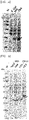
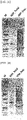
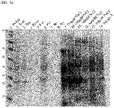
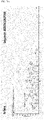
- the culture was centrifuged at 4,000 rpm for 15 minutes, and the cells were harvested by removing the supernatant. The harvested cells were added to SDS-sample buffer, heated at 95°C for 5 min, and centrifuged at 15,000 rpm for 5 min. Using the prepared samples, the expression and size of each target protein were analyzed through SDS-PAGE gel analysis ( FIG. 1 ).
- a strain confirmed as positive for extended spectrum ⁇ -lactamase was collected.
- the collected strain was subjected to colony PCR using the primers used for KPC and OXA gene amplification.
- the resulting amplified PCR products were subjected to agar gel electrophoresis to confirm the sizes of the target genes.
- the PCR products whose sizes have been confirmed were subjected to DNA sequencing to confirm the exact genotypes of the KPC and OXA genes.
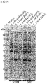
- the strain whose genotypes have been confirmed was cultured in LB liquid medium, and SDS-PAGE gel analysis was performed to examine whether KPC and OXA proteins would be expressed.
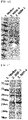
- a recombinant strain containing the KPC and OXA genes derived from the clinical strain was constructed in the same manner as the existing recombinant strain comprising the vector containing the KPC and OXA genes, and the recombinant strain was also subjected to SDS-PAGE gel analysis in the same manner as the clinical strain to confirm the sizes of the actually expressed KPC and OXA proteins ( FIG. 2 ).
- E. coli transformed with the plasmid containing the target MBL gene was inoculated into Luria-bertani liquid medium containing 50 mg/L of ampicillin antibiotic and zinc sulfate (0.01 mM to 1 mM ZnSO 4 ), and cultured at 37°C for 16 hours or more.
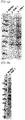
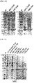
- the culture was centrifuged at 4,000 rpm for 15 minutes, and the cells were harvested by removing the supernatant. The harvested cells were added to SDS-sample buffer, heated at 95°C for 5 min, and centrifuged at 15,000 rpm for 5 min. Using the prepared sample, the expression and size of the target protein were analyzed through SDS-PAGE gel analysis ( FIG. 3 ).
- the culture was centrifuged at 4,000 rpm for 15 minutes, and the cells were harvested by removing the supernatant.
- the cells were treated with a buffer solution (0.25 mM Tris-HCl, 2% OG) and incubated at room temperature for 10 minutes.
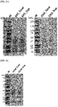
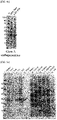
- the prepared crude extract was separated into a supernatant (hereinafter referred to as crude enzyme solution) and a precipitate by centrifugation at 15,000 rpm at 4°C for 10 minutes. From the crude enzyme solution, the expression and size of the target protein were analyzed by SDS-PAGE analysis ( FIG. 4 ).
- Sample pretreatment was performed by the following steps: 1) 100 ml of the expressed cell culture was centrifuged and to recover the cells, and 2) the supernatant was removed, and then a buffer solution (0.25 mM Tris-HCl, pH 8.0 and 2% DOC) containing 2% sodium deoxycholate (DOC) as a nonionic surfactant was added to the cells. 3) The suspension was incubated at room temperature for 10 minutes, and 4) centrifuged at 15,000 rpm at 4°C for 10 minutes to obtain a crude enzyme solution.
- a buffer solution (0.25 mM Tris-HCl, pH 8.0 and 2% DOC) containing 2% sodium deoxycholate (DOC) as a nonionic surfactant was added to the cells.
- DOC sodium deoxycholate
- the sample pretreatment method is as follows: 1) 50 mM ammonium bicarbonate (hereinafter referred to as ABC) solution was added to the harvested cells, 2) the cells were resuspended by pipetting, and then 3) the suspension was centrifuged at 12,000 rpm and 4°C for 10 minutes to recover the supernatant. From the crude extract and crude enzyme solution obtained by treatment, the expression and size of the protein were analyzed by SDS-PAGE gel analysis ( FIGS. 4c and 4e ).
- ABC ammonium bicarbonate
- the cells in the suspensions treated by the methods (1) to (3) above were disrupted by sonication using a Sonic bath (JAC 2010, Hansol Tech, Korea) at 40 Hz and 200 W for 5 to 10 minutes, and centrifuged at 15,000 rpm for 10 min, and the supernatant was recovered.
- a Sonic bath JOC 2010, Hansol Tech, Korea
- Ion exchange chromatography was used to separate/purify each target protein.
- anion exchange chromatography a column containing Q-resin was used, and for cation exchange chromatography, a column containing SP-resin was used.
- the crude enzyme solution was loaded into a column containing Q-resin, and then the eluted solution was collected.
- the column was washed with 1 ml of 20 mM Tris-HCl (pH 8.0) buffer, and elution solutions containing 100 mM NaCl, 200 mM NaCl, 300 mM NaCl, 400 mM NaCl and 500 mM NaCl 1M, respectively, in buffer, were sequentially loaded into the column in an amount of 250 ⁇ l for each elution solution, and the eluate was collected for each zone.
- each target protein was separated/purified in the same manner as (1) anion exchange resin chromatography.
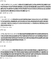
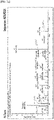
- the proteins whose expression and size were confirmed on the SDS-PAGE gel were identified using the in-gel digestion method and the nano-LC-MS/MS method, thereby confirming the type of antibiotic resistance protein actually expressed in the strain ( FIG. 7 ).
- nano liquid chromatography and high-resolution mass spectrometry were performed (Q-Exactive HF-X mass spectrometry system).
- the desalted peptide sample was dissolved with 0.1% formic acid solution and then loaded into a column.
- the peptide sample was separated using a C18 column (75 ⁇ m ⁇ 70 cm) and nanoflow liquid chromatography. Examples of the gradient conditions for sample loading and separation used in this case are as follows:
- FIG. 6a For the KPC protein, a peptide in which one methionine among three methionine residues (49, 116 and 151) was in an oxidized form was identified ( FIG. 6a ).
- OXA a peptide in which one methionine or two or three methionine residues among six methionine residues (115, 138, 195, 237, 239 and 241) were in an oxidized form was identified (FIG. 6b).
- N-terminal sequence peptides consisting of residues 1-18 and residues 1-26, respectively were not detected, and for GES, an N-terminal sequence peptide consisting of residues 1-18 was not detected.
- the identified sequence coverages were 97.2 (243/250) for NDM, 97.8% (223/228) for IMP, 100% (240/240) for VIM, and 98.5% (265/269) for GES.
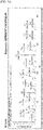
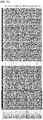
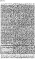
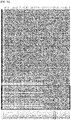
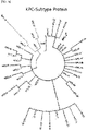
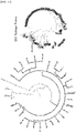
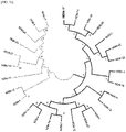
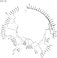
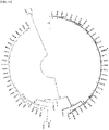
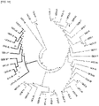
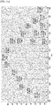
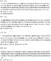
- FIG. 8a Based on the MS2 results obtained in Example 5, multiple alignment analysis was performed on a total of 43 KPC subtype proteins ( FIG. 8a ), 660 OXA subtype proteins ( FIG. 8b ), 27 NDM subtype proteins ( FIG. 8c ), 79 IMP subtype proteins ( FIG. 8d ), 66 VIM subtype proteins ( FIG. 8e ) and 43 GES subtype proteins ( FIG. 8f ) known to date in the National Center for Biotechnology Information (NCBI) database.
- NCBI National Center for Biotechnology Information
- OXA For OXA, 30 OXA proteins comprising the same N-terminal peptide (1 to 22 a.a.) were identified, and 23 OXA proteins were characterized by the same sequence as OXA-48 composed of a sequence consisting of 265 amino acids.
- NDM nuclear magnetic resonance
- IMP 63.5%
- VIM 63.5%
- GES 86.5%
- 27 NDM subtype proteins As subtype proteins having the same N-terminus, 27 NDM subtype proteins, 23 IMP subtype proteins (89.4%), 26 VIM subtype proteins (92.1%), and 35 GES subtype proteins (87.9%) were identified.
- 26 NDM subtype proteins As subtype proteins having the same peptide number as the N-terminal sequence and the same full-length amino acid sequence, 26 NDM subtype proteins (92.6%), 23 IMP subtype proteins (89.4%), 25 VIM subtype proteins (93.6%), and 34 GES subtype proteins (88.9%) were identified.
- Example 7 Target protein identification using mass spectrometry (top-down method)
- top-down mass spectrometry was performed.
- a sample obtained by partial purification from the crude protein extract derived from the strain was used, and an LC-MS/MS system (Nano-LC and Q-Exactive HF-X mass spectrometry system) capable of top-down analysis was used.
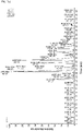
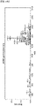
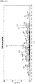
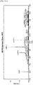
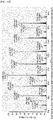
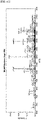
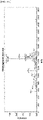
- FIGS. 10c , 10e , 10g and 10i Several peaks of multi-charged MBL proteins or GES protein appeared ( FIGS. 10c , 10e , 10g and 10i ), and it was confirmed that, for NDM, a representative mass value obtained by deconvolution of the peaks was an average molecular weight of 26,724 m/z ⁇ z, corresponding to a monoisotopic mass of 26,707 m/z ⁇ z.
- the average molecular weight for IMP was 25082.68 m/z ⁇ z, corresponding to a monoisotopic mass of 25,067.19 m/z ⁇ z
- the average molecular weight for VIM was 25,515.42 m/z ⁇ z, corresponding to a monoisotopic mass of 25,499.92 m/z ⁇ z
- the average molecular weight for GES was 29,245.15 m/z ⁇ z, corresponding to a monoisotopic mass of 29,226.92 m/z ⁇ z.
- the identified representative mass value for NDM corresponded to a palmitoylated protein type ( FIG. 11a ).
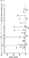
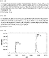
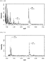
- the mass spectrometry spectrum of each protein was obtained using a low-resolution mass spectrometer (1, SciEX 4800; 2, Bruker Biotyper MALDI-TOF MS system).
- a low-resolution mass spectrometer (1, SciEX 4800; 2, Bruker Biotyper MALDI-TOF MS system).
- SA sinapinic acid
- 100 ng of each protein were placed on the plate spot, dried completely, and subjected to mass spectrometry.
- protein samples all of the colonies cultured in solid culture, cells harvested in liquid culture, crude extract and crude enzyme solution after cell lysis, and purified proteins obtained after purification can be used.
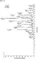
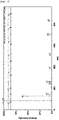
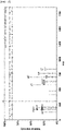
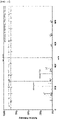
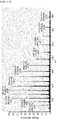
- Mass spectrometry spectra were obtained for the range of 10,000 to 40,000 m/z (KPC and OXA) or 35,000 m/z (MBL and GES), and each target protein with a charge state of +1 as well as each target protein with a charge state of +2 were simultaneously detected ( FIG. 13 ). As a result of low-resolution mass spectrometry, both charge states of +1 and +2 were detected for all the three MBL proteins and the GES protein.
- Example 8 Genotype identification and protein identification of MBL proteins and GES derived from clinical strains
- IMP and VIM MBL proteins
- GES Class A carbapenem protein
- the strain whose genotype was confirmed was cultured using LB liquid medium, and whether MBL proteins (NDM, IMP and VIM) and GES protein were expressed was analyzed by SDS-PAGE gel analysis ( FIG. 2 ).
- the clinical strain-derived MBL proteins and GES protein were digested by the in-gel digestion method and identified by the Bottom-Up method using the Q-Exactive HF-X plus system. It was confirmed that, for NDM, the sequence coverage for the identified peptides was 61.11% (165/270) in the full-length protein sequence and 65.74% (165/251) in the active form from which the N-terminus has been removed. It was confirmed that, for IMP and VIM, the sequence coverages for the identified peptides were 22.76% (56/246) and 44.74% (119/266), respectively, in the full-length protein sequences, and 24.56% (56/228) and 49.58% (119/240), respectively, in the active forms from which the N-terminus has been removed. In addition, it was confirmed that, for GES, the sequence coverage for the identified peptides were 67.25% (193/287) in the full-length amino acid sequence, and 71.75% (193/269) in the active protein sequence.
Landscapes
- Life Sciences & Earth Sciences (AREA)
- Health & Medical Sciences (AREA)
- Engineering & Computer Science (AREA)
- Chemical & Material Sciences (AREA)
- Molecular Biology (AREA)
- Physics & Mathematics (AREA)
- Immunology (AREA)
- Biochemistry (AREA)
- General Health & Medical Sciences (AREA)
- Analytical Chemistry (AREA)
- Bioinformatics & Cheminformatics (AREA)
- Proteomics, Peptides & Aminoacids (AREA)
- Organic Chemistry (AREA)
- Hematology (AREA)
- Biomedical Technology (AREA)
- Urology & Nephrology (AREA)
- General Physics & Mathematics (AREA)
- Pathology (AREA)
- Biotechnology (AREA)
- Microbiology (AREA)
- Bioinformatics & Computational Biology (AREA)
- Biophysics (AREA)
- Wood Science & Technology (AREA)
- Zoology (AREA)
- Food Science & Technology (AREA)
- Cell Biology (AREA)
- Spectroscopy & Molecular Physics (AREA)
- Medicinal Chemistry (AREA)
- General Engineering & Computer Science (AREA)
- Genetics & Genomics (AREA)
- Toxicology (AREA)
- Optics & Photonics (AREA)
- Virology (AREA)
- Tropical Medicine & Parasitology (AREA)
- Other Investigation Or Analysis Of Materials By Electrical Means (AREA)
- Investigating Or Analysing Biological Materials (AREA)
- Measuring Or Testing Involving Enzymes Or Micro-Organisms (AREA)
Applications Claiming Priority (3)
| Application Number | Priority Date | Filing Date | Title |
|---|---|---|---|
| KR20190177683 | 2019-12-30 | ||
| KR20200012108 | 2020-01-31 | ||
| PCT/KR2020/019464 WO2021137645A1 (fr) | 2019-12-30 | 2020-12-30 | Procédé de détection directe de souche pathogène présentant une résistance aux antibiotiques carbapénèmes |
Publications (2)
| Publication Number | Publication Date |
|---|---|
| EP4086632A1 true EP4086632A1 (fr) | 2022-11-09 |
| EP4086632A4 EP4086632A4 (fr) | 2024-02-28 |
Family
ID=76686703
Family Applications (1)
| Application Number | Title | Priority Date | Filing Date |
|---|---|---|---|
| EP20908619.8A Pending EP4086632A4 (fr) | 2019-12-30 | 2020-12-30 | Procédé de détection directe de souche pathogène présentant une résistance aux antibiotiques carbapénèmes |
Country Status (5)
| Country | Link |
|---|---|
| US (1) | US20230059795A1 (fr) |
| EP (1) | EP4086632A4 (fr) |
| KR (1) | KR20220123542A (fr) |
| CN (1) | CN115104030B (fr) |
| WO (1) | WO2021137645A1 (fr) |
Families Citing this family (2)
| Publication number | Priority date | Publication date | Assignee | Title |
|---|---|---|---|---|
| CN118006813B (zh) * | 2023-09-14 | 2025-07-29 | 北京金匙医学检验实验室有限公司 | 预测铜绿假单胞菌碳青霉烯类药物药敏表型的特征基因 |
| CN117761311B (zh) * | 2023-12-25 | 2025-10-03 | 威尔塔斯生物工程技术(上海)有限公司 | 用于检测细菌内碳青霉烯酶的免疫层析装置及检测方法 |
Family Cites Families (8)
| Publication number | Priority date | Publication date | Assignee | Title |
|---|---|---|---|---|
| FI119190B (fi) * | 2006-06-21 | 2008-08-29 | Ipsat Therapies Oy | Modifioitu beta-laktamaasi ja menetelmä sen valmistamiseksi |
| EP2576811A1 (fr) * | 2010-06-02 | 2013-04-10 | Johns Hopkins University | Système et procédés pour déterminer la résistance aux médicaments dans des micro-organismes |
| CN103765216B (zh) * | 2011-04-21 | 2017-08-11 | 生物梅里埃公司 | 使用质谱检测至少一种碳青霉烯抗性机制的方法 |
| FR3024465B1 (fr) * | 2014-07-30 | 2018-03-23 | Biomerieux | Caracterisation de micro-organismes par maldi-tof |
| WO2017127733A1 (fr) * | 2016-01-20 | 2017-07-27 | Thermo Finnigan Llc | Procédés de spectrométrie de masse rapide pour test de sensibilité aux antimicrobiens faisant appel à la spectrométrie de masse descendante |
| JP6232115B2 (ja) * | 2016-10-26 | 2017-11-15 | エラスムス ユニバーシティ メディカル センター ロッテルダムErasmus University Medical Center Rotterdam | 微生物が潜在的に抗菌剤化合物に対して耐性を有するか否かを決定するための方法 |
| AR112775A1 (es) * | 2017-08-17 | 2019-12-11 | Consejo Nacional De Investigaciones Cientificas Y Tecn Conicet | Detección directa de mecanismos de resistencia por espectrometría de masa maldi-tof |
| KR101999574B1 (ko) * | 2018-08-03 | 2019-09-27 | 다이아텍코리아 주식회사 | 질량분석법을 이용한 베타락탐 항생제 가수분해 효소의 활성형 직접 검출법 |
-
2020
- 2020-12-30 EP EP20908619.8A patent/EP4086632A4/fr active Pending
- 2020-12-30 KR KR1020227026396A patent/KR20220123542A/ko active Pending
- 2020-12-30 US US17/758,235 patent/US20230059795A1/en active Pending
- 2020-12-30 WO PCT/KR2020/019464 patent/WO2021137645A1/fr not_active Ceased
- 2020-12-30 CN CN202080096367.1A patent/CN115104030B/zh active Active
Also Published As
| Publication number | Publication date |
|---|---|
| US20230059795A1 (en) | 2023-02-23 |
| CN115104030A (zh) | 2022-09-23 |
| KR20220123542A (ko) | 2022-09-07 |
| WO2021137645A1 (fr) | 2021-07-08 |
| CN115104030B (zh) | 2025-11-21 |
| EP4086632A4 (fr) | 2024-02-28 |
Similar Documents
| Publication | Publication Date | Title |
|---|---|---|
| Ali et al. | Characterization of mutant MUTYH proteins associated with familial colorectal cancer | |
| Everett et al. | Contributions of individual mechanisms to fluoroquinolone resistance in 36 Escherichia coli strains isolated from humans and animals | |
| Lee et al. | Proteomic and transcriptional analysis of Lactobacillus johnsonii PF01 during bile salt exposure by iTRAQ shotgun proteomics and quantitative RT-PCR | |
| JPH10506263A (ja) | コンドロイチンリアーゼ酵素 | |
| EP2844762B1 (fr) | Test de sensibilité aux antibiotiques utilisant des sondes pour de l'arn pré-ribosomique | |
| JP2010515434A5 (fr) | ||
| Saves et al. | Mass spectral kinetic study of acylation and deacylation during the hydrolysis of penicillins and cefotaxime by. beta.-lactamase TEM-1 and the G238S mutant | |
| EP4086632A1 (fr) | Procédé de détection directe de souche pathogène présentant une résistance aux antibiotiques carbapénèmes | |
| Townsend et al. | Identification of protein kinase A phosphorylation sites on NBD1 and R domains of CFTR using electrospray mass spectrometry with selective phosphate ion monitoring | |
| WO2015013324A1 (fr) | Amdinocilline permettant la détermination rapide de la sensibilité à des antibiotiques bêta-lactames | |
| US7786103B2 (en) | Method for isolating an intestinal cholesterol binding protein | |
| Holberger et al. | Ribosomal protein S12 and aminoglycoside antibiotics modulate A-site mRNA cleavage and transfer-messenger RNA activity in Escherichia coli | |
| Van Oudenhove et al. | Differential proteomic analysis of the response of Stenotrophomonas maltophilia to imipenem | |
| JP2002541775A (ja) | フォンヒッペルリンドウ腫瘍サプレッサー複合体およびscfユビキチンリガーゼの新規成分 | |
| US5863762A (en) | Nucleic acids encoding TFIIB transcription factor from candida albicans | |
| EP4009052A1 (fr) | Procédé pour détecter directement une souche pathogène présentant une résistance aux antibiotiques bêta-lactame | |
| Bertrand et al. | N*-methyl coprogen B, a potential marker of the airway colonization by Scedosporium apiospermum in patients with cystic fibrosis | |
| JP2004520010A (ja) | ヌクレオチド配列中の突然変異を検出するための方法 | |
| JP2000509605A (ja) | テイコ酸酵素および検定法 | |
| Wang et al. | Exploring membrane and cytoplasm proteomic responses of Alkalimonas amylolytica N10 to different external pHs with combination strategy of de novo peptide sequencing | |
| US6232095B1 (en) | Recombinant helix modification recognition proteins and uses thereof | |
| KR20240037468A (ko) | 그람 음성균 단백질의 분리를 위한 순차적 전처리 방법 | |
| HUT69971A (en) | Purified pala-nitrobenzyl esterase from bacillus | |
| JP4468299B2 (ja) | 細菌の増殖抑制剤のスクリーニング方法 | |
| US20240051995A1 (en) | Antibacterial agents |
Legal Events
| Date | Code | Title | Description |
|---|---|---|---|
| STAA | Information on the status of an ep patent application or granted ep patent |
Free format text: STATUS: THE INTERNATIONAL PUBLICATION HAS BEEN MADE |
|
| PUAI | Public reference made under article 153(3) epc to a published international application that has entered the european phase |
Free format text: ORIGINAL CODE: 0009012 |
|
| STAA | Information on the status of an ep patent application or granted ep patent |
Free format text: STATUS: REQUEST FOR EXAMINATION WAS MADE |
|
| 17P | Request for examination filed |
Effective date: 20220728 |
|
| AK | Designated contracting states |
Kind code of ref document: A1 Designated state(s): AL AT BE BG CH CY CZ DE DK EE ES FI FR GB GR HR HU IE IS IT LI LT LU LV MC MK MT NL NO PL PT RO RS SE SI SK SM TR |
|
| DAV | Request for validation of the european patent (deleted) | ||
| DAX | Request for extension of the european patent (deleted) | ||
| A4 | Supplementary search report drawn up and despatched |
Effective date: 20240129 |
|
| RIC1 | Information provided on ipc code assigned before grant |
Ipc: G01N 33/569 20060101ALI20240123BHEP Ipc: C12Q 1/34 20060101ALI20240123BHEP Ipc: C12Q 1/10 20060101ALI20240123BHEP Ipc: G01N 30/88 20060101ALI20240123BHEP Ipc: C12Q 1/04 20060101ALI20240123BHEP Ipc: G01N 27/64 20060101ALI20240123BHEP Ipc: G01N 30/96 20060101ALI20240123BHEP Ipc: G01N 30/72 20060101ALI20240123BHEP Ipc: G01N 33/68 20060101AFI20240123BHEP |