EP4134095A1 - Thérapie par anti-cd47 synergique pour les cancers hématologiques - Google Patents
Thérapie par anti-cd47 synergique pour les cancers hématologiques Download PDFInfo
- Publication number
- EP4134095A1 EP4134095A1 EP22185805.3A EP22185805A EP4134095A1 EP 4134095 A1 EP4134095 A1 EP 4134095A1 EP 22185805 A EP22185805 A EP 22185805A EP 4134095 A1 EP4134095 A1 EP 4134095A1
- Authority
- EP
- European Patent Office
- Prior art keywords
- cells
- antibody
- cell
- human
- expression
- Prior art date
- Legal status (The legal status is an assumption and is not a legal conclusion. Google has not performed a legal analysis and makes no representation as to the accuracy of the status listed.)
- Pending
Links
Images












Classifications
-
- C—CHEMISTRY; METALLURGY
- C07—ORGANIC CHEMISTRY
- C07K—PEPTIDES
- C07K16/00—Immunoglobulins [IG], e.g. monoclonal or polyclonal antibodies
- C07K16/18—Immunoglobulins [IG], e.g. monoclonal or polyclonal antibodies against material from animals or humans
- C07K16/28—Immunoglobulins [IG], e.g. monoclonal or polyclonal antibodies against material from animals or humans against receptors, cell surface antigens or cell surface determinants
- C07K16/2803—Immunoglobulins [IG], e.g. monoclonal or polyclonal antibodies against material from animals or humans against receptors, cell surface antigens or cell surface determinants against the immunoglobulin superfamily
-
- A—HUMAN NECESSITIES
- A61—MEDICAL OR VETERINARY SCIENCE; HYGIENE
- A61K—PREPARATIONS FOR MEDICAL, DENTAL OR TOILETRY PURPOSES
- A61K39/00—Medicinal preparations containing antigens or antibodies
- A61K39/395—Antibodies; Immunoglobulins; Immune serum, e.g. antilymphocytic serum
- A61K39/39533—Antibodies; Immunoglobulins; Immune serum, e.g. antilymphocytic serum against materials from animals
- A61K39/39558—Antibodies; Immunoglobulins; Immune serum, e.g. antilymphocytic serum against materials from animals against tumor tissues, cells, antigens
-
- A—HUMAN NECESSITIES
- A61—MEDICAL OR VETERINARY SCIENCE; HYGIENE
- A61K—PREPARATIONS FOR MEDICAL, DENTAL OR TOILETRY PURPOSES
- A61K51/00—Preparations containing radioactive substances for use in therapy or testing in vivo
- A61K51/02—Preparations containing radioactive substances for use in therapy or testing in vivo characterised by the carrier, i.e. characterised by the agent or material covalently linked or complexing the radioactive nucleus
- A61K51/04—Organic compounds
- A61K51/08—Peptides, e.g. proteins, carriers being peptides, polyamino acids, proteins
- A61K51/10—Antibodies or immunoglobulins; Fragments thereof, the carrier being an antibody, an immunoglobulin or a fragment thereof, e.g. a camelised human single domain antibody or the Fc fragment of an antibody
- A61K51/1093—Antibodies or immunoglobulins; Fragments thereof, the carrier being an antibody, an immunoglobulin or a fragment thereof, e.g. a camelised human single domain antibody or the Fc fragment of an antibody conjugates with carriers being antibodies
-
- A—HUMAN NECESSITIES
- A61—MEDICAL OR VETERINARY SCIENCE; HYGIENE
- A61P—SPECIFIC THERAPEUTIC ACTIVITY OF CHEMICAL COMPOUNDS OR MEDICINAL PREPARATIONS
- A61P35/00—Antineoplastic agents
-
- A—HUMAN NECESSITIES
- A61—MEDICAL OR VETERINARY SCIENCE; HYGIENE
- A61P—SPECIFIC THERAPEUTIC ACTIVITY OF CHEMICAL COMPOUNDS OR MEDICINAL PREPARATIONS
- A61P35/00—Antineoplastic agents
- A61P35/02—Antineoplastic agents specific for leukemia
-
- C—CHEMISTRY; METALLURGY
- C07—ORGANIC CHEMISTRY
- C07K—PEPTIDES
- C07K16/00—Immunoglobulins [IG], e.g. monoclonal or polyclonal antibodies
- C07K16/18—Immunoglobulins [IG], e.g. monoclonal or polyclonal antibodies against material from animals or humans
- C07K16/28—Immunoglobulins [IG], e.g. monoclonal or polyclonal antibodies against material from animals or humans against receptors, cell surface antigens or cell surface determinants
- C07K16/2887—Immunoglobulins [IG], e.g. monoclonal or polyclonal antibodies against material from animals or humans against receptors, cell surface antigens or cell surface determinants against CD20
-
- A—HUMAN NECESSITIES
- A61—MEDICAL OR VETERINARY SCIENCE; HYGIENE
- A61K—PREPARATIONS FOR MEDICAL, DENTAL OR TOILETRY PURPOSES
- A61K39/00—Medicinal preparations containing antigens or antibodies
- A61K2039/505—Medicinal preparations containing antigens or antibodies comprising antibodies
- A61K2039/507—Comprising a combination of two or more separate antibodies
-
- C—CHEMISTRY; METALLURGY
- C07—ORGANIC CHEMISTRY
- C07K—PEPTIDES
- C07K2317/00—Immunoglobulins specific features
- C07K2317/70—Immunoglobulins specific features characterized by effect upon binding to a cell or to an antigen
- C07K2317/73—Inducing cell death, e.g. apoptosis, necrosis or inhibition of cell proliferation
- C07K2317/732—Antibody-dependent cellular cytotoxicity [ADCC]
-
- C—CHEMISTRY; METALLURGY
- C07—ORGANIC CHEMISTRY
- C07K—PEPTIDES
- C07K2317/00—Immunoglobulins specific features
- C07K2317/70—Immunoglobulins specific features characterized by effect upon binding to a cell or to an antigen
- C07K2317/73—Inducing cell death, e.g. apoptosis, necrosis or inhibition of cell proliferation
- C07K2317/734—Complement-dependent cytotoxicity [CDC]
-
- C—CHEMISTRY; METALLURGY
- C07—ORGANIC CHEMISTRY
- C07K—PEPTIDES
- C07K2317/00—Immunoglobulins specific features
- C07K2317/70—Immunoglobulins specific features characterized by effect upon binding to a cell or to an antigen
- C07K2317/77—Internalization into the cell
Definitions
- Therapeutic antibodies have proven effective at fighting cancer, especially in cases where conventional therapy fails: Out of the 21 marketed therapeutic antibodies, 9 are for the treatment of cancer. Even more encouraging is that antibodies for cancer generally operate in a distinct mechanism from traditional chemotherapy or radiotherapy, so they can often be combined with traditional therapies to generate a synergistic effect. However, there is still a lot of room for improvement, in that even the most effective cancer antibodies rarely cure the disease, especially in its more advanced stages.
- the first patient treated in the United States with monoclonal antibody therapy was a patient with non-Hodgkin's lymphoma, using a murine monoclonal antibody designated AB 89.
- treatment was not successful in inducing a significant clinical response, it did represent the first proof of principle in humans that a monoclonal antibody could induce transient decreases in the number of circulating tumor cells, induce circulating dead cells, and form complexes with circulating antigen, all with minimal toxicity to the patient.
- a monoclonal antibody could induce transient decreases in the number of circulating tumor cells, induce circulating dead cells, and form complexes with circulating antigen, all with minimal toxicity to the patient.
- there was much excitement about this new treatment modality there were also problems and limitations, including the modulation of antigens off the cell surface and into circulation or internalization.
- Monoclonal antibodies achieve their therapeutic effect through various mechanisms. They can have direct effects in producing apoptosis or programmed cell death. They can block growth factor receptors, effectively arresting proliferation of tumor cells. In cells that express monoclonal antibodies, they can bring about anti-idiotype antibody formation. Indirect effects include recruiting cells that have cytotoxicity, such as monocytes and macrophages. This type of antibody-mediated cell kill is called antibody-dependent cell mediated cytotoxicity (ADCC). Monoclonal antibodies also bind complement, leading to direct cell toxicity, known as complement dependent cytotoxicity (CDC).
- ADCC antibody-dependent cell mediated cytotoxicity
- Rituximab became the first monoclonal antibody approved specifically for cancer therapy, and a number of other naked or immunoconjugated antibodies have followed, including a humanized monoclonal antibody to CD33 conjugated to calicheamicin, and radiolabeled antibodies specific for CD20 (ytrrium-90 ibritumomab tiuxetan and iodine-131 tositumomab).
- Rituximab has become the largest-selling biologic drug in clinical oncology, and is active in a variety of human lymphomas and chronic lymphocytic leukemia. This is a chimeric monoclonal antibody targeting the CD20 antigen found on both normal B cells and on most low-grade and some higher grade B-cell lymphomas. It can be effective as a single agent in induction and maintenance therapy. It is primarily used, however, in combination with standard chemotherapies in the treatment of patients with non-Hodgkin's B-cell lymphomas and chronic lymphocytic leukemia.
- Alemtuzumab is a humanized monoclonal antibody targeting the CD52 antigen found on B lymphocytes and is used primarily for chronic lymphocytic leukemia, and is effective as induction and maintenance therapy. Alemtuzumab is also reactive with T lymphocytes, and is typically not combined with chemotherapy because of the increased risk of infection.
- the CD22 antigen is also targeted by a number of antibodies, and has recently demonstrated efficacy combined with toxin in chemotherapy-resistant hairy cell leukemia.
- compositions are provided for the treatment of hematologic tumors with a synergistic combination of agents.
- One of the agents targets the tumor cells for phagocytosis by blocking CD47 on the cell surface. It is shown that leukemia or lymphoma cells evade macrophage surveillance by upregulation of CD47 expression.
- Administration of agents that mask the CD47 protein e.g. antibodies or small molecules that bind to CD47 and prevent interaction between CD47 and SIRP ⁇ , are administered to a patient, which increases the clearance of cancer cells via phagocytosis.
- the agent that masks CD47 is combined with monoclonal antibodies directed against one or more additional cancer cell markers, e.g. CD96, CD20, CD22, CD33, CD52, CD123, CD44, etc., which compositions can be synergistic in enhancing phagocytosis and elimination of cancer cells as compared to the use of single agents.
- the therapy provides for a combination of an agent that blocks CD47, and a second agent that is directed to a second cancer cell marker.
- Specific combinations of interest include anti-CD47 and anti-CD20, e.g. rituximab, tiuxetan, tositumomab, etc., which combination finds particular use in the treatment of non-Hodgkin's B cell lymphomas and chronic lymphocytic leukemia (CLL).
- a combination of anti-CD47 and anti-CD22 e.g. Epratuzumab, etc. finds particular use in the treatment of B cell leukemias and hairy cell leukemias.
- a combination of anti-CD47 and anti-CD52 e.g. alemtuzumab, etc., finds particular use in the treatment of B cell and T cell leukemias, including without limitation chronic lymphocytic leukemia.
- a combination of anti-CD47 and anti-CD33 finds particular use in the treatment of myeloid leukemias such as acute myelogenous leukemias.
- Other combination of interest for treatment of myelogenous leukemias include, without limitation, anti-CD47 and anti-CD96; anti-CD47 and anti-CD44; anti-CD47 and anti-CD123.
- the therapy provides for a multispecific antibody that targets CD47 and a second cancer cell marker, including multispecific antibodies that target CD47 and CD20; CD47 and CD22; CD47 and CD52; CD47 and CD33; CD47 and CD96; CD47 and CD44; Cd47 and CD123; and the like.
- Compositions of such multispecific antibodies are also provided, where the multispecific antibody is desirably human or humanized; and may be modified to extend the blood half-life, e.g. by pegylation, etc.
- Methods and compositions are provided for the treatment of hematologic tumors with a synergistic combination of agents targeting CD47 and a second cancer cell marker, e.g. CD96, CD20, CD22, CD33, CD52, CD123, CD44, etc.
- a synergistic combination of agents targeting CD47 and a second cancer cell marker e.g. CD96, CD20, CD22, CD33, CD52, CD123, CD44, etc.
- Synergistic combination may provide for a therapeutic effect that is comparable to the effectiveness of a monotherapy, while reducing adverse side effects, e.g. damage to non-targeted tissues, immune status, and other clinical indicia.
- synergistic combinations may provide for an improved effectiveness, which effect may be measured by total tumor cell number; length of time to relapse; and other indicia of patient health.
- Synergistic combinations of the present invention combine an agent that is targeted to inhibit or block CD47 function; and an agent that is targeted to inhibit or block a second cancer cell marker, usually a cell surface marker.
- the combination may be provided with a combination of agents, e.g. two distinct antibodies, each of which is specific for a different marker; or may be provided as a multispecific agent, e.g. antibody, that combines specificity for two or more different markers.
- CD47 polypeptides The three transcript variants of human CD 47 (variant 1, NM 001777; variant 2, NM 198793; and variant 3, NM 001025079) encode three isoforms of CD47 polypeptide.
- CD47 isoform 1 (NP 001768), the longest of the three isoforms, is 323 amino acids long.
- CD47 isoform 2 (NP 942088) is 305 amino acid long.
- CD47 isoform 3 is 312 amino acids long.
- the three isoforms are identical in sequence in the first 303 amino acids.
- Amino acids 1-8 comprise the signal sequence
- amino acids 9-142 comprise the CD47 immunoglobulin like domain, which is the soluble fragment
- amino acids 143-300 is the transmembrane domain.
- a “fusion” polypeptide is a polypeptide comprising a polypeptide or portion (e.g., one or more domains) thereof fused or bonded to heterologous polypeptide.
- a fusion soluble CD47 protein for example, will share at least one biological property in common with a native sequence soluble CD47 polypeptide.
- Examples of fusion polypeptides include immunoadhesins, as described above, which combine a portion of the CD47 polypeptide with an immunoglobulin sequence, and epitope tagged polypeptides, which comprise a soluble CD47 polypeptide or portion thereof fused to a "tag polypeptide".
- the tag polypeptide has enough residues to provide an epitope against which an antibody can be made, yet is short enough such that it does not interfere with biological activity of the CD47 polypeptide.
- Suitable tag polypeptides generally have at least six amino acid residues and usually between about 6-60 amino acid residues.
- a “functional derivative” of a native sequence polypeptide is a compound having a qualitative biological property in common with a native sequence polypeptide.
- “Functional derivatives” include, but are not limited to, fragments of a native sequence and derivatives of a native sequence polypeptide and its fragments, provided that they have a biological activity in common with a corresponding native sequence polypeptide.
- the term “derivative” encompasses both amino acid sequence variants of polypeptide and covalent modifications thereof. Derivatives and fusion of soluble CD47 find use as CD47 mimetic molecules.
- the first 142 amino acids of CD47 polypeptide comprise the extracellular region of CD47 (SEQ ID NO: 1).
- the three isoforms have identical amino acid sequence in the extracellular region, and thus any of the isoforms are can be used to generate soluble CD47.
- “Soluble CD47” is a CD47 protein that lacks the transmembrane domain. Soluble CD47 is secreted out of the cell expressing it instead of being localized at the cell surface.
- I n vitro assays for CD47 biological activity include, e.g. inhibition of phagocytosis of porcine cells by human macrophages, binding to SIRP ⁇ receptor, SIRP ⁇ tyrosine phosphorylation, etc.
- An exemplary assay for CD47 biological activity contacts a human macrophage composition in the presence of a candidate agent. The cells are incubated with the candidate agent for about 30 minutes and lysed. The cell lysate is mixed with anti-human SIRP ⁇ antibodies to immunoprecipitate SIRP ⁇ . Precipitated proteins are resolved by SDS PAGE, then transferred to nitrocellulose and probed with antibodies specific for phosphotyrosine.
- a candidate agent useful as a CD47 mimetic increases SIRP ⁇ tyrosine phosphorylation by at least 10%, or up to 20%, or 50%, or 70% or 80% or up to about 90% compared to the level of phosphorylation observed in the absence of candidate agent.
- Another exemplary assay for CD47 biological activity measures phagocytosis of hematopoietic cells by human macrophages.
- a candidate agent useful as a CD47 mimetic results in the down regulation of phagocytosis by at least about 10%, at least about 20%, at least about 50%, at least about 70%, at least about 80%, or up to about 90% compared to level of phagocytosis observed in absence of candidate agent.
- manipulating phagocytosis is meant an up-regulation or a down-regulation in phagocytosis by at least about 10%, or up to 20%, or 50%, or 70% or 80% or up to about 90% compared to level of phagocytosis observed in absence of intervention.
- manipulating phagocytosis means a down-regulation in phagocytosis by at least about 10%, or up to 20%, or 50%, or 70% or 80% or up to about 90% compared to level of phagocytosis observed in absence of intervention.
- CD47 inhibitors Agents of interest as CD47 inhibitors include specific binding members that prevent the binding of CD47 with SIRP ⁇ receptor.
- the term "specific binding member” or “binding member” as used herein refers to a member of a specific binding pair, i.e. two molecules, usually two different molecules, where one of the molecules (i.e., first specific binding member) through chemical or physical means specifically binds to the other molecule (i.e., second specific binding member).
- CD47 inhibitors useful in the methods of the invention include analogs, derivatives and fragments of the original specific binding member.
- the specific binding member is an antibody.
- antibody or “antibody moiety” is intended to include any polypeptide chain-containing molecular structure with a specific shape that fits to and recognizes an epitope, where one or more non-covalent binding interactions stabilize the complex between the molecular structure and the epitope.
- Antibodies utilized in the present invention may be polyclonal antibodies, although monoclonal antibodies are preferred because they may be reproduced by cell culture or recombinantly, and can be modified to reduce their antigenicity.
- Polyclonal antibodies can be raised by a standard protocol by injecting a production animal with an antigenic composition. See, e.g., Harlow and Lane, Antibodies: A Laboratory Manual, Cold Spring Harbor Laboratory, 1988 .
- antibodies may be raised by immunizing the production animal with the protein and a suitable adjuvant (e.g., Freund's, Freund's complete, oil-in-water emulsions, etc.)
- a suitable adjuvant e.g., Freund's, Freund's complete, oil-in-water emulsions, etc.
- conjugate proteins that are commercially available for such use include bovine serum albumin (BSA) and keyhole limpet hemocyanin (KLH).
- BSA bovine serum albumin
- KLH keyhole limpet hemocyanin
- peptides derived from the full sequence may be utilized.
- a superior immune response may be elicited if the polypeptide is joined to a carrier protein, such as ovalbumin, BSA or KLH.
- hybridomas may be formed by isolating the stimulated immune cells, such as those from the spleen of the inoculated animal.
- immortalized cells such as myeloma cells or transformed cells, which are capable of replicating indefinitely in cell culture, thereby producing an immortal, immunoglobulin-secreting cell line.
- immortalized cells such as myeloma cells or transformed cells, which are capable of replicating indefinitely in cell culture, thereby producing an immortal, immunoglobulin-secreting cell line.
- the antibodies or antigen binding fragments may be produced by genetic engineering. Humanized, chimeric, or xenogeneic human antibodies, which produce less of an immune response when administered to humans, are preferred for use in the present invention.
- immunoglobulin fragments comprising the epitope binding site (e.g., Fab', F(ab') 2 , or other fragments) are useful as antibody moieties in the present invention.
- Such antibody fragments may be generated from whole immunoglobulins by ricin, pepsin, papain, or other protease cleavage.
- "Fragment,” or minimal immunoglobulins may be designed utilizing recombinant immunoglobulin techniques.
- Fv immunoglobulins for use in the present invention may be produced by linking a variable light chain region to a variable heavy chain region via a peptide linker (e.g., poly-glycine or another sequence which does not form an alpha helix or beta sheet motif).
- a peptide linker e.g., poly-glycine or another sequence which does not form an alpha helix or beta sheet motif.
- Antibodies include free antibodies and antigen binding fragments derived therefrom, and conjugates, e.g. pegylated antibodies, drug, radioisotope, or toxin conjugates, and the like. Monoclonal antibodies directed against a specific epitope, or combination of epitopes, will allow for the targeting and/or depletion of cellular populations expressing the marker.
- Various techniques can be utilized using monoclonal antibodies to screen for cellular populations expressing the marker(s), and include magnetic separation using antibody-coated magnetic beads, "panning" with antibody attached to a solid matrix (i.e., plate), and flow cytometry (See, e.g., U.S. Pat. No. 5,985,660 ; and Morrison et al. Cell, 96:737-49 (1999 )). These techniques allow for the screening of particular populations of cells; in immunohistochemistry of biopsy samples; in detecting the presence of markers shed by cancer cells into the blood and other biologic fluids, and the like.
- Humanized versions of such antibodies are also within the scope of this invention. Humanized antibodies are especially useful for in vivo applications in humans due to their low antigenicity.
- multispecific or bispecific antibody refers to a synthetic or recombinant antibody that recognizes more than one protein.
- Examples include bispecific antibodies 2B1, 520C9xH22, mDX-H210, and MDX447.
- Bispecific antibodies directed against a combination of epitopes will allow for the targeting and/or depletion of cellular populations expressing the combination of epitopes.
- Exemplary bi-specific antibodies include those targeting a combination of CD47 and a cancer cell marker, such as, CD20, CD22, CD96, CD97, CD99, PTHR2, HAVCR2 etc.
- Generation of bi-specific antibodies are described in the literature, for example, in USPN 5989830, USPN 5798229, which are incorporated herein by reference. Higher order specificities, e.g. trispecific antibodies, are described by Holliger and Hudson (2005) Nature Biotechnology 23:1126-1136 .
- the efficacy of a CD47 inhibitor is assessed by assaying CD47 activity.
- the above-mentioned assays or modified versions thereof are used.
- AML SCs are incubated with bone marrow derived macrophages, in the presence or absence of the candidate agent.
- An inhibitor of the cell surface CD47 will up-regulate phagocytosis by at least about 10%, or up to 20%, or 50%, or 70% or 80% or up to about 90% compared to the phagocytosis in absence of the candidate agent.
- an in vitro assay for levels of tyrosine phosphorylation of SIRP ⁇ will show a decrease in phosphorylation by at least about 10%, or up to 20%, or 50%, or 70% or 80% or up to about 90% compared to phosphorylation observed in absence of the candidate agent.
- the agent, or a pharmaceutical composition comprising the agent is provided in an amount effective to detectably inhibit the binding of CD47 to SIRP ⁇ receptor present on the surface of phagocytic cells.
- the effective amount is determined via empirical testing routine in the art. The effective amount may vary depending on the number of cells being transplanted, site of transplantation and factors specific to the transplant recipient.
- phagocytic cells and “phagocytes” are used interchangeably herein to refer to a cell that is capable of phagocytosis.
- phagocytes There are four main categories of phagocytes: macrophages, mononuclear cells (histiocytes and monocytes); polymorphonuclear leukocytes (neutrophils) and dendritic cells.
- biological sample encompasses a variety of sample types obtained from an organism and can be used in a diagnostic or monitoring assay.
- the term encompasses blood and other liquid samples of biological origin, solid tissue samples, such as a biopsy specimen or tissue cultures or cells derived therefrom and the progeny thereof.
- the term encompasses samples that have been manipulated in any way after their procurement, such as by treatment with reagents, solubilization, or enrichment for certain components.
- the term encompasses a clinical sample, and also includes cells in cell culture, cell supernatants, cell lysates, serum, plasma, biological fluids, and tissue samples.
- treatment means treating, “treating”, “treat” and the like to generally refer to obtaining a desired pharmacologic and/or physiologic effect.
- the effect may be prophylactic in terms of completely or partially preventing a disease or symptom thereof and/or may be therapeutic in terms of a partial or complete stabilization or cure for a disease and/or adverse effect attributable to the disease.
- Treatment covers any treatment of a disease in a mammal, e.g.
- mice, rat, rabbit, pig, primate including humans and other apes, particularly a human, and includes: (a) preventing the disease or symptom from occurring in a subject which may be predisposed to the disease or symptom but has not yet been diagnosed as having it; (b) inhibiting the disease symptom, i.e., arresting its development; or (c) relieving the disease symptom, i.e., causing regression of the disease or symptom.
- a "host cell”, as used herein, refers to a microorganism or a eukaryotic cell or cell line cultured as a unicellular entity which can be, or has been, used as a recipient for a recombinant vector or other transfer polynucleotides, and include the progeny of the original cell which has been transfected. It is understood that the progeny of a single cell may not necessarily be completely identical in morphology or in genomic or total DNA complement as the original parent, due to natural, accidental, or deliberate mutation.
- cancer neoplasm
- tumor tumor
- tumor tumor
- tumor tumor
- tumor tumor
- tumor tumor
- cells of interest for detection or treatment in the present application include precancerous (e.g., benign), malignant, pre-metastatic, metastatic, and non-metastatic cells. Detection of cancerous cells is of particular interest.
- normal as used in the context of "normal cell,” is meant to refer to a cell of an untransformed phenotype or exhibiting a morphology of a non-transformed cell of the tissue type being examined.
- Cancerous phenotype generally refers to any of a variety of biological phenomena that are characteristic of a cancerous cell, which phenomena can vary with the type of cancer.
- the cancerous phenotype is generally identified by abnormalities in, for example, cell growth or proliferation (e.g., uncontrolled growth or proliferation), regulation of the cell cycle, cell mobility, cell-cell interaction, or metastasis, etc.
- “Therapeutic target” refers to a gene or gene product that, upon modulation of its activity (e.g., by modulation of expression, biological activity, and the like), can provide for modulation of the cancerous phenotype.
- modulation is meant to refer to an increase or a decrease in the indicated phenomenon (e.g., modulation of a biological activity refers to an increase in a biological activity or a decrease in a biological activity).
- Acute leukemias are rapidly progressing leukemia characterized by replacement of normal bone marrow by blast cells of a clone arising from malignant transformation of a hematopoietic cell.
- the acute leukemias include acute lymphoblastic leukemia (ALL) and acute myelogenous leukemia (AML). ALL often involves the CNS, whereas acute monoblastic leukemia involves the gums, and AML involves localized collections in any site (granulocytic sarcomas or chloromas).
- the presenting symptoms are usually nonspecific (e.g., fatigue, fever, malaise, weight loss) and reflect the failure of normal hematopoiesis.
- Anemia and thrombocytopenia are very common (75 to 90%).
- the WBC count may be decreased, normal, or increased.
- Blast cells are usually found in the blood smear unless the WBC count is markedly decreased.
- the blasts of ALL can be distinguished from those of AML by histochemical studies, cytogenetics, immunophenotyping, and molecular biology studies. In addition to smears with the usual stains, terminal transferase, myeloperoxidase, Sudan black B, and specific and nonspecific esterase.
- ALL is the most common malignancy in children, with a peak incidence from ages 3 to 5 yr. It also occurs in adolescents and has a second, lower peak in adults.
- Typical treatment emphasizes early introduction of an intensive multidrug regimen, which may include prednisone, vincristine, anthracycline or asparaginase.
- Other drugs and combinations are cytarabine and etoposide, and cyclophosphamide.
- Relapse usually occurs in the bone marrow but may also occur in the CNS or testes, alone or concurrent with bone marrow. Although second remissions can be induced in many children, subsequent remissions tend to be brief.
- AML AML may be associated with chemotherapy or irradiation (secondary AML).
- Remission induction rates are lower than with ALL, and long-term disease-free survival reportedly occurs in only 20 to 40% of patients.
- Treatment differs most from ALL in that AML responds to fewer drugs.
- the basic induction regimen includes cytarabine; along with daunorubicin or idarubicin. Some regimens include 6-thioguanine, etoposide, vincristine, and prednisone.
- PV Polycythemia vera
- Hb concentration and RBC mass erythrocytosis
- PV occurs in about 2.3/100,000 people per year; more often in males (about 1.4:1).
- the mean age at diagnosis is 60 yr (range, 15 to 90 yr; rarely in childhood); 5% of patients are ⁇ 40 yr at onset.
- the bone marrow sometimes appears normal but usually is hypercellular; hyperplasia involves all marrow elements and replaces marrow fat.
- RBCs, neutrophils, and platelets Increased megakaryocytes may be present in clumps.
- Marrow iron is absent in > 90% of patients, even when phlebotomy has not been performed.
- Myelodysplastic syndrome is a group of syndromes (preleukemia, refractory anemias, Ph-negative chronic myelocytic leukemia, chronic myelomonocytic leukemia, myeloid metaplasia) commonly seen in older patients. Exposure to carcinogens may by be implicated. MDS is characterized by clonal proliferation of hematopoietic cells, including erythroid, myeloid, and megakaryocytic forms. The bone marrow is normal or hypercellular, and ineffective hematopoiesis causes variable cytopenias, the most frequent being anemia. The disordered cell production is also associated with morphologic cellular abnormalities in marrow and blood.
- Extramedullary hematopoiesis may occur, leading to hepatomegaly and splenomegaly.
- Myelofibrosis is occasionally present at diagnosis or may develop during the course of MDS.
- the MDS clone is unstable and tends to progress to AML.
- Non-Hodgkin lymphomas are a heterogeneous group of disorders involving malignant monoclonal proliferation of lymphoid cells in lymphoreticular sites, including lymph nodes, bone marrow, the spleen, the liver, and the Gl tract. It is the 6th most common cancer in the US; about 65,000 new cases are diagnosed annually in all age groups. Presenting symptoms usually include peripheral lymphadenopathy. However, some patients present without adenopathy but with abnormal lymphocytes in circulation. Compared with Hodgkin lymphoma, there is a greater likelihood of disseminated disease at the time of diagnosis. Diagnosis is usually based on lymph node or bone marrow biopsy or both. Treatment involves radiation therapy, chemotherapy, or both. Stem cell transplantation is usually reserved for salvage therapy after incomplete remission or relapse.
- NHLs arise from B cells; the remainder arise from T cells or natural killer cells. Either precursor or mature cells may be involved. Overlap exists between leukemia and NHL because both involve proliferation of lymphocytes or their precursors. A leukemia-like picture with peripheral lymphocytosis and bone marrow involvement may be present in up to 50% of children and in about 20% of adults with some types of NHL.
- lymphomas within this group are: precursor B-lymphoblastic leukemia/lymphoma; B-cell chronic lymphocytic leukemia/small lymphocytic lymphoma; B-cell prolymphocytic leukemia; Lymphoplasmacytic lymphoma; Splenic marginal zone B-cell lymphoma ( ⁇ villous lymphocytes); Hairy cell leukemia; Plasma cell myeloma/plasmacytoma; Extranodal marginal zone B-cell lymphoma of the MALT type; Nodal marginal zone ⁇ -cell lymphoma ( ⁇ monocytoid B cells); Follicular lymphoma; Mantle cell lymphoma; Diffuse large B-cell lymphomas; Burkitt's lymphoma; Precursor T-lymphoblastic lymphoma/leukemia; T-cell prolymphocytic leukemia; T-cell granular lymphocytic leukemia; Aggressive NK cell leukemia
- B cell hematologic cancers within the NHL group include, without limitation, diffuse large B cell lymphoma, chronic lymphocytic leukemia, mantle cell lymphoma, B lymphoblastic leukemia/lymphoma, and Burkitt's lymphoma. Diffuse large B cell lymphomas are of particular interest in this group.
- Lymphomas are commonly also categorized as indolent or aggressive. Indolent lymphomas are slowly progressive and responsive to therapy but are often not curable with conventional approaches. Aggressive lymphomas are rapidly progressive but responsive to therapy and often curable.
- NHL lymphoblastic lymphoma
- Gl tract involvement particularly in the terminal ileum
- meningeal spread requiring CSF prophylaxis or treatment
- other sanctuary sites of involvement such as testis or brain.
- treatment adverse effects as well as outcome must be considered, including late risks of secondary cancer.
- Rituximab is included in the standard chemotherapeutic regimen. Patients in the highest risk groups (patients with 4 or 5 risk factors) now have a 50% 5-yr survival. In patients with the aggressive B-cell lymphomas (eg, diffuse large B cell), the standard drug combination is rituximab plus cyclophosphamide, hydroxydaunorubicin, vincristine, prednisone(R-CHOP). Complete disease regression is expected in ⁇ 70% of patients, depending on the IPI category.
- Chronic leukemia usually manifests as abnormal leukocytosis with or without cytopenia in an otherwise asymptomatic person. Findings and management differ significantly between chronic lymphocytic leukemia (CLL) and chronic myelocytic leukemia (CML).
- CLL chronic lymphocytic leukemia
- CML chronic myelocytic leukemia
- CLL The most common type of leukemia in the Western world, CLL involves mature-appearing defective neoplastic lymphocytes (almost always B cells) with an abnormally long life span.
- the peripheral blood, bone marrow, spleen, and lymph nodes undergo leukemic infiltration. Symptoms may be absent or may include lymphadenopathy, splenomegaly, hepatomegaly, and nonspecific symptoms attributable to anemia (fatigue, malaise). Diagnosis is by examination of peripheral smear and bone marrow aspirate.
- Treatment delayed until symptoms develop, is aimed at lengthening life and decreasing symptoms and may involve chlorambucil or fludarabine, prednisone, and cyclophosphamide or doxorubicin or both.
- Monoclonal antibodies such as alemtuzumab and rituximab, are increasingly being used.
- Palliative radiation therapy is reserved for patients whose lymphadenopathy or splenomegaly interferes with other organs.
- CD5+ B cells undergo malignant transformation, with lymphocytes initially accumulating in the bone marrow and then spreading to lymph nodes and other lymphoid tissues, eventually inducing splenomegaly and hepatomegaly.
- abnormal hematopoiesis results in anemia, neutropenia, thrombocytopenia, and decreased immunoglobulin production.
- Many patients develop hypogammaglobulinemia and impaired antibody response, perhaps related to increased T-suppressor cell activity.
- Patients have increased susceptibility to autoimmune disease characterized by immunohemolytic anemias (usually Coombs' test-positive) or thrombocytopenia and a modest increase in risk of developing other cancers.
- Rituximab is the first monoclonal antibody used in the successful treatment of lymphoid cancers.
- the partial response rate with conventional doses in CLL is 10 to 15%.
- the response rate is 75%, with 20% of patients achieving complete remission.
- Alemtuzumab has a 33% response rate in previously treated patients refractory to fludarabine and a 75 to 80% response rate in previously untreated patients.
- Multiple myeloma is a cancer of plasma cells that produce monoclonal immunoglobulin and invade and destroy adjacent bone tissue. Common manifestations include bone pain, renal insufficiency, hypercalcemia, anemia, and recurrent infections. Diagnosis requires demonstration of M-protein (sometimes present in urine and not serum) and either lytic bone lesions, light-chain proteinuria, or excessive marrow plasma cells. A bone marrow biopsy is usually needed. Specific treatment includes conventional chemotherapy with the addition of bortezomib, lenalidomide, thalidomide, corticosteroids, and high-dose melphalan followed by autologous peripheral blood stem cell transplantation.
- the M-protein produced by the malignant plasma cells is IgG in about 55% of myeloma patients and IgA in about 20%; of patients producing either IgG or IgA, 40% also have Bence Jones proteinuria, which is free monoclonal ⁇ or ⁇ light chains in the urine. In 15 to 20% of patients, plasma cells secrete only Bence Jones protein. IgD myeloma accounts for about 1% of cases.
- the invention provides methods for reducing growth of hematologic cancer cells through the introduction of a CD47 blocking agent, e.g. an anti-CD47 antibody, small molecule, etc., in combination with a second agent that block a second cancer cell marker.
- a CD47 blocking agent e.g. an anti-CD47 antibody, small molecule, etc.
- the cancer is AML, where the second cancer associated marker may be CD33, CD44, CD123, CD96, CD97, CD99, PTHR2, HAVCR2, etc.
- the cancer is diffuse large B cell lymphoma, where the second cancer associated marker may be CD20, CD22, etc.
- the cancer is CLL, where the second marker may be CD20, CD22, CD52, etc.
- Reducing growth of cancer cells includes, but is not limited to, reducing proliferation of cancer cells, and reducing the incidence of a non-cancerous cell becoming a cancerous cell. Whether a reduction in cancer cell growth has been achieved can be readily determined using any known assay, including, but not limited to, [ 3 H]-thymidine incorporation; counting cell number over a period of time; detecting and/or measuring a marker associated with AML, etc.
- Whether a substance, or a specific amount of the substance, is effective in treating cancer can be assessed using any of a variety of known diagnostic assays for cancer, including, but not limited to biopsy, contrast radiographic studies, CAT scan, and detection of a tumor marker associated with cancer in the blood of the individual.
- the substance can be administered systemically or locally, usually systemically.
- an agent e.g. a chemotherapeutic drug that reduces cancer cell growth
- a chemotherapeutic drug that reduces cancer cell growth can be targeted to a cancer cell by conjugation to a CD47 specific antibody.
- the invention provides a method of delivering a drug to a cancer cell, comprising administering a drug-antibody complex to a subject, wherein the antibody is specific for a cancer-associated polypeptide, and the drug is one that reduces cancer cell growth, a variety of which are known in the art.
- Targeting can be accomplished by coupling (e.g., linking, directly or via a linker molecule, either covalently or non-covalently, so as to form a drug-antibody complex) a drug to an antibody specific for a cancer-associated polypeptide.
- Methods of coupling a drug to an antibody are well known in the art and need not be elaborated upon herein.
- a bi-specific antibody may be used.
- a bi-specific antibody in which one antigen binding domain is directed against CD47 and the other antigen binding domain is directed against a cancer cell marker, such as, CD20, CD22, CD33, CD44, CD52, CD123, CD96, CD97, CD99, PTHR2, HAVCR2 etc. may be used.
- antibody or “antibody moiety” is intended to include any polypeptide chain-containing molecular structure that has a specific shape which fits to and recognizes an epitope, where one or more non-covalent binding interactions stabilize the complex between the molecular structure and the epitope.
- hybridomas may be formed by isolating the stimulated immune cells, such as those from the spleen of the inoculated animal. These cells are then fused to immortalized cells, such as myeloma cells or transformed cells, which are capable of replicating indefinitely in cell culture, thereby producing an immortal, immunoglobulin-secreting cell line.
- the immortal cell line utilized is preferably selected to be deficient in enzymes necessary for the utilization of certain nutrients.
- Many such cell lines (such as myelomas) are known to those skilled in the art, and include, for example: thymidine kinase (TK) or hypoxanthine-guanine phosphoriboxyl transferase (HGPRT). These deficiencies allow selection for fused cells according to their ability to grow on, for example, hypoxanthine aminopterinthymidine medium (HAT).
- TK thymidine kinase
- HGPRT hypoxanthine-guanine phosphoriboxyl transferase
- Antibodies which have a reduced propensity to induce a violent or detrimental immune response in humans (such as anaphylactic shock), and which also exhibit a reduced propensity for priming an immune response which would prevent repeated dosage with the antibody therapeutic or imaging agent (e.g., the human-anti-murine-antibody "HAMA" response), are preferred for use in the invention. These antibodies are preferred for all administrative routes. Thus, humanized, chimeric, or xenogenic human antibodies, which produce less of an immune response when administered to humans, are preferred for use in the present invention.
- Chimeric antibodies may be made by recombinant means by combining the murine variable light and heavy chain regions (VK and VH), obtained from a murine (or other animal-derived) hybridoma clone, with the human constant light and heavy chain regions, in order to produce an antibody with predominantly human domains.
- VK and VH murine variable light and heavy chain regions
- the production of such chimeric antibodies is well known in the art, and may be achieved by standard means (as described, e.g., in U.S. Pat. No. 5,624,659 , incorporated fully herein by reference).
- Humanized antibodies are engineered to contain even more human-like immunoglobulin domains, and incorporate only the complementarity-determining regions of the animal-derived antibody.
- polyclonal or monoclonal antibodies may be produced from animals which have been genetically altered to produce human immunoglobulins.
- the transgenic animal may be produced by initially producing a "knock-out" animal which does not produce the animal's natural antibodies, and stably transforming the animal with a human antibody locus (e.g., by the use of a human artificial chromosome). Only human antibodies are then made by the animal. Techniques for generating such animals, and deriving antibodies therefrom, are described in U.S. Pat. Nos. 6,162,963 and 6,150,584 , incorporated fully herein by reference. Such fully human xenogenic antibodies are a preferred antibody for use in the methods and compositions of the present invention.
- single chain antibodies can be produced from phage libraries containing human variable regions. See U.S. Pat. No. 6,174,708 , incorporated fully herein by reference.
- immunoglobulin fragments comprising the epitope binding site (e.g., Fab', F(ab') 2 , or other fragments) are useful as antibody moieties in the present invention.
- Such antibody fragments may be generated from whole immunoglobulins by ficin, pepsin, papain, or other protease cleavage. "Fragment,” or minimal immunoglobulins may be designed utilizing recombinant immunoglobulin techniques.
- Fv immunoglobulins for use in the present invention may be produced by linking a variable light chain region to a variable heavy chain region via a peptide linker (e.g., poly-glycine or another sequence which does not form an alpha helix or beta sheet motif).
- a peptide linker e.g., poly-glycine or another sequence which does not form an alpha helix or beta sheet motif.
- Fv fragments are heterodimers of the variable heavy chain domain (V H ) and the variable light chain domain (V L ).
- the heterodimers of heavy and light chain domains that occur in whole IgG, for example, are connected by a disulfide bond.
- Recombinant Fvs in which V H and V L are connected by a peptide linker are typically stable, see, for example, Huston et al., Proc. Natl. Acad, Sci. USA 85:5879 5883 (1988 ) and Bird et al., Science 242:423 426 (1988 ), both fully incorporated herein, by reference.
- derivatized immunoglobulins with added chemical linkers detectable moieties such as fluorescent dyes, enzymes, substrates, chemiluminescent moieties, specific binding moieties such as streptavidin, avidin, or biotin, or drug conjugates may be utilized in the methods and compositions of the present invention.
- the antibodies are coupled or conjugated to one or more therapeutic cytotoxic or imaging moieties.
- cytotoxic moiety simply means a moiety which inhibits cell growth or promotes cell death when proximate to or absorbed by the cell.
- Suitable cytotoxic moieties include radioactive isotopes (radionuclides), chemotoxic agents such as differentiation inducers and small chemotoxic drugs, toxin proteins, and derivatives thereof.
- Agents may be conjugated to the antibody by any suitable technique, with appropriate consideration of the need for pharmokinetic stability and reduced overall toxicity to the patient.
- a therapeutic agent may be coupled to a suitable antibody moiety either directly or indirectly (e.g. via a linker group).
- a direct reaction between an agent and an antibody is possible when each possesses a functional group capable of reacting with the other.
- a nucleophilic group such as an amino or sulfhydryl group
- a carbonyl-containing group such as an anhydride or an acid halide, or with an alkyl group containing a good leaving group (e.g., a halide).
- a suitable chemical linker group may be used.
- a linker group can function as a spacer to distance an antibody from an agent in order to avoid interference with binding capabilities.
- a linker group can also serve to increase the chemical reactivity of a substituent on a moiety or an antibody, and thus increase the coupling efficiency. An increase in chemical reactivity may also facilitate the use of moieties, or functional groups on moieties, which otherwise would not be possible.
- Suitable linkage chemistries include maleimidyl linkers and alkyl halide linkers (which react with a sulfhydryl on the antibody moiety) and succinimidyl linkers (which react with a primary amine on the antibody moiety).
- Several primary amine and sulfhydryl groups are present on immunoglobulins, and additional groups may be designed into recombinant immunoglobulin molecules. It will be evident to those skilled in the art that a variety of bifunctional or polyfunctional reagents, both homo- and hetero-functional (such as those described in the catalog of the Pierce Chemical Co., Rockford, III.), may be employed as a linker group.
- Coupling may be effected, for example, through amino groups, carboxyl groups, sulfhydryl groups or oxidized carbohydrate residues.
- cytotoxic moieties may be coupled to the antibody through a an oxidized carbohydrate group at a glycosylation site, as described in U.S. Pat. Nos. 5,057,313 and 5,156,840 .
- Yet another alternative method of coupling the antibody moiety to the cytotoxic moiety is by the use of a non-covalent binding pair, such as streptavidin/biotin, or avidin/biotin. In these embodiments, one member of the pair is covalently coupled to the antibody moiety and the other member of the binding pair is covalently coupled to the cytotoxic or imaging moiety.
- a cytotoxic moiety is more potent when free from the antibody portion of the immunoconjugates of the present invention
- a number of different cleavable linker groups have been described.
- the mechanisms for the intracellular release of a cytotoxic moiety agent from these linker groups include cleavage by reduction of a disulfide bond (e.g., U.S. Pat. No. 4,489,710 ), by irradiation of a photolabile bond (e.g., U.S. Pat. No.
- immunoconjugates with more than one moiety may be prepared in a variety of ways. For example, more than one moiety may be coupled directly to an antibody molecule, or linkers which provide multiple sites for attachment (e.g., dendrimers) can be used. Alternatively, a carrier with the capacity to hold more than one cytotoxic or imaging moiety can be used.
- a carrier may bear the agents in a variety of ways, including covalent bonding either directly or via a linker group, and non-covalent associations.
- Suitable covalent-bond carriers include proteins such as albumins (e.g., U.S. Pat. No. 4,507,234 ), peptides, and polysaccharides such as aminodextran (e.g., U.S. Pat. No. 4,699,784 ), each of which have multiple sites for the attachment of moieties.
- a carrier may also bear an agent by non-covalent associations, such as non-covalent bonding or by encapsulation, such as within a liposome vesicle (e.g., U.S. Pat. Nos.
- Encapsulation carriers are especially useful for imaging moiety conjugation to antibody moieties for use in the invention, as a sufficient amount of the imaging moiety (dye, magnetic resonance contrast reagent, etc.) for detection may be more easily associated with the antibody moiety.
- encapsulation carriers are also useful in chemotoxic therapeutic embodiments, as they can allow the therapeutic compositions to gradually release a chemotoxic moiety over time while concentrating it in the vicinity of the tumor cells.
- radionuclides for use as cytotoxic moieties are radionuclides which are suitable for pharmacological administration.
- radionuclides include 123 I, 125 I, 131 I, 90 Y, 211 At, 67 CU, 186 Re, 188 Re, 212 Pb, and 212 Bi.
- Iodine and astatine isotopes are more preferred radionuclides for use in the therapeutic compositions of the present invention, as a large body of literature has been accumulated regarding their use.
- Preferred chemotoxic agents include small-molecule drugs such as carboplatin, cisplatin, vincristine, taxanes such as paclitaxel and doceltaxel, hydroxyurea, gemcitabine, vinorelbine, irinotecan, tirapazamine, matrilysin, methotrexate, pyrimidine and purine analogs, and other suitable small toxins known in the art.
- Preferred chemotoxin differentiation inducers include phorbol esters and butyric acid. Chemotoxic moieties may be directly conjugated to the antibody moiety via a chemical linker, or may be encapsulated in a carrier, which is in turn coupled to the antibody.
- Preferred toxin proteins for use as cytotoxic moieties include ricins A and B, abrin, diphtheria toxin, bryodin 1 and 2, momordin, trichokirin, cholera toxin, gelonin, Pseudomonas exotoxin, Shigella toxin, pokeweed antiviral protein, and other toxin proteins known in the medicinal biochemistry arts.
- ricins A and B abrin, diphtheria toxin, bryodin 1 and 2, momordin, trichokirin, cholera toxin, gelonin, Pseudomonas exotoxin, Shigella toxin, pokeweed antiviral protein, and other toxin proteins known in the medicinal biochemistry arts.
- these toxin agents may elicit undesirable immune responses in the patient, especially if injected intravascularly, it is preferred that they be encapsulated in a carrier for coupling to the antibody.
- the anti-CD47 and the second agent may be administered separately or together; and will generally be administered within the same general time frame, e.g. within a week, within 3-4 days, within 1 day or simultaneously with each other.
- the agent or agents are mixed, prior to administration, with a non-toxic, pharmaceutically acceptable carrier substance.
- a non-toxic, pharmaceutically acceptable carrier substance usually, this will be an aqueous solution, such as normal saline or phosphate-buffered saline (PBS), Ringer's solution, lactate-Ringer's solution, or any isotonic physiologically acceptable solution for administration by the chosen means.
- PBS normal saline or phosphate-buffered saline
- Ringer's solution such as normal saline or phosphate-buffered saline (PBS), Ringer's solution, lactate-Ringer's solution, or any isotonic physiologically acceptable solution for administration by the chosen means.
- PBS normal saline or phosphate-buffered saline
- Ringer's solution such as normal saline or phosphate-buffered saline (PBS), Ringer's solution, lactate-Ringer's solution, or
- the agents may be stabilized against aggregation and polymerization with amino acids and non-ionic detergents, polysorbate, and polyethylene glycol.
- additional stabilizers may include various physiologically-acceptable carbohydrates and salts.
- polyvinylpyrrolidone may be added in addition to the amino acid.
- Suitable therapeutic immunoglobulin solutions which are stabilized for storage and administration to humans are described in U.S. Pat. No. 5,945,098 , incorporated fully herein by reference.
- Other agents such as human serum albumin (HSA), may be added to the therapeutic or imaging composition to stabilize the antibody conjugates.
- HSA human serum albumin
- compositions of the invention may be administered using any medically appropriate procedure, e.g., intravascular (intravenous, intraarterial, intracapillary) administration, injection into the lymph nodes, etc.
- Intravascular injection may be by intravenous or intraarterial injection.
- the effective amount of the therapeutic compositions to be given to a particular patient will depend on a variety of factors, several of which will be different from patient to patient.
- a competent clinician will be able to determine an effective amount of a therapeutic composition to administer to a patient to retard the growth and promote the death of tumor cells. Dosage of the agents will depend on the treatment of the tumor, route of administration, the nature of the therapeutics, sensitivity of the tumor to the therapeutics, etc.
- a clinician can determine the maximum safe dose for an individual, depending on the route of administration. For instance, an intravenously administered dose may be more than an locally administered dose, given the greater body of fluid into which the therapeutic composition is being administered. Similarly, compositions which are rapidly cleared from the body may be administered at higher doses, or in repeated doses, in order to maintain a therapeutic concentration. Utilizing ordinary skill, the competent clinician will be able to optimize the dosage of a particular therapeutic or imaging composition in the course of routine clinical trials.
- the dosage will be 0.001 to 100 milligrams of antibody per kilogram subject body weight.
- the ratio of anti-CD47 to the second agent may range from 1:100; 1:50; 1:10; 1:5; 1:2; 1:1; 2:1; 5:1; 10:1; 50:1; 100:1.
- the agents can be administered to the subject in a series of more than one administration.
- regular periodic administration e.g., every 2-3 days
- antibody moieties which do not provoke HAMA or other immune responses are preferred.
- depletion of cancer stem cells is useful in the treatment of cancer.
- Depletion can be achieved by several methods. Depletion is defined as a reduction in the target population by up to about 30%, or up to about 40%, or up to about 50%, or up to about 75% or more.
- An effective depletion is usually determined by the sensitivity of the particular disease condition to the levels of the target population. Thus in the treatment of certain conditions a depletion of even about 20% could be beneficial.
- a CD47 specific agent that specifically depletes the targeted cancer stem cells is used to contact the patient blood in vitro or in vivo, wherein after the contacting step, there is a reduction in the number of viable cancer stem cells in the targeted population.
- An effective dose of antibodies for such a purpose is sufficient to decrease the targeted population to the desired level, for example as described above.
- Antibodies for such purposes may have low antigenicity in humans or may be humanized antibodies.
- antibodies for depleting target population are added to patient blood in vivo.
- the antibodies are added to the patient blood ex vivo.
- Beads coated with the antibody of interest can be added to the blood, target cells bound to these beads can then be removed from the blood using procedures common in the art.
- the beads are magnetic and are removed using a magnet.
- the antibody is biotinylated, it is also possible to indirectly immobilize the antibody onto a solid phase which has adsorbed avidin, streptavidin, or the like.
- the solid phase usually agarose or sepharose beads are separated from the blood by brief centrifugation. Multiple methods for tagging antibodies and removing such antibodies and any cells bound to the antibodies are routine in the art.
- the blood is returned to the patient.
- Depletion of target cells ex vivo decreases the side effects such as infusion reactions associated with the intravenous administration.
- An additional advantage is that the repertoire of available antibodies is expanded significantly as this procedure does not have to be limited to antibodies with low antigenicity in humans or humanized antibodies.
- CD47 IS A MARKER OF MYELOID LEUKEMIAS
- Cytospins of double sorted myeloid progenitor populations (CMP, GMP), IL-3R ⁇ high CD45 RA+ cells and CD14+c-kit+lin- cells were performed using a Shandon cytospin apparatus. Cytospins were stained with Giemsa diluted 1/5 with H20 for 10 min followed by staining with May-Grunwald for 20 minutes. Cytospins were analyzed with the aid of a Zeiss microscope.
- CMML bone marrow samples were obtained with informed consent, from previously untreated patients, at Stanford University Medical Center.
- myeloid progenitors were stained with lineage marker specific phycoerythrin (PE)-Cy5-conjugated antibodies including CD2 RPA-2.10; CD11b, ICRF44; CD20, 2H7; CD56, B159; GPA, GA-R2 (Becton Dickinson - PharMingen, San Diego), CD3,S4.1;CD4, S3.5; CD7, CD7-6B7; CD8, 3B5; CD10, 5-1B4, CD14, TUK4; CD19, SJ25-C1 (Caltag, South San Francisco, CA) and APC-conjugated anti-CD34, HPCA-2 (Becton Dickinson-PharMingen), biotinylated anti-CD38, HIT2 (Caltag) in addition to PE-conjugated anti-IL-3R ⁇ , 9F5 (Becton Dickinson- ParMingen) and FITC-conjugated anti-CD45RA,
- PE lineage marker specific
- HSC Double sorted progenitor cells
- CMP Common myeloid progenitors
- GMP granulocyte/macrophage progenitors
- MEP megakaryocyte/erythrocyte progenitors
- Peripheral blood and bone marrow samples were obtained with informed consent from patients with myeloproliferative disorders and acute myelogenous leukemia at Stanford University Medical Center according to Stanford IRB and HIPAA regulations.
- Peripheral blood or bone marrow mononuclear cells (1 - 5 ⁇ 10 6 cells) were stained with lineage cocktail as above but excluding CD7, CD11b and CD14.
- samples were stained with CD14 PE (1/25), CD47 FITC (1/25), CD38 Bio (Bio) and c-kit APC (1/25) or CD34 APC or FITC (1/50) for 45 min followed by washing and staining with Streptavidin Texas Red (1/25) for 45 min and finally resuspension in propidium iodide.
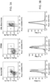
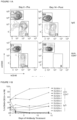
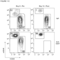
- CD47 overexpression is characteristic of progression of human myeloproliferative disorders to AML (see Figures 1 - 5B ).
- CD47 controls integrin function but also the ability of macrophages to phagocytose cells, depending on the level of CD47 expression.
- aberrant CD47 expression may allow LSC to evade both innate and adaptive host immunity.
- PV polycythemia vera
- PPMM/MF post-polycythemic myeloid metaplasia/myelofibrosis
- ET essential thrombocythemia
- CML chronic myelogenous leukemia
- CML chronic myeosinophilic leukemia
- CMML chronic myelomonocytic
- CD47 Facilitates Engraftment, Inhibits Phagocytosis, and is More Highly Expressed on AML LSC.
- HSC Long-CD34+CD38-CD90+
- AML LSC Long-CD34+CD38-CD90-
- AML LSC In order to test the model that CD47 overexpression on AML LSC prevents phagocytosis of these cells through its interaction with SIRP ⁇ on effector cells, we have utilized a monoclonal antibody directed against CD47 known to disrupt the CD47-SIRP ⁇ interaction.
- B6H12 mouse-anti-human CD47 monoclonal antibody
- CD47 a ligand for the macrophage inhibitory signal regulatory protein alpha (SIRP ⁇ ) receptor
- SIRP ⁇ macrophage inhibitory signal regulatory protein alpha
- CD47 also known as integrin associated protein (IAP)
- IAP integrin associated protein
- CD47 functions in promoting leukemogenesis by inhibiting phagocytosis of the leukemic cells by macrophages.
- CD47 is significantly upregulated in leukemic Fas lpr/lpr x hMRP8bcl2 transgenic bone marrow, and in leukemic hMRP8bcr/abl x hMRP8bcl2 mice.
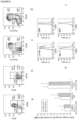
- Transcripts for CD47 are increased in leukemic hMRP8bcr/abl x hMRP8bcl2 bone marrow 3-4 fold by quantitative RT-PCR and 6-7 fold in c-Kit enriched leukemic marrow relative to healthy hMRP8bcl2+ bone marrow ( Figure 8e ).
- Leukemic spleen had an expansion of the granulocyte macrophage progenitor (GMP) population as well as c-Kit+ Sca-1+ Lin-stem and progenitor subsets relative to control mice, which were of the same genotype as leukemic mice but failed to develop disease ( Figure 8a-d ).
- GMP granulocyte macrophage progenitor
- Expression levels for CD47 protein were found to begin increasing in leukemic mice relative to control mice at the stage of the Flk2- CD34- c-Kit+ Sca-1+ Lin- long-term hematopoietic stem cell (LT-HSC) ( Figure 8f ).
- PV polycythemia vera
- MF myelofibrosis
- E essential thrombocythemia
- CMML chronic myelomonocytic leukaemia
- GMP granulocyte-macrophage progenitors
- AML HSC and progenitors uniformly exhibited higher levels of CD47 expression compared with normal controls ( Figure 9b ); every sample from BC-CML and AML had elevated levels of CD47.
- progression from chronic phase CML to blast crisis was associated with a significant increase in CD47 expression ( Figure 9c ).
- MOLM-13 cells which are derived from a patient with AML 5a, were transduced with Tet-MCS-IRES-GFP (Tet) or Tet-CD47-MCS-IRES-GFP (Tet-CD47), and stable integrants were propagated on the basis of GFP expression.
- Tet Tet-MCS-IRES-GFP
- Tet-CD47-MCS-IRES-GFP Tet-CD47
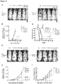
- Tet-CD47 MOLM-13 cells In addition to developing tumor nodules, the Tet-CD47 MOLM-13 cells exhibited patterns of hepatic involvement typically seen with human AML, with leukemic cells infiltrating the liver with a sinusoidal and perivenous pattern. Overall, Tet-CD47 MOLM-13 transplanted mice died more quickly than Tet MOLM-13 transplanted mice, which had virtually no engraftment of leukemic cells in hematopoietic tissues. Tet-MOLM-13 mice still had significant mortality, most likely due to localized growth at the site of injection (retro-orbital sinus) with extension into the brain.
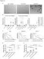
- CD47 dramatically lowered macrophage clearance of these cells at all time points tested, whereas Tet-MOLM-13 were quickly phagocytosed in a manner that increased over time.
- MOLM-13 hematopoietic organs 2 hours later for evidence of macrophage phagocytosis. Macrophages in bone marrow, spleen, and liver all had higher GFP+ fraction when injected with Tet MOLM-13 cells as compared to CD47 expressing cells. This indicates that CD47 over-expression can compensate for pro-phagocytic signals already present on leukemic cells, allowing them to survive when they would otherwise be cleared by macrophages.
- CD47 density on the CD47'° MOLM-13 cells was approximately equal to mouse bone marrow cells, whereas CD47 hi MOLM-13 cells had approximately 9 fold higher expression, an increase commensurate with the change seen in CD47 expression on primary leukemic cells compared to their normal counterparts.
- high or low expressing cells were transplanted into recipients, only mice transplanted with high expressing cells succumbed to disease by 75 days of age. Furthermore, organomegaly was more pronounced in mice transplanted with high expressing cells. Mice receiving CD47'° MOLM-13 cells still had notable liver masses.
- the masses were invariably 1-2 large nodes that were well-encapsulated and physically segregated from the liver parenchyma, in marked contrast to tumor masses from CD47hi MOLM-13 cells which consisted of hundreds of small masses scattered throughout the parenchyma.
- these large tumor masses consist of cells which have found macrophage free-niches to grow in separate from the main organ body.
- the infiltration of MOLM-13 cells in bone marrow and spleen of recipient mice was much higher for mice transplanted with CD47 hi MOLM-13 cells as well.
- CD47 expression level would provide added protection against macrophage phagocytosis.
- RFP red fluorescent protein
- CD47 is expressed in MOLM-13 cells using a Tet-OFF system
- Tet-inducible promoter element to control expression of CD47 in MOLM-13 cells.
- a cohort of mice was given doxycycline and followed for up to 75 days post-transplant.
- none of the mice given doxycycline succumbed to disease or had large infiltration of MOLM-13 cells in hematopoietic organs.
- muCD47 expression in MOLM-13 cells was reduced to levels below that of normal mouse bone marrow, but notably not completely absent.
- a sustained high level of CD47 expression is required for robust MOLM-13 survival in hematopoietic organs.
- CD47 was first cloned as an ovarian tumor cell marker, indicating that it may play a role in preventing phagocytosis of other tissue cancers as well. Furthermore, solid tumors often metastasize to macrophage rich tissues such as liver, lung, bone marrow, and lymph nodes, indicating that they must be able to escape macrophage-mediated killing in those tissues. Finding methods to disrupt CD47-SIRP ⁇ interaction may thus prove broadly useful in developing novel therapies for cancer. Preventing CD47-SIRP ⁇ interaction is doubly effective since antigens from phagocytosed tumor cells may be presented by macrophages to activate an adaptive immune response, leading to further tumor destruction.
- mice hMRP8bcrabl, hMRP8bcl2, and Fas lpr/lpr transgenic mice were created as previously described and crossed to obtain double transgenics.
- hMRP8bcl2 homozygotes were obtained by crossing heterozygote mice to each other.
- C57BI/6 Ka mice from our colony were used as a source of wild-type cells.
- cells were transplanted into C57BI/6 RAG2 -/- common gamma chain (Gc) -/- mice given a radiation dose of 4 Gy using gamma rays from a cesium irradiator (Phillips).
- Primary mouse leukemias were transplanted into CD45.2 C57BI6/Ka mice given a radiation dose of 9.5 Gy. Mice were euthanized when moribund.
- Bone marrow was obtained from leukemic hMRP8bcr/abl x hMRP8bcl2 mice or hMRP8bcl2 control mice.
- Cells were c-Kit enriched using c-Kit microbeads and an autoMACS column (Miltenyi).
- RNA was extracted using Trizol reagent (Invitrogen) and reverse transcription performed using SuperScriptll reverse polymerase (Invitrogen). cDNA corresponding to approximately 1000 cells was used per PCR reaction.
- Quantitative PCR was performed with a SYBR green kit on an ABI Prism 7000 PCR (Applied Biosystems) machine at 50°C for 2 minutes, followed by 95°C for 10 minutes and then 40 cycles of 95°C for 15 minutes followed by 60°C for 1 minute.
- Beta-actin and 18S RNA were used as controls for cDNA quantity and results of CD47 expression were normalized. Sequences for 18S RNA forward and reverse primers were TTGACGGAAGGGCACCACCAG and GCACCACCACCCACGGAATCG, respectively, for beta-actin were TTCCTTCTTGGGTATGGAAT and GAGCAATGATCTTGATCCTC, and for CD47 were AGGCCAAGTCCAGAAGCATTC and AATCATTCTGCTGCTCGTTGC.
- CMML chronic myelomonocytic leukemia
- CML chronic myeloid leukemia
- AML acute myelogenous leukemia
- MOLM-13 cells were obtained from DSMZ. HL-60 and Jurkat cells were obtained from ATCC. Cells were maintained in Iscove's modified Dulbecco's media (IMDM) plus 10% fetal bovine serum (FBS) (Hyclone). To fractionate MOLM-13 cells into those with high and low CD47 expression, Tet-CD47 MOLM-13 cells were stained with anti-mouse CD47 Alexa-680 antibody (mlAP301). The highest and lowest 5% of mouse CD47 expressing cells was sorted on a BD FACSAria and re-grown in IMDM+10%FCS for 2 weeks. The cells were sorted for three more rounds of selection following the same protocol to obtain the high and low expressing cells used in this study. To obtain red fluorescent protein (RFP) constructs, the mCherry RFP DNA was cloned into Lentilox 3.7 (pLL3.7) empty vector. Lentivirus obtained from this construct was then used to infect cell lines.
- IMDM Iscove's
- Mouse CD47 antibody (clone mIAP301) was assessed using biotinylated antibody produced in our lab. Cells were then stained with streptavidin conjugated Quantum Dot 605 (Chemicon). Samples were analyzed using a FACSAria (Beckton Dickinson).
- CD34+ cells were enriched from mononuclear fractions with the aid of immunomagnetic beads (CD34+ Progenitor Isolation Kit, Miltenyi Biotec, Bergisch-Gladbach, Germany).
- myeloid progenitors were stained with lineage marker specific phycoerythrin (PE)-Cy5-conjugated antibodies including CD2 RPA-2.10; CD11b, ICRF44; CD20, 2H7; CD56, B159; GPA, GA-R2 (Becton Dickinson - PharMingen, San Diego), CD3,S4.1;CD4, S3.5; CD7, CD7-6B7; CD8, 3B5; CD10, 5-1B4, CD14, TUK4; CD19, SJ25-C1 (Caltag, South San Francisco, CA) and APC-conjugated anti-CD34, HPCA-2 (Becton Dickinson-PharMingen), biotinylated anti-CD38, HIT2 (Caltag) in addition to PE-conjugated anti-IL-3R ⁇ , 9F5 (Becton Dickinson- ParMingen) and FITC-conjugated anti-CD45RA,
- PE lineage marker specific
- HSC Hematopoietic stem cells
- Anti-human CD47 FITC (clone B6H12, Pharmingen) was used to assess CD47 expression in all human samples. Fold change for CD47 expression was determined by dividing the average mean fluorescence intensity of CD47 for all the samples of CML-BC, CML-CP, or AML by the average mean fluorescence intensity of normal cells for a given cell population.
- CMP Common myeloid progenitors
- GMP granulocyte/macrophage progenitors
- MEP megakaryocyte/erythrocyte progenitors
- Engraftment of MOLM-13 cells was assessed by using anti-human CD45 PE-Cy7 (Pharmingen), anti-mouse CD45.2 APC (clone AL1-4A2), and anti-mouse CD47 Alexa-680 (mlAP301). All samples were resuspended in propidium iodide containing buffer before analysis to exclude dead cells. FACS data was analyzed using FloJo software (Treestar).
- pRRL.sin-18.PPT.Tet07.IRES.GFP.pre, CMV, VSV, and tet trans-activator (tTA) plasmids were obtained from Luigi Naldini.
- the full length murine cDNA for CD47 form 2 was provided by Eric Brown (UCSF).
- the CD47 cDNA construct was ligated into the BamHI/Nhel site of Tet-MCS-IRES-GFP. Plasmid DNA was transfected into 293T cells using standard protocols. The supernatant was harvested and concentrated using a Beckman LM-8 centrifuge (Beckman). Cells were transduced with Tet or Tet-CD47-MCS-IRES-GFP and tTA lentivirus for 48 hours. GFP+ cells were sorted to purity and grown for several generations to ensure stability of the transgenes.
- MOLM-13 cell engraftment Animals were euthanized when moribund and bone marrow, spleen, and liver harvested. Peripheral blood was obtained by tail bleed of the animals 1 hour prior to euthanization. Engraftment of MOLM-13 cells in marrow, spleen, and peripheral blood was determined as described above. Tumor burden in the liver was determined by calculating the area of each visible tumor nodule using the formula ((length in mm + width in mm)/2) * ⁇ . Area of each nodule was then added together per liver.
- Doxycycline administration Doxycycline hydrochloride (Sigma) was added to drinking water at a final concentration of 1 mg/mL. Drinking water was replaced every 4 days and protected from light. In addition, mice received a 10 ⁇ g bolus of doxycycline by i.p. injection once a week.
- Bone marrow derived macrophages Femurs and tibias were harvested from C57BI/6 Ka mice and the marrow was flushed and placed into a sterile suspension of PBS. The bone marrow suspension was grown in IMDM plus 10% FBS with 10 ng/mL of recombinant murine macrophage colony stimulating factor (MCSF, Peprotech) for 7-10 days.
- MCSF murine macrophage colony stimulating factor
- BMDM In vitro phagocytosis assays.
- BMDM were harvested by incubation in trypsin/EDTA (Gibco) for 5 minutes and gentle scraping. Macrophages were plated at 5 ⁇ 10 4 cells per well in a 24-well tissue culture plate (Falcon). After 24 hours, media was replaced with serum-free IMDM. After an additional 2 hours, 2.5 ⁇ 10 5 Tet or Tet-CD47 MOLM-13 cells were added to the macrophage containing wells and incubated at 37 C° for the indicated times. After co-incubation, wells were washed thoroughly with IMDM 3 times and examined under an Eclipse T5100 (Nikon) using an enhanced green fluorescent protein (GFP) or Texas Red filter set (Nikon).
- GFP enhanced green fluorescent protein
- Nikon Texas Red filter set
- phagocytic index number of ingested cells/(number of macrophages/100). At least 200 macrophages were counted per well.
- phagocytic index number of ingested cells/(number of macrophages/100). At least 200 macrophages were counted per well.
- For flow cytometry analysis of phagocytosis macrophages were harvested after incubation with MOLM-13 cells using trypsin/EDTA and gentle scraping. Cells were stained with anti-Mac-1 PE antibody and analyzed on a BD FACSAria.
- Fluorescent and brightfield images were taken separately using an Eclipse T5100 (Nikon), a super high pressure mercury lamp (Nikon), an endow green fluorescent protein (eGFP) bandpass filter (Nikon) a Texas Red bandpass filter (Nikon), and a RT Slider (Spot Diagnostics) camera. Images were merged with Photoshop software (Adobe).
- marrow from leg long bones, spleen, and liver were harvested 2 hours after injecting target cells into RAG2 -/- ,Gc -/- mice. They were prepared into single cell suspensions in PBS plus 2% FCS. Cells were labeled with anti-human CD45 Cy7-PE and anti-mouse F4/80 biotin (eBiosciences). Secondary stain was performed with Streptavidin-APC (eBiosciences). Cells that were human CD45-, F4/80+ were considered to be macrophages, and GFP+ cells in this fraction was assessed.
- CD47 is an Independent Prognostic Factor and Therapeutic Antibody Target on Human Acute Myeloid Leukemia Cells
- AML Acute myelogenous leukemia
- LSC self-renewing leukemia stem cells
- CD47 expression is an independent poor prognostic factor that can be targeted on human AML stem cells with monoclonal antibodies capable of stimulating phagocytosis of LSC.
- CD47 is More Highly Expressed on AML LSC than Their Normal Counterparts and is Associated with the FLT3-ITD Mutation.
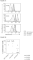
- flow cytometry CD47 was more highly expressed on multiple specimens of AML LSC than normal bone marrow HSC and MPP ( Figure 5 ). This increased expression extended to the bulk leukemia cells, which expressed CD47 similarly to the LSC-enriched fraction.
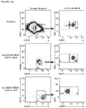
- CD47'° cells engrafted with normal myelo-lymphoid hematopoiesis in vivo and formed numerous morphologically normal myeloid colonies in vitro ( Figure 10B,C ).
- Monoclonal Antibodies Directed Against Human CD47 Preferentially Enable Phagocytosis of AML LSC by Human Macrophages.
- CD47 expression on human AML contributes to pathogenesis by inhibiting phagocytosis of leukemia cells, leading us to predict that disruption of the CD47-SIRP ⁇ interaction with a monoclonal antibody directed against CD47 will preferentially enable the phagocytosis of AML LSC.
- Several anti-human CD47 monoclonal antibodies have been generated including some capable of blocking the CD47-SIRP ⁇ interaction (B6H12.2 and BRIC126) and others unable to do so (2D3) ( Subramanian et al., 2006 Blood 107, 2548-2556 ).
- a Monoclonal Antibody Directed Against Human CD47 Inhibits AML LSC Engraftment and Eliminates AML in Vivo.
- the ability of the blocking anti-CD47 antibody B6H12.2 to target AML LSC in vivo was tested.
- AML LSC were purified by FACS and incubated with IgG1 isotype control, anti-human CD45, or anti-human CD47 antibody.
- An aliquot of the cells was analyzed for coating by staining with a secondary antibody demonstrating that both anti-CD45 and anti-CD47 antibody bound the cells.
- the remaining cells were transplanted into newborn NOG mice that were analyzed for leukemic engraftment 13 weeks later.
- the isotype control and anti-CD45 antibody coated cells exhibited long-term leukemic engraftment.
- most mice transplanted with cells coated with anti-CD47 antibody had no detectable leukemia engraftment.
- mice were first engrafted with human AML LSC and then administered daily intraperitoneal injections of 100 micrograms of either mouse IgG or anti-CD47 antibody for 14 days, with leukemic engraftment determined pre- and post-treatment.
- Analysis of the peripheral blood showed near complete elimination of circulating leukemia in mice treated with anti-CD47 antibody, often after a single dose, with no response in control mice ( Figure 11A,B ).
- there was a significant reduction in leukemic engraftment in the bone marrow of mice treated with anti-CD47 antibody, while leukemic involvement increased in control IgG-treated mice ( Figure 11C , D ).
- CD47 contributes to pathogenesis via a distinct mechanism, conferring a survival advantage to LSC and progeny blasts through evasion of phagocytosis by the innate immune system.
- increased CD47 expression represents the first described immune evasion mechanism with prognostic and therapeutic implications for human AML.
- AML LSC are enriched in the Lin-CD34+CD38- fraction, which in normal bone marrow contains HSC and MPP.
- MPP minimal residual disease
- Increased CD47 expression is independently predictive of a worse clinical outcome in AML patients with a normal karyotype, including the subset without the FLT3-ITD mutation, which is the largest subgroup of AML patients.
- a quantitative PCR assay for AML prognosis may be based on the level of CD47 expression. Such an assay could be utilized in risk adapted therapeutic decision making, particularly in the large subgroup of AML patients with normal karyotypes who lack the FLT3-ITD mutation.
- CD47 Cell surface molecules preferentially expressed on LSC compared to their normal counterparts are suitable for targeting with therapeutic monoclonal antibodies. Thus far, several molecules have been targeted on AML including CD33, CD44, CD123, and now CD47.
- CD33 is the target of the monoclonal antibody conjugate gemtuzumab ozogamicin (Mylotarg), which is approved for the treatment of relapsed AML in older patients.
- Mylotarg gemtuzumab ozogamicin
- Targeting of CD44 with a monoclonal antibody was shown to markedly reduce AML engraftment in mice, with evidence that it acts specifically on LSC to induce differentiation.
- a monoclonal antibody directed against CD123 was recently reported to have efficacy in reducing AML LSC function in vivo.
- a monoclonal antibody directed against CD47 is able to stimulate phagocytosis of AML LSC in vitro and inhibit engraftment in vivo.
- a combination of CD47 with CD33; with CD44 or with CD123 may provide for synergistic results.
- the experimental evidence presented here provides for anti-CD47 monoclonal antibodies as monotherapy or a combination strategy for treatment of leukemia.
- the combination of an anti-CD47 antibody, able to block a strong inhibitory signal for phagocytosis, with a second antibody able to bind a LSC-specific molecule (for example CD96, CD33, CD123, etc. ) and engage Fc receptors on phagocytes, thereby delivering a strong positive signal for phagocytosis, may result in a synergistic stimulus for phagocytosis and specific elimination of LSC.
- combinations of monoclonal antibodies to LSC that include blocking anti-CD47 and human IgG1 antibodies directed against two other cell surface antigens will be more likely to eliminate leukemia cells with pre-existing epitope variants or antigen loss that are likely to recur in patients treated with a single antibody.
- Human Samples Normal human bone marrow mononuclear cells were purchased from AllCells Inc. (Emeryville, CA). Human acute myeloid leukemia samples (Figure 1A) were obtained from patients at the Stanford University Medical Center with informed consent, according to an IRB-approved protocol (Stanford IRB# 76935 and 6453). Human CD34- positive cells were enriched with magnetic beads (Miltenyi Biotech).
- a panel of antibodies was used for analysis and sorting of AML LSC (Lin-CD34+CD38-CD90-, where lineage included CD3, CD19, and CD20), HSC (Lin-CD34+CD38-CD90+), and MPP (Lin-CD34+CD38-CD90-CD45RA-) as previously described (Majeti et al., 2007).
- Analysis of CD47 expression was performed with an anti-human CD47 PE antibody (clone B6H12, BD Biosciences, San Jose CA).
- Genomic DNA Preparation and Analysis of FLT3-ITD by PCR Genomic DNA was isolated from cell pellets using the Gentra Puregene Kit according to the manufacturer's protocol (Gentra Systems, Minneapolis, MN). FLT3-ITD status was screened by PCR using primers that generated a wild-type product of 329 bp and ITD products of variable larger sizes.
- Anti-Human CD47 Antibodies Monoclonal mouse anti-human CD47 antibodies included: BRIC126, IgG2b (Abcam, Cambridge, MA), 2D3, IgG1 (Ebiosciences. San Diego, CA), and B6H12.2, lgG1.
- B6H12.2 hybridoma was obtained from the American Type Culture Collection (Rockville, MD).
- Antibody was either purified from hybridoma supernatant using protein G affinity chromatography according to standard procedures or obtained from BioXCell (Lebanon, NH).
- Methylcellulose Colony Assay Methylcellulose colony formation was assayed by plating sorted cells into a 6-well plate, each well containing 1 ml of complete methylcellulose (Methocult GF+ H4435, Stem Cell Technologies). Plates were incubated for 14 days at 37°C, then scored based on morphology.
- mice were sacrificed 13 weeks post-transplantation and bone marrow was analyzed for human leukemia engraftment (hCD45+hCD33+) by flow cytometry ( Majeti et al., 2007 Cell Stem Cell 1, 635-645 ). The presence of human leukemia was confirmed by Wright-Giemsa staining of hCD45+ cells and FLT3-ITD PCR. Statistical analysis using Student's t-test was performed with GraphPad Prism (San Diego, CA).
- AML Patients, Microarray Gene Expression Data, and Statistical Analysis Gene expression and clinical data were analyzed for three previously described cohorts of adult AML patients: (1) a training dataset of 285 patients with diverse cytogenetic and molecular abnormalities described by Valk et al., (2) a test dataset of 242 patients with normal karyotypes described by Metzeler et al., and (3) a validation dataset of 137 patients with normal karyotypes described by Bullinger et al.
- the clinical end points analyzed included overall and event-free survival, with events defined as the interval between study enrollment and removal from the study owing to a lack of complete remission, relapse, or death from any cause, with data censored for patients who did not have an event at the last follow-up visit.
- FLT3-ITD PCR All reactions were performed in a volume of 50 ⁇ l containing 5 ⁇ l of 10x PCR buffer (50mM KCL/10nM Tris/2mM MgCl2/0.01% gelatin), 1 ⁇ l of 10mM dNTPs, 2 units of Taq polymerase (Invitrogen), 1 ul of 10 ⁇ M forward primer 11F (5'-GCAATTTAGGTATGAAAGCCAGC-3') and reverse primer 12R (5'-CTTTCAGCATTTTGACGGCAACC-3'), and 10-50 ng of genomic DNA.
- PCR conditions for amplification of the FLT3 gene were 40 cycles of denaturation (30 sec at 95°C) annealing (30 sec at 62°C), and extension (30 sec at 72°C).
- BALB/c mouse bone marrow mononuclear cells were harvested and grown in IMDM containing 10% FBS supplemented with 10 ng/mL recombinant murine macrophage colony stimulating factor (M-CSF, Peprotech, Rocky Hill, NJ) for 7-10 days to allow terminal differentiation of monocytes to macrophages.
- M-CSF murine macrophage colony stimulating factor
- Human peripheral blood mononuclear cells were prepared from discarded normal blood from the Stanford University Medical Center. Monocytes were isolated by adhering mononuclear cells to culture plates for one hour at 37°C, after which non-adherent cells were removed by washing. The remaining cells were >95% CD14 and CD11b positive. Adherent cells were then incubated in IMDM plus 10% human serum (Valley Biomedical, Winchester, VA) for 7-10 days to allow terminal differentiation of monocytes to macrophages.
- BMDM or peripheral blood macrophages were harvested by incubation in trypsin/EDTA (Gibco/Invitrogen) for 5 minutes followed by gentle scraping. 5 ⁇ 10 4 macrophages were plated in each well of a 24-well tissue culture plate in 10% IMDM containing 10% FBS. After 24 hours, media was replaced with serum-free IMDM and cells were cultured an additional 2 hours. LSC were fluorescently labeled with CFSE according to the manufacturer's protocol (Invitrogen).
- CFSE-labeled LSC 2 ⁇ 10 4 CFSE-labeled LSC were added to the macrophage-containing wells along with 7 ⁇ g/mL of IgG1 isotype (Ebiosciences), anti-CD45 (clone HI30, Ebiosciences), or anti-CD47 antibody, and incubated for 2 hours. Wells were then washed 3 times with IMDM and examined under an Eclipse T5100 immunofluorescent microscope (Nikon) using an enhanced green fluorescent protein filter able to detected CFSE fluorescence. The number of CFSE positive cells within macrophages was counted and the phagocytic index was determined as the number of ingested cells per 100 macrophages. At least 200 macrophages were counted per well.
- Microarray Gene Expression Data Training Set: Gene expression data, cytogenetics data, and molecular data for the 285 and 465 patients with AML profiled with Affymetrix HG-U133A and HG-U133 Plus 2.0 microarrays by Valk et al. and Jongen-Lavrencic et al. respectively, were obtained from the Gene Expression Omnibus using the corresponding accession numbers (GSE1159 and GSE6891). Outcome data were only available for the former dataset, and the corresponding clinical information were kindly provided by the authors. This cohort is presented as the "training" dataset. The latter dataset was used to confirm univariate associations with karyotype and molecular mutations described in the former.
- probe set 213857_s_at from the Affymetrix HG-U133A and HG-U133 Plus 2.0 microarrays, given its similar expression distribution (Supplemental Figure 3B ), and considering its position within the mRNA transcript as compared with cDNA clones on the Stanford cDNA microarrays as annotated within the NetAffx resource.
- Test Set Gene expression and clinical data for the 242 adult patients with NKAML profiled with Affymetrix HG-U133A and HG-U133 Plus 2.0 microarrays by Metzeler et al. were obtained from the Gene Expression Omnibus using the corresponding accession numbers (GSE12417). Since raw data were not available for this dataset, for purposes of assessing the prognostic value of CD47, we employed the normalized datasets provided by the authors (base 2 logarithms) and assessed expression of CD47 using the probe set 213857_s_at on the corresponding microarrays.
- Validation Set Gene expression data for the 137 patients with normal karyotype AML profiled with cDNA microarrays by Bullinger et al. were obtained from the Stanford Microarray Database10. The corresponding clinical information including outcome data and FLT3 mutation status were kindly provided by the authors. Using the original annotations of microarray features as well as SOURCE11, RefSeq5, and the UCSC Genome Browser6, we identified IMAGE:811819 as a sequence verified cDNA clone mapping to the constitutively transcribed 3' terminal exon of CD47 on the corresponding cDNA microarrays.
- AML patients described by Valk et al. (training set), were treated according to several protocols of the Dutch-Belgian Hematology-Oncology Cooperative group. The majority (90%) of the NK-AML patients described by Metzeler et al. (test set) were treated per protocol AMLCG-1999 of the German AML Cooperative Group, with all patients receiving intensive double-induction and consolidation chemotherapy. All 137 NK-AML patients described by Bullinger et al.
- validation set received standard-of-care intensified treatment regimens (protocol AML HD98A), which included 2 courses of induction therapy with idarubicin, cytarabine, and etoposide, one consolidation cycle of high-dose cytarabine and mitoxantrone (HAM), followed by random assignment to a late consolidation cycle of HAM versus autologous hematopoietic cell transplantation in case no HLA identical family donor was available for allogeneic hematopoietic cell transplantation.
- protocol AML HD98A standard-of-care intensified treatment regimens
- HAM high-dose cytarabine and mitoxantrone
- the prognostic value of CD47 expression was measured through comparison of the event-free and overall survival of patients with estimation of survival curves by the Kaplan-Meier product limit method and the log-rank test.
- This stratification segregates the 261 AML patients with available outcome data into two unequally sized groups, with 72% of patients with lowest expression considered CD47 low, and 28% with highest expression considered CD47 high. These two groups have different overall survival with a hazard ratio of 1.42 for the CD47 high group, and a corresponding uncorrected p-value of 0.033, which requires cross-validation to avoid the risk of overfitting.
- DLBCL diffuse large B cell lymphoma
- B-CLL B cell chronic lymphocytic leukemia
- MCL mantle cell lymphoma
- FL follicular lymphoma
- MZL marginal zone lymphoma
- pre-B ALL pre-B acute lymphoblastic leukemia
- CD47 expression was associated with known adverse molecular features in NHL.
- DLBCL normal germinal center B cells
- ABC activated blood memory B cells
- CD47 expression was significantly higher in ABC-like DLBCL ( Figure 12F ).
- CD47 expression was not found to have independent prognostic value within GCB and ABC subtypes, suggesting a strong association with the cell-of-origin classification of DLBCL.
- CD47 expression was also associated with unmutated immunoglobulin heavy chain variable regions (IgVH) in CLL ( Figure 12G ) and significantly correlated with the proliferative index in MCL ( Figure 12H ), both known adverse prognostic factors.
- IgVH immunoglobulin heavy chain variable regions
- MCL proliferative index
- CD47 expression remained prognostic of disease progression independent from the international prognostic index in DLBCL and two major prognostic factors in CLL: IgVH mutation status and ZAP-70 status.
- a multivariate model did not find independent prognostic value for CD47 when considering the proliferation index.
- Incubation of NHL cells in the presence of IgG1 isotype control or anti-CD45 IgG1 antibody did not result in significant phagocytosis; however, two different blocking anti-CD47 antibodies (B6H12.2 and BRIC126) enabled phagocytosis of NHL cells but not NPB cells ( Figures 13A and 13B ).
- CD47 expression levels correlated with the degree of anti-CD47 antibody-mediated phagocytosis.
- lentiviral shRNA vectors were used to knock down expression of CD47 in Raji cells.
- Several clones were generated with a range of CD47 knockdown. Those clones with a greater than 50% reduction in CD47 expression (shCD47-1 and shCD47-2) demonstrated a significant reduction in anti-CD47 antibody-mediated phagocytosis.
- shCD47-1 and shCD47-2 demonstrated a significant reduction in anti-CD47 antibody-mediated phagocytosis.
- a statistical analysis demonstrated a positive correlation between CD47 expression and degree of anti-CD47 antibody-mediated phagocytosis with both mouse and human macrophage effector cells.
- anti-CD47 antibodies might be predicted to directly induce apoptosis of NHL cells that are then recognized by macrophages and phagocytosed. Contrary to this prediction, when NHL cells were incubated with anti-CD47 antibody in the absence of macrophages, no induction of apoptosis was observed when cells were incubated in suspension for either 2 hr ( Figure 13B , right) or 8 hr. Incubation of NHL cells with immobilized anti-CD47 antibody resulted in increased apoptosis compared to controls, consistent with prior reports. Given that phagocytosis of NHL cells occurs in the presence of soluble anti-CD47 mAbs, it is unlikely that these mAbs induce apoptosis of NHL cells that are then secondarily phagocytosed.
- lymphoma Using Raji, SUDHL4, and NHL17 cells, which express varying levels of both CD47 and CD20, anti-CD47 antibody synergized with rituximab or anti-human CD20 (mouse IgG2a) antibody, as indicated by combination indices less than 1 ( Figure 13C ).
- in vitro phagocytosis assays were conducted with primary NHL cells incubated with either anti-CD47 antibody or rituximab alone, or both in combination at half of the single agent dose. NHL cells exhibited a significant increase in phagocytosis when incubated with the combination compared to either antibody alone when using mouse ( Figure 13D ) or human ( Figure 13E ) macrophage effectors. No phagocytosis of NPB cells was observed with either antibody alone or in combination with human macrophages ( Figure 13E ).
- Combination Therapy with Anti-CD47 Antibody and Rituximab Eliminates Lymphoma in Both Disseminated and Localized Human NHL Xenotransplant Models.
- mice were first engrafted with NHL and then administered single or combination antibody therapy.
- To best model NHL we established disseminated and localized xenotransplantation models in NSG mice that are deficient in T, B, and NK cells but retain phagocyte effector cells.
- luciferase expressing Raji cells were transplanted intravenously into adult NSG mice.
- mice Two weeks later, these mice were administered daily injections of either control mouse IgG, anti-CD47 antibody, rituximab, or anti-CD47 antibody and rituximab.
- Anti-CD47 antibody treatment decreased the lymphoma burden in these mice ( Figures 15A and 15B ) and significantly prolonged survival compared to control IgG, although all mice eventually died ( Figure 15C ). Similar results were seen with rituximab and were not statistically different compared to anti-CD47 antibody ( Figures 15A-15C ).
- combination therapy with anti-CD47 antibody and rituximab eliminated lymphoma in 60% of mice as indicated by long-term survival ( Figure 15C ) and the absence of luciferase-positive lymphoma more than 4 months after the end of treatment.
- rituximab efficacy is thought to be primarily mediated by both macrophages and NK cells.
- NSG mice lack NK cells, we conducted a similar experiment in NK cell-containing SCID mice and observed similar therapeutic responses as in NSG mice.
- luciferase-expressing Raji cells were transplanted subcutaneously in the right flank of NSG mice. Once tumors were palpable (approximately 2 weeks), mice were treated with antibody therapy. Mice treated with anti-CD47 antibody or rituximab demonstrated a decreased rate of lymphoma growth compared to control IgGtreated mice as measured by both luciferase signal and tumor volume ( Figures 15D-15F ) but, like controls, eventually had to be sacrificed due to enlarged tumor growth.
- mice treated with the combination of anti-CD47 antibody and rituximab demonstrated complete elimination of lymphoma, with 86% of treated mice having no measurable mass or luciferase-positive lymphoma 4 weeks after the end of therapy ( Figures 15D-15F ). Moreover, all showed no evidence of tumor growth, remained relapse free, and were alive at over 197 days after tumor engraftment. Out of six mice achieving a complete remission, five remained relapse free whereas one mouse died of nontumor- related causes ( Figure 15E ). For both disseminated and localized xenograft models, expression of CD47 and CD20 in transplanted Raji cells did not differ from Raji cells in culture.
- NHL cell lines have been valuable for the evaluation of therapeutics, but they may not accurately recapitulate the heterogeneity of the primary disease.
- NHL cell lines have been valuable for the evaluation of therapeutics, but they may not accurately recapitulate the heterogeneity of the primary disease.
- two new mouse xenograft models for NHL in which intravenous transplantation of cells from a DLBCL patient (NHL7/SUNHL7) and a FL patient (NHL31/SUNHL31) give rise to robust lymphoma engraftment in the bone marrow and/or peripheral blood.
- Primary DLBCL cells were transplanted into mice, which 2 weeks later were treated with daily injections of antibodies for 14 days.
- mice 8 out of 9 mice (89%) administered combination antibody treatment were cured of lymphoma, as indicated by long-term disease-free survival more than 4 months after the end of treatment ( Figure 16C ) with no detectable lymphoma in the bone marrow.
- FL cells were transplanted intravenously in a similar manner.
- mice were treated with a single intraperitoneal injection of either control IgG, anti-CD47 antibody, rituximab, or the combination and then followed for disease progression.
- a single dose of anti-CD47 antibody alone or in combination with rituximab eliminated lymphoma both in the peripheral blood ( Figure 16D ) and in bone marrow ( Figure 16E ).
- a single dose of rituximab enabled a partial reduction in tumor burden that rebounded back to baseline levels in the peripheral blood with no tumor reduction observed in the bone marrow.
- the difference in anti-CD47 antibody clearance of lymphoma as a single dose in FL-engrafted mice may be due to cell-intrinsic differences in antibody sensitivity between different NHL subtypes or due to different anti-CD47 antibody potencies in distinct tissue compartments (peripheral blood versus bone marrow (BM) versus soft tissue compartments).
- BM bone marrow
- NHL cells were incubated with either anti-CD47 antibody/rituximab alone or in combination without macrophages, and cell death was measured.
- No synergistic apoptosis was observed when NHL cells were incubated with soluble ( Figure 17A ) or immobilized antibodies.
- crosslinking of soluble anti-CD47 antibody alone or in combination with rituximab by macrophages did not induce increased apoptosis of nonphagocytosed NHL cells compared to IgG1 isotype control, whereas rituximab induced a slight increase in apoptosis.
- No synergistic apoptosis was observed in this context. The small increase in apoptosis upon antibody treatment was not FcR dependent given that results were similar with macrophages lacking the Fcg subunit.
- NK cells could mediate tumor elimination by anti-CD47 antibody alone or in synergy with rituximab.
- the ability of anti-CD47 antibody to induce NK cell-mediated ADCC through FcRs or by CD47-SIRP ⁇ blockade was investigated. Utilizing human NK cells as effectors, anti-CD47 antibody did not induce increased ADCC of Raji or SUDHL4 cells compared to IgG1 isotype control ( Figure 17C ).
- the F(ab') 2 fragment did not enable ADCC, indicating that the increased ADCC was likely mediated through FcRs ( Figure 17D ).
- the combination of anti-CD47 antibody or F(ab') 2 fragment with rituximab did not induce a statistically significant increase in ADCC compared to single agent therapy, indicating no synergistic effect.
- NK cell and complement depletion Compared to vehicle control, NK cell and complement depletion had no effect on tumor elimination by combination antibody therapy. In contrast, macrophage depletion significantly abrogated the therapeutic effect, indicating that macrophages, and not NK cells or complement, are required for combination anti-CD47 antibody and rituximab-mediated elimination of NHL in vivo.
- Anti-CD47 Antibody Synergizes with Rituximab through FcR-Independent and FcR-Dependent Mechanisms.
- FcR-Independent and FcR-Dependent Mechanisms we hypothesize that the observed synergy between an anti-CD47 antibody and rituximab occurs through the combination of two separate mechanisms for stimulating phagocytosis: (1) FcR-independent through blockade of an inhibitory phagocytic signal by anti-CD47 antibody, and (2) FcR-dependent through delivery of a positive phagocytic signal by rituximab.
- FcR-independent through blockade of an inhibitory phagocytic signal by anti-CD47 antibody
- FcR-dependent FcR-dependent through delivery of a positive phagocytic signal by rituximab.
- F(ab') 2 fragments of both anti-CD47 antibody and rituximab were generated and utilized in phagocytosis assays with NHL cells and wild-type macrophages.
- anti-CD47 F(ab') 2 , but not rituximab F(ab') 2 enabled phagocytosis of NHL cells ( Figure 18E ).
- a bispecific FcR-engaging antibody with one arm binding and blocking CD47 and the other binding to a validated cancer antibody target (CD20) could reduce potential antibody toxicity, while retaining the synergy effect, especially as CD47 is expressed in multiple normal tissue types.
- a Burkitt's lymphoma cell line (Raji) and a DLBCL cell line (SUDHL4) were obtained from the American Type Culture Collection or generated in the lab.
- the NHL17* cell line was generated from a patient with DLBCL by culturing bulk cells in vitro with IMDM supplemented with 10% human AB serum for 1.5 months.
- Normal human peripheral blood and human NHL samples were obtained from the Stanford University Medical Center (Stanford, CA, USA) with informed consent, according to an IRB-approved protocol or with informed consent from the Norwegian Radium Hospital (Oslo, Norway) according to a Regional Ethic Committee (REK)-approved protocol.
- Normal tonsils for germinal center B cell analysis were obtained from discarded tonsillectomy specimens from consented pediatric patients at Stanford University Medical Center according to an IRB-approved protocol.
- CD19, CD20, CD3, CD10, CD45, CD5, CD38 (Invitrogen, Carlsbad, CA, USA and BD Biosciences, San Jose, CA, USA). Analysis of CD47 expression was performed with an antihuman CD47 FITC antibody (clone B6H12.2, BD Biosciences). Cell staining and flow cytometry analysis was performed as previously described.
- Rituximab anti-CD20, human IgG1 was obtained from the Stanford University Medical Center, mouse anti-human CD20, IgG2a from Beckman Coulter (Miami, FL, USA), and anti-CD47 antibody BRIC126, IgG2b from AbD Serotec (Raleigh, NC, USA). Other anti-CD47 antibodies were used as in Majeti et al. (2009). All in vivo antibody experiments were performed using the anti-CD47 B6H12.2 antibody.
- a CI of less than, equal to, and greater than 1 indicates synergy, additivity, and antagonism, respectively.
Landscapes
- Health & Medical Sciences (AREA)
- Chemical & Material Sciences (AREA)
- Immunology (AREA)
- Life Sciences & Earth Sciences (AREA)
- Organic Chemistry (AREA)
- Medicinal Chemistry (AREA)
- General Health & Medical Sciences (AREA)
- Proteomics, Peptides & Aminoacids (AREA)
- Veterinary Medicine (AREA)
- Pharmacology & Pharmacy (AREA)
- Animal Behavior & Ethology (AREA)
- Public Health (AREA)
- Genetics & Genomics (AREA)
- Biochemistry (AREA)
- Biophysics (AREA)
- Molecular Biology (AREA)
- Epidemiology (AREA)
- Engineering & Computer Science (AREA)
- Oncology (AREA)
- Chemical Kinetics & Catalysis (AREA)
- Nuclear Medicine, Radiotherapy & Molecular Imaging (AREA)
- General Chemical & Material Sciences (AREA)
- Bioinformatics & Cheminformatics (AREA)
- Physics & Mathematics (AREA)
- Biomedical Technology (AREA)
- Optics & Photonics (AREA)
- Microbiology (AREA)
- Mycology (AREA)
- Hematology (AREA)
- Medicines Containing Antibodies Or Antigens For Use As Internal Diagnostic Agents (AREA)
- Medicines That Contain Protein Lipid Enzymes And Other Medicines (AREA)
- Peptides Or Proteins (AREA)
- Pharmaceuticals Containing Other Organic And Inorganic Compounds (AREA)
Applications Claiming Priority (3)
| Application Number | Priority Date | Filing Date | Title |
|---|---|---|---|
| US27672609P | 2009-09-15 | 2009-09-15 | |
| EP10817793.2A EP2477648B1 (fr) | 2009-09-15 | 2010-09-15 | Thérapie par anti-cd47 synergique pour les cancers hématologiques |
| PCT/US2010/048992 WO2011034969A1 (fr) | 2009-09-15 | 2010-09-15 | Thérapie par anti-cd47 synergique pour les cancers hématologiques |
Related Parent Applications (1)
| Application Number | Title | Priority Date | Filing Date |
|---|---|---|---|
| EP10817793.2A Division EP2477648B1 (fr) | 2009-09-15 | 2010-09-15 | Thérapie par anti-cd47 synergique pour les cancers hématologiques |
Publications (1)
| Publication Number | Publication Date |
|---|---|
| EP4134095A1 true EP4134095A1 (fr) | 2023-02-15 |
Family
ID=43758999
Family Applications (2)
| Application Number | Title | Priority Date | Filing Date |
|---|---|---|---|
| EP22185805.3A Pending EP4134095A1 (fr) | 2009-09-15 | 2010-09-15 | Thérapie par anti-cd47 synergique pour les cancers hématologiques |
| EP10817793.2A Active EP2477648B1 (fr) | 2009-09-15 | 2010-09-15 | Thérapie par anti-cd47 synergique pour les cancers hématologiques |
Family Applications After (1)
| Application Number | Title | Priority Date | Filing Date |
|---|---|---|---|
| EP10817793.2A Active EP2477648B1 (fr) | 2009-09-15 | 2010-09-15 | Thérapie par anti-cd47 synergique pour les cancers hématologiques |
Country Status (9)
| Country | Link |
|---|---|
| US (4) | US8758750B2 (fr) |
| EP (2) | EP4134095A1 (fr) |
| AU (1) | AU2010295661B2 (fr) |
| CA (2) | CA2771336C (fr) |
| ES (1) | ES2927646T3 (fr) |
| PL (1) | PL2477648T3 (fr) |
| PT (1) | PT2477648T (fr) |
| SI (1) | SI2477648T1 (fr) |
| WO (1) | WO2011034969A1 (fr) |
Families Citing this family (69)
| Publication number | Priority date | Publication date | Assignee | Title |
|---|---|---|---|---|
| US11072655B2 (en) | 2008-01-15 | 2021-07-27 | The Board Of Trustees Of The Leland Stanford Junior University | Markers of acute myeloid leukemia stem cells |
| ES2582340T3 (es) | 2008-01-15 | 2016-09-12 | The Board Of Trustees Of The Leland Stanford Junior University | Métodos para manipular fagocitosis mediada por CD47 |
| EP3043181B1 (fr) | 2008-01-15 | 2020-04-08 | The Board of Trustees of the Leland Stanford Junior University | Marqueurs de cellules souches de leucémie myéloïde aiguë |
| EP2111869A1 (fr) * | 2008-04-23 | 2009-10-28 | Stichting Sanquin Bloedvoorziening | Compositions et procédés pour renforcer le système immunitaire |
| US9873918B2 (en) | 2011-08-11 | 2018-01-23 | Albert Einstein College Of Medicine, Inc. | Treatment of acute myeloid leukemia and myelodysplastic syndromes |
| KR102100388B1 (ko) | 2012-02-06 | 2020-04-13 | 인히브릭스, 인크. | Cd47 항체 및 그 사용 방법 |
| PT2850106T (pt) * | 2012-05-18 | 2022-07-18 | Aptevo Res & Development Llc | Imunofusão biespecífica (bif) de scfv de ligação a cd123 e cd3 |
| DK3725807T3 (da) | 2012-12-03 | 2026-01-12 | Novimmune Sa | Anti-cd47 antistoffer og fremgangsmåder til anvendelse deraf |
| US9221908B2 (en) | 2012-12-12 | 2015-12-29 | Vasculox, Inc. | Therapeutic CD47 antibodies |
| BR112015013431A2 (pt) | 2012-12-12 | 2017-11-14 | Vasculox Inc | anticorpos monoclonais ou respectivos fragmentos ligantes de antígenos, composição farmacêutica, e usos de anticorpo monoclonal ou respectivo fragmento ligante de antígenos |
| WO2014121093A1 (fr) | 2013-01-31 | 2014-08-07 | Thomas Jefferson University | Protéines de fusion qui facilitent la destruction de cellules cancéreuses |
| AU2014244083B2 (en) * | 2013-03-13 | 2018-09-27 | The United States Of America, As Represented By The Secretary, Department Of Health And Human Services | Methods for modulating chemotherapeutic cytotoxicity |
| CN112245586A (zh) * | 2013-03-15 | 2021-01-22 | 小利兰·斯坦福大学托管委员会 | 用于实现治疗有效剂量的抗cd47剂的方法 |
| US9945870B2 (en) | 2013-05-17 | 2018-04-17 | The Board Of Trustees Of The Leland Stanford Junior University | Methods for determining responsiveness to an anti-CD47 agent |
| NL2011904C2 (en) * | 2013-12-06 | 2015-06-09 | Stichting Vu Vumc | Leukemic stem cell markers. |
| CN106456748A (zh) * | 2014-01-08 | 2017-02-22 | 小利兰·斯坦福大学托管委员会 | 小细胞肺癌的靶向疗法 |
| CN106456749B (zh) | 2014-03-11 | 2021-03-30 | 小利兰·斯坦福大学托管委员会 | 抗SIRPα抗体和双特异性巨噬细胞增强型抗体 |
| WO2016022971A1 (fr) | 2014-08-08 | 2016-02-11 | The Board Of Trustees Of The Leland Stanford Junior University | Protéines de fusion sirp alpha-anticorps |
| PL3180363T3 (pl) | 2014-08-15 | 2020-02-28 | Merck Patent Gmbh | Białka fuzyjne immunoglobulin sirp-alfa |
| EP4257145B1 (fr) | 2014-08-26 | 2025-06-25 | The Board of Trustees of the Leland Stanford Junior University | Greffe de cellules souches avec combinaison d'un agent ciblant des cellules souches et modulation de la signalisation immunorégulatrice |
| EP3204420B1 (fr) * | 2014-10-10 | 2020-09-02 | The United States of America, as represented by The Secretary, Department of Health and Human Services | Procédés d'élimination de cellules souches cancéreuses par ciblage de cd47 |
| HUE064960T2 (hu) * | 2014-12-05 | 2024-04-28 | Regeneron Pharma | Differenciációs 47 gén humanizált klasztert tartalmazó nem-humán állatok |
| MX391051B (es) | 2014-12-30 | 2025-03-21 | Celgene Corp | Anticuerpos anti-cd47 y usos de los mismos. |
| CN107614531A (zh) * | 2015-03-18 | 2018-01-19 | 纪念斯隆凯特琳癌症中心 | 在造血和淋巴恶性肿瘤中靶向cd99的组合物和方法 |
| CN107921148A (zh) | 2015-05-08 | 2018-04-17 | 哈佛学院校长同事会 | 通用供体干细胞和相关方法 |
| AU2016310348B2 (en) | 2015-08-26 | 2023-01-05 | The Board Of Trustees Of The Leland Stanford Junior University | Enhanced depletion of targeted cells with CD47 blockade and an immune costimulatory agonist |
| RU2748401C2 (ru) | 2015-09-18 | 2021-05-25 | Арч Онколоджи, Инк. | Терапевтические антитела к CD47 |
| WO2017053423A1 (fr) | 2015-09-21 | 2017-03-30 | Erasmus University Medical Center | Anticorps anti-cd47 et méthodes d'utilisation |
| US20170189548A1 (en) * | 2015-11-25 | 2017-07-06 | Immunogen, Inc. | Pharmaceutical formulations and methods of use thereof |
| AU2016361451B2 (en) | 2015-11-27 | 2024-01-25 | Cartherics Pty. Ltd. | Genetically modified cells and uses thereof |
| US10946042B2 (en) | 2015-12-01 | 2021-03-16 | The Trustees Of The University Of Pennsylvania | Compositions and methods for selective phagocytosis of human cancer cells |
| PT3402820T (pt) | 2016-01-11 | 2020-08-20 | Forty Seven Inc | Anticorpos monoclonais anti-cd47 quiméricos, de ratinho ou humanizados |
| US20170210803A1 (en) * | 2016-01-21 | 2017-07-27 | The Board Of Trustees Of The Leland Stanford Junior University | Treatment of cancer with combinations of immunoregulatory agents |
| AU2017250029C1 (en) * | 2016-04-15 | 2022-03-24 | Pfizer Inc. | Macrophage stimulation in CD47 blockade therapy |
| JP7160683B2 (ja) | 2016-04-29 | 2022-10-25 | ザ ボード オブ トラスティーズ オブ ザ レランド スタンフォード ジュニア ユニバーシティー | 術後癒着の予防及び治療のための方法及び組成物 |
| EP3464360B1 (fr) | 2016-05-27 | 2025-11-12 | Agenus Inc. | Anticorps anti-tim-3 et leurs méthodes d'utilisation |
| CN117717604A (zh) | 2016-07-19 | 2024-03-19 | 梯瓦制药澳大利亚股份有限公司 | 抗cd47联合治疗 |
| CN109862910A (zh) | 2016-08-03 | 2019-06-07 | 小利兰·斯坦福大学托管委员会 | 破坏巨噬细胞上的Fc受体接合增强抗SIRPα抗体疗法的功效 |
| US20200181713A1 (en) * | 2016-08-03 | 2020-06-11 | Cbmed Gmbh Center For Biomarker Research In Medicine | Method for prognosing and diagnosing tumors |
| CN109952112B (zh) | 2016-09-21 | 2024-09-06 | 阿帕特夫研究和发展有限公司 | Cd123结合蛋白和相关的组合物和方法 |
| EP3529276A4 (fr) | 2016-10-21 | 2020-06-17 | Arch Oncology, Inc. | Anticorps cd47 thérapeutiques |
| EP3532497B1 (fr) | 2016-10-26 | 2024-07-24 | The Board of Trustees of the Leland Stanford Junior University | Régions charnières d'immunoglobuline modifiées pour réduire l'hémagglutination |
| EP3345924A1 (fr) | 2017-01-10 | 2018-07-11 | Universität Duisburg-Essen | Emploi d'anticorps cd47 |
| WO2018165015A1 (fr) | 2017-03-09 | 2018-09-13 | The Board Of Trustees Of The Leland Stanford Junior University | Traitement de tumeurs cérébrales pédiatriques avec ciblage de la voie cd47 |
| WO2018237168A1 (fr) | 2017-06-21 | 2018-12-27 | The Board Of Trustees Of The Leland Stanford Junior University | Paramètres de dosage pour des thérapies ciblant cd47 à des malignités hématologiques |
| JP7262440B2 (ja) | 2017-08-02 | 2023-04-21 | フェインズ セラピューティクス,インコーポレーテッド | 抗cd47抗体及びその使用 |
| CN109422811A (zh) | 2017-08-29 | 2019-03-05 | 信达生物制药(苏州)有限公司 | 抗cd47抗体及其用途 |
| JP7308191B2 (ja) | 2017-10-18 | 2023-07-13 | フォーティ セブン, インコーポレイテッド | 抗cd47剤ベースの卵巣癌療法 |
| AU2018358004A1 (en) | 2017-11-01 | 2020-05-28 | Hummingbird Bioscience Pte. Ltd. | CD47 antigen-binding molecules |
| WO2019086574A1 (fr) * | 2017-11-01 | 2019-05-09 | Hummingbird Bioscience Holdings Pte. Ltd. | Molécules de liaison à l'antigène cd47 et cd33 |
| CN111699005A (zh) | 2018-02-12 | 2020-09-22 | 四十七公司 | 使用抗cd47抗体和抗cd20抗体的抗癌方案 |
| CN110305212A (zh) | 2018-03-27 | 2019-10-08 | 信达生物制药(苏州)有限公司 | 抗cd47抗体及其用途 |
| CA3109078A1 (fr) * | 2018-07-17 | 2020-01-23 | The Regents Of The University Of California | Cellules differenciees de cellules pluripotentes obtenues par immuno-ingenierie |
| US11591390B2 (en) | 2018-09-27 | 2023-02-28 | Celgene Corporation | SIRP-α binding proteins and methods of use thereof |
| BR112021005585A2 (pt) | 2018-09-27 | 2021-06-29 | Celgene Corporation | proteínas de ligação a sirpa e métodos de uso das mesmas |
| US12331320B2 (en) | 2018-10-10 | 2025-06-17 | The Research Foundation For The State University Of New York | Genome edited cancer cell vaccines |
| CN113260632A (zh) * | 2018-10-29 | 2021-08-13 | 蒂嘉特克斯公司 | 包含IgA抗体构建体的组合物和方法 |
| EA202190986A1 (ru) | 2018-11-26 | 2021-12-14 | Фоти Севен, Инк. | ГУМАНИЗИРОВАННЫЕ АНТИТЕЛА К c-Kit |
| AR117327A1 (es) | 2018-12-20 | 2021-07-28 | 23Andme Inc | Anticuerpos anti-cd96 y métodos de uso de estos |
| TW202104260A (zh) | 2019-04-05 | 2021-02-01 | 美商西建公司 | 腫瘤選擇性結合cd47之抗體之工程 |
| US12221622B2 (en) | 2019-05-10 | 2025-02-11 | The Regents Of The University Of California | Modified pluripotent cells |
| US11162079B2 (en) | 2019-05-10 | 2021-11-02 | The Regents Of The University Of California | Blood type O Rh-hypo-immunogenic pluripotent cells |
| JP2022552748A (ja) * | 2019-10-31 | 2022-12-19 | フォーティ セブン, インコーポレイテッド | 抗cd47及び抗cd20による血液癌の治療 |
| CN110755635B (zh) * | 2019-11-14 | 2022-10-11 | 天津大学 | Flu@α-cd47@RGD制剂的合成方法 |
| US20210221886A1 (en) * | 2019-12-05 | 2021-07-22 | Sorrento Therapeutics, Inc. | Compositions and Methods Comprising an Anti-CD47 Antibody in Combination with a Tumor Targeting Antibody |
| EP4262828A1 (fr) | 2020-12-18 | 2023-10-25 | Instil Bio (Uk) Limited | Lymphocytes infiltrant les tumeurs et agents thérapeutiques anti-cd47 |
| US12606936B2 (en) | 2021-07-15 | 2026-04-21 | Vcreate, Inc. | Compositions and methods for making novel T-cell receptors |
| WO2023070353A1 (fr) * | 2021-10-27 | 2023-05-04 | Adagene Pte. Ltd. | Anticorps anti-cd47 et leurs procédés d'utilisation |
| WO2023165618A1 (fr) * | 2022-03-04 | 2023-09-07 | Nanjing University | Méthodes de traitement du cancer |
Citations (23)
| Publication number | Priority date | Publication date | Assignee | Title |
|---|---|---|---|---|
| US4429008A (en) | 1981-12-10 | 1984-01-31 | The Regents Of The University Of California | Thiol reactive liposomes |
| US4489710A (en) | 1981-06-23 | 1984-12-25 | Xoma Corporation | Composition and method for transplantation therapy |
| US4507234A (en) | 1982-12-24 | 1985-03-26 | Teijin Limited | Conjugate having cytotoxicity and process for the preparation thereof |
| US4569789A (en) | 1984-08-29 | 1986-02-11 | Dana-Farber Cancer Institute, Inc. | Acid-cleavable compound, use in protein conjugates and drug delivery systems |
| US4625014A (en) | 1984-07-10 | 1986-11-25 | Dana-Farber Cancer Institute, Inc. | Cell-delivery agent |
| US4638045A (en) | 1985-02-19 | 1987-01-20 | Massachusetts Institute Of Technology | Non-peptide polyamino acid bioerodible polymers |
| US4671958A (en) | 1982-03-09 | 1987-06-09 | Cytogen Corporation | Antibody conjugates for the delivery of compounds to target sites |
| US4699784A (en) | 1986-02-25 | 1987-10-13 | Center For Molecular Medicine & Immunology | Tumoricidal methotrexate-antibody conjugate |
| US4873088A (en) | 1983-09-06 | 1989-10-10 | Liposome Technology, Inc. | Liposome drug delivery method and composition |
| US5057313A (en) | 1986-02-25 | 1991-10-15 | The Center For Molecular Medicine And Immunology | Diagnostic and therapeutic antibody conjugates |
| US5156840A (en) | 1982-03-09 | 1992-10-20 | Cytogen Corporation | Amine-containing porphyrin derivatives |
| US5624659A (en) | 1993-03-19 | 1997-04-29 | Duke University | Method of treating brain tumors expressing tenascin |
| US5798229A (en) | 1993-08-02 | 1998-08-25 | Merck Patent Gesellschaft Mit Beschrankter Haftung | Bispecific molecules recognizing lymphocyte antigen CD2 and tumor antigens |
| US5945098A (en) | 1990-02-01 | 1999-08-31 | Baxter International Inc. | Stable intravenously-administrable immune globulin preparation |
| US5985660A (en) | 1994-06-15 | 1999-11-16 | Systemix, Inc. | Method of identifying biological response modifiers involved in dendritic and/or lymphoid progenitor cell proliferation and/or differentiation |
| US5989830A (en) | 1995-10-16 | 1999-11-23 | Unilever Patent Holdings Bv | Bifunctional or bivalent antibody fragment analogue |
| US6147203A (en) | 1993-06-14 | 2000-11-14 | The United States Of America As Represented By The Department Of Health And Human Services | Recombinant disulfide-stabilized polypeptide fragments having binding specificity |
| US6150584A (en) | 1990-01-12 | 2000-11-21 | Abgenix, Inc. | Human antibodies derived from immunized xenomice |
| US6162963A (en) | 1990-01-12 | 2000-12-19 | Abgenix, Inc. | Generation of Xenogenetic antibodies |
| US6174708B1 (en) | 1995-12-04 | 2001-01-16 | Pasteur Merieux Serums & Vaccins | Preparation of a multicombinatorial library of antibody gene expression vectors |
| US6187287B1 (en) | 1994-08-12 | 2001-02-13 | Immunomedics, Inc. | Immunoconjugates and humanized antibodies specific for B-cell lymphoma and leukemia cells |
| WO2009091601A1 (fr) * | 2008-01-15 | 2009-07-23 | The Board Of Trustees Of The Leland Stanford Junior University | Procédés pour manipuler une phagocytose à médiation par cd47 |
| WO2009091547A1 (fr) * | 2008-01-15 | 2009-07-23 | The Board Of Trustees Of The Leland Stanford Junior University | Marqueurs des cellules souches de la leucémie myéloïde aiguë |
Family Cites Families (9)
| Publication number | Priority date | Publication date | Assignee | Title |
|---|---|---|---|---|
| CA2226962A1 (fr) * | 1998-02-16 | 1999-08-16 | Marie Sarfati | Utilisation d'agents liants a cd47 et ces ligands pour le traitement ou prophylaxie de maladies inflammatoire, autoimmunitaire et allergique et pour le traitement de rejet de greffons |
| GB9809951D0 (en) | 1998-05-08 | 1998-07-08 | Univ Cambridge Tech | Binding molecules |
| WO2003072736A2 (fr) * | 2002-02-21 | 2003-09-04 | Duke University | Reactifs et methodes therapeutiques de maladies auto-immunes |
| US20050180972A1 (en) * | 2002-07-31 | 2005-08-18 | Wahl Alan F. | Anti-CD20 antibody-drug conjugates for the treatment of cancer and immune disorders |
| CA2545166A1 (fr) | 2003-11-11 | 2005-05-19 | Chugai Seiyaku Kabushiki Kaisha | Anticorps anti-cd47 humanise |
| US7601355B2 (en) | 2005-06-01 | 2009-10-13 | Northwestern University | Compositions and methods for altering immune function |
| US20090191202A1 (en) | 2005-09-29 | 2009-07-30 | Jamieson Catriona Helen M | Methods for manipulating phagocytosis mediated by CD47 |
| US20100189716A1 (en) | 2007-03-28 | 2010-07-29 | Biogen Idec Inc. | Treatment of hodgkins lymphoma |
| EP2111869A1 (fr) * | 2008-04-23 | 2009-10-28 | Stichting Sanquin Bloedvoorziening | Compositions et procédés pour renforcer le système immunitaire |
-
2010
- 2010-09-15 CA CA2771336A patent/CA2771336C/fr active Active
- 2010-09-15 EP EP22185805.3A patent/EP4134095A1/fr active Pending
- 2010-09-15 PT PT108177932T patent/PT2477648T/pt unknown
- 2010-09-15 AU AU2010295661A patent/AU2010295661B2/en active Active
- 2010-09-15 ES ES10817793T patent/ES2927646T3/es active Active
- 2010-09-15 CA CA 3057650 patent/CA3057650A1/fr not_active Abandoned
- 2010-09-15 EP EP10817793.2A patent/EP2477648B1/fr active Active
- 2010-09-15 WO PCT/US2010/048992 patent/WO2011034969A1/fr not_active Ceased
- 2010-09-15 PL PL10817793.2T patent/PL2477648T3/pl unknown
- 2010-09-15 SI SI201032114T patent/SI2477648T1/sl unknown
- 2010-09-15 US US13/394,060 patent/US8758750B2/en active Active
-
2014
- 2014-05-12 US US14/275,628 patent/US20140369924A1/en not_active Abandoned
-
2018
- 2018-04-03 US US15/944,669 patent/US20190048075A1/en not_active Abandoned
-
2021
- 2021-01-15 US US17/150,881 patent/US20210214437A1/en not_active Abandoned
Patent Citations (24)
| Publication number | Priority date | Publication date | Assignee | Title |
|---|---|---|---|---|
| US4489710A (en) | 1981-06-23 | 1984-12-25 | Xoma Corporation | Composition and method for transplantation therapy |
| US4429008A (en) | 1981-12-10 | 1984-01-31 | The Regents Of The University Of California | Thiol reactive liposomes |
| US4429008B1 (en) | 1981-12-10 | 1995-05-16 | Univ California | Thiol reactive liposomes |
| US4671958A (en) | 1982-03-09 | 1987-06-09 | Cytogen Corporation | Antibody conjugates for the delivery of compounds to target sites |
| US5156840A (en) | 1982-03-09 | 1992-10-20 | Cytogen Corporation | Amine-containing porphyrin derivatives |
| US4507234A (en) | 1982-12-24 | 1985-03-26 | Teijin Limited | Conjugate having cytotoxicity and process for the preparation thereof |
| US4873088A (en) | 1983-09-06 | 1989-10-10 | Liposome Technology, Inc. | Liposome drug delivery method and composition |
| US4625014A (en) | 1984-07-10 | 1986-11-25 | Dana-Farber Cancer Institute, Inc. | Cell-delivery agent |
| US4569789A (en) | 1984-08-29 | 1986-02-11 | Dana-Farber Cancer Institute, Inc. | Acid-cleavable compound, use in protein conjugates and drug delivery systems |
| US4638045A (en) | 1985-02-19 | 1987-01-20 | Massachusetts Institute Of Technology | Non-peptide polyamino acid bioerodible polymers |
| US4699784A (en) | 1986-02-25 | 1987-10-13 | Center For Molecular Medicine & Immunology | Tumoricidal methotrexate-antibody conjugate |
| US5057313A (en) | 1986-02-25 | 1991-10-15 | The Center For Molecular Medicine And Immunology | Diagnostic and therapeutic antibody conjugates |
| US6150584A (en) | 1990-01-12 | 2000-11-21 | Abgenix, Inc. | Human antibodies derived from immunized xenomice |
| US6162963A (en) | 1990-01-12 | 2000-12-19 | Abgenix, Inc. | Generation of Xenogenetic antibodies |
| US5945098A (en) | 1990-02-01 | 1999-08-31 | Baxter International Inc. | Stable intravenously-administrable immune globulin preparation |
| US5624659A (en) | 1993-03-19 | 1997-04-29 | Duke University | Method of treating brain tumors expressing tenascin |
| US6147203A (en) | 1993-06-14 | 2000-11-14 | The United States Of America As Represented By The Department Of Health And Human Services | Recombinant disulfide-stabilized polypeptide fragments having binding specificity |
| US5798229A (en) | 1993-08-02 | 1998-08-25 | Merck Patent Gesellschaft Mit Beschrankter Haftung | Bispecific molecules recognizing lymphocyte antigen CD2 and tumor antigens |
| US5985660A (en) | 1994-06-15 | 1999-11-16 | Systemix, Inc. | Method of identifying biological response modifiers involved in dendritic and/or lymphoid progenitor cell proliferation and/or differentiation |
| US6187287B1 (en) | 1994-08-12 | 2001-02-13 | Immunomedics, Inc. | Immunoconjugates and humanized antibodies specific for B-cell lymphoma and leukemia cells |
| US5989830A (en) | 1995-10-16 | 1999-11-23 | Unilever Patent Holdings Bv | Bifunctional or bivalent antibody fragment analogue |
| US6174708B1 (en) | 1995-12-04 | 2001-01-16 | Pasteur Merieux Serums & Vaccins | Preparation of a multicombinatorial library of antibody gene expression vectors |
| WO2009091601A1 (fr) * | 2008-01-15 | 2009-07-23 | The Board Of Trustees Of The Leland Stanford Junior University | Procédés pour manipuler une phagocytose à médiation par cd47 |
| WO2009091547A1 (fr) * | 2008-01-15 | 2009-07-23 | The Board Of Trustees Of The Leland Stanford Junior University | Marqueurs des cellules souches de la leucémie myéloïde aiguë |
Non-Patent Citations (14)
| Title |
|---|
| BAEUERLE PATRICK A ET AL: "Bispecific T-cell engaging antibodies for cancer therapy", CANCER RESEARCH, AMERICAN ASSOCIATION FOR CANCER RESEARCH, US, vol. 69, no. 12, 15 June 2009 (2009-06-15), pages 4941 - 4944, XP002665118, ISSN: 1538-7445, [retrieved on 20090609], DOI: 10.1158/0008-5472.CAN-09-0547 * |
| BIRD ET AL., SCIENCE, vol. 242, 1988 |
| BONAVIDA BENJAMIN: "Postulated Mechanisms of Resistance of B-Cell Non-Hodgkin Lymphoma to Rituximab Treatment Regimens: Strategies to Overcome Resistance", SEMINARS IN ONCOLOGY, vol. 41, no. 5, 1 October 2014 (2014-10-01), pages 667 - 677, XP055789553, ISSN: 0093-7754, DOI: 10.1053/j.seminoncol.2014.08.006 * |
| BROWN ERIC J ET AL: "Integrin-associated protein (CD47) and its ligands", TRENDS IN CELL BIOLOGY, ELSEVIER SCIENCE LTD, XX, vol. 11, no. 3, 1 March 2001 (2001-03-01), pages 130 - 135, XP002413983, ISSN: 0962-8924, DOI: 10.1016/S0962-8924(00)01906-1 * |
| BULLINGER ET AL., BLOOD, vol. 111, 2008, pages 4490 - 4495 |
| CHAO M P ET AL: "CD47 IS AN ADVERSE PROGNOSTIC FACTOR IN NON-HODGKIN LYMPHOMA AND A THERAPEUTIC ANTIBODY TARGET THAT SYNERGIZES WITH RITUXIMAB", EXPERIMENTAL HEMATOLOGY (NEW YORK), vol. 37, no. 9, Suppl. 1, 9 September 2009 (2009-09-09) - 12 September 2009 (2009-09-12), & 38TH ANNUAL SCIENTIFIC MEETING OF THE ISEH-SOCIETY-FOR-HEMATOLOGY-AND-STEM-CELLS; ATHENS, GREECE; SEPTEMBER 09 -12, 2009, pages S8 - S9, XP009168476, ISSN: 0301-472X * |
| CHAO MARK P ET AL: "Therapeutic Antibody Targeting of CD47 Synergizes with Rituximab to Completely Eradicate Human B-Cell Lymphoma Xenografts", BLOOD, vol. 114, no. 22, November 2009 (2009-11-01), & 51ST ANNUAL MEETING OF THE AMERICAN-SOCIETY-OF-HEMATOLOGY; NEW ORLEANS, LA, USA; DECEMBER 05 -08, 2009, pages 1063 - 1064, XP009168477 * |
| EMILY C PICCIONE ET AL: "A bispecific antibody targeting CD47 and CD20 selectively binds and eliminates dual antigen expressing lymphoma cells", MABS, vol. 7, no. 5, 17 June 2015 (2015-06-17), US, pages 946 - 956, XP055314609, ISSN: 1942-0862, DOI: 10.1080/19420862.2015.1062192 * |
| HOLLIGERHUDSON, NATURE BIOTECHNOLOGY, vol. 23, 2005, pages 1126 - 1136 |
| HUSTON ET AL., PROC. NATL. ACAD, SCI. USA, vol. 85, 1988, pages 5879 - 5883 |
| MAJETI ET, CELL STEM CELL, vol. 1, 2007, pages 635 - 645 |
| MORRISON ET AL., CELL, vol. 96, 1999, pages 737 - 49 |
| SUBRAMANIAN ET AL., BLOOD, vol. 107, 2006, pages 2548 - 2556 |
| VALK ET AL., N ENGL J MED, vol. 350, 2004, pages 1617 - 1628 |
Also Published As
| Publication number | Publication date |
|---|---|
| US20190048075A1 (en) | 2019-02-14 |
| US20210214437A1 (en) | 2021-07-15 |
| US8758750B2 (en) | 2014-06-24 |
| WO2011034969A1 (fr) | 2011-03-24 |
| CA3057650A1 (fr) | 2011-03-24 |
| PT2477648T (pt) | 2022-10-27 |
| US20140369924A1 (en) | 2014-12-18 |
| ES2927646T3 (es) | 2022-11-08 |
| EP2477648A1 (fr) | 2012-07-25 |
| SI2477648T1 (sl) | 2022-09-30 |
| US20120282174A1 (en) | 2012-11-08 |
| CA2771336C (fr) | 2019-11-26 |
| AU2010295661A1 (en) | 2012-03-22 |
| CA2771336A1 (fr) | 2011-03-24 |
| EP2477648A4 (fr) | 2013-05-08 |
| PL2477648T3 (pl) | 2022-11-07 |
| AU2010295661B2 (en) | 2015-04-30 |
| EP2477648B1 (fr) | 2022-07-20 |
Similar Documents
| Publication | Publication Date | Title |
|---|---|---|
| US20210214437A1 (en) | Synergistic anti-cd47 therapy for hematologic cancers | |
| US20250333502A1 (en) | Methods for Manipulating Phagocytosis Mediated by CD47 | |
| EP3056515A1 (fr) | Procédés permettant de manipuler la phagocytose médiée par cd47 | |
| HK40088027A (en) | Synergistic anti-cd47 therapy for hematologic cancers | |
| AU2021240137B2 (en) | Methods for manipulating phagocytosis mediated by CD74 | |
| HK40088830A (en) | Methods for manipulating phagocytosis mediated by cd47 | |
| HK1227902A1 (en) | Methods for manipulating phagocytosis mediated by cd47 | |
| HK1227900A1 (en) | Methods for manipulating phagocytosis mediated by cd47 |
Legal Events
| Date | Code | Title | Description |
|---|---|---|---|
| PUAI | Public reference made under article 153(3) epc to a published international application that has entered the european phase |
Free format text: ORIGINAL CODE: 0009012 |
|
| STAA | Information on the status of an ep patent application or granted ep patent |
Free format text: STATUS: THE APPLICATION HAS BEEN PUBLISHED |
|
| AC | Divisional application: reference to earlier application |
Ref document number: 2477648 Country of ref document: EP Kind code of ref document: P |
|
| AK | Designated contracting states |
Kind code of ref document: A1 Designated state(s): AL AT BE BG CH CY CZ DE DK EE ES FI FR GB GR HR HU IE IS IT LI LT LU LV MC MK MT NL NO PL PT RO SE SI SK SM TR |
|
| STAA | Information on the status of an ep patent application or granted ep patent |
Free format text: STATUS: REQUEST FOR EXAMINATION WAS MADE |
|
| 17P | Request for examination filed |
Effective date: 20230815 |
|
| RBV | Designated contracting states (corrected) |
Designated state(s): AL AT BE BG CH CY CZ DE DK EE ES FI FR GB GR HR HU IE IS IT LI LT LU LV MC MK MT NL NO PL PT RO SE SI SK SM TR |
|
| REG | Reference to a national code |
Ref country code: HK Ref legal event code: DE Ref document number: 40088027 Country of ref document: HK |
|
| STAA | Information on the status of an ep patent application or granted ep patent |
Free format text: STATUS: EXAMINATION IS IN PROGRESS |
|
| 17Q | First examination report despatched |
Effective date: 20241125 |