EP4134452A1 - Verfahren zur klassifizierung von krebs - Google Patents
Verfahren zur klassifizierung von krebs Download PDFInfo
- Publication number
- EP4134452A1 EP4134452A1 EP21190938.7A EP21190938A EP4134452A1 EP 4134452 A1 EP4134452 A1 EP 4134452A1 EP 21190938 A EP21190938 A EP 21190938A EP 4134452 A1 EP4134452 A1 EP 4134452A1
- Authority
- EP
- European Patent Office
- Prior art keywords
- cancer
- gene sites
- biological
- gene
- tumour
- Prior art date
- Legal status (The legal status is an assumption and is not a legal conclusion. Google has not performed a legal analysis and makes no representation as to the accuracy of the status listed.)
- Withdrawn
Links
Images














Classifications
-
- G—PHYSICS
- G16—INFORMATION AND COMMUNICATION TECHNOLOGY [ICT] SPECIALLY ADAPTED FOR SPECIFIC APPLICATION FIELDS
- G16H—HEALTHCARE INFORMATICS, i.e. INFORMATION AND COMMUNICATION TECHNOLOGY [ICT] SPECIALLY ADAPTED FOR THE HANDLING OR PROCESSING OF MEDICAL OR HEALTHCARE DATA
- G16H50/00—ICT specially adapted for medical diagnosis, medical simulation or medical data mining; ICT specially adapted for detecting, monitoring or modelling epidemics or pandemics
- G16H50/20—ICT specially adapted for medical diagnosis, medical simulation or medical data mining; ICT specially adapted for detecting, monitoring or modelling epidemics or pandemics for computer-aided diagnosis, e.g. based on medical expert systems
-
- C—CHEMISTRY; METALLURGY
- C12—BIOCHEMISTRY; BEER; SPIRITS; WINE; VINEGAR; MICROBIOLOGY; ENZYMOLOGY; MUTATION OR GENETIC ENGINEERING
- C12Q—MEASURING OR TESTING PROCESSES INVOLVING ENZYMES, NUCLEIC ACIDS OR MICROORGANISMS; COMPOSITIONS OR TEST PAPERS THEREFOR; PROCESSES OF PREPARING SUCH COMPOSITIONS; CONDITION-RESPONSIVE CONTROL IN MICROBIOLOGICAL OR ENZYMOLOGICAL PROCESSES
- C12Q1/00—Measuring or testing processes involving enzymes, nucleic acids or microorganisms; Compositions therefor; Processes of preparing such compositions
- C12Q1/68—Measuring or testing processes involving enzymes, nucleic acids or microorganisms; Compositions therefor; Processes of preparing such compositions involving nucleic acids
- C12Q1/6876—Nucleic acid products used in the analysis of nucleic acids, e.g. primers or probes
- C12Q1/6883—Nucleic acid products used in the analysis of nucleic acids, e.g. primers or probes for diseases caused by alterations of genetic material
- C12Q1/6886—Nucleic acid products used in the analysis of nucleic acids, e.g. primers or probes for diseases caused by alterations of genetic material for cancer
-
- G—PHYSICS
- G16—INFORMATION AND COMMUNICATION TECHNOLOGY [ICT] SPECIALLY ADAPTED FOR SPECIFIC APPLICATION FIELDS
- G16B—BIOINFORMATICS, i.e. INFORMATION AND COMMUNICATION TECHNOLOGY [ICT] SPECIALLY ADAPTED FOR GENETIC OR PROTEIN-RELATED DATA PROCESSING IN COMPUTATIONAL MOLECULAR BIOLOGY
- G16B20/00—ICT specially adapted for functional genomics or proteomics, e.g. genotype-phenotype associations
-
- G—PHYSICS
- G16—INFORMATION AND COMMUNICATION TECHNOLOGY [ICT] SPECIALLY ADAPTED FOR SPECIFIC APPLICATION FIELDS
- G16B—BIOINFORMATICS, i.e. INFORMATION AND COMMUNICATION TECHNOLOGY [ICT] SPECIALLY ADAPTED FOR GENETIC OR PROTEIN-RELATED DATA PROCESSING IN COMPUTATIONAL MOLECULAR BIOLOGY
- G16B40/00—ICT specially adapted for biostatistics; ICT specially adapted for bioinformatics-related machine learning or data mining, e.g. knowledge discovery or pattern finding
- G16B40/20—Supervised data analysis
-
- G—PHYSICS
- G16—INFORMATION AND COMMUNICATION TECHNOLOGY [ICT] SPECIALLY ADAPTED FOR SPECIFIC APPLICATION FIELDS
- G16B—BIOINFORMATICS, i.e. INFORMATION AND COMMUNICATION TECHNOLOGY [ICT] SPECIALLY ADAPTED FOR GENETIC OR PROTEIN-RELATED DATA PROCESSING IN COMPUTATIONAL MOLECULAR BIOLOGY
- G16B40/00—ICT specially adapted for biostatistics; ICT specially adapted for bioinformatics-related machine learning or data mining, e.g. knowledge discovery or pattern finding
- G16B40/30—Unsupervised data analysis
-
- C—CHEMISTRY; METALLURGY
- C12—BIOCHEMISTRY; BEER; SPIRITS; WINE; VINEGAR; MICROBIOLOGY; ENZYMOLOGY; MUTATION OR GENETIC ENGINEERING
- C12Q—MEASURING OR TESTING PROCESSES INVOLVING ENZYMES, NUCLEIC ACIDS OR MICROORGANISMS; COMPOSITIONS OR TEST PAPERS THEREFOR; PROCESSES OF PREPARING SUCH COMPOSITIONS; CONDITION-RESPONSIVE CONTROL IN MICROBIOLOGICAL OR ENZYMOLOGICAL PROCESSES
- C12Q2600/00—Oligonucleotides characterized by their use
- C12Q2600/112—Disease subtyping, staging or classification
-
- C—CHEMISTRY; METALLURGY
- C12—BIOCHEMISTRY; BEER; SPIRITS; WINE; VINEGAR; MICROBIOLOGY; ENZYMOLOGY; MUTATION OR GENETIC ENGINEERING
- C12Q—MEASURING OR TESTING PROCESSES INVOLVING ENZYMES, NUCLEIC ACIDS OR MICROORGANISMS; COMPOSITIONS OR TEST PAPERS THEREFOR; PROCESSES OF PREPARING SUCH COMPOSITIONS; CONDITION-RESPONSIVE CONTROL IN MICROBIOLOGICAL OR ENZYMOLOGICAL PROCESSES
- C12Q2600/00—Oligonucleotides characterized by their use
- C12Q2600/118—Prognosis of disease development
-
- C—CHEMISTRY; METALLURGY
- C12—BIOCHEMISTRY; BEER; SPIRITS; WINE; VINEGAR; MICROBIOLOGY; ENZYMOLOGY; MUTATION OR GENETIC ENGINEERING
- C12Q—MEASURING OR TESTING PROCESSES INVOLVING ENZYMES, NUCLEIC ACIDS OR MICROORGANISMS; COMPOSITIONS OR TEST PAPERS THEREFOR; PROCESSES OF PREPARING SUCH COMPOSITIONS; CONDITION-RESPONSIVE CONTROL IN MICROBIOLOGICAL OR ENZYMOLOGICAL PROCESSES
- C12Q2600/00—Oligonucleotides characterized by their use
- C12Q2600/154—Methylation markers
-
- C—CHEMISTRY; METALLURGY
- C12—BIOCHEMISTRY; BEER; SPIRITS; WINE; VINEGAR; MICROBIOLOGY; ENZYMOLOGY; MUTATION OR GENETIC ENGINEERING
- C12Q—MEASURING OR TESTING PROCESSES INVOLVING ENZYMES, NUCLEIC ACIDS OR MICROORGANISMS; COMPOSITIONS OR TEST PAPERS THEREFOR; PROCESSES OF PREPARING SUCH COMPOSITIONS; CONDITION-RESPONSIVE CONTROL IN MICROBIOLOGICAL OR ENZYMOLOGICAL PROCESSES
- C12Q2600/00—Oligonucleotides characterized by their use
- C12Q2600/158—Expression markers
Definitions
- the present disclosure pertains to the classification of cancer, in particular to a computer-implemented method for the diagnostic classification of cancer and/or an in vitro method for classification of cancer based on the biological state of specific genomic DNA sites or transcripts.
- the disclosure provides a method that allows for a classification of a tumour sample obtained from a patient by analysing a multitude, preferably genome wide, of gene sites, combining the biological state of the analysed gene sites into a biological state pattern and comparing it directly and/or indirectly with pre-determined biological state patterns pertaining to different cancer types or tumour species.
- the disclosure is in particular useful for classifying cancer of the central nervous system, i.e.
- tumour samples and/or tumours of the spinal cord since these need to be correctly identified from a large variety of distinct tumour species which have different prognostic values and require a developed treatment regime for each species in the clinical context.
- other cancers could similarly profit from the disclosure, for example sarcomas.
- tumours of the spinal cord and tumours originating in tissues outside the central nervous system there are more than 100 different entities listed in the World Health Organisation classification. Many of these show complex patterns of potentially overlapping histological features. Moreover, even histologically identical tumours can belong to different molecular groups with very different treatment requirements and prognosis. The same is true for tumours of the spinal cord and tumours originating in tissues outside the central nervous system. Therefore, more advanced diagnostic tools are needed.
- Epigenetic patterns for example the epigenetic states of different gene sites, play a critical role in development, differentiation and pathogenesis of diseases such as multiple sclerosis, diabetes, schizophrenia, aging, and multiple forms of cancer including tumours of the central nervous system.
- Tumour entities originate from different precursor-cell populations which are transformed by genetic and epigenetic alterations. It is now recognized that many tumour entities, including the ones of the central nervous system, that are of distinct biological groups are not always distinguishable by their histology. Most tumour entities display varied histological spectra with no clear boundaries. Epigenetic modifications, such as methylation, preserve the information of the cell of origin, its original identity. Therefore, methylation data, for example DNA methylation patterns, have a great potential to identify molecular subgroups of tumours, such as tumours of the central nervous system. Similar results can be obtained by analysing the transcripts of the respective genes of interest.
- the present disclosure seeks to provide a strategy and method for the diagnostic classification of cancer samples with higher specificity and sensitivity.
- a computer-implemented method for diagnostic classification of cancer includes classifying a cancer using a classification algorithm based on biological states or biological state patterns of a set of gene sites of a cancer sample.
- the classification algorithm is trained using biological data derived from classified cancer types, such as pre-classified cancer types.
- the cancer types can be pre-classified and/or can be new cancer types which are identified using the classification algorithm.
- the classification algorithm may classify a cancer sample as unknown, wherein such unknown cancer samples can then be further analysed to determine a cancer type thereof. The further analysis may be conducted by various means, such as software and/or medical personnel.
- the set of gene sites comprises at least 3 gene sites of the cancer sample genome selected from a list consisting of the gene sites in Table 1 of this document.
- the biological states of the gene sites comprise the biological states of the gene sites as listed in Table 1 of this document and preferably up to 20 (or 15 or 12 kb) upstream and/or downstream of each of said gene sites.
- the biological states of the gene sites comprise the biological states of the gene sites as listed in Table 1 and up to 10 kb, preferably up to 8 kb or up to 6 kb or up to 4 kb or up to 2 kb, upstream and/or downstream of the gene sites.
- the method further includes determining biological states pertaining to the at least 3 gene sites of the cancer sample genome.
- the method further includes determining a biological state pattern of the set of gene sites based on the determined biological state(s) of each of the at least 3 gene sites.
- a method for diagnostic classification of cancer is provided.
- the method for diagnostic classification of cancer is an in-vitro method.
- the method includes:
- the biological states of the gene sites comprise the biological states of the gene sites as listed in Table 1 of this document and preferably up to 20 kb (or 15 or 12 kb) upstream and/or downstream of each of said gene sites.
- the biological states of the gene sites comprise the biological states of the gene sites as listed in Table 1 and preferably up to 10 kb, preferably up to 8 kb or up to 6 kb or up to 4 kb or up to 2 kb, upstream and/or downstream of the gene sites.
- the step of determining a biological state pattern comprises combining the biological state(s) of the gene sites into the biological state pattern.
- classifying a cancer type comprises comparing the biological state pattern of the set of gene sites with pre-determined biological state patterns derived from the biological state data pertaining to different cancer types.
- the cancer is classified as a specific cancer type if the biological state pattern of the set of gene sites differs from the biological state data derived from the pre-classified cancer type by at most 5 %, preferably at most 4 % or at most 3 % or at most 2 % or at most 1 %.
- the biological state is selected from a group including, or consisting of, epigenetic state, mutation state, copy number and RNA expression.
- the epigenetic state is a methylation state.
- the set of gene sites comprises at least 10, preferably at least 20 or at least 30 or at least 40 or at least 50 or at least 60 or at least 70 or at least 80 or at least 90 or at least 100 or all gene sites of the cancer sample genome in Table 1.
- the gene sites are the ones with the highest values of variable importance (imp_sum) in Tables 3 to 172 of this document.
- the biological states of the gene sites comprise exclusively the biological states of the gene sites as listed in Table 1 without any bases upstream and/or downstream of the gene sites.
- the biological state is a methylation state and/or the biological state pattern is a methylation state pattern.
- the cancer is a cancer of the central nervous system or a sarcoma.
- the cancer is a cancer listed in Table 2.
- the method further includes determining a further (second) biological state different from the (first) biological state and pertaining to at least one of the gene sites pertaining to the cancer sample genome.
- the method further includes correlating the further (second) biological state of the at least one gene site pertaining to the cancer sample genome with the classified cancer type.
- the method further includes defining the at least one gene site with the determined further (second) biological state as an alternative or additional biomarker in the diagnosis of the classified cancer types.
- the further (second) biological state is selected from the group including, or consisting of, epigenetic state, mutation state, RNA expression and copy number.
- a computer-readable storage medium has computer-executable instructions stored, that, when executed, cause a computer to perform the methods described herein.
- computer-readable storage medium may refer to any storage device used for storing data accessible by a computer, as well as any other means for providing access to data by a computer.
- Examples of a storage device-type computer-readable medium include: a magnetic hard disk; a floppy disk; an optical disk, such as a CD-ROM and a DVD; a magnetic tape; a memory chip.
- a system for diagnosing cancer includes one or more processors and a memory coupled to the one or more processors and comprising instructions executable by the one or more processors to implement the methods described herein.
- the system may be a computer system.
- the term a "computer system" may refer to a system having a computer, where the computer comprises a computer-readable storage medium embodying software to operate the computer.
- software is used interchangeably herein with "program” and refers to prescribed rules to operate a computer. Examples of software include: software; code segments; instructions; computer programs; and programmed logic.
- the embodiments of the present disclosure provide a classification of cancer samples in cancer diagnosis using a classification algorithm, which is a machine learning (ML) algorithm.
- a classification algorithm which is a machine learning (ML) algorithm.
- classification refers to a procedure and/or algorithm in which individual items are placed into groups or classes based on quantitative information on one or more characteristics inherent in the items (referred to as traits, variables, characters, features, etc.) and based on a statistical model and/or a training set of previously labelled items. Specifically in the context of the present disclosure, classification preferably means determining which specific cancer type, for example determined by its epigenetic features, a cancer sample belongs to.
- machine learning algorithm refers to an algorithm that builds a model based on training data, in order to make predictions or decisions without being explicitly programmed to do so.
- classification refers to a machine learning algorithm in which individual items are placed into groups or classes based on quantitative information on one or more characteristics inherent in the items (referred to as traits, variables, characters, features, etc.) and based on a statistical model and/or a training data set of previously labelled items.
- classification preferably means determining which specific cancer type, for example determined by its epigenetic state pattern, a cancer sample belongs to.
- training data set in context of the invention refers to a set of biological state data, such as genomic methylation data, of a multitude of tumours that were classified by prior art methods, and therefore are of known tumour species.
- the classification algorithm can be any appropriate algorithm for establishing a correlation between datasets, namely the biological state(s) or biological state pattern(s) of the cancer sample and the biological state data derived from pre-classified cancer types, which can be pre-determined biological state(s) or biological state patterns.
- Methods for establishing correlation between datasets include, but are not limited to, discriminant analysis (DA) (e.g., linear-, quadratic-, regularized-DA), Discriminant Functional Analysis (DFA), Kernel Methods (e.g., SVM), Multidimensional Scaling (MDS), Nonparametric Methods (e.g., k-Nearest-Neighbour Classifiers), PLS (Partial Least Squares), Tree-Based Methods (e.g., Logic Regression, CART, Random Forest Methods, Boosting/Bagging Methods), Generalized Linear Models (e.g., Logistic Regression), Principal Components based Methods (e.g., SIMCA), Generalized Additive Models, Fuzzy Logic based Methods, Neural Networks and Genetic Algorithms based methods.
- DA discriminant analysis
- DFA Discriminant Functional Analysis
- Kernel Methods e.g., SVM
- MDS Multidimensional Scaling
- the method/algorithm used in a correlating the biological state(s) or biological state pattern(s) of the cancer sample and the biological state data derived from pre-classified cancer types of the present invention is selected from the group including (or consisting of) DA (e.g., Linear-, Quadratic-, Regularized Discriminant Analysis), DFA, Kernel Methods (e.g., SVM), MDS, Nonparametric Methods (e.g., k-Nearest-Neighbour Classifiers), PLS (Partial Least Squares), Tree-Based Methods (e.g., Logic Regression, CART, Random Forest Methods, Boosting Methods), Generalized Linear Models (e.g., Logistic Regression), and Principal
- the classification algorithm uses random forest analysis.
- random forest analysis refers to a computational method that is based on the idea of using multiple different decision trees to compute the overall most predicted class (the mode).
- the mode will be either tumour species or class based on how many decision trees predicted the samples to match a specific class.
- the class predicted by the majority is selected as the predicted class for the sample.
- the different decision trees used in this algorithm are trained on a randomly generated subset of the training data set and on a randomly selected set of the variables. This is why this algorithm relies on two hyperparameters: the number of random trees to use, and the number of random variables used to train the different trees.
- biological state may refer to an epigenetic state, mutation state, RNA expression or copy number of a gene or gene site.
- epigenetic state refers to a measure for epigenetic changes (or for functionally relevant changes of an upregulation and/or downregulation) of the gene activity of a particular gene site and/or gene in the genome of a cancer sample.
- the epigenetic state comprises an epigenetic downregulation and/or upregulation of the gene site's activity in the cancer sample in comparison to that same gene site's activity in physiological tissue.
- Such downregulation and/or upregulation can for example be due to DNA methylation, histone modification or other epigenetic effects.
- epigenetic state pattern refers to a combination of the epigenetic state(s) of a plurality of gene sites and/or genes. It comprises an overview of the epigenetic state(s) of the gene sites and/or genes.
- An epigenetic state pattern can therefore in its simplest form comprise information about which of the gene sites and/or genes of the plurality in question have an activation which is epigenetically modified in comparison to the physiological state and which do not.
- the epigenetic state pattern could also comprise information about which of the gene sites and/or genes are epigenetically upregulated and/or downregulated, for example in terms of hypermethylation (resulting in downregulation) or hypomethylation (resulting in upregulation) when DNA methylation is used as measure for epigenetic influence on gene or gene site activity.
- the classification algorithm of the present disclosure can be trained using epigenetic data derived from classified cancer types, such as pre-classified cancer types.
- the epigenetic data may be provided in the form of a predetermined pattern or predetermined epigenetic state pattern.
- predetermined pattern or "predetermined epigenetic state pattern” refers to an epigenetic state pattern that has been determined beforehand and that is typical of a specific cancer type, for example one of the types mentioned in Table 2 (and, for example, Tables 3 to 172). The first iteration of predetermined patterns has been determined by the inventors and has been used to train the classification algorithm.
- the predetermined epigenetic state patterns pertain to the cancer types listed in Table 2 (and, for example, Tables 3 to 172, respectively). Furthermore, the predetermined epigenetic state pattern comprises essentially the same gene sites as the set of gene sites of the cancer sample being analysed. If, by determining the epigenetic state of the set of gene sites, as explained in more detail below, an epigenetic state pattern is obtained that corresponds to one of the predetermined patterns, the cancer pertaining to the sample can be classified as pertaining to this cancer type.
- the predetermined epigenetic state patterns are preferably determined by the classification algorithm.
- the predetermined epigenetic state patterns are not in itself accessible by a user, but contained in the results of the classification algorithm, which, for example, employs machine learning and continually updates its own reference material.
- the predetermined epigenetic state patterns determined by the classification algorithm therefore change over time in an effort to increase sensitivity and specificity even further. It is therefore neither feasible nor useful to give an example of the predetermined patterns used in the invention as they are subject to continuous change.
- the skilled person is familiar with these aspects of machine learning and can easily provide for a classification algorithm to establish its own predetermined epigenetic state patterns as used herein.
- Biological changes, such as epigenetic changes, in cancer tissue are known to be specific for certain cancer types or subtypes.
- the biological state(s) of a gene site can be determined using different methods known to the skilled person.
- the biological state, such as the epigenetic state, of a gene site can be determined by assaying histone modifications, proteomics or transcriptomics.
- One approach could, for example, be based on an Assay for Trans-posase-Accessible Chromatin using sequencing (ATAC-seq).
- Another approach is assaying DNA methylation.
- determining the epigenetic state of gene sites through determining methylation is the preferred approach used in the disclosure.
- the type of the cancer is determined by the epigenetic downregulation or upregulation of its gene sites, which in turn determines the metabolism and phenotype of the cancer.
- Gene site activation can, however, be determined by a number of different epigenetic approaches, as outlined above. To classify cancer types, it is therefore more prudent to determine the effect of the epigenetic changes on gene site activity rather than rely on the specific epigenetic changes measured by a specific type of assay.
- all of the epigenetic approaches should in the end imply the same gene sites as having a pathological activity, provided that all gene sites and their activity are equally accessible through the various assays. This pathological gene site activity is what makes and defines the cancer types.
- the methods of the present disclosure classify a cancer based on the biological state of specific genomic DNA sites or transcripts.
- the inventors used DNA methylation to find gene sites with pathological activity within the cancer genome. The epigenetic state of these gene sites was then used to find patterns typical for different cancer types. Thus, the inventors found a set of gene sites having the biggest impact on differentiating between different cancer types.
- Illumina's HumanMethylation450 (450k) BeadChip allows to assays DNA methylation at 482,421 CpG dinucleotides.
- the platform measures DNA methylation by genotyping sodium bisulfite treated DNA.
- FFPE frozen and paraffin
- the classification according to the disclosure also means that a stratification and/or a diagnosis of the cancer is achieved.
- stratification refers to the classification or grouping of patients according to one or more predetermined criteria.
- stratification is performed in a diagnostic setting in order to group a patient according to the prognosis of disease progression, either with or without treatment.
- stratification is used in order to distribute patients enrolled for a clinical study according to their individual characteristics.
- stratification is used in order to identify the best suitable treatment option for a patient.
- diagnosis or “diagnostic” is used herein to refer to the identification or classification of a molecular or pathological state, disease or condition.
- diagnosis may refer to identification of a particular type of cancer, e.g., a lung cancer.
- Diagnosis may also refer to the classification of a particular type of cancer, e.g., by histology (e.g., a non-small cell lung carcinoma), by molecular features (e.g., a lung cancer characterized by nucleotide and/or amino acid variation(s) in a particular gene or protein), or both.
- histology e.g., a non-small cell lung carcinoma
- molecular features e.g., a lung cancer characterized by nucleotide and/or amino acid variation(s) in a particular gene or protein
- cancer type shall refer to a specific kind of a tumour or subcategory of a tumour that can be classified based on its tissue origin, genetic makeup, histology etc.
- various distinct tumour species or classes of the central nervous system exist that can be differentiated via for example histopathology (1. Acta Neuropathol. 2007 Aug; 114(2):97-109. Epub 2007 Jul 6.
- the 2007 WHO classification of tumours of the central nervous system Louis DN(1), Ohgaki H, Wiestler OD, Cavenee WK, Burger PC, Jouvet A, Scheithauer BW, Kleihues P. ).
- the disclosure pertains to the cancer types as listed in Table 2.
- tumour sample refers to a sample obtained from a patient.
- the tumour sample can be obtained from the patient by routine measures known to the person skilled in the art, i.e., biopsy (taken by aspiration or punctuation, excision or by any other surgical method leading to biopsy or resected cellular material).
- biopsy taken by aspiration or punctuation, excision or by any other surgical method leading to biopsy or resected cellular material.
- stereotaxic instrumentation allows the surgeon to precisely position a biopsy probe in three-dimensional space to allow access almost anywhere in the brain. Therefore, it is possible to obtain tissue for the diagnostic method of the present disclosure.
- the actual removal of the sample from the patient is, however, not part of the inventive method. "Providing a cancer sample” therefore merely pertains to making a sample available for laboratory use without the step of obtaining it from a patient in the first place.
- cancer or “tumour” is not limited to any stage, grade, histomorphological feature, invasiveness, aggressiveness or malignancy of an affected tissue or cell aggregation.
- stage 0 cancer stage I cancer, stage II cancer, stage III cancer, stage IV cancer, grade I cancer, grade II cancer, grade III cancer, malignant cancer, primary carcinomas, and all other types of cancers, malignancies etc. are included.
- the term “gene site” refers to a region of DNA comprising or consisting of a gene, particularly a gene or gene site as listed in Table 1.
- the term “gene site” refers to a DNA sequence with a genetic locus as defined in Table 1.
- a gene site may comprise additional base pairs upstream and/or downstream of a gene, for example up to 12 kb, preferably up to 10 kb up to 8 kb or up to 6 kb or up to 4 kb or up to 2 kb upstream and/or downstream.
- a biological state, such as an epigenetic state, of a gene site may therefore refer to the biological state of the gene itself and additionally to the biological state of the additional string of base pairs upstream and/or downstream of the gene.
- the biological state of the gene sites in the set comprises the biological state of the gene sites as listed in Table 1 and up to 10 kb, preferably up to 8 kb or up to 6 kb or up to 4 kb or up to 2 kb, upstream and/or downstream of the genes.
- the biological state of the gene sites comprises exclusively the biological state of the gene sites as listed in Table 1 without any bases upstream and/or downstream of the gene sites. In this case, only the biological state of the gene sites themselves are being used and the gene sites do not comprise any bases outside of the gene sites as listed in Table 1.
- set of gene sites refers to a number of gene sites being grouped together. For example, it is the epigenetic state(s) of this set of gene sites that is being evaluated in the disclosure, then combined into a pattern and analysed by the classification algorithm.
- CpG site or "CpG position” refers to a region of DNA where a cytosine nucleotide occurs next to a guanine nucleotide in the linear sequence of bases along its length, the cytosine (C) being separated by only one phosphate (p) from the guanine (G).
- C cytosine
- p phosphate
- G guanine
- CpG islands Cytosines in CpG dinucleotides can be methylated to form 5-methylcytosine.
- Methylation of (i.e., introduction of a methyl group in) the cytosines of CpG site within the promoters of genes can lead to gene silencing, a feature found in a number of human cancers.
- hypomethylation of CpG sites has generally been associated with the over-expression of oncogenes within cancer cells.
- independent genomic CpG positions shall in the context of the present disclosure mean that each CpG position of a group of genomic CpG positions can be probed separately for its methylation state.
- methylation state describes the state of methylation of a CpG position, thus refers to the presence or absence of 5-methylcytosine at one CpG site within genomic DNA.
- the position is 0% methylated.
- the position is 100% methylated.
- the CpG position is said to be 50%, 75%, or 80%, methylated, respectively.
- methylation state reflects any relative or absolute amount of methylation of a CpG position. Methylation of CpG positions can be assessed by any method used in the art.
- methylation and “hypermethylation” are used herein interchangeably. When used in reference to a CpG positions, they refer to the methylation state corresponding to an increased presence of 5-methylcytosine at a CpG site within the DNA of a biological sample obtained from a patient, relative to the amount of 5-methylcytosine found at the CpG site within the same genomic position of a biological sample obtained from a healthy individual, or alternatively from an individual suffering from a tumour of a different class or species.
- biological sample is used herein in its broadest sense.
- a biological sample is generally obtained from a subject.
- a sample may be any biological tissue or fluid with which the biological state(s) of gene sites of the present disclosure may be assayed.
- a sample will be a "clinical sample” (i.e., a sample obtained or derived from a patient to be tested).
- the sample may also be an archival sample with known diagnosis, treatment, and/or outcome history.
- biological samples suitable for use in the practice of the present disclosure include, but are not limited to, bodily fluids, e.g., blood samples (e.g., blood smears), and cerebrospinal fluid, brain tissue samples, spinal cord tissue samples or bone marrow tissue samples such as tissue or fine needle biopsy samples.
- Biological samples may also include sections of tissues such as frozen sections taken for histological purposes.
- the term "biological sample” also encompasses any material derived by processing a biological sample. Derived materials include, but are not limited to, cells (or their progeny) isolated from the sample, as well as nucleic acid molecules (DNA and/or RNA) extracted from the sample. Processing of a biological sample may involve one or more of: filtration, distillation, extraction, concentration, inactivation of interfering components, addition of reagents, and the like.
- the method according to some embodiments of the present disclosure includes a step of "determining an epigenetic state" of a set of gene sites. This can be achieved through any means suitable to assay epigenetically modified activity of the gene sites.
- the epigenetic state of a set of gene sites is determined by assessing the DNA methylation state of a multitude of independent genomic CpG positions, particularly CpG positions within the gene sites as mentioned above, preferably within the gene sites listed in Table 1, in a biological sample obtained from a patient. Determination of the methylation state may be performed using any method known in the art to be suitable for assessing the methylation of cytosine residues in DNA. Such methods are known in the art and have been described; and one skilled in the art will know how to select the most suitable method depending on the number of samples to be tested, the quantity of sample available, and the like.
- the methylation state of a genomic CpG position or a combination of genomic CpG positions can be determined using any of a wide variety of methods that are generally divided into strategies based on methylation-specific PCR (MSP), and strategies employing PCR performed under methylation-independent conditions (MIP).
- MSP methylation-specific PCR
- MIP methylation-independent PCR primers are used in most of the available PCR-based methods. They are designed for proportional amplification of methylated and unmethylated DNA.
- MSP methylation-specific PCR
- methylation-independent PCR based techniques include, but are not limited to, direct bisulfite direct sequencing ( Frommer et al., PNAS USA, 1992, 89: 1827-1831 ), pyrosequencing ( Collela et al., Biotechniques, 2003, 35: 146-150 ; Uhlmann et al., Electrophoresis, 2002, 23: 4072-4079 ; Tost et al., Biotechniques, 2003, 35: 152-156 ), Combined Bisulfite Restriction Analysis or "COBRA” ( Xiong et al., Nucleic Acids Res., 1997, 25: 2532-2534 ), Methylation-Sensitive Single-Nucleotide Primer Extension or "MS-SnuPE” ( Gonzalgo et al., Nucleic Acids Res., 1997, 25: 2529-2531 ), Methylation-Sensitive Melting Curve Analysis or "MS-MSA”
- methylation-specific PCR based techniques include for example methylation specific PCR or "MSP" ( Herman et al., PNAS USA, 1996, 93: 9821-9826 ; Mackay et al., Hum. Genet., 2006, 120: 262-269 ; Mackay et al., Hum.
- MSP methylation specific PCR
- the step of determining the epigenetic state can be achieved by determining the methylation state of a gene promoter, or of a combination of gene promoters of the disclosure. It may be performed using any of the techniques described above or any combination of these techniques. One skilled in the art will recognized that when the methylation state of a combination of gene promoters has to be determined, the determinations may be performed using the same DNA methylation analysis technique or different DNA methylation analysis techniques.
- oligonucleotide methylation tiling arrays BeadChip assays, HPLC/MS methods, methylation-specific multiplex ligation-dependent probe amplification (MS-MPLA), bisulphite sequencing, and assays using antibodies to DNA methylation, i.e., ELISA assays.
- MS-MPLA methylation-specific multiplex ligation-dependent probe amplification
- the inventors found that the gene sites comprising the gene sites listed in Table 1 are sufficient to classify cancer samples into a large number of different cancer types. While it may be possible to classify even more cancer types by analysing the named gene sites, this has been validated for the cancer types listed in Table 2. To classify a cancer type, according to the disclosure, it is therefore only necessary to determine the epigenetic state of these selected gene sites, in particular of at least 3 gene sites. A full analysis of the whole genome of the cancer type can therefore be avoided. For a sufficiently specific classification, only those gene sites listed in Table 1 must be analysed, resulting in quicker and less laborious diagnosis.
- the set of gene sites thus comprises at least 10, preferably at least 20 or at least 30 or at least 40 or at least 50 or at least 60 or at least 70 or at least 80 or at least 90 or at least 100, genes of the sample genome of the cancer being classified.
- a set of gene sites preferably comprising 100 or less, 90 or less, 80 or less, 70 or less, 60 or less, 50 or less, 40 or less, 30 or less, 20 or less, or 10 or less gene sites provide for a good balance between accuracy and work necessary.
- the embodiments of the present disclosure are not limited thereto, and the set may comprise more than 100 gene sites or all gene sites listed in Table 1.
- the predetermined pattern for a cancer type as listed in Table 2 comprises at least 3 gene sites for that specific cancer type. It is further preferred that the predetermined pattern for a cancer type comprises at least 3 gene sites for that specific cancer type selected from the gene sites listed in Tables 3 to 172, respectively.
- the set of gene sites of the cancer sample genome being analysed comprises the exact same gene sites or genes as the predetermined pattern.
- the statistical model employed by the inventors provides for a measure of the variable importance of the gene sites for each cancer.
- the different gene sites have different importance for the classification.
- the epigenetic data for a cancer type comprises those gene sites listed in Tables 3 to 172 for that cancer type that are the ones with the highest values of variable importance for that cancer type.
- the set of gene sites of the cancer sample genome being analysed comprises the same genes or gene sites as the epigenetic data derived from pre-classified cancer types (predetermined pattern).
- the set of gene sites of the cancer sample genome being analysed, and the epigenetic data derived from pre-classified cancer types therefore preferably also comprise the same number of genes or gene sites.
- the predetermined pattern for a cancer type listed in Table 2 comprises at least 10, preferably at least 20 or at least 30 or at least 40 or at least 50 or at least 60 or at least 70 or at least 80 or at least 90 or at least 100, gene sites or genes listed in Table 1.
- the preferred gene sites or genes "for a cancer type" are the ones listed for each cancer type in in Tables 3 to 172, respectively. As explained above for the set, 80 to 100 genes provide for a good balance between accuracy and workload.
- the classification may include a direct or indirect comparison of the epigenetic state pattern of the set of gene sites with predetermined epigenetic state patterns, e.g. by determining the overlap of the two patterns, i.e., how much the two patterns are similar to or different from each other. This may, for example, be statistically determined and may be represented as a numerical value. Specifically, the difference between the patterns may be represented by a percentage.
- the accuracy of the classification can be influenced by allowing patterns with higher or lower difference from a predetermined pattern to still be classified as the cancer type the predetermined pattern pertains to.
- the cancer is classified as the cancer to which the predetermined pattern pertains if the epigenetic state pattern of the set of gene sites differs from the predetermined pattern by at most 5 %, preferably at most 4 % or at most 3 % or at most 2 % or at most 1 %. These values are both useful in practice and achievable by the inventive method.
- the predetermined epigenetic state patterns used for comparison have been determined by the inventors by analysing more than 90000 cancer samples from a range of different sources. As this process is also part of the present disclosure, it is explained in detail below.
- the method of the disclosure is performed as an ex-vivo or in-vitro method.
- the tumour preferably is a brain tumour or a spinal cord tumour
- the tumour species is a brain tumour species or a spinal cord tumour species.
- these tumours are characterized by a huge epigenetic variety which has a significant impact on the development of treatment regimes in order to allow for the best treatment of the patient.
- the tumour disease is a tumour of the central nervous system (CNS)
- it is preferred that said tumour species comprises at least 184 different classes of CNS tumours.
- the disclosure is also applicable to sarcomas.
- said CNS tumours are selected form the list of cancer types or tumour species of Table 2.
- DNA methylation levels of the disclosure is performed preferably with a genomic array or chip comprising probes which are specific for the methylation of at least 1000 CpG positions. It is preferred to test as many positions as possible in order to allow for the generation of a highly specific classification. Genome-wide DNA methylation assays are therefore preferred, such as the HumanMethylation450k-chip (Illumina ® ).
- the classification algorithm may be based on random forest (RF).
- the training of the RF-based classification algorithm may comprise a preceding step of selecting CpG position which of all CpG positions used provide the purest splitting rules, and using said selected CpG positions as a training-data-optimization-set to train a classification rule.
- the training of the RF-based classification algorithm may comprise a step of down-sampling for each tumour species the number of bootstrap samples to the minority class, the minority class being the lowest sample size of a tumour species in the training dataset.
- Another embodiment of the disclosure provides the above method and comprises the further step of including the methylation data of the tumour sample as classified into the training-data-set to obtain an enhanced-training-data-set and computing an enhanced-classification-rule by random forest analysis based on the enhanced-training-dataset.
- the classification of said tumour sample may be repeated with the enhanced-classification-rule.
- This embodiment serves the continuous development and improvement of the original training data set.
- Each further classified tumour species will have a genomic DNA methylation profile or epigenetic state pattern that further enhances the classification for that tumour species and that can then be used as a predetermined epigenetic state pattern in the present disclosure. Therefore, the disclosure in one preferred embodiment provides a classification system characterized by a self-learning classification rule.
- the predetermined methylation data / epigenetic state pattern used in context of the present disclosure includes for each pre-classified cancer type the methylation state/levels at said CpG position of at least one, two, three, four, five, six or more independent samples.
- Another aspect of the present disclosure pertains to a method for stratifying the treatment of a tumour patient, comprising the classification of the tumour species / cancer type of the tumour of the patient according to the classification methods of the disclosure and stratifying the treatment of the patient in accordance with the diagnosed tumour species.
- Yet a further aspect of the disclosure pertains to a computer-implemented method for generating a classification-rule for aiding the classification of tumour samples in cancer diagnosis, the method comprising providing DNA methylation data of a multitude of independent genomic CpG positions of genomes of a multitude of diverse pre-classified tumour species of the same tumour type (for example brain cancer, lung cancer, leukaemia, etc.); computing a random forest of binary decision trees from the DNA methylation data, wherein in each binary decision tree of said random forest each node is a CpG position, and each terminal leave a specific tumour species, and each binary splitting rule is a methylation state at said CpG position.
- This method can be used to create the predetermined epigenetic state patterns as explained above.
- the RF algorithm is a so-called ensemble method that combines the predictions of several 'weak' classifiers to achieve improved prediction accuracy.
- the RF uses binary classification trees ( Classification and Regression Trees (CART); Breiman et al., 1983 ) as 'weak' classifiers. Each of these trees is a sequence of binary splitting rules that are learned by recursive binary splitting.
- the CART algorithm starts with all samples assigned to a 'root' node and tries to find the variable, e.g., a measured CpG probe, and a corresponding cut-off that results in the purest split into the different classes.
- the variable e.g., a measured CpG probe
- a corresponding cut-off that results in the purest split into the different classes.
- a classical statistical measure for inequality may be used.
- To fit a tree the CART algorithm iteratively repeats these steps until no further improvements can be made, i.e., only samples of the same class are assigned to the final 'leaf' node, or a pre-specified node size is achieved.
- the binary splitting rules are compared with the new data starting in the root node down to one of the leaf nodes. The tree then predicts or votes for the class dominating that leaf node.
- Decision trees have the advantage that they are non-parametric and do not rely on any distributional assumptions. Moreover, trees allow to learn complex non-linear relationships and interactions, they are easy to interpret and can be efficiently fitted in large data sets. The main disadvantages of decision trees are that they often tend to overfit the data and that they have a weak prediction performance.
- the RF algorithm combines thousands of trees by bootstrap aggregation (bagging).
- each tree is fitted using training data sets that are generated by drawing bootstrap samples, i.e., randomly selecting two-third of the data with replacement.
- bootstrap samples i.e., randomly selecting two-third of the data with replacement.
- at each node only a random subset of the available variables is used to find an optimal splitting rule.
- This additional source of randomization allows selecting variables with lower predictive value that would otherwise be ruled out by the most prominent variables.
- This feature guarantees that the resulting trees are decorrelated, i.e., they use different variables to find an optimal prediction rule.
- Taking the majority vote over thousands of bootstrap aggregated and decorrelated trees greatly improves the prediction accuracy of the RF.
- the majority vote i.e., the proportion of trees voting for a class, can be used as empirical class probabilities or scores that turned out to be a very useful tool for diagnosis.
- the resulting RF classifier is validated by a repeated threefold cross-validation.
- the reference set is randomly split into three parts. Then two-third of the data is used to train the RF classifier and one-third is used for prediction.
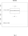
- the estimated test error of the classifier is around 4.9%.
- the classification scores generated by the RF i.e., the proportion of trees voting for a class
- the scores often fail to estimate the actual class probabilities and are thus said to be not well-calibrated.
- the uncertainties associated with an individual prediction in terms of a confidence scores, or estimated class probability is needed.
- the inventors fit a calibration model to raw RF scores. This calibration model is a multinomial logistic regression model, which takes the tumour subclasses as response variable and the 'raw' RF scores as explanatory variables.
- this model is fitted by incorporating a small ridge-penalty on the likelihood to prevent the model from over fitting as well as to stabilize estimation in situations where classes are perfectly separable.
- the amount of this regularization i.e., the penalization parameter, is determined by running a ten-fold cross-validation and choosing the value that minimizes the misclassification error.
- 'raw' RF scores are needed, i.e., the scores need to be generated by an RF classifier that was not trained using the same samples, otherwise the RF scores will be systematically biased and not comparable to scores of unseen cases.
- the inventors apply a three-fold cross validation.
- a three-fold nested cross-validation is applied.
- the reference set is randomly split into three parts.
- two-third of the data is used to train the RF classifier and one-third is used for prediction.
- a nested three-fold cross-validation is applied to generate independent RF scores, which are used to train a calibration model.
- the predicted RF scores resulting from the outer cross-validation loop are then recalibrated by using the suitable calibration model, i.e. a model that was fitted using the RF scores generated by using the other two-third of the data in the inner loop.
- the suitable calibration model i.e. a model that was fitted using the RF scores generated by using the other two-third of the data in the inner loop.
- the estimated test error of the classifier when using the recalibrated scores for prediction is around 3.2%.
- Some embodiments of the disclosure pertain to a method where the diverse tumour species are selected from metastatic tumours, tumours stemming from specific tissues, tumours in a specific stage, recurrent tumours, tumours having a specific genetic mutation, tumours of patients having different gender, age or genetic background.
- Genome-wide screening of DNA methylation patterns was performed by using the Infinium HumanMethylation450 BeadChips (Illumina, San Diego, US), allowing the simultaneous quantitative measurement of the methylation state at 485,577 CpG sites.
- the BeadChip provides coverage of 99% of RefSeq genes and 96 % of CpG islands.
- DNA concentrations were determined using PicoGreen (Life Technologies, Darmstadt, Germany). The quality of genomic DNA samples was checked by agarose-gel analysis, and sampies with an average fragment size >3kb were selected for methylation analysis.
- FFPE formalin-fixed paraffin-embedded
- DNA 500 ng genomic DNA and 250 ng FFPE DNA, respectively
- EZ-96 DNA Methylation Kit Zinc Research Corporation, Orange, US
- Bisulfite treatment leads to the deamination of non-methylated cytosines to uracils, while methylated cytosines are refractory to the effects of bisulfite and remain cytosine.
- FFPE samples were treated with the Infinium HD DNA Restoration Kit (Illumina) according to the manufacturer recommendations. By using enzymatic reactions, degraded FFPE DNA is restored in preparation for the whole genome amplification.
- Each sample was whole genome amplified and enzymatically fragmented following the instructions in the Illumina Infinium HD Assay Methylation Protocol Guide (genomic DNA) or Infinium HD FFPE Methylation Guide (FFPE DNA), respectively.
- the DNA was applied to Infinium HumanMethylation450 BeadChip and hybridization is performed for 16-24h at 48°C. During hybridization, the DNA molecules anneal to locus-specific DNA oligomers linked to individual bead types. One or two probes are used to interrogate CpG locus, depending on the probe design for a particular CpG site.
- Allele-specific primer annealing is followed by single-base extension using DNP- and Biotin- labeled ddNTPs.
- both bead types one each for the methylated and unmethylated states
- Infinium II uses only one bead type with a unique type of probe allowing detection of both alleles. The methylated and unmethylated signals are generated respectively in the green and the red channels.
- DNA methylation values are recorded for each locus in each sample.
- DNA methylation beta values are continuous variables between 0 and 1, representing the percentage of methylation of a given cytosine corresponding to the ratio of the methylated signal over the sum of the methylated and unmethylated signals.
- the Random Forest (RF) algorithm implemented in the R package randomForest (Liaw and Wiener, 2002) was used.
- the RF algorithm is a so-called ensemble method that combines the predictions of several 'weak' classifiers to achieve improved prediction accuracy.
- the RF uses binary classification trees ( Classification and Regression Trees (CART); Breiman et al., 1983 ) as 'weak' classifiers. Each of these trees represents a sequence of binary splitting rules that are learned by recursive binary splitting.
- the CART algorithm starts with all samples assigned to a 'root' node and tries to find the variable, e.g., a measured CpG probe, and a corresponding cut-off that results in the purest split into the different classes.
- the variable e.g., a measured CpG probe
- a corresponding cut-off that results in the purest split into the different classes.
- the Gini index a classical statistical measure for inequality.
- To fit a tree the CART algorithm iteratively repeats these steps until no further improvements can be made, i.e., only samples of the same class are assigned to the final 'leaf' node, or a pre-specified node size is achieved.
- the binary splitting rules are compared with the new data starting in the root node down to one of the leaf nodes.
- the tree then predicts or votes for the class dominating that leaf node.
- the RF algorithm combines thousands of trees by bootstrap aggregation (bagging).
- each tree is fitted using training data sets that are generated by drawing bootstrap samples, i.e., randomly selecting two-third of the data with replacement.
- bootstrap samples i.e., randomly selecting two-third of the data with replacement.
- at each node only a random subset of the available variables are used to find an optimal splitting rule.
- the RF takes the majority vote of all trees in the forest.
- variable selection was performed, i.e. in a first step the algorithm is used to calculate the variable importance, e.g. the average improvement in Gini purity of a CpG probe when used for a splitting rule.
- the final classifier was trained using only the 30,000 CpG probes with highest variable importance measure.
- Example 1 Distinguishing WNT medulloblastoma from Group 3 medulloblastoma
- Medulloblastoma is the most common malignant paediatric brain tumour and comprises four distinct molecular variants. These variants are known as WNT, SHH, Group 3, and Group 4. These variants are histologically indistinguishable, but clearly separable by DNA methylation patterns (see Figure 4 ). WNT tumours show activated Wnt signalling pathway and carry a favourable prognosis. SHH medulloblastoma show Hedgehog signalling pathway activation and are known to have an intermediate to good prognosis. While both WNT and SSH variants are molecularly already well characterised, the genetic programs driving the pathogenesis of Group 3 and Group 4 medulloblastoma remain largely unknown.
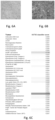
- a 1944 born female brain tumour patient was diagnosed based on histology (see Figure 5A ) to suffer from an anaplastic astrocytoma WHO III.
- a classifier score of 0.335 changed the diagnosis to Glioblastoma WHO IV (see Figure 5B ).
- Example 4 DNA methylation-based classification of tumour entities using three gene sites
- ATRT Atypical teratoid rhabdoid tumour (ATRT) is a rare paediatric brain tumour that can be subdivided into three molecular subgroups: ATRT-TYR, ATRT-SHH and ATRT-MYC (Ho et al. 2020, PMID: 31889194).
- the inventors have identified genes that include CpG sites that are most important for the classification of brain tumours and molecular subtypes.
- the importance of these CpGs for the classification has been measured by applying the permutation-based variable importance measure of the Random Forest (RF) algorithm (Strobl et al. 2007, PMID: 17254353).
- RF Random Forest
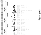
- PAX6, PTPRN2 and OSTM1 include many important CpGs for the classification.
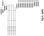
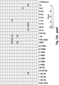
- Figure 8 displays the PTPRN2 gene and the CpG sites located on it. Most of the CpGs have a positive variable importance measure, indicating that these CpGs are predictive for the classification of brain tumours.
- PCA Next principal component analysis
- t-SNE stochastic neighbour embedding
- MDS multidimensional scaling
- FA factor analysis
- NMF non-negative matrix factorization
- SNE stochastic neighbour embedding
- UMAP uniform manifold approximation and projection for dimension reduction
- LDA linear discriminant analysis
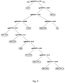
- a classification and regression tree (CART) has been applied to methylation data ( Figure 10C ).
- the CART algorithm automatically tries out all available 1022 CpGs probes and possible cut-offs and selects the CpG probe and corresponding cut-off that leads to the purest split into the ATRT subtypes.
- the algorithm stops, as soon as the class purity measured by the Gini coefficient cannot be further improved.
- the CART algorithm found two sequential splitting rules ( Figure 10D ) that involve only two CpG probes that result in an almost perfect separation of the ATRT subclasses.
- Random Forests usually combine hundreds or thousands of CART trees by bootstrap aggregation (bagging) to achieve an improved prediction accuracy.
- Other supervised methods that can be applied to fit models with comparable prediction performance, are for example gradient boosting machines (GBM), support vector machines (SVM), multinomial logistic regression models and (deep) neural nets.
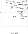
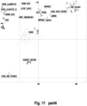
- Figure 11 shows the 1167 samples projected onto a t-distributed stochastic neighbour embedding (tSNE) that was applied to the gene expression data of the 688 most important genes identified in the methylation data.
- the colouring and the labelling of the groups are according to the class, and the samples are classified by the DNA-methylation classifier.
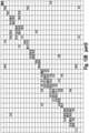
- the general clustering of the classes is very similar to a tSNE performed on DNA-methylation and even new sub-entities such as the medulloblastoma (MB) group 3 and 4 subtypes I-VIII can be identified. This proves that the gene expression data of the 688 identified genes is highly predictive for the 184 classes.
- the gene expression data can also be used to train supervised machine learning models
- the gene expression data set was reduced to 1057 samples belonging to 50 classes with a minimal class sample size of 7 samples.
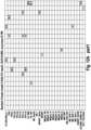
- the inventors then trained a basic random forest (RF) model and a lasso-penalized multinomial logistic regression model to this data set and validated the performance of both models by 3-fold cross-validation (CV).
- the CV estimated an accuracy of 0.788 for the RF ( Figure 12B ) and an accuracy 0.766 for the logistic regression model ( Figure 12A ), what proves that gene expression can be used to train similar classification models.
- the biological state used to train the classification algorithm is not limited to methylation, but can also be another biological state such as gene expression.
- Table 1 List of gene sites according to the disclosure including their genetic locus and Sequence ID in the sequence listing. The sequence listing associated with this application is filed in electronic format and hereby incorporated by reference into the specification in its entirety. Seq. ID No. Gene site Chromosome Sequence start Sequence end 1 ABAT chr16 8766944 8879932 2 ABLIM2 chr4 7965537 8162059 3 ABR chr17 905258 1092116 4 ACAD10 chr12 112122357 112196411 5 ACMSD chr2 135594686 135661102 6 ACOT7 chr1 6322832 6455326 7 ACOX3 chr4 8366509 8443952 8 ACSL1 chr4 185675249 185748715 9 ACTR3C chr7 149942801 150022258 10 ADAMTS17 chr15 100510143 100883683 11 ADAMTS2 chr5 178536352 178773931 12
- the WHO 2020 entity or cancer type names are shown in Column 2.
- Column 3 provides a descriptor for the molecular class and
- Column 4 lists the PubMed Number (PMID). Where no PMID number appears in Column 4 the method according to the disclosure uncovered cancer subspecies that where not known or published before and thus have no PMID.
- Tables 3 to 172 Classification of cancer types listed in Table 2 according to the disclosure.
- Table 2 The classification data for each cancer type as listed in Table 2 is shown in an individual table. Each table comprises the following columns:
Landscapes
- Engineering & Computer Science (AREA)
- Health & Medical Sciences (AREA)
- Life Sciences & Earth Sciences (AREA)
- Medical Informatics (AREA)
- Physics & Mathematics (AREA)
- Chemical & Material Sciences (AREA)
- Data Mining & Analysis (AREA)
- Bioinformatics & Cheminformatics (AREA)
- General Health & Medical Sciences (AREA)
- Public Health (AREA)
- Biophysics (AREA)
- Biotechnology (AREA)
- Proteomics, Peptides & Aminoacids (AREA)
- Evolutionary Biology (AREA)
- Bioinformatics & Computational Biology (AREA)
- Spectroscopy & Molecular Physics (AREA)
- Epidemiology (AREA)
- Databases & Information Systems (AREA)
- Theoretical Computer Science (AREA)
- Genetics & Genomics (AREA)
- Pathology (AREA)
- Analytical Chemistry (AREA)
- Organic Chemistry (AREA)
- Bioethics (AREA)
- Software Systems (AREA)
- Computer Vision & Pattern Recognition (AREA)
- Evolutionary Computation (AREA)
- Biomedical Technology (AREA)
- Artificial Intelligence (AREA)
- Molecular Biology (AREA)
- Wood Science & Technology (AREA)
- Zoology (AREA)
- Immunology (AREA)
- Primary Health Care (AREA)
- Microbiology (AREA)
- Oncology (AREA)
- Biochemistry (AREA)
- Hospice & Palliative Care (AREA)
- General Engineering & Computer Science (AREA)
- Measuring Or Testing Involving Enzymes Or Micro-Organisms (AREA)
Priority Applications (8)
| Application Number | Priority Date | Filing Date | Title |
|---|---|---|---|
| EP21190938.7A EP4134452A1 (de) | 2021-08-12 | 2021-08-12 | Verfahren zur klassifizierung von krebs |
| US18/683,110 US20260058010A1 (en) | 2021-08-12 | 2022-08-12 | Method for classification of cancer |
| JP2024508310A JP2024537596A (ja) | 2021-08-12 | 2022-08-12 | 癌の分類のための方法 |
| PCT/US2022/074920 WO2023019254A2 (en) | 2021-08-12 | 2022-08-12 | Method for classification of cancer |
| AU2022326568A AU2022326568A1 (en) | 2021-08-12 | 2022-08-12 | Method for classification of cancer |
| CA3228765A CA3228765A1 (en) | 2021-08-12 | 2022-08-12 | Method for classification of cancer |
| EP22856839.0A EP4384959A4 (de) | 2021-08-12 | 2022-08-12 | Verfahren zur klassifizierung von krebs |
| CN202280068424.4A CN118475939A (zh) | 2021-08-12 | 2022-08-12 | 用于癌症分类的方法 |
Applications Claiming Priority (1)
| Application Number | Priority Date | Filing Date | Title |
|---|---|---|---|
| EP21190938.7A EP4134452A1 (de) | 2021-08-12 | 2021-08-12 | Verfahren zur klassifizierung von krebs |
Publications (1)
| Publication Number | Publication Date |
|---|---|
| EP4134452A1 true EP4134452A1 (de) | 2023-02-15 |
Family
ID=77316835
Family Applications (2)
| Application Number | Title | Priority Date | Filing Date |
|---|---|---|---|
| EP21190938.7A Withdrawn EP4134452A1 (de) | 2021-08-12 | 2021-08-12 | Verfahren zur klassifizierung von krebs |
| EP22856839.0A Pending EP4384959A4 (de) | 2021-08-12 | 2022-08-12 | Verfahren zur klassifizierung von krebs |
Family Applications After (1)
| Application Number | Title | Priority Date | Filing Date |
|---|---|---|---|
| EP22856839.0A Pending EP4384959A4 (de) | 2021-08-12 | 2022-08-12 | Verfahren zur klassifizierung von krebs |
Country Status (7)
| Country | Link |
|---|---|
| US (1) | US20260058010A1 (de) |
| EP (2) | EP4134452A1 (de) |
| JP (1) | JP2024537596A (de) |
| CN (1) | CN118475939A (de) |
| AU (1) | AU2022326568A1 (de) |
| CA (1) | CA3228765A1 (de) |
| WO (1) | WO2023019254A2 (de) |
Families Citing this family (2)
| Publication number | Priority date | Publication date | Assignee | Title |
|---|---|---|---|---|
| US20250270650A1 (en) * | 2023-12-12 | 2025-08-28 | Flagship Pioneering Innovations Vi, Llc | Methods for identifying genomic regions of low background methylation |
| CN120822085A (zh) * | 2024-04-11 | 2025-10-21 | 中国科学院深圳先进技术研究院 | 肿瘤分类方法、装置、终端设备以及存储介质 |
Citations (2)
| Publication number | Priority date | Publication date | Assignee | Title |
|---|---|---|---|---|
| EP3067432A1 (de) * | 2015-03-11 | 2016-09-14 | Deutsches Krebsforschungszentrum Stiftung des Öffentlichen Rechts | Auf DNA-Methylierung basierendes Verfahren zur Klassifizierung von Tumorspezies des Gehirns |
| WO2020210754A1 (en) * | 2019-04-10 | 2020-10-15 | University Of Pittsburgh - Of The Commonwealth System Of Higher Education | Computational filtering of methylated sequence data for predictive modeling |
Family Cites Families (1)
| Publication number | Priority date | Publication date | Assignee | Title |
|---|---|---|---|---|
| US20160275604A1 (en) * | 2015-03-17 | 2016-09-22 | Accenture Global Services Limited | Adaptive quality assurance management system |
-
2021
- 2021-08-12 EP EP21190938.7A patent/EP4134452A1/de not_active Withdrawn
-
2022
- 2022-08-12 EP EP22856839.0A patent/EP4384959A4/de active Pending
- 2022-08-12 WO PCT/US2022/074920 patent/WO2023019254A2/en not_active Ceased
- 2022-08-12 US US18/683,110 patent/US20260058010A1/en active Pending
- 2022-08-12 CN CN202280068424.4A patent/CN118475939A/zh active Pending
- 2022-08-12 AU AU2022326568A patent/AU2022326568A1/en active Pending
- 2022-08-12 JP JP2024508310A patent/JP2024537596A/ja active Pending
- 2022-08-12 CA CA3228765A patent/CA3228765A1/en active Pending
Patent Citations (2)
| Publication number | Priority date | Publication date | Assignee | Title |
|---|---|---|---|---|
| EP3067432A1 (de) * | 2015-03-11 | 2016-09-14 | Deutsches Krebsforschungszentrum Stiftung des Öffentlichen Rechts | Auf DNA-Methylierung basierendes Verfahren zur Klassifizierung von Tumorspezies des Gehirns |
| WO2020210754A1 (en) * | 2019-04-10 | 2020-10-15 | University Of Pittsburgh - Of The Commonwealth System Of Higher Education | Computational filtering of methylated sequence data for predictive modeling |
Non-Patent Citations (26)
| Title |
|---|
| ACTA NEUROPATHOL., vol. 114, no. 2, 6 July 2007 (2007-07-06), pages 97 - 109 |
| AKEY ET AL., GENOMICS, vol. 80, 2002, pages 376 - 384 |
| ANNE ROCHTUS ET AL: "Methylome analysis for spina bifida shows hypomethylation as a risk factor with evidence for a complex (epi)genetic interplay to affect neural tube development", CLINICAL EPIGENETICS, BIOMED CENTRAL LTD, LONDON, UK, vol. 8, no. 1, 13 October 2016 (2016-10-13), pages 1 - 12, XP021267782, ISSN: 1868-7075, DOI: 10.1186/S13148-016-0272-8 * |
| BO-NANNO ET AL., CLIN. CHEM., vol. 53, 2007, pages 2119 - 2127 |
| CAPPER DAVID ET AL: "DNA methylation-based classification of central nervous system tumours", NATURE, vol. 555, no. 7697, 14 March 2018 (2018-03-14), London, pages 469 - 474, XP055879741, ISSN: 0028-0836, Retrieved from the Internet <URL:http://www.nature.com/articles/nature26000> DOI: 10.1038/nature26000 * |
| CAPPER DAVID ET AL: "Practical implementation of DNA methylation and copy-number-based CNS tumor diagnostics: the Heidelberg experience", ACTA NEUROPATHOLOGICA, SPRINGER VERLAG, BERLIN, DE, vol. 136, no. 2, 2 July 2018 (2018-07-02), pages 181 - 210, XP036555932, ISSN: 0001-6322, [retrieved on 20180702], DOI: 10.1007/S00401-018-1879-Y * |
| CLARK ET AL., NUCLEIC ACIDS RES., vol. 22, 1994, pages 2990 - 2997 |
| COLLELA ET AL., BIOTECHNIQUES, vol. 35, 2003, pages 152 - 156 |
| COTTRELL ET AL., NUCLEIC ACIDS RES., no. 32, 2004, pages e10 |
| EADS ET AL., CANCER RES., vol. 59, 1999, pages 3899 - 3903 |
| EADS ET AL., NUCLEIC ACIDS RES., vol. 28, 2000, pages e32 |
| EHRICH ET AL., PNAS USA, vol. 102, 2005, pages 15785 - 15790 |
| FROMMER ET AL., PNAS USA, vol. 89, 1992, pages 1827 - 1831 |
| GENTLEMAN, V. J.CAREY, D. M.BATES, B.BOLSTAD, M.DETTLING, S.DUDOIT, B.ELLIS, L.GAUTIER, Y.GE, GENOME BIOLOGY, vol. 5, 2004, pages R80 |
| GONZALGO ET AL., NUCLEIC ACIDS RES., vol. 25, 1997, pages 2529 - 2531 |
| HERMAN ET AL., PNAS USA, vol. 93, 1996, pages 9821 - 9826 |
| KRISTENSEN ET AL., NUCLEIC ACIDS RES., vol. 36, 2008, pages e42 |
| LOUIS DNOHGAKI HWIESTLER ODCAVENEE WKBURGER PCJOUVET ASCHEITHAUER BWKLEIHUES P., THE 2007 WHO CLASSIFICATION OF TUMOURS OF THE CENTRAL NERVOUS SYSTEM |
| MACKAY ET AL., HUM. GENET., vol. 116, 2005, pages 255 - 261 |
| MACKAY ET AL., HUM. GENET., vol. 120, 2006, pages 262 - 269 |
| PALMISANO ET AL., CANCER RES., vol. 60, 2000, pages 5954 - 5958 |
| UHLMANN ET AL., ELECTROPHORESIS, vol. 23, 2002, pages 4072 - 4079 |
| VOSO ET AL., BLOOD, vol. 103, 2004, pages 698 - 700 |
| WOJDACZ ET AL., NUCLEIC ACIDS RES., vol. 35, 2007, pages e41 |
| WORM ET AL., CLIN. CHEM., vol. 47, 2001, pages 1183 - 1189 |
| ZHAO GUANGJIE ET AL: "High methylation of the 4-aminobutyrate aminotransferase gene predicts a poor prognosis in patients with myelodysplastic syndrome", INTERNATIONAL JOURNAL OF ONCOLOGY, 4 December 2018 (2018-12-04), GR, XP055880436, ISSN: 1019-6439, DOI: 10.3892/ijo.2018.4652 * |
Also Published As
| Publication number | Publication date |
|---|---|
| JP2024537596A (ja) | 2024-10-16 |
| CN118475939A (zh) | 2024-08-09 |
| CA3228765A1 (en) | 2023-02-16 |
| AU2022326568A1 (en) | 2024-02-01 |
| US20260058010A1 (en) | 2026-02-26 |
| WO2023019254A3 (en) | 2023-04-20 |
| EP4384959A2 (de) | 2024-06-19 |
| EP4384959A4 (de) | 2025-11-05 |
| WO2023019254A2 (en) | 2023-02-16 |
Similar Documents
| Publication | Publication Date | Title |
|---|---|---|
| JP7681145B2 (ja) | 生体試料の多検体アッセイのための機械学習実装 | |
| AU2020283042B2 (en) | Methods and systems for high-depth sequencing of methylated nucleic acid | |
| EP3268492B1 (de) | Auf dna-methylierung basierendes verfahren zur klassifizierung von tumorspezies | |
| US20230349000A1 (en) | Classification and prognosis of cancer | |
| US12454725B2 (en) | Method of detecting cancer through generalized loss of stability of epigenetic domains and compositions thereof | |
| WO2012104642A1 (en) | Method for predicting risk of developing cancer | |
| WO2019195941A1 (en) | Dna methylation assays and methods | |
| US20160186270A1 (en) | Signature of cycling hypoxia and use thereof for the prognosis of cancer | |
| Cheng et al. | Integrated analysis of DNA methylation and mRNA expression profiles to identify key genes involved in the regrowth of clinically non-functioning pituitary adenoma | |
| US20260058010A1 (en) | Method for classification of cancer | |
| EP4162277A1 (de) | Zelluläre reaktionstests für lungenkrebs | |
| US20250188543A1 (en) | Methods for methylation analysis of cell-free dna | |
| WO2017046714A1 (en) | Methylation signature in squamous cell carcinoma of head and neck (hnscc) and applications thereof | |
| US20220290243A1 (en) | Identification of patients that will respond to chemotherapy | |
| US20240379236A1 (en) | Method for the diagnosis and/or classification of a disease in a subject | |
| US20250342959A1 (en) | Prediction of Alzheimer's Disease | |
| US20260015674A1 (en) | Use of pa-tn5 to generate dna libraries | |
| Almeida | Molecular diagnosis of autism spectrum disorder through whole exome sequencing | |
| WO2025062136A1 (en) | Chromosome detection assay | |
| Bizet | Bioinformatic inference of a prognostic epigenetic signature of immunity in breast cancers | |
| Wang | A Study of Normalization Methods of 450K Methylation Microarrays and Association of a FTO Gene Variant and Exercises with Epigenetic Changes by Modeling |
Legal Events
| Date | Code | Title | Description |
|---|---|---|---|
| PUAI | Public reference made under article 153(3) epc to a published international application that has entered the european phase |
Free format text: ORIGINAL CODE: 0009012 |
|
| STAA | Information on the status of an ep patent application or granted ep patent |
Free format text: STATUS: THE APPLICATION HAS BEEN PUBLISHED |
|
| AK | Designated contracting states |
Kind code of ref document: A1 Designated state(s): AL AT BE BG CH CY CZ DE DK EE ES FI FR GB GR HR HU IE IS IT LI LT LU LV MC MK MT NL NO PL PT RO RS SE SI SK SM TR |
|
| STAA | Information on the status of an ep patent application or granted ep patent |
Free format text: STATUS: THE APPLICATION IS DEEMED TO BE WITHDRAWN |
|
| 18D | Application deemed to be withdrawn |
Effective date: 20230817 |