EP4136972A1 - Compositions and methods for stabilizing circulating tumor cells - Google Patents
Compositions and methods for stabilizing circulating tumor cells Download PDFInfo
- Publication number
- EP4136972A1 EP4136972A1 EP22199049.2A EP22199049A EP4136972A1 EP 4136972 A1 EP4136972 A1 EP 4136972A1 EP 22199049 A EP22199049 A EP 22199049A EP 4136972 A1 EP4136972 A1 EP 4136972A1
- Authority
- EP
- European Patent Office
- Prior art keywords
- protective agent
- sample
- blood
- circulating tumor
- agent composition
- Prior art date
- Legal status (The legal status is an assumption and is not a legal conclusion. Google has not performed a legal analysis and makes no representation as to the accuracy of the status listed.)
- Pending
Links
Images


Classifications
-
- A—HUMAN NECESSITIES
- A01—AGRICULTURE; FORESTRY; ANIMAL HUSBANDRY; HUNTING; TRAPPING; FISHING
- A01N—PRESERVATION OF BODIES OF HUMANS OR ANIMALS OR PLANTS OR PARTS THEREOF; BIOCIDES, e.g. AS DISINFECTANTS, AS PESTICIDES OR AS HERBICIDES; PEST REPELLANTS OR ATTRACTANTS; PLANT GROWTH REGULATORS
- A01N1/00—Preservation of bodies of humans or animals, or parts thereof
- A01N1/10—Preservation of living parts
- A01N1/12—Chemical aspects of preservation
- A01N1/122—Preservation or perfusion media
- A01N1/126—Physiologically active agents, e.g. antioxidants or nutrients
-
- G—PHYSICS
- G01—MEASURING; TESTING
- G01N—INVESTIGATING OR ANALYSING MATERIALS BY DETERMINING THEIR CHEMICAL OR PHYSICAL PROPERTIES
- G01N33/00—Investigating or analysing materials by specific methods not covered by groups G01N1/00 - G01N31/00
- G01N33/48—Biological material, e.g. blood, urine; Haemocytometers
- G01N33/50—Chemical analysis of biological material, e.g. blood, urine; Testing involving biospecific ligand binding methods; Immunological testing
- G01N33/53—Immunoassay; Biospecific binding assay; Materials therefor
- G01N33/575—Immunoassay; Biospecific binding assay; Materials therefor for cancer
Definitions
- the teachings herein relate to devices and methods for stabilizing and preserving circulating tumor cells without damaging the tumor cell integrity for improved protection and regulation of circulating tumor cells during collection, storage, and shipment.
- CTC circulating tumor cell
- CTCs are detectable in the blood of patients with metastatic cancer using a number of different technologies. Since CTCs are rare they need to be enriched from patient blood for accurate enumeration and characterization. Most of the CTC enrichment and identification assays available today are based on enrichment with anti-EpCAM antibodies and subsequent identification using anti-cytokeratin antibodies. An example is the CellSearch ® instrument system available from Janssen Diagnostics, Raritan, NJ.
- CTCs Factors that limit the utility of CTCs in cancer diagnosis and prognosis are the low abundance and the fragility of the CTCs that may introduce variability in the evaluation of CTCs using different assay platforms.
- Transportation of blood samples from the site of phlebotomy to another facility is commonly required for CTC enumeration and characterization.
- fragile CTCs may degrade and compromise the accuracy of CTC enumeration and characterization.
- CTCs non-invasive diagnosis, prognosis and monitoring of treatment regimens.
- the low abundance of the CTCs and their fragile nature may introduce variability in the evaluation of CTCs using different assay platforms.
- This fragile nature of CTCs arises due to the apoptosis of CTCs which begins after separation from the tumor of origin and after removal of blood from a patient. Therefore, it is necessary to address several pre-analytical issues that arise during the time between blood draw and CTC enrichment and characterization in order to effectively preserve the CTCs for analysis. These include delays in blood processing, blood storage temperature, and agitation of the sample during transport and shipment of blood. Such conditions may affect the integrity of already fragile CTCs causing accurate enumeration and characterization of CTCs difficult. As a result, it is important to consider the type of blood collection device and post-phlebotomy conditions while working with CTC samples.
- the teachings herein employ a protocol using a unique protective agent composition that successfully preserves samples while stabilizing CTC integrity for a prolonged period (e.g., which may be at least 14 days, and which may be at room temperature).
- the present teachings provide a consistent and efficient method for preserving CTCs in biological samples.
- Data demonstrated herein describes a method that reduces cell lysis and nuclease activity, and also permits accurate and precise analytical analysis by virtue of the preservation of the final concentration of recoverable CTCs over time. In so doing, the teachings provide a novel approach that improves the downstream clinical analysis of CTCs.
- the present teachings describe protecting the CTCs by inhibiting all cellular metabolic activity in CTCs in blood.
- the present teachings contemplate a method for blood sample treatment comprising locating a protective agent into the blood collection devices described herein.
- the protective agent may include a preservative.
- a blood sample may be drawn into the blood collection device, the blood sample having a first CTC concentration.
- the blood collection device containing a blood sample may be transported from a first location to a second location, wherein at least a portion of the transporting occurs at a temperature of greater than about 0°C.
- the CTCs from the sample may be isolated at least 24 hours after blood draw, the sample having a second CTC concentration, wherein the second CTC concentration is not lower or higher than the first CTC concentration by any statistically significant value.
- the preservative may be selected from the group consisting of diazolidinyl urea and imidazolidinyl urea.
- the concentration of the preservative prior to the contacting step may be between about 0.1 g/ml and about 3 g/ml.
- the circulating tumor cells may be isolated from the sample at least 3 days after blood draw.
- the circulating tumor cells may be isolated from the sample at least 7 days after blood draw.
- the circulating tumor cells may be isolated from the sample at least 14 days after blood draw.
- the transporting step may occur without freezing the blood sample to a temperature colder than about -30°C.
- the protective agent may contact the circulating tumor cells so that after a period of at least 7 days from the time the blood sample is drawn, the amount of circulating tumor cells is at Least about 90% of the amount of circulating tumor cells at the time the blood sample is drawn.
- the protective agent may contact the circulating tumor cells so that after a period of at least 7 days from the time the blood sample is drawn, the amount of circulating tumor cells present in the sample is about 100% of the amount of circulating tumor cells present in the sample at the time the blood sample is drawn.
- the protective agent compositions will generally include a preservative agent as described herein, and a quenching agent for substantially abating any free aldehyde (e.g., formaldehyde) from reacting with DNA within a sample.
- the protective agent composition may also include one or more nuclease inhibitors.
- the protective agent composition may also include one or more metabolic inhibitors.
- the methods described herein may comprise a step of obtaining in blood collection device a biological sample from a subject.
- the biological sample may include at least one circulating tumor cell from the subject.
- the methods may include a step of contacting the biological sample while within the blood collection device with a protective agent composition that includes a preservative agent, an optional anticoagulant, and a quenching agent to form a mixture that includes the protective agent composition and the sample.
- the methods may include a step of quenching any free formaldehyde that may be present with the quenching agent from the protective agent composition so that the free formaldehyde reacts to form a reaction product that is inert to the CTCs within the biological sample.
- the resulting mixture may be devoid of any aldehyde, and CTCs within the sample may be suitable for downstream applications.
- a blood draw step of drawing blood from a patient into a blood collection device that has the protective agent composition loaded therein prior to the blood draw step.
- the method may include a step of transporting the sample while it is contacted with the protective agent composition from a blood draw site to a clinical laboratory (e.g., one located at least 100 meters, 1000 meters, 10,000 meters from the blood draw site) at which a sample analysis will occur. The quenching occurs prior to and/or substantially simultaneously with the contacting step.
- the methods may include a step of isolating circulating tumor cells from the sample. The methods may be free of any step of centrifugation of the sample.
- the methods may be free of any step of isolating cell-free fetal DNA, cell-free DNA, cell-free RNA or cellular RNA from a blood sample.
- the methods may be free of any step of refrigerating the sample (e.g., to a temperature below room temperature, such as about 10°C or cooler) after it has been contacted with the protective agent composition.
- the teachings herein provide for advantageous treatment of CTC-containing samples and provide stabilized samples that are essentially free of detectable covalent modifications that inhibit downstream testing of the characteristics of CTCs.
- the present teachings contemplate a non-invasive screening method for the identification of circulating tumor cells that are potentially indicative of cancer diagnosis and progression.
- the teachings herein envision not only preserving the state of any cells in a sample but also envisions protecting the circulating tumor cells from any adverse effects during any delay between sample collection and processing and/or testing.
- the methods of the teachings herein generally involve steps of contacting a biological sample (which may include multiple blood cells and cell-free biomolecules), with an aldehyde-free (e.g., formaldehyde-free) protective agent composition in an amount and time that is sufficient to prevent degradation of the circulating tumor cells.
- a biological sample which may include multiple blood cells and cell-free biomolecules
- the treatment is such that it substantially prevents any free aldehyde from adversely reacting with the CTCs of the sample such as by employing a quenching agent.
- substantial quantities of CTCs can be maintained within and isolated from the sample.
- the CTC integrity is substantially preserved in its as provided state (e.g., the state at the time of blood draw) by avoiding the damaging effects of any aldehyde (e.g., formaldehyde).
- the method may further include steps of analyzing the CTCs from a sample that has been treated in accordance with the above, or that have otherwise been contacted with the protective agent composition and the quenching agent therein.
- the teachings herein permit identification of CTC characteristics for prognostic and diagnostic use of various pathological conditions in the clinic.
- Protective Agent Compositions A & B as described in the examples below and shown in the drawings include a formaldehyde free stabilization reagent that stabilizes CTCs in a blood sample for up to 14 days at room temperature. They do so by inhibiting cell metabolism in CTCs in blood and preventing cellular apoptosis and necrosis that degrade the CTCs in blood. These examples were designed to investigate the effectiveness of this blood collection devices for the stabilization of CTCs in a blood sample for an extended period of time at room temperature.
- MCF-7 cells was obtained from American Type Culture Collection (Rockville, MD, USA) and routinely passaged in Eagle's MEM medium containing 10% fetal bovine serum at 37°C in humidified atmosphere of 5% CO2.
- MCF-7 cells 2000 cells 10 mL blood
- All samples were shipped at ambient temperature to Geneuity Clinical Research Services (Maryville, TN, USA) and analyzed on the CellSearch system on days 1 and 4 post phlebotomy to determine the stability of spiked MCF-7 cells. Blood samples were maintained at room temperature during the entire process.
- MCF-7 cells in K 3 EDTA blood showed a significant drop in recovered MCF-7 cells at day 1 and 4 compared to values obtained for Protective Agent Compositions A & B.
- MCF-7 cell count was stable up to 4 days at room temperature.
- EpCAM Epithelial cell adhesion molecule
- CK cytokeratin
- the CK protein expression showed no significant change over 4 days in tubes containing Protective Agent Compositions A & B.
- Protective Agent Composition A showed improved stabilizing of c-fos mRNA as compared to K 3 EDTA tubes. No significant change in cyclin D1 mRNA expression was observed in all tubes.
- Protective Agent Compositions A & B provide preservation and stabilization of CTCs in blood samples for at least 4 days at room temperature. In doing so, it facilitates the development of new non-invasive diagnostic and prognostic methodologies for CTC enumeration as well as characterization.
- Plasma samples were drawn from each donor into ono 10 ml K 3 EDTA tube, one 10 mL tube containing Protective Agent Composition A, and one 10 mL tube containing Protective Agent Composition B.
- Plasma was separated from blood within 2 h post collection. To separate plasma, blood samples were centrifuged at 300 x g for 20 min at room temperature. The upper plasma layer was carefully removed without disturbing the buffy coat and transferred to a new tube that was then centrifuged at 5000 x g for 10 min. The cell-free plasma was then spiked with MCF-7 cells ( ⁇ 2,000 cells / 4 - 5 mL of plasma) and stored at room temperature.
- MCF-7 cells were washed with phosphate buffered saline solution and centrifuged at 500 rpm for 7 minutes on glass slides using Shandon Cytospin ® 3 cytocentrifuge. Slides were then dried and immunostaining for EpCAM and Cytokeratin were carried out with a primary antibody cocktail containing a mouse anti-EpCAM antibody (VU-1D9, #sc-51681, 1:1:100) and a mouse anti-CK antibody (T-13, #sc-241376, 1:100).
- a primary antibody cocktail containing a mouse anti-EpCAM antibody (VU-1D9, #sc-51681, 1:1:100) and a mouse anti-CK antibody (T-13, #sc-241376, 1:100).
- coverslips were mounted onto slides with UltraCruz TM mounting medium (#sc- 24941 ) containing 4',6-diamidino-2-phenylindole (DAPI) to counterstain cell nuclei. All antibodies and mounting medium were purchased from Santa Cruz Biotechnology, Inc. (Dallas, TX, USA) and manufacturer's protocol was followed. Fluorescent images were obtained using Zeiss LS 510 META NLO laser scanning confocal microscope (Oberkochen, Germany).
- Cytospin slides of spiked MCF-7 cells were prepared as described above. Cells on the slides were treated with ice cold methanol (-10 °C) for 5 to 10 min. After air drying, the slides were stained with a mixture of 200 nmol/L of fluorescent-tagged molecular beacons targeting c-fos or cyclin D1 mRNAs in Opti-MEM (Invitrogen) at 37 °C for 1 h. Stained cells on the slides were then washed, counterstained with DAPI and examined using a confocal microscope. The molecular beacons were purchased from Eurofins MWG Operon (Huntsville, AL).
- K 3 EDTA tubes failed to preserve CTCs resulting in a much lower recovery rates for both day 1 and 4 as compared to Protective Agent Compositions A & B.
- FIG. 2 illustrates the effects of room temperature storage on the stability of tumor-associated transmembrane protein EpCAM and cytoskeleton protein cytokeratin of MCF-7 tumor cells spiked into blood plasma.
- EpCAM protein which is expressed on the cell membrane of MCF-7 cells are stable up to 4 days at room temperature in tubes containing Protective Agent Compositions A & B whereas in K 3 EDTA tubes this membrane protein was partially degraded by day 4.
- fluorescence signal for EpCAM cell membrane protein is very- weak and diffused in MCF-7 cells spiked into K 3 EDTA blood at day 4, CK protein is stabilized in tubes containing Protective Agent Compositions A & B at day 4, however this protein was less stable in K 3 EDTA tubes at day 4 as evidenced from reduced fluorescence intensity for this protein in spiked MCF-7 cells recovered from - K 3 EDTA tubes.
- Staining of cells with DAPI shows that nucleus and nuclear content is stable in cells recovered from tubes containing Protective Agent Compositions A & B but not from cells recovered from K 3 EDTA tubes after 4 days of storage at room temperature ( Figure 2 ).
- the molecular beacon example shows that both c-fos and cycling D1 mRNA are stable in Protective Agent Composition A at room for up to 4 days ( Figure 3 ).
- CTCs recovered from K 3 EDTA tubes showed CTCs with degrading c-fos and cyclin D1 mRNAs as shown in Figure 3 .
- Protective Agent Compositions A & B provide preservation and stabilization of CTCs in blood samples for up to 4 days at room temperature. In doing so, the protective agent compositions support the development of new non-invasive diagnostic and prognostic methodologies for CTC enumeration as well as characterization.
- a single protective agent composition may be employed that includes a preservative composition and a quenching agent.
- Such protective agent composition may be preloaded into a sample collection device, such as a blood collection tube (which may be evacuated to a pressure below atmospheric pressure after loading).
- a sample may be taken from a subject directly into the sample collection device (e.g., a blood collection tube), at which time it will be contacted with the protective agent composition.
- a sample can be taken from a subject into a first container and the sample subsequently transferred to one or more second containers in which the protective agent composition is present.
- the aldehyde-free (e.g., formaldehyde-free) protective agent composition may include a preservative composition such as one selected from the group consisting of: diazolidinyl urea, imidazolidinyl urea, dimethoylol-5,5-dimethylhydantoin, diethyl urea, 2-bromo-2-nitropropane-1,3-diol, oxazolidines, sodium hydroxymethyl glycinate, 5-hydroxymethoxymethyl-1-1aza-3,7-dioxabicyclo [3.3.0] octane, 5-hydroxymethyl-1-1aza-3,7-dioxabicyclo [3.3.0] octane, 5-hydroxypoly[methyleneoxy]methyl-1-1aza-3,7-dioxabicyclo [3.3.0] octane, quaternary adamantine and any combination thereof.
- a preservative composition such as one selected
- aldehyde-free (e.g., formaldehyde-free) protective agent composition may release an aldehyde (e.g., formaldehyde)
- the teachings herein envision a specific step of quenching the aldehyde to render it inert to the CTCs
- the preservative composition is desirably used in combination with a quenching agent for helping to assure that nucleic acid (e.g., DNA) in the sample avoids being subjected to free aldehyde (e.g., free formaldehyde) which may cause one or more deleterious effects upon the nucleic acid.
- a quenching agent for helping to assure that nucleic acid (e.g., DNA) in the sample avoids being subjected to free aldehyde (e.g., free formaldehyde) which may cause one or more deleterious effects upon the nucleic acid.
- free aldehyde e.g., free formaldehyde
- the resulting mixture is preferably devoid of any aldehyde, and nucleic acids within the sample are suitable for polymerase chain reaction and DNA sequencing, and will exhibit structural integrity comparable to native nucleic acids (e.g., DNA will exhibit ellipticity that is substantially similar that of untreated native DNA, as measured by circular dichroism spectroscopy or will exhibit DNA-dye fluorescence that is substantially similar to that of untreated native DNA, as measured by fluorescence spectroscopy).
- native nucleic acids e.g., DNA will exhibit ellipticity that is substantially similar that of untreated native DNA, as measured by circular dichroism spectroscopy or will exhibit DNA-dye fluorescence that is substantially similar to that of untreated native DNA, as measured by fluorescence spectroscopy).
- the concentration of the preservative composition after sample contact may be greater than about 20 mg/ml, 10 mg/ml, 5 mg/ml, 2 mg/ml, less than about 0.8 g/ml of the mixture of protective agent composition and biological (e.g., blood) sample.
- the concentration of the preservative composition after sample contact may be more than about 0.1 g/ml of the mixture of protective agent composition and biological (e.g., blood) sample.
- the concentration of the preservative composition after sample contact may be between approximately 0.1 g/ml to approximately 0.8 g/ml of the mixture of protective agent composition and biological (e.g., blood) sample.
- the concentration of the preservative composition after sample contact may be between approximately 0.3 g/ml to approximately 0.6 g/ml of the mixture of protective agent composition and biological (e.g., blood) sample.
- the concentration of the preservative composition both before and after contact with a blood sample may be modified depending upon what diagnostic procedures a sample may undergo. As an example, the concentration may be modified in the event that a sample is to undergo flow cytometry analysis. More specifically, the concentration may be increased in the event that a sample is to undergo flow cytometry analysis.
- the concentration of the preservative composition after sample contact may be greater than about 15 mg/ml, greater than about 25 mg/ml, or even greater than about 30 mg/ml after sample contact.
- the formulation of the protective agent composition may also be modified such that a sample that will undergo flow cytometry analysis may contain diazolidinyl urea.
- the protective agent composition may also include a quenching agent.
- the protective agent composition may also include one or more metabolic inhibitors, one or more nuclease inhibitors and EDTA.
- the quenching agent may be one or more compounds that include at least one functional group capable of reacting with an electron deficient functional group of an aldehyde (e.g., an amine compound that reacts with formaldehyde to form methylol and/or imine Schiff base or a cis-diol compound that reacts with formaldehyde to form a cyclic acetal).
- the quenching agent may be selected from amino acids, alkyl amines, polyamines, primary amines, secondary amines, ammonium salts, nucleobases or any combination thereof.
- the quenching agent may be selected from glycine, lysine, ethylene diamine, arginine, urea, adenine, guanine, cytosine, thymine, spermidine, or any combination thereof.
- the concentration of the quenching agent is an amount that is sufficiently large that after contacting the sample with the protective agent composition, there is an absence of free aldehyde (e.g., an absence of free formaldehyde). However, the concentration is sufficiently small that dilution of the sample will not materially impact any analyzed characteristic of the sample.
- the concentration of the formaldehyde-quenching reagent after the sample contacting step may be above about 0.001 g/ml, 0.002 g/ml or even about 0.004 g/ml of the mixture of protective agent composition and biological (e.g., blood) sample.
- the concentration of the formaldehyde-quenching reagent after the sample contacting step may be below about 0.03 g/ml, 0.01 g/ml, or even about 0.008 g/ml of the mixture of protective agent composition and biological (e.g., blood) sample.
- the concentration of the formaldehyde-quenching reagent after the sample contacting step may be between about 0.004 g/ml to about 0.008 g/ml.
- the protective agent composition may be present in an overall small fraction of the mixture volume. For example, it may be present in an amount that is less than about 5%, 2%, 0.5% or even less than about 0.3% of the overall mixture volume.
- the protective agent composition may be present in an amount of from about 1:20 parts by volume to about 1:300 parts by volume of the mixture.
- the amount of the protective agent composition may be present from about 1:50 parts by volume to about 1:200 parts by volume of the mixture.
- the amount of the protective agent composition is present from about 1:20 (1 part protective agent composition to 20 parts total mixture) parts by volume to about 1:300 parts by volume of the total mixture (which includes both the protective agent composition and the biological sample).
- the amount of the protective agent composition is present from about 1:100 parts by volume to about 1:200 parts by volume of the mixture.
- the protective agent composition may include at least one preservative composition selected from diazolidinyl urea, imidazolidinyl urea, dimethoylol-5,5-dimethylhydantoin, dimethylol urea, 2-bromo-2-nitropropane-1,3-diol, oxazolidines, sodium hydroxymethyl glycinate, 5-hydroxymethoxymethyl-1-1aza-3,7-dioxabicyclo [3.3.0] octane, 5-hydroxymethyl-1-1aza-3,7-dioxabicyclo [3.3.0] octane, 5-hydroxypoly[methyleneoxy]methyl-1-1aza-3,7-dioxabicyclo [3.3.0] octane, quaternary adamantine, 2-aminoacetic acid or any combination thereof.
- preservative composition selected from diazolidinyl urea, imidazolidinyl urea, dimetho
- the contacting step may include employing as the protective agent composition, a composition that includes imidazolidinyl urea in an amount of about 0.1 to about 2.0% by weight of the total mixture of the protective agent composition plus a biological sample; optionally, ethylenediaminetetraacetic acid (EDTA) in an amount of about 0.05 to about 0.75% by weight of the total mixture of the protective agent composition plus a biological sample; and a quenching agent in an amount sufficient to react with any free aldehyde (e.g., formaldehyde) that may arise from the imidazolidinyl urea to form a reaction product that will not react to denature any protein of the biological sample.
- EDTA ethylenediaminetetraacetic acid
- the protective agent composition (prior to contact with any biological sample) may include from about 20% to about 60% by weight imidazolidinyl urea.
- the protective agent composition may include at least about 30% by weight imidazolidinyl urea.
- the protective agent composition may include at least about 40% by weight imidazolidinyl urea and less than about 55% by weight imidazolidinyl urea.
- the protective agent composition may include from about 1% to about 10% by weight of the quenching agent.
- the protective agent composition may include at least about 2% by weight of the quenching agent.
- the protective agent composition may include at least about 4% by weight of the quenching agent and less than about 8% by weight of the quenching agent.
- the protective agent composition may include from about 1% to about 20% by weight EDTA.
- the protective agent composition may include at least about 5% by weight EDTA.
- the protective agent composition may include at least about 7% by weight EDTA and less than about 10% by weight EDTA.
- the protective agent composition may be pre-loaded into a tube and may be pre-loaded in amount of from about 50 to about 400 ⁇ l of protective agent composition.
- the pre-loaded amount may be at least about 100 ⁇ l and less than about 300 ⁇ l.
- the pre-loaded amount may be at least about 150 ⁇ l and less than about 250 ⁇ l.
- the protective agent composition may comprise at least about 80 mg and less than about 100 mg of the protective agent composition.
- the quenching agent may comprise at least about 1 mg and less than about 15 mg of the protective agent composition.
- EDTA may comprise at least about 10 mg and less than about 25 mg of the protective agent composition.
- it may include an amount of about 10 parts by weight of the protective agent composition per about 1 parts by weight of the quenching agent.
- the quenching agent may include a compound that includes at least one functional group capable of reacting with an electron deficient functional group of formaldehyde (e.g., an amine compound that reacts with formaldehyde to form methylol or imine Schiff base or a cis-diol compound that reacts with formaldehyde to form a cyclic acetal).
- formaldehyde e.g., an amine compound that reacts with formaldehyde to form methylol or imine Schiff base or a cis-diol compound that reacts with formaldehyde to form a cyclic acetal.
- the quenching agent may be an ingredient selected from amino acids, alkyl amines, polyamines, primary amines, secondary amines, ammonium salts, or a combination thereof, it may be an ingredient selected from glycine, lysine, ethylene diamine, arginine, urea, adenine, guanine, cytosine, thymine, spermidine, or any combination thereof. It may be an ingredient selected from glycine, lysine, ethylene diamine, urea or any combination thereof.
- the quenching step may include reacting any free aldehyde (e.g., formaldehyde) for forming a methylol, imine Schiff base, a Schiff base-quencher crosslink reaction product, a Schiff base dimer, or any combination thereof.
- any free aldehyde e.g., formaldehyde
- the protective agent may include one or more preservative agents, one or more nuclease inhibitors, one or more metabolic inhibitors, or any combination thereof.
- the one or more nuclease inhibitors may be selected from the group consisting of: diethyl pyrocarbonate, ethanol, aurintricarboxylic acid (ATA), glyceraldehydes, sodium fluoride, ethylenediamine tetraacetic acid (EDTA), formamide, vanadyl-ribonucleoside complexes, macaloid, heparin, hydroxylamine-oxygen-cupric ion, bentonite, ammonium sulfate, dithiothreitol (DTT), beta-mercaptoethanol, cysteine, dithioerythritol, tris (2-carboxyethyl) phosphene hydrochloride, a divalent cation such as Mg +2 , Mn +2 , Zn +2 ,
- the one or more metabolic inhibitors may be selected from the group consisting of: glyceraldehyde, dihydroxyacetone phosphate, glyceraldehyde 3-phosphate, 1,3-bisphosphoglycerate, 3-phosphoglycerate, 2-phosphoglycerate, phosphoenolpyruvate, pyruvate and glycerate dihydroxyacetate, sodium fluoride, K 2 C 2 O 4 , and any combination thereof.
- the amount of any active ingredient within the protective agent may generally be at least about 0.01% by weight.
- the amount of any active ingredient within the protective agent may generally be less than about 70% by weight.
- the protective agent may comprise at least about 10% diazolidinyl urea.
- the protective agent may comprise less than about 40% diazolidinyl urea.
- the protective agent may further contain at least about 1% of one or more enzyme inhibitors (e.g., nuclease inhibitors) such as EDTA and ATA.
- the protective agent may contain less than about 30% of one or more enzyme inhibitors.
- the protective agent may also contain at least about 1 % of one or more metabolic inhibitors.
- the protective agent may contain less than about 20% of one or more metabolic inhibitors.
- the protective agent composition optionally may include a nuclease inhibitor selected from the group consisting of: diethyl pyrocarbonate, ethanol, aurintricarboxylic acid (ATA), formamide, vanadyl-ribonucleoside complexes, macaloid, ethylenediamine tetraacetic acid (EDTA), proteinase K, heparin, hydroxylamine-oxygen-cupric ion, bentonite, ammonium sulfate, dithiothreitol (DTT), beta-mercaptoethanol (BME), cysteine, dithioerythritol, tris(2-carboxyethyl) phosphene hydrochloride, a divalent cation such as Mg 2+ , Mn 2+ , Zn 2+ , Fe 2+ , Ca 2+ , Cu 2+ , and any combination thereof.
- a nuclease inhibitor selected from the group consisting of: diethy
- the protective agent composition may include a preservative composition, an aldehyde quenching agent, and an anticoagulant.
- the protective agent composition may include imidazolidinyl urea, glycine, and ethylenediamine tetraacetic acid.
- teachings herein contemplate applications including but not limited to extracting circulating tumor cells for use in detecting cancer (including but not limited to carcinomas, leukemia, and/or lymphoma).
- cancer including but not limited to carcinomas, leukemia, and/or lymphoma.
- teachings herein may be employed for detecting abnormal methylation for breast cancer, prostate cancer, gastric cancer, ovarian, colorectal cancer, bladder cancer, testicular cancer, esophogeal cancer, melanoma or other cancers.
- any numerical values recited herein include all values from the lower value to the upper value in increments of one unit provided that there is a separation of at least 2 units between any lower value and any higher value.
- the amount of a component, a property, or a value of a process variable such as, for example, temperature, pressure, time and the like is, for example, from 1 to 90, preferably from 20 to 80, more preferably from 30 to 70, it is intended that intermediate range values (for example, 15 to 85, 22 to 68, 43 to 51 , 30 to 32 etc.) are within the teachings of this specification.
- individual intermediate values are also within the present teachings.
Landscapes
- Health & Medical Sciences (AREA)
- Life Sciences & Earth Sciences (AREA)
- Engineering & Computer Science (AREA)
- Immunology (AREA)
- Urology & Nephrology (AREA)
- Biomedical Technology (AREA)
- General Health & Medical Sciences (AREA)
- Hematology (AREA)
- Chemical & Material Sciences (AREA)
- Molecular Biology (AREA)
- Physics & Mathematics (AREA)
- Zoology (AREA)
- Food Science & Technology (AREA)
- Medicinal Chemistry (AREA)
- Cell Biology (AREA)
- Analytical Chemistry (AREA)
- Biochemistry (AREA)
- Biotechnology (AREA)
- General Physics & Mathematics (AREA)
- Pathology (AREA)
- Environmental Sciences (AREA)
- Microbiology (AREA)
- Wood Science & Technology (AREA)
- Dentistry (AREA)
- Investigating Or Analysing Biological Materials (AREA)
- Measuring Or Testing Involving Enzymes Or Micro-Organisms (AREA)
- Physiology (AREA)
- Biophysics (AREA)
- Oncology (AREA)
- Hospice & Palliative Care (AREA)
- Medicines Containing Material From Animals Or Micro-Organisms (AREA)
Abstract
Description
- The teachings herein relate to devices and methods for stabilizing and preserving circulating tumor cells without damaging the tumor cell integrity for improved protection and regulation of circulating tumor cells during collection, storage, and shipment.
- In the peripheral blood of patients with solid tumors of epithelial origin, studies have identified circulating cells that have characteristics of tumor cells. These cells that are present in the bloodstream of cancer patients (referred to as circulating tumor cells or CTCs) are thought to play an important role in cancer metastasis by breaking loose from a solid tumor, entering the circulation, and then migrating to distant organs to develop secondary tumors. As such, circulating tumor cell (CTC) enumeration and characterization in the blood of cancer patients is useful for cancer prognostic and treatment monitoring purposes. Even though the number of CTCs present in patient blood is very low, robust technologies have been developed to enumerate and characterize CTCs in patient blood samples. CTCs are detectable in the blood of patients with metastatic cancer using a number of different technologies. Since CTCs are rare they need to be enriched from patient blood for accurate enumeration and characterization. Most of the CTC enrichment and identification assays available today are based on enrichment with anti-EpCAM antibodies and subsequent identification using anti-cytokeratin antibodies. An example is the CellSearch® instrument system available from Janssen Diagnostics, Raritan, NJ.
- While the presence of CTCs in patients with cancer has been known for over a century, utilization of these rare cells in cancer diagnosis and prognosis was not feasible since methodologies to detect, isolate and characterize CTCs have not been developed until recently. With the development of robust methodologies to enrich, isolate and characterize CTCs in different types of cancers that are found in solid organs, several clinical studies have been conducted to investigate the possible use of CTCs in cancer diagnosis and prognosis. Assays that enumerate CTCs using the Cell Search® system have been developed for use as an aid to monitor patients with metastatic breast, colorectal, and prostate cancers. It has also been shown the potential usefulness of CTC enumeration using the Cell Search™ system for monitoring patients with melanoma, urothelial, and lung cancer.
- Factors that limit the utility of CTCs in cancer diagnosis and prognosis are the low abundance and the fragility of the CTCs that may introduce variability in the evaluation of CTCs using different assay platforms. Transportation of blood samples from the site of phlebotomy to another facility is commonly required for CTC enumeration and characterization. During post-phlebotomy blood sample transportation/storage, fragile CTCs may degrade and compromise the accuracy of CTC enumeration and characterization.
- There is a growing interest in the use of CTCs in non-invasive diagnosis, prognosis and monitoring of treatment regimens. The low abundance of the CTCs and their fragile nature may introduce variability in the evaluation of CTCs using different assay platforms. This fragile nature of CTCs arises due to the apoptosis of CTCs which begins after separation from the tumor of origin and after removal of blood from a patient. Therefore, it is necessary to address several pre-analytical issues that arise during the time between blood draw and CTC enrichment and characterization in order to effectively preserve the CTCs for analysis. These include delays in blood processing, blood storage temperature, and agitation of the sample during transport and shipment of blood. Such conditions may affect the integrity of already fragile CTCs causing accurate enumeration and characterization of CTCs difficult. As a result, it is important to consider the type of blood collection device and post-phlebotomy conditions while working with CTC samples.
- There is thus a need for methods of stabilizing and protecting circulating tumor cells whereby structural integrity is maintained so that shipping and storage is possible with minimal deleterious effect on the circulating tumor cells. There is a further need for such methods where the detrimental effects of aldehyde fixation are avoided.
- The teachings herein employ a protocol using a unique protective agent composition that successfully preserves samples while stabilizing CTC integrity for a prolonged period (e.g., which may be at least 14 days, and which may be at room temperature). The present teachings provide a consistent and efficient method for preserving CTCs in biological samples. Data demonstrated herein describes a method that reduces cell lysis and nuclease activity, and also permits accurate and precise analytical analysis by virtue of the preservation of the final concentration of recoverable CTCs over time. In so doing, the teachings provide a novel approach that improves the downstream clinical analysis of CTCs. The present teachings describe protecting the CTCs by inhibiting all cellular metabolic activity in CTCs in blood. As a result of metabolic inhibition of CTCs in blood all apoptotic and necrotic pathways are inhibited and CTCs are protected from cell degradation. Therefore it is no longer necessary to isolate and characterize CTCs immediately after venipuncture. Furthermore, samples can be stored at room temperature for up to 14 days without deleterious effects to sample integrity, which eliminates the need for cold storage of the blood sample.
- In one aspect, the present teachings contemplate a method for blood sample treatment comprising locating a protective agent into the blood collection devices described herein. The protective agent may include a preservative. A blood sample may be drawn into the blood collection device, the blood sample having a first CTC concentration. The blood collection device containing a blood sample may be transported from a first location to a second location, wherein at least a portion of the transporting occurs at a temperature of greater than about 0°C. The CTCs from the sample may be isolated at least 24 hours after blood draw, the sample having a second CTC concentration, wherein the second CTC concentration is not lower or higher than the first CTC concentration by any statistically significant value.
- The teachings herein further include that the preservative may be selected from the group consisting of diazolidinyl urea and imidazolidinyl urea. The concentration of the preservative prior to the contacting step may be between about 0.1 g/ml and about 3 g/ml. The circulating tumor cells may be isolated from the sample at least 3 days after blood draw. The circulating tumor cells may be isolated from the sample at least 7 days after blood draw. The circulating tumor cells may be isolated from the sample at least 14 days after blood draw. The transporting step may occur without freezing the blood sample to a temperature colder than about -30°C. The protective agent may contact the circulating tumor cells so that after a period of at least 7 days from the time the blood sample is drawn, the amount of circulating tumor cells is at Least about 90% of the amount of circulating tumor cells at the time the blood sample is drawn. The protective agent may contact the circulating tumor cells so that after a period of at least 7 days from the time the blood sample is drawn, the amount of circulating tumor cells present in the sample is about 100% of the amount of circulating tumor cells present in the sample at the time the blood sample is drawn.
- The teachings herein contemplate improved protective agent compositions, and methods of stabilizing a biological sample for analysis. The protective agent compositions will generally include a preservative agent as described herein, and a quenching agent for substantially abating any free aldehyde (e.g., formaldehyde) from reacting with DNA within a sample. The protective agent composition may also include one or more nuclease inhibitors. The protective agent composition may also include one or more metabolic inhibitors. The methods described herein may comprise a step of obtaining in blood collection device a biological sample from a subject. The biological sample may include at least one circulating tumor cell from the subject. The methods may include a step of contacting the biological sample while within the blood collection device with a protective agent composition that includes a preservative agent, an optional anticoagulant, and a quenching agent to form a mixture that includes the protective agent composition and the sample. The methods may include a step of quenching any free formaldehyde that may be present with the quenching agent from the protective agent composition so that the free formaldehyde reacts to form a reaction product that is inert to the CTCs within the biological sample. The resulting mixture may be devoid of any aldehyde, and CTCs within the sample may be suitable for downstream applications.
- For samples derived from blood, there may be a blood draw step of drawing blood from a patient into a blood collection device that has the protective agent composition loaded therein prior to the blood draw step. The method may include a step of transporting the sample while it is contacted with the protective agent composition from a blood draw site to a clinical laboratory (e.g., one located at least 100 meters, 1000 meters, 10,000 meters from the blood draw site) at which a sample analysis will occur. The quenching occurs prior to and/or substantially simultaneously with the contacting step. The methods may include a step of isolating circulating tumor cells from the sample. The methods may be free of any step of centrifugation of the sample. The methods may be free of any step of isolating cell-free fetal DNA, cell-free DNA, cell-free RNA or cellular RNA from a blood sample. The methods may be free of any step of refrigerating the sample (e.g., to a temperature below room temperature, such as about 10°C or cooler) after it has been contacted with the protective agent composition.
- As can be appreciated from the above, the teachings herein provide for advantageous treatment of CTC-containing samples and provide stabilized samples that are essentially free of detectable covalent modifications that inhibit downstream testing of the characteristics of CTCs.
-
-
Fig. 1 shows a graph displaying the effect of two exemplary protective agent compositions and a standard K3EDTA composition on MCF-7 cell recovery atDay 1 andDay 4 post blood draw. -
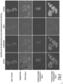
Fig. 2 shows immunofluorescence cell staining results (EpCAM and Cytokeratin) of two exemplary protective agent compositions and a standard K3EDTA composition. -
Fig. 3 shows fluorescence cell staining results showing levels of mRNA expression after contact with one exemplary protective agent composition and a standard K3EDTA composition. - This application is related to and claims the benefit of the filing date of
U.S. Provisional Application Serial No. 61/857,847 filed July 24, 2013 - The explanations and illustrations presented herein are intended to acquaint others skilled in the art with the teachings, its principles, and its practical application. Those skilled in the art may adapt and apply the teachings in its numerous forms, as may be best suited to the requirements of a particular use. Accordingly, the specific embodiments of the present teachings as set forth are not intended as being exhaustive or limiting of the teachings. The scope of the teachings should, therefore, be determined not with reference to the above description, but should instead be determined with reference to the appended claims, along with the full scope of equivalents to which such claims are entitled. The disclosures of all articles and references, including patent applications and publications, are incorporated by reference for ail purposes. Other combinations are also possible as will be gleaned from the following claims, which are also hereby incorporated by reference into this written description.
- Unless otherwise stated, percentages as set forth herein refer to percent by weight. Further, unless the context of the discussion makes clear to the contrary, references to circulating tumor cells refers not only to intact circulating tumor cells, but to tumor cell fragments.
- The present teachings contemplate a non-invasive screening method for the identification of circulating tumor cells that are potentially indicative of cancer diagnosis and progression. The teachings herein envision not only preserving the state of any cells in a sample but also envisions protecting the circulating tumor cells from any adverse effects during any delay between sample collection and processing and/or testing. The methods of the teachings herein generally involve steps of contacting a biological sample (which may include multiple blood cells and cell-free biomolecules), with an aldehyde-free (e.g., formaldehyde-free) protective agent composition in an amount and time that is sufficient to prevent degradation of the circulating tumor cells. The treatment is such that it substantially prevents any free aldehyde from adversely reacting with the CTCs of the sample such as by employing a quenching agent. In this manner, substantial quantities of CTCs can be maintained within and isolated from the sample. The CTC integrity is substantially preserved in its as provided state (e.g., the state at the time of blood draw) by avoiding the damaging effects of any aldehyde (e.g., formaldehyde). Thus, accurate and precise analytical analysis of the CTCs of the sample can be achieved. The method may further include steps of analyzing the CTCs from a sample that has been treated in accordance with the above, or that have otherwise been contacted with the protective agent composition and the quenching agent therein. As noted, the teachings herein permit identification of CTC characteristics for prognostic and diagnostic use of various pathological conditions in the clinic.
- Protective Agent Compositions A & B as described in the examples below and shown in the drawings include a formaldehyde free stabilization reagent that stabilizes CTCs in a blood sample for up to 14 days at room temperature. They do so by inhibiting cell metabolism in CTCs in blood and preventing cellular apoptosis and necrosis that degrade the CTCs in blood. These examples were designed to investigate the effectiveness of this blood collection devices for the stabilization of CTCs in a blood sample for an extended period of time at room temperature.
- The study was approved by the institutional review board of the Methodist Hospital, Omaha, NE, USA and informed consent was obtained from all donors prior to blood draw. Blood specimens were collected from apparently healthy adult donors by standard phlebotomy techniques.
- Breast cancer cell line, MCF-7 cells was obtained from American Type Culture Collection (Rockville, MD, USA) and routinely passaged in Eagle's MEM medium containing 10% fetal bovine serum at 37°C in humidified atmosphere of 5% CO2.
- For the MCF-7 cell spiking experiment, blood from each healthy donor (7 donors in total) was drawn into 10 mL K3EDTA tubes (BD Vacutainer®, Becton Dickinson, Franklin Lakes, NJ, USA), and 10 mL tubes containing Protective Agent A and 10mL tubes containing Protective Agent B. The blood volume was as close to 10 mL as possible for all tube types. MCF-7 cells (2000
cells 10 mL blood) were then spiked into all tubes and blood was mixed immediately after spiking by inverting 10 times each, All samples were shipped at ambient temperature to Geneuity Clinical Research Services (Maryville, TN, USA) and analyzed on the CellSearch system ondays - While the CellSearch system was utilized for the examples herein, it is envisioned that the protective agent compositions described herein could be utilized with any device and/or platform equipped for tumor cell enrichment and characterization.
- Blood was drawn from each donor into multiple 10 mL K3EDTA tubes and multiple 10 mL tubes for each of Protective Agent Compositions A & B. All tube types were spiked with MCF-7 breast cancer cells and stored at room temperature. Spiked MCF-7 cells were enumerated using the CellSearch™ system on
days - Overall enumeration of MCF-7 cells in K3EDTA blood showed a significant drop in recovered MCF-7 cells at
day - As further discussed in the details below, Protective Agent Compositions A & B provide preservation and stabilization of CTCs in blood samples for at least 4 days at room temperature. In doing so, it facilitates the development of new non-invasive diagnostic and prognostic methodologies for CTC enumeration as well as characterization.
- Blood was drawn from each donor into
ono 10 ml K3EDTA tube, one 10 mL tube containing Protective Agent Composition A, and one 10 mL tube containing Protective Agent Composition B. Plasma was separated from blood within 2 h post collection. To separate plasma, blood samples were centrifuged at 300 x g for 20 min at room temperature. The upper plasma layer was carefully removed without disturbing the buffy coat and transferred to a new tube that was then centrifuged at 5000 x g for 10 min. The cell-free plasma was then spiked with MCF-7 cells (~ 2,000 cells / 4 - 5 mL of plasma) and stored at room temperature. Ondays - Cytospin slides of spiked MCF-7 cells were prepared as described above. Cells on the slides were treated with ice cold methanol (-10 °C) for 5 to 10 min. After air drying, the slides were stained with a mixture of 200 nmol/L of fluorescent-tagged molecular beacons targeting c-fos or cyclin D1 mRNAs in Opti-MEM (Invitrogen) at 37 °C for 1 h. Stained cells on the slides were then washed, counterstained with DAPI and examined using a confocal microscope. The molecular beacons were purchased from Eurofins MWG Operon (Huntsville, AL).
- Experiments were carried out to determine the ability of Protective Agent Compositions A & B to stabilize CTCs during blood sample storage and transportation as compared to regular K3EDTA blood collection tubes. Parallel blood samples drawn into K3EDTA and tubes containing Protective Agent Compositions A & B and spiked with MCF-7 cells were analyzed using the CellSearch system for spiked tumor cell recovery. As shown in
Figure 1 , Protective Agent Compositions A & B demonstrated desired percentage recovery of the tumor cells at room temperature for up to 4 days. In Protective Agent Compositions A & B, atday day 4 it was 58% (SD = 8%. CV = 14.3%). In contrast, K3EDTA tubes failed to preserve CTCs resulting in a much lower recovery rates for bothday day 1 , recovery rate was 32% (SD = 12%, CV = 36.3%) of the spiked MCF-7 cells and atday 4 it was 16% (SD = 14%, CV = 87%). -
Figure 2 illustrates the effects of room temperature storage on the stability of tumor-associated transmembrane protein EpCAM and cytoskeleton protein cytokeratin of MCF-7 tumor cells spiked into blood plasma. According toFigure 2 , EpCAM protein which is expressed on the cell membrane of MCF-7 cells are stable up to 4 days at room temperature in tubes containing Protective Agent Compositions A & B whereas in K3EDTA tubes this membrane protein was partially degraded byday 4. According toFigure 2 , fluorescence signal for EpCAM cell membrane protein is very- weak and diffused in MCF-7 cells spiked into K3EDTA blood atday 4, CK protein is stabilized in tubes containing Protective Agent Compositions A & B atday 4, however this protein was less stable in K3EDTA tubes atday 4 as evidenced from reduced fluorescence intensity for this protein in spiked MCF-7 cells recovered from - K3EDTA tubes. Staining of cells with DAPI shows that nucleus and nuclear content is stable in cells recovered from tubes containing Protective Agent Compositions A & B but not from cells recovered from K3EDTA tubes after 4 days of storage at room temperature (Figure 2 ). - Experiments were carried out to study the stability of mRNA in spiked MCF- 7 cells recovered from Protective Agent Composition A and K3EDTA tubes. Cytospins of recovered MCF-7 cells were prepared as described above. To detect mRNA in situ, fluorescent-labeled molecular beacons and a scanning confocal microscope was used. As shown in
Figure 3 , c-fos mRNA (green fluorescence) and cyclin D1 mRNA (red fluorescence) were both stable in Protective Agent Composition A up to 4 days at room temperature. However in K3EDTA tubes c-fos mRNA was not stable after 4 days of storage at room temperature, indicating that c-fos mRNA expression was significantly degraded or downregulated. The change in cyclin D1 mRNA level in MCF-7 cells recovered from K3EDAT tube was minimal after 4 days incubation at room temperature. - In the example with results shown at
Figure 1 , 2000 MCF-7 cells were spiked into each K3EDTA tube and tubes containing Protective Agent Compositions A & B and shipped from Omaha NE to Maryville, TN by overnight shipping for analysis by the CellSearch™ System. Thus, this example represents a combination of transportation as well as storage effect on CTCs in blood samples. As shown inFigure 1 , our CTC recovery study conducted using spiked MCF-7 cells and analyzed by CellSearch™ System provided evidence that Protective Agent Compositions A & B are able to preserve CTCs during transportation and storage at room temperature for up to 4 days. Previous studies using CellSearch™ System have shown that the recovery rate for MCF-7 cells shortly after blood draw is between 62 - 89%. Our results show that in tubes containing Protective Agent Compositions A & B, postshipping day 1 andday 4 recovery rates of 61% and 57% respectively. There was no statistically significant difference between these two values indicating that CTCs are stable in Protective Agent Compositions A & B for 4 days after shipping at room temperature. However in K3EDTA tubes, CTC recovery rate was very low compared to Protective Agent Compositions A & B. in K3EDTA tubes postshipping day 1 andday 4 recovery rates were 32% and 16% respectively. There was a statistically significant decrease in CTC recovery in K3EDTA tube atday 1 andday 4 compared to Protective Agent Compositions A & B. As shown inFigure 2 . immunofluorescence staining of recovered CTCs for EpCAM and CK showed stability of these proteins in CTCs recovered from Protective Agent Compositions A &B 4 days after storage at room temperature. However cells recovered from K3EDTA tubes showed degrading EpCAM and CK proteins after 4 days at the same temperature. DAPI staining of cells showed stable nucleus and nuclear content in CTCs recovered from Protective Agent Compositions A & B whereas CTCs recovered from K3EDTA tubes showed degrading nucleus and nuclear content (Figure 2 ). - The molecular beacon example shows that both c-fos and cycling D1 mRNA are stable in Protective Agent Composition A at room for up to 4 days (
Figure 3 ). However, CTCs recovered from K3EDTA tubes showed CTCs with degrading c-fos and cyclin D1 mRNAs as shown inFigure 3 . - Analysis of the stabilizing reagent present in Protective Agent Compositions A & B by 13C-NMR has shown that the reagent is free of formaldehyde. Aldehyde based chemicais traditionally used in cell stabilization, such as formaldehyde and glutaraldehyde, are known to damage DNA and RNA by causing chemical modifications in nucleic acids and making nucleic acid-protein cross-links which make extraction of nucleic acids difficult. Application of such aldehyde based chemicals for CTC stabilization may cause problems for CTC characterization studies. Cell stabilizing reagent present in Protective Agent Compositions A & B have an advantage over aldehyde based stabilizing agents because of it has no negative effect on either nucleic acid extraction or amplification by PCR.
- In these examples, Protective Agent Compositions A & B provide preservation and stabilization of CTCs in blood samples for up to 4 days at room temperature. In doing so, the protective agent compositions support the development of new non-invasive diagnostic and prognostic methodologies for CTC enumeration as well as characterization.
- The teachings herein envision that a single protective agent composition may be employed that includes a preservative composition and a quenching agent. Such protective agent composition may be preloaded into a sample collection device, such as a blood collection tube (which may be evacuated to a pressure below atmospheric pressure after loading). Thus, it is possible that a sample may be taken from a subject directly into the sample collection device (e.g., a blood collection tube), at which time it will be contacted with the protective agent composition. It is also possible that a sample can be taken from a subject into a first container and the sample subsequently transferred to one or more second containers in which the protective agent composition is present.
- The aldehyde-free (e.g., formaldehyde-free) protective agent composition may include a preservative composition such as one selected from the group consisting of: diazolidinyl urea, imidazolidinyl urea, dimethoylol-5,5-dimethylhydantoin, diethyl urea, 2-bromo-2-nitropropane-1,3-diol, oxazolidines, sodium hydroxymethyl glycinate, 5-hydroxymethoxymethyl-1-1aza-3,7-dioxabicyclo [3.3.0] octane, 5-hydroxymethyl-1-1aza-3,7-dioxabicyclo [3.3.0] octane, 5-hydroxypoly[methyleneoxy]methyl-1-1aza-3,7-dioxabicyclo [3.3.0] octane, quaternary adamantine and any combination thereof. Though the aldehyde-free (e.g., formaldehyde-free) protective agent composition may release an aldehyde (e.g., formaldehyde), the teachings herein envision a specific step of quenching the aldehyde to render it inert to the CTCs,
- The preservative composition is desirably used in combination with a quenching agent for helping to assure that nucleic acid (e.g., DNA) in the sample avoids being subjected to free aldehyde (e.g., free formaldehyde) which may cause one or more deleterious effects upon the nucleic acid. Accordingly, the teachings herein contemplate the use of at least one aldehyde quenching agent, which is employed in an amount and in a manner sufficient so that any free aldehyde (e.g., formaldehyde) released from the protective agent composition reacts to form a reaction product that is inert to the nucleic acid of the biological sample. Further, the resulting mixture is preferably devoid of any aldehyde, and nucleic acids within the sample are suitable for polymerase chain reaction and DNA sequencing, and will exhibit structural integrity comparable to native nucleic acids (e.g., DNA will exhibit ellipticity that is substantially similar that of untreated native DNA, as measured by circular dichroism spectroscopy or will exhibit DNA-dye fluorescence that is substantially similar to that of untreated native DNA, as measured by fluorescence spectroscopy).
- The concentration of the preservative composition after sample contact may be greater than about 20 mg/ml, 10 mg/ml, 5 mg/ml, 2 mg/ml, less than about 0.8 g/ml of the mixture of protective agent composition and biological (e.g., blood) sample. The concentration of the preservative composition after sample contact may be more than about 0.1 g/ml of the mixture of protective agent composition and biological (e.g., blood) sample. By way of example, the concentration of the preservative composition after sample contact may be between approximately 0.1 g/ml to approximately 0.8 g/ml of the mixture of protective agent composition and biological (e.g., blood) sample. The concentration of the preservative composition after sample contact may be between approximately 0.3 g/ml to approximately 0.6 g/ml of the mixture of protective agent composition and biological (e.g., blood) sample. The concentration of the preservative composition both before and after contact with a blood sample may be modified depending upon what diagnostic procedures a sample may undergo. As an example, the concentration may be modified in the event that a sample is to undergo flow cytometry analysis. More specifically, the concentration may be increased in the event that a sample is to undergo flow cytometry analysis. Thus, the concentration of the preservative composition after sample contact may be greater than about 15 mg/ml, greater than about 25 mg/ml, or even greater than about 30 mg/ml after sample contact. The formulation of the protective agent composition (and the preservative composition contained therein) may also be modified such that a sample that will undergo flow cytometry analysis may contain diazolidinyl urea. The protective agent composition may also include a quenching agent. The protective agent composition may also include one or more metabolic inhibitors, one or more nuclease inhibitors and EDTA.
- The quenching agent may be one or more compounds that include at least one functional group capable of reacting with an electron deficient functional group of an aldehyde (e.g., an amine compound that reacts with formaldehyde to form methylol and/or imine Schiff base or a cis-diol compound that reacts with formaldehyde to form a cyclic acetal). The quenching agent may be selected from amino acids, alkyl amines, polyamines, primary amines, secondary amines, ammonium salts, nucleobases or any combination thereof. The quenching agent may be selected from glycine, lysine, ethylene diamine, arginine, urea, adenine, guanine, cytosine, thymine, spermidine, or any combination thereof.
- The concentration of the quenching agent is an amount that is sufficiently large that after contacting the sample with the protective agent composition, there is an absence of free aldehyde (e.g., an absence of free formaldehyde). However, the concentration is sufficiently small that dilution of the sample will not materially impact any analyzed characteristic of the sample. The concentration of the formaldehyde-quenching reagent after the sample contacting step may be above about 0.001 g/ml, 0.002 g/ml or even about 0.004 g/ml of the mixture of protective agent composition and biological (e.g., blood) sample. The concentration of the formaldehyde-quenching reagent after the sample contacting step may be below about 0.03 g/ml, 0.01 g/ml, or even about 0.008 g/ml of the mixture of protective agent composition and biological (e.g., blood) sample. By way of example, the concentration of the formaldehyde-quenching reagent after the sample contacting step may be between about 0.004 g/ml to about 0.008 g/ml.
- Upon being brought into contact with a sample to form a mixture of the sample and protective agent composition (e.g., at time of a blood draw into a blood collection device containing a protective agent composition of the teachings herein), the protective agent composition may be present in an overall small fraction of the mixture volume. For example, it may be present in an amount that is less than about 5%, 2%, 0.5% or even less than about 0.3% of the overall mixture volume. For example, the protective agent composition may be present in an amount of from about 1:20 parts by volume to about 1:300 parts by volume of the mixture. The amount of the protective agent composition may be present from about 1:50 parts by volume to about 1:200 parts by volume of the mixture.
- During at least the contacting step, the amount of the protective agent composition is present from about 1:20 (1 part protective agent composition to 20 parts total mixture) parts by volume to about 1:300 parts by volume of the total mixture (which includes both the protective agent composition and the biological sample). For instance, during at least the contacting step, the amount of the protective agent composition is present from about 1:100 parts by volume to about 1:200 parts by volume of the mixture.
- The protective agent composition may include at least one preservative composition selected from diazolidinyl urea, imidazolidinyl urea, dimethoylol-5,5-dimethylhydantoin, dimethylol urea, 2-bromo-2-nitropropane-1,3-diol, oxazolidines, sodium hydroxymethyl glycinate, 5-hydroxymethoxymethyl-1-1aza-3,7-dioxabicyclo [3.3.0] octane, 5-hydroxymethyl-1-1aza-3,7-dioxabicyclo [3.3.0] octane, 5-hydroxypoly[methyleneoxy]methyl-1-1aza-3,7-dioxabicyclo [3.3.0] octane, quaternary adamantine, 2-aminoacetic acid or any combination thereof. By way of illustration, the contacting step may include employing as the protective agent composition, a composition that includes imidazolidinyl urea in an amount of about 0.1 to about 2.0% by weight of the total mixture of the protective agent composition plus a biological sample; optionally, ethylenediaminetetraacetic acid (EDTA) in an amount of about 0.05 to about 0.75% by weight of the total mixture of the protective agent composition plus a biological sample; and a quenching agent in an amount sufficient to react with any free aldehyde (e.g., formaldehyde) that may arise from the imidazolidinyl urea to form a reaction product that will not react to denature any protein of the biological sample. The protective agent composition (prior to contact with any biological sample) may include from about 20% to about 60% by weight imidazolidinyl urea. The protective agent composition may include at least about 30% by weight imidazolidinyl urea. The protective agent composition may include at least about 40% by weight imidazolidinyl urea and less than about 55% by weight imidazolidinyl urea. The protective agent composition may include from about 1% to about 10% by weight of the quenching agent. The protective agent composition may include at least about 2% by weight of the quenching agent. The protective agent composition may include at least about 4% by weight of the quenching agent and less than about 8% by weight of the quenching agent. The protective agent composition may include from about 1% to about 20% by weight EDTA. The protective agent composition may include at least about 5% by weight EDTA. The protective agent composition may include at least about 7% by weight EDTA and less than about 10% by weight EDTA.
- The protective agent composition may be pre-loaded into a tube and may be pre-loaded in amount of from about 50 to about 400µl of protective agent composition. The pre-loaded amount may be at least about 100µl and less than about 300µl. The pre-loaded amount may be at least about 150µl and less than about 250µl. Within the pre-loaded protective agent composition, the protective agent composition may comprise at least about 80 mg and less than about 100 mg of the protective agent composition. The quenching agent may comprise at least about 1 mg and less than about 15 mg of the protective agent composition. EDTA may comprise at least about 10 mg and less than about 25 mg of the protective agent composition. For the protective agent composition, it may include an amount of about 10 parts by weight of the protective agent composition per about 1 parts by weight of the quenching agent. The quenching agent may include a compound that includes at least one functional group capable of reacting with an electron deficient functional group of formaldehyde (e.g., an amine compound that reacts with formaldehyde to form methylol or imine Schiff base or a cis-diol compound that reacts with formaldehyde to form a cyclic acetal). The quenching agent may be an ingredient selected from amino acids, alkyl amines, polyamines, primary amines, secondary amines, ammonium salts, or a combination thereof, it may be an ingredient selected from glycine, lysine, ethylene diamine, arginine, urea, adenine, guanine, cytosine, thymine, spermidine, or any combination thereof. It may be an ingredient selected from glycine, lysine, ethylene diamine, urea or any combination thereof. The quenching step may include reacting any free aldehyde (e.g., formaldehyde) for forming a methylol, imine Schiff base, a Schiff base-quencher crosslink reaction product, a Schiff base dimer, or any combination thereof.
- The protective agent may include one or more preservative agents, one or more nuclease inhibitors, one or more metabolic inhibitors, or any combination thereof. The one or more nuclease inhibitors may be selected from the group consisting of: diethyl pyrocarbonate, ethanol, aurintricarboxylic acid (ATA), glyceraldehydes, sodium fluoride, ethylenediamine tetraacetic acid (EDTA), formamide, vanadyl-ribonucleoside complexes, macaloid, heparin, hydroxylamine-oxygen-cupric ion, bentonite, ammonium sulfate, dithiothreitol (DTT), beta-mercaptoethanol, cysteine, dithioerythritol, tris (2-carboxyethyl) phosphene hydrochloride, a divalent cation such as Mg+2, Mn+2, Zn+2, Fe+2, Ca+2, Cu+2 and any combination thereof. The one or more metabolic inhibitors may be selected from the group consisting of: glyceraldehyde, dihydroxyacetone phosphate, glyceraldehyde 3-phosphate, 1,3-bisphosphoglycerate, 3-phosphoglycerate, 2-phosphoglycerate, phosphoenolpyruvate, pyruvate and glycerate dihydroxyacetate, sodium fluoride, K2C2O4, and any combination thereof.
- The amount of any active ingredient within the protective agent may generally be at least about 0.01% by weight. The amount of any active ingredient within the protective agent may generally be less than about 70% by weight. The protective agent may comprise at least about 10% diazolidinyl urea. The protective agent may comprise less than about 40% diazolidinyl urea. The protective agent may further contain at least about 1% of one or more enzyme inhibitors (e.g., nuclease inhibitors) such as EDTA and ATA. The protective agent may contain less than about 30% of one or more enzyme inhibitors. The protective agent may also contain at least about 1 % of one or more metabolic inhibitors. The protective agent may contain less than about 20% of one or more metabolic inhibitors.
- The protective agent composition optionally may include a nuclease inhibitor selected from the group consisting of: diethyl pyrocarbonate, ethanol, aurintricarboxylic acid (ATA), formamide, vanadyl-ribonucleoside complexes, macaloid, ethylenediamine tetraacetic acid (EDTA), proteinase K, heparin, hydroxylamine-oxygen-cupric ion, bentonite, ammonium sulfate, dithiothreitol (DTT), beta-mercaptoethanol (BME), cysteine, dithioerythritol, tris(2-carboxyethyl) phosphene hydrochloride, a divalent cation such as Mg2+, Mn2+, Zn2+, Fe2+, Ca2+, Cu2+, and any combination thereof. The protective agent composition may include a preservative composition, an aldehyde quenching agent, and an anticoagulant. In one preferred embodiment, the protective agent composition may include imidazolidinyl urea, glycine, and ethylenediamine tetraacetic acid.
- The teachings herein contemplate applications including but not limited to extracting circulating tumor cells for use in detecting cancer (including but not limited to carcinomas, leukemia, and/or lymphoma). For instance, the teachings herein may be employed for detecting abnormal methylation for breast cancer, prostate cancer, gastric cancer, ovarian, colorectal cancer, bladder cancer, testicular cancer, esophogeal cancer, melanoma or other cancers.
- The explanations and illustrations presented herein are intended to acquaint others skilled in the art with the invention, its principles, and its practical application. Those skilled in the art may adapt and apply the invention in its numerous forms, as may be best suited to the requirements of a particular use. Accordingly, the specific embodiments of the present invention as set forth are not intended as being exhaustive or limiting of the invention. The scope of the invention should, therefore, be determined not only with reference to the above description, but should instead be determined with reference to the appended claims, along with the full scope of equivalents to which such claims are entitled. The disclosures of all articles and references, including patent applications and publications, are incorporated by reference for all purposes. Other combinations are also possible as will be gleaned from the following claims, which are also hereby incorporated by reference into this written description. As to all of the foregoing general teachings, as used herein, unless otherwise stated, the teachings envision that any member of a genus (list) may be excluded from the genus; and/or any member of a Markush grouping may be excluded from the grouping.
- Unless otherwise stated, any numerical values recited herein include all values from the lower value to the upper value in increments of one unit provided that there is a separation of at least 2 units between any lower value and any higher value. As an example, if it is stated that the amount of a component, a property, or a value of a process variable such as, for example, temperature, pressure, time and the like is, for example, from 1 to 90, preferably from 20 to 80, more preferably from 30 to 70, it is intended that intermediate range values (for example, 15 to 85, 22 to 68, 43 to 51 , 30 to 32 etc.) are within the teachings of this specification. Likewise, individual intermediate values are also within the present teachings. For values which are less than one, one unit is considered to be 0.0001, 0.001, 0.01 or 0.1 as appropriate. These are only examples of what is specifically intended and all possible combinations of numerical values between the lowest value and the highest value enumerated are to be considered to be expressly stated in this application in a similar manner. As can be seen, the teaching of amounts expressed as "parts by weight" herein also contemplates the same ranges expressed in terms of percent by weight and vice versa. Thus, an expression in the Detailed Description of the Invention of a range in terms of at "x" parts by weight of the resulting polymeric blend composition" also contemplates a teaching of ranges of same recited amount of "x" in percent by weight of the resulting polymeric blend composition."
- Unless otherwise stated, all ranges include both endpoints and all numbers between the endpoints. The use of "about" or "approximately" in connection with a range applies to both ends of the range. Thus, "about 20 to 30" is intended to cover "about 20 to about 30", inclusive of at least the specified endpoints. Concentrations of ingredients identified in Tables herein may vary ±10%, or even 20% or more and remain within the teachings.
- The disclosures of all articles and references, including patent applications and publications, are incorporated by reference for ail purposes. The term "consisting essentially of' to describe a combination shall include the elements, ingredients, components or steps Identified, and such other elements ingredients, components or steps that do not materially affect the basic and novel characteristics of the combination. The use of the terms "comprising" or "including" to describe combinations of elements, ingredients, components or steps herein also contemplates embodiments that consist essentially of, or even consist of the elements, ingredients, components or steps. Plural elements, ingredients, components or steps can be provided by a single integrated element, ingredient, component or step. Alternatively, a single integrated element, ingredient, component or step might be divided into separate plural elements, ingredients, components or steps. The disclosure of "a" or "one" to describe an element, ingredient, component or step is not intended to foreclose additional elements, ingredients, components or steps. All references herein to elements or metals belonging to a certain Group refer to the Periodic Table of the Elements published and copyrighted by CRC Press, inc., 1989. Any reference to the Group or Groups shall be to the Group or Groups as reflected in this Periodic Table of the Elements using the IUPAC system for numbering groups.
- Even if not expressly stated, teachings from a description of one embodiment may be combined with teachings for other embodiments unless the description makes clear that such embodiments are mutually exclusive, or that the resulting combination would be clearly inoperative in the absence of unreasonable experimentation.
- It is understood that the above description is intended to be illustrative and not restrictive. Many embodiments as well as many applications besides the examples provided will be apparent to those of skill in the art upon reading the above description. The scope of the invention should, therefore, be determined not with reference to the above description, but should instead be determined with reference to the appended claims, along with the full scope of equivalents to which such claims are entitled. The disclosures of all articles and references, including patent applications and publications, are incorporated by reference for all purposes. The omission in the following claims of any aspect of subject matter that is disclosed herein is not a disclaimer of such subject matter, nor should it be regarded that the inventors did not consider such subject matter to be part of the disclosed inventive subject matter.
- Preferred embodiments of the invention are compiled as
Clauses 1 to 20 hereinafter: - Clause 1 : A method for blood sample treatment comprising: a. locating a protective agent into a tube, the protective agent including a preservative, EDTA and glycine; b. drawing a blood sample into the tube, the blood sample having a first circulating tumor cell concentration; c. isolating circulating tumor cells from the sample at least 24 hours after blood draw, the sample having a second circulating tumor cell concentration, wherein the second circulating tumor cell concentration is not lower or higher than the first circulating tumor cell concentration by any statistically significant value.
- Clause 2; The method of
clause 1, wherein the preservative is selected from the group consisting of; diazolidinyl urea and imidazolidinyl urea. - Clause 3: The method of
clause 1 or clause 2, wherein the concentration of the preservative prior to the contacting step is between about 0.1 g/ml and about 3 g/ml. -
Clause 4; The method of any ofclauses 1 through 3, wherein the circulating tumor cells are isolated from the sample at least 3 days after blood draw. - Clause 5; The method of any of
clauses 1 through 4, wherein the circulating tumor cells are isolated from the sample at least 7 days after blood draw. - Clause 6: The method of any of
clauses 1 through 5, wherein the circulating tumor cells are isolated from the sample at least 14 days after blood draw. -
Clause 7; The method of any ofclauses 1 through 6, wherein the transporting step occurs without freezing the blood sample to a temperature colder than about -30°C. - Clause 8; The method of any of
clauses 1 through 7, wherein the protective agent contacts the circulating tumor cells so that after a period of at least 7 days from the time the blood sample is drawn, the amount of circulating tumor cells is at least about 90% of the amount of circulating tumor cells at the time the blood sample is drawn. - Clause 9: The method of any of
clauses 1 through 8, wherein the protective agent contacts the circulating tumor cells so that after a period of at least 7 days from the time the blood sample is drawn, the amount of circulating tumor cells present in the sample is about 100% of the amount of circulating tumor cells present in the sample at the time the blood sample is drawn. - Clause 10: The method of any of
clauses 1 through 9, wherein the protective agent composition includes: a. imidazolidinyl urea in an amount of about 0.1 to about 1.0 % by weight of the total composition; b. optionally, ethylenediaminetetraacetic acid in an amount of about 0.05 to about 0.75% by weight of the total composition; and c. a quenching agent in an amount sufficient to react with an free formaldehyde that arises from the imidazolidinyl urea form a reaction product that will not react to denature any protein of the biological sample. - Clause 11: The method of any of
clauses 1 through 10, wherein the protective agent composition includes an amount of about 10 parts by weight of the preservative agent per about 1 parts by weight of the quenching agent. - Clause 12: The method of any of
clauses 1 through 11, wherein there is no significant degradation of EpCAM proteins in the blood sample after 4 days at room temperature. - Clause 13: The method of any of
clauses 1 through 12, wherein there is no significant degradation of CK proteins in the blood sample after 4 days at room temperature. - Clause 14: The method of any of
clauses 1 through 13, wherein at least 40% of the circulating tumor cells are recovered for isolation afterday 1 . - Clause 15: The method of any of
clauses 1 through 14, wherein at least 40% of the circulating tumor cells are recovered for isolation afterday 4. - Clause 16; The method of any of
clauses 1 through 15, wherein at least 50% of the circulating tumor cells are recovered for isolation afterday 1. - Clause 17: The method of any of
clauses 1 through 16, wherein at least 50% of the circulating tumor cells are recovered for isolation afterday 4. - Clause 18: The method of any of
clauses 1 through 17, wherein CK protein in the blood sample is stabilized. - Clause 19: The method of any of
clauses 1 through 18, wherein EpCAM protein in the blood sample is stabilized. - Clause 20: The method of any of
clauses 1 through 19, wherein c-fos mRNA in the circulating tumor cells remains stable after 4 days at room temperature.
Claims (10)
- A method for blood sample treatment comprising the steps oflocating a protective agent into a blood collection device, wherein the protective agent includes a preservative selected from the group consisting of diazolidinyl urea and imidazolidinyl urea; anddrawing a blood sample into the blood collection device, the blood sample having a first circulating tumor cell concentration.
- The method according to claim 1, wherein the protective agent includes the preservative agent, an optional anticoagulant, and a quenching agent.
- The method according to claim 1 or 2, wherein the protective agent includes a quenching agent in an amount and in a manner sufficient so that any free aldehyde released from the protective agent composition reacts to form a reaction product that is inert to nucleic acid of a biological sample; wherein the resulting mixture is devoid of any aldehyde, and nucleic acids within the sample are suitable for polymerase chain reaction and DNA sequencing, and exhibit structural integrity comparable to native nucleic acids.
- The method according to claim 2 or 3, wherein the quenching agent is selected from amino acids, alkyl amines, polyamines, primary amines, secondary amines, ammonium salts, nucleobases or any combination thereof.
- The method according to any of claims 2 to 4, wherein the quenching agent is selected from glycine, lysine, ethylene diamine, arginine, urea, adenine, guanine, cytosine, thymine, spermidine, or any combination thereof.
- The method according to any of the preceding claims, which comprises the additional step of transporting the blood collection device containing the blood sample from a first location to a second location, wherein at least a portion of the transporting occurs at a temperature of greater than about 0°C.
- The method according to any of the pcedeing claims, which comprises the additional step of isolating circulating tumor cells from the sample at least 24 hours after blood draw, the sample having a second circulating tumor cell concentration, wherein the second circulating tumor cell concentration is not lower or higher than the first circulating tumor cell concentration by any statistically significant value.
- The method according to any of the preceding claims, wherein the concentration of the preservative prior to the contacting step is between about 0.1 g/ml and about 3 g/ml.
- The method according to any of the preceding claims, wherein the protective agent includes a nuclease inhibitor selected from the group consisting of: diethyl pyrocarbonate, ethanol, aurintricarboxylic acid (ATA), formamide, vanadyl-ribonucleoside complexes, macaloid, efhylenediamine tetraacetic acid (EDTA), proteinase K, heparin, hydroxylamine-oxygen-cupric ion, bentonite, ammonium sulfate, dithiothreitol (DTT), beta-mercaptoethanol (BME), cysteine, dithioerythritol, tris(2-carboxyethyl) phosphene hydrochloride, a divalent cation such as Mg2+, Mn2+, Zn2+, Fe2+, Ca2+, Cu2+, and any combination thereof.
- The method according to any of the preceding claims, wherein the protective agent includes imidazolidinyl urea, glycine, and ethylenediamine tetraacetic acid.
Applications Claiming Priority (3)
| Application Number | Priority Date | Filing Date | Title |
|---|---|---|---|
| US201361857847P | 2013-07-24 | 2013-07-24 | |
| PCT/US2014/047551 WO2015013244A1 (en) | 2013-07-24 | 2014-07-22 | Compositions and methods for stabilizing circulating tumor cells |
| EP14750092.0A EP3024323B1 (en) | 2013-07-24 | 2014-07-22 | Compositions and methods for stabilizing circulating tumor cells |
Related Parent Applications (1)
| Application Number | Title | Priority Date | Filing Date |
|---|---|---|---|
| EP14750092.0A Division EP3024323B1 (en) | 2013-07-24 | 2014-07-22 | Compositions and methods for stabilizing circulating tumor cells |
Publications (1)
| Publication Number | Publication Date |
|---|---|
| EP4136972A1 true EP4136972A1 (en) | 2023-02-22 |
Family
ID=51300872
Family Applications (2)
| Application Number | Title | Priority Date | Filing Date |
|---|---|---|---|
| EP22199049.2A Pending EP4136972A1 (en) | 2013-07-24 | 2014-07-22 | Compositions and methods for stabilizing circulating tumor cells |
| EP14750092.0A Active EP3024323B1 (en) | 2013-07-24 | 2014-07-22 | Compositions and methods for stabilizing circulating tumor cells |
Family Applications After (1)
| Application Number | Title | Priority Date | Filing Date |
|---|---|---|---|
| EP14750092.0A Active EP3024323B1 (en) | 2013-07-24 | 2014-07-22 | Compositions and methods for stabilizing circulating tumor cells |
Country Status (5)
| Country | Link |
|---|---|
| US (4) | US10091984B2 (en) |
| EP (2) | EP4136972A1 (en) |
| CA (1) | CA2917912C (en) |
| ES (1) | ES2938048T3 (en) |
| WO (1) | WO2015013244A1 (en) |
Families Citing this family (19)
| Publication number | Priority date | Publication date | Assignee | Title |
|---|---|---|---|---|
| ES2784011T3 (en) | 2002-10-16 | 2020-09-21 | Streck Inc | Procedure and device to collect and preserve cells for analysis |
| US11634747B2 (en) | 2009-01-21 | 2023-04-25 | Streck Llc | Preservation of fetal nucleic acids in maternal plasma |
| EP3290530B1 (en) | 2009-02-18 | 2020-09-02 | Streck Inc. | Preservation of cell-free nucleic acids |
| EP4136972A1 (en) * | 2013-07-24 | 2023-02-22 | Streck, Inc. | Compositions and methods for stabilizing circulating tumor cells |
| WO2016077709A1 (en) | 2014-11-14 | 2016-05-19 | Liquid Genomics, Inc. | Use of circulating cell-free rna for diagnosis and/or monitoring cancer |
| US11168351B2 (en) | 2015-03-05 | 2021-11-09 | Streck, Inc. | Stabilization of nucleic acids in urine |
| JP6617495B2 (en) * | 2015-09-18 | 2019-12-11 | 東ソー株式会社 | Method for detecting tumor cells |
| US20170145475A1 (en) | 2015-11-20 | 2017-05-25 | Streck, Inc. | Single spin process for blood plasma separation and plasma composition including preservative |
| AU2017277626A1 (en) * | 2016-06-08 | 2019-01-24 | Cf Genome, Llc | A chemical composition to stabilize extracellular vesicles in a blood sample and method of use thereof |
| WO2018022991A1 (en) | 2016-07-29 | 2018-02-01 | Streck, Inc. | Suspension composition for hematology analysis control |
| EP3512433B1 (en) * | 2016-09-15 | 2023-06-07 | Biora Therapeutics, Inc. | Fluid sampling device |
| CN110520045A (en) * | 2017-02-03 | 2019-11-29 | 斯特里克公司 | Sampling pipe with preservative |
| CN110997933B (en) | 2017-08-02 | 2024-01-09 | 萨斯特德特股份有限两合公司 | Methods and compositions for stabilizing cell-free nucleic acids and cells |
| US12378543B2 (en) | 2017-10-19 | 2025-08-05 | Streck Llc | Compositions for hemolysis and coagulation regulation and stabilization of extracellular vesicles |
| DE102018218739B3 (en) * | 2018-11-01 | 2020-03-19 | Sarstedt Ag & Co. Kg | Blood collection tube and preparation method |
| CN111334560A (en) * | 2018-12-19 | 2020-06-26 | 合肥铼科生物科技有限公司 | Composition and method for preserving nucleic acid in urine |
| US20230314442A1 (en) * | 2020-07-01 | 2023-10-05 | Streck, Inc. | Methods of preparing samples for proteomic analysis |
| EP4145105A1 (en) | 2021-09-06 | 2023-03-08 | Anacyte Laboratories GmbH | Cell asservation solution |
| CN116686824A (en) * | 2023-06-07 | 2023-09-05 | 晶准生物医学(深圳)有限公司 | Blood sample preservation solution and its preparation method and application |
Citations (6)
| Publication number | Priority date | Publication date | Assignee | Title |
|---|---|---|---|---|
| WO1998002740A1 (en) * | 1996-07-16 | 1998-01-22 | Streck Laboratories, Inc. | Method and composition for preserving antigens and nucleic acids and process for utilizing cytological material produced by same |
| WO2003018757A2 (en) * | 2001-08-23 | 2003-03-06 | Immunivest Corporation | Stabilization of cells and biological specimens for analysis |
| US20050181353A1 (en) * | 2004-02-17 | 2005-08-18 | Rao Galla C. | Stabilization of cells and biological specimens for analysis |
| EP2228453A1 (en) * | 2009-01-21 | 2010-09-15 | Streck Inc. | Preservation of fetal nucleic acids in maternal plasma |
| WO2011014741A1 (en) * | 2009-07-31 | 2011-02-03 | Artemis Health, Inc. | Methods and compositions for cell stabilization |
| WO2013086428A1 (en) * | 2011-12-09 | 2013-06-13 | The Scripps Research Institute | Apparatus, system and method for identifying circulating tumor cells |
Family Cites Families (184)
| Publication number | Priority date | Publication date | Assignee | Title |
|---|---|---|---|---|
| US1432249A (en) | 1922-02-06 | 1922-10-17 | Howard S Borden | Article of manufacture |
| US1922799A (en) | 1932-12-16 | 1933-08-15 | George E Gaus | Method for affixing identification tags |
| US2250666A (en) | 1938-10-31 | 1941-07-29 | Godefroy Mfg Company | Combined label, cap loosener, and auxiliary container |
| US2690624A (en) | 1949-09-15 | 1954-10-05 | Ollie W Phillips | Sign |
| US2930570A (en) | 1959-01-30 | 1960-03-29 | Herbert R Leedy | Pinch clamp |
| US3867521A (en) | 1970-08-26 | 1975-02-18 | Scherer Corp R P | Method for absorption of drugs |
| US3781120A (en) | 1970-09-14 | 1973-12-25 | Technicon Instr | Self-locating sample receptacle having integral identification label |
| US3874384A (en) | 1971-11-01 | 1975-04-01 | American Hospital Supply Corp | Improved blood storage unit and method of storing blood |
| US3872730A (en) | 1972-03-10 | 1975-03-25 | Becton Dickinson Co | Sampling apparatus |
| US3879295A (en) | 1973-08-17 | 1975-04-22 | Eastman Kodak Co | Vacutainer with positive separation barrier |
| US3973913A (en) | 1976-01-29 | 1976-08-10 | Louderback Allan Lee | Blood control standard |
| US3994085A (en) | 1976-03-29 | 1976-11-30 | Groselak Robert E | Baggage tag |
| US4043453A (en) | 1976-05-11 | 1977-08-23 | The Wright Tool & Forge Company | Holder and display device for wrench sockets |
| US4318090A (en) | 1980-10-27 | 1982-03-02 | Sensormatic Electronics Corporation | Apparatus for deactivating a surveillance tag |
| US4513522A (en) | 1982-09-16 | 1985-04-30 | Selenke William M | Label with particular application to laboratory specimen container identification |
| US4515890A (en) | 1982-11-08 | 1985-05-07 | Abbott Laboratories | Immunoassay of terminal deoxynucleotidyl transferase |
| DE8407967U1 (en) | 1984-03-15 | 1984-06-07 | Idento - Gesellschaft für industrielle Kennzeichnung mbH, 6074 Rödermark | Labelable cable marking strips |
| US4584219A (en) | 1984-11-09 | 1986-04-22 | Baartmans Hans R | Web of labels |
| US4675159A (en) | 1985-09-29 | 1987-06-23 | Al Sioufi Habib | Method and device for disinfecting biological fluids and container for same |
| US4818700A (en) * | 1985-10-25 | 1989-04-04 | Phillips Petroleum Company | Pichia pastoris argininosuccinate lyase gene and uses thereof |
| US5110908A (en) | 1986-12-31 | 1992-05-05 | Praxis Biologics, Inc. | Haemophilus influenzae peptides and proteins |
| US4884827A (en) | 1988-01-22 | 1989-12-05 | Norfolk Scientific Inc. | Partially transparent label |
| US4921277A (en) | 1988-10-24 | 1990-05-01 | Academy Of Applied Science, Inc. | Method of labeling needle syringes and medication vials and novel labels therefor |
| HU201865B (en) | 1989-04-28 | 1991-01-28 | Pecsi Dohanygyar | Tobacco-smoke filter of high efficiency |
| US5000484A (en) | 1989-08-30 | 1991-03-19 | Phelan James C | Identification and monitoring system for surgical specimens |
| US5153117A (en) | 1990-03-27 | 1992-10-06 | Genetype A.G. | Fetal cell recovery method |
| JP2540649B2 (en) | 1990-04-27 | 1996-10-09 | テルモ株式会社 | Blood collection tube |
| US5250438A (en) | 1990-05-09 | 1993-10-05 | Streck Laboratories, Inc. | Method for differential determination of white blood cells using diazolidinyl urea to stabilize white blood cells |
| US5135125A (en) | 1991-02-15 | 1992-08-04 | Tapecon, Inc. | Hanging label |
| US5460797A (en) | 1991-05-08 | 1995-10-24 | Streck Laboratories, Inc. | Method for fixing tissues and cells for analysis using oxazolidine compounds |
| US5459073A (en) | 1991-05-08 | 1995-10-17 | Streck Laboratories, Inc. | Method and composition for preserving antigens and process for utilizing cytological material produced by same |
| US5260048A (en) | 1991-05-08 | 1993-11-09 | Streck Laboratories, Inc. | Tissue fixative solution and method |
| US5196182A (en) | 1991-05-08 | 1993-03-23 | Streck Laboratories, Inc. | Tissue fixative |
| US5459253A (en) | 1991-07-19 | 1995-10-17 | Pharmacia P-L Biochemicals Inc. | M-RNA purification |
| US5343647A (en) | 1991-09-03 | 1994-09-06 | Moore Business Forms, Inc. | Pressure sensitive pricing tag/label |
| AU669673B2 (en) | 1991-09-20 | 1996-06-20 | Gerald W. Camiener | Methods and compositions with aldehyde stabilizing solution |
| US5998483A (en) | 1991-09-20 | 1999-12-07 | Camiener; Gerald W. | Glyoxal-containing preservative compositions |
| DE4134231A1 (en) | 1991-10-16 | 1993-04-22 | Fix Gmbh | LUGGAGE STRIP TAG FOR INDIVIDUAL LABELING |
| US5257633A (en) | 1992-06-23 | 1993-11-02 | Becton, Dickinson And Company | Surface modified blood collection tubes |
| JPH07509136A (en) | 1992-07-17 | 1995-10-12 | バイシス,インク. | Enrichment and identification of fetal cells in maternal blood for in situ (IN SITU) hybridization |
| US5629147A (en) | 1992-07-17 | 1997-05-13 | Aprogenex, Inc. | Enriching and identifying fetal cells in maternal blood for in situ hybridization |
| GB9223035D0 (en) | 1992-11-03 | 1992-12-16 | Kiessling Cooper Ann A | Preservation of peripheral blood & semen in fixatives that inactivate human pathogens |
| US5688516A (en) | 1992-11-12 | 1997-11-18 | Board Of Regents, The University Of Texas System | Non-glycopeptide antimicrobial agents in combination with an anticoagulant, an antithrombotic or a chelating agent, and their uses in, for example, the preparation of medical devices |
| AU669754B2 (en) | 1992-12-18 | 1996-06-20 | Becton Dickinson & Company | Barrier coating |
| US5457024A (en) | 1993-01-22 | 1995-10-10 | Aprogenex, Inc. | Isolation of fetal erythrocytes |
| AU6230594A (en) | 1993-02-01 | 1994-08-29 | University Of Iowa Research Foundation, The | Quartenary amine surfactants and methods of using same in isolation of rna |
| ATE241368T1 (en) | 1993-07-05 | 2003-06-15 | Sheffield Teaching Hospitals | PREPARATION AND STABILIZATION OF CELLS |
| US6043032A (en) | 1993-09-22 | 2000-03-28 | Tosoh Corporation | Method of extracting nucleic acids and method of detecting specified nucleic acid sequences |
| EP0804614A1 (en) | 1994-03-29 | 1997-11-05 | Genzyme Corporation | Culture and isolation of fetal cells from maternal peripheral blood |
| US5858699A (en) | 1994-04-05 | 1999-01-12 | Northern General Hospital Nhs Trust | Method to stabilize cell suspensions using aged heavy metal solution and paraformaldehyde |
| US5501954A (en) | 1994-06-13 | 1996-03-26 | Genzyme Corporation | Method of detecting cellular material |
| US5468022A (en) | 1994-06-14 | 1995-11-21 | Massachusetts Institute Of Technology | Sample tube identification flag |
| FR2722506B1 (en) | 1994-07-13 | 1996-08-14 | Rhone Poulenc Rorer Sa | COMPOSITION CONTAINING NUCLEIC ACIDS, PREPARATION AND USES |
| US5512343A (en) | 1994-08-01 | 1996-04-30 | Diagraph Corporation | Label assembly |
| US5540358A (en) | 1994-12-19 | 1996-07-30 | The Procter And Gamble Company | Flexible planar gusseted package for dispensing a product through a fitment |
| US5490658A (en) | 1995-03-02 | 1996-02-13 | Avery Dennison Corporation | Label hangers for intravenous bottles |
| US5560657A (en) | 1995-03-08 | 1996-10-01 | Morgan; Brian R. | Tamper-indicating label |
| USD382343S (en) | 1995-06-06 | 1997-08-12 | Home Access Health Corporation | Filmstrip for enhancing blood flow |
| US6527957B1 (en) | 1995-08-09 | 2003-03-04 | Baxter International Inc. | Methods for separating, collecting and storing red blood cells |
| US5817519A (en) | 1995-12-28 | 1998-10-06 | Bayer Corporation | Automated method and device for identifying and quantifying platelets and for determining platelet activation state using whole blood samples |
| US6630301B1 (en) | 1997-03-14 | 2003-10-07 | The Penn State Research Foundation | Detection of extracellular tumor-associated nucleic acid in blood plasma or serum |
| US6156504A (en) | 1996-03-15 | 2000-12-05 | The Penn State Research Foundation | Detection of extracellular tumor-associated nucleic acid in blood plasma or serum using nucleic acid amplification assays |
| US6759217B2 (en) | 1996-03-26 | 2004-07-06 | Oncomedx, Inc. | Method enabling use of extracellular RNA extracted from plasma or serum to detect, monitor or evaluate cancer |
| ES2347854T5 (en) | 1996-03-26 | 2014-07-24 | Michael S. Kopreski | Method that allows the use of extracellular RNA extracted from plasma or serum to detect, monitor or evaluate a cancer |
| US6072086A (en) | 1996-04-12 | 2000-06-06 | Intergen Company | Method and composition for controlling formaldehyde fixation by delayed quenching |
| GB2313288B (en) | 1996-05-24 | 2000-07-12 | North Gen Hospital Nhs Trust | Specimen collection fluid |
| IT1294964B1 (en) | 1996-07-12 | 1999-04-23 | Domenico Valerio | INSULATION AND CULTURE OF FETAL CELLS FROM THE MATERNAL PERIPHERAL BLOOD |
| US6074827A (en) | 1996-07-30 | 2000-06-13 | Aclara Biosciences, Inc. | Microfluidic method for nucleic acid purification and processing |
| US5731156A (en) | 1996-10-21 | 1998-03-24 | Applied Imaging, Inc. | Use of anti-embryonic hemoglobin antibodies to identify fetal cells |
| US6190609B1 (en) | 1996-11-19 | 2001-02-20 | Baxter International Inc. | Methods and apparatus for inactivating contaminants in biological fluid |
| US5783093A (en) | 1997-01-02 | 1998-07-21 | Haemonetics Corporation | Blood cell concentrates using a single solution for anticoagulation and preservation |
| EP0973383A4 (en) | 1997-01-21 | 2001-08-22 | American Nat Red Cross | INTRACELLULAR AND EXTRACELLULAR DECONTAMINATION OF TOTAL BLOOD AND BLOOD COMPONENTS USING AMPHIPHILIC DYES PHENOTIAZIN-5-IUM AND LIGHT |
| US6125563A (en) | 1997-02-14 | 2000-10-03 | Girerd; Philippe H. | Container label with handle flap |
| US20010051341A1 (en) | 1997-03-04 | 2001-12-13 | Isis Innovation Limited | Non-invasive prenatal diagnosis |
| GB9704444D0 (en) | 1997-03-04 | 1997-04-23 | Isis Innovation | Non-invasive prenatal diagnosis |
| WO1998043067A1 (en) | 1997-03-25 | 1998-10-01 | Immunivest Corporation | Apparatus and methods for capture and analysis of particulate entities |
| SE514350C2 (en) | 1997-04-07 | 2001-02-12 | Resemino System Ab | The method for affixing and the system adapted for the affixing method |
| US5939259A (en) | 1997-04-09 | 1999-08-17 | Schleicher & Schuell, Inc. | Methods and devices for collecting and storing clinical samples for genetic analysis |
| US5906744A (en) | 1997-04-30 | 1999-05-25 | Becton Dickinson And Company | Tube for preparing a plasma specimen for diagnostic assays and method of making thereof |
| US5976014A (en) | 1997-05-28 | 1999-11-02 | Moore U.S.A., Inc. | Integrity seal form/label combination |
| DK0920539T3 (en) | 1997-05-30 | 2006-07-24 | Xenomics | Methods for Detecting Nucleic Acid Sequences in Urine |
| US6083731A (en) | 1997-06-24 | 2000-07-04 | Washington State University Research Foundation | Recombinant materials and methods for the production of limonene hydroxylases |
| EP1023460A4 (en) | 1997-06-25 | 2003-09-10 | Promega Corp | Method of isolating rna |
| US5962234A (en) | 1997-10-20 | 1999-10-05 | Applied Imaging Corporation | Use of anti-embryonic epsilon hemoglobin antibodies to identify fetal cells |
| CA2260402C (en) | 1998-01-22 | 2001-10-23 | Pierre Boisvert | Display card |
| US6210889B1 (en) | 1998-01-28 | 2001-04-03 | The Universite Laval | Method for enrichment of fetal cells from maternal blood and use of same in determination of fetal sex and detection of chromosomal abnormalities |
| DE69941689D1 (en) | 1998-02-12 | 2010-01-07 | Univ Texas | METHOD AND REAGENTS FOR RAPID AND EFFICIENT INSULATION OF CIRCULATING CANCER CELLS |
| US20010018192A1 (en) | 1998-02-12 | 2001-08-30 | Terstappen Leon W.M.M. | Labeled cells for use as an internal functional control in rare cell detection assays |
| US6177163B1 (en) | 1998-06-22 | 2001-01-23 | Tricor Direct, Inc. | Markable repositionable adhesive sheet dispensing roll for use in an industrial setting |
| US6204375B1 (en) | 1998-07-31 | 2001-03-20 | Ambion, Inc. | Methods and reagents for preserving RNA in cell and tissue samples |
| US20090233276A1 (en) | 1998-09-22 | 2009-09-17 | Oncomedx, Inc. | Method Enabling the Use of Extracellular Ribonucleic Acid (RNA) Extracted from Plasma or Serum to Detect, Monitor or Evaluate Cancer or Premalignant Conditions |
| US6077235A (en) | 1999-02-23 | 2000-06-20 | Becton, Dickinson And Company | Blood collection assembly and method therefor |
| CA2299119C (en) | 1999-02-23 | 2013-02-05 | Qiagen Gmbh | A method of stabilizing and/or isolating nucleic acids |
| EP1203227A4 (en) | 1999-06-04 | 2003-01-02 | Bioseparations Inc | System and method for prenatal diagnostic screening |
| WO2000077235A1 (en) | 1999-06-16 | 2000-12-21 | University Of Cincinnati | Agent and process for isolation of extra-chromosomal nucleic acids |
| DE19928820A1 (en) | 1999-06-17 | 2000-12-21 | Ulrich Groth | Fixative for cells and tissues, useful in cytological or histological diagnosis, comprises mixture of alcohols, acetone, evaporation retardant and pH regulator |
| AU6498600A (en) | 1999-07-29 | 2001-02-19 | Baxter International Inc. | Sampling tube holder for blood sampling system |
| US6664379B1 (en) | 1999-09-24 | 2003-12-16 | Ambion, Inc. | Nuclease inhibitor cocktail |
| US20010049895A1 (en) | 2000-01-06 | 2001-12-13 | Burke Doyle G. | Promotional hanger |
| US6337189B1 (en) | 2000-02-08 | 2002-01-08 | Streck Laboratories, Inc. | Fixative system, method and composition for biological testing |
| DE10006662A1 (en) | 2000-02-15 | 2001-08-23 | Antigen Produktions Gmbh | Sample vessel for stabilizing and isolating nucleic acid, contains a lytic solution that stabilizes nucleic acid and a solid phase that binds it, especially for sampling whole blood |
| GB0009179D0 (en) | 2000-04-13 | 2000-05-31 | Imp College Innovations Ltd | Non-invasive prenatal diagnosis |
| GB0009784D0 (en) | 2000-04-20 | 2000-06-07 | Simeg Limited | Methods for clinical diagnosis |
| US20020045196A1 (en) | 2000-05-12 | 2002-04-18 | Walt Mahoney | Methods of isolating trophoblast cells from maternal blood |
| WO2001094126A1 (en) | 2000-06-08 | 2001-12-13 | Kaufman Stacy R | Verification of prescription information and warning label |
| AU1548802A (en) | 2000-06-21 | 2002-01-02 | Digene Corp | Universal collection medium |
| DE60131986T2 (en) | 2000-08-10 | 2008-08-28 | Baxa Corp., Englewood | ARTICLES FOR HANDLING, LABELING, FILLING AND CLOSING OF SPRAY |
| US6664056B2 (en) | 2000-10-17 | 2003-12-16 | The Chinese University Of Hong Kong | Non-invasive prenatal monitoring |
| ATE396255T1 (en) | 2000-10-17 | 2008-06-15 | Zeptometrix Corp | NON-PATHOGENIC MICROORGANISMS |
| US6551267B1 (en) | 2000-10-18 | 2003-04-22 | Becton, Dickinson And Company | Medical article having blood-contacting surface |
| JP5192631B2 (en) | 2000-11-08 | 2013-05-08 | ベクトン・ディキンソン・アンド・カンパニー | Method and apparatus for collecting and stabilizing biological samples |
| US6602718B1 (en) | 2000-11-08 | 2003-08-05 | Becton, Dickinson And Company | Method and device for collecting and stabilizing a biological sample |
| WO2002055985A2 (en) | 2000-11-15 | 2002-07-18 | Roche Diagnostics Corp | Methods and reagents for identifying rare fetal cells in the material circulation |
| EP1207208A3 (en) | 2000-11-15 | 2003-12-10 | Becton Dickinson and Company | Method for preservation of cells and nucleic acid targets |
| US20020066216A1 (en) | 2000-12-01 | 2002-06-06 | Delacruz Cedric G. | Baby birth announcement |
| US6794152B2 (en) | 2000-12-22 | 2004-09-21 | Streck Laboratories Inc. | Flow cytometry reagent and system |
| US6581973B2 (en) | 2001-03-30 | 2003-06-24 | Cheringal Associates, Inc. | Double blind study label |
| US6617180B1 (en) | 2001-04-16 | 2003-09-09 | Taiwan Semiconductor Manufacturing Company | Test structure for detecting bridging of DRAM capacitors |
| FR2824144B1 (en) | 2001-04-30 | 2004-09-17 | Metagenex S A R L | METHOD OF PRENATAL DIAGNOSIS ON FETAL CELLS ISOLATED FROM MATERNAL BLOOD |
| DK1421215T3 (en) | 2001-07-25 | 2011-06-27 | Oncomedx Inc | Methods for evaluating pathological conditions using extracellular RNA |
| WO2008111981A1 (en) | 2007-03-14 | 2008-09-18 | Sierra Molecular Corporation | Compositions, systems, and methods for preservation of macromolecules |
| US7863012B2 (en) | 2004-02-17 | 2011-01-04 | Veridex, Llc | Analysis of circulating tumor cells, fragments, and debris |
| US6884573B2 (en) | 2001-08-31 | 2005-04-26 | The University Of North Carolina At Chapel Hill | Fixed dried red blood cells and method of use |
| US6527242B1 (en) | 2001-10-01 | 2003-03-04 | Netta Kennedy | Brace for a picture frame |
| EP1476208B1 (en) | 2002-02-01 | 2011-10-05 | CaridianBCT, Inc. | Whole blood collection and processing system |
| GB0203280D0 (en) | 2002-02-12 | 2002-03-27 | Ic Innovations Ltd | Anti-glycolytic composition |
| CA2515243A1 (en) | 2002-02-22 | 2003-08-28 | Klaus Strebhardt | Agent for inhibiting development or progress of proliferative diseases and especially cancer diseases and pharmaceutical composition containing said agent |
| GB2386038A (en) | 2002-02-27 | 2003-09-03 | Motorola Inc | Channel estimation in a radio receiver |
| US6977162B2 (en) | 2002-03-01 | 2005-12-20 | Ravgen, Inc. | Rapid analysis of variations in a genome |
| WO2003095974A2 (en) | 2002-05-07 | 2003-11-20 | Becton, Dickinson And Company | Collection assembly |
| US20070178478A1 (en) | 2002-05-08 | 2007-08-02 | Dhallan Ravinder S | Methods for detection of genetic disorders |
| US7442506B2 (en) | 2002-05-08 | 2008-10-28 | Ravgen, Inc. | Methods for detection of genetic disorders |
| US7022478B2 (en) | 2002-05-14 | 2006-04-04 | The Chinese University Of Hong Kong | Methods for evaluating stroke or cardiac ischemia by nucleic acid detection |
| AU2003239543A1 (en) | 2002-05-22 | 2003-12-12 | Duke University | Rnp immunoprecipitation assay |
| AU2003243475A1 (en) | 2002-06-13 | 2003-12-31 | New York University | Early noninvasive prenatal test for aneuploidies and heritable conditions |
| US6913932B2 (en) | 2002-08-23 | 2005-07-05 | Beckman Coulter, Inc. | Formaldehyde-ammonium salt complexes for the stabilization of blood cells |
| US20060008807A1 (en) | 2002-08-23 | 2006-01-12 | O'hara Shawn M | Multiparameter analysis of comprehensive nucleic acids and morphological features on the same sample |
| EP1816461B1 (en) | 2002-10-16 | 2020-01-08 | Streck Laboratories, Inc. | Method and device for collecting and preserving cells for analysis |
| ES2784011T3 (en) | 2002-10-16 | 2020-09-21 | Streck Inc | Procedure and device to collect and preserve cells for analysis |
| KR100471183B1 (en) | 2002-10-29 | 2005-03-10 | 삼성전자주식회사 | Semiconductor memory device having offset transistor and method of fabricating the same |
| WO2004078999A1 (en) | 2003-03-05 | 2004-09-16 | Genetic Technologies Limited | Identification of fetal dna and fetal cell markers in maternal plasma or serum |
| US7267980B1 (en) | 2003-04-04 | 2007-09-11 | Research & Diagnostic Systems, Inc. | Stabilizing solution for cells and tissues |
| US20050277204A1 (en) | 2003-08-12 | 2005-12-15 | Massachusetts Institute Of Technology | Sample preparation methods and devices |
| US7318293B2 (en) | 2003-09-19 | 2008-01-15 | Ardern Ii William B | Binder clip sleeve |
| US20070111233A1 (en) | 2003-10-30 | 2007-05-17 | Bianchi Diana W | Prenatal diagnosis using cell-free fetal DNA in amniotic fluid |
| US20100216153A1 (en) | 2004-02-27 | 2010-08-26 | Helicos Biosciences Corporation | Methods for detecting fetal nucleic acids and diagnosing fetal abnormalities |
| WO2005108411A2 (en) | 2004-05-05 | 2005-11-17 | Isis Pharmaceuticals, Inc. | Substituted pixyl protecting groups for oligonucleotide synthesis |
| US8497134B2 (en) | 2004-08-19 | 2013-07-30 | Blood Cell Storage, Inc. | Fluorescent detector systems for the detection of chemical perturbations in sterile storage devices |
| ES2251307B2 (en) | 2004-10-05 | 2006-12-16 | Universidad Complutense De Madrid | SOLUTION FOR THE UNDEFINED MAINTENANCE OF NUCLEIC ACIDS IN ITS ORIGIN CELL. |
| CA2586532C (en) | 2004-11-05 | 2014-03-25 | Qiagen North American Holdings, Inc. | Compositions and methods for purifying nucleic acids from stabilization reagents |
| ATE481504T1 (en) | 2004-12-10 | 2010-10-15 | Siemens Healthcare Diagnostics | GENETIC MODIFICATION SUITABLE FOR PREDICTING THE RESPONSE OF MALIGNANT NEOPLASIA TO TAXAN-BASED MEDICAL TREATMENT |
| WO2006091918A2 (en) | 2005-02-23 | 2006-08-31 | Streck, Inc. | Process, composition and kit for providing a stable whole blood calibrator/control |
| US7419832B2 (en) | 2005-03-10 | 2008-09-02 | Streck, Inc. | Blood collection tube with surfactant |
| DE102005013261A1 (en) | 2005-03-22 | 2006-09-28 | Adnagen Ag | Reagent and method for preventing time-dependent expression in biological cells |
| EP1915447A4 (en) | 2005-08-19 | 2008-07-30 | Bioventures Inc | METHOD AND DEVICE FOR COLLECTING AND ISOLATING NUCLEIC ACID |
| WO2007053491A2 (en) | 2005-10-28 | 2007-05-10 | The Trustees Of Columbia University In The City Of New York | Method and kit for evaluating rna quality |
| HUE030215T2 (en) | 2006-02-02 | 2017-04-28 | Univ Leland Stanford Junior | Non-invasive fetal genetic screening by digital analysis |
| US7799531B2 (en) | 2006-02-28 | 2010-09-21 | University Of Louisville Research Foundation | Detecting fetal chromosomal abnormalities using tandem single nucleotide polymorphisms |
| EP1998789A4 (en) * | 2006-02-28 | 2009-04-22 | Metabolon Inc | BIOMARKERS FOR AMYOTROPHIC LATERAL SCLEROSIS |
| US20070243548A1 (en) | 2006-03-28 | 2007-10-18 | Elias Georges | Calumenin-directed diagnostics and therapeutics for cancer and chemotherapeutic drug resistance |
| US20070243549A1 (en) | 2006-04-12 | 2007-10-18 | Biocept, Inc. | Enrichment of circulating fetal dna |
| ES2264403B1 (en) | 2006-06-22 | 2007-11-01 | Grifols S.A. | HEMATIES SUSPENSION MEDIA. |
| WO2008006086A2 (en) | 2006-07-07 | 2008-01-10 | Atlantic City Coin & Slot Service Company, Inc. | Gaming device and method of use |
| CN101528763B (en) | 2006-08-30 | 2012-06-27 | 生物风险公司 | Methods and substances for isolation and detection of small polynucleotides |
| US8080645B2 (en) | 2007-10-01 | 2011-12-20 | Longhorn Vaccines & Diagnostics Llc | Biological specimen collection/transport compositions and methods |
| US20080108071A1 (en) | 2006-10-10 | 2008-05-08 | Katherine Thompson | Methods and Systems to Determine Fetal Sex and Detect Fetal Abnormalities |
| WO2008067196A2 (en) * | 2006-11-16 | 2008-06-05 | The Regents Of The University Of California | Methods for identifying inhibitors of solute transporters |
| GB0704488D0 (en) | 2007-03-08 | 2007-04-18 | Univ Nottingham | Platelet viability kit |
| US8537789B2 (en) | 2007-08-01 | 2013-09-17 | Harris Corporation | Mobile ad-hoc network providing communication latency reduction features and related methods |
| EP2191012A1 (en) | 2007-09-21 | 2010-06-02 | Streck, Inc. | Nucleic acid isolation in preserved whole blood |
| US20090308303A1 (en) | 2008-06-13 | 2009-12-17 | Burlando Albert A | Identification marker |
| US20100167271A1 (en) | 2008-12-30 | 2010-07-01 | Streck, Inc. | Method for screening blood using a preservative that may be in a substantially solid state form |
| EP3290530B1 (en) | 2009-02-18 | 2020-09-02 | Streck Inc. | Preservation of cell-free nucleic acids |
| CA2756493C (en) | 2009-03-24 | 2019-07-02 | Biocept, Inc. | Devices and methods of cell capture and analysis |
| US20110053208A1 (en) | 2009-08-31 | 2011-03-03 | Streck, Inc. | Biological sample identification system |
| CA2780536C (en) | 2009-11-09 | 2018-01-02 | Streck, Inc. | Stabilization of rna in and extracting from intact cells within a blood sample |
| EP2521778B1 (en) | 2010-01-04 | 2014-03-19 | QIAGEN Gaithersburg, Inc. | Methods, compositions, and kits for recovery of nucleic acids or proteins from fixed tissue samples |
| US9040255B2 (en) | 2010-09-23 | 2015-05-26 | Biocept, Inc. | Use of diazolidinyl urea for anti-clumping of biological samples |
| WO2012145662A1 (en) | 2011-04-21 | 2012-10-26 | Streck, Inc. | Improved sample tube having particular utility for nucleic acid amplification |
| WO2012166913A1 (en) | 2011-06-01 | 2012-12-06 | Streck, Inc. | Rapid thermocycler system for rapid amplification of nucleic acids and related methods |
| EP2814981B2 (en) | 2012-02-13 | 2021-04-28 | Streck, Inc. | Blood collection method for stabilizing cell-free plasma dna |
| US20150225712A1 (en) | 2012-08-21 | 2015-08-13 | Qiagen Gmbh | Method for isolating nucleic acids from a formaldehyde releaser stabilized sample |
| CA2839051A1 (en) * | 2013-01-14 | 2014-07-14 | Streck, Inc. | Blood collection device for stabilizing cell-free rna in blood during sample shipping and storage |
| EP4136972A1 (en) * | 2013-07-24 | 2023-02-22 | Streck, Inc. | Compositions and methods for stabilizing circulating tumor cells |
-
2014
- 2014-07-22 EP EP22199049.2A patent/EP4136972A1/en active Pending
- 2014-07-22 WO PCT/US2014/047551 patent/WO2015013244A1/en not_active Ceased
- 2014-07-22 ES ES14750092T patent/ES2938048T3/en active Active
- 2014-07-22 CA CA2917912A patent/CA2917912C/en active Active
- 2014-07-22 EP EP14750092.0A patent/EP3024323B1/en active Active
- 2014-07-22 US US14/907,167 patent/US10091984B2/en active Active
-
2018
- 2018-10-05 US US16/153,273 patent/US10674721B2/en active Active
-
2020
- 2020-06-08 US US16/895,292 patent/US11547111B2/en active Active
-
2023
- 2023-01-09 US US18/094,770 patent/US12114654B2/en active Active
Patent Citations (6)
| Publication number | Priority date | Publication date | Assignee | Title |
|---|---|---|---|---|
| WO1998002740A1 (en) * | 1996-07-16 | 1998-01-22 | Streck Laboratories, Inc. | Method and composition for preserving antigens and nucleic acids and process for utilizing cytological material produced by same |
| WO2003018757A2 (en) * | 2001-08-23 | 2003-03-06 | Immunivest Corporation | Stabilization of cells and biological specimens for analysis |
| US20050181353A1 (en) * | 2004-02-17 | 2005-08-18 | Rao Galla C. | Stabilization of cells and biological specimens for analysis |
| EP2228453A1 (en) * | 2009-01-21 | 2010-09-15 | Streck Inc. | Preservation of fetal nucleic acids in maternal plasma |
| WO2011014741A1 (en) * | 2009-07-31 | 2011-02-03 | Artemis Health, Inc. | Methods and compositions for cell stabilization |
| WO2013086428A1 (en) * | 2011-12-09 | 2013-06-13 | The Scripps Research Institute | Apparatus, system and method for identifying circulating tumor cells |
Also Published As
| Publication number | Publication date |
|---|---|
| US20200315163A1 (en) | 2020-10-08 |
| WO2015013244A1 (en) | 2015-01-29 |
| US12114654B2 (en) | 2024-10-15 |
| US20160174544A1 (en) | 2016-06-23 |
| US20230180742A1 (en) | 2023-06-15 |
| US10091984B2 (en) | 2018-10-09 |
| CA2917912C (en) | 2019-09-17 |
| EP3024323A1 (en) | 2016-06-01 |
| US11547111B2 (en) | 2023-01-10 |
| ES2938048T3 (en) | 2023-04-04 |
| US10674721B2 (en) | 2020-06-09 |
| US20190075785A1 (en) | 2019-03-14 |
| EP3024323B1 (en) | 2022-10-19 |
| CA2917912A1 (en) | 2015-01-29 |
Similar Documents
| Publication | Publication Date | Title |
|---|---|---|
| US12114654B2 (en) | Compositions and methods for stabilizing circulating tumor cells | |
| US20240026340A1 (en) | Blood collection device for improved nucleic acid regulation | |
| US11612163B2 (en) | Stabilization of whole blood samples | |
| Bæk et al. | The impact of various preanalytical treatments on the phenotype of small extracellular vesicles in blood analyzed by protein microarray | |
| JP6608280B2 (en) | Biological sample stabilization | |
| AU2008288601B2 (en) | Stabilization of cells and biological specimens for analysis | |
| EP3572531A1 (en) | Single spin process for blood plasma separation and plasma composition including preservative | |
| AU2004221625B2 (en) | Tissue binding composition | |
| US20060194192A1 (en) | Stabilization of cells and biological specimens for analysis | |
| US12453346B2 (en) | Stabilization of whole blood at room temperature | |
| US20220349014A1 (en) | Multimodal analysis of stabilized cell-containing bodily fluid samples | |
| EP4356731A1 (en) | Medium and device for collecting, storing and transporting biological samples, as well as their use for the stabilization of extracellular nucleic acids | |
| Votteler et al. | RNA isolation from fetal and adult human tissues for transcriptional profiling | |
| ES3018258T3 (en) | Preservation of cellular components from cells with a preservative | |
| Salamin | RNA stabilized blood tubes to measure haemoglobin concentration |
Legal Events
| Date | Code | Title | Description |
|---|---|---|---|
| PUAI | Public reference made under article 153(3) epc to a published international application that has entered the european phase |
Free format text: ORIGINAL CODE: 0009012 |
|
| STAA | Information on the status of an ep patent application or granted ep patent |
Free format text: STATUS: THE APPLICATION HAS BEEN PUBLISHED |
|
| AC | Divisional application: reference to earlier application |
Ref document number: 3024323 Country of ref document: EP Kind code of ref document: P |
|
| AK | Designated contracting states |
Kind code of ref document: A1 Designated state(s): AL AT BE BG CH CY CZ DE DK EE ES FI FR GB GR HR HU IE IS IT LI LT LU LV MC MK MT NL NO PL PT RO RS SE SI SK SM TR |
|
| P01 | Opt-out of the competence of the unified patent court (upc) registered |
Effective date: 20230523 |
|
| STAA | Information on the status of an ep patent application or granted ep patent |
Free format text: STATUS: REQUEST FOR EXAMINATION WAS MADE |
|
| 17P | Request for examination filed |
Effective date: 20230821 |
|
| RBV | Designated contracting states (corrected) |
Designated state(s): AL AT BE BG CH CY CZ DE DK EE ES FI FR GB GR HR HU IE IS IT LI LT LU LV MC MK MT NL NO PL PT RO RS SE SI SK SM TR |
|
| STAA | Information on the status of an ep patent application or granted ep patent |
Free format text: STATUS: EXAMINATION IS IN PROGRESS |
|
| 17Q | First examination report despatched |
Effective date: 20250729 |