EP4194002A1 - Tgf-? rii-mutante und fusionsprotein davon - Google Patents
Tgf-? rii-mutante und fusionsprotein davon Download PDFInfo
- Publication number
- EP4194002A1 EP4194002A1 EP21846357.8A EP21846357A EP4194002A1 EP 4194002 A1 EP4194002 A1 EP 4194002A1 EP 21846357 A EP21846357 A EP 21846357A EP 4194002 A1 EP4194002 A1 EP 4194002A1
- Authority
- EP
- European Patent Office
- Prior art keywords
- tgf
- rii
- antibody
- mutant
- protein
- Prior art date
- Legal status (The legal status is an assumption and is not a legal conclusion. Google has not performed a legal analysis and makes no representation as to the accuracy of the status listed.)
- Pending
Links
Images


Classifications
-
- C—CHEMISTRY; METALLURGY
- C07—ORGANIC CHEMISTRY
- C07K—PEPTIDES
- C07K14/00—Peptides having more than 20 amino acids; Gastrins; Somatostatins; Melanotropins; Derivatives thereof
- C07K14/435—Peptides having more than 20 amino acids; Gastrins; Somatostatins; Melanotropins; Derivatives thereof from animals; from humans
- C07K14/705—Receptors; Cell surface antigens; Cell surface determinants
- C07K14/71—Receptors; Cell surface antigens; Cell surface determinants for growth factors; for growth regulators
-
- A—HUMAN NECESSITIES
- A61—MEDICAL OR VETERINARY SCIENCE; HYGIENE
- A61P—SPECIFIC THERAPEUTIC ACTIVITY OF CHEMICAL COMPOUNDS OR MEDICINAL PREPARATIONS
- A61P31/00—Antiinfectives, i.e. antibiotics, antiseptics, chemotherapeutics
-
- A—HUMAN NECESSITIES
- A61—MEDICAL OR VETERINARY SCIENCE; HYGIENE
- A61P—SPECIFIC THERAPEUTIC ACTIVITY OF CHEMICAL COMPOUNDS OR MEDICINAL PREPARATIONS
- A61P35/00—Antineoplastic agents
-
- A—HUMAN NECESSITIES
- A61—MEDICAL OR VETERINARY SCIENCE; HYGIENE
- A61P—SPECIFIC THERAPEUTIC ACTIVITY OF CHEMICAL COMPOUNDS OR MEDICINAL PREPARATIONS
- A61P37/00—Drugs for immunological or allergic disorders
- A61P37/02—Immunomodulators
-
- C—CHEMISTRY; METALLURGY
- C07—ORGANIC CHEMISTRY
- C07K—PEPTIDES
- C07K16/00—Immunoglobulins [IG], e.g. monoclonal or polyclonal antibodies
- C07K16/18—Immunoglobulins [IG], e.g. monoclonal or polyclonal antibodies against material from animals or humans
- C07K16/28—Immunoglobulins [IG], e.g. monoclonal or polyclonal antibodies against material from animals or humans against receptors, cell surface antigens or cell surface determinants
- C07K16/2803—Immunoglobulins [IG], e.g. monoclonal or polyclonal antibodies against material from animals or humans against receptors, cell surface antigens or cell surface determinants against the immunoglobulin superfamily
- C07K16/2827—Immunoglobulins [IG], e.g. monoclonal or polyclonal antibodies against material from animals or humans against receptors, cell surface antigens or cell surface determinants against the immunoglobulin superfamily against B7 molecules, e.g. CD80, CD86
-
- C—CHEMISTRY; METALLURGY
- C07—ORGANIC CHEMISTRY
- C07K—PEPTIDES
- C07K16/00—Immunoglobulins [IG], e.g. monoclonal or polyclonal antibodies
- C07K16/18—Immunoglobulins [IG], e.g. monoclonal or polyclonal antibodies against material from animals or humans
- C07K16/28—Immunoglobulins [IG], e.g. monoclonal or polyclonal antibodies against material from animals or humans against receptors, cell surface antigens or cell surface determinants
- C07K16/2896—Immunoglobulins [IG], e.g. monoclonal or polyclonal antibodies against material from animals or humans against receptors, cell surface antigens or cell surface determinants against molecules with a "CD"-designation, not provided for elsewhere
-
- C—CHEMISTRY; METALLURGY
- C12—BIOCHEMISTRY; BEER; SPIRITS; WINE; VINEGAR; MICROBIOLOGY; ENZYMOLOGY; MUTATION OR GENETIC ENGINEERING
- C12N—MICROORGANISMS OR ENZYMES; COMPOSITIONS THEREOF; PROPAGATING, PRESERVING, OR MAINTAINING MICROORGANISMS; MUTATION OR GENETIC ENGINEERING; CULTURE MEDIA
- C12N15/00—Mutation or genetic engineering; DNA or RNA concerning genetic engineering, vectors, e.g. plasmids, or their isolation, preparation or purification; Use of hosts therefor
- C12N15/09—Recombinant DNA-technology
- C12N15/63—Introduction of foreign genetic material using vectors; Vectors; Use of hosts therefor; Regulation of expression
-
- A—HUMAN NECESSITIES
- A61—MEDICAL OR VETERINARY SCIENCE; HYGIENE
- A61K—PREPARATIONS FOR MEDICAL, DENTAL OR TOILETRY PURPOSES
- A61K39/00—Medicinal preparations containing antigens or antibodies
- A61K2039/505—Medicinal preparations containing antigens or antibodies comprising antibodies
-
- A—HUMAN NECESSITIES
- A61—MEDICAL OR VETERINARY SCIENCE; HYGIENE
- A61K—PREPARATIONS FOR MEDICAL, DENTAL OR TOILETRY PURPOSES
- A61K38/00—Medicinal preparations containing peptides
-
- C—CHEMISTRY; METALLURGY
- C07—ORGANIC CHEMISTRY
- C07K—PEPTIDES
- C07K2317/00—Immunoglobulins specific features
- C07K2317/50—Immunoglobulins specific features characterized by immunoglobulin fragments
- C07K2317/56—Immunoglobulins specific features characterized by immunoglobulin fragments variable (Fv) region, i.e. VH and/or VL
- C07K2317/565—Complementarity determining region [CDR]
-
- C—CHEMISTRY; METALLURGY
- C07—ORGANIC CHEMISTRY
- C07K—PEPTIDES
- C07K2317/00—Immunoglobulins specific features
- C07K2317/50—Immunoglobulins specific features characterized by immunoglobulin fragments
- C07K2317/56—Immunoglobulins specific features characterized by immunoglobulin fragments variable (Fv) region, i.e. VH and/or VL
- C07K2317/569—Single domain, e.g. dAb, sdAb, VHH, VNAR or nanobody®
-
- C—CHEMISTRY; METALLURGY
- C07—ORGANIC CHEMISTRY
- C07K—PEPTIDES
- C07K2317/00—Immunoglobulins specific features
- C07K2317/70—Immunoglobulins specific features characterized by effect upon binding to a cell or to an antigen
- C07K2317/76—Antagonist effect on antigen, e.g. neutralization or inhibition of binding
-
- C—CHEMISTRY; METALLURGY
- C07—ORGANIC CHEMISTRY
- C07K—PEPTIDES
- C07K2317/00—Immunoglobulins specific features
- C07K2317/90—Immunoglobulins specific features characterized by (pharmaco)kinetic aspects or by stability of the immunoglobulin
- C07K2317/92—Affinity (KD), association rate (Ka), dissociation rate (Kd) or EC50 value
-
- C—CHEMISTRY; METALLURGY
- C07—ORGANIC CHEMISTRY
- C07K—PEPTIDES
- C07K2317/00—Immunoglobulins specific features
- C07K2317/90—Immunoglobulins specific features characterized by (pharmaco)kinetic aspects or by stability of the immunoglobulin
- C07K2317/94—Stability, e.g. half-life, pH, temperature or enzyme-resistance
-
- C—CHEMISTRY; METALLURGY
- C07—ORGANIC CHEMISTRY
- C07K—PEPTIDES
- C07K2319/00—Fusion polypeptide
-
- C—CHEMISTRY; METALLURGY
- C07—ORGANIC CHEMISTRY
- C07K—PEPTIDES
- C07K2319/00—Fusion polypeptide
- C07K2319/30—Non-immunoglobulin-derived peptide or protein having an immunoglobulin constant or Fc region, or a fragment thereof, attached thereto
-
- C—CHEMISTRY; METALLURGY
- C07—ORGANIC CHEMISTRY
- C07K—PEPTIDES
- C07K2319/00—Fusion polypeptide
- C07K2319/32—Fusion polypeptide fusions with soluble part of a cell surface receptor, "decoy receptors"
-
- C—CHEMISTRY; METALLURGY
- C07—ORGANIC CHEMISTRY
- C07K—PEPTIDES
- C07K2319/00—Fusion polypeptide
- C07K2319/50—Fusion polypeptide containing protease site
Definitions
- the invention belongs to the field of biological pharmacy, in particular to the field of therapeutic protein medicines for tumor. Specifically, the invention relates to a TGF- ⁇ RII mutant and a fusion protein and applications thereof.
- PD-L1 Programmed death factor ligand 1
- CD274 cluster of differentiation 274
- B7-H1 B7 homologous protein 1
- the mature PD-L1 protein has a size of 40 kDa, is a type I transmembrane protein consisting of 272 amino acids, and is inducibly expressed on the surface of activated T cells, B cells, dendritic cells, macrophages, mesenchymal stem cells, bone marrow-derived mast cells and non-hematopoietic cells; and is also widely expressed on tumor tissues, e.g., lung cancer, liver cancer, bladder cancer, and the like.
- tumor tissues e.g., lung cancer, liver cancer, bladder cancer, and the like.
- PD-L1 may be rapidly up-regulated in tumor tissues and other tissues in response to the stimulation by interferons and other inflammatory factors.
- the receptor for PD-L1 is programmed death protein 1 (PD-1).
- PD-1 also known as CD279, is a member of the CD28 family of T cell receptors and is expressed on the surface of a variety of immune cells, e.g., activated T cells, B cells, monocytes, and the like.
- PD-L1 binds to its receptor PD-1, T cell apoptosis, anergy and exhaustion can be induced, and then the activation, proliferation and anti-tumor function of tumor antigen specific T cells are inhibited, resulting in tumor immune escape.
- PD-1/PD-L1 blocking antibodies can relieve the immunosuppression effect of PD-L1, and enhance the recognition and killing of tumor cells by in vivo immune cells such as T cells, thereby achieving the effect of killing tumors.
- So far a plurality of antibody medicines targeting PD-1/PD-L1 are marketed worldwide, and show clinically good therapeutic effects on various tumors including melanoma, lung cancer, kidney cancer, Hodgkin lymphoma, head and neck squamous cell carcinoma, urothelial carcinoma and the like.
- therapeutic PD-1/PD-L1 antibodies there are still certain problems with therapeutic PD-1/PD-L1 antibodies, and the main problem is the low effective rate when such an antibody is used alone clinically. For most cancers, only an effective rate of 10% - 20% on average can be achieved if a therapeutic PD-1/PD-L1 antibody is used alone indiscriminately. Therefore, it is desirable to develop more effective antibody molecules to meet clinical needs.
- TGF- ⁇ Transforming growth factor- ⁇
- TGF- ⁇ is a member of the TGF- ⁇ superfamily, whose major function is to regulate cell growth and differentiation.
- TGF- ⁇ has high affinity receptors (TGF- ⁇ Rs) on the cell surface which are divided into 3 subtypes: TGF- ⁇ RI, TGF- ⁇ RII, and TGF- ⁇ RIII.
- TGF- ⁇ Rs high affinity receptors
- TGF- ⁇ RII having a serine/threonine protein kinase activity on the surface of the cell membrane
- TGF- ⁇ forms a complex with TGF- ⁇ RII which in turn complexed with TGF- ⁇ RI, thereby transmitting signals to cause the activation of corresponding signaling pathways and playing roles in regulating cell proliferation, differentiation and apoptosis.
- TGF- ⁇ has the effects of regulating the development of regulatory T cells (Tregs), inhibiting the maturation of DC cells, inhibiting the generation of IgA by B cells, inhibiting the activation of NK cells, and the like, thereby resulting in immunosuppression to promote the growth and metastasis of tumor cells.
- Tregs regulatory T cells
- TGF- ⁇ RII the tumor-promoting activity of TGF- ⁇ can be inhibited. Therefore, the development of TGF- ⁇ blocking medicines is an important direction for tumor treatment.
- TGF- ⁇ RII Although M7824 has a good anti-tumor effect, a problem that it is easy to break at TGF- ⁇ RII exists in production, which brings some difficulties to the processing and quality control during its production.
- Hengrui Medicine designed a truncated form of TGF- ⁇ RII; the study shown in the patent document WO2018/205985A1 found, a truncated form comprising a deletion of the first 26 amino acids, especially a deletion of amino acids at positions 14-21 at the N-terminus of TGF- ⁇ RII maintains physiological functions of TGF- ⁇ RII and is more stable.
- the TGF- ⁇ RII comprised in an antibody/TGF- ⁇ RII fusion protein is smaller than the antibody, so the function of TGF- ⁇ RII in the fusion protein is susceptible to interference from the antibody molecule, even is shielded by the antibody molecule.
- Such a problem can only be alleviated to a certain extent even with a flexible linker; while the truncation at the N-terminus of TGF- ⁇ RII would further exacerbate the difference in molecule size between the two functional parts of an antibody/TGF- ⁇ RII fusion protein molecule.
- antibody/TGF- ⁇ RII fusion proteins in prior arts are susceptible to breakage and degradation during production, and the difference in molecule size between the two functional parts of the antibody/TGF- ⁇ RII fusion proteins will be further aggravated if an N-terminally truncated TGF- ⁇ RII is used.
- the inventors discovered from research that the main reason behind the instability of an antibody/TGF- ⁇ RII fusion protein is as follows: after the C terminus of the antibody is linked to TGF- ⁇ RII via a linker peptide, a breakage may occur between the antibody and the TGF- ⁇ RII and lead to an incomplete structure of the fusion protein, thereby affecting the purity and the quality uniformity of the fusion protein, and in turn causing medicine safety and effectiveness concerns.
- a technical solution for performing mutation modification on TGF- ⁇ RII is provided herein, which can overcome the defect of being susceptible to breakage and degradation of the antibody/TGF- ⁇ RII fusion protein and also balance the molecule sizes of the two functional parts, to provide an antibody/TGF- ⁇ RII fusion protein which is more convenient for large-scale production with more stable quality.
- technical solutions of the invention are presented below.
- the invention provides a TGF- ⁇ RII mutant, characterized in that compared with wild-type TGF- ⁇ RII, the TGF- ⁇ RII mutant comprises mutation(s) at one or more amino acid residue positions selected from the group consisting of position 6, position 12, and position 20; wherein the numbering of the amino acid residues of the wild-type TGF- ⁇ RII refers to SEQ ID NO. 6.
- the TGF- ⁇ RII mutant according to the invention is characterized in that compared with the wild-type TGF- ⁇ RII, the mutant comprises one or more mutations selected from the group consisting of Q6N, D12T, and G20T.
- the TGF- ⁇ RII mutant according to the invention is characterized in that the mutant is capable of binding to TGF- ⁇ , and the TGF- ⁇ includes TGF- ⁇ 1, TGF- ⁇ 2 and TGF- ⁇ 3.
- the TGF- ⁇ RII mutant according to the invention is characterized in that compared with the wild-type TGF- ⁇ RII, the TGF- ⁇ RII mutant has less breakage and/or degradation when recombinantly expressed.
- the invention provides a fusion protein comprising two or more functional fragments, characterized in that at least one of the functional fragments has the amino acid sequence of the TGF- ⁇ RII mutant as described in the first aspect of the invention.
- the fusion protein according to the invention is characterized in that the two or more functional fragments function independently from each other.
- the functional fragments are linked to each other via a polypeptide linker (Linker).
- Linker polypeptide linker
- the fusion protein according to the invention is characterized in that the functional fragments further include an antibody or an antigen binding portion thereof, a receptor or a ligand binding portion thereof, a cytokine or a fragment thereof, a cytotoxin or a variant thereof, a label, or a tracer, or the like.
- the fusion protein according to the invention is characterized in that the functional fragments specifically bind to a target selected from the group consisting of a target for tumor or cancer immunotherapy, a target for chronic infectious disease immunotherapy, and a target for autoimmune disease therapy.
- the fusion protein according to the invention is characterized in that the target includes epidermal growth factor receptor (EGFR), vascular endothelial growth factor receptor (VEGFR), platelet-derived growth factor receptor (PDGFR), fibroblast growth factor receptor (FGFR), insulin receptor (InsR), Bruton's tyrosine kinase (BTK), HER2, CTLA, CD20, CD52, CD30, CD33, CD133, PD-1, PD-L1, Src, Abl, phosphatidylinositol 3-kinase (PI3K), protein kinase B (PKB/Akt), rapamycin target protein (mTOR), serine threonine protein kinase Ras, mitogen activated protein kinase (MAPK), STAT1, STAT3, STATS, and the like.
- EGFR epidermal growth factor receptor
- VEGFR vascular endothelial growth factor receptor
- PDGFR platelet-derived growth factor receptor
- the invention provides a multifunctional active molecule having two or more functional activities, characterized in that at least one of the functional activities is a TGF- ⁇ binding activity which is conferred by a polypeptide fragment having the amino acid sequence of the TGF- ⁇ RII mutant as described in the first aspect of the invention.
- the multifunctional active molecule according to the invention is characterized in that the functional activities further include an antigen binding activity, a ligand binding activity, a cytokine activity, a cytotoxicity, or a labeling activity.
- the multifunctional active molecule according to the invention is characterized in that the functional activities further include binding activities to the following molecules: epidermal growth factor receptor (EGFR), vascular endothelial growth factor receptor (VEGFR), platelet-derived growth factor receptor (PDGFR), fibroblast growth factor receptor (FGFR), insulin receptor (InsR), Bruton's tyrosine kinase (BTK), HER2, CTLA, CD20, CD52, CD30, CD33, CD133, PD-1, PD-L1, Src, Abl, phosphatidylinositol 3-kinase (PI3K), protein kinase B (PKB/Akt), rapamycin target protein (mTOR), serine threonine protein kinase Ras, mitogen activated protein kinase (MAPK), STAT1, STAT3, STATS, and the like.
- EGFR epidermal growth factor receptor
- VEGFR vascular endothelial growth factor receptor
- the invention provides an antibody-TGF- ⁇ RII conjugate molecule, characterized in that the antibody targets a target for tumor therapy, and the TGF- ⁇ RII has the amino acid sequence of the TGF- ⁇ RII mutant as described in the first aspect of the invention.
- the antibody-TGF- ⁇ RII conjugate molecule is characterized in that the antibody specifically targets epidermal growth factor receptor (EGFR), vascular endothelial growth factor receptor (VEGFR), platelet-derived growth factor receptor (PDGFR), fibroblast growth factor receptor (FGFR), insulin receptor (InsR), Bruton's tyrosine kinase (BTK), HER2, CTLA, CD20, CD52, CD30, CD33, CD133, PD-1, PD-L1, Src, Abl, phosphatidylinositol 3-kinase (PI3K), protein kinase B (PKB/Akt), rapamycin target protein (mTOR), serine threonine protein kinase Ras, mitogen activated protein kinase (MAPK), STAT1, STAT3, or STAT5, preferably EGFR, VEGFR, PDGFR, FGFR, HER2, CTLA, CD20, CD133, PD
- the antibody-TGF- ⁇ RII conjugate molecule according to the invention is characterized in that the antibody is a murine antibody, a chimeric antibody, a humanized antibody, an Fab antibody, an Fab' antibody, an F(ab')2 antibody, an Fv antibody, a scFv antibody, or a nanobody.
- the antibody or fragment thereof provided by the invention is in any form, e.g., a monoclonal antibody, a single chain antibody, a single domain antibody, a bifunctional antibody, a nanobody, a fully or partially humanized antibody, or a chimeric antibody or the like.
- the antibody or fragment thereof may be a half-antibody or an antigen-binding fragment of the half-antibody, e.g., a scFv, a BsFv, a dsFv, a (dsFv) 2 , an Fab, an Fab', an F(ab') 2 , or a Fv.
- the fragment may be any fragment of the antibody capable of binding to PD-L1.
- the antibody or antigen binding fragment thereof according to the invention may be a murine antibody, a chimeric antibody, a humanized antibody, an Fab, an Fab', an F(ab')2, an Fv, or a scFv.
- the antibody provided by the invention is IgA, IgD, IgE, IgG or IgM, more preferably IgG1.
- the fragment of the antibody is selected from the group consisting of a scFv, an Fab, an F(ab')2 and an Fv fragment of the antibody.
- the antibody or fragment thereof further comprises a human or murine constant region, preferably a human or murine light chain constant region (CL) and/or heavy chain constant region (CH). More preferably, the antibody or fragment thereof comprises a heavy chain constant region selected from the group consisting of IgG, IgA, IgM, IgD and IgE and/or a kappa or lambda type light chain constant region.
- the antibody may be a monoclonal antibody, preferably a murine, chimeric, or humanized monoclonal antibody. More preferably, the heavy chain constant region of the monoclonal antibody is of an IgG1 or IgG4 subtype.
- the antibody-TGF- ⁇ RII conjugate molecule according to the invention is characterized in that the antibody is an anti-human PD-L1 antibody or antigen binding fragment thereof, wherein the anti-human PD-L1 antibody or antigen binding fragment thereof has CDR1 as shown in SEQ ID NO. 25, CDR2 as shown in SEQ ID NO. 26, and CDR3 as shown in SEQ ID NO. 27 in the heavy chain, and has CDR1 as shown in SEQ ID NO. 28, CDR2 as shown in SEQ ID NO. 29, and CDR3 as shown in SEQ ID NO. 30 in the light chain.
- the antibody-TGF- ⁇ RII conjugate molecule according to the invention is characterized in that the antibody is an anti-human PD-L1 nanobody, wherein the amino acid sequence of the anti-human PD-L1 nanobody is as shown in SEQ ID NO. 20.
- the antibody-TGF- ⁇ RII conjugate molecule according to the invention is characterized in that the TGF- ⁇ RII is linked to the anti-human PD-L1 antibody via a linker peptide, wherein the linker peptide preferably comprises (G 4 S)n, in which n is an integer from 1 to 4.
- the present invention provides a composition comprising the TGF- ⁇ RII mutant as described in the first aspect of the invention, the fusion protein as described in the second aspect of the invention, the multifunctional active molecule as described in the third aspect of the invention, or the conjugate molecule as described in the fourth aspect of the invention, and a pharmaceutically acceptable excipient.
- the invention provides a nucleic acid encoding the TGF- ⁇ RII mutant as described in the first aspect of the invention, the fusion protein as described in the second aspect of the invention, the multifunctional active molecule as described in the third aspect of the invention, or the conjugate molecule as described in the fourth aspect of the invention.
- the invention provides a recombinant vector comprising the nucleic acid as described in the sixth aspect of the invention.
- the invention provides a recombinant host cell comprising the nucleic acid as described in the sixth aspect of the invention, or the recombinant vector as described in the seventh aspect of the invention.
- the invention provides a method for producing a product, characterized in that the method includes producing a TGF- ⁇ RII mutant, a fusion protein thereof, a multifunctional active molecule thereof, or a conjugate molecule thereof using the nucleic acid as described in the sixth aspect, the recombinant vector as described in the seventh aspect, or the recombinant host cell as described in the eighth aspect of the invention.
- the invention provides a method for reducing or eliminating degradation or breakage of a recombinant protein comprising a TGF- ⁇ RII fragment, characterized in that the method comprises subjecting the coding region encoding the TGF- ⁇ RII fragment in the recombinant protein to mutagenesis, such that compared with wild-type TGF- ⁇ RII, the TGF- ⁇ RII encoded by the coding region comprises mutation(s) at one or more amino acid residue positions selected from the group consisting of position 6, position 12, and position 20; wherein the numbering of the amino acid residues of the wild-type TGF- ⁇ RII refers to SEQ ID NO. 6.
- the method for reducing or eliminating degradation or breakage of a recombinant protein according to the invention is characterized in that compared with the wild-type TGF- ⁇ RII, the TGF- ⁇ RII fragment comprises one or more mutations selected from the group consisting of Q6N, D12T, and G20T.
- the invention provides a method for treating a disease, characterized in that the method comprises administering to a subject in need thereof an effective amount of a product selected from the group consisting of the TGF- ⁇ RII mutant as described in the first aspect, the fusion protein as described in the second aspect, the multifunctional active molecule as described in the third aspect, the conjugate molecule as described in the fourth aspect, the composition as described in the fifth aspect, the nucleic acid as described in the sixth aspect, the recombinant vector as described in the seventh aspect, or the recombinant cell as described in the eighth aspect of the invention.
- a product selected from the group consisting of the TGF- ⁇ RII mutant as described in the first aspect, the fusion protein as described in the second aspect, the multifunctional active molecule as described in the third aspect, the conjugate molecule as described in the fourth aspect, the composition as described in the fifth aspect, the nucleic acid as described in the sixth aspect, the recombinant vector as described in the seventh aspect, or the recombinant cell
- the method according to the invention is characterized in that the method is for preventing or treating a tumor or cancer, a chronic infectious disease, or an autoimmune disease.
- the method according to the invention is characterized in that the tumor or cancer is preferably selected from the group consisting of pharyngeal squamous carcinoma, non-small cell lung carcinoma, pancreatic cancer, liver cancer, urothelial cancer, colorectal cancer, and gastric cancer.
- the invention provides use of a product in manufacturing a medicament, characterized in that the product comprises the TGF- ⁇ RII mutant as described in the first aspect, the fusion protein as described in the second aspect, the multifunctional active molecule as described in the third aspect, the conjugate molecule as described in the fourth aspect, the composition as described in the fifth aspect, the nucleic acid as described in the sixth aspect, the recombinant vector as described in the seventh aspect, or the recombinant cell as described in the eighth aspect of the invention.
- the use of a product in manufacturing a medicament according to the invention is characterized in that the medicament is for preventing or treating a tumor or cancer, a chronic infectious disease, or an autoimmune disease.
- the use of a product in manufacturing a medicament according to the invention is characterized in that the tumor or cancer is preferably selected from the group consisting of pharyngeal squamous carcinoma, non-small cell lung carcinoma, pancreatic cancer, liver cancer, urothelial cancer, colorectal cancer, and gastric cancer.
- antibody as used herein is intended to encompass a full-length antibody and any antigen-binding fragment (i.e., an antigen-binding portion) or single chain thereof.
- the full-length antibody refers to a glycoprotein comprising at least two heavy (H) chains and two light (L) chains, the heavy and light chains being linked by disulfide bonds.
- Each heavy chain is composed of a heavy chain variable region (abbreviated as VH) and a heavy chain constant region.
- the heavy chain constant region is composed of three domains, i.e., CH1, CH2, and CH 3.
- Each light chain is composed of a light chain variable region (abbreviated as VL) and a light chain constant region.
- the light chain constant region is composed of one domain CL.
- the VH and VL regions can further be divided into hypervariable regions termed Complementarity Determining Regions (CDRs) and more conserved Framework Regions (FRs) which separate the CDRs.
- CDRs Complementarity Determining Regions
- FRs Framework Regions
- Each VH and VL is composed of three CDRs and four FRs, arranged in the order FR1, CDR1, FR2, CDR2, FR3, CDR3, FR4 from the amino terminus to the carboxyl terminus.
- the variable regions of the heavy and light chains comprise binding domains that interact with an antigen.
- the constant regions of an antibody can mediate the binding of the immunoglobulin to a tissue or factor in a host, including various immune system cells (e.g., effector cells) and the first component of the classical complement system (C1q).
- isolated antibody refers to an antibody that is substantially free of other antibodies having specificities for different antigens.
- Term "antigen-binding fragment" of an antibody refers to one or more fragments of an antibody that retain the ability to specifically bind antigen. It has been demonstrated that the antigen binding function of an antibody can be implemented by a fragment of the full-length antibody.
- Examples of a binding fragment encompassed within the "antigen binding portion" of an antibody include: (i) an Fab fragment, a monovalent fragment composed of VL, VH, CL and CH1; (ii) an F(ab')2 fragment, a bivalent fragment comprising two Fab fragments linked by a disulfide bridge at the hinge region; (iii) an Fd fragment composed of VH and CH1; (iv) an Fv fragment composed of the VL and VH of a single arm of an antibody; (v) a dAb fragment composed of VH ( Ward et al, (1989) Nature 341: 544-546 ); (vi) an isolated Complementarity Determining Region (CDR); and (vii) a nanobody, a heavy chain variable region comprising a single variable domain and two constant domains.
- CDR Complementarity Determining Region
- the two domains VL and VH of an Fv fragment are encoded by different genes, they may be joined recombinantly via a synthetic linker to form a single protein chain, in which the VL and VH regions pair to form a monovalent molecule (referred to as a single chain Fc (scFv); see, e.g., Bird et al, (1988) Science 242: 423-426 ; and Huston et al., (1988) Proc. Natl. Acad. Sci. USA 85: 5879-5883 ).
- scFv single chain Fc
- These single chain antibodies are also intended to be encompassed within the meaning of the term.
- These antibody fragments can be obtained using conventional techniques known to those skilled in the art, and can be functionally screened in the same manner as intact antibodies.
- the antigen binding fragment of the invention includes those capable of specifically binding to an antigen.
- an antibody binding fragment include, for example, but are not limited to, an Fab, an Fab', an F(ab')2, an Fv fragment, a single chain Fv (scFv), and a single domain fragment.
- An Fab fragment contains the constant domain of the light chain and the first constant domain (CH1) of the heavy chain.
- An Fab' fragments differs from an Fab fragment by the addition of a few residues at the carboxyl terminus of the heavy chain CH1 domain including one or more cysteines from the antibody hinge region.
- An F(ab') fragment is produced by cleavage of the disulfide bond at the hinge cysteines of an F(ab')2 pepsin digestion product. Additional chemical couplings of antibody fragments are known to those of ordinary skill in the art.
- Fab and F(ab')2 fragments lack the fragment crystallizable (Fc) region of an intact antibody, clear more rapidly from the circulation of animals, and may have less non-specific tissue binding than an intact antibody (see, e.g. , Wahl et al, 1983, J. Nucl. Med. 24:316 ).
- an "Fc” region is a fragment crystallizable constant region of an antibody that comprises no an antigen specific binding region.
- the Fc region consists of two identical protein fragments derived from the second and third constant domains of the two heavy chains of an antibody (CH2 and CH3 domains, respectively).
- the Fc regions of IgM and IgE contain three heavy chain constant domains (CH2, CH3, and CH4 domains) in each polypeptide chain.
- an “Fv” fragment is the minimum fragment of an antibody that contains a complete target recognition and binding site.
- This region consists of a dimer of one heavy chain variable domain and one light chain variable domain in a tight, non-covalent association (VH-VL dimer).
- VH-VL dimer the three CDRs of each variable domain interact to define a target binding site on the surface of the VH-VL dimer.
- the six CDRs confer target binding specificity to the antibody.
- even a single variable domain or half of an Fv comprising only three CDRs specific for a target
- a “single-chain Fv” or “scFv” antibody binding fragment comprises the VH and VL domains of an antibody, which are present in a single polypeptide chain.
- an Fv polypeptide further comprises a polypeptide linker between the VH and VL domains which enables the scFv to form a structure desired for target binding.
- a "single domain fragment” is composed of a single VH or VL domain which exhibits sufficient affinity to an antigen.
- the single domain fragment is camelized (see, e.g., Riechmann, 1999, Journal of Immunological Methods 231:25-38 ).
- the antibody of the invention encompasses derivatized antibodies.
- derivatized antibodies are typically modified by glycosylation, acetylation, pegylation, phosphorylation, amidation, derivatization by known protecting/blocking groups, proteolytic cleavage, or attachment to cellular ligands or other proteins. Any of a number of chemical modifications can be made by known techniques including, but not limited to, specific chemical cleavage, acetylation, formylation, metabolic synthesis of tunicamycin, and the like.
- the derivatives may contain one or more unnatural amino acids, e.g., using the ambrx technique (see, e.g., Wolfson, 2006, Chem. Biol. 13 (10): 1011-2 ).
- the polypeptide Linker also called polypeptide linking arm and polypeptide Linker, is a polypeptide molecule used for linking two functional active molecules. Whether the two active components in a fusion protein can respectively form correct spacial structures and better exert biological functions is closely related to the linker sequence linking the two components in the fusion protein. Recombinantly produced fusion proteins require the Linker inserted into the fusion protein not to hinder the respective functions of the proteins of interest.
- Linker in the form of a helix such as [A(EAAAK)nA]; and (2) Linker with low hydrophobicity and low charge effect, including peptide chains of different lengths which can form helices, flexible Linkers, staphylococcal protein A, and the like.
- the length of the Linker is another important factor, and a too long Linker may make the fusion protein sensitive to proteases, resulting in a reduced yield of active fusion protein during production; while a shorter Linker when used, may make the two molecules fused too close, affecting the function of the protein.
- the inventors identified the sites where antibody/TGF- ⁇ RII fusion protein molecules break and degrade, according to mass spectrometry analysis results of recombinant antibody/TGF- ⁇ RII fusion proteins in combination with bioinformatics analysis, and further verified the breaking and degrading sites through designing amino acid mutations. Accordingly, the technical problem that antibody/TGF- ⁇ RII fusion proteins are susceptible to degradation during recombinant expression is overcome.
- TGF- ⁇ RII was modified by the inventors through site-directed mutagenesis. Without changing the number of the amino acids of TGF- ⁇ RII and the length of TGF- ⁇ RII, the shielding or loss of function caused by too large difference in molecular weight when the TGF- ⁇ RII is fused/conjugated with a multi-subunit protein such as an antibody is avoided.
- Site-directed mutagenesis is performed on the amino acids at positions 6, 12, and 20 of TGF- ⁇ RII in the present disclosure, and when the TGF- ⁇ RII obtained is used as a component of a fusion protein, it not only maintains the specific binding activity to TGF- ⁇ , thereby capable of binding to TGF- ⁇ 1, TGF- ⁇ 2 and TGF- ⁇ 3 effectively, but also retain the biological functions of TGF- ⁇ R, thereby capable of blocking the binding of TGF- ⁇ to TGF- ⁇ R in vivo, inhibiting the tumor-promoting activity of TGF- ⁇ , and having anti-tumor effects and functions.
- the antibody to be fused with TGF- ⁇ RII was engineered and screened also by the inventors.
- An nanobody with a relatively small molecular weight was adopted to fuse with TGF- ⁇ RII via a linker, so that both the difference in molecule size between the two components in the bifunctional fusion protein and the structure complexity of the recombinant bifunctional fusion protein are reduced, thereby providing an antibody/TGF- ⁇ RII bifunctional fusion protein which is more convenient for large-scale production with more stable quality and a preparation method thereof.
- the invention provides a TGF- ⁇ RII mutant and a fusion protein thereof, wherein compared with the extracellular domain of the wild-type TGF- ⁇ RII as shown in SEQ ID NO. 6, the TGF- ⁇ RII mutant has mutation(s) at the position(s) selected from positions 6 (Gln), 12 (Asp) and 20 (Gly).
- the TGF- ⁇ RII mutant is capable of binding to TGF- ⁇ .
- the TGF- ⁇ RII mutant has less breakage and/or degradation when recombinantly expressed, compared with the wild-type TGF- ⁇ RII.
- An antibody/TGF- ⁇ RII bifunctional protein and a fusion protein thereof which are more convenient for large-scale production with more stable quality are provided.
- Example 1 Preparation of samples of anti-human PD-L1 antibody/TGF- ⁇ RII fusion protein and control
- nucleotide sequence (SEQ ID NO. 1) encoding the heavy chain of the PD-L1 antibody H182-MUT4 (MW22, from the patent application No. CN201911419802.5 ) was C-terminally linked to the nucleotide sequence (SEQ ID NO. 5) encoding the TGF- ⁇ RII extracellular domain via the nucleotide sequence (SEQ ID NO. 3) encoding a linker peptide, to obtain a sequence (SEQ ID NO. 9) encoding H182-MUT4-H-TGF- ⁇ RII comprising PD-L1 antibody heavy chain - TGF- ⁇ RII.
- the nucleotide sequence encoding H182-MUT4-H-TGF- ⁇ RII and the nucleotide sequence (SEQ ID NO. 7) encoding the light chain of the PD-L1 antibody (H182-MUT4-L) were ligated by enzymatic digestion and cloned into a stable expression vector containing a Glutamine Synthetase (GS) gene, to construct a eukaryotic expression vector of H182-MUT4-TGF- ⁇ RII for stable transfection.
- the vector was transferred into Escherichia coli cells for expansion, and a large amount of eukaryotic expression plasmids of H182-MUT4-TGF- ⁇ RII were isolated and obtained.
- the prepared eukaryotic expression plasmids of H182-MUT4-TGF- ⁇ RII were electrically transfected (Nucleofector IIb, Lonza) into CHO-K1 cells in suspension culture, and cells stably expressing fusion protein H182-MUT4-TGF- ⁇ RII were obtained through MSX pressure screening.
- the cells were cultured in fed-batch culture and observed for cell density and activity every day. Portions of culture supernatant of the cells were collected every day since day 9, and the supernatant was collected by high-speed centrifugation of all the cell culture broth when the cell activity was less than 20%. Part of the expression supernatant was purified using a Protein A affinity chromatography column and the anti-human PD-L1 antibody/TGF- ⁇ RII fusion protein H182-MUT4-TGF- ⁇ RII was obtained.
- the genes of the light chain and the heavy chain - TGF- ⁇ RII of M7824 were totally synthesized artificially, ligated by enzymatic digestion and cloned into a stable expression vector containing a Glutamine Synthetase (GS) gene, to construct a eukaryotic expression vector for stable transfection.
- the vector was electrically transfected (Nucleofector IIb, Lonza) into CHO-K1 cells in suspension culture, and cells stably expressing the recombinant protein M7824 were obtained through MSX pressure screening. The cells were cultured for expressing the protein, and the recombinant protein M7824 was obtained and used as a control.
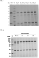
- the expression supernatant of H182-MUT4-TGF-P RII was subjected to electrophoretic analysis by reducing SDS-PAGE.
- the results showed ( FIG 1 ) there were three distinct bands in electrophoresis gel, of which the band having a molecular weight greater than 70 kDa was the protein chain with heavy chain fused of H182-MUT4-TGF- ⁇ RII, the band having a molecular weight between 20 and 30 kDa was the light chain of the anti-PD-L1 antibody H182-MUT4, while the distinct protein band between 50-70 kDa of the protein marker was presumably the heavy chain of H182-MUT4 off which TGF- ⁇ RII had shed.
- the proportion of the band of the broken protein increased with the culture time prolonging.
- the analysis results of M7824 were similar to those of H182-MUT4-TGF-P RII and had the problem of band due to breakage also.
- the bands having a molecular weight of 50-70 KDa in SDS-PAGE electrophoresis gel were recovered.
- Decolorizing buffer was added into the sample for an adequate decoloration, and reducing buffer was added to perform reducing treatment.
- tyrisin was added and enzymatic digestion was performed at 37 °C overnight. Then the enzymatic digestion products were extracted and desalted; and finally the peptide fragments were re-dissolved in 0.1% formic acid aqueous solution for subsequent mass spectrometry.
- an appropriate amount of the sample of peptide fragments was subjected to chromatographic resolution using an Easy nLC 1200 system (Thermo Scientific) at nanoliter flow rate.
- H182-MUT4-TGF- ⁇ RII by Mass Spectrometry Sequence Length Proteins Start position End position WSVLTVLHQDWLNGK 16 H182-MUT4-H-TGF- ⁇ RII 304 319 WQQGNVFSCSVMHEALHNHYTQK 23 H182-MUT4-H-TGF- ⁇ RII 419 441 NQVSLTCLVK 10 H182-MUT4-H-TGF- ⁇ RII 363 372 FNWYVDGVEVHNAK 14 H182-MUT4-H-TGF- ⁇ RII 277 290 DTLMISR 7 H182-MUT4-H-TGF- ⁇ RII 251 257 GPSVFPLAPSSK 12 H182-MUT4-H-TGF- ⁇ RII 124 135 EVQLVQSGAEVK 12 H182-MUT4-H-TGF- ⁇ RII 1 12 VTITADTSTNTAYMELSSLR 20 H
- Example 3 Design, expression and analysis of anti-human PD-L1 antibody/TGF- ⁇ RII fusion protein mutants
- glycosylation sites before and after the N-terminal enzyme cleavage sites of TGF- ⁇ RII were designed to realize the protection of the cleavage sites and prevent the occurrence of cleavage.
- the design of mutants is provided in Table 2.
- the nucleotide sequence encoding the TGF- ⁇ RII extracellular domain in the expression vector of anti-human PD-L1 antibody/TGF- ⁇ RII fusion protein H182-MUT4-TGF- ⁇ RII was subjected to site-directed mutagenesis using StarMut gene site-directed mutagenesis kit (Cat: T111-01, GenStar), to obtain expression plasmids of the mutants.
- the plasmids were then transferred into Escherichia coli cells for expansion, and plasmids of anti-human PD-L1 antibody/TGF- ⁇ RII fusion protein mutants, i.e., H182-MUT4-TGF- ⁇ RIIm1, H182-MUT4-TGF- ⁇ RIIm2, and H182-MUT4-TGF- ⁇ RIIm3, were obtained.
- H182-MUT4-TGF- ⁇ RIIm1, H182-MUT4-TGF- ⁇ RIIm2 and H182-MUT4-TGF- ⁇ RIIm3 were electrically transfected (Nucleofector IIb, Lonza) into CHO-K1 cells in suspension culture, and cells stably expressing PD-L1 antibody/TGF- ⁇ RII fusion protein mutants were obtained through MSX pressure screening.
- H182-MUT4-TGF- ⁇ RII and its mutants H182-MUT4-TGF- ⁇ RIIm1, H182-MUT4-TGF- ⁇ RIIm2 and H182-MUT4-TGF- ⁇ RIIm3 were cultured in fed-batch culture and observed for cell density and activity every day. The supernatants were collected by high-speed centrifugation of all the cell culture broths when the cell activity was less than 20%. Parts of the supernatants were purified using a Protein A affinity chromatography column and the fusion protein H182-MUT4-TGF- ⁇ RII and its mutants were obtained. Table 2.
- H182-MUT4-TGF- ⁇ RII Samples of the proteins H182-MUT4-TGF- ⁇ RII and its mutants H182-MUT4-TGF- ⁇ RIIm1, H182-MUT4-TGF- ⁇ RIIm2 and H182-MUT4-TGF- ⁇ RIIm3 were subjected to SEC-HPLC analysis.
- Table 3 Table 3.
- Example 4 Stability detection of anti-human PD-L1 antibody/TGF- ⁇ RII fusion protein mutant
- Samples of the bifunctional protein H182-MUT4-TGF- ⁇ RIIm2 were placed at 40 °C for three weeks, and one tube of the samples was taken and detected for stability each week.
- samples of the protein were repeatedly freezed and thawed at -20 °C for 3 times, and one of the samples was detected for freeze-thaw stability each time.
- the samples treated and a control sample always placed at 4 °C were detected for purity by SEC-HPLC, to verify their stability. The results are shown in FIG 5 and FIG 6 . As shown, the purity of the samples placed at the high temperature and subjected to repeatedly freezing-thawing was basically consistent with that of the control sample, indicating the product was stable and no breakage occurred.
- Example 5 Affinity detection of the anti-human PD-L1 antibody/TGF- ⁇ RII fusion proteins
- Antibody affinity was detected by an assay including capturing antibody Fc fragment with anti-human IgG Fc capture (AHC) biosensors on Octet QKe system instrument from Fortebio.
- AHC anti-human IgG Fc capture
- each of the bifunctional proteins H182-MUT4-TGF- ⁇ RII, H182-MUT4-TGF- ⁇ RIIm1, H182-MUT4-TGF- ⁇ RIIm2, and H182-MUT4-TGF- ⁇ RIIm3, as well as H182-MUT4 and TGF- ⁇ RII-hFc (Cat: CC10, Novoprotein Scientific Inc.) was diluted to 5 ⁇ g/ml in PBS, and was allowed to flow through the surface of an AHC biosensor (Cat.: 18-0015, PALL) for 120 s.
- Recombinant human PD-L1-his protein and recombinant human TGF- ⁇ 1 protein were used as the mobile phase in a concentration of 60 nM.
- the binding time was 300 s and the dissociation time was 300 s.
- data from which the response values of blank control had been deducted were fitted to a 1:1 Langmuir binding model using software, and then kinetic parameters for antigen-antibody binding were calculated. The results are provided in Table 4.
- Example 6 Design and expression of anti-human PD-L1 nanobody/TGF- ⁇ RII fusion protein mutant
- nucleotide sequence (SEQ ID NO. 19) encoding the humanized anti-human PD-L1 nanobody hzF2 was C-terminally linked to the nucleotide sequence (SEQ ID NO. 13) encoding TGF- ⁇ RIIm2 via the nucleotide sequence encoding a linker peptide (SEQ ID NO. 2), to obtain a sequence (SEQ ID NO. 23) encoding hzF2-TGF- ⁇ RIIm2 comprising PD-L1 nanobody - TGF- ⁇ RIIm2.
- the nucleotide sequence was cloned into a stable expression vector containing a Glutamine Synthetase (GS) gene through enzyme digestion sites, to construct a eukaryotic expression vector of hzF2-TGF- ⁇ RIIm2 for stable transfection.
- the vector was transferred into Escherichia coli cells for expansion, and a large amount of eukaryotic expression plasmids of fusion protein hzF2-TGF- ⁇ RIIm2 were isolated and obtained.
- the prepared eukaryotic expression plasmids were electrically transfected (Nucleofector IIb, Lonza) into CHO-K1 cells in suspension culture, and cells stably expressing the fusion protein hzF2-TGF- ⁇ RIIm2 were obtained through MSX pressure screening. The cells were cultured for expression and purification, and the fusion protein mutant hzF2-TGF- ⁇ RIIm2 was obtained.
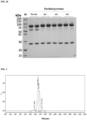
- Example 7 Purity detection of the fusion protein hzF2 TGF- ⁇ RIIm2
- Cells expressing M7824 and hzF2-TGF- ⁇ RIIm2 were cultured in fed-batch culture and observed for cell density and activity every day. Portions of cell culture broths were collected in the midterm of culture (day 10), and the remaining cell culture broths were collected in the late-term of culture when the cell activity was less than 20% (day 15). Supernatants were collected by high-speed centrifugation of the culture broths, and then purified using a Protein A affinity chromatography column. The purified expression supernatants were then subjected to protein quantification, and subpackaged for use. The expression supernatants were detected by reducing SDS-PAGE electrophoresis and SEC-HPLC.
- the SDS-PAGE results showed there were three distinct bands in electrophoresis gel of M7824, in which the band having a molecular weight greater than 70 kDa was the protein chain with fused heavy chain of M7824, the band having a molecular weight between 20 and 30 kDa was the light chain of M7824, while the distinct protein band between 50-70 kDa of the protein marker was presumably the heavy chain of M7824 off which TGF- ⁇ RII had shed. The proportion of the band of the broken protein increased with the extension of culture time. In contrast, in the electrophoresis gel of hzF2-TGF- ⁇ RIIm2, there was one distinct main band between 50 and 70 kDa, without any other obvious protein bands.
- Example 8 Affinity analysis of the fusion protein hzF2-TGF- ⁇ RIIm2
- Antibody affinity was detected by an assay including capturing antibody Fc fragment with anti-human IgG Fc capture (AHC) biosensors on Octet QKe system instrument from Fortebio.
- AHC anti-human IgG Fc capture
- each of the proteins was diluted to 4 ⁇ g/ml in PBS, and was allowed to flow through the surface of an AHC biosensor (Cat.: 18-0015, PALL) for 120 s.
- Recombinant human PD-L1-his protein accesion No.: NP_054862.1, 19 aa-238 aa
- human TGF- ⁇ 1 Cat: CA59, Novoprotein Scientific Inc.
- the binding time was 300 s and the dissociation time was 300 s.
- data from which the response values of blank control had been deducted were fitted to a 1:1 Langmuir binding model using software, and then kinetic constants for antigen-antibody binding were calculated.
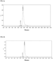
- the response curves of hzF2-TGF- ⁇ RIIm2 and the control protein M7824 to the recombinant human PD-L1 protein are shown in FIG 10 and FIG 11 , and the response curves of them to the recombinant human TGF- ⁇ 1 protein are shown in FIG 12 and FIG 13 .
- the curves were fitted and affinities were calculated.
- hzF2-TGF- ⁇ RIIm2 exhibited an affinity to PD-L1 with a KD of 1.58E-09 M and an affinity to TGF- ⁇ 1 with a KD of 2.46E-09 M; and M7824 exhibited an affinity to PD-L1 with a KD of 3.21E-09 M and an affinity to TGF- ⁇ 1 with a KD of 2.81E-09 M.
- the kinetic parameters in detailed are provided in Table 6 below.
- the results showed that hzF2-TGF- ⁇ RIIm2 had high affinity to both human PD-L1 and TGF- ⁇ 1. Table 6.
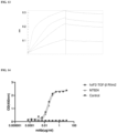
- Example 9 Binding activity of the fusion protein hzF2-TGF- ⁇ RIIm2 detected by ELISA
- Plates were coated with recombinant human PD-L1-his protein (Accession No.: NP_054861.2, 19 aa-238 aa), human TGF- ⁇ 1 (Cat: CA59, Novoprotein Scientific Inc.), human TGF- ⁇ 2 (Cat: CJ79, Novoprotein Scientific Inc.), and human TGF- ⁇ 3 (Cat: CJ44, Novoprotein Scientific Inc.) respectively overnight at 4 °C, each protein having a concentration of 1 ⁇ g/ml. Afterwards, the plates were blocked with 5% BSA in a constant temperature incubator at 37 °C for 60 min.
- human TGF- ⁇ 1 Cat: CA59, Novoprotein Scientific Inc.
- human TGF- ⁇ 2 Cat: CJ79, Novoprotein Scientific Inc.
- human TGF- ⁇ 3 Cat: CJ44, Novoprotein Scientific Inc.
- HzF2-TGF- ⁇ RIIm2 and the control protein M7824 as well as an isotype control NC-hIgG1 were added into the plates which were then incubated in a constant temperature incubator at 37 °C for 60 min, then the plates were washed 4 times with PBST.
- 1:5000 dilution of HRP-anti-human Fc (Cat.: 109-035-098, Jackson ImmunoResearch) was added into the plates for incubation for 45 min, and then the plates were washed with PBST 4 times.
- TMB substrate Cat: ME142, GalaxyBio, Beijing
- 2 M HCl was added to stop reaction; and absorbances of the plates at 450 nm were read and recorded.
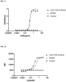
- hzF2-TGF- ⁇ RIIm2 had binding activities to human PD-L1, TGF- ⁇ 1, TGF- ⁇ 2 and TGF- ⁇ 3, which were comparable to those of M7824: the half-maximal effective concentration (EC50) values were 0.261 nM, 0.394 nM, 18.045 nM and 1.121 nM respectively, while M7824 had half-maximal effective concentration (EC50) values of 0.209 nM, 0.472 nM, 18.172 nM and 0.981 nM respectively for human PD-L1, TGF- ⁇ 1, TGF- ⁇ 2 and TGF- ⁇ 3 ( FIG 14 to FIG 17 ).
- EC50 half-maximal effective concentration
- Example 10 Binding activity of the fusion protein hzF2-TGF- ⁇ RIIm2 detected by FACS
- Human breast cancer cells MDA-MB-231 naturally expressing human PD-L1 were added into 96-well plates at 2 ⁇ 10 4 cells/well, and then were blocked with added 5% BSA at room temperature for 30 min. Afterwards, hzF2-TGF- ⁇ RIIm2 and the control protein M7824 as well as an isotype control NC-hIgG1 (dilutions of 12 concentrations obtained through 3-fold serially diluting from an initial concentration of 10 nM), were added into the plates which were then incubated on ice for 1 h.
- the cells were washed with ice-cold PBS (containing 0.05% Tween) 2 times, and then 1:200 dilution of Goat Anti human IgG Fc-FITC (Cat: F9512, Sigma) was added into the plates which were then incubated on ice for 45 min. Then the cells were washed with ice-cold PBS (containing 0.05% Tween) 2 times and then resuspended in 200 ⁇ L PBS.
- Mean Fluorescence Intensity (MFI) values were measured by a flow cytometer.
- Example 11 Blocking activity of the fusion protein hzF2-TGF- ⁇ RIIm2 detected by ELISA
- Plates were coated with recombinant human PD-1-hFc protein (Accession No.: NP_005009.2, 21 aa-167 aa) overnight at 4 °C, the protein having a concentration of 1 ⁇ g/ml. Afterwards, the plates were blocked with 5% BSA in a constant temperature incubator at 37 °C for 60 min.
- hzF2-TGF- ⁇ RIIm2 and the control protein M7824 as well as an isotype control NC-hIgG1 were added into the plates, followed by the addition of 50 ⁇ L of PD-L1-mFc (Accession No.: NP_054862.1, 19 aa-238 aa) at a concentration of 1 ⁇ g/ml.
- the plates were then incubated in a constant temperature incubator at 37 °C for 60 min, and then were washed 4 times with PBST.
- hzF2-TGF- ⁇ RIIm2 could effectively block the binding of the recombinant human PD-L1 to its receptor PD-1.
- the competitive inhibition effects of hzF2m9-TGF- ⁇ RIIm2 and M7824 on the binding of human PD-L1 to its receptor PD-1 were detected by ELISA, and the half-maximal inhibitory concentration (IC50) values of them were 7.533 nM and 6.935 nM, respectively ( FIG 19 ).
- Example 12 Observation of cytological activity of the fusion protein hzF2-TGF- ⁇ RIIm2 to block the binding of PD-L1 to its receptor PD1
- CHO cells recombinantly expressing human PD-L1 and anti-CD3-ScFv (CHO-PD-L1-CD3L, JIANGSU T-MAB BIOPHARMA CO., LTD.) were inoculated into 96-well plates (Cat: 3917, Corning) at 5000 cells/well and incubated overnight in a cell incubator, and then supernants were discarded.
- HzF2-TGF- ⁇ RIIm2 and the control protein M7824 (dilutions of 8 concentrations obtained through 2.5-fold serially diluting from an initial concentration of 5 ⁇ g/ml) (25 ⁇ L/well) and 50 ⁇ L of a suspension (1 ⁇ 10 6 cells/mL) of Jurkat cells recombinantly expressing human PD-1 and luciferase (Jurkat-PD1-NFAT, JIANGSU T-MAB BIOPHARMA CO., LTD.) were added into the plates which in turned were incubated in a cell incubator for 6 h.
- Bio-Turbo firefly luciferase substrate (Cat: RA-GL03, RHINOZYME BIOTECHNOLOGY) was added to the plates at 125 ⁇ L/well, and the plates were placed in a microplate constant temperature oscillator and incubated at 800 rpm for 5 min in dark.
- a multifunctional microplate reader was set to operate under the Luminescence mode, with the Interpretation being 500 (a default value of the instrument). RLU values were read, and the detection results are shown in FIG 20 .
- hzF2-TGF- ⁇ RIIm2 and the control protein M7824 showed blocking effects on the PD-L1/PD1 pathway with EC50 values of 10.122 nM and 8.537 nM, respectively.
- Table 7 Table 7.
- Example 13 Evaluation of antitumor efficacy of the fusion protein hzF2-TGF- ⁇ RIIm2 in a model of PBMC immune reconstitution mice subcutaneously transplantated with human pharyngeal squamous cell carcinoma Fadu cells
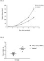
- Human pharyngeal squamous carcinoma (FaDu) cells were inoculated subcutaneously in the right flank of 5-6 week old male NCG mice at a concentration of 5 ⁇ 10 6 cells/0.1 mL, and human PBMCs were inoculated into the mice at 2 ⁇ 10 6 cells/mouse.
- mice with tumor volume meeting the requirement were randomly grouped into 2 groups, 6 mice each group, and then were administered with hzF2-TGF- ⁇ RIIm2 and an isotype control hIgG1 respectively.
- the administration schedule is provided in Table 8. The results are shown in FIG 21 and FIG 22 .
- HzF2-TGF- ⁇ RIIm2 significantly inhibited tumor growth and had a definite antitumor efficacy, achieving a tumor growth inhibition (TGI; Tumor weight) rate of 53%.
- Table 8 Administration schedule for the subcutaneous transplantation model in PBMC immune reconstitution mice Group Animals Treatment Dosage (mg/kg) Administration route Administration period 1 6 hzF2-TGF- ⁇ RIIm2 8 i.p. BIW ⁇ 5 2 6 hIgG1 10 i.p.
- BIW ⁇ 5 Note: the administration volume was 10 ⁇ l/g body weight, and the administration dosage can be adjusted when the body weight is reduced by 15-20%; i.p.: intraperitoneal injection; BIW ⁇ 5: twice per week for 5 times in total.
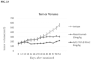
- Example 14 Evaluation of antitumor activity of the fusion protein hzF2-TGF- ⁇ RIIm2 in a tumor model of human CD34+ cord blood stem cell humanized mice subcutaneously inoculated with HCC827 cells
- Human non-small cell lung cancer HCC827 cell line was recovered and cultured, and cultured and passaged in RMPI 1640 medium (with addition of inactivated 10% FBS) in a 37 °C, 5% CO 2 incubator.
- the tumor cells in logarithmic growth phase were used for in vivo tumor inoculation.
- 20 qualified female human CD34+ umbilical cord blood stem cell humanized mice, after 1 week of adaptive feeding, were inoculated with the human non-small cell lung cancer HCC827 cells and were observed for tumor volume and body weight after the inoculation of tumor cells. Mice with tumor volume between 120-200 mm 3 were selected and randomly grouped into 3 groups according to their tumor volume and body weight, 5 mice each group.
Landscapes
- Health & Medical Sciences (AREA)
- Chemical & Material Sciences (AREA)
- Life Sciences & Earth Sciences (AREA)
- Organic Chemistry (AREA)
- Immunology (AREA)
- General Health & Medical Sciences (AREA)
- Medicinal Chemistry (AREA)
- Genetics & Genomics (AREA)
- Molecular Biology (AREA)
- Biochemistry (AREA)
- Biophysics (AREA)
- General Chemical & Material Sciences (AREA)
- Chemical Kinetics & Catalysis (AREA)
- Veterinary Medicine (AREA)
- Pharmacology & Pharmacy (AREA)
- Nuclear Medicine, Radiotherapy & Molecular Imaging (AREA)
- Public Health (AREA)
- Animal Behavior & Ethology (AREA)
- Proteomics, Peptides & Aminoacids (AREA)
- Zoology (AREA)
- Engineering & Computer Science (AREA)
- Bioinformatics & Cheminformatics (AREA)
- Gastroenterology & Hepatology (AREA)
- Toxicology (AREA)
- Cell Biology (AREA)
- Biotechnology (AREA)
- General Engineering & Computer Science (AREA)
- Biomedical Technology (AREA)
- Wood Science & Technology (AREA)
- Oncology (AREA)
- Communicable Diseases (AREA)
- Microbiology (AREA)
- Physics & Mathematics (AREA)
- Plant Pathology (AREA)
- Peptides Or Proteins (AREA)
- Medicinal Preparation (AREA)
- Medicines That Contain Protein Lipid Enzymes And Other Medicines (AREA)
- Medicines Containing Antibodies Or Antigens For Use As Internal Diagnostic Agents (AREA)
- Micro-Organisms Or Cultivation Processes Thereof (AREA)
Applications Claiming Priority (2)
| Application Number | Priority Date | Filing Date | Title |
|---|---|---|---|
| CN202010721371 | 2020-07-24 | ||
| PCT/CN2021/108065 WO2022017487A1 (zh) | 2020-07-24 | 2021-07-23 | TGF-β RII突变体及其融合蛋白 |
Publications (2)
| Publication Number | Publication Date |
|---|---|
| EP4194002A1 true EP4194002A1 (de) | 2023-06-14 |
| EP4194002A4 EP4194002A4 (de) | 2024-08-14 |
Family
ID=79586333
Family Applications (1)
| Application Number | Title | Priority Date | Filing Date |
|---|---|---|---|
| EP21846357.8A Pending EP4194002A4 (de) | 2020-07-24 | 2021-07-23 | Tgf-beta rii-mutante und fusionsprotein davon |
Country Status (5)
| Country | Link |
|---|---|
| US (1) | US20230365653A1 (de) |
| EP (1) | EP4194002A4 (de) |
| JP (1) | JP7766672B2 (de) |
| CN (1) | CN113968903A (de) |
| WO (1) | WO2022017487A1 (de) |
Families Citing this family (6)
| Publication number | Priority date | Publication date | Assignee | Title |
|---|---|---|---|---|
| CN113527488B (zh) * | 2020-04-22 | 2026-04-17 | 迈威(上海)生物科技股份有限公司 | 一种靶向人程序性死亡配体1(pd-l1)的单可变域抗体及其衍生物 |
| EP4578877A1 (de) * | 2022-08-03 | 2025-07-02 | Nanjing Leads Biolabs Co., Ltd. | Antikörperfusionsprotein zum targeting von fap und tgf-beta und verwendung davon |
| JP2024096518A (ja) * | 2023-01-03 | 2024-07-16 | エフビーデー バイオロジクス リミテッド | 操作されたtgfbriiバリアント及びその使用方法 |
| EP4676977A1 (de) * | 2023-03-09 | 2026-01-14 | Telix Pharmaceuticals (Innovations) Pty Ltd | Multifunktionelle antikörper |
| EP4490201A4 (de) | 2023-05-31 | 2026-03-11 | Fbd Biologics Ltd | Gegen cd47/pd-l1 gerichteter proteinkomplex und verfahren zur verwendung davon |
| WO2025004683A1 (ja) * | 2023-06-30 | 2025-01-02 | 国立大学法人大阪大学 | がん検査方法及びがん治療剤 |
Family Cites Families (16)
| Publication number | Priority date | Publication date | Assignee | Title |
|---|---|---|---|---|
| AU671606B2 (en) | 1991-10-31 | 1996-09-05 | Whitehead Institute For Biomedical Research | TGF-beta type receptor cDNAs and uses therefor |
| DE69332026T2 (de) | 1992-10-29 | 2002-10-31 | Celtrix Pharmaceuticals, Inc. | Typ ii tgf-beta-bindendes rezeptorfragment als therapeutisches mittel |
| NZ500284A (en) * | 1997-04-18 | 2001-09-28 | Biogen Inc | Type II TGF-beta receptor fusion proteins |
| CA2593648A1 (en) | 2005-01-10 | 2006-07-13 | Research Development Foundation | Targeted chimeric molecules for cancer therapy |
| WO2009152610A1 (en) | 2008-06-20 | 2009-12-23 | The Royal Institution For The Advancement Of Learning/Mcgill University | Interleukin-2/soluble tgf-beta type ii receptor b conjugates and methods and uses thereof |
| BR112016014952A2 (pt) | 2014-02-10 | 2017-09-19 | Merck Patent Gmbh | Inibição direcionada de tgfbeta |
| CN106397592A (zh) * | 2015-07-31 | 2017-02-15 | 苏州康宁杰瑞生物科技有限公司 | 针对程序性死亡配体(pd-l1)的单域抗体及其衍生蛋白 |
| AU2017310027A1 (en) * | 2016-08-12 | 2019-01-31 | Merck Patent Gmbh | Combination therapy for cancer |
| MY202480A (en) * | 2017-05-12 | 2024-04-30 | Shanghai hengrui pharmaceutical co ltd | Fusion protein containing tgf- receptor and medicinal uses thereof |
| WO2019222252A1 (en) * | 2018-05-15 | 2019-11-21 | Merck Patent Gmbh | Dosing regimens for targeted tgf-b inhibition for use in treating cancer in treatment naive subjects |
| AU2019299318A1 (en) * | 2018-07-02 | 2021-01-21 | Merck Patent Gmbh | Combination therapy with targeted TGF-B inhibition for treatment of advanced non-small cell lung cancer |
| WO2020041607A1 (en) * | 2018-08-22 | 2020-02-27 | Merck Patent Gmbh | Treatment of triple negative breast cancer with targeted tgf-b inhibition |
| US12559539B2 (en) * | 2018-11-09 | 2026-02-24 | Jiangsu Hengrui Medicine Co., Ltd. | TGF-beta receptor fusion protein pharmaceutical composition and use thereof |
| CN114340735B (zh) * | 2019-06-28 | 2024-11-12 | 璟尚生物制药公司 | 突变的TGFβ1-RII胞外域和免疫球蛋白支架组成的抗肿瘤拮抗剂 |
| CN111978412B (zh) * | 2020-08-13 | 2021-04-30 | 南京凯地生物科技有限公司 | 武装靶向TGF-β的特异性嵌合抗原受体细胞及其制备方法和应用 |
| EP4165086A4 (de) * | 2020-09-16 | 2024-07-31 | Suzhou Neologics Bioscience Co., Ltd. | Pd-l1-antikörper, fusionsproteine und verwendungen davon |
-
2021
- 2021-07-23 CN CN202110835893.1A patent/CN113968903A/zh active Pending
- 2021-07-23 US US18/006,571 patent/US20230365653A1/en active Pending
- 2021-07-23 EP EP21846357.8A patent/EP4194002A4/de active Pending
- 2021-07-23 WO PCT/CN2021/108065 patent/WO2022017487A1/zh not_active Ceased
- 2021-07-23 JP JP2023504523A patent/JP7766672B2/ja active Active
Also Published As
| Publication number | Publication date |
|---|---|
| EP4194002A4 (de) | 2024-08-14 |
| JP2023534738A (ja) | 2023-08-10 |
| JP7766672B2 (ja) | 2025-11-10 |
| CN113968903A (zh) | 2022-01-25 |
| US20230365653A1 (en) | 2023-11-16 |
| WO2022017487A1 (zh) | 2022-01-27 |
Similar Documents
| Publication | Publication Date | Title |
|---|---|---|
| EP4194002A1 (de) | Tgf-? rii-mutante und fusionsprotein davon | |
| US11739148B2 (en) | Human CD3 binding antibody | |
| TWI851585B (zh) | 抗steap1抗原結合蛋白 | |
| US20180305464A1 (en) | Pdl-1 antibody, pharmaceutical composition thereof, and uses thereof | |
| US20250340631A1 (en) | Cldn18.2-targeting antibody, bispecific antibody and use thereof | |
| EP4155318A1 (de) | Bispezifischer antikörper und verwendung davon | |
| US20230357398A1 (en) | Novel human antibodies binding to human cd3 epsilon | |
| JP7822576B2 (ja) | 抗cldn4-抗cd137二重特異性抗体 | |
| CN111819200A (zh) | 抗c-met抗体 | |
| US20250188166A1 (en) | Il-18 polypeptides fused to immune cell antigen specific binding polypeptides and uses thereof | |
| CN111818972A (zh) | 去免疫的抗erbb3抗体 | |
| CN113214402A (zh) | 分离的抗原结合蛋白及其应用 | |
| KR20230030626A (ko) | 루이스 y에 대한 인간화된 항체 | |
| CN121646609A (zh) | Tnf超家族成员免疫细胞因子及其用途 | |
| CN119421903A (zh) | 抗tigit抗体与il2的融合蛋白或其变体及其应用 | |
| EP4613774A1 (de) | Gegen menschliches ror1 gerichteter einzeldomänenantikörper | |
| WO2024054418A1 (en) | Sequence optimization of a pd1 blocking antibody | |
| NZ739165B2 (en) | Human cd3 binding antibody |
Legal Events
| Date | Code | Title | Description |
|---|---|---|---|
| STAA | Information on the status of an ep patent application or granted ep patent |
Free format text: STATUS: THE INTERNATIONAL PUBLICATION HAS BEEN MADE |
|
| PUAI | Public reference made under article 153(3) epc to a published international application that has entered the european phase |
Free format text: ORIGINAL CODE: 0009012 |
|
| STAA | Information on the status of an ep patent application or granted ep patent |
Free format text: STATUS: REQUEST FOR EXAMINATION WAS MADE |
|
| 17P | Request for examination filed |
Effective date: 20230224 |
|
| AK | Designated contracting states |
Kind code of ref document: A1 Designated state(s): AL AT BE BG CH CY CZ DE DK EE ES FI FR GB GR HR HU IE IS IT LI LT LU LV MC MK MT NL NO PL PT RO RS SE SI SK SM TR |
|
| DAV | Request for validation of the european patent (deleted) | ||
| DAX | Request for extension of the european patent (deleted) | ||
| A4 | Supplementary search report drawn up and despatched |
Effective date: 20240716 |
|
| RIC1 | Information provided on ipc code assigned before grant |
Ipc: A61P 35/00 20060101ALI20240710BHEP Ipc: C07K 19/00 20060101ALI20240710BHEP Ipc: A61K 38/17 20060101ALI20240710BHEP Ipc: A61K 38/16 20060101AFI20240710BHEP |
|
| STAA | Information on the status of an ep patent application or granted ep patent |
Free format text: STATUS: EXAMINATION IS IN PROGRESS |
|
| 17Q | First examination report despatched |
Effective date: 20250407 |