EP4194875A1 - Tomographe à résonance magnétique à suppression active des parasites et procédé d'élimination des parasites dans un tomographe à résonance magnétique - Google Patents
Tomographe à résonance magnétique à suppression active des parasites et procédé d'élimination des parasites dans un tomographe à résonance magnétique Download PDFInfo
- Publication number
- EP4194875A1 EP4194875A1 EP23155132.6A EP23155132A EP4194875A1 EP 4194875 A1 EP4194875 A1 EP 4194875A1 EP 23155132 A EP23155132 A EP 23155132A EP 4194875 A1 EP4194875 A1 EP 4194875A1
- Authority
- EP
- European Patent Office
- Prior art keywords
- magnetic resonance
- signal
- interference
- interference suppression
- resonance tomograph
- Prior art date
- Legal status (The legal status is an assumption and is not a legal conclusion. Google has not performed a legal analysis and makes no representation as to the accuracy of the status listed.)
- Granted
Links
Images



Classifications
-
- G—PHYSICS
- G01—MEASURING; TESTING
- G01R—MEASURING ELECTRIC VARIABLES; MEASURING MAGNETIC VARIABLES
- G01R33/00—Arrangements or instruments for measuring magnetic variables
- G01R33/20—Arrangements or instruments for measuring magnetic variables involving magnetic resonance
- G01R33/28—Details of apparatus provided for in groups G01R33/44 - G01R33/64
- G01R33/32—Excitation or detection systems, e.g. using radio frequency signals
- G01R33/36—Electrical details, e.g. matching or coupling of the coil to the receiver
- G01R33/3692—Electrical details, e.g. matching or coupling of the coil to the receiver involving signal transmission without using electrically conductive connections, e.g. wireless communication or optical communication of the MR signal or an auxiliary signal other than the MR signal
-
- G—PHYSICS
- G01—MEASURING; TESTING
- G01R—MEASURING ELECTRIC VARIABLES; MEASURING MAGNETIC VARIABLES
- G01R33/00—Arrangements or instruments for measuring magnetic variables
- G01R33/20—Arrangements or instruments for measuring magnetic variables involving magnetic resonance
- G01R33/44—Arrangements or instruments for measuring magnetic variables involving magnetic resonance using nuclear magnetic resonance [NMR]
- G01R33/48—NMR imaging systems
-
- A—HUMAN NECESSITIES
- A61—MEDICAL OR VETERINARY SCIENCE; HYGIENE
- A61B—DIAGNOSIS; SURGERY; IDENTIFICATION
- A61B5/00—Measuring for diagnostic purposes; Identification of persons
- A61B5/05—Detecting, measuring or recording for diagnosis by means of electric currents or magnetic fields; Measuring using microwaves or radio waves
- A61B5/055—Detecting, measuring or recording for diagnosis by means of electric currents or magnetic fields; Measuring using microwaves or radio waves involving electronic [EMR] or nuclear [NMR] magnetic resonance, e.g. magnetic resonance imaging
-
- A—HUMAN NECESSITIES
- A61—MEDICAL OR VETERINARY SCIENCE; HYGIENE
- A61B—DIAGNOSIS; SURGERY; IDENTIFICATION
- A61B5/00—Measuring for diagnostic purposes; Identification of persons
- A61B5/70—Means for positioning the patient in relation to the detecting, measuring or recording means
- A61B5/704—Tables
-
- G—PHYSICS
- G01—MEASURING; TESTING
- G01R—MEASURING ELECTRIC VARIABLES; MEASURING MAGNETIC VARIABLES
- G01R33/00—Arrangements or instruments for measuring magnetic variables
- G01R33/20—Arrangements or instruments for measuring magnetic variables involving magnetic resonance
- G01R33/28—Details of apparatus provided for in groups G01R33/44 - G01R33/64
- G01R33/32—Excitation or detection systems, e.g. using radio frequency signals
- G01R33/36—Electrical details, e.g. matching or coupling of the coil to the receiver
-
- G—PHYSICS
- G01—MEASURING; TESTING
- G01R—MEASURING ELECTRIC VARIABLES; MEASURING MAGNETIC VARIABLES
- G01R33/00—Arrangements or instruments for measuring magnetic variables
- G01R33/20—Arrangements or instruments for measuring magnetic variables involving magnetic resonance
- G01R33/28—Details of apparatus provided for in groups G01R33/44 - G01R33/64
- G01R33/32—Excitation or detection systems, e.g. using radio frequency signals
- G01R33/36—Electrical details, e.g. matching or coupling of the coil to the receiver
- G01R33/3614—RF power amplifiers
-
- G—PHYSICS
- G01—MEASURING; TESTING
- G01R—MEASURING ELECTRIC VARIABLES; MEASURING MAGNETIC VARIABLES
- G01R33/00—Arrangements or instruments for measuring magnetic variables
- G01R33/20—Arrangements or instruments for measuring magnetic variables involving magnetic resonance
- G01R33/28—Details of apparatus provided for in groups G01R33/44 - G01R33/64
- G01R33/32—Excitation or detection systems, e.g. using radio frequency signals
- G01R33/36—Electrical details, e.g. matching or coupling of the coil to the receiver
- G01R33/3628—Tuning/matching of the transmit/receive coil
- G01R33/3635—Multi-frequency operation
-
- G—PHYSICS
- G01—MEASURING; TESTING
- G01R—MEASURING ELECTRIC VARIABLES; MEASURING MAGNETIC VARIABLES
- G01R33/00—Arrangements or instruments for measuring magnetic variables
- G01R33/20—Arrangements or instruments for measuring magnetic variables involving magnetic resonance
- G01R33/28—Details of apparatus provided for in groups G01R33/44 - G01R33/64
- G01R33/38—Systems for generation, homogenisation or stabilisation of the main or gradient magnetic field
- G01R33/385—Systems for generation, homogenisation or stabilisation of the main or gradient magnetic field using gradient magnetic field coils
- G01R33/3854—Systems for generation, homogenisation or stabilisation of the main or gradient magnetic field using gradient magnetic field coils means for active and/or passive vibration damping or acoustical noise suppression in gradient magnet coil systems
-
- G—PHYSICS
- G01—MEASURING; TESTING
- G01R—MEASURING ELECTRIC VARIABLES; MEASURING MAGNETIC VARIABLES
- G01R33/00—Arrangements or instruments for measuring magnetic variables
- G01R33/20—Arrangements or instruments for measuring magnetic variables involving magnetic resonance
- G01R33/28—Details of apparatus provided for in groups G01R33/44 - G01R33/64
- G01R33/42—Screening
- G01R33/422—Screening of the radio frequency field
-
- G—PHYSICS
- G01—MEASURING; TESTING
- G01R—MEASURING ELECTRIC VARIABLES; MEASURING MAGNETIC VARIABLES
- G01R33/00—Arrangements or instruments for measuring magnetic variables
- G01R33/20—Arrangements or instruments for measuring magnetic variables involving magnetic resonance
- G01R33/44—Arrangements or instruments for measuring magnetic variables involving magnetic resonance using nuclear magnetic resonance [NMR]
- G01R33/48—NMR imaging systems
- G01R33/4818—MR characterised by data acquisition along a specific k-space trajectory or by the temporal order of k-space coverage, e.g. centric or segmented coverage of k-space
-
- G—PHYSICS
- G01—MEASURING; TESTING
- G01R—MEASURING ELECTRIC VARIABLES; MEASURING MAGNETIC VARIABLES
- G01R33/00—Arrangements or instruments for measuring magnetic variables
- G01R33/20—Arrangements or instruments for measuring magnetic variables involving magnetic resonance
- G01R33/44—Arrangements or instruments for measuring magnetic variables involving magnetic resonance using nuclear magnetic resonance [NMR]
- G01R33/48—NMR imaging systems
- G01R33/483—NMR imaging systems with selection of signals or spectra from particular regions of the volume, e.g. in vivo spectroscopy
- G01R33/4833—NMR imaging systems with selection of signals or spectra from particular regions of the volume, e.g. in vivo spectroscopy using spatially selective excitation of the volume of interest, e.g. selecting non-orthogonal or inclined slices
-
- G—PHYSICS
- G01—MEASURING; TESTING
- G01R—MEASURING ELECTRIC VARIABLES; MEASURING MAGNETIC VARIABLES
- G01R33/00—Arrangements or instruments for measuring magnetic variables
- G01R33/20—Arrangements or instruments for measuring magnetic variables involving magnetic resonance
- G01R33/44—Arrangements or instruments for measuring magnetic variables involving magnetic resonance using nuclear magnetic resonance [NMR]
- G01R33/48—NMR imaging systems
- G01R33/54—Signal processing systems, e.g. using pulse sequences ; Generation or control of pulse sequences; Operator console
- G01R33/543—Control of the operation of the MR system, e.g. setting of acquisition parameters prior to or during MR data acquisition, dynamic shimming, use of one or more scout images for scan plane prescription
-
- G—PHYSICS
- G01—MEASURING; TESTING
- G01R—MEASURING ELECTRIC VARIABLES; MEASURING MAGNETIC VARIABLES
- G01R33/00—Arrangements or instruments for measuring magnetic variables
- G01R33/20—Arrangements or instruments for measuring magnetic variables involving magnetic resonance
- G01R33/44—Arrangements or instruments for measuring magnetic variables involving magnetic resonance using nuclear magnetic resonance [NMR]
- G01R33/48—NMR imaging systems
- G01R33/54—Signal processing systems, e.g. using pulse sequences ; Generation or control of pulse sequences; Operator console
- G01R33/56—Image enhancement or correction, e.g. subtraction or averaging techniques, e.g. improvement of signal-to-noise ratio and resolution
- G01R33/5608—Data processing and visualization specially adapted for MR, e.g. for feature analysis and pattern recognition on the basis of measured MR data, segmentation of measured MR data, edge contour detection on the basis of measured MR data, for enhancing measured MR data in terms of signal-to-noise ratio by means of noise filtering or apodization, for enhancing measured MR data in terms of resolution by means for deblurring, windowing, zero filling, or generation of gray-scaled images, colour-coded images or images displaying vectors instead of pixels
-
- G—PHYSICS
- G01—MEASURING; TESTING
- G01R—MEASURING ELECTRIC VARIABLES; MEASURING MAGNETIC VARIABLES
- G01R33/00—Arrangements or instruments for measuring magnetic variables
- G01R33/20—Arrangements or instruments for measuring magnetic variables involving magnetic resonance
- G01R33/44—Arrangements or instruments for measuring magnetic variables involving magnetic resonance using nuclear magnetic resonance [NMR]
- G01R33/48—NMR imaging systems
- G01R33/54—Signal processing systems, e.g. using pulse sequences ; Generation or control of pulse sequences; Operator console
- G01R33/56—Image enhancement or correction, e.g. subtraction or averaging techniques, e.g. improvement of signal-to-noise ratio and resolution
- G01R33/565—Correction of image distortions, e.g. due to magnetic field inhomogeneities
- G01R33/5659—Correction of image distortions, e.g. due to magnetic field inhomogeneities caused by a distortion of the RF magnetic field, e.g. spatial inhomogeneities of the RF magnetic field
-
- G—PHYSICS
- G01—MEASURING; TESTING
- G01R—MEASURING ELECTRIC VARIABLES; MEASURING MAGNETIC VARIABLES
- G01R33/00—Arrangements or instruments for measuring magnetic variables
- G01R33/20—Arrangements or instruments for measuring magnetic variables involving magnetic resonance
- G01R33/44—Arrangements or instruments for measuring magnetic variables involving magnetic resonance using nuclear magnetic resonance [NMR]
- G01R33/48—NMR imaging systems
- G01R33/58—Calibration of imaging systems, e.g. using test probes, Phantoms; Calibration objects or fiducial markers such as active or passive RF coils surrounding an MR active material
- G01R33/583—Calibration of signal excitation or detection systems, e.g. for optimal RF excitation power or frequency
Definitions
- the invention relates to a method for active interference suppression in a magnetic resonance tomograph and a magnetic resonance tomograph with a receiver.
- Magnetic resonance tomographs are imaging devices which, in order to image an examination object, align nuclear spins of the examination object with a strong external magnetic field and excite them to precess around this alignment by means of an alternating magnetic field. The precession or return of the spins from this excited state to a lower-energy state in turn generates an alternating magnetic field in response, which is received by antennas.
- a spatial coding is impressed on the signals, which subsequently enables the received signal to be assigned to a volume element.
- the signal received is then evaluated and a three-dimensional imaging representation of the examination object is provided.
- local receiving antennas so-called local coils, are preferably used, which are arranged directly on the examination object to achieve a better signal-to-noise ratio.
- the receiving antennas can also be installed in a patient couch.
- Magnetic resonance tomographs require radiofrequency shielding in two ways.
- high-frequency pulses with outputs in the kilowatt range are generated, which are only partially absorbed in the patient. Radio waves leaving the patient passageway are radiated into the room and must therefore be shielded to meet emission limits.
- the magnetic resonance signals to be received for imaging are extremely weak.
- SNR signal-to-noise ratio
- the object is achieved by a magnetic resonance tomograph according to claim 1 and claim 2 according to the invention and by a method according to the invention for operating the magnetic resonance tomograph according to claim 6 and claim 7 .
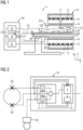
- the inventive magnetic resonance tomograph has a patient tunnel, a first receiving antenna for receiving a magnetic resonance signal from a patient, the patient being in the patient tunnel, a second receiving antenna for receiving a signal with the Larmor frequency of the magnetic resonance signal, and a receiver.
- the receiver is in signal communication with the first receiving antenna and the second receiving antenna and is preferably designed to process the magnetic resonance signal for imaging.
- the second receiving antenna is arranged outside or near an opening of the patient tunnel.
- the openings through which the patient couch with the patient is moved into the patient tunnel and, depending on the area to be examined, also exits the patient tunnel again at the opposite end are regarded as the opening of the patient tunnel.
- a distance of less than 0.1 m, 0.2 m, 0.5 m, 1 m or 2 m from the opening is considered.
- Proximity can also be considered as a distance from the aperture less than a quarter wavelength or a half wavelength of a radio wave in air at a Larmor frequency of magnetic resonance imaging.
- the first reception antenna preferably receives the magnetic resonance signal, which, however, always also has a small proportion of the interference signal.
- the second receiving antenna not only receives the interference signal, but also a minimal or negligible portion of the magnetic resonance signal.
- the signal received by the first receiving antenna is referred to below as a magnetic resonance signal (albeit with a portion of the interfering signal to be eliminated) and the signal received by the second receiving antenna or antennas is referred to as an interfering signal.
- the receiver is designed to suppress an interference signal received with the second receiving antenna in a magnetic resonance signal received by the first receiving antenna. Exemplary embodiments are described in more detail in the dependent claims.
- the interference signal has a broader bandwidth than the magnetic resonance signal.
- the components outside the frequency range of the magnetic resonance signal correlate with the components within the frequency range of the magnetic resonance signal.
- the second receiving antenna receives the broadband interference signal and the receiver is designed to evaluate this signal only partially, for example in a frequency range that is unequal to or outside the frequency range of the magnetic resonance signal, and then, depending on this partial signal, To suppress interference signal in a received from the first receiving antenna magnetic resonance signal.
- the second receiving antenna it would also be conceivable as an alternative or in addition for the second receiving antenna to only transmit frequencies outside the frequency range of the magnetic resonance signal and forwards it to the receiver.
- the amplitude of the frequency components of the interference signal in the frequency range of the magnetic resonance signal can be linked to an amplitude outside.
- the second receiving antenna in connection with the receiver only monitors frequency components outside the frequency range of the magnetic resonance signal for interference signals during a magnetic resonance measurement and suppresses the interference signal in the signals of the first receiving antenna depending on this.
- the receiver determines a relationship between the interference signal from the first receiving antenna and the interference signal component from the second receiving antenna, also referred to as a transfer function, for an interference signal in the frequency range of the magnetic resonance signals between acquisitions of magnetic resonance signals and, for example, the amplitude during the acquisition of magnetic resonance signals or scaling based on the components of the interference signal received by the second receiving antenna outside the frequency range of the magnetic resonance signals.
- a transfer function for an interference signal in the frequency range of the magnetic resonance signals between acquisitions of magnetic resonance signals and, for example, the amplitude during the acquisition of magnetic resonance signals or scaling based on the components of the interference signal received by the second receiving antenna outside the frequency range of the magnetic resonance signals.
- a filter to be provided between the second receiving antenna and the receiver, which filter preferably lets the interference signal through and suppresses magnetic resonance signals.
- a filter that suppresses the magnetic resonance signals could be provided for an interferer with components outside the frequency band of the magnetic resonance signal.
- the filter could also be adaptive or controllable. So it would be conceivable that the interference suppression control adapts the filter to the Larmor frequency of a layer that is currently being recorded
- the receiver can have a summation device that generates a linear combination of the magnetic resonance signal that is dependent on one or more parameters and generates an interference signal.
- the receiver can have an interference suppression controller that is designed to vary the parameter or parameters in such a way that the energy of the interference signal in the linear combination is minimal.
- One or more parameters can be complex in order to model a phase shift or a phase shift can be specified by a separate parameter. In particular, several parameters allow the effective suppression of different sources of interference.
- the interference suppression control could also weight interference signals with a particularly large amplitude compared to weaker interference signals in the parameters in the interference suppression, because these strong interference signals can be suppressed particularly well by a particularly large difference between the signal level and statistical background noise.
- interference signals are considered to be different interference signals that can be separated by means of a plurality of second receiving antennas due to their different origins, occupy different frequency ranges or differ in terms of a different temporal behavior.
- the receiver can be designed to perform this inventive processing of the received magnetic resonance signal and the interference signal to suppress the interference signal in real time, for example by means of a programmable logic (FPGA) or a signal processor (DSP).
- FPGA programmable logic
- DSP signal processor
- the receiver it is also possible for the receiver to have a memory and initially store the received interference signal and the received magnetic resonance signal, with the interference signal only being suppressed at a later point in time with a delay, for example of the duration of an echo sequence, an excitation sequence or an entire image acquisition a single slice or the entire image acquisition sequence.
- a delay for example of the duration of an echo sequence, an excitation sequence or an entire image acquisition a single slice or the entire image acquisition sequence.
- the delay may be greater than 50ms, 100ms, 0.5s, 1s, 10s 1 min or more.
- the hardware for analog and/or digital high-frequency processing such as amplifiers, filters and mixers in real time, but also an image evaluation unit for later generation of an image from the received magnetic resonance signals can be regarded as a receiver within the meaning of the invention.
- the method according to the invention for operating the magnetic resonance tomograph according to the invention has the step of receiving the interference signal via the second reception antenna by means of the receiver. Due to the arrangement of the second antenna in the vicinity of the opening, the interference signal received by the second antenna has hardly any or no components of the magnetic resonance signal.
- the receiver receives a magnetic resonance signal via the first receiving antenna.
- the first receiving antenna can be, for example, a body coil or a local coil of the magnetic resonance tomograph.
- the receiver processes the magnetic resonance signal as a function of the interference signal by the receiver to form a received signal, with the dependence depending on a parameter.
- a parameter For example, it is conceivable that the receiver forms a linear combination of the interference signal and the magnetic resonance signal with the parameter as a factor, with the parameter being able to be complex in order to map a phase shift.
- the receiver forms a linear combination of the interference signal and the magnetic resonance signal with the parameter as a factor, with the parameter being able to be complex in order to map a phase shift.
- the receiver uses the second receiving antenna to transmit components of a broadband interference signal outside the frequency range of the magnetic resonance signal.
- components of the interference signal outside the frequency range of the magnetic resonance signal correlate with components within the frequency range.
- the amplitudes of the interference signal inside and outside the frequency range of the magnetic resonance signal are proportional to one another if the source is the same.
- scaling with the amplitude of the signal from the second receiving antenna can then be provided, for example, as a dependency.
- the separate frequency ranges can advantageously prevent the suppression of the interference signal from being influenced by the magnetic resonance signals during image acquisition.
- the evaluation of the signal received by the second receiving antenna by the receiver in frequency ranges unequal to the frequencies of the magnetic resonance signals can prevent stray magnetic resonance signals from being evaluated as an interference signal and being suppressed by the interference suppression control.
- the interference suppression control preferably also takes into account different properties of signals in the frequency band of the magnetic resonance signals and outside. This can be, for example, different attenuation or signal delays, which can be taken into account by other amplification factors and phase shifts.
- the different properties can be determined, for example, by calibration measurements to determine transfer functions, as explained below with regard to the dependent claims.
- interference signals are detected by the receiver between and/or also during MR signal detections. Permanent detection advantageously makes it possible to better detect the time profile of an interference signal and thus also easier to estimate for the future, which allows more precise suppression.
- an interim acquisition i.e. between the acquisition of MR signals, makes it possible to avoid the assessment of MR signals as interference signals. It is also conceivable that not only the second or the second receiving antennas detect the interference signals, but also the first receiving antenna or first receiving antennas at the same time, so that it is known exactly how an interference signal received by the second receiving antennas is detected by the first receiving antennas becomes. This relationship, also referred to as a transfer function, then allows a more precise determination of parameters for interference suppression and thus better suppression of the interference by the receiver.
- the different methods can also be combined with one another and/or used alternately in order to achieve improved interference suppression overall.
- results can be averaged over longer or more recording periods and/or also between the different methods.
- the receiver sets the parameter, for example by means of an interference suppression controller, in such a way that a proportion of the interference signal in the received signal is reduced. It is conceivable, for example, for the parameter to be set by the interference suppression control in an optimization method in such a way that the energy of the interference signal in the received signal is minimized.
- the receiver can also store the magnetic resonance signal and/or the interference signal, so that the steps of processing and setting the parameter can also be performed at a time interval from receiving the signals can take place through the first receiving antenna and the second receiving antenna.
- the magnetic resonance tomograph according to the invention and the method for operation by the second receiving antenna and the receiver according to the invention advantageously make it possible to reduce the proportion of external interference signals in the magnetic resonance signal and therefore make do with simpler and more cost-effective shielding measures.
- the magnetic resonance tomograph is designed to receive magnetic resonance signals with a Larmor frequency in an industrial band.
- a resonant frequency of the nuclear spins used for imaging in the magnetic resonance tomograph in a static magnetic field B0 of a field magnet of the magnetic resonance tomograph is referred to as the Larmor frequency of the magnetic resonance tomograph.
- the industry band refers to frequency bands released for use by medical or technical devices, for which lighter regulations are available for emission and approval. These are also known as ISM bands (Industrial, Scientific, Medical Band).
- ISM bands Industrial, Scientific, Medical Band
- An example of a frequency band in which high powers can be emitted is between 26.9 and 27.3 MHz.
- Other such frequency bands are between 6.7MHz and 6.8MHz, 13.5MHz and 13.6MHz, 40.6MHz and 40.7MHz, and 433.0MHz and 434.8MHz.
- a magnetic resonance tomograph is not only dependent on receiving as little interference as possible, but the strong excitation pulses must not interfere with other devices either.
- the legal tolerance limits are significantly higher, so that a legally compliant limitation by shielding the emitted excitation pulses at this frequency easier to do or not necessary.
- a magnetic resonance tomograph can be implemented in an advantageous manner without a shielded cabin.
- the magnetic resonance tomograph has a transmission path for transmitting an excitation pulse with a filter.
- the filter is designed to suppress signals outside the ISM band.
- the filter may be a bandpass filter for the ISM band used, which rejects frequencies outside the ISM band by more than 12dB, 24dB, 40dB or 60dB relative to a signal with minimal attenuation within the ISM band. band dampens.
- the filter can be arranged, for example, between the output stage and a hybrid coupler, between the hybrid coupler and the transmit/receive splitter, or between the transmit/receive splitter and the transmit antenna.
- the filter advantageously makes it possible to restrict transmitted high-frequency power essentially to the ISM band and thus to comply with the stricter limits outside the ISM band. This means that the magnetic resonance tomograph can also be operated in the ISM band without an RF cabin.
- the magnetic resonance tomograph has a transmission antenna for emitting an excitation pulse
- the Magnetic resonance imaging has non-linear components for detuning the transmitting antenna.
- These can be PIN diodes, for example, but also other diodes or active components such as transistors or FETs.
- the nonlinear components are arranged in an area of the magnetic resonance tomograph that is shielded from the patient tunnel for radio frequency and the filter for the ISM band or ISM filter is arranged in the signal connection between the nonlinear component and antenna, preferably in the shielded area.
- Shielding the nonlinear components from the patient tunnel and the environment advantageously avoids emissions of frequency components which are generated by the nonlinearity during the excitation pulse of the components used for detuning and are no longer in the ISM band as harmonics.
- the filter prevents harmonics from being transmitted via the signal connection between the non-linear component and the transmitting antenna.
- the arrangement of the non-linear components thus contributes to compliance with emission limit values and enables or simplifies the elimination of shielding of the entire magnetic resonance tomograph with a high-frequency cabin.
- the magnetic resonance tomograph has a high-frequency unit with a predistortion device.
- the predistorter is designed to predistort an excitation pulse to excite the nuclear spins in such a way that signal components of the excitation pulse are reduced outside the ISM band compared to an excitation pulse without predistortion when it is emitted, ie in particular after amplification by a high-frequency power amplifier.
- the predistorter generates and mixes in signal components which, after amplification by the radio-frequency unit, correspond to the harmonics generated from the excitation pulse by the non-linearity of the radio-frequency unit, but have the opposite sign and thus reduce the harmonics or wipe out.
- the predistortion device can be implemented, for example, in a digital signal generator, or analog components can be used to generate corresponding signals from an input signal for a power amplifier.
- the predistorter can also be adaptive, for example its characteristics can be controlled as a function of the loading of the patient tunnel. Partial regulation by rapid feedback from a sensor in the transmission path, such as a directional coupler, is also conceivable.
- the predistorter reduces harmonics outside the ISM band in an advantageous manner and thus facilitates or enables compliance with the emission limit values even without a shielding booth.
- a limit frequency for a propagation of a radio wave in the patient tunnel is greater than a Larmor frequency of the magnetic resonance tomograph.
- the frequency at which a radio wave propagating in the longitudinal direction (z-direction) through the patient tunnel can still form in the patient tunnel as a waveguide is regarded as the limit frequency.
- the limit frequency is also referred to as the cut-off frequency for a waveguide, here for the patient tunnel as a waveguide.
- the field strength of an interference signal coming from outside advantageously drops exponentially with the distance from the opening inside the patient tunnel, so that the interference signal is significantly reduced in the examination area (Field of View, FoV).
- the second receiving antenna is arranged at an opening in the patient tunnel or the patient couch.
- the second receiving antenna directly placed at an edge of an opening or under the patient support surface.
- the patient and the patient tunnel form a coaxial conductor for the interfering signal.
- a second receiving antenna in the vicinity of the patient and the opening can advantageously detect the interference signal coupled into the patient tunnel by the patient and thus enable particularly effective suppression by the receiver.
- the magnetic resonance tomograph has a waveguide that surrounds the magnetic resonance tomograph, the waveguide having a limit frequency or cut-off frequency that is greater than the Larmor frequency of the magnetic resonance tomograph.
- a waveguide is any electrically conductive structure that surrounds the magnetic resonance tomograph on the outside in at least four spatial directions, for example in the form of a tube or a prism, and its conductivity at the Larmor frequency of the magnetic resonance tomograph allows radio waves or electric fields to propagate through the conductive structure essentially suppressed throughout.
- a signal with Larmor frequency is attenuated by more than 30 dB, 40 dB, 60 dB or more compared to a signal on the side facing the magnetic resonance tomograph.
- the magnetic resonance tomograph according to the invention is surrounded by a waveguide according to the invention or also by a classic shielded cabin.
- a high-frequency-tight door to the shielded cabin or the waveguide is replaced by an electrically conductive tunnel, which in turn represents a waveguide with a cut-off frequency greater than the Larmor frequency.
- the damping of the alternating fields in the tunnel reduces radiation into the environment, so that an expensive and cumbersome and error-prone HF-tight door can be dispensed with, especially with the higher limit values for frequencies in ISM bands.
- the waveguide has an electrically conductive connection to the patient tunnel. It is conceivable that the waveguide forms a continuous or continuous waveguide together with the patient tunnel. It is also possible for the waveguide to be electrically connected to the patient tunnel at both ends of the patient tunnel, or for two waveguides to be electrically conductively connected to the patient tunnel at opposite ends thereof.
- the waveguide can also have a limit frequency or cut-off frequency that is different from the limit frequency of the patient tunnel, but also above the Larmor frequency of the magnetic resonance tomograph.
- the second reception antenna has an essentially all-round reception characteristic.
- a sensitivity distribution of the receiving antenna in all spatial directions in which the difference between a maximum sensitivity and a minimum sensitivity depending on the different directions is less than 6 dB, 12 dB, 18 dB or 24 dB, is regarded as an essentially all-round reception characteristic. This preferably also applies to different polarizations of the interference signal
- the interference signal can come from different directions and with different polarizations, so an antenna with directional characteristics or preferential polarization cannot detect all interference signals.
- An antenna with an all-round reception characteristic can advantageously detect all interference signals.
- the magnetic resonance tomograph has a plurality of second antennas and the receiver is designed to suppress the interference signal in the magnetic resonance signal as a function of received signals from the plurality of second receiving antennas.
- the plurality of second receiving antennas are arranged at a distance from one another, for example at a distance greater than a quarter of a wavelength or a half wavelength of a radio wave having the Larmor frequency.
- a plurality of spatially distributed antennas is also advantageously suitable for better detecting one or more interference sources and thus improving the interference suppression.
- the plurality of receiving antennas are arranged in a symmetrical arrangement with respect to the patient tunnel.
- a symmetrical arrangement with respect to the patient tunnel.
- an arrangement at the edge of the opening of the patient's tunnel at two opposite points, at the corner points of a regular polygon or polyhedron, is conceivable.
- a symmetrical relationship of the parameters can also be produced in an advantageous manner by means of the symmetry of the antennas, and the optimization method for reducing the interference signal can thus be simplified and/or accelerated.
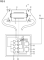
- the magnetic resonance tomograph has an interference suppression transmitter and an interference suppression antenna.
- the interference suppression antenna is at a distance to placed in the patient tunnel.
- the interference suppression transmitter is designed to generate a signal in a frequency range of an excitation pulse of the magnetic resonance tomograph and to output it via the interference suppression antenna such that a field strength of the excitation pulse is reduced by destructive interference in an area surrounding the magnetic resonance tomograph.
- a reduction in the field strength or attenuation of the signal of the excitation pulse in the range by more than 6 dB, 12 dB, 24 dB, 40 dB or 60 dB is considered reduced.
- the field strength of the electromagnetic waves emitted by the excitation pulse in the vicinity of the magnetic resonance tomograph can advantageously be reduced by the interference suppression transmitter and the interference suppression antenna in such a way, in particular in synergy with the other proposed measures, that the legal limit values can be complied with even without an RF cabin .
- the magnetic resonance tomograph has a large number of interference suppression antennas, the interference suppression antennas being arranged at a distance from the patient tunnel and relative to one another.
- the distances are preferably smaller than a wavelength of a free radio wave with Larmor frequency and/or larger than one tenth of the wavelength.
- the interference suppression antennas can be arranged in a plane around the opening of the patient tunnel.
- the interference suppression transmitter is designed to emit signals in a frequency range of an excitation pulse of the magnetic resonance tomograph via the interference suppression antennas, so that a field strength of the excitation pulse is reduced by destructive interference in several areas surrounding the magnetic resonance tomograph.
- the electromagnetic fields in several areas or to reduce the radiation in several directions or, ideally, to bring it to zero.
- the number of directions without radiation corresponds to the zeros of the radiation diagram of a multipole.
- a resulting electromagnetic wave from the signal of the excitation pulse from the local transmission antenna and the interference suppression antenna is thus a multipole field (e.g. quadrupole field), which advantageously decreases significantly faster than a dipole field, for example, with increasing distance from the source and thus makes it possible to set limit values for radiation as well without complying with RF cabin.
- the interference suppression transmitter is designed to generate the signals for the interference suppression antenna or antennas by phase shifting and/or amplitude adjustment as a function of one or more transmission interference suppression parameters.
- the signal or signals for the interference suppression antenna or antennas are preferably generated from the excitation pulse by adjustable amplifiers or attenuators and phase shifting elements in the interference suppression transmitter.
- the amplitude ratios and phase shifts can represent the transmission interference suppression parameters or can be derived from the transmission interference suppression parameter or parameters, for example using analytical functions, tables or iteration processes.
- the excitation pulse can be detected, for example, via a sensor such as a directional coupler in the line between the high-frequency power amplifier and the transmitting antenna or a sensor antenna in the patient tunnel. It is also conceivable to generate the signals for the interference suppression antennas directly from the digital data of the excitation pulse by scaling and phase shifting with an AD converter and amplifier.
- Transmission interference suppression parameters advantageously simplify a following by reducing the number of variables specified determination of the setting of the interference suppression transmitter and allow faster adaptation to changing conditions such as other excitation pulses or environment by patient or operator.
- the transmission interference suppression parameters are set during manufacture or installation.
- the magnetic resonance tomograph has a calibration element in the surroundings of the magnetic resonance tomograph and an interference suppression control.
- the calibration element is preferably an antenna or sensor, with which an electric and/or magnetic field strength of an electric and/or magnetic alternating field with a frequency of the excitation pulse can be detected and forwarded to the interference suppression control.
- the interference suppression control is designed to use the calibration element to detect a field strength in a frequency range of an excitation pulse at the location of the calibration element.
- the interference suppression control can have a calibration receiver.
- the interference suppression control is also designed to set the transmission interference suppression parameter as a function of the detected field strength in such a way that a field strength of the excitation pulse is reduced in a predetermined area surrounding the calibration element.
- the interference suppression control can vary or optimize an amplitude and/or phase of the interference suppression antenna(s) in such a way that a reduction and/or a local minimum of the field strength at the location of the calibration element is achieved through destructive interference.
- the transmission can be interrupted and/or a new determination of the parameter can be initiated.
- the interference suppression antenna has a high-frequency power amplifier.
- the high-frequency power amplifier is preferably designed to generate an electromagnetic alternating field sufficient to suppress stray fields of the excitation pulse via the interference suppression antenna from a drive signal with low high-frequency power, for example less than 10 mW, 50 mW or 100 mW.
- the high-frequency power amplifier makes it possible to connect it to the interference suppression transmitter using a thin, flexible high-frequency cable, thus simplifying installation.
- the patient tunnel as a waveguide and the plurality of interference suppression antennas only a power in the range of a few watts is required, so that the locally arranged high-frequency power amplifiers can also be small and light, which simplifies installation.
- the transmission interference suppression according to the invention by means of destructive interference can also be used independently of the other features of the magnetic resonance tomograph according to the invention.
- the use of an active interference suppression transmitter is also conceivable without active interference suppression at the receiving end, in particular if the permissible electromagnetic radiation is the limiting factor.
- the step of setting the parameter includes the step of averaging over time with the formation of a mean over time as a function of the interference signal.
- the amplitude and/or phase of the interference signal can be detected and averaged using a low-pass filter or by averaging over a window, so that the parameter follows only slow changes.
- the averaging over time advantageously means that the interference suppression is not falsified by short-term, possibly sporadic influences and no higher-frequency noise components are artificially generated by the interference suppression.
- the adjustment step has the following sub-steps:
- the receiver measures a first transfer function between a first receiving antenna and the calibration element.
- the measurement can be carried out, for example, by the receiver or the interference suppression controller instructing the interference suppression transmitter via a signal connection to emit a signal with a predetermined amplitude and/or predetermined phase in a frequency range of the magnetic resonance signal and/or in an adjacent frequency range via the calibration antenna.
- a signal connection is required between the interference suppression transmitter and the calibration element.
- the receiver can then receive the signal via the first receiving antenna and thus determine a first transfer function. This can also happen simultaneously for several first receiving antennas. In principle, however, it would also be conceivable to record the transfer function by transmitting via the first receiving antenna and receiving via the calibration element.
- the receiver measures a second Transfer function between the second receiving antenna and the calibration element. This can be done in the same way as previously described. If the signal is sent out via the calibration element, both transfer functions can advantageously be detected simultaneously.
- the signals sent out to record the first and second transfer functions are encoded in such a way that the receiver can easily determine the amplitude and phase relationship.
- a pseudo-random code would be conceivable, for example, which would allow the signals to be autocorrelated quickly and reliably.
- the signal can be modulated in terms of amplitude, frequency and/or phase.
- Spread spectrum modulations are also conceivable, in which the signal for determining the transfer function can also remain below the noise limit of the MR signals and simultaneous transmission while an MR signal is being acquired is possible. In this way, changes in the environment can be permanently addressed.
- the permanent emission of the signal can also be achieved by using a frequency range that is adjacent to the MR signals. Then, however, the different propagation conditions due to the different frequencies must be taken into account when determining the transfer functions.
- the interference suppression parameter or, in the case of a plurality of first and second receiving antennas, the interference suppression parameters are set as a function of the measured first transfer function or transfer functions and second transfer function or transfer functions, so that a proportion of an interference signal received by the second receiving antenna or antennas is reduced in a signal received by the receiver via the first receiving antenna.
- the interference signal received by the second receiving antenna or antennas is preferably also taken into account.
- the transfer functions are used to determine the form in which the signal from an individual receiving antenna is taken into account. This can be achieved, for example, by Interference signal separated via an autocorrelation in the received MR signal by a variation method or linear optimization method by the interference suppression control so that the interference signal component is minimized.
- the transfer functions are included as predetermined attenuations and phase shifts between receiving antennas and receiver. Strictly speaking, the transfer function for a calibration element is only valid for a source of interference at a specific location or direction. However, when using a sufficiently large number of suitably positioned calibration elements to determine the transfer functions, transfer functions can also be determined that are independent of the respective location of the calibration element.
- Determining the transfer functions using the calibration element advantageously enables the reception interference suppression to react to different conditions in the environment, such as the position of people or equipment and the propagation conditions that have changed as a result, and to adapt the interference suppression.
- the step of setting takes place in a time period of a sequence in which no magnetic resonance signal is received, in particular no signal for exciting the spins is sent either.
- time periods in which no excitation pulses are emitted and no acquisition of a magnetic resonance signal for imaging takes place are also periods of time in the sequence in which the examination subject or the patient does not emit any significant magnetic resonance signal, i.e. the level of the signal is at least 12 dB, 24 dB, 36 dB, 48 dB or 60 dB below a maximum magnetic resonance signal.
- the setting step takes place in such a period of time.
- the receiver's anti-interference control is designed accordingly, the step of adjusting in such a period of time to execute. For example, you can receive a trigger signal from the control of the magnetic resonance tomograph.
- the setting of the parameters can advantageously be simplified without a magnetic resonance signal, for example by minimizing an energy of the signal received by the first receiving antenna as a function of the parameters. Even if the interference signal changes over time, for example in terms of amplitude, the set parameters are still valid and effective given the same spatial arrangement.
- the parameter is set permanently, i.e. at short intervals of 1 ms, 10 ms or 100 ms, or in particular in real time with a delay of less than 10, 100 or 500 microseconds occurs.
- the permanent setting of the parameter in real time or almost real time advantageously allows a quick reaction to newly occurring interference signals and to minimize their negative effect on the imaging as far as possible.
- the receiver has a memory and stores the received magnetic resonance signal and the received interference signal.
- the receiver can also include an image evaluation computer, for example.
- the receiver in the narrower sense ie the devices used for processing the received high-frequency magnetic resonance signals, to have the memory.
- the setting of the parameter and the processing of the magnetic resonance signal with the interference signal as a function of the parameter for reducing the interference signal by the receiver into a received signal can then take place with a delay in relation to the reception.
- the delay can be, for example, the duration of an echo sequence, an excitation sequence or an entire image acquisition sequence, for example more than 10 ms, 100 ms, 0.5 s, 10 s or also several minutes, hours or in principle also days.
- the storage makes it possible to also use existing resources of the magnetic resonance tomograph or to provide the interference suppression more cost-effectively with less computing power because real-time processing is not required.
- Subsequent interference suppression also makes it possible to compare the results of different parameter settings and suppression processes and thus optimize interference suppression.
- the receiver or the interference suppression control has an autocorrelation device.
- the interference suppression controller can use the autocorrelation device to determine a component of the interference signal in the magnetic resonance signal, for example an amplitude and a phase shift.
- the interference suppression control has an estimation device that determines the component of the interference signal, for example using an optimization method in which the interference component in the magnetic resonance signal is minimized by varying the parameter or parameters, such as Least Mean Square Root (LSR) or similar methods.
- LSR Least Mean Square Root
- the interference suppression control optimizes the majority of the parameters in such a way that the largest possible proportion of the interference signal is reduced. This can be particularly advantageous when there are several sources of interference or reflections.
- the captured image space or the associated volume is larger than the examination object. Areas are then detected in the image space that do not have a magnetic resonance signal, but only interference signals. The interference signal can be separated and determined by this segmentation.
- the interference signals separated in the image space are transformed back into a raw data space or k-space, for example again using a Fourier transformation.
- the parameters for the suppression of the interference signals are determined from the transformed interference signals in the raw data space.
- the coordinates in k-space provide information on the phase and frequency of an interfering signal, so that, taking into account the arrangement of the first receiving antenna and the second receiving antenna, as well as damping factors and phase shift of the signal paths, an attenuation and signal delay can be determined after their application to the interfering signal of the second Receiving antenna in a sum signal with the received signal of the first receiving antenna, the interference in the magnetic resonance signal is reduced.
- the interference signals can be suppressed directly in the image space, for example by hiding the corresponding image data. This can be used in particular if the image data is not in the examination area or can be replaced by already recorded, undisturbed data from this area. It is also conceivable to mark the disturbed data in the image space with a special identifier, e.g. a color or brightness value, so that image artifacts caused by disturbance cannot be mistaken for features of the examination object.
- a special identifier e.g. a color or brightness value
- Recognizing the interference signals in the image space advantageously allows a separation or segmentation of magnetic resonance data and interference. This allows the interference signals to be detected separately in a simple manner and the properties to be better identified, resulting in better and more effective suppression.
- the partial steps of transforming, separating, inverse transforming and determining the parameters are carried out on lines of data of the received magnetic resonance signals in the raw data space.
- the parameters can advantageously be changed more quickly than, for example, when capturing an entire layer, and the response to changes in the interference signals can thus be quicker. A repetition of the detection for an entire shift can thus be avoided.
- the receiver monitors the interference signal for changes in one step and, if there is a change, adapts the parameter in a further step. For example, it is due to a movement of the interference source or a reflecting object in the vicinity of the magnetic resonance tomograph possible that the field distribution of the interference source changes in phase and amplitude.
- the receiver can then detect such changes in the interference signal and adjust the parameter or parameters accordingly, so that, for example, the interference signal received by the second receiving antenna or the second receiving antennas is added to the MRT signal with an adjusted gain and/or phase shift. It is conceivable that the receiver takes threshold values into account during the monitoring and only when a threshold value is exceeded is regarded as a change.
- the receiver carries out averaging over time in order to hide short-term fluctuations.
- the averaging can relate to properties of the interference signal such as amplitude, phase, frequency and/or frequency distribution, for example, in order not to react to short-term fluctuations and to introduce as few additional noise components into the magnetic resonance signal as possible through the interference suppression.
- monitoring of the interference signal by the receiver makes it possible to react to changes in the interference signal and thus ensure effective interference suppression of a variable interference signal over a longer period of time. Thresholds and averaging limit the changes and avoid oscillations caused by instabilities or artefacts caused by excessive compensation.
- the receiver stores a first received magnetic resonance signal in a memory in a partial step.
- the receiver stores a second received magnetic resonance signal in a memory.
- the receiver compares the first received magnetic resonance signal and the second received magnetic resonance signal. If the receiver detects a deviation that can be traced back to external interference, it leads the receiver issues an interference suppression measure or signals a fault to the controller of the magnetic resonance tomograph, so that the latter triggers the interference suppression measure. Sudden amplitude differences in magnetic resonance signals from slices that are close together or measurements of the same slice, for example from a calibration measurement, can possibly be interference signals.
- Interfering signals can advantageously be detected by comparing the magnetic resonance signals of adjacent areas or of the same areas at different times. This is also conceivable in an application without a second receiving antenna for receiving the interference signal.
- the interference suppression measure is the discarding of the first and/or second received magnetic resonance signal. It is also conceivable that additionally or alternatively a detection of the first and/or second magnetic resonance signal is repeated.
- the interference suppression measure can also include adjusting the parameter.
- the proposed interference suppression measures can be used to avoid image artifacts in the magnetic resonance recordings after an interference signal has been detected.
- the magnetic resonance tomograph has a patient tunnel, a first receiving antenna for receiving a magnetic resonance signal from a patient in the patient tunnel, a second receiving antenna for receiving a signal with the Larmor frequency of the magnetic resonance signal and a receiver, the the second receiving antenna is arranged outside the patient tunnel or in the vicinity of an opening of the patient tunnel.
- the method includes the step of receiving an interfering signal by the receiver via the second receiving antenna.
- the arrangement of the second receiving antenna makes it more sensitive to signals outside the patient tunnel, which cannot be magnetic resonance signals simply because of their spatial origin.
- the signal from the second receiving antenna can also have small portions of a magnetic resonance signal, which, however, can initially be ignored in the suppression of interference due to their small portion or can be further reduced or avoided using means and methods specified in the subclaims
- the receiver receives a magnetic resonance signal via the first receiving antenna.
- the signal that is primarily used for image acquisition is referred to as the magnetic resonance signal.
- the first receiving antennas can be local coils on the patient's body.
- the magnetic resonance signal can contain components of the interference signal, which are further reduced with the device or method according to the invention, as described below
- the magnetic resonance signal received from the first receiving antenna is discarded as a function of the interference signal from received from the second antenna.
- the interference signal received by the second receiving antenna can be above a threshold level, so that interference with an image obtained from the magnetic resonance signal is to be expected despite the propagation attenuation between the location of the second receiving antenna and the location of the first receiving antenna.
- the receiver or the controller of the magnetic resonance tomograph can then reject the signal from the first receiving antenna. It is then conceivable that the magnetic resonance tomograph then repeats the detection of the rejected signal.
- an interference signal can advantageously already be recognized on the basis of the signal amplitudes.
- the method has the step of determining an excitation pulse for exciting nuclear spins in an examination subject.
- the determination takes place as a function of predetermined frequency limits of the ISM band.
- the excitation pulse is preferably determined in such a way that it has spectral components outside the ISM band only below predetermined limit values.
- the limitation to the ISM band can be achieved, for example, by the measures described in relation to the following claims.
- the magnetic resonance tomograph transmits the excitation pulse.
- the magnetic resonance tomograph receives a magnetic resonance signal and, in another step, determines an image of a distribution of nuclear spins in the examination subject. The image can then be output on a display.
- excitation pulses can become so broad-banded that they have portions outside the ISM band, even if the Larmor frequency is within the ISM band. If this is avoided by the measures explained below, additional shielding measures such as an RF cabin can be dispensed with without exceeding the limit values.
- the step of determining an excitation pulse has the sub-steps of generating an excitation pulse to excite the nuclear spins in a slice of the examination object as a function of a relative position of the slice to a magnet unit, a predetermined gradient strength, a thickness of the slice and the determine the type of measurement. This can be done, for example, by using parameterized libraries of excitation pulses.
- the magnetic resonance tomograph determines whether the excitation pulse lies within the predetermined frequency limits. This can be done, for example, by spectral analysis using FFT.
- the step of determining is repeated.
- a pulse parameter is varied which, when the excitation pulse is determined, has an effect on a spectral frequency distribution of the excitation pulse.
- the excitation pulse is within the predetermined frequency limits, it is emitted by the magnetic resonance tomograph in a further step.
- the pulse parameter affecting the determination of the excitation pulse is one of the parameters duration of the excitation pulse, thickness of the slice, relative position of the slice or strength of the gradient.
- the duration of the excitation pulse, the thickness of the slice to be excited or the position of the slice or the strength of the gradient can be changed.
- a simultaneous variation of several parameters is also conceivable.
- an excitation pulse can be determined in an advantageous manner that does not exceed the limits of the frequency band and thus enables operation within the permissible range.
- the step of emitting includes the sub-step of changing the position of the examination object relative to the magnet unit before the step of emitting the pulse.
- the magnetic resonance tomograph has a control unit for controlling the image acquisition, which in particular parameters of the image acquisition such as the time of the excitation pulse and/or the time of the acquisition of the magnetic resonance signals or also the frequency of the excitation pulse and/or the frequency range of the receiver when the magnetic resonance signal is received can affect.
- the magnetic resonance tomograph has an interface with a signal connection to the control unit.
- the interface can be an interface for data exchange with other magnetic resonance systems or also a high-frequency interface.
- the control unit is designed to synchronize an image acquisition as a function of a signal received from another magnetic resonance tomograph via the interface. Any activity that reduces mutual interference is seen as synchronizing. This can include temporal coordination, but also, for example, a change in frequencies.
- control unit can also be designed to send a signal containing information about imminent image acquisition to another magnetic resonance tomograph. Comparable to a plug-socket combination, this feature supplements the previous magnetic resonance tomograph, which is designed to receive a signal from another magnetic resonance tomograph.
- the information preferably relates to a point in time of the transmission and/or frequency of an excitation pulse.
- a magnetic resonance tomograph according to the invention is preferably designed both for transmitting and for receiving a signal.
- the magnetic resonance tomograph is also referred to below as the first magnetic resonance tomograph.
- the first magnetic resonance tomograph according to the invention can also be signal-connected to a second magnetic resonance tomograph according to the invention.
- the first magnetic resonance tomograph and the second magnetic resonance tomograph each have an interface and a control unit.
- the first magnetic resonance tomograph and the second magnetic resonance tomograph have a signal connection via the interfaces.
- the signal connection enables at least the transmission of a signal from the first magnetic resonance tomograph to the second magnetic resonance tomograph, but a bidirectional exchange of information is also conceivable.
- Point-to-point connections by electrical, optical or wireless means are conceivable.
- Networks such as LAN, WAN, and especially TCP/IP are also possible as signal connections.
- the control unit of the first magnetic resonance tomograph is designed to send information about an upcoming image acquisition process to the second magnetic resonance tomograph via the interface.
- a point in time of a planned transmission of an excitation pulse is conceivable in absolute time or relative to the signal, or also to the frequency or a frequency range of the excitation pulse.
- the control unit of the second magnetic resonance tomograph is designed to receive the information via the interface and to carry out image acquisition as a function of the information received.
- the controller of the second magnetic resonance tomograph can be designed to temporally shift a magnetic resonance signal acquisition, ie a sequence or a part thereof, so that the excitation pulse of the first magnetic resonance tomograph does not interfere.
- the second magnetic resonance tomograph is also referred to below as another magnetic resonance tomograph.
- the method according to the invention for operating a magnetic resonance tomograph can also be expanded to operate a first magnetic resonance tomograph with a control unit for controlling the image acquisition and an interface with a signal connection to the control unit.
- the method then includes the step of receiving a signal by the control unit via the interface from a second magnetic resonance tomograph.
- This can be a targeted exchange of information, in which the first magnetic resonance tomograph receives information or a message from the second magnetic resonance tomograph via a data interface, which has parameters such as the time and/or frequency of an intended image acquisition.
- the first magnetic resonance tomograph to monitor the environment, for example via a receiver for magnetic resonance signals.
- the control unit of the first magnetic resonance tomograph sets a parameter of the image acquisition as a function of the received signal. For example, it is conceivable that a sequence is shifted in time.
- the first magnetic resonance tomograph carries out an image acquisition according to the set parameters.
- the sequence can be started at a different point in time, so that the excitation pulses of both magnetic resonance tomographs occur simultaneously or the excitation pulse of the first magnetic resonance tomograph occurs at a point in time at which the second magnetic resonance tomograph does not receive any image-relevant magnetic resonance signals.
- the magnetic resonance tomograph according to the invention in combination with a second magnetic resonance tomograph and the method for operation make it possible to reduce mutual interference between two magnetic resonance tomographs during simultaneous operation.
- the interface is designed for data exchange.
- the first magnetic resonance tomograph is designed both to send data to a second magnetic resonance tomograph via the interface and to receive data from a second magnetic resonance tomograph via this interface.
- the control unit is designed to synchronize image acquisition with a second magnetic resonance tomograph by means of an exchange of information via the interface.
- the control units use messages to coordinate how the image is captured, so that mutual interference is reduced.
- the signal has information about a point in time and/or frequency of a transmission process.
- the time can be specified in absolute terms, for example, or relative to the time the message was sent.
- a center frequency and/or a bandwidth, a frequency range or else a channel specification that encodes a frequency range can be used as the frequency specification.
- the method also has the step of determining information about impending image acquisition by the second magnetic resonance tomograph by a control unit of the second magnetic resonance tomograph and, in a further step, generating a signal with the Send information to the first magnetic resonance tomograph.
- control unit can receive a message from a second magnetic resonance tomograph via the interface that this will send an excitation pulse with a center frequency equal to the Larmor frequency +100 kHz with a bandwidth of 200 kHz in 2 seconds.
- the first magnetic resonance tomograph can then, for example, interrupt and continue a sequence before its own excitation pulse after the second magnetic resonance tomograph has completed its sequence, or transmit the next excitation pulse at the same time as the excitation pulse of the second magnetic resonance tomograph.
- Mutual interference from an excitation pulse of the second magnetic resonance tomograph during a reception phase can thus be avoided in an advantageous manner.
- the signal has information about a point in time and/or frequency of a reception process.
- the information can indicate, for example, a start and a duration and frequency, such that the second magnetic resonance tomograph is received starting in one second for 2 seconds at a center frequency equal to the Larmor frequency minus 300 kHz with a bandwidth of 200 kHz.
- the first magnetic resonance tomograph can then, for example, interrupt a sequence in such a way that it does not transmit any excitation pulse in the mentioned frequency band during this time.
- transmission of the first magnetic resonance tomograph can advantageously be postponed in such a way that interference is reduced.
- control unit of the first magnetic resonance tomograph is designed to change the frequency of an image acquisition process as a function of the information received.
- an image acquisition includes different slices, these being differentiated by a gradient magnetic field in the z-axis and thus in the effective Larmor frequency of the magnetic resonance signal. Simultaneous operation with reduced interaction is possible in this way, provided that the order of the slices during image acquisition is arranged in such a way that acquisition in the same frequency range never takes place in both systems at the same time.
- the differentiation via the frequency enables a simultaneous detection of magnetic resonance signals in neighboring magnetic resonance tomographs and thus a better use of the examination time.
- the first magnetic resonance tomograph has a receiver as an interface.
- a receiver for magnetic resonance signals including an antenna such as a local coil or body coil is considered here as a receiver.
- the first magnetic resonance tomograph is designed to acquire an excitation pulse of a second magnetic resonance tomograph outside of an image acquisition and to carry out the image acquisition as a function of the acquired excitation pulse.
- the first magnetic resonance tomograph first acquires magnetic resonance signals of a slice with a different effective Larmor frequency or waits until a maximum duration for acquiring magnetic resonance signals in the second magnetic resonance tomograph emitting the excitation pulse has elapsed
- the synchronization can thus advantageously also take place without a data connection or changes in the second magnetic resonance tomograph.
- the method also has the step of determining information about impending image acquisition by the second magnetic resonance tomograph by a control unit of the second magnetic resonance tomograph and, in a further step, generating a signal with the Send information to the first magnetic resonance tomograph.
- the magnetic resonance tomograph has a patient tunnel, a first receiving antenna for receiving a magnetic resonance signal from a patient in the patient tunnel, a second receiving antenna for receiving a signal with the Larmor frequency of the magnetic resonance signal, and a receiver, the second receiving antenna being outside or in the vicinity of an opening of the patient tunnel is arranged, wherein the receiver is in signal connection with the first receiving antenna and the second receiving antenna and the receiver is designed to suppress an interference signal received with the second receiving antenna in a magnetic resonance signal received by the first receiving antenna.
- the magnetic resonance tomograph has a patient tunnel, a first receiving antenna for receiving a magnetic resonance signal from a patient in the patient tunnel, a second receiving antenna for receiving a signal close to the Larmor frequency of the magnetic resonance signal and a receiver, with the second receiving antenna being outside or in the is arranged in the vicinity of an opening of the patient tunnel, the receiver being in signal connection with the first receiving antenna and the second receiving antenna and the receiver being designed to convert a broadband interference signal received with the second receiving antenna outside a frequency range of the magnetic resonance signal into a magnetic resonance signal received by the first receiving antenna suppress.
- the magnetic resonance tomograph according to the invention is designed to receive magnetic resonance signals with a Larmor frequency in an industrial band.
- the magnetic resonance tomograph according to the invention has a transmission path for emitting an excitation pulse with an ISM filter, the ISM filter being designed to suppress signals outside the ISM band.
- the magnetic resonance tomograph according to the invention has a transmitting antenna for emitting an excitation pulse, the magnetic resonance tomograph having non-linear components for detuning the transmitting antenna, the non-linear components being arranged in an area of the magnetic resonance tomograph shielded from the patient tunnel for radio frequency, the ISM -Filter is arranged between non-linear component and antenna.
- the magnetic resonance tomograph according to the invention has a radio-frequency unit, the radio-frequency unit having a predistortion device that is designed to predistort an excitation pulse for exciting the nuclear spins in such a way that emitted signal components of the excitation pulse outside the ISM band are reduced compared to an excitation pulse without predistortion .
- a limit frequency for a propagation of a radio wave in the patient tunnel is greater than a Larmor frequency of the magnetic resonance tomograph.
- the magnetic resonance tomograph has a waveguide that surrounds the magnetic resonance tomograph, the waveguide having a limit frequency that is greater than the Larmor frequency of the magnetic resonance tomograph.
- the waveguide has an electrically conductive connection to the patient tunnel.
- the second receiving antenna is arranged at an opening in the patient tunnel or on the patient couch.
- the second reception antenna has an all-round reception characteristic.
- the magnetic resonance tomograph has a plurality of second receiving antennas and the receiver is designed to suppress the interference signal in the magnetic resonance signal as a function of received signals from the plurality of second receiving antennas.
- the plurality of second receiving antennas is arranged in a symmetrical arrangement with respect to the patient tunnel.
- the receiver has an autocorrelation device and the autocorrelation device is designed to determine a proportion of the signal received by the second receiving antenna in the magnetic resonance signal received by the first receiving antenna.
- the receiver has an estimation device and the estimation device is designed to estimate a proportion of the signal received by the second receiving antenna in the magnetic resonance signal received by the first receiving antenna.
- the magnetic resonance tomograph has an interference suppression transmitter and an interference suppression antenna, the interference suppression antenna being arranged at a distance from the patient tunnel, the interference suppression transmitter being designed to transmit a signal in a frequency range of an excitation pulse of the magnetic resonance tomograph via the interference suppression antenna depending on to output a transmission interference suppression parameter, so that a field strength of the excitation pulse is reduced by destructive interference in a predetermined area of an area surrounding the magnetic resonance tomograph.
- the magnetic resonance tomograph has a calibration element in the vicinity of the magnetic resonance tomograph and an interference suppression controller, the interference suppression controller being designed to use the calibration element to detect a field strength in a frequency range of an excitation pulse at the location of the calibration element and as a function of the detected Field strength to set the transmission interference suppression parameters such that a field strength of the excitation pulse is reduced in a predetermined environment of the calibration element.
- the interference suppression transmitter is designed to generate the signals for the interference suppression antenna or antennas by phase shifting and/or amplitude adjustment as a function of one or more transmission interference suppression parameters.
- the interference suppression antenna has a high-frequency power amplifier.
- the magnetic resonance tomograph Calibration element in an environment of the magnetic resonance tomograph, wherein the receiver is designed to measure a first transfer function between the first receiving antenna and the calibration element and a second transfer function between the second receiving antenna and the calibration element and depending on the measured first transfer function and the second transfer function or set the interference suppression parameters in such a way that an interference signal received with the second receiving antenna is reduced in a magnetic resonance signal received by the first receiving antenna.
- the step of setting the parameter includes the step of averaging over time with the formation of a mean over time as a function of the interference signal.
- the step of receiving the interference signal takes place a period of time of a sequence in which no magnetic resonance signal for imaging is received.
- the receiver has a memory and the method has a step of storing, in which the receiver stores the interference signal and the magnetic resonance signal in the memory, wherein the step of processing occurs with a delay relative to receiving the jamming signal and/or magnetic resonance signal.
- the receiver has an autocorrelation device and in the step of setting the parameter the autocorrelation device determines a component of the interference signal in the magnetic resonance signal and the parameter is set as a function of the determined component of the interference signal.
- the receiver has an estimation device and in the step of setting the parameter, the estimation device determines a proportion of the interference signal in the magnetic resonance signal and the parameter is set as a function of the determined proportion of the interference signal.
- the steps of transforming, separating, inverse transforming and determining the parameter are carried out on lines of data of the received magnetic resonance signals in the raw data space.
- the receiver monitors the interference signal for changes in one step and adapts the parameter in the event of a change in one step.
- the receiver stores a first received magnetic resonance signal in a memory in one step, in one step a second received magnetic resonance signal and in one step compares the first received magnetic resonance signal and the second received magnetic resonance signal and carries out an interference suppression measure in the event of a deviation which is attributable to external interference.
- the interference suppression measure is one of discarding the first and/or second received magnetic resonance signal, repeating a detection of the first and/or second magnetic resonance signal or setting the parameter.
- the pulse parameter has an effect on the determination of the excitation pulse one of the parameters duration of the excitation pulse, thickness of the slice, relative position of the slice or strength of the gradient.
- the step of emitting includes the sub-step of changing the position of the examination object relative to the magnet unit before the step of emitting the pulse.
- the magnetic resonance tomograph has a control unit for controlling the image acquisition and an interface with a signal connection to the control unit, the control unit being designed to synchronize an image acquisition as a function of a signal received via the interface from a second magnetic resonance tomograph.
- the magnetic resonance tomograph has a control unit for controlling the image acquisition and an interface with a signal connection to the control unit, the control unit being designed to send a signal with information about imminent image acquisition to a second magnetic resonance tomograph.
- the interface is designed for data exchange, with the control unit being designed to synchronize image acquisition with a second magnetic resonance tomograph by means of information exchange via the interface.
- the signal has information about a point in time and/or frequency of a transmission process.
- the signal has information about a point in time and/or frequency of a reception process.
- a possible embodiment of the invention has a computer program product that can be loaded directly into a processor of a programmable controller, with program code means to execute all steps of a method according to the invention for operating a magnetic resonance tomograph when the program product is executed on the controller.
- a possible embodiment of the invention relates to a computer-readable storage medium with electronically readable control information stored thereon, which is designed such that when the storage medium is used in a controller of a magnetic resonance tomograph according to the invention, the method according to the invention for operating a magnetic resonance tomograph is carried out.
- FIG. 1 shows a schematic representation of an embodiment of a magnetic resonance tomograph 1 with a local coil 50 according to the invention.
- the magnet unit 10 has a field magnet 11 that generates a static magnetic field B0 for aligning nuclear spins of samples or of the patient 100 in a recording area.
- the recording area is characterized by an extremely homogeneous static magnetic field B0, with the homogeneity particularly affecting the magnetic field strength or the amount.
- the recording area is almost spherical and is arranged in a patient tunnel 16 which extends through the magnet unit 10 in a longitudinal direction 2 .
- a patient bed 30 can be moved in the patient tunnel 16 by the traversing unit 36 .
- the field magnet 11 is usually a superconducting magnet that can provide magnetic fields with a magnetic flux density of up to 3T, and even more in the case of the latest devices. For lower field strengths, however, permanent magnets or electromagnets with normally conducting coils can also be used.
- the magnet unit 10 has gradient coils 12 which are designed to superimpose variable magnetic fields in three spatial directions on the magnetic field B0 for spatial differentiation of the recorded imaging regions in the examination volume.
- the gradient coils 12 are usually coils made from normally conducting wires, which can generate mutually orthogonal fields in the examination volume.
- the magnet unit 10 also has a body coil 14 which is designed to emit a high-frequency signal supplied via a signal line into the examination volume and to receive resonance signals emitted by the patient 100 and emit them via a signal line.
- a control unit 20 supplies the magnet unit 10 with the various signals for the gradient coils 12 and the body coil 14 and evaluates the received signals.
- control unit 20 has a gradient control 21, which is designed to supply the gradient coils 12 via supply lines with variable currents which vary over time coordinates to provide the desired gradient fields in the examination volume.
- control unit 20 has a high-frequency unit 22 which is designed to generate a high-frequency pulse with a predetermined time profile, amplitude and spectral power distribution for exciting a magnetic resonance of the nuclear spins in the patient 100 .
- Pulse powers in the kilowatt range can be achieved.
- the excitation pulses can be radiated into the patient 100 via the body coil 14 or via a local transmitting antenna.
- a controller 23 communicates with the gradient controller 21 and the radio-frequency unit 22 via a signal bus 25.
- a local coil 50 is arranged on the patient 100 as a first receiving coil and is connected to the radio-frequency unit 22 and its receiver via a connecting line 33 .
- the body coil 14 it is also conceivable for the body coil 14 to be a first receiving antenna within the meaning of the invention.
- second receiving antennas 60 Arranged at an edge of the opening of the patient tunnel 16 are four second receiving antennas 60 which are arranged at the corners of a square inscribed in the circular opening so that the corners come to rest on the edge of the opening.
- the four second receiving antennas 60 are signal-connected to the receiver 70 of the radio-frequency unit 22. Due to the plurality of second receiving antennas 60, it is conceivable that these do not all have an all-round reception characteristic, but are dipoles, for example, and, due to the different alignment, become one Complete all-round characteristics. However, it would also be conceivable, for example, to provide a crossed dipole as the only second antenna with an all-round characteristic.
- a second receiving antenna 60 may be arranged in the patient bed 30 as an alternative or in addition.
- the patient tunnel preferably has a radius R for which the following applies: R ⁇ lambda L * 1,841 / 2 * pi
- lambda L indicates the wavelength of a radio wave in air at the Larmor frequency of the magnetic resonance tomograph 1 . If the radius R is smaller than the right-hand term, the radio wave propagates in the patient tunnel 16 with exponential attenuation and the interference signal is strongly attenuated in the center in the examination area FoV. Lambda L is also referred to as the cut-off wavelength of a circular waveguide, the associated frequency as the cut-off frequency.
- the patient 100 acts as the core of a coaxial cable, whose jacket is the wall of the patient tunnel 16, and forwards an electromagnetic signal coupled in at the legs or the head end into the examination area.
- the second receiving antenna 70 or the second receiving antennas 70 in the vicinity of the opening or in the patient bed 30 advantageously pick up the interference signal forwarded by the patient 100 into the FoV and thereby make the compensation in the receiver 70 particularly effective.
- FIG. 12 shows a schematic representation of the functional units of a possible embodiment of a receiver 70.
- the summation device 71 weights the signals arriving from the first reception antenna or local coil 50 and from the second reception antenna 60 with parameters, which can also be complex, in order to indicate a phase shift. In an analog receiver 70, this can be done by an adjustable amplifier in conjunction with an adjustable phase shifter. The real part of the parameter then corresponds to the gain factor and the imaginary part to the phase shift. Subsequently, in a preferred Embodiment summed the weighted signals, but there are also other non-linear signal operations for combining the individual signals conceivable.
- An interference suppression controller 72 receives the combined signal and the individual signals from the first receiving antenna and the second receiving antennas 60. In order to determine a proportion of the interference signal in the combined signal, it is conceivable, for example, for the interference suppression controller 72 to perform an autocorrelation of the signals. However, it is also conceivable that the energy of the combined signal is determined. In a conceivable embodiment, the interference suppression controller 72 determines the proportion of the interference signal in sections of sequences of the magnetic resonance tomograph in which no magnetic resonance signal is expected for imaging, so that the combined signal only has the interference signal. This can be the case, for example, in dephased sections of an echo sequence, since the amplitudes of individual nuclear spins cancel out due to different phases and do not generate a signal overall.
- the interference suppression controller 72 then optimizes the parameters in the summation device, for example using a least square root method (LSR), so that the proportion or the energy of the interference signal in the combined signal is minimized.
- LSR least square root method
- the receiver 70 can be implemented using analog signal processing technology, so that, for example, gain control and phase shift are controlled by a parameter and then implemented in analog form, or as a digital receiver that either receives digitized signals from the first receiving antenna and/or the second receiving antenna 60 or they are already digitized at the signal input using an A/D converter.
- the receiver 70 forwards the combined signal, in which the interference signal is largely suppressed, to the controller 23 of the magnetic resonance tomograph for imaging.
- the interference signal has a broader bandwidth than the magnetic resonance signal.
- the components outside the frequency range of the magnetic resonance signal usually correlate with the components within the frequency range of the magnetic resonance signal. It can therefore also be sufficient according to the invention if the second receiving antenna 60 receives the broadband interference signal only partially, for example in a frequency range that is unequal to or outside the frequency range of the magnetic resonance signal.
- the receiver 70 can also be designed to only pick up frequencies in this frequency range from the second receiving antenna. The receiver 70 is designed to suppress the interference signal in a magnetic resonance signal received by the first receiving antenna as a function of this partial signal.
- the amplitude of the frequency components of the interference signal in the frequency range of the magnetic resonance signal can be linked to an amplitude outside of the frequency range.
- the second receiving antenna 60 in connection with the receiver 70 only monitors frequency components outside the frequency range of the magnetic resonance signal for interference signals during a magnetic resonance measurement and, depending on this, suppresses the interference signal in the signals of the first receiving antenna.
- the receiver 70 determines a relationship between the interference signal from the first receiving antenna and the interference signal component from the second receiving antenna 60, also referred to as a transfer function, for an interference signal in the frequency range of the magnetic resonance signals between acquisitions of magnetic resonance signals and during the acquisition of magnetic resonance signals, for example adjusts the amplitude or scaling based on the components of the interference signal received by the second receiving antenna outside the frequency range of the magnetic resonance signals.
- the interference signal in the received signal is not suppressed in real time, ie not immediately upon receipt of the interference signal and/or the magnetic resonance signal, but the magnetic resonance signal and the interference signal are stored in a memory by the receiver 70, which can also include parts of the controller 23 of the magnetic resonance tomograph 1 or an image evaluation.
- the receiver 70 can also include parts of the controller 23 of the magnetic resonance tomograph 1 or an image evaluation. The steps set out below for the method then no longer take place in real time or almost in real time, but can be carried out on the stored data with a delay, for example in advance of an image evaluation.
- a combination of reception with the second antenna outside the frequency band of the MR signals with real-time interference suppression or suppression at a later point in time, for example the image evaluation, is also conceivable.
- interference suppression in the receiver can also be performed with a single second antenna 60.
- the receiver 70 it is possible for the receiver 70 to have a plurality of channels or for a plurality of receivers 70 to be provided in the magnetic resonance tomograph 1 in order to suppress interference from the magnetic resonance signals of a plurality of local coils 50 . It is conceivable that the signals of the second receiving antennas 60 are used by a plurality of receivers 70 or channels of the receivers 70 for interference suppression.
- the dependency between the interference signal received by the second antenna and the interference suppression is both linear and non-linear.
- linear dependencies can be a phase shift by a specific value or a linear scaling with a value determined from the signal of the second antenna.
- the transfer function for the interference signal on the way from the first receiving antenna and/or second receiving antenna has non-linearities, for example due to mixers or non-linear amplifiers, so that the interference signal received by the second receiving antenna 60 is also affected non-linear operations for interference suppression in the receiver 70 must be applied.
- FIG. 3 shows a schematic flow chart of a method according to the invention.
- the receiver 70 receives an interference signal via the second receiving antenna 60 or via the plurality of second antennas 60.
- the interference signal is transmitted to the receiver 70 via a signal connection. It is also conceivable that the interference signal is first digitized by an A/D converter before it is transmitted to the receiver 70, which is designed as a digital receiver 70 in this case.
- the receiver 70 receives a magnetic resonance signal via the first receiving antenna, for example the local coil 50. If there are several local coils 50, a corresponding number of receivers 70 can be provided, or else a receiver 70 with a number of channels, each with a summation device 71.
- the interference suppression control 72 can each be provided separately or together, which can speed up the subsequent setting because of similar parameters for different channels.
- the receiver 70 processes the magnetic resonance signal as a function of the interference signal or the interference signals at a plurality of second receiving antennas 60 to form a received signal.
- the interference signal or signals of the second receiving antenna or second receiving antennas 60 and the magnetic resonance signal of the first receiving antenna are weighted and delayed with different parameters and then combined. For example, this can be the generation of a linear combination.
- the received signal or sum signal generated depends on one or more parameters.
- the parameter or parameters are set by the receiver 70, in particular the interference suppression controller 72, in such a way that a proportion of the interference signal in the received signal is reduced. If, for example, the interference signal received by the second receiving antenna 60 is scaled by the set parameter in such a way that it has the same amplitude as the interference signal component via the first receiving antenna and if it is provided with a phase shift of 180 degrees relative thereto, the interference signal in the generated signal is increased receive signal exactly.
- the parameter or parameters can be determined using optimization methods such as Least Square Root (LSR) or Wiener filters.
- step S40 can also include the sub-step S41 of forming a time mean value and setting the parameter for interference suppression as a function of this mean value.
- an amplitude or phase of the interference signal can be averaged in order to compensate for statistical fluctuations and introduce less noise into the magnetic resonance signal as a result of the interference suppression.
- steps S10 to S30 are each performed on received magnetic resonance signals and received interference signals in real time, in particular with analog receivers 70.
- steps S10 to S30 are each performed on stored interference signals and magnetic resonance signals , which are digitized, for example, for a single sequence or individual sections of it.
- step S40 is carried out with interference signals from a time period of a sequence in which no magnetic resonance signal for imaging is received.
- the parameters with interference signals from the second receiving antenna 60 and a signal from the first receiving antenna can be determined by the interference suppression control 72 in a period of time in which the nuclear spins are dephased and do not generate a magnetic resonance signal.
- the interference signal and the signal of the first receiving antenna are only recorded digitally and evaluated later.
- the interference suppression control 72 monitors the interference signal for changes, for example different amplitude, frequency or phase. If such a change is detected or if the change exceeds a predetermined threshold value, the interference suppression controller 72 can change steps S10 of receiving the interference signal and S20 of receiving the magnetic resonance signal and setting a parameter for interference suppression depending on the interference signal received in a step S51 in order to adapt the interference suppression to the changed interference signal.
- the received interference signal and/or magnetic resonance signal is stored by the receiver 70 in a step S25.
- the receiver 70 can also include parts of the controller 23 of the magnetic resonance tomograph 1 or an external image evaluation computer.
- steps S20 to S40 are carried out subsequently, for example at the end of an echo sequence, an excitation sequence, a signal acquisition for a slice of the examination object or after acquisition of all data.
- the decoupling of the signal detection of the interference signal and magnetic resonance signal from the interference suppression makes it possible in an advantageous manner to use more cost-effective components with less computing power or to use existing resources twice, for example from image evaluation. It is then also conceivable to compare and select different parameter settings or to subsequently optimize them.
- the application can also be limited to periods of disruption.
- FIG. 4 shows a schematic representation of a high-frequency unit of a magnetic resonance tomograph according to the invention. The illustration does not show all the details of the high-frequency unit, but only those relevant to interference suppression on the transmission path according to the invention.
- the transmission path of the high-frequency unit 22 has a pulse generator 220 , a predistorter 221 , a power amplifier 222 and an ISM filter 223 .
- the pulse generator 220 can have, for example, an oscillator, a modulator and a mixer, with which a pulse is generated in the baseband and then converted to the Larmor frequency.
- the predistorter 221 is designed to predistort an excitation pulse for exciting the nuclear spins in such a way that signal components of the excitation pulse outside the ISM band are reduced compared to an excitation pulse without predistortion. It is conceivable, for example, that the predistorter 221 generates and mixes in signal components which, after amplification by the power amplifier, correspond to the harmonics generated from the excitation pulse due to the nonlinearity of the power amplifier, but have the opposite sign and thus reduce or eliminate the harmonics. The same is conceivable for intermodulation of signal components due to the non-linearity.
- the predistorter 221 can be realized, for example, in a digital signal generator or analog components can also be used to generate corresponding signals from an input signal for a power amplifier. It is also conceivable to integrate the predistorter 221 in a digital pulse generator 220, for example.
- the output of the predistorter 221 is amplified in the power amplifier 222 .
- a power amplifier 22 with a linear characteristic is conceivable.
- the predistorter 222 uses the input signal of the Power amplifier 222 just changed so that after amplification by the power amplifier 222, a signal without unwanted harmonics is generated.
- the characteristic curve of the predistorter 221 multiplied by the characteristic curve of the power amplifier 222 ideally gives a linear characteristic curve, so that the system made up of the predistorter 221 and the power amplifier 222 amplifies the pulse of the pulse generator without undesired harmonics.
- the predistorter 221 can, in an embodiment as shown in 4 represented by the dashed line from the output of the ISM filter 223, may also be adaptive in the sense that by monitoring the output of the power amplifier 222 it adjusts the predistortion so that the overall predistorter/power amplifier system has a linear characteristic.
- the signal is preferably also filtered by an ISM filter 223.
- the filter preferably suppresses frequency components outside of the ISM band, which the magnetic resonance tomograph 1 uses for image acquisition.
- the filter may be a bandpass filter for the ISM band used, which rejects frequencies outside the ISM band by more than 12dB, 24dB, 40dB or 60dB relative to a signal with minimal attenuation within the ISM band. band dampens. A low pass is also possible.
- the filter can be arranged, for example, between the power amplifier 222 and a hybrid coupler (not shown), between the hybrid coupler and a transmit/receive switch (not shown) or between the transmit/receive switch and transmit antenna.
- the magnetic resonance tomograph also has detuning elements for the transmission antenna.
- detuning elements for the transmission antenna.
- These detuning elements are provided to detune the transmitting antenna when receiving and to avoid interactions with the receiving antennas.
- the detuning elements usually have non-linear characteristics and can therefore generate harmonics when transmitting.
- the ISM filter 223 is therefore arranged between the detuning elements and the transmission antenna.
- the non-linear components are also preferably arranged in a region of the magnetic resonance tomograph that is shielded from high-frequency by the patient tunnel.
- the arrangement of the non-linear components thus contributes to compliance with emission limit values and enables or simplifies the elimination of shielding of the entire magnetic resonance tomograph with a high-frequency cabin.
- FIG 5 1 is a schematic representation of an embodiment of a magnetic resonance tomograph 1 according to the invention, surrounded by a waveguide.
- the waveguide 260 can be provided by any electrically conductive surface that surrounds the magnetic resonance tomograph 1 on the outside at least in 4 spatial directions.
- a metallic or metalized surface or fabric is regarded as an electrically conductive surface, which dampens an electromagnetic wave with the Larmor frequency by 60 dB, 80 dB, 100 dB or more in one passage.
- the conductivity of the surface can also be anisotropic through geometric subdivision such as slits, as long as the conductivity parallel to the electric field vector of the alternating field is sufficient to achieve the damping.
- the surface preferably forms the waveguide 260 as a tunnel around the magnetic resonance tomograph in the form of, for example, a cylinder, cuboid, prism with a width that does not allow the formation of a free wave with Larmor frequency.
- a cuboid for example, this is the case if the longer dimension of the cross section is less than half a wavelength of an electromagnetic wave with the Larmor frequency.
- the cut-off frequency of the waveguide 260 is greater than the Larmor frequency.
- the electromagnetic field drops exponentially with the distance from the source, so that alternating fields leaking out of the patient tunnel 16 drop off quickly.
- the waveguide 260 is open at one or both ends, since the exponential attenuation is already strong enough due to the distance from the patient tunnel 16 to comply with the limit values permissible in the ISM band.
- the magnetic resonance tomograph 1 is surrounded by a shield that has larger dimensions than half the wavelength.
- a tunnel-shaped access opening 261 made of conductive material with a correspondingly small cross section to the magnetic resonance tomograph 1 can be provided, the dimensions of which prevent the free propagation of the wave through a cut-off frequency greater than the Larmor frequency.
- the access opening 261 is preferably electrically conductively connected to the shielding and/or the waveguide for high frequency.
- the waveguide 260 is also electrically conductively connected to the patient tunnel 16 for radio frequency.
- FIG. 6 shows a schematic representation of an embodiment of a magnetic resonance tomograph 1 according to the invention with an interference suppression transmitter 80.
- Electrical waves or alternating fields can also be suppressed by electrical fields with the same frequency and amplitude, but opposite polarity or a phase shift of 180 degrees. If the amplitude amounts or phase do not match exactly, then at least a reduction is achieved by the destructive interference.
- a magnetic resonance tomograph 1 according to the invention has interference suppression antennas 81 which are arranged around the source of the fields, here the patient tunnel 16 .
- the interference suppression antennas cover 81 covers all spatial directions around the opening and symmetry is used, such as equal distances from the opening of the patient tunnel 16 and/or distribution at equal angular distances from the opening, in order to simplify control of the individual interference suppression antennas 81 .
- any desired distribution is also conceivable due to the amplitude and phase that can be set individually for each interference suppression antenna 81 .
- the alignment of the antennas or the polarization of the generated field is preferably oriented to the field directions of the alternating fields to be suppressed.
- the signal emitted by the interference suppression antennas 81 is intended to reduce the radiation of the excitation pulse and must therefore have a predetermined amplitude and phase relationship with the excitation pulse.
- the signals are therefore preferably derived from the excitation pulse in analog form or also from the digital pulse generation. However, it is also conceivable for the signals to be provided by separate units independently of the pulse generation, as long as the necessary amplitude and phase relationship is established.
- connection line between the body coil 14 as the source of the electromagnetic waves and the interference suppression transmitter is indicated symbolically.
- a direct connection via a power divider or, for example, a directional coupler would be conceivable, and a sensor in the patient tunnel for direct detection of the electromagnetic field would also be possible.
- the reference signal for interference suppression derived from the excitation pulse is then used by adjustable phase adjusters 82 for the individual interference suppression antennas 81 delayed or shifted in phase and then amplified in amplitude by adjustable amplifier 83 before it is emitted via the interference suppression antennas 81 .
- the phase adjuster 82 and the amplifier 83 are adjusted by an interference suppression control 84 via a signal connection. It is conceivable that the interference suppression controller 84 sets predetermined phase shifts and amplitudes that are determined, for example, when the magnetic resonance tomograph 1 is installed.
- the setting can take place by means of a calibration measurement. It is conceivable that a calibration receiver 85 picks up the alternating field to be suppressed by means of one or preferably a plurality of calibration elements 86 distributed in space. At the same time, the calibration receiver 85 detects the signals supplied to the interference suppression antennas 81 and transmits the detected values to the interference suppression controller 84.
- the interference suppression setting 84 can then, for example, use a linear optimization method such as LSR to adjust the phases and amplitudes of the individual interference suppression antennas such that at the location of the calibration antennas 86 the field strength becomes zero.
- the resulting alternating field from body coil 14 and interference suppression antennas 81 can be changed to a multipole field with n zero points or radiation lobes, which decrease to a high power with distance and enable effective suppression.
- the propagation of the fields is reversible.
- the calibration element or elements 86 emit a signal and the body coil 14 and the interference suppression antennas 84 receive the signal and then the interference suppression controller 84 determine a suitable phase relationship and amplitudes.
- the calibration element 86 could also be used to transmit a reference signal for receiving interference suppression.
- the reference signal would have to be encoded or modulated in such a way that it can be distinguished from a magnetic resonance signal by the receiver 70 . This could, for example, also be achieved below the noise limit of the magnetic resonance signal with a spread spectrum modulation. Transmission in a neighboring frequency range would also be conceivable. It is necessary for the receiver 70 to be able to establish a correlation between the reference signal and the signals received via the second and first receiving antennas in order to optimize interference suppression. In this way, for example, settings for suppressing interference signals from certain directions could be determined.
- step S130 shows a schematic flowchart of a possible embodiment of a method according to the invention for operating the method according to the invention.
- the aspect of how an excitation pulse must be designed and sent out is considered in order to comply with regulatory limit values for high-frequency radiation even without a shielding cabin, especially if the Larmor frequency is in an ISM band.
- the already too 3 The steps of a method according to the invention are explained in 7 summarized under step S130 and not explained again. In principle, however, it is also possible to 7 explained measures to limit the emission of the excitation pulse even without the reception interference suppression 3 to implement.
- an excitation pulse for exciting nuclear spins in an examination subject is determined by the controller 23 .
- an excitation pulse for excitation of the nuclear spins in a slice of the examination object is first determined by the controller 23 in a 5111 . This can be done, for example, depending on the selected sequence or type of examination by selecting from a library of excitation pulses take place.
- the frequency, the duration, the power and the spectral distribution depend on several parameters.
- the center frequency results from the nuclear spins to be recorded, the strength of the homogeneous static magnetic field B0, the position of the slice in relation to the gradient field and the strength of the gradient field.
- the spectral distribution and bandwidth results in turn from the strength of the gradient field and the thickness of the layer in the direction of the field gradient.
- the amplitude in turn depends on the duration of the excitation pulse, the volume to be excited and the desired excitation strength, also known as the flip angle.
- a set of parameters that describes a possible excitation pulse for these boundary conditions is determined as a function of these boundary conditions. It is conceivable that a library or table of different sets of parameters for specific standard situations, such as image acquisition of specific organs, is specified and selected from these.
- the controller 23 checks whether the determined excitation pulse is within the predetermined frequency limits.
- the top and bottom frequencies of the excitation pulse can be calculated using the center frequency and the spectral frequency distribution. It is conceivable to also calculate the power distribution and to evaluate limit values for a frequency-dependent power.
- Sub-step S111 is repeated if the evaluation establishes that the determined excitation pulse exceeds limit values, in particular limit values for permissible radiation from high-frequency lines.
- limit values in particular limit values for permissible radiation from high-frequency lines.
- sub-step S111 When sub-step S111 is repeated, parameters that influence it are varied. a longer one Pulse, for example, can achieve the same excitation with less power. With a lower gradient, a smaller frequency bandwidth is required to excite the same slice thickness.
- step S111 If the excitation pulse determined in step S111 complies with the limit values, then it is emitted by the high-frequency unit 22 in a step S120 of the method.
- the magnetic resonance signal is received by the receiver 70 .
- a step S140 an image of a distribution of nuclear spins is determined by the controller 23 from the received magnetic resonance signal.
- the image is preferably then reproduced on a display.
- FIG. 8 shows a flow chart of a further aspect of the in 3 illustrated method according to the invention, here a possible interference suppression by previously recorded image information.
- a step S21 the receiver 70 receives a first received magnetic resonance signal and stores it in a memory. It is also conceivable that the signal recorded in step S21 originates from a calibration measurement or a pre-scan, which are also recorded with other parameters or a lower resolution.
- the receiver 70 receives a second received magnetic resonance signal and stores it.
- the second magnetic resonance signal is preferably a signal for image acquisition.
- the first received magnetic resonance signal and the second received magnetic resonance signal are compared. This can take place, for example, in the raw data or only in the image space, for example after a Fourier transform. In a preferred embodiment, the comparison is done on a row basis in k-space. If the first magnetic resonance signal and the second magnetic resonance signal differ significantly, in particular if a possibly different recording situation is already taken into account in the comparison, then an interference suppression measure is carried out in the event of a deviation that can be attributed to external interference. Interfering signals can be distinguished, for example, by the frequency, amplitude or a characteristic course or duration.
- An interference suppression measure can be, for example, a repetition of the detection, which leads to shorter delays, particularly in the case of a line in k-space. It is also conceivable to set the signal to zero, especially if it is an area from which no image signal is to be expected.
- FIG. 9 shows a schematic flow chart of a further partial aspect of a method according to the invention for interference suppression, here a possible interference suppression by analyzing the captured image information in an image space.
- the magnetic resonance signal is examined in the image space to identify the interference signal and to determine the parameters for interference suppression.
- the received magnetic resonance signals are transformed by the controller 23 into an image space, for example by a Fourier transformation.
- this step can also be carried out on individual lines of the raw data space, so that recognition and correction can be carried out more quickly.
- the interference signals are separated from the magnetic resonance data by the controller 23. This is possible, for example, if by segmentation from a pre-scan or other additional information about the patient and location of areas in the image space can be defined for which no magnetic resonance signals from nuclear spins are to be expected. Signals occurring there in the image space can then be assigned to a disturbance.
- the interference signals are transformed back into a raw data space, for example again by a Fourier transformation.
- the parameters for interference suppression can then be determined from the interference signals in the raw data space, separated from the useful signal and transformed back, for example as phase and amplitude for destructive interference in the receiver from the signals of the first and second receiving antennas. It is also conceivable that the steps of inverse transformation and the determination of the parameters are linked to one another, since the frequency and phase in the raw data space are linked to the position in the image space.
- FIG. 10 schematically shows a flow chart of a further partial aspect of a method according to the invention for interference suppression, which determines transfer functions between first receiving antennas and second receiving antennas 60 with one or more additional calibration elements 86.
- a transfer function between a first receiving antenna and the calibration element 86 is determined.
- a signal to be emitted by the interference suppression control via the calibration element 86 under the coordination of the control 23, which signal is received and evaluated by the first receiving antenna.
- the signal is encoded using a pseudo-random sequence or in some other way so that a correlation between the transmitted and received signals can be easily determined by the receiver 70 .
- a transfer function is created in the same way determined between a first receiving antenna and the calibration element 86 .
- a signal it is conceivable for a signal to be emitted by the interference suppression control via the calibration element 86 under the coordination of the control 23 , which signal is received by the second receiving antenna 60 and evaluated in the receiver 70 .
- signals could also be sent from the first reception antenna and the second reception antenna 62 and are received by the calibration element 86 .
- At least one parameter for receiving interference suppression is set as a function of the measured transfer functions such that a proportion of an interference signal received by second receiving antenna 60 is reduced in a signal received by receiver 70 via the first receiving antenna.
- the transfer functions can be used to determine how an interference signal from the direction of the calibration element 86 arrives at the input of the receiver 70 via the first receiving antenna and the second receiving antenna 60, in particular with which amplitude and phase shift.
- An additional phase shift can thus be set in the receiver 70, for example, so that the signals from the first receiving antenna and the second receiving antenna are superimposed destructively in the receiver and the interference is suppressed.
- the amplification of the amplitude can be set in such a way that an interfering signal from a point in space is canceled out. If there are several first reception antennas and second reception antennas 60, more parameters or pairs of parameters must be adjusted accordingly, which can be done, for example, by linear optimization methods such as LSR.
- the control unit 20 of the first magnetic resonance tomograph 1 receives a signal from a second magnetic resonance tomograph via an interface 101.
- the control unit 21 is designed to synchronize an image acquisition as a function of a signal received from the second magnetic resonance tomograph via the interface.
- the interface can be the LAN interface 26 via which the magnetic resonance tomograph 1 has a signal connection with the second magnetic resonance tomograph 101 .
- the interface can be the LAN interface 26 via which the magnetic resonance tomograph 1 has a signal connection with the second magnetic resonance tomograph 101 .
- all other interfaces for exchanging information such as WIFI, WAN or serial or parallel point-to-point connections are also conceivable.
- the second magnetic resonance tomograph 101 sends a message about a planned image acquisition with the signal.
- the message can indicate, for example, that at a specific point in time t an excitation pulse of duration d will be transmitted by the second magnetic resonance tomograph 101 at the frequency f.
- the control unit 20 of the first magnetic resonance tomograph 1 then synchronizes its own image acquisition as a function of this information.
- control unit 20 synchronizes its own excitation pulse in such a way that it takes place at the same time, because due to the extremely high field strengths required for excitation, the excitation pulses of neighboring magnetic resonance tomographs interfere with one another due to the damping already given by the construction of the magnetic resonance tomograph not.
- the reception of magnetic resonance signals from the examination volume or the patient 100 is more sensitive to interference. Since there is an attenuation compared to the excitation pulse of more than 100 dB, an excitation pulse from a neighboring magnetic resonance tomograph 101 can interfere with the reception of an MR signal even if there is shielding.
- the control unit 20 of the magnetic resonance tomograph 1 can therefore plan and carry out the image acquisition in such a way that it does not coincide with the excitation pulse of the second magnetic resonance tomograph 101 .
- own excitation pulses and the readout sequences dependent on them can be arranged in such a way that the reception time windows of the first magnetic resonance tomograph 1 do not coincide with the excitation pulses of the second magnetic resonance tomograph 101 .
- the second magnetic resonance tomograph 101 can send information about a planned reception.
- the message can indicate, for example, that an MR signal is to be recorded by the second magnetic resonance tomograph 101 at a specific point in time t at the frequency f for the duration d.
- the first magnetic resonance tomograph 1 can then set its own transmission process in such a way that no transmission process takes place in the time window specified in the message, at least not on a frequency band that includes the frequency f including a bandwidth specified in the message.
- the signal is a radio wave of an excitation pulse itself and the interface, for example, the local coil 50 with the high-frequency unit 22.
- reception also takes place or especially at times when the first magnetic resonance tomograph 1 itself does not have an MR -Signal records.
- the control unit 20 can detect that a second magnetic resonance tomograph 101 has just sent an excitation pulse and therefore a magnetic resonance signal is subsequently detected plans. It is then conceivable, for example, that the first magnetic resonance tomograph 1 itself does not emit any excitation pulses for a certain time.
- the first magnetic resonance tomograph 1 it would also be possible for the first magnetic resonance tomograph 1 to use the excitation pulse of the second magnetic resonance tomograph 101 itself as a trigger pulse and to transmit its own excitation pulse almost synchronously, since there are usually pauses between the excitation pulse and reception of the magnetic resonance signal without reception and thus possible mutual interference.
- control unit 20 changes the frequency of the next excitation pulse depending on the signal.
- individual slices are differentiated in terms of frequency along the direction of the B0 field, usually along the z-axis 2, by means of a superimposed gradient field in the z-direction and are therefore distinguishable.
- the control unit 20 can, for example, change the order in which individual slices are scanned, so that the first magnetic resonance tomograph 1 and the second magnetic resonance tomograph 101 each record slices with different center frequencies and crosstalk or interaction due to the different frequencies is thus avoided.
- An additional degree of freedom that the control unit 20 can use is also the position of the patient 100 on the movable patient bed 30 relative to the isocenter of the field magnet 10. By moving the patient 100 a little along the z-axis, there is a change also the Larmor frequency for a slice due to the different position in relation to the z-gradient field.
- the first magnetic resonance tomograph 1 can thus also acquire the same slice in the body of the patient 100 with different frequencies by a relative movement of the patient along the z-axis, so that an interaction with the second magnetic resonance tomograph 101 can be avoided.
Landscapes
- Physics & Mathematics (AREA)
- Health & Medical Sciences (AREA)
- Life Sciences & Earth Sciences (AREA)
- Condensed Matter Physics & Semiconductors (AREA)
- General Physics & Mathematics (AREA)
- Engineering & Computer Science (AREA)
- High Energy & Nuclear Physics (AREA)
- Nuclear Medicine, Radiotherapy & Molecular Imaging (AREA)
- General Health & Medical Sciences (AREA)
- Radiology & Medical Imaging (AREA)
- Biomedical Technology (AREA)
- Animal Behavior & Ethology (AREA)
- Biophysics (AREA)
- Pathology (AREA)
- Veterinary Medicine (AREA)
- Heart & Thoracic Surgery (AREA)
- Medical Informatics (AREA)
- Molecular Biology (AREA)
- Surgery (AREA)
- Public Health (AREA)
- Signal Processing (AREA)
- Epidemiology (AREA)
- Optics & Photonics (AREA)
- Spectroscopy & Molecular Physics (AREA)
- Computer Networks & Wireless Communication (AREA)
- Artificial Intelligence (AREA)
- Computer Vision & Pattern Recognition (AREA)
- Magnetic Resonance Imaging Apparatus (AREA)
Applications Claiming Priority (3)
| Application Number | Priority Date | Filing Date | Title |
|---|---|---|---|
| EP17194969.6A EP3467531A1 (fr) | 2017-10-05 | 2017-10-05 | Appareil d'imagerie par résonance magnétique pourvu de dispositif d'antiparasitage actif et procédé d'antiparasitage dans un appareil d'imagerie par résonance magnétique |
| EP18785882.4A EP3658946B1 (fr) | 2017-10-05 | 2018-10-02 | Appareil d'imagerie par résonance magnétique pourvu de dispositif d'antiparasitage actif et procédé d'antiparasitage dans un appareil d'imagerie par résonance magnétique |
| PCT/EP2018/076746 WO2019068687A2 (fr) | 2017-10-05 | 2018-10-02 | Tomographe à résonance magnétique avec réduction active de parasites et procédé de suppression de parasites dans un tomographe à résonance magnétique |
Related Parent Applications (2)
| Application Number | Title | Priority Date | Filing Date |
|---|---|---|---|
| EP18785882.4A Division EP3658946B1 (fr) | 2017-10-05 | 2018-10-02 | Appareil d'imagerie par résonance magnétique pourvu de dispositif d'antiparasitage actif et procédé d'antiparasitage dans un appareil d'imagerie par résonance magnétique |
| EP18785882.4A Division-Into EP3658946B1 (fr) | 2017-10-05 | 2018-10-02 | Appareil d'imagerie par résonance magnétique pourvu de dispositif d'antiparasitage actif et procédé d'antiparasitage dans un appareil d'imagerie par résonance magnétique |
Publications (3)
| Publication Number | Publication Date |
|---|---|
| EP4194875A1 true EP4194875A1 (fr) | 2023-06-14 |
| EP4194875C0 EP4194875C0 (fr) | 2024-05-22 |
| EP4194875B1 EP4194875B1 (fr) | 2024-05-22 |
Family
ID=60022000
Family Applications (3)
| Application Number | Title | Priority Date | Filing Date |
|---|---|---|---|
| EP17194969.6A Withdrawn EP3467531A1 (fr) | 2017-10-05 | 2017-10-05 | Appareil d'imagerie par résonance magnétique pourvu de dispositif d'antiparasitage actif et procédé d'antiparasitage dans un appareil d'imagerie par résonance magnétique |
| EP23155132.6A Active EP4194875B1 (fr) | 2017-10-05 | 2018-10-02 | Tomographe à résonance magnétique à suppression active des parasites et procédé d'élimination des parasites dans un tomographe à résonance magnétique |
| EP18785882.4A Active EP3658946B1 (fr) | 2017-10-05 | 2018-10-02 | Appareil d'imagerie par résonance magnétique pourvu de dispositif d'antiparasitage actif et procédé d'antiparasitage dans un appareil d'imagerie par résonance magnétique |
Family Applications Before (1)
| Application Number | Title | Priority Date | Filing Date |
|---|---|---|---|
| EP17194969.6A Withdrawn EP3467531A1 (fr) | 2017-10-05 | 2017-10-05 | Appareil d'imagerie par résonance magnétique pourvu de dispositif d'antiparasitage actif et procédé d'antiparasitage dans un appareil d'imagerie par résonance magnétique |
Family Applications After (1)
| Application Number | Title | Priority Date | Filing Date |
|---|---|---|---|
| EP18785882.4A Active EP3658946B1 (fr) | 2017-10-05 | 2018-10-02 | Appareil d'imagerie par résonance magnétique pourvu de dispositif d'antiparasitage actif et procédé d'antiparasitage dans un appareil d'imagerie par résonance magnétique |
Country Status (4)
| Country | Link |
|---|---|
| US (2) | US11199598B2 (fr) |
| EP (3) | EP3467531A1 (fr) |
| CN (2) | CN111386470B (fr) |
| WO (1) | WO2019068687A2 (fr) |
Families Citing this family (52)
| Publication number | Priority date | Publication date | Assignee | Title |
|---|---|---|---|---|
| KR20170048560A (ko) | 2014-09-05 | 2017-05-08 | 하이퍼파인 리서치, 인크. | 낮은 필드 자기 공명 이미징 방법들 및 장치 |
| US10539637B2 (en) | 2016-11-22 | 2020-01-21 | Hyperfine Research, Inc. | Portable magnetic resonance imaging methods and apparatus |
| US10627464B2 (en) | 2016-11-22 | 2020-04-21 | Hyperfine Research, Inc. | Low-field magnetic resonance imaging methods and apparatus |
| US10955504B2 (en) | 2016-11-22 | 2021-03-23 | Hyperfine Research, Inc. | Systems and methods for automated detection in magnetic resonance images |
| EP3467531A1 (fr) | 2017-10-05 | 2019-04-10 | Siemens Healthcare GmbH | Appareil d'imagerie par résonance magnétique pourvu de dispositif d'antiparasitage actif et procédé d'antiparasitage dans un appareil d'imagerie par résonance magnétique |
| DE102018220351A1 (de) * | 2018-11-27 | 2020-05-28 | Siemens Healthcare Gmbh | Pilotton-Kennzeichnung |
| DE102018221695A1 (de) * | 2018-12-13 | 2020-06-18 | Siemens Healthcare Gmbh | Verfahren zur quantitativen Magnetresonanzbildgebung, Magnetresonanzeinrichtung, Computerprogramm und elektronisch lesbarer Datenträger |
| EP3723037B1 (fr) * | 2019-04-10 | 2024-05-15 | Canon Medical Systems Corporation | Appareil et procédé de traitement d'informations médicales |
| DE102019208583B4 (de) * | 2019-06-13 | 2023-01-26 | Siemens Healthcare Gmbh | Pilottonvorrichtung, Magnetresonanztomograph mit Pilottonvorrichtung und Verfahren zum Betrieb |
| EP3770624B1 (fr) * | 2019-07-25 | 2023-03-22 | Siemens Healthcare GmbH | Procédés et dispositifs de prise en compte du signal de résonance magnétique lors d'un antiparasitage |
| EP3796020B1 (fr) | 2019-09-17 | 2024-07-17 | Siemens Healthineers AG | Réception des signaux utilisateur dans une bande de fréquences de résonance magnétique |
| EP3796021A1 (fr) | 2019-09-18 | 2021-03-24 | Siemens Healthcare GmbH | Suppression de signaux parasites haute fréquence dans des installations à résonance magnétique |
| EP3800479B1 (fr) * | 2019-10-02 | 2023-03-22 | Siemens Healthcare GmbH | Tomographe à résonance magnétique avec un conduit pourvu de capteur permettant de détecter des perturbations de conduction |
| EP4575540A3 (fr) | 2019-10-08 | 2025-09-03 | Hyperfine Operations, Inc. | Système et procédés de détection d'interférence électromagnétique chez des patients pendant une imagerie par résonance magnétique |
| DE102020200013A1 (de) | 2020-01-03 | 2021-07-08 | Siemens Healthcare Gmbh | Magnetresonanzeinrichtung und Verfahren zum Betrieb einer Magnetresonanzeinrichtung, Computerprogramm und elektronisch lesbarer Datenträger |
| EP3865890B1 (fr) | 2020-02-13 | 2023-05-24 | Siemens Healthcare GmbH | Tomographe à résonance magnétique à modulation b0 et procédé de fonctionnement |
| DE102020205037A1 (de) | 2020-04-21 | 2021-10-21 | Siemens Healthcare Gmbh | Vorrrichtung und Verfahren zur patientenspezifischen Störunterdrückung |
| DE102020208816A1 (de) | 2020-07-15 | 2022-01-20 | Siemens Healthcare Gmbh | Vorrichtung und Verfahren zu aktiven lokalen Empfangsunterdrückung bei Magnetresonanzaufnahmen |
| CN114076910B (zh) * | 2020-08-18 | 2024-06-25 | 西门子(深圳)磁共振有限公司 | 导频音信号处理方法、装置、电子设备、存储介质以及磁共振成像设备 |
| DE102020211439B4 (de) | 2020-09-11 | 2022-05-12 | Siemens Healthcare Gmbh | Verfahren und Vorrichtung zur aktiven Unterdrückung von bei Magnetresonanzaufnahmen emittierten elektrischen und/oder magnetischen Feldern auf Grundlage eines angepassten Referenz- Feldprofils |
| US11940513B2 (en) | 2020-09-16 | 2024-03-26 | Siemens Healthineers Ag | Method and apparatus for suppressing interference emissions in magnetic resonance systems |
| DE102020211606A1 (de) | 2020-09-16 | 2022-03-17 | Siemens Healthcare Gmbh | Verfahren und Vorrichtung zur Unterdrückung von Störemissionen in Magnetresonanzsystemen |
| DE102020211602A1 (de) | 2020-09-16 | 2022-03-03 | Siemens Healthcare Gmbh | Vorrichtung und Verfahrten zur frequenzkompensierten Störunterdrückung in Magnetresonanzsystemen |
| DE102020211608A1 (de) | 2020-09-16 | 2022-03-17 | Siemens Healthcare Gmbh | Vorrichtung und Verfahren zur Fernfeld-Störunterdrückung für ein Magnetresonanz-System |
| DE102020213938A1 (de) | 2020-11-05 | 2022-05-05 | Siemens Healthcare Gmbh | Verfahren und Vorrichtung zur Störunterdrückung für MR-Ganzkörperantennen |
| CN112509074B (zh) * | 2020-11-09 | 2025-01-28 | 杭州微影医疗科技有限公司 | 伪影消除方法、系统、终端及存储介质 |
| EP4012436A1 (fr) | 2020-12-11 | 2022-06-15 | Siemens Healthcare GmbH | Tomographie par résonance magnétique à réduction des interférences par simulation |
| DE102020215738B4 (de) | 2020-12-11 | 2024-08-22 | Siemens Healthineers Ag | Verfahren zum Betreiben eines Magnetresonanztomographie-systems mit iterativer Störungsreduktion mittels wenigstens einer Hilfsempfangsantenne und einer Hauptempfangsantenne |
| EP4012437B1 (fr) | 2020-12-11 | 2026-02-11 | Siemens Healthineers AG | Tomographie par résonance magnétique à double enregistrement destiné à la réduction des interférences |
| US11860252B2 (en) * | 2021-03-09 | 2024-01-02 | Siemens Healthcare Gmbh | MR system with partial shielding cabin and method for operation |
| DE102021202236A1 (de) | 2021-03-09 | 2022-09-15 | Siemens Healthcare Gmbh | Kalibrierverfahren und Vorrichtung zum Betrieb eines MRSystems ohne HF-Kabine |
| DE102021202235A1 (de) | 2021-03-09 | 2022-09-15 | Siemens Healthcare Gmbh | MR System mit partieller Schirmkabine und Verfahren zum Betrieb |
| EP4063890B1 (fr) | 2021-03-23 | 2024-07-03 | Siemens Healthineers AG | Détection d'interférences rf dans un système de tomographie par résonance magnétique |
| CN113176528B (zh) * | 2021-04-29 | 2025-03-18 | 杭州微影医疗科技有限公司 | 干扰消除方法、介质及设备 |
| CN113203969B (zh) * | 2021-04-29 | 2022-08-16 | 杭州微影医疗科技有限公司 | 干扰消除方法、介质及设备 |
| DE102021205090A1 (de) | 2021-05-19 | 2022-11-24 | Siemens Healthcare Gmbh | Magnetresonanztomographie mit Signalentstörung |
| DE102021208598A1 (de) * | 2021-08-06 | 2023-02-09 | Siemens Aktiengesellschaft | Verfahren zur störungsbeaufschlagten Durchflussmessung, magnetisch-induktiver Durchflussmesser und Computerprogrammprodukt |
| EP4152028A1 (fr) | 2021-09-20 | 2023-03-22 | Siemens Healthcare GmbH | Procédé et dispositif de suppression des interférences dans l'imagerie par résonance magnétique |
| EP4152034A1 (fr) | 2021-09-20 | 2023-03-22 | Siemens Healthcare GmbH | Procédé et dispositif de suppression des interférences dans l'imagerie par résonance magnétique |
| DE102021210497A1 (de) | 2021-09-21 | 2023-03-23 | Siemens Healthcare Gmbh | Entstörung von Magnetresonanztomographen |
| DE102021210499B3 (de) | 2021-09-21 | 2023-02-23 | Siemens Healthcare Gmbh | Verfahren und Vorrichtung zur Unterdrückung von bei Magnetresonanzaufnahmen emittierten elektrischen und/oder magnetischen Feldern |
| WO2023049506A1 (fr) | 2021-09-27 | 2023-03-30 | Hyperfine Operations, Inc. | Bobine de courant à contre-rotation pour imagerie par résonance magnétique |
| EP4198544B1 (fr) * | 2021-12-20 | 2024-09-18 | Siemens Healthineers AG | Instrument, tomographe à résonance magnétique et procédé de suivi de l'instrument |
| DE102022202078A1 (de) | 2022-03-01 | 2023-09-07 | Siemens Healthcare Gmbh | Sensor zur Detektion von gradientengebundenen Störungen eines Magnetresonanztomographen und Verfahren zum Betrieb |
| CN116794578A (zh) * | 2022-03-18 | 2023-09-22 | 通用电气精准医疗有限责任公司 | 磁共振成像系统以及补偿参数确定方法,扫描成像方法 |
| WO2023244980A1 (fr) * | 2022-06-13 | 2023-12-21 | Hyperfine Operations, Inc. | Techniques de suppression d'interférence électromagnétique pour imagerie par résonance magnétique |
| CN115267399A (zh) * | 2022-08-11 | 2022-11-01 | 上海联影医疗科技股份有限公司 | 一种射频接收线圈故障检测方法和系统 |
| EP4328611A1 (fr) | 2022-08-25 | 2024-02-28 | Siemens Healthineers AG | Dispositif et procédé de suppression des parasites dans un tomographe à résonance magnétique |
| DE202022104969U1 (de) | 2022-09-02 | 2022-10-04 | Siemens Healthcare Gmbh | Magnetresonanzvorrichtung mit einer Bedienkonsole |
| CN116236161A (zh) * | 2023-03-03 | 2023-06-09 | 芯海科技(深圳)股份有限公司 | 消减电流调整方法、装置、芯片及电子设备 |
| US20250038775A1 (en) * | 2023-07-28 | 2025-01-30 | Qualcomm Incorporated | Robust low complexity digital post distortion technique |
| CN119672043B (zh) * | 2025-02-24 | 2025-05-13 | 脉得智能科技(无锡)有限公司 | 图像分割模型训练方法、图像分割方法、装置及电子设备 |
Citations (4)
| Publication number | Priority date | Publication date | Assignee | Title |
|---|---|---|---|---|
| US20080048658A1 (en) * | 2006-08-24 | 2008-02-28 | Stephen Gerard Hushek | Automatic noise cancellation for unshielded mr systems |
| DE102012203450A1 (de) * | 2012-03-05 | 2013-09-05 | Siemens Aktiengesellschaft | Magnetresonanzeinrichtung mit einer zylindrischen Patientenaufnahme und Patientenkapsel |
| WO2015150236A1 (fr) * | 2014-03-31 | 2015-10-08 | Koninklijke Philips N.V. | Imagerie par résonance magnétique au moyen de bobines de détection de bruit rf |
| DE102014207843A1 (de) | 2014-04-25 | 2015-10-29 | Siemens Aktiengesellschaft | Knie-Spule |
Family Cites Families (10)
| Publication number | Priority date | Publication date | Assignee | Title |
|---|---|---|---|---|
| US5125108A (en) * | 1990-02-22 | 1992-06-23 | American Nucleonics Corporation | Interference cancellation system for interference signals received with differing phases |
| JP2001149331A (ja) | 1999-11-19 | 2001-06-05 | Ge Medical Systems Global Technology Co Llc | 磁気共鳴信号受信装置および磁気共鳴撮像装置 |
| GB0820687D0 (en) * | 2008-11-12 | 2008-12-17 | Siemens Ag | Upconversion method |
| US8816684B2 (en) * | 2009-11-09 | 2014-08-26 | Vista Clara Inc. | Noise canceling in-situ NMR detection |
| WO2013016639A1 (fr) * | 2011-07-28 | 2013-01-31 | Brigham And Women's Hospital, Inc. | Systèmes et procédés pour mesures par résonance magnétique portables de propriétés pulmonaires |
| US9678182B2 (en) * | 2014-03-14 | 2017-06-13 | Schlumberger Technology Corporation | System and method for processing magnetic resonance signals |
| DE102014206311B3 (de) * | 2014-04-02 | 2015-10-08 | Siemens Aktiengesellschaft | Empfangssystem für Lokalspulen eines Magnetresonanztomographiesystems |
| KR20170048560A (ko) * | 2014-09-05 | 2017-05-08 | 하이퍼파인 리서치, 인크. | 낮은 필드 자기 공명 이미징 방법들 및 장치 |
| EP3467531A1 (fr) | 2017-10-05 | 2019-04-10 | Siemens Healthcare GmbH | Appareil d'imagerie par résonance magnétique pourvu de dispositif d'antiparasitage actif et procédé d'antiparasitage dans un appareil d'imagerie par résonance magnétique |
| US10649054B2 (en) | 2017-11-13 | 2020-05-12 | Siemens Healthcare Gmbh | Signal-preserving noise decorrelation |
-
2017
- 2017-10-05 EP EP17194969.6A patent/EP3467531A1/fr not_active Withdrawn
-
2018
- 2018-10-02 EP EP23155132.6A patent/EP4194875B1/fr active Active
- 2018-10-02 CN CN201880075857.6A patent/CN111386470B/zh active Active
- 2018-10-02 WO PCT/EP2018/076746 patent/WO2019068687A2/fr not_active Ceased
- 2018-10-02 US US16/753,796 patent/US11199598B2/en active Active
- 2018-10-02 EP EP18785882.4A patent/EP3658946B1/fr active Active
- 2018-10-02 CN CN202210965913.1A patent/CN115389994B/zh active Active
-
2021
- 2021-10-27 US US17/512,177 patent/US11698424B2/en active Active
Patent Citations (4)
| Publication number | Priority date | Publication date | Assignee | Title |
|---|---|---|---|---|
| US20080048658A1 (en) * | 2006-08-24 | 2008-02-28 | Stephen Gerard Hushek | Automatic noise cancellation for unshielded mr systems |
| DE102012203450A1 (de) * | 2012-03-05 | 2013-09-05 | Siemens Aktiengesellschaft | Magnetresonanzeinrichtung mit einer zylindrischen Patientenaufnahme und Patientenkapsel |
| WO2015150236A1 (fr) * | 2014-03-31 | 2015-10-08 | Koninklijke Philips N.V. | Imagerie par résonance magnétique au moyen de bobines de détection de bruit rf |
| DE102014207843A1 (de) | 2014-04-25 | 2015-10-29 | Siemens Aktiengesellschaft | Knie-Spule |
Non-Patent Citations (2)
| Title |
|---|
| BURL M ET AL: "EXAMPLES OF THE DESIGN OF SCREENED AND SHIELDED RF RECEIVER COILS", MAGNETIC RESONANCE IN MEDICINE, WILEY-LISS, US, vol. 36, no. 2, 1 August 1996 (1996-08-01), pages 326 - 330, XP000625808, ISSN: 0740-3194 * |
| KUZMIN V V ET AL: "An improved shielded RF transmit coil for low-frequency NMR and MRI", JOURNAL OF MAGNETIC RESONANCE, ACADEMIC PRESS, ORLANDO, FL, US, vol. 256, 14 May 2015 (2015-05-14), pages 70 - 76, XP029224704, ISSN: 1090-7807, DOI: 10.1016/J.JMR.2015.05.001 * |
Also Published As
| Publication number | Publication date |
|---|---|
| CN115389994B (zh) | 2025-10-03 |
| EP4194875C0 (fr) | 2024-05-22 |
| CN111386470B (zh) | 2022-11-25 |
| WO2019068687A3 (fr) | 2019-05-31 |
| WO2019068687A2 (fr) | 2019-04-11 |
| US11698424B2 (en) | 2023-07-11 |
| US11199598B2 (en) | 2021-12-14 |
| EP3658946B1 (fr) | 2023-04-26 |
| US20200249292A1 (en) | 2020-08-06 |
| US20220050158A1 (en) | 2022-02-17 |
| EP3658946A2 (fr) | 2020-06-03 |
| EP4194875B1 (fr) | 2024-05-22 |
| CN111386470A (zh) | 2020-07-07 |
| EP3467531A1 (fr) | 2019-04-10 |
| CN115389994A (zh) | 2022-11-25 |
Similar Documents
| Publication | Publication Date | Title |
|---|---|---|
| EP3658946B1 (fr) | Appareil d'imagerie par résonance magnétique pourvu de dispositif d'antiparasitage actif et procédé d'antiparasitage dans un appareil d'imagerie par résonance magnétique | |
| EP3770624B1 (fr) | Procédés et dispositifs de prise en compte du signal de résonance magnétique lors d'un antiparasitage | |
| DE112011104494T5 (de) | Verfahren zur Reduzierung der deponierten Leistung bei Magnetresonanz- Tomografie unter Verwendung von Vielband-Pulsen und Vielkanal-Übertragung | |
| DE102011086288A1 (de) | Magnetresonanztomographie-System, Empfangsvorrichtung für ein solches System sowie Verfahren zum Gewinnen eines Bildsignals in dem System | |
| DE102013220301B4 (de) | Ermittlung einer Ansteuersequenz für ein Magnetresonanzbildgebungssystem unter Verwendung eines Ausführbarkeitskriteriums | |
| DE102016215044A1 (de) | Erzeugung einer Bewegungsinformation | |
| DE102020208816A1 (de) | Vorrichtung und Verfahren zu aktiven lokalen Empfangsunterdrückung bei Magnetresonanzaufnahmen | |
| DE102012210827B4 (de) | Bestimmung einer Kommunikationslatenz in einem Magnetresonanztomographen | |
| EP4369017B1 (fr) | Appareil d'imagerie par résonance magnétique et procédé de réduction de perturbations d'image par des champs magnétiques basse fréquence | |
| DE102020215738B4 (de) | Verfahren zum Betreiben eines Magnetresonanztomographie-systems mit iterativer Störungsreduktion mittels wenigstens einer Hilfsempfangsantenne und einer Hauptempfangsantenne | |
| DE102014204665B4 (de) | Geräuschoptimierung einer Magnetresonanzanlage | |
| EP3796021A1 (fr) | Suppression de signaux parasites haute fréquence dans des installations à résonance magnétique | |
| DE102015218331A1 (de) | Verfahren und Vorrichtung zur Signalübertragung in Magnetresonanztomographen | |
| DE102011086566B4 (de) | Anpassung der Mittenfrequenzen und der Bandbreiten bei der MR-Bildgebung | |
| DE102021210497A1 (de) | Entstörung von Magnetresonanztomographen | |
| DE102020211606A1 (de) | Verfahren und Vorrichtung zur Unterdrückung von Störemissionen in Magnetresonanzsystemen | |
| DE102020204167A1 (de) | Magnetresonanztomograph sowie System und Verfahren zum Verhindern von Störungen | |
| DE102020211602A1 (de) | Vorrichtung und Verfahrten zur frequenzkompensierten Störunterdrückung in Magnetresonanzsystemen | |
| DE102021202236A1 (de) | Kalibrierverfahren und Vorrichtung zum Betrieb eines MRSystems ohne HF-Kabine | |
| DE102020211608A1 (de) | Vorrichtung und Verfahren zur Fernfeld-Störunterdrückung für ein Magnetresonanz-System | |
| DE102020208232A1 (de) | Vorrichtung, System und Verfahren zur Datenübertragung mittels Breitbandsignalen für Lokalspulen | |
| EP4012437B1 (fr) | Tomographie par résonance magnétique à double enregistrement destiné à la réduction des interférences | |
| EP4198544B1 (fr) | Instrument, tomographe à résonance magnétique et procédé de suivi de l'instrument | |
| EP3819653B1 (fr) | Procédé d'essai d'une chaîne de réception d'un tomographe à résonance magnétique | |
| EP4386410A1 (fr) | Bobines locales avec capteurs de champ d'excitation intégrés |
Legal Events
| Date | Code | Title | Description |
|---|---|---|---|
| PUAI | Public reference made under article 153(3) epc to a published international application that has entered the european phase |
Free format text: ORIGINAL CODE: 0009012 |
|
| STAA | Information on the status of an ep patent application or granted ep patent |
Free format text: STATUS: THE APPLICATION HAS BEEN PUBLISHED |
|
| AC | Divisional application: reference to earlier application |
Ref document number: 3658946 Country of ref document: EP Kind code of ref document: P |
|
| AK | Designated contracting states |
Kind code of ref document: A1 Designated state(s): AL AT BE BG CH CY CZ DE DK EE ES FI FR GB GR HR HU IE IS IT LI LT LU LV MC MK MT NL NO PL PT RO RS SE SI SK SM TR |
|
| STAA | Information on the status of an ep patent application or granted ep patent |
Free format text: STATUS: REQUEST FOR EXAMINATION WAS MADE |
|
| 17P | Request for examination filed |
Effective date: 20231120 |
|
| RBV | Designated contracting states (corrected) |
Designated state(s): AL AT BE BG CH CY CZ DE DK EE ES FI FR GB GR HR HU IE IS IT LI LT LU LV MC MK MT NL NO PL PT RO RS SE SI SK SM TR |
|
| GRAP | Despatch of communication of intention to grant a patent |
Free format text: ORIGINAL CODE: EPIDOSNIGR1 |
|
| STAA | Information on the status of an ep patent application or granted ep patent |
Free format text: STATUS: GRANT OF PATENT IS INTENDED |
|
| RAP1 | Party data changed (applicant data changed or rights of an application transferred) |
Owner name: SIEMENS HEALTHINEERS AG |
|
| RIC1 | Information provided on ipc code assigned before grant |
Ipc: G01R 33/422 20060101ALN20240122BHEP Ipc: G01R 33/565 20060101ALI20240122BHEP Ipc: G01R 33/36 20060101AFI20240122BHEP |
|
| INTG | Intention to grant announced |
Effective date: 20240205 |
|
| GRAS | Grant fee paid |
Free format text: ORIGINAL CODE: EPIDOSNIGR3 |
|
| GRAA | (expected) grant |
Free format text: ORIGINAL CODE: 0009210 |
|
| STAA | Information on the status of an ep patent application or granted ep patent |
Free format text: STATUS: THE PATENT HAS BEEN GRANTED |
|
| AC | Divisional application: reference to earlier application |
Ref document number: 3658946 Country of ref document: EP Kind code of ref document: P |
|
| AK | Designated contracting states |
Kind code of ref document: B1 Designated state(s): AL AT BE BG CH CY CZ DE DK EE ES FI FR GB GR HR HU IE IS IT LI LT LU LV MC MK MT NL NO PL PT RO RS SE SI SK SM TR |
|
| REG | Reference to a national code |
Ref country code: GB Ref legal event code: FG4D Free format text: NOT ENGLISH |
|
| REG | Reference to a national code |
Ref country code: CH Ref legal event code: EP |
|
| REG | Reference to a national code |
Ref country code: DE Ref legal event code: R096 Ref document number: 502018014664 Country of ref document: DE |
|
| REG | Reference to a national code |
Ref country code: IE Ref legal event code: FG4D Free format text: LANGUAGE OF EP DOCUMENT: GERMAN |
|
| U01 | Request for unitary effect filed |
Effective date: 20240523 |
|
| U07 | Unitary effect registered |
Designated state(s): AT BE BG DE DK EE FI FR IT LT LU LV MT NL PT SE SI Effective date: 20240603 |
|
| PG25 | Lapsed in a contracting state [announced via postgrant information from national office to epo] |
Ref country code: IS Free format text: LAPSE BECAUSE OF FAILURE TO SUBMIT A TRANSLATION OF THE DESCRIPTION OR TO PAY THE FEE WITHIN THE PRESCRIBED TIME-LIMIT Effective date: 20240922 |
|
| PG25 | Lapsed in a contracting state [announced via postgrant information from national office to epo] |
Ref country code: HR Free format text: LAPSE BECAUSE OF FAILURE TO SUBMIT A TRANSLATION OF THE DESCRIPTION OR TO PAY THE FEE WITHIN THE PRESCRIBED TIME-LIMIT Effective date: 20240522 |
|
| PG25 | Lapsed in a contracting state [announced via postgrant information from national office to epo] |
Ref country code: GR Free format text: LAPSE BECAUSE OF FAILURE TO SUBMIT A TRANSLATION OF THE DESCRIPTION OR TO PAY THE FEE WITHIN THE PRESCRIBED TIME-LIMIT Effective date: 20240823 |
|
| PG25 | Lapsed in a contracting state [announced via postgrant information from national office to epo] |
Ref country code: ES Free format text: LAPSE BECAUSE OF FAILURE TO SUBMIT A TRANSLATION OF THE DESCRIPTION OR TO PAY THE FEE WITHIN THE PRESCRIBED TIME-LIMIT Effective date: 20240522 |
|
| PG25 | Lapsed in a contracting state [announced via postgrant information from national office to epo] |
Ref country code: PL Free format text: LAPSE BECAUSE OF FAILURE TO SUBMIT A TRANSLATION OF THE DESCRIPTION OR TO PAY THE FEE WITHIN THE PRESCRIBED TIME-LIMIT Effective date: 20240522 |
|
| PG25 | Lapsed in a contracting state [announced via postgrant information from national office to epo] |
Ref country code: PL Free format text: LAPSE BECAUSE OF FAILURE TO SUBMIT A TRANSLATION OF THE DESCRIPTION OR TO PAY THE FEE WITHIN THE PRESCRIBED TIME-LIMIT Effective date: 20240522 Ref country code: NO Free format text: LAPSE BECAUSE OF FAILURE TO SUBMIT A TRANSLATION OF THE DESCRIPTION OR TO PAY THE FEE WITHIN THE PRESCRIBED TIME-LIMIT Effective date: 20240822 Ref country code: IS Free format text: LAPSE BECAUSE OF FAILURE TO SUBMIT A TRANSLATION OF THE DESCRIPTION OR TO PAY THE FEE WITHIN THE PRESCRIBED TIME-LIMIT Effective date: 20240922 Ref country code: HR Free format text: LAPSE BECAUSE OF FAILURE TO SUBMIT A TRANSLATION OF THE DESCRIPTION OR TO PAY THE FEE WITHIN THE PRESCRIBED TIME-LIMIT Effective date: 20240522 Ref country code: GR Free format text: LAPSE BECAUSE OF FAILURE TO SUBMIT A TRANSLATION OF THE DESCRIPTION OR TO PAY THE FEE WITHIN THE PRESCRIBED TIME-LIMIT Effective date: 20240823 Ref country code: ES Free format text: LAPSE BECAUSE OF FAILURE TO SUBMIT A TRANSLATION OF THE DESCRIPTION OR TO PAY THE FEE WITHIN THE PRESCRIBED TIME-LIMIT Effective date: 20240522 Ref country code: RS Free format text: LAPSE BECAUSE OF FAILURE TO SUBMIT A TRANSLATION OF THE DESCRIPTION OR TO PAY THE FEE WITHIN THE PRESCRIBED TIME-LIMIT Effective date: 20240822 |
|
| U20 | Renewal fee for the european patent with unitary effect paid |
Year of fee payment: 7 Effective date: 20241018 |
|
| PG25 | Lapsed in a contracting state [announced via postgrant information from national office to epo] |
Ref country code: CZ Free format text: LAPSE BECAUSE OF FAILURE TO SUBMIT A TRANSLATION OF THE DESCRIPTION OR TO PAY THE FEE WITHIN THE PRESCRIBED TIME-LIMIT Effective date: 20240522 |
|
| PG25 | Lapsed in a contracting state [announced via postgrant information from national office to epo] |
Ref country code: RO Free format text: LAPSE BECAUSE OF FAILURE TO SUBMIT A TRANSLATION OF THE DESCRIPTION OR TO PAY THE FEE WITHIN THE PRESCRIBED TIME-LIMIT Effective date: 20240522 Ref country code: SK Free format text: LAPSE BECAUSE OF FAILURE TO SUBMIT A TRANSLATION OF THE DESCRIPTION OR TO PAY THE FEE WITHIN THE PRESCRIBED TIME-LIMIT Effective date: 20240522 |
|
| PG25 | Lapsed in a contracting state [announced via postgrant information from national office to epo] |
Ref country code: SK Free format text: LAPSE BECAUSE OF FAILURE TO SUBMIT A TRANSLATION OF THE DESCRIPTION OR TO PAY THE FEE WITHIN THE PRESCRIBED TIME-LIMIT Effective date: 20240522 Ref country code: RO Free format text: LAPSE BECAUSE OF FAILURE TO SUBMIT A TRANSLATION OF THE DESCRIPTION OR TO PAY THE FEE WITHIN THE PRESCRIBED TIME-LIMIT Effective date: 20240522 Ref country code: CZ Free format text: LAPSE BECAUSE OF FAILURE TO SUBMIT A TRANSLATION OF THE DESCRIPTION OR TO PAY THE FEE WITHIN THE PRESCRIBED TIME-LIMIT Effective date: 20240522 |
|
| REG | Reference to a national code |
Ref country code: DE Ref legal event code: R097 Ref document number: 502018014664 Country of ref document: DE |
|
| PLBE | No opposition filed within time limit |
Free format text: ORIGINAL CODE: 0009261 |
|
| STAA | Information on the status of an ep patent application or granted ep patent |
Free format text: STATUS: NO OPPOSITION FILED WITHIN TIME LIMIT |
|
| 26N | No opposition filed |
Effective date: 20250225 |
|
| REG | Reference to a national code |
Ref country code: CH Ref legal event code: PL |
|
| PG25 | Lapsed in a contracting state [announced via postgrant information from national office to epo] |
Ref country code: MC Free format text: LAPSE BECAUSE OF FAILURE TO SUBMIT A TRANSLATION OF THE DESCRIPTION OR TO PAY THE FEE WITHIN THE PRESCRIBED TIME-LIMIT Effective date: 20240522 |
|
| PG25 | Lapsed in a contracting state [announced via postgrant information from national office to epo] |
Ref country code: CH Free format text: LAPSE BECAUSE OF NON-PAYMENT OF DUE FEES Effective date: 20241031 |
|
| PG25 | Lapsed in a contracting state [announced via postgrant information from national office to epo] |
Ref country code: IE Free format text: LAPSE BECAUSE OF NON-PAYMENT OF DUE FEES Effective date: 20241002 |
|
| U20 | Renewal fee for the european patent with unitary effect paid |
Year of fee payment: 8 Effective date: 20251020 |
|
| PGFP | Annual fee paid to national office [announced via postgrant information from national office to epo] |
Ref country code: GB Payment date: 20251111 Year of fee payment: 8 |
|
| PG25 | Lapsed in a contracting state [announced via postgrant information from national office to epo] |
Ref country code: CY Free format text: LAPSE BECAUSE OF FAILURE TO SUBMIT A TRANSLATION OF THE DESCRIPTION OR TO PAY THE FEE WITHIN THE PRESCRIBED TIME-LIMIT; INVALID AB INITIO Effective date: 20181002 |
|
| PG25 | Lapsed in a contracting state [announced via postgrant information from national office to epo] |
Ref country code: HU Free format text: LAPSE BECAUSE OF FAILURE TO SUBMIT A TRANSLATION OF THE DESCRIPTION OR TO PAY THE FEE WITHIN THE PRESCRIBED TIME-LIMIT; INVALID AB INITIO Effective date: 20181002 |
|
| PG25 | Lapsed in a contracting state [announced via postgrant information from national office to epo] |
Ref country code: SM Free format text: LAPSE BECAUSE OF FAILURE TO SUBMIT A TRANSLATION OF THE DESCRIPTION OR TO PAY THE FEE WITHIN THE PRESCRIBED TIME-LIMIT; INVALID AB INITIO Effective date: 20181002 |