EP4218604A1 - Dispositif de traitement d'images, procédé d'utilisation de dispositif de traitement d'images et programme d'utilisation de traitement d'images - Google Patents
Dispositif de traitement d'images, procédé d'utilisation de dispositif de traitement d'images et programme d'utilisation de traitement d'images Download PDFInfo
- Publication number
- EP4218604A1 EP4218604A1 EP21872192.6A EP21872192A EP4218604A1 EP 4218604 A1 EP4218604 A1 EP 4218604A1 EP 21872192 A EP21872192 A EP 21872192A EP 4218604 A1 EP4218604 A1 EP 4218604A1
- Authority
- EP
- European Patent Office
- Prior art keywords
- image
- medical
- new
- medical image
- examination
- Prior art date
- Legal status (The legal status is an assumption and is not a legal conclusion. Google has not performed a legal analysis and makes no representation as to the accuracy of the status listed.)
- Withdrawn
Links
Images



Classifications
-
- G—PHYSICS
- G06—COMPUTING OR CALCULATING; COUNTING
- G06T—IMAGE DATA PROCESSING OR GENERATION, IN GENERAL
- G06T7/00—Image analysis
- G06T7/0002—Inspection of images, e.g. flaw detection
- G06T7/0012—Biomedical image inspection
-
- A—HUMAN NECESSITIES
- A61—MEDICAL OR VETERINARY SCIENCE; HYGIENE
- A61B—DIAGNOSIS; SURGERY; IDENTIFICATION
- A61B5/00—Measuring for diagnostic purposes; Identification of persons
- A61B5/0033—Features or image-related aspects of imaging apparatus, e.g. for MRI, optical tomography or impedance tomography apparatus; Arrangements of imaging apparatus in a room
- A61B5/004—Features or image-related aspects of imaging apparatus, e.g. for MRI, optical tomography or impedance tomography apparatus; Arrangements of imaging apparatus in a room adapted for image acquisition of a particular organ or body part
- A61B5/0042—Features or image-related aspects of imaging apparatus, e.g. for MRI, optical tomography or impedance tomography apparatus; Arrangements of imaging apparatus in a room adapted for image acquisition of a particular organ or body part for the brain
-
- A—HUMAN NECESSITIES
- A61—MEDICAL OR VETERINARY SCIENCE; HYGIENE
- A61B—DIAGNOSIS; SURGERY; IDENTIFICATION
- A61B5/00—Measuring for diagnostic purposes; Identification of persons
- A61B5/05—Detecting, measuring or recording for diagnosis by means of electric currents or magnetic fields; Measuring using microwaves or radio waves
- A61B5/055—Detecting, measuring or recording for diagnosis by means of electric currents or magnetic fields; Measuring using microwaves or radio waves involving electronic [EMR] or nuclear [NMR] magnetic resonance, e.g. magnetic resonance imaging
-
- A—HUMAN NECESSITIES
- A61—MEDICAL OR VETERINARY SCIENCE; HYGIENE
- A61B—DIAGNOSIS; SURGERY; IDENTIFICATION
- A61B5/00—Measuring for diagnostic purposes; Identification of persons
- A61B5/40—Detecting, measuring or recording for evaluating the nervous system
- A61B5/4076—Diagnosing or monitoring particular conditions of the nervous system
- A61B5/4088—Diagnosing of monitoring cognitive diseases, e.g. Alzheimer, prion diseases or dementia
-
- A—HUMAN NECESSITIES
- A61—MEDICAL OR VETERINARY SCIENCE; HYGIENE
- A61B—DIAGNOSIS; SURGERY; IDENTIFICATION
- A61B5/00—Measuring for diagnostic purposes; Identification of persons
- A61B5/72—Signal processing specially adapted for physiological signals or for diagnostic purposes
- A61B5/7235—Details of waveform analysis
- A61B5/7264—Classification of physiological signals or data, e.g. using neural networks, statistical classifiers, expert systems or fuzzy systems
- A61B5/7267—Classification of physiological signals or data, e.g. using neural networks, statistical classifiers, expert systems or fuzzy systems involving training the classification device
-
- G—PHYSICS
- G06—COMPUTING OR CALCULATING; COUNTING
- G06T—IMAGE DATA PROCESSING OR GENERATION, IN GENERAL
- G06T7/00—Image analysis
- G06T7/30—Determination of transform parameters for the alignment of images, i.e. image registration
-
- G—PHYSICS
- G06—COMPUTING OR CALCULATING; COUNTING
- G06V—IMAGE OR VIDEO RECOGNITION OR UNDERSTANDING
- G06V10/00—Arrangements for image or video recognition or understanding
- G06V10/20—Image preprocessing
- G06V10/24—Aligning, centring, orientation detection or correction of the image
- G06V10/243—Aligning, centring, orientation detection or correction of the image by compensating for image skew or non-uniform image deformations
-
- G—PHYSICS
- G06—COMPUTING OR CALCULATING; COUNTING
- G06V—IMAGE OR VIDEO RECOGNITION OR UNDERSTANDING
- G06V10/00—Arrangements for image or video recognition or understanding
- G06V10/20—Image preprocessing
- G06V10/32—Normalisation of the pattern dimensions
-
- G—PHYSICS
- G06—COMPUTING OR CALCULATING; COUNTING
- G06V—IMAGE OR VIDEO RECOGNITION OR UNDERSTANDING
- G06V10/00—Arrangements for image or video recognition or understanding
- G06V10/70—Arrangements for image or video recognition or understanding using pattern recognition or machine learning
- G06V10/74—Image or video pattern matching; Proximity measures in feature spaces
- G06V10/761—Proximity, similarity or dissimilarity measures
-
- G—PHYSICS
- G06—COMPUTING OR CALCULATING; COUNTING
- G06V—IMAGE OR VIDEO RECOGNITION OR UNDERSTANDING
- G06V10/00—Arrangements for image or video recognition or understanding
- G06V10/70—Arrangements for image or video recognition or understanding using pattern recognition or machine learning
- G06V10/764—Arrangements for image or video recognition or understanding using pattern recognition or machine learning using classification, e.g. of video objects
-
- G—PHYSICS
- G06—COMPUTING OR CALCULATING; COUNTING
- G06V—IMAGE OR VIDEO RECOGNITION OR UNDERSTANDING
- G06V10/00—Arrangements for image or video recognition or understanding
- G06V10/70—Arrangements for image or video recognition or understanding using pattern recognition or machine learning
- G06V10/77—Processing image or video features in feature spaces; using data integration or data reduction, e.g. principal component analysis [PCA] or independent component analysis [ICA] or self-organising maps [SOM]; Blind source separation
- G06V10/774—Generating sets of training patterns; Bootstrap methods, e.g. bagging or boosting
-
- G—PHYSICS
- G06—COMPUTING OR CALCULATING; COUNTING
- G06T—IMAGE DATA PROCESSING OR GENERATION, IN GENERAL
- G06T2207/00—Indexing scheme for image analysis or image enhancement
- G06T2207/10—Image acquisition modality
- G06T2207/10072—Tomographic images
- G06T2207/10081—Computed x-ray tomography [CT]
-
- G—PHYSICS
- G06—COMPUTING OR CALCULATING; COUNTING
- G06T—IMAGE DATA PROCESSING OR GENERATION, IN GENERAL
- G06T2207/00—Indexing scheme for image analysis or image enhancement
- G06T2207/10—Image acquisition modality
- G06T2207/10072—Tomographic images
- G06T2207/10088—Magnetic resonance imaging [MRI]
-
- G—PHYSICS
- G06—COMPUTING OR CALCULATING; COUNTING
- G06T—IMAGE DATA PROCESSING OR GENERATION, IN GENERAL
- G06T2207/00—Indexing scheme for image analysis or image enhancement
- G06T2207/10—Image acquisition modality
- G06T2207/10072—Tomographic images
- G06T2207/10104—Positron emission tomography [PET]
-
- G—PHYSICS
- G06—COMPUTING OR CALCULATING; COUNTING
- G06T—IMAGE DATA PROCESSING OR GENERATION, IN GENERAL
- G06T2207/00—Indexing scheme for image analysis or image enhancement
- G06T2207/10—Image acquisition modality
- G06T2207/10072—Tomographic images
- G06T2207/10108—Single photon emission computed tomography [SPECT]
-
- G—PHYSICS
- G06—COMPUTING OR CALCULATING; COUNTING
- G06T—IMAGE DATA PROCESSING OR GENERATION, IN GENERAL
- G06T2207/00—Indexing scheme for image analysis or image enhancement
- G06T2207/20—Special algorithmic details
- G06T2207/20076—Probabilistic image processing
-
- G—PHYSICS
- G06—COMPUTING OR CALCULATING; COUNTING
- G06T—IMAGE DATA PROCESSING OR GENERATION, IN GENERAL
- G06T2207/00—Indexing scheme for image analysis or image enhancement
- G06T2207/20—Special algorithmic details
- G06T2207/20081—Training; Learning
-
- G—PHYSICS
- G06—COMPUTING OR CALCULATING; COUNTING
- G06T—IMAGE DATA PROCESSING OR GENERATION, IN GENERAL
- G06T2207/00—Indexing scheme for image analysis or image enhancement
- G06T2207/20—Special algorithmic details
- G06T2207/20084—Artificial neural networks [ANN]
-
- G—PHYSICS
- G06—COMPUTING OR CALCULATING; COUNTING
- G06T—IMAGE DATA PROCESSING OR GENERATION, IN GENERAL
- G06T2207/00—Indexing scheme for image analysis or image enhancement
- G06T2207/30—Subject of image; Context of image processing
- G06T2207/30004—Biomedical image processing
- G06T2207/30016—Brain
-
- G—PHYSICS
- G06—COMPUTING OR CALCULATING; COUNTING
- G06V—IMAGE OR VIDEO RECOGNITION OR UNDERSTANDING
- G06V2201/00—Indexing scheme relating to image or video recognition or understanding
- G06V2201/03—Recognition of patterns in medical or anatomical images
- G06V2201/031—Recognition of patterns in medical or anatomical images of internal organs
Definitions
- a technique of the present disclosure relates to an image processing apparatus, an operation method of an image processing apparatus, and an operation program of an image processing apparatus.
- the data expansion method for example, a method of generating a new image by performing, on one image, various processing such as parallel displacement, rotation, enlargement/reduction, inversion, cropping, and noise addition as described in ⁇ C. Shorten, T. M. Khoshgofttar: A survey on Image Data Augmentation, Journal of Big Data, 2019> is well known.
- T. M. Khoshgofttar A survey on Image Data Augmentation, Journal of Big Data, 2019 >.
- only an image similar to one target image is generated. As a result, a variation of the learning image is not increased.
- the new image in this case is, for example, an image obtained by averaging pixel values of pixels of two different images.
- An embodiment according to the technique of the present disclosure provides an image processing apparatus, an operation method of an image processing apparatus, and an operation program of an image processing apparatus capable of generating examination results of new medical examinations while maintaining consistency in the examination results.
- an image processing apparatus including: a processor; and a memory connected to or built in the processor, in which the processor is configured to generate, as learning data used for training a machine learning model for medical images and examination results of medical examinations, new medical images from a first medical image and a second medical image among a plurality of the medical images according to a generation condition, and generate new examination results by performing calculation based on the generation condition on a first examination result of the medical examination corresponding to the first medical image and a second examination result of the medical examination corresponding to the second medical image.
- the processor is configured to perform non-linear registration processing on the first medical image and the second medical image, generate a first new medical image and a second new medical image by transforming the first medical image and the second medical image under the generation condition according to a result of the non-linear registration processing, and generate a first new examination result corresponding to the first new medical image and a second new examination result corresponding to the second new medical image from the first examination result and the second examination result by using a calculation equation based on the generation condition.
- the processor is configured to set the first medical image as the first new medical image by applying, to the first medical image, a corrected transformation amount ⁇ T_12 obtained by multiplying a transformation amount T_12 from the first medical image to the second medical image in the non-linear registration processing by a transformation coefficient ⁇ , and convert the first examination result and the second examination result into the first new examination result by calculating a first calculation equation including the transformation coefficient ⁇ or a value similar to the transformation coefficient ⁇ , and set the second medical image as the second new medical image by applying, to the second medical image, a corrected transformation amount ⁇ T_21 obtained by multiplying a transformation amount T_21 from the second medical image to the first medical image in the non-linear registration processing by a transformation coefficient ⁇ , and convert the first examination result and the second examination result into the second new examination result by calculating a second calculation equation including the transformation coefficient ⁇ or a value similar to the transformation coefficient ⁇ .
- the medical images are classified into classes, and the processor is configured to change at least one of values of the transformation coefficients ⁇ and ⁇ , the value similar to the transformation coefficient ⁇ , or the value similar to the transformation coefficient ⁇ depending on whether classes of the first medical image and the second medical image are the same or different from each other.
- the processor is configured to set a class of the first new medical image to be the same as the class of the first medical image, and set a class of the second new medical image to be the same as the class of the second medical image.
- the transformation coefficients ⁇ and ⁇ are random numbers according to a normal distribution.
- a mean of a normal distribution in a case where the classes of the first medical image and the second medical image are different from each other is smaller than a mean of a normal distribution in a case where the classes of the first medical image and the second medical image are the same.
- the processor is configured to generate the new medical images and the new examination results in accordance with a tendency of the examination results of a population of patients treated in the machine learning model.
- the processor is configured to perform normalization processing of matching the first medical image and the second medical image with a reference medical image prior to generation of the new medical images.
- the medical image is an image in which a head of a patient appears
- the machine learning model is a model that outputs a dementia opinion on the patient.
- an operation method of an image processing apparatus including: generating, as learning data used for training a machine learning model for medical images and examination results of medical examinations, new medical images from a first medical image and a second medical image among a plurality of the medical images according to a generation condition; and generating new examination results by performing calculation based on the generation condition on a first examination result of the medical examination corresponding to the first medical image and a second examination result of the medical examination corresponding to the second medical image.
- an operation program of an image processing apparatus the program causing a computer to execute a process including: generating, as learning data used for training a machine learning model for medical images and examination results of medical examinations, new medical images from a first medical image and a second medical image among a plurality of the medical images according to a generation condition; and generating new examination results by performing calculation based on the generation condition on a first examination result of the medical examination corresponding to the first medical image and a second examination result of the medical examination corresponding to the second medical image.
- an image processing apparatus an operation method of an image processing apparatus, and an operation program of an image processing apparatus capable of generating examination results of new medical examinations while maintaining consistency in the examination results.
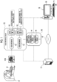
- an image processing apparatus 10 is connected to a medical information database server 11 via a network 12.
- the medical information database server 11 stores and manages various medical information uploaded from a plurality of medical facilities via the network 12.
- the medical information includes a head magnetic resonance imaging (MRI) image 13 and an examination result 14 of a medical examination.
- the network 12 is, for example, a wide area network (WAN) such as the Internet or a public communication network.
- WAN wide area network
- VPN virtual private network
- HTTPS hypertext transfer protocol secure
- the image processing apparatus 10 receives a head MRI image group 15 and an examination result group 16 distributed from the medical information database server 11.
- the head MRI image group 15 includes a plurality of head MRI images 13 that are allowed to be provided from medical facilities, for example, head MRI images 13 that are captured at a plurality of medical facilities from ten years ago to two years ago.
- the examination result group 16 includes a plurality of examination results 14 that are allowed to be provided from medical facilities and are examination results 14 of the patient P same as the patient of the head MRI images 13 of the head MRI image group 15.
- the head MRI image 13 is obtained by imaging a head of a patient P by an MRI apparatus 17.
- the head MRI image 13 is voxel data representing a three-dimensional shape of the head of the patient P.
- a head MRI image 13S having a sagittal cross section is illustrated.
- the head MRI image 13 is an example of a "medical image” and an "image in which a head of a patient appears" according to the technique of the present disclosure.
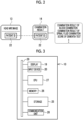
- a class 18 is associated with the head MRI image 13, and thus the head MRI images 13 are classified into classes.
- "A” is registered in a case where the patient P of the head MRI image 13 develops dementia two years later
- "B” is registered in a case where the patient P of the head MRI image 13 does not develop dementia two years later.
- the registration of the class 18 is performed by a doctor in charge of the patient P.
- the image processing apparatus 10 is, for example, a desktop personal computer, and includes a display 19 and an input device 20.
- the input device 20 is a keyboard, a mouse, a touch panel, a microphone, or the like.
- An operator of the image processing apparatus 10 transmits a distribution request of the head MRI image group 15 and the examination result group 16 to the medical information database server 11 by operating the input device 20.
- the medical information database server 11 searches for the head MRI image group 15 and the examination result group 16 of which the distribution request is received, and distributes the head MRI image group 15 and the examination result group 16 to the image processing apparatus 10.
- the head MRI image 13 and the examination result 14 are associated with each other by, for example, patient identification data (ID) 22 for uniquely identifying the patient P.
- the examination result 14 includes an examination result of a blood examination, an examination result of a spinal fluid examination, a score of a dementia test, and the like.
- the examination result of the blood examination includes, for example, a measured value of apolipoprotein, a measured value of complement protein, a measured value of transthyretin, and the like.
- the examination result of the spinal fluid examination includes, for example, a measured value of amyloid ⁇ , a measured value of tau protein, and the like.
- the score of the dementia test includes, for example, a score of revised Hasegawa's dementia scale (HDS-R), a score of mini-mental state examination (MMSE), a score of a rivermead behavioural memory test (RBMT), clinical dementia rating (CDR), activities of daily living (ADL), Alzheimer's disease assessment scale-cognitive subscale (ADAS-Cog), and the like.
- HDS-R Hasegawa's dementia scale
- MMSE mini-mental state examination
- RBMT rivermead behavioural memory test
- CDR clinical dementia rating
- ADL Alzheimer's disease assessment scale-cognitive subscale
- ADAS-Cog Alzheimer's disease assessment scale-cognitive subscale
- a computer including the image processing apparatus 10 includes a storage 25, a memory 26, a central processing unit (CPU) 27, and a communication unit 28, in addition to the display 19 and the input device 20.
- the components are connected to each other via a bus line 29.
- the CPU 27 is an example of a "processor" according to the technique of the present disclosure.
- the storage 25 is a hard disk drive that is built in the computer including the image processing apparatus 10 or is connected via a cable or a network. Alternatively, the storage 25 is a disk array in which a plurality of hard disk drives are connected in series. The storage 25 stores a control program such as an operating system, various application programs, and various data associated with the programs. A solid state drive may be used instead of the hard disk drive.
- the memory 26 is a work memory which is necessary to execute processing by the CPU 27.
- the CPU 27 loads the program stored in the storage 25 into the memory 26, and executes processing according to the program. Thereby, the CPU 27 collectively controls each unit of the computer.
- the communication unit 28 controls transmission of various types of information to an external apparatus such as the medical information database server 11.
- the memory 26 may be built in the CPU 27.
- an operation program 30 is stored in the storage 25 of the image processing apparatus 10.
- the operation program 30 is an application program for causing the computer to function as the image processing apparatus 10. That is, the operation program 30 is an example of "the operation program of the image processing apparatus" according to the technique of the present disclosure.
- a reference head MRI image (hereinafter, abbreviated as a reference image) 35 and a learning data group 36 are also stored in the storage 25.
- the learning data group 36 is a set of a plurality of learning head MRI images (hereinafter, abbreviated as learning images) 81 (refer to Fig. 11 ), which are used for training a dementia opinion derivation model 80 (refer to Fig. 11 ), and a plurality of learning examination results 82 (refer to Fig. 11 ).
- the learning data group 36 includes a normalization head MRI image group (hereinafter, abbreviated as a normalization image group) 37, a new head MRI image group (hereinafter, abbreviated as a new image group) 38, an examination result group 16, and a new examination result group 39.
- the normalization image group 37 includes a plurality of normalization head MRI images (hereinafter, abbreviated as normalization images) 40.
- the new image group 38 includes a plurality of first new head MRI images (hereinafter, abbreviated as first new images) 41_1 and a plurality of second new head MRI images (hereinafter, abbreviated as second new images) 41_2 (refer to Fig. 11 ).
- the new examination result group 39 includes a plurality of first new examination results 42_1 and a plurality of second new examination results 42_2 (refer to Fig. 11 ).
- the first new image 41_1 and the second new image 41_2 are an example of "new medical images” according to the technique of the present disclosure.
- the first new examination result 42_1 and the second new examination result 42_2 are an example of "new examination results" according to the technique of the present disclosure.
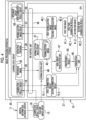
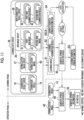
- the CPU 27 of the computer including the image processing apparatus 10 functions as a read/write (hereinafter, abbreviated as RW) control unit 45, a normalization unit 46, a non-linear registration unit 47, and a generation unit 48, in cooperation with the memory 26 and the like.
- RW read/write
- the RW control unit 45 controls storing of various types of data in the storage 25 and reading of various types of data in the storage 25. For example, the RW control unit 45 reads the reference image 35 from the storage 25, and outputs the read reference image 35 to the normalization unit 46. In addition, the RW control unit 45 receives the examination result group 16 from the medical information database server 11, and stores the received examination result group 16 in the storage 25, as a part of the learning data group 36. In addition, the RW control unit 45 receives the normalization image group 37 from the normalization unit 46, and stores the received normalization image group 37 in the storage 25, as a part of the learning data group 36.
- the RW control unit 45 reads a first examination result 14_1 and a second examination result 14_2, which are two examination results 14 among the plurality of examination results 14 of the examination result group 16, from the storage 25, and outputs the read first examination result 14_1 and the read second examination result 14_2 to the generation unit 48.
- the RW control unit 45 reads the first normalization image 40_1 and the second normalization image 40_2, which are two normalization images 40 among the plurality of normalization images 40 of the normalization image group 37 and correspond to the first examination result 14_1 and the second examination result 14_2, from the storage 25, and outputs the read first normalization image 40_1 and the read second normalization image 40_2 to the non-linear registration unit 47 and the generation unit 48.
- the first examination result 14_1 and the second examination result 14_2 and the first normalization image 40_1 and the second normalization image 40_2 are the examination results 14 and the normalization images 40 of two patients P having the same attributes such as a gender and an age.
- the normalization image 40 is associated with the class 18 based on the original head MRI image 13 (refer to Figs. 9A to 10B ).
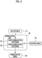
- the normalization unit 46 performs normalization processing of matching the head MRI image 13 with the reference image 35, and sets the head MRI image 13 as the normalization image 40.
- the normalization unit 46 performs normalization processing on all of the plurality of head MRI images 13 included in the head MRI image group 15. Thereby, a plurality of normalization images 40 corresponding to the plurality of head MRI images 13 included in the head MRI image group 15 are generated.
- the normalization unit 46 outputs a normalization image group 37 including the plurality of normalization images 40 to the RW control unit 45.
- the reference image 35 is a head MRI image in which a brain having a reference shape, a reference size, and a reference shade (pixel value) appears.
- the reference image 35 is, for example, an image generated by averaging head MRI images 13 of a plurality of healthy persons, or an image generated by computer graphics.
- the reference image 35 is an example of a "reference medical image" according to the technique of the present disclosure.
- the non-linear registration unit 47 performs non-linear registration processing on the first normalization image 40_1 and the second normalization image 40_2.
- the non-linear registration unit 47 outputs a processing result 50, which is a result of the non-linear registration processing, to the generation unit 48.
- the generation unit 48 generates a first new image 41_1 from the first normalization image 40_1 by transforming the first normalization image 40_1 under a generation condition according to the processing result 50, and generates a second new image 41_2 from the second normalization image 40_2 by transforming the second normalization image 40_2 under a generation condition according to the processing result 50.
- the generation unit 48 generates a first new examination result 42_1 corresponding to the first new image 41_1 from the first examination result 14_1, and generates a second new examination result 42_2 corresponding to the second new image 41_2 from the second examination result 14_2.
- the generation unit 48 outputs the first new image 41_1, the first new examination result 42_1, the second new image 41_2, and the second new examination result 42_2 to the RW control unit 45.
- the RW control unit 45 stores the first new image 41_1 and the second new image 41_2 in the storage 25, as a part of the new image group 38 and a part of the learning data group 36. Further, the RW control unit 45 stores the first new examination result 42_1 and the second new examination result 42_2 in the storage 25, as a part of the new examination result group 39 and a part of the learning data group 36.
- the first new image 41_1 is an example of a "first new medical image” according to the technique of the present disclosure.
- the second new image 41_2 is an example of a "second new medical image” according to the technique of the present disclosure.
- the normalization unit 46 performs, as normalization processing, shape normalization processing 55 and shade normalization processing 56 on the head MRI image 13.
- the shape normalization processing 55 is processing of extracting, for example, landmarks serving as references for registration from the head MRI image 13 and the reference image 35, and performing parallel displacement, rotation, and/or enlargement/reduction of the head MRI image 13 in accordance with the reference image 35 such that a correlation between the landmark of the head MRI image 13 and the landmark of the reference image 35 is maximized.
- the shade normalization processing 56 is, for example, processing of correcting a shade histogram of the head MRI image 13 in accordance with a shade histogram of the reference image 35.
- the non-linear registration unit 47 sets, on the first normalization image 40_1, a plurality of control points 60 which are arranged in a grid pattern at equal intervals. In addition, the non-linear registration unit 47 moves each control point 60 to a position at which a local similarity between the first normalization image 40_1 and the second normalization image 40_2 is increased.
- the non-linear registration unit 47 derives, from a movement amount of each control point 60, a transformation amount T_12 (X, Y, Z) of each pixel in a case where the first normalization image 40_1 is registered with the second normalization image 40_2, by using an interpolation approximation curve such as a B-Spline curve.
- the non-linear registration unit 47 outputs the derived transformation amount T_12 (X, Y, Z) as a processing result 50.
- the non-linear registration unit 47 sets, on the second normalization image 40_2, control points 63 which are same as the control points 60.
- the non-linear registration unit 47 moves each control point 63 as in the case of Fig. 6 .
- the non-linear registration unit 47 derives, from a movement amount of each control point 63, a transformation amount T_21 (X, Y, Z) of each pixel in a case where the second normalization image 40_2 is registered with the first normalization image 40_1.
- the non-linear registration unit 47 outputs the derived transformation amount T_21 (X, Y, Z) as a processing result 50. In the following, (X, Y, Z) may be omitted.
- Fig. 6 and Fig. 7 illustrate a state where the control points 60 and 63 are set in a two-dimensional shape on the first normalization image 40_1 and the second normalization image 40_2 having axial cross sections. On the other hand, the control points 60 and 63 are actually set in a three-dimensional shape.
- the transformation amount T_12 (X, Y, Z) and the transformation amount T_21 (X, Y, Z) have an inverse function relationship with each other.
- the other of the transformation amount T_12 (X, Y, Z) and the transformation amount T_21 (X, Y, Z) may be derived by obtaining an inverse function of one of the transformation amount T_12 (X, Y, Z) and the transformation amount T_21 (X, Y, Z).
- the generation unit 48 includes a transformation coefficient generation unit 70 and a calculation unit 71.
- the transformation coefficient generation unit 70 generates transformation coefficients ⁇ (X, Y, Z) and ⁇ (X, Y, Z) based on a normal distribution group 72.
- the transformation coefficient generation unit 70 outputs the generated transformation coefficients ⁇ (X, Y, Z) and ⁇ (X, Y, Z) to the calculation unit 71.
- the transformation coefficients ⁇ (X, Y, Z) and ⁇ (X, Y, Z) are values larger than 0 and smaller than 1 (0 ⁇ ⁇ , ⁇ ⁇ 1).
- the normal distribution group 72 includes a first ⁇ normal distribution 73, a first ⁇ normal distribution 74, a second ⁇ normal distribution 75, and a second ⁇ normal distribution 76.
- the transformation coefficient generation unit 70 outputs, as the transformation coefficients ⁇ (X, Y, Z) and ⁇ (X, Y, Z), a random number according to any one normal distribution of the first ⁇ normal distribution 73, the first ⁇ normal distribution 74, the second ⁇ normal distribution 75, and the second ⁇ normal distribution 76.
- the frequency of occurrence of the random number is, for example, for each pixel.
- the first ⁇ normal distribution 73 and the first ⁇ normal distribution 74 are used in a case where the classes 18 of the first normalization image 40_1 and the second normalization image 40_2 are the same.
- the second ⁇ normal distribution 75 and the second ⁇ normal distribution 76 are used in a case where the classes 18 of the first normalization image 40_1 and the second normalization image 40_2 are different from each other.
- the means ⁇ of the second ⁇ normal distribution 75 and the second ⁇ normal distribution 76 are smaller than the means ⁇ of the first ⁇ normal distribution 73 and the first ⁇ normal distribution 74. Therefore, values of the transformation coefficients ⁇ (X, Y, Z) and ⁇ (X, Y, Z) are changed depending on whether the classes 18 of the first normalization image 40_1 and the second normalization image 40_2 are the same or different from each other.
- the first ⁇ normal distribution 73 and the first ⁇ normal distribution 74 are examples of "normal distributions in a case where the classes of the first medical image and the second medical image are the same" according to the technique of the present disclosure.
- the second ⁇ normal distribution 75 and the second ⁇ normal distribution 76 are examples of "normal distributions in a case where the classes of the first medical image and the second medical image are different from each other" according to the technique of the present disclosure.
- the calculation unit 71 multiplies the transformation coefficient ⁇ (X, Y, Z) by the transformation amount T_12 (X, Y, Z) from the first normalization image 40_1 to the second normalization image 40_2, and sets a value obtained by the multiplication, as a corrected transformation amount ⁇ T_12 (X, Y, Z).
- the calculation unit 71 multiplies the transformation coefficient ⁇ (X, Y, Z) by the transformation amount T_21 (X, Y, Z) from the second normalization image 40_2 to the first normalization image 40_1, and sets a value obtained by the multiplication, as a corrected transformation amount ⁇ T_21 (X, Y, Z).
- the calculation unit 71 sets the first normalization image 40_1 as the first new image 41_1 (denoted as I_1N (X, Y, Z)) by applying the corrected transformation amount ⁇ T_12 (X, Y, Z) to the first normalization image 40_1.
- the calculation unit 71 sets the second normalization image 40_2 as the second new image 41_2 (denoted as I_2N (X, Y, Z)) by applying the corrected transformation amount ⁇ T_21 (X, Y, Z) to the second normalization image 40_2.
- the corrected transformation amount ⁇ T_12 (X, Y, Z) and the corrected transformation amount ⁇ T_21 (X, Y, Z) are an example of a "generation condition" according to the technique of the present disclosure.
- the calculation unit 71 converts the first examination result 14_1 and the second examination result 14_2 into a first new examination result 42_1 (denoted as MTR_1N) by calculating Equation (1), which indicates a weighted average of the first examination result 14_1 (denoted as MTR_1) and the second examination result 14_2 (denoted as MTR_2) and indicates a weighted average by the coefficient ⁇ F.
- the coefficient ⁇ F is a fixed value, and is an example of a "value similar to the transformation coefficient ⁇ " according to the technique of the present disclosure.
- the coefficient ⁇ F is, for example, 0.5 which is the same as the mean ⁇ of the first ⁇ normal distribution 73.
- the calculation unit 71 converts the first examination result 14_1 and the second examination result 14_2 into a second new examination result 42_2 (denoted as MTR_2N) by calculating Equation (2), which indicates a weighted average of the first examination result 14_1 and the second examination result 14_2 and indicates a weighted average by the coefficient ⁇ F.
- the coefficient ⁇ F is also a fixed value, and is an example of a "value similar to the transformation coefficient ⁇ " according to the technique of the present disclosure.
- the coefficient ⁇ F is, for example, 0.5 which is the same as the mean ⁇ of the first ⁇ normal distribution 74.
- the generation unit 48 sets the class of the first new image 41_1 to be the same as the class 18 of the first normalization image 40_1. More specifically, in a case where the class 18 of the first normalization image 40_1 is "A" as illustrated in Fig. 9A , the class 18 of the first new image 41_1 is also set to "A". In addition, in a case where the class 18 of the first normalization image 40_1 is "B" as illustrated in Fig. 9B , the class 18 of the first new image 41_1 is also set to "B".
- the generation unit 48 sets the class of the second new image 41_2 to be the same as the class 18 of the second normalization image 40_2. More specifically, in a case where the class 18 of the second normalization image 40_2 is "A" as illustrated in Fig. 10A , the class 18 of the second new image 41_2 is also set to "A". In addition, in a case where the class 18 of the second normalization image 40_2 is "B" as illustrated in Fig. 10B , the class 18 of the second new image 41_2 is also set to "B".
- the plurality of normalization images 40 included in the normalization image group 37, and one image among the plurality of first new images 41_1 and the plurality of second new images 41_2 included in the new image group 38 are provided as the learning images 81 of the dementia opinion derivation model 80.
- one examination result among the plurality of examination results 14 included in the examination result group 16 and the plurality of first new examination results 42_1 and the plurality of second new examination results 42_2 included in the new examination result group 39 is provided as a learning examination result 82 of the dementia opinion derivation model 80.
- the dementia opinion derivation model 80 is a machine learning model that receives the normalization images 40 and the examination results 14 as input data and outputs dementia opinion information 83 as output data, the dementia opinion information 83 being information representing a dementia opinion of the patient P of the normalization images 40.
- the dementia opinion information 83 is any one of normal control (NC), mild cognitive impairment (MCI), and Alzheimer's disease (AD).
- the dementia opinion derivation model 80 is configured by, for example, any method of a neural network, a support vector machine, and boosting.
- the dementia opinion derivation model 80 is trained by inputting, to the dementia opinion derivation model 80, learning data 84 which is a set of the learning image 81, the learning examination result 82, and correct dementia opinion information 83CA.
- the correct dementia opinion information 83CA is a dementia opinion which is for the patient P of the normalization image 40 and the examination result 14 and is actually obtained by a doctor in charge of the patient P.
- the correct dementia opinion information 83CA is a dementia opinion which is actually obtained by a doctor who receives a request with reference to the new image 41 and the new examination result 42.
- the learning images 81 and the learning examination results 82 are input to the dementia opinion derivation model 80.
- the dementia opinion derivation model 80 outputs learning dementia opinion information 83L in response to the learning images 81 and the learning examination results 82.
- a loss calculation of the dementia opinion derivation model 80 is performed based on the learning dementia opinion information 83L and the correct dementia opinion information 83CA.
- update settings of various coefficients of the dementia opinion derivation model 80 are performed according to a result of the loss calculation, and the dementia opinion derivation model 80 is updated according to the update settings.
- a series of pieces of processing which includes inputting of the learning image 81 and the learning examination result 82 to the dementia opinion derivation model 80, outputting of the learning dementia opinion information 83L from the dementia opinion derivation model 80, the loss calculation, the update settings, and updating of the dementia opinion derivation model 80, is repeatedly performed.
- the repetition of the series of pieces of processing is ended in a case where prediction accuracy of the learning dementia opinion information 83L with respect to the correct dementia opinion information 83CA reaches a predetermined set level.
- the dementia opinion derivation model 80 of which the prediction accuracy reaches the set level in this way is used as a trained model in an operation phase.
- the CPU 27 of the image processing apparatus 10 functions as the RW control unit 45, the normalization unit 46, the non-linear registration unit 47, and the generation unit 48.
- the generation unit 48 includes the transformation coefficient generation unit 70 and the calculation unit 71.
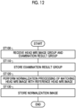
- the normalization unit 46 receives the head MRI image group 15 from the medical information database server 11.
- the RW control unit 45 receives the examination result group 16 from the medical information database server 11 (step ST100).
- the examination result group 16 is stored in the storage 25 by the RW control unit 45 (step ST110).
- the normalization unit 46 performs normalization processing (shape normalization processing 55 and shade normalization processing 56) of matching the head MRI image 13 with the reference image 35 (step ST120). Thereby, the head MRI image 13 is set as the normalization image 40.
- the normalization image 40 is output from the normalization unit 46 to the RW control unit 45, and is stored in the storage 25 by the RW control unit 45 (step ST130).
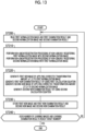
- the RW control unit 45 reads the first normalization image 40_1 and the first examination result 14_1, which are associated with each other by the patient ID 22, and the second normalization image 40_2 and the second examination result 14_2, which are associated with each other by the patient ID 22, from the storage 25 (step ST200).
- the first normalization image 40_1 and the second normalization image 40_2 are output from the RW control unit 45 to the non-linear registration unit 47 and the generation unit 48.
- the first examination result 14_1 and the second examination result 14_2 are output from the RW control unit 45 to the generation unit 48.
- the non-linear registration unit 47 performs non-linear registration processing of non-linearly registering the first normalization image 40_1 with the second normalization image 40_2.
- the non-linear registration unit 47 performs non-linear registration processing of non-linearly registering the second normalization image 40_2 with the first normalization image 40_1 (step ST210).
- the transformation amounts T_12 and T_21 derived by the non-linear registration processing are output from the non-linear registration unit 47 to the generation unit 48, as the processing result 50.
- the generation unit 48 applies, to the first normalization image 40_1, the corrected transformation amount ⁇ T_12 obtained by multiplying the transformation amount T_12 by the transformation coefficient ⁇ , and generates the first new image 41_1 from the first normalization image 40_1.
- the generation unit 48 applies, to the second normalization image 40_2, the corrected transformation amount ⁇ T_21 obtained by multiplying the transformation amount T_21 by the transformation coefficient ⁇ , and generates the second new image 41_2 from the second normalization image 40_2.
- the generation unit 48 generates the first new examination result 42_1 corresponding to the first new image 41_1 by using Equation (1), that is, the first calculation equation.
- the generation unit 48 generates the second new examination result 42_2 corresponding to the second new image 41_2 by using Equation (2), that is, the second calculation equation (step ST220).
- the first new image 41_1 and the first new examination result 42_1 and the second new image 41_2 and the second new examination result 42_2 are output from the generation unit 48 to the RW control unit 45, and are stored in the storage 25 by the RW control unit 45 (step ST230).
- step ST240 a series of processing of step ST200 to step ST230 is repeatedly performed while changing a combination of the first normalization image 40_1 and the first examination result 14_1 and the second normalization image 40_2 and the second examination result 14_2.
- the generation unit 48 of the CPU 27 of the image processing apparatus 10 generates the first new image 41_1 and the second new image 41_2 under the generation condition, from the first normalization image 40_1 and the second normalization image 40_2 among the plurality of normalization images 40, as the learning data 84 used for training the dementia opinion derivation model 80 for the normalization image 40 and the examination result 14.
- the generation unit 48 generates, as the learning data 84, the first new examination result 42_1 and the second new examination result 42_2 by performing, based on the generation condition, calculation on the first examination result 14_1 of the medical examination that corresponds to the first normalization image 40_1 and the second examination result 14_2 of the medical examination that corresponds to the second normalization image 40_2. Therefore, it is possible to generate an examination result of a new medical examination in which consistency is maintained.
- the non-linear registration unit 47 performs non-linear registration processing on the first normalization image 40_1 and the second normalization image 40_2.
- the generation unit 48 generates the first new image 41_1 and the second new image 41_2 by transforming the first normalization image 40_1 and the second normalization image 40_2 under the generation condition according to the processing result 50 of the non-linear registration processing.
- the generation unit 48 generates a first new examination result 42_1 corresponding to the first new image 41_1 from the first examination result 14_1, and generates a second new examination result 42_2 corresponding to the second new image 41_2 from the second examination result 14_2, by using Equation (1) and Equation (2) based on the generation condition.
- the first new image 41_1 and the second new image 41_2 are generated based on the first normalization image 40_1 and the second normalization image 40_2. Therefore, it is possible to increase a variation of the learning image 81, as compared to a method described in ⁇ C. Shorten, T. M. Khoshgofttar: A survey on Image Data Augmentation, Journal of Big Data, 2019>, which generates the learning image 81 from one image.
- the first new image 41_1 and the second new image 41_2 are not obtained by mixing pixel values of the first normalization image 40_1 and the second normalization image 40_2.
- a form having an anatomical structure is not blurred as in a method described in ⁇ Y. Tokozume, Y.
- the generation unit 48 sets the first normalization image 40_1 as the first new image 41_1 by applying, to the first normalization image 40_1, the corrected transformation amount ⁇ T_12 obtained by multiplying the transformation amount T_12 from the first normalization image 40_1 to the second normalization image 40_2 in the non-linear registration processing by the transformation coefficient ⁇ . Further, the generation unit 48 sets the second normalization image 40_2 as the second new image 41_2 by applying, to the second normalization image 40_2, the corrected transformation amount ⁇ T_21 obtained by multiplying the transformation amount T_21 from the second normalization image 40_2 to the first normalization image 40_1 in the non-linear registration processing by the transformation coefficient ⁇ .
- the desired first new image 41_1 and the desired second new image 41_2 can be obtained.
- the transformation coefficient ⁇ is set to a value close to 0
- the first new image 41_1 that is relatively similar to the first normalization image 40_1 is obtained.
- the transformation coefficient ⁇ is set to a value close to 1
- the first new image 41_1 that is relatively similar to the second normalization image 40_2 is obtained.
- the generation unit 48 converts the first examination result 14_1 and the second examination result 14_2 into the first new examination result 42_1 by calculating Equation (1) including the coefficient ⁇ F. In addition, the generation unit 48 converts the first examination result 14_1 and the second examination result 14_2 into the second new examination result 42_2 by calculating Equation (2) including the coefficient ⁇ F. Therefore, in a case where the coefficients ⁇ F and ⁇ F are set to appropriate values, the desired first new examination result 42_1 and the desired second new examination result 42_2 can be obtained. For example, in a case where the coefficient ⁇ F is set to a value close to 0, the first new examination result 42_1 that is relatively similar to the first examination result 14_1 is obtained. On the contrary, in a case where the coefficient ⁇ F is set to a value close to 1, the first new examination result 42_1 that is relatively similar to the second examination result 14_2 is obtained.
- the head MRI image 13 and the normalization image 40 are classified into classes.
- the generation unit 48 changes the values of the transformation coefficients ⁇ and ⁇ and the values of the coefficients ⁇ F and ⁇ F depending on whether the classes 18 of the first normalization image 40_1 and the second normalization image 40_2 are the same or different from each other. Therefore, it is possible to generate the first new image 41_1 and the first new examination result 42_1 and the second new image 41_2 and the second new examination result 42_2 that are suitable depending on whether the classes 18 of the first normalization image 40_1 and the second normalization image 40_2 are the same or different from each other.
- the generation unit 48 sets the class of the first new image 41_1 to be the same as the class 18 of the first normalization image 40_1, and sets the class of the second new image 41_2 to be the same as the class 18 of the second normalization image 40_2. Therefore, the class 18 that is suitable for a purpose of each of the first new image 41_1 and the second new image 41_2 can be associated.
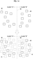
- the new image 41 can be used to fill a data blank in each class 18 of "A" and "B” that cannot be filled by only the normalization image 40. Further, the new image 41 can be used to fill a data blank near a boundary 90 of each class 18 of "A" and "B".
- the transformation coefficients ⁇ and ⁇ are random numbers according to any one of the normal distributions 73 to 76. Therefore, it is possible to generate the first new image 41_1 and the second new image 41_2 that cannot be predicted from the first normalization image 40_1 and the second normalization image 40_2. Thereby, it is possible to increase a variation of the learning image 81.
- the means ⁇ of the second ⁇ normal distribution 75 and the second ⁇ normal distribution 76 in a case where the classes of the first normalization image 40_1 and the second normalization image 40_2 are different from each other are smaller than the means ⁇ of the first ⁇ normal distribution 73 and the first ⁇ normal distribution 74 in a case where the classes of the first normalization image 40_1 and the second normalization image 40_2 are the same.
- the classes of the first normalization image 40_1 and the second normalization image 40_2 are different from each other, as compared with a case where the classes of the first normalization image 40_1 and the second normalization image 40_2 are the same, it is possible to generate the first new image 41_1, which is relatively similar to the first normalization image 40_1, and the second new image 41_2, which is relatively similar to the second normalization image 40_2. Therefore, even in a case where the class of the first new image 41_1 is set to be the same as the class 18 of the first normalization image 40_1 and the class of the second new image 41_2 is set to be the same as the class 18 of the second normalization image 40_2, there is no sense of incongruity.
- the normalization unit 46 performs normalization processing of matching the head MRI image 13 with the reference image 35, prior to the generation of the new image 41. Therefore, after an individual difference of the patient P and an apparatus difference of the MRI apparatus 17 are substantially eliminated, subsequent processing can be performed. Thereby, it is possible to improve reliability of the dementia opinion information 83.
- a medical image is the head MRI image 13 and a machine learning model for the medical image is the dementia opinion derivation model 80 which outputs the dementia opinion information 83 is a form matching with the current social problem.
- a form having an anatomical structure such as a degree of atrophy of a hippocampus, a parahippocampal gyrus, an amygdala, or the like, provides a particularly important key in the dementia opinion.
- an effect of capable of generating the learning image 81 in which a form having an anatomical structure is maintained can be further exhibited.
- the dementia opinion information 83 is not limited to the content illustrated in Fig. 11 (normal control/mild cognitive impairment/Alzheimer's disease).
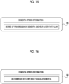
- the dementia opinion information may indicate whether a degree of progression of dementia of the patient P one year later is fast or slow.
- the dementia opinion information 98 illustrated in Fig. 16 the dementia opinion information may be a type of dementia, such as Alzheimer's disease, dementia with Lewy body, or vascular dementia.
- both the first new image 41_1 and the first new examination result 42_1 and the second new image 41_2 and the second new examination result 42_2 are generated has been described.
- the present invention is not limited thereto. Any one set of the first new image 41_1 and the first new examination result 42_1 and the second new image 41_2 and the second new examination result 42_2 may be generated. For example, only the first new image 41_1 and the first new examination result 42_1 are generated.
- the new image 41 and the new examination result 42 are generated in accordance with a tendency of the examination results 14 of a population of the patients P treated in the dementia opinion derivation model 80.
- Fig. 17 in Japan nationwide statistics 100 of MMSE scores of patients P, a case where 400000 patients have scores equal to or higher than 24 points, 200000 patients have scores equal to or higher than 10 points and lower than 20 points, and 100000 patients have scores lower than 10 points is considered. Further, in statistics 101 of the examination result group 16, a case where 10000 patients have scores equal to or higher than 24 points, 20000 patients have scores equal to or higher than 10 points and lower than 20 points, and 30000 patients have scores lower than 10 points is considered.
- the generation unit 48 in order to match statistics 102 of the new examination result group 39 with the nationwide statistics 100, the generation unit 48 generates the new examination results 42 of 110000 patients having MMSE scores equal to or higher than 24 points (also generates the new images 41 of 110000 patients), and generates the new examination results 42 of 40000 patients having MMSE scores equal to or higher than 10 points and lower than 20 points (also generates the new images 41 of 40000 patients).
- the nationwide statistics 100 are an example of a "tendency of examination results of a population" according to the technique of the present disclosure.
- attributes of a patient P of the head MRI image 13 and the examination result 14 are different from attributes of a patient P treated in the dementia opinion derivation model 80, such as a case where the head MRI image 13 of the head MRI image group 15 and the examination result 14 of the examination result group 16 are, for example, a head MRI image and an examination result of a patient P in the United States of America and the patient P treated in the dementia opinion derivation model 80 is a Japanese.
- the dementia opinion derivation model 80 deviates from the tendency of the population of the patients P.
- the new image 41 and the new examination result 42 are generated in accordance with the tendency of the examination results 14 of the population of the patients P treated in the dementia opinion derivation model 80. Thereby, it is possible to generate the dementia opinion derivation model 80 that matches the tendency of the population of the patients P.
- the non-linear registration processing may be performed on two new images 41, and the first new image 41_1 and the second new image 41_2 may be generated based on the processing result 50. Similarly, the first new image 41_1 and the second new image 41_2 may be generated from the normalization image 40 and the new image 41.
- the class 18 is not limited to whether or not the patient develops dementia two years after in the example.
- any one of normal control/mild cognitive impairment/Alzheimer's disease may be registered as the class 18.
- the transformation coefficients ⁇ and ⁇ may be fixed values instead of random numbers according to the normal distribution.
- the means ⁇ of the first ⁇ normal distribution 73 and the first ⁇ normal distribution 74 in a case where the classes of the first normalization image 40_1 and the second normalization image 40_2 are the same are not limited to 0.5 in the example.
- the mean ⁇ may be 0.4 or 0.6.
- the coefficients ⁇ F and ⁇ F may also be, for example, 0.4 or 0.6.
- the means ⁇ of the second ⁇ normal distribution 75 and the second ⁇ normal distribution 76 in a case where the classes of the first normalization image 40_1 and the second normalization image 40_2 are different from each other are not limited to 0.2 in the example.
- the mean ⁇ may be 0.1 or 0.3.
- the coefficients ⁇ F and ⁇ F may also be, for example, 0.1 or 0.3.
- the standard deviation ⁇ is also not limited to 0.2 in the example.
- the new image 41 may be generated by using a data expansion method described in ⁇ Y. Tokozume, Y. Ushiku, T. Harada: Between-class Learning for Image Classification, CVPR, 2018>.
- a calculation equation of the new examination result 42 (denoted as MTR_N) is represented as Equation (3).
- the training of the dementia opinion derivation model 80 illustrated in Fig. 11 may be performed by the image processing apparatus 10, or may be performed by an apparatus other than the image processing apparatus 10.
- an operation of actually inputting the normalization image 40 and the examination result 14 to the dementia opinion derivation model 80 and outputting the dementia opinion information 83 from the dementia opinion derivation model 80 may also be performed in the image processing apparatus 10, or may be performed by an apparatus other than the image processing apparatus 10.
- the training of the dementia opinion derivation model 80 may be continued even after the operation.
- the medical information database server 11 may perform some or all functions of each of the processing units 45 to 48.
- the normalization unit 46 may be included in the CPU of the medical information database server 11
- the non-linear registration unit 47 and the generation unit 48 may be included in the CPU of the image processing apparatus 10.
- the medical image is not limited to the head MRI image 13 in the example.
- the medical image may be a positron emission tomography (PET) image, a single photon emission computed tomography (SPECT) image, a computed tomography (CT) image, an endoscopic image, an ultrasound image, or the like.
- PET positron emission tomography
- SPECT single photon emission computed tomography
- CT computed tomography
- endoscopic image an ultrasound image, or the like.
- the subject is not limited to the head in the example, and may be a chest, an abdomen, or the like.
- the disease is not limited to dementia in the example, and may be a heart disease, pneumonia, dyshepatia, or the like.
- the various processors include, as described above, the CPU 27 which is a general-purpose processor that functions as various processing units by executing software (an operation program 30), a programmable logic device (PLD) such as a field programmable gate array (FPGA) which is a processor capable of changing a circuit configuration after manufacture, a dedicated electric circuit such as an application specific integrated circuit (ASIC) which is a processor having a circuit configuration specifically designed to execute specific processing, and the like.
- PLD programmable logic device
- FPGA field programmable gate array
- ASIC application specific integrated circuit
- One processing unit may be configured by one of these various processors, or may be configured by a combination of two or more processors having the same type or different types (for example, a combination of a plurality of FPGAs and/or a combination of a CPU and an FPGA). Further, the plurality of processing units may be configured by one processor.
- the plurality of processing units are configured by one processor
- a computer such as a client and a server
- a form in which one processor is configured by a combination of one or more CPUs and software and the processor functions as the plurality of processing units may be adopted.
- SoC system on chip
- a processor that realizes the functions of the entire system including a plurality of processing units with one integrated circuit (IC) chip is used.
- the various processing units are configured by using one or more various processors as a hardware structure.
- an electric circuit in which circuit elements such as semiconductor elements are combined may be used.
- the technique of the present disclosure can also appropriately combine the various embodiments and/or the various modification examples.
- the technique of the present disclosure is not limited to each embodiment, and various configurations may be adopted without departing from the scope of the present disclosure.
- the technique of the present disclosure extends to a program and a storage medium for non-temporarily storing the program.
- the described contents and the illustrated contents are detailed explanations of a part according to the technique of the present disclosure, and are merely examples of the technique of the present disclosure.
- the descriptions related to the configuration, the function, the operation, and the effect are descriptions related to examples of a configuration, a function, an operation, and an effect of a part according to the technique of the present disclosure. Therefore, it goes without saying that, in the described contents and illustrated contents, unnecessary parts may be deleted, new components may be added, or replacements may be made without departing from the spirit of the technique of the present disclosure. Further, in order to avoid complications and facilitate understanding of the part according to the technique of the present disclosure, in the described contents and illustrated contents, descriptions of technical knowledge and the like that do not require particular explanations to enable implementation of the technique of the present disclosure are omitted.
- a and/or B is synonymous with "at least one of A or B”. That is, “A and/or B” means that only A may be included, that only B may be included, or that a combination of A and B may be included. Further, in this specification, even in a case where three or more matters are expressed by being connected using "and/or", the same concept as "A and/or B" is applied.
Landscapes
- Engineering & Computer Science (AREA)
- Health & Medical Sciences (AREA)
- Physics & Mathematics (AREA)
- Theoretical Computer Science (AREA)
- Life Sciences & Earth Sciences (AREA)
- Medical Informatics (AREA)
- General Health & Medical Sciences (AREA)
- Computer Vision & Pattern Recognition (AREA)
- General Physics & Mathematics (AREA)
- Artificial Intelligence (AREA)
- Multimedia (AREA)
- Nuclear Medicine, Radiotherapy & Molecular Imaging (AREA)
- Evolutionary Computation (AREA)
- Radiology & Medical Imaging (AREA)
- Computing Systems (AREA)
- Databases & Information Systems (AREA)
- Software Systems (AREA)
- Heart & Thoracic Surgery (AREA)
- Biophysics (AREA)
- Pathology (AREA)
- Biomedical Technology (AREA)
- Neurology (AREA)
- Molecular Biology (AREA)
- Surgery (AREA)
- Animal Behavior & Ethology (AREA)
- Public Health (AREA)
- Veterinary Medicine (AREA)
- Quality & Reliability (AREA)
- Physiology (AREA)
- Psychiatry (AREA)
- Neurosurgery (AREA)
- Signal Processing (AREA)
- Psychology (AREA)
- Mathematical Physics (AREA)
- Hospice & Palliative Care (AREA)
- Developmental Disabilities (AREA)
- Child & Adolescent Psychology (AREA)
- High Energy & Nuclear Physics (AREA)
- Fuzzy Systems (AREA)
- Measuring And Recording Apparatus For Diagnosis (AREA)
Applications Claiming Priority (2)
| Application Number | Priority Date | Filing Date | Title |
|---|---|---|---|
| JP2020162679 | 2020-09-28 | ||
| PCT/JP2021/033190 WO2022065061A1 (fr) | 2020-09-28 | 2021-09-09 | Dispositif de traitement d'images, procédé d'utilisation de dispositif de traitement d'images et programme d'utilisation de traitement d'images |
Publications (2)
| Publication Number | Publication Date |
|---|---|
| EP4218604A1 true EP4218604A1 (fr) | 2023-08-02 |
| EP4218604A4 EP4218604A4 (fr) | 2024-04-10 |
Family
ID=80846522
Family Applications (1)
| Application Number | Title | Priority Date | Filing Date |
|---|---|---|---|
| EP21872192.6A Withdrawn EP4218604A4 (fr) | 2020-09-28 | 2021-09-09 | Dispositif de traitement d'images, procédé d'utilisation de dispositif de traitement d'images et programme d'utilisation de traitement d'images |
Country Status (5)
| Country | Link |
|---|---|
| US (1) | US12597130B2 (fr) |
| EP (1) | EP4218604A4 (fr) |
| JP (1) | JP7440655B2 (fr) |
| CN (1) | CN116209396A (fr) |
| WO (1) | WO2022065061A1 (fr) |
Families Citing this family (2)
| Publication number | Priority date | Publication date | Assignee | Title |
|---|---|---|---|---|
| EP3901963B1 (fr) * | 2020-04-24 | 2024-03-20 | Cognes Medical Solutions AB | Procédé et dispositif permettant d'estimer la progression précoce de la démence à partir d'images de tête humaine |
| JP2025087448A (ja) * | 2023-11-29 | 2025-06-10 | 国立研究開発法人情報通信研究機構 | プログラム及び情報処理方法 |
Family Cites Families (13)
| Publication number | Priority date | Publication date | Assignee | Title |
|---|---|---|---|---|
| JP3974946B2 (ja) | 1994-04-08 | 2007-09-12 | オリンパス株式会社 | 画像分類装置 |
| JP2008048880A (ja) * | 2006-08-24 | 2008-03-06 | Ge Medical Systems Global Technology Co Llc | 画像作成方法 |
| US9595120B2 (en) * | 2015-04-27 | 2017-03-14 | Siemens Healthcare Gmbh | Method and system for medical image synthesis across image domain or modality using iterative sparse representation propagation |
| JP6461743B2 (ja) * | 2015-08-03 | 2019-01-30 | 株式会社日立製作所 | 医用画像処理装置および医用画像処理方法 |
| JP6930283B2 (ja) * | 2017-08-18 | 2021-09-01 | コニカミノルタ株式会社 | 画像処理装置、画像処理装置の作動方法、及び画像処理プログラム |
| WO2019172181A1 (fr) * | 2018-03-09 | 2019-09-12 | 日本メジフィジックス株式会社 | Dispositif d'aide au diagnostic, programme, modèle appris et dispositif d'apprentissage |
| JP2019198376A (ja) | 2018-05-14 | 2019-11-21 | キヤノンメディカルシステムズ株式会社 | 医用画像処理装置、医用画像処理方法、及び医用画像処理システム |
| JP7210175B2 (ja) * | 2018-07-18 | 2023-01-23 | キヤノンメディカルシステムズ株式会社 | 医用情報処理装置、医用情報処理システム及び医用情報処理プログラム |
| JP7175682B2 (ja) * | 2018-09-06 | 2022-11-21 | キヤノンメディカルシステムズ株式会社 | 診断支援装置、診断支援システム、診断支援方法、及び診断支援プログラム |
| CN112702947A (zh) * | 2018-09-12 | 2021-04-23 | 株式会社Splink | 诊断辅助系统以及方法 |
| KR20200089146A (ko) * | 2019-01-16 | 2020-07-24 | 삼성전자주식회사 | 의료 영상 처리 장치 및 방법 |
| US11969239B2 (en) * | 2019-03-01 | 2024-04-30 | Siemens Healthineers Ag | Tumor tissue characterization using multi-parametric magnetic resonance imaging |
| JP7218215B2 (ja) * | 2019-03-07 | 2023-02-06 | 株式会社日立製作所 | 画像診断装置、画像処理方法及びプログラム |
-
2021
- 2021-09-09 CN CN202180064618.2A patent/CN116209396A/zh active Pending
- 2021-09-09 EP EP21872192.6A patent/EP4218604A4/fr not_active Withdrawn
- 2021-09-09 JP JP2022551872A patent/JP7440655B2/ja active Active
- 2021-09-09 WO PCT/JP2021/033190 patent/WO2022065061A1/fr not_active Ceased
-
2023
- 2023-03-21 US US18/187,649 patent/US12597130B2/en active Active
Also Published As
| Publication number | Publication date |
|---|---|
| WO2022065061A1 (fr) | 2022-03-31 |
| US20230222656A1 (en) | 2023-07-13 |
| EP4218604A4 (fr) | 2024-04-10 |
| JP7440655B2 (ja) | 2024-02-28 |
| US12597130B2 (en) | 2026-04-07 |
| CN116209396A (zh) | 2023-06-02 |
| JPWO2022065061A1 (fr) | 2022-03-31 |
Similar Documents
| Publication | Publication Date | Title |
|---|---|---|
| US11080895B2 (en) | Generating simulated body parts for images | |
| JP2022550688A (ja) | 低投与量容積造影mriを改良するためのシステム及び方法 | |
| US11978208B2 (en) | Trained model, learning method, learning program, medical information acquisition device, medical information acquisition method, and medical information acquisition program | |
| US12597130B2 (en) | Image processing apparatus, operation method of image processing apparatus, and operation program of image processing apparatus for generating examination results of new medical examinations | |
| Kamiya | Deep learning technique for musculoskeletal analysis | |
| Parreño et al. | Deidentifying MRI data domain by iterative backpropagation | |
| Dong et al. | MRI-CORE: a foundation model for magnetic resonance imaging | |
| Nechyporenko et al. | Deep learning based image segmentation for detection of odontogenic maxillary sinusitis | |
| WO2022138960A1 (fr) | Dispositif d'aide au diagnostic, procédé de fonctionnement pour dispositif d'aide au diagnostic, et programme de fonctionnement pour dispositif d'aide au diagnostic | |
| Wang | MRI-based diagnosis of Alzheimer’s disease using deep learning with CycleGAN for data augmentation | |
| US9224229B2 (en) | Process and apparatus for data registration | |
| US12505544B2 (en) | Image processing apparatus, image processing method, and image processing program | |
| US12608828B2 (en) | Image processing apparatus, operation method of image processing apparatus, operation program of image processing apparatus, and trained model | |
| Nambiar et al. | Tooth Image Synthesis Using Customized GAN Architectures: A Deep Learning-Based Solution for Data Scarcity in Dentistry | |
| Abdo et al. | Teeth and Jaw Segmentation From CBCT Images Using 3D Deep Learning Models | |
| US20230230261A1 (en) | Diagnosis support device, operation method of diagnosis support device, operation program of diagnosis support device | |
| EP4485352A1 (fr) | Segmentation d'image à l'aide d'un modèle de distribution de points | |
| Said et al. | Tooth-Diffusion: Guided 3D CBCT Synthesis with Fine-Grained Tooth Conditioning | |
| EP4270307A1 (fr) | Dispositif de traitement d'informations, procédé d'exploitation de dispositif de traitement d'informations et programme d'exploitation de dispositif de traitement d'informations | |
| Hanaf et al. | 10 Advanced Neuroimaging | |
| Caesarardhi et al. | Gender Detection on Cephalogram Images using Entropy Equalization Technique and Deep Learning Convolutional Neural Networks | |
| WO2026022501A1 (fr) | Modèles osseux à haute résolution à partir de scanners irm préopératoires à basse résolution | |
| Sithi Banu et al. | Enhanced U-Net Model for Hippocampus Segmentation in Magnetic Resonance Brain Images to Detect Alzheimer’s Disease | |
| EP2643811B1 (fr) | Procédé et appareil destinés à faire coïncider des données | |
| Vaidya | Perceptually Improved Medical Image Translations Using Conditional Generative Adversarial Networks |
Legal Events
| Date | Code | Title | Description |
|---|---|---|---|
| STAA | Information on the status of an ep patent application or granted ep patent |
Free format text: STATUS: THE INTERNATIONAL PUBLICATION HAS BEEN MADE |
|
| PUAI | Public reference made under article 153(3) epc to a published international application that has entered the european phase |
Free format text: ORIGINAL CODE: 0009012 |
|
| STAA | Information on the status of an ep patent application or granted ep patent |
Free format text: STATUS: REQUEST FOR EXAMINATION WAS MADE |
|
| 17P | Request for examination filed |
Effective date: 20230323 |
|
| AK | Designated contracting states |
Kind code of ref document: A1 Designated state(s): AL AT BE BG CH CY CZ DE DK EE ES FI FR GB GR HR HU IE IS IT LI LT LU LV MC MK MT NL NO PL PT RO RS SE SI SK SM TR |
|
| DAV | Request for validation of the european patent (deleted) | ||
| DAX | Request for extension of the european patent (deleted) | ||
| A4 | Supplementary search report drawn up and despatched |
Effective date: 20240312 |
|
| RIC1 | Information provided on ipc code assigned before grant |
Ipc: G06V 10/774 20220101ALI20240305BHEP Ipc: G06V 10/74 20220101ALI20240305BHEP Ipc: G06V 10/32 20220101ALI20240305BHEP Ipc: G06V 10/24 20220101ALI20240305BHEP Ipc: A61B 5/00 20060101ALI20240305BHEP Ipc: A61B 5/055 20060101ALI20240305BHEP Ipc: G06T 7/00 20170101ALI20240305BHEP Ipc: A61B 10/00 20060101AFI20240305BHEP |
|
| STAA | Information on the status of an ep patent application or granted ep patent |
Free format text: STATUS: THE APPLICATION IS DEEMED TO BE WITHDRAWN |
|
| 18D | Application deemed to be withdrawn |
Effective date: 20241003 |