EP4218739A2 - Mesoporöse siliciumdioxidnanopartikel mit lipiddoppelschichtbeschichtung zur frachtlieferung - Google Patents
Mesoporöse siliciumdioxidnanopartikel mit lipiddoppelschichtbeschichtung zur frachtlieferung Download PDFInfo
- Publication number
- EP4218739A2 EP4218739A2 EP23158990.4A EP23158990A EP4218739A2 EP 4218739 A2 EP4218739 A2 EP 4218739A2 EP 23158990 A EP23158990 A EP 23158990A EP 4218739 A2 EP4218739 A2 EP 4218739A2
- Authority
- EP
- European Patent Office
- Prior art keywords
- drug
- nanoparticle
- cancer
- irinotecan
- carrier
- Prior art date
- Legal status (The legal status is an assumption and is not a legal conclusion. Google has not performed a legal analysis and makes no representation as to the accuracy of the status listed.)
- Pending
Links
Images


Classifications
-
- A—HUMAN NECESSITIES
- A61—MEDICAL OR VETERINARY SCIENCE; HYGIENE
- A61K—PREPARATIONS FOR MEDICAL, DENTAL OR TOILETRY PURPOSES
- A61K9/00—Medicinal preparations characterised by special physical form
- A61K9/48—Preparations in capsules, e.g. of gelatin, of chocolate
- A61K9/50—Microcapsules having a gas, liquid or semi-solid filling; Solid microparticles or pellets surrounded by a distinct coating layer, e.g. coated microspheres, coated drug crystals
- A61K9/51—Nanocapsules; Nanoparticles
- A61K9/5107—Excipients; Inactive ingredients
- A61K9/5115—Inorganic compounds
-
- A—HUMAN NECESSITIES
- A61—MEDICAL OR VETERINARY SCIENCE; HYGIENE
- A61K—PREPARATIONS FOR MEDICAL, DENTAL OR TOILETRY PURPOSES
- A61K31/00—Medicinal preparations containing organic active ingredients
- A61K31/33—Heterocyclic compounds
- A61K31/335—Heterocyclic compounds having oxygen as the only ring hetero atom, e.g. fungichromin
- A61K31/337—Heterocyclic compounds having oxygen as the only ring hetero atom, e.g. fungichromin having four-membered rings, e.g. taxol
-
- A—HUMAN NECESSITIES
- A61—MEDICAL OR VETERINARY SCIENCE; HYGIENE
- A61K—PREPARATIONS FOR MEDICAL, DENTAL OR TOILETRY PURPOSES
- A61K31/00—Medicinal preparations containing organic active ingredients
- A61K31/33—Heterocyclic compounds
- A61K31/395—Heterocyclic compounds having nitrogen as a ring hetero atom, e.g. guanethidine or rifamycins
- A61K31/435—Heterocyclic compounds having nitrogen as a ring hetero atom, e.g. guanethidine or rifamycins having six-membered rings with one nitrogen as the only ring hetero atom
- A61K31/47—Quinolines; Isoquinolines
- A61K31/4738—Quinolines; Isoquinolines ortho- or peri-condensed with heterocyclic ring systems
- A61K31/4745—Quinolines; Isoquinolines ortho- or peri-condensed with heterocyclic ring systems condensed with ring systems having nitrogen as a ring hetero atom, e.g. phenantrolines
-
- A—HUMAN NECESSITIES
- A61—MEDICAL OR VETERINARY SCIENCE; HYGIENE
- A61K—PREPARATIONS FOR MEDICAL, DENTAL OR TOILETRY PURPOSES
- A61K45/00—Medicinal preparations containing active ingredients not provided for in groups A61K31/00 - A61K41/00
- A61K45/06—Mixtures of active ingredients without chemical characterisation, e.g. antiphlogistics and cardiaca
-
- A—HUMAN NECESSITIES
- A61—MEDICAL OR VETERINARY SCIENCE; HYGIENE
- A61K—PREPARATIONS FOR MEDICAL, DENTAL OR TOILETRY PURPOSES
- A61K47/00—Medicinal preparations characterised by the non-active ingredients used, e.g. carriers or inert additives; Targeting or modifying agents chemically bound to the active ingredient
- A61K47/50—Medicinal preparations characterised by the non-active ingredients used, e.g. carriers or inert additives; Targeting or modifying agents chemically bound to the active ingredient the non-active ingredient being chemically bound to the active ingredient, e.g. polymer-drug conjugates
- A61K47/51—Medicinal preparations characterised by the non-active ingredients used, e.g. carriers or inert additives; Targeting or modifying agents chemically bound to the active ingredient the non-active ingredient being chemically bound to the active ingredient, e.g. polymer-drug conjugates the non-active ingredient being a modifying agent
- A61K47/62—Medicinal preparations characterised by the non-active ingredients used, e.g. carriers or inert additives; Targeting or modifying agents chemically bound to the active ingredient the non-active ingredient being chemically bound to the active ingredient, e.g. polymer-drug conjugates the non-active ingredient being a modifying agent the modifying agent being a protein, peptide or polyamino acid
-
- A—HUMAN NECESSITIES
- A61—MEDICAL OR VETERINARY SCIENCE; HYGIENE
- A61K—PREPARATIONS FOR MEDICAL, DENTAL OR TOILETRY PURPOSES
- A61K47/00—Medicinal preparations characterised by the non-active ingredients used, e.g. carriers or inert additives; Targeting or modifying agents chemically bound to the active ingredient
- A61K47/50—Medicinal preparations characterised by the non-active ingredients used, e.g. carriers or inert additives; Targeting or modifying agents chemically bound to the active ingredient the non-active ingredient being chemically bound to the active ingredient, e.g. polymer-drug conjugates
- A61K47/69—Medicinal preparations characterised by the non-active ingredients used, e.g. carriers or inert additives; Targeting or modifying agents chemically bound to the active ingredient the non-active ingredient being chemically bound to the active ingredient, e.g. polymer-drug conjugates the conjugate being characterised by physical or galenical forms, e.g. emulsion, particle, inclusion complex, stent or kit
- A61K47/6921—Medicinal preparations characterised by the non-active ingredients used, e.g. carriers or inert additives; Targeting or modifying agents chemically bound to the active ingredient the non-active ingredient being chemically bound to the active ingredient, e.g. polymer-drug conjugates the conjugate being characterised by physical or galenical forms, e.g. emulsion, particle, inclusion complex, stent or kit the form being a particulate, a powder, an adsorbate, a bead or a sphere
- A61K47/6923—Medicinal preparations characterised by the non-active ingredients used, e.g. carriers or inert additives; Targeting or modifying agents chemically bound to the active ingredient the non-active ingredient being chemically bound to the active ingredient, e.g. polymer-drug conjugates the conjugate being characterised by physical or galenical forms, e.g. emulsion, particle, inclusion complex, stent or kit the form being a particulate, a powder, an adsorbate, a bead or a sphere the form being an inorganic particle, e.g. ceramic particles, silica particles, ferrite or synsorb
-
- A—HUMAN NECESSITIES
- A61—MEDICAL OR VETERINARY SCIENCE; HYGIENE
- A61K—PREPARATIONS FOR MEDICAL, DENTAL OR TOILETRY PURPOSES
- A61K47/00—Medicinal preparations characterised by the non-active ingredients used, e.g. carriers or inert additives; Targeting or modifying agents chemically bound to the active ingredient
- A61K47/50—Medicinal preparations characterised by the non-active ingredients used, e.g. carriers or inert additives; Targeting or modifying agents chemically bound to the active ingredient the non-active ingredient being chemically bound to the active ingredient, e.g. polymer-drug conjugates
- A61K47/69—Medicinal preparations characterised by the non-active ingredients used, e.g. carriers or inert additives; Targeting or modifying agents chemically bound to the active ingredient the non-active ingredient being chemically bound to the active ingredient, e.g. polymer-drug conjugates the conjugate being characterised by physical or galenical forms, e.g. emulsion, particle, inclusion complex, stent or kit
- A61K47/6921—Medicinal preparations characterised by the non-active ingredients used, e.g. carriers or inert additives; Targeting or modifying agents chemically bound to the active ingredient the non-active ingredient being chemically bound to the active ingredient, e.g. polymer-drug conjugates the conjugate being characterised by physical or galenical forms, e.g. emulsion, particle, inclusion complex, stent or kit the form being a particulate, a powder, an adsorbate, a bead or a sphere
- A61K47/6927—Medicinal preparations characterised by the non-active ingredients used, e.g. carriers or inert additives; Targeting or modifying agents chemically bound to the active ingredient the non-active ingredient being chemically bound to the active ingredient, e.g. polymer-drug conjugates the conjugate being characterised by physical or galenical forms, e.g. emulsion, particle, inclusion complex, stent or kit the form being a particulate, a powder, an adsorbate, a bead or a sphere the form being a solid microparticle having no hollow or gas-filled cores
- A61K47/6929—Medicinal preparations characterised by the non-active ingredients used, e.g. carriers or inert additives; Targeting or modifying agents chemically bound to the active ingredient the non-active ingredient being chemically bound to the active ingredient, e.g. polymer-drug conjugates the conjugate being characterised by physical or galenical forms, e.g. emulsion, particle, inclusion complex, stent or kit the form being a particulate, a powder, an adsorbate, a bead or a sphere the form being a solid microparticle having no hollow or gas-filled cores the form being a nanoparticle, e.g. an immuno-nanoparticle
-
- A—HUMAN NECESSITIES
- A61—MEDICAL OR VETERINARY SCIENCE; HYGIENE
- A61K—PREPARATIONS FOR MEDICAL, DENTAL OR TOILETRY PURPOSES
- A61K9/00—Medicinal preparations characterised by special physical form
- A61K9/0012—Galenical forms characterised by the site of application
- A61K9/0019—Injectable compositions; Intramuscular, intravenous, arterial, subcutaneous administration; Compositions to be administered through the skin in an invasive manner
-
- A—HUMAN NECESSITIES
- A61—MEDICAL OR VETERINARY SCIENCE; HYGIENE
- A61K—PREPARATIONS FOR MEDICAL, DENTAL OR TOILETRY PURPOSES
- A61K9/00—Medicinal preparations characterised by special physical form
- A61K9/10—Dispersions; Emulsions
- A61K9/127—Synthetic bilayered vehicles, e.g. liposomes or liposomes with cholesterol as the only non-phosphatidyl surfactant
-
- A—HUMAN NECESSITIES
- A61—MEDICAL OR VETERINARY SCIENCE; HYGIENE
- A61K—PREPARATIONS FOR MEDICAL, DENTAL OR TOILETRY PURPOSES
- A61K9/00—Medicinal preparations characterised by special physical form
- A61K9/48—Preparations in capsules, e.g. of gelatin, of chocolate
- A61K9/50—Microcapsules having a gas, liquid or semi-solid filling; Solid microparticles or pellets surrounded by a distinct coating layer, e.g. coated microspheres, coated drug crystals
- A61K9/51—Nanocapsules; Nanoparticles
- A61K9/5107—Excipients; Inactive ingredients
- A61K9/5123—Organic compounds, e.g. fats, sugars
-
- A—HUMAN NECESSITIES
- A61—MEDICAL OR VETERINARY SCIENCE; HYGIENE
- A61K—PREPARATIONS FOR MEDICAL, DENTAL OR TOILETRY PURPOSES
- A61K9/00—Medicinal preparations characterised by special physical form
- A61K9/48—Preparations in capsules, e.g. of gelatin, of chocolate
- A61K9/50—Microcapsules having a gas, liquid or semi-solid filling; Solid microparticles or pellets surrounded by a distinct coating layer, e.g. coated microspheres, coated drug crystals
- A61K9/51—Nanocapsules; Nanoparticles
- A61K9/5192—Processes
-
- A—HUMAN NECESSITIES
- A61—MEDICAL OR VETERINARY SCIENCE; HYGIENE
- A61P—SPECIFIC THERAPEUTIC ACTIVITY OF CHEMICAL COMPOUNDS OR MEDICINAL PREPARATIONS
- A61P31/00—Antiinfectives, i.e. antibiotics, antiseptics, chemotherapeutics
-
- A—HUMAN NECESSITIES
- A61—MEDICAL OR VETERINARY SCIENCE; HYGIENE
- A61P—SPECIFIC THERAPEUTIC ACTIVITY OF CHEMICAL COMPOUNDS OR MEDICINAL PREPARATIONS
- A61P31/00—Antiinfectives, i.e. antibiotics, antiseptics, chemotherapeutics
- A61P31/04—Antibacterial agents
-
- A—HUMAN NECESSITIES
- A61—MEDICAL OR VETERINARY SCIENCE; HYGIENE
- A61P—SPECIFIC THERAPEUTIC ACTIVITY OF CHEMICAL COMPOUNDS OR MEDICINAL PREPARATIONS
- A61P31/00—Antiinfectives, i.e. antibiotics, antiseptics, chemotherapeutics
- A61P31/10—Antimycotics
-
- A—HUMAN NECESSITIES
- A61—MEDICAL OR VETERINARY SCIENCE; HYGIENE
- A61P—SPECIFIC THERAPEUTIC ACTIVITY OF CHEMICAL COMPOUNDS OR MEDICINAL PREPARATIONS
- A61P31/00—Antiinfectives, i.e. antibiotics, antiseptics, chemotherapeutics
- A61P31/12—Antivirals
-
- A—HUMAN NECESSITIES
- A61—MEDICAL OR VETERINARY SCIENCE; HYGIENE
- A61P—SPECIFIC THERAPEUTIC ACTIVITY OF CHEMICAL COMPOUNDS OR MEDICINAL PREPARATIONS
- A61P35/00—Antineoplastic agents
-
- A—HUMAN NECESSITIES
- A61—MEDICAL OR VETERINARY SCIENCE; HYGIENE
- A61P—SPECIFIC THERAPEUTIC ACTIVITY OF CHEMICAL COMPOUNDS OR MEDICINAL PREPARATIONS
- A61P35/00—Antineoplastic agents
- A61P35/02—Antineoplastic agents specific for leukemia
-
- A—HUMAN NECESSITIES
- A61—MEDICAL OR VETERINARY SCIENCE; HYGIENE
- A61K—PREPARATIONS FOR MEDICAL, DENTAL OR TOILETRY PURPOSES
- A61K9/00—Medicinal preparations characterised by special physical form
- A61K9/10—Dispersions; Emulsions
- A61K9/127—Synthetic bilayered vehicles, e.g. liposomes or liposomes with cholesterol as the only non-phosphatidyl surfactant
- A61K9/1277—Preparation processes; Proliposomes
- A61K9/1278—Post-loading, e.g. by ion or pH gradient
-
- Y—GENERAL TAGGING OF NEW TECHNOLOGICAL DEVELOPMENTS; GENERAL TAGGING OF CROSS-SECTIONAL TECHNOLOGIES SPANNING OVER SEVERAL SECTIONS OF THE IPC; TECHNICAL SUBJECTS COVERED BY FORMER USPC CROSS-REFERENCE ART COLLECTIONS [XRACs] AND DIGESTS
- Y10—TECHNICAL SUBJECTS COVERED BY FORMER USPC
- Y10S—TECHNICAL SUBJECTS COVERED BY FORMER USPC CROSS-REFERENCE ART COLLECTIONS [XRACs] AND DIGESTS
- Y10S977/00—Nanotechnology
- Y10S977/70—Nanostructure
- Y10S977/773—Nanoparticle, i.e. structure having three dimensions of 100 nm or less
-
- Y—GENERAL TAGGING OF NEW TECHNOLOGICAL DEVELOPMENTS; GENERAL TAGGING OF CROSS-SECTIONAL TECHNOLOGIES SPANNING OVER SEVERAL SECTIONS OF THE IPC; TECHNICAL SUBJECTS COVERED BY FORMER USPC CROSS-REFERENCE ART COLLECTIONS [XRACs] AND DIGESTS
- Y10—TECHNICAL SUBJECTS COVERED BY FORMER USPC
- Y10S—TECHNICAL SUBJECTS COVERED BY FORMER USPC CROSS-REFERENCE ART COLLECTIONS [XRACs] AND DIGESTS
- Y10S977/00—Nanotechnology
- Y10S977/902—Specified use of nanostructure
- Y10S977/904—Specified use of nanostructure for medical, immunological, body treatment, or diagnosis
- Y10S977/906—Drug delivery
- Y10S977/907—Liposome
Definitions
- Pancreatic ductal adenocarcinoma is a fatal disease with a 5-year survival rate of less than 6% ( Siegel et al. (2014) CA Cancer J. Clin. 64(1): 9-29 ).
- the major treatment regimens for chemotherapy include either a single reagent, gemcitabine (GEM), or a four-drug regimen,).
- GEM gemcitabine
- FOLFIRINOX has a better response rate than GEM (31.6% versus 9.4%), with improved survival (11 months versus 6.8 months), the former combination is significantly more toxic and restricted to a minority of PDAC patients with good performance status ( Conroy et al. (2011) N. Engl. J. Med. 364(19): 1817-1825 ).
- Irinotecan contributes significantly to this toxicity, including a severe impact on the bone marrow (e.g. neutropenia), liver (e.g. necrosis and steatosis) and the gastrointestinal (GI) tract ( e.g. vomiting, diarrhea) ( Conroy et al. (2011) N. Engl. J. Med. 364(19): 1817-1825 ; Ueno et al. (2007) Cancer Chemother. Pharmacol. 59(4): 447-454 ; Loupakis et al. (2013) Br. J. Cancer, 108(12): 2549-2556 ).
- GI gastrointestinal
- liposomes could achieve high irinotecan loading capacity through the use of ammonium sulfate or proton entrapment agents ( Chou et al. (2003) J. Biosci. Bioeng., 95(4): 405-408 ; Messerer et al. (2004) Clin. Cancer Res, 10(19): 6638-6649 ; Drummond et al. (2006) Cancer Res., 66(6): 3271-3277 ; Sadzuka et al. (1998) Cancer Lett., 127(1): 99-106 ; Ramsay et al. (2008) Eur. J. Pharm. Biopharm.
- these carriers are spherical particles or supramolecular assemblies in the size range of 80-200 nm, often containing PEG coating on the surface to prolong circulatory half-life, and typically exhibiting loading capacities from ⁇ 5 w/w% (e.g., polymer-based nanoparticles) to ⁇ 50 w/w% ( e.g., liposomal carrier).
- ⁇ 5 w/w% e.g., polymer-based nanoparticles
- ⁇ 50 w/w% e.g., liposomal carrier.
- the potential benefits of these nanocarriers in animal studies, including murine PDAC models have been shown to include a reduction in in vivo toxicity, enhanced antitumor efficacy, and improved survival rate.
- Nanocarriers including an ionophore (A23187, also known as calimycin) enabled irinotecan delivery liposomal formulation (Irinophore C) and a protonating agent irinotecan delivering liposomal formulation (MM-398) ( Baker et al. (2008) Clin. Cancer Res. 14: 7260-7271 ; Drummond et al. (2006) Cancer Res. 15(66): 3271-3277 ).
- the Irinophore C formulation (Champions Biotechnology) is a liposomal carrier that makes use of active irinotecan loading through the generation of transmembrane proton gradients, using the ionophore, A23187, or ammonium sulfate ( Ramsay et al. (2008) Eur. J. Pharm. Biopharm. 68: 607-617 ).
- the Irinophore C formulation was used in a clinical study that commenced in 2011, but there have been no updated information about the outcome of the study or the results.
- MM-398 Human subjects participating in MM-398 clinical trials also showed significant elevations of liver enzymes, including alanine aminotransferase (ALT) (see, e.g., www.accessdata.fda.gov/drugsatfda_docs/label/2015/207793LB.pdf). Nonetheless, MM-398 received FDA approval for use in PDAC for patients failing to respond to GEM therapy, and is marketed as Onivyde ® ( see, e.g., www.fda.gov/newsevents/newsroom/pressannouncements/ucm468654.htm).
- Onivyde ® see, e.g., www.fda.gov/newsevents/newsroom/pressannouncements/ucm468654.htm).
- the MM-398 liposomal formulation incorporates irinotecan hydrochloride with the assistance of a polyanionic trapping agent (ESMO Gl 2014, www.merrimackpharma.com). More specifically, irinotecan loading into the MM-398 liposome was achieved by intra-liposomal drug encapsulation of a multivalent anionic trapping agent, triethylammonium sucrose octasulfate (TEA 8 SOS). This chemical leads to irinotecan protonation and entrapment at more than 10 times the loading that can be achieved through passive drug encapsulation ( Drummond et al. (2006) Cancer Res. 66(6): 3271-3277 ).
- MM-398 liposome IV administration of MM-398 liposome has been shown to induce complete tumor regression in various PDAC tumor models in mice, including inhibition of metastatic tumor foci ( Paz et al. (2102) Cancer Res. 72(12 Suppl): Abstract A63 ).
- MM-398 is currently in a Phase 3 clinical trial and lays claim to providing improved tumor inhibition, pharmacokinetics and efficacy compared to free irinotecan in animal and human studies ( Kalra (2012) AACR meeting, Abstract #5622 ). This includes experimental data claiming complete PDAC regression using a dose of 20 mg/kg MM-398 (human equivalent dose 60-120 mg/m2) in murine xenograft studies ( Paz et al. (2102) Cancer Res.
- MM-398 also increases the maximum tolerated dose (MTD) of free irinotecan from 80 to 324 mg/kg in mice ( Drummond et al. (2006) Cancer Res. 66(6): 3271-3277 ). Additionally, in the Phase 3 clinical trial by Merrimack Pharmaceuticals ( Hoff et al. ESMO GI 2014, www.merrimackpharma.com ) involving 417 PDAC patients, the combination of MM-398, 5-FU and leucovorin resulted in an overall survival (OS) of 6.1 months, which is 1.9 months longer than the control arm receiving 5-FU and leucovorin.
- OS overall survival
- MM-398 received FDA approval for use in PDAC for patients failing to respond to GEM therapy, and is marketed as ONIVYDE ® .
- the use of polyanionic polymers to increase drug entrapment in liposomes leads to ⁇ 80 nm drug precipitation (Zhu et al. (1996) 39(1): 138-142; Colbern et al.(1998) Clin. Cancer Res. 4(12): 3077-3082 ), which constitutes one of the reasons for slow irinotecan release from the liposomal carrier compared to LB-MSNP pores.
- nanocarriers and delivery methods that enable efficient drug delivery, including chemotherapy such as irinotecan chemotherapy, with an improved margin of safety and reduced toxicity.
- Urgent intervention is required to improve the 5-year survival rate of pancreatic ductal adenocarcinoma (PDAC). While the 4-drug regimen, FOLFIRINOX (comprised of irinotecan (IRIN), 5-fluorouracil (5-FU), oxaliplatin (OX), and leucovorin (LV)), has a better survival outcome than the more frequently used gemcitabine (GEM), the former treatment regimen is highly toxic and restricted for use in a patient with good performance status.
- FOLFIRINOX compact of irinotecan (IRIN), 5-fluorouracil (5-FU), oxaliplatin (OX), and leucovorin (LV)
- one specific aim of the present invention is to reduce the toxicity of the former drug with a custom-designed mesoporous silica nanoparticle (MSNP) platform, which uses a proton gradient for high-dose irinotecan loading across a coated lipid bilayer (LB).
- MSNP mesoporous silica nanoparticle
- LB-MSNP coated lipid bilayer
- the improved stability of the LB-coated MSNP (LB-MSNP) carrier allows for better protected irinotecan delivery and increased tumor drug concentrations when compared, for example, to a liposomal equivalent in a Kras-derived orthotopic PDAC model in immunocompetent mice.
- the LB-MSNP nanocarrier is also more efficient for treating tumor metastases.
- the reduced leakage and slower rate of drug release by the LB-MSNP carrier dramatically reduces the rate of bone marrow, gastrointestinal and liver toxicity compared to a liposomal carrier.
- the combination of high efficacy and reduced toxicity by the LB-MSNP carrier facilitates the use of irinotecan as a first-line therapeutic to improve PDAC survival.
- a nanocarrier comprising a silica body having a surface and defining a plurality of pores that are suitable to receive molecules therein, a lipid bilayer (e.g., a phospholipid bilayer) coating the surface, and a cargo-trapping agent contained in the pores by the coated lipid bilayer, where the submicron structure has a maximum dimension of less than one micron, and wherein the phospholipid bilayer stably seals the plurality of pores and can serve as a basis for additional packaging, targeting, imaging and functionalization.
- a lipid bilayer e.g., a phospholipid bilayer
- One significant aspect of the present invention includes new methods for making improved cargo/agent (e.g . irinotecan or other molecule(s) with the chemical structures described herein) nanocarriers. These methods generally include providing an unloaded nanocarrier comprising a silica body having a surface with a plurality of pores that are suitable to receive molecules therein, and encapsulating a cargo-trapping, targeting or imaging agent within the pores by a lipid bilayer (e.g., a phospholipid bilayer). As discussed in detail below, working examples include, but are not limted to new methods of making concentrated irinotecan nanocarriers.
- tese methods include selecting a nanocarrier comprising a silica body having a surface including a plurality of pores suitable to receive irinotecan therein.
- an agent having an ability to specifically influence the diffusion of and/or trap the irinotecan molecule e.g. triethylammonium sucrose octasulfate
- the nanocarrier and pores are completely coated with a lipid bilayer (for example using a sonication process).
- the silica body nanocarrier is not dried and/or washed immediately prior to this coating step.
- this lipid bilayer can comprise one or more bioactive molecules selected to facilitate the nanocarrier function (such as polyethylene glycol, and/or targeting ligands, and/or paclitaxel and/or activated irinotecan, SN38).
- bioactive molecules selected to facilitate the nanocarrier function
- irinotecan is then allowed to migrate across the lipid bilayer into the pores where it is entrapped. This is also known as "remote drug loading”.
- irinotecan nanocarriers with surprisingly high loading capacities e.g . 40 wt% or greater than 40% drug/MSNP
- irinotecan nanocarriers formed using these methods have a constellation of other desirable properties, for example exhibiting ⁇ 5% irinotecan leakage over 24 hours in a biological buffer with pH of 7.4 at 37 °C.
- Various embodiments contemplated herein may include, but need not be limited to, one or more of the following:
- subject may be used interchangeably and refer to humans, as well as non-human mammals (e.g., non-human primates, canines, equines, felines, porcines, bovines, ungulates, lagomorphs, and the like).
- non-human mammals e.g., non-human primates, canines, equines, felines, porcines, bovines, ungulates, lagomorphs, and the like.
- the subject can be a human (e.g., adult male, adult female, adolescent male, adolescent female, male child, female child) under the care of a physician or other health worker in a hospital, as an outpatient, or other clinical context.
- the subject may not be under the care or prescription of a physician or other health worker.
- the phrase "a subject in need thereof” refers to a subject, as described infra, that suffers from, or is at risk for a pathology to which the nanoparticle drug carriers described herein (silicasomes) are directed.
- the subject is a subject with a cancer (e.g., pancreatic ductal adenocarcinoma (PDAC), breast cancer (e.g., drug-resistant breast cancer), colon cancer, brain cancer, and the like).
- PDAC pancreatic ductal adenocarcinoma
- breast cancer e.g., drug-resistant breast cancer
- colon cancer e.g., brain cancer, and the like.
- the subject is a subject with a microbial infection including, but not limited to drug-resistant microbial infections.
- treat when used with reference to treating, e.g., a pathology or disease refers to the mitigation and/or elimination of one or more symptoms of that pathology or disease, and/or a delay in the progression and/or a reduction in the rate of onset or severity of one or more symptoms of that pathology or disease, and/or the prevention of that pathology or disease.
- treat can refer to prophylactic treatment which includes a delay in the onset or the prevention of the onset of a pathology or disease.
- coadministration indicates that the first compound and the second compound are administered so that there is at least some chronological overlap in the biological activity of first compound and the second compound in the organism to which they are administered.
- Coadministration can include simultaneous administration or sequential administration. In sequential administration there may even be some substantial delay (e.g., minutes or even hours) between administration of the first compound and the second compound as long as their biological activities overlap.
- the coadminstration is over a time frame that permits the first compound and second compound to produce an enhanced therapeutic or prophylactic effect on the organism.
- the enhanced effect is a synergistic effect.
- nanoparticle drug carrier and “silicasome” are used interchangeably and refer to a nanostructure having a porous particle core, which is interchangeable with the term “porous nanoparticle” as used herein, and a lipid bilayer encasing (or surrounding or enveloping) the porous particle core.
- silica nanoparticle is a porous silica nanoparticle (e.g., mesoporous silica nanoparticle (MSNP)).
- lipid refers to conventional lipids, phospholipids, cholesterol, chemically functionalized lipids for attachment of PEG and ligands, etc.
- lipid bilayer or "LB” refers to any double layer of oriented amphipathic lipid molecules in which the hydrocarbon tails face inward to form a continuous non-polar phase.
- liposome refers to an aqueous compartment enclosed by a lipid bilayer, as being conventionally defined (see, e.g., Stryer (1981) Biochemistry, 2d Edition, W. H. Freeman & Co., p. 213 ).
- the lipid bilayer in a liposome can be referred to as an "unsupported lipid bilayer” and the liposome itself (when unloaded) can be referred to as an "empty liposome".
- the lipid bilayer in a silicasome can be referred to as a "supported lipid bilayer” because the lipid bilayer in a silicasome is located on the surface and supported by a porous particle core.
- the lipid bilayer can have a thickness ranging form about 6 nm to about 7 nm which includes a 3-4 nm thickness of the hydrophobic core, plus the hydrated hydrophilic head group layers (each about 0.9 nm) plus two partially hydated regions of about 0.3 nm each.
- the term "selective targeting” or “specific binding” refers to use of targeting ligands on the surface of silicasomes (empty or loaded), in particular, on the surface of the lipid bilayer of the silicasomes, wherein the ligands interact specifically/selectively with receptors or other biomolecular components expressed on the target, e.g ., a cell surface of interest.
- the targeting ligands can include such molecules and/or materials as peptides, antibodies, aptamers, targeting peptides, polysaccarides, and the like.
- a silicasome having targeting ligands can be referred to as a "targeted silicasome”.
- silica refers to a drug containing (drug delivery) silica nanoparticle in which the silica nanoparticle is fully covered with a lipid bilayer (e.g., a phospholipid bilayer).
- a lipid bilayer e.g., a phospholipid bilayer
- the silica nanoparticle is a porous silica nanoparticle (e.g., mesoporous silica nanoparticle).
- the term can mean within an order of magnitude, preferably within 5-fold, and more preferably within 2-fold, of a value.
- the term "about” meaning within an acceptable error range for the particular value should be assumed.
- drug refers to a chemical entity of varying molecular size, small and large, naturally occurring or synthetic, that exhibits a therapeutic effect in animals and humans.
- a drug may include, but is not limited to, an organic molecule (e.g., a small organic molecule), a therapeutic protein, peptide, antigen, or other biomolecule, an oligonucleotide, an siRNA, a construct encoding CRISPR cas9 components and, optionally one or more guide RNAs, and the like.
- a "pharmaceutically acceptable carrier” as used herein is defined as any of the standard pharmaceutically acceptable carriers.
- the pharmaceutical compositions of the subject invention can be formulated according to known methods for preparing pharmaceutically useful compositions.
- the pharmaceutically acceptable carrier can include diluents, adjuvants, and vehicles, as well as carriers, and inert, non-toxic solid or liquid fillers, diluents, or encapsulating material that does not react with the active ingredients of the invention. Examples include, but are not limited to, phosphate buffered saline, physiological saline, water, and emulsions, such as oil/water emulsions.
- the carrier can be a solvent or dispersing medium containing, for example, ethanol, polyol (for example, glycerol, propylene glycol, liquid polyethylene glycol, and the like), suitable mixtures thereof, and vegetable oils.
- ethanol for example, ethanol, polyol (for example, glycerol, propylene glycol, liquid polyethylene glycol, and the like), suitable mixtures thereof, and vegetable oils.
- polyol for example, glycerol, propylene glycol, liquid polyethylene glycol, and the like
- suitable mixtures thereof for example, glycerol, propylene glycol, liquid polyethylene glycol, and the like
- an “antibody” refers to a protein consisting of one or more polypeptides substantially encoded by immunoglobulin genes or fragments of immunoglobulin genes or derived therefrom that is capable of binding ( e.g., specifically binding) to a target ( e.g., to a target polypeptide).
- the recognized immunoglobulin genes include the kappa, lambda, alpha, gamma, delta, epsilon and mu constant region genes, as well as myriad immunoglobulin variable region genes.
- Light chains are classified as either kappa or lambda.
- Heavy chains are classified as gamma, mu, alpha, delta, or epsilon, which in turn define the immunoglobulin classes, IgG, IgM, IgA, IgD and IgE, respectively.
- a typical immunoglobulin (antibody) structural unit is known to comprise a tetramer.
- Each tetramer is composed of two identical pairs of polypeptide chains, each pair having one "light” (about 25 kD) and one "heavy” chain (about 50-70 kD).
- the N-terminus of each chain defines a variable region of about 100 to 110 or more amino acids primarily responsible for antigen recognition.
- the terms variable light chain (V L ) and variable heavy chain (V H ) refer to these light and heavy chains respectively.
- Antibodies exist as intact immunoglobulins or as a number of well characterized fragments produced by digestion with various peptidases.
- pepsin digests an antibody below the disulfide linkages in the hinge region to produce F(ab)' 2 , a dimer of Fab which itself is a light chain joined to V H -C H 1 by a disulfide bond.
- the F(ab)' 2 may be reduced under mild conditions to break the disulfide linkage in the hinge region thereby converting the (Fab') 2 dimer into a Fab' monomer.
- the Fab' monomer is essentially a Fab with part of the hinge region ( see, Fundamental Immunology, W.E. Paul, ed., Raven Press, N.Y.
- antibody fragments are defined in terms of the digestion of an intact antibody, one of skill will appreciate that such Fab' fragments may be synthesized de novo either chemically or by utilizing recombinant DNA methodology.
- the term antibody as used herein also includes antibody fragments either produced by the modification of whole antibodies or synthesized de novo using recombinant DNA methodologies.
- Certain preferred antibodies include single chain antibodies (antibodies that exist as a single polypeptide chain), more preferably single chain Fv antibodies (sFv or scFv) in which a variable heavy and a variable light chain are joined together (directly or through a peptide linker) to form a continuous polypeptide.
- the single chain Fv antibody is a covalently linked V H- V L heterodimer which may be expressed from a nucleic acid including V H - and V L - encoding sequences either joined directly or joined by a peptide-encoding linker.
- the first functional antibody molecules to be expressed on the surface of filamentous phage were single-chain Fv's (scFv), however, alternative expression strategies have also been successful.
- Fab molecules can be displayed on a phage if one of the chains (heavy or light) is fused to g3 capsid protein and the complementary chain exported to the periplasm as a soluble molecule.
- the two chains can be encoded on the same or on different replicons; the important point is that the two antibody chains in each Fab molecule assemble post-translationally and the dimer is incorporated into the phage particle via linkage of one of the chains to, e.g., g3p ( see, e.g., U.S. Patent No: 5733743 ).
- scFv antibodies and a number of other structures converting the naturally aggregated, but chemically separated light and heavy polypeptide chains from an antibody V region into a molecule that folds into a three dimensional structure substantially similar to the structure of an antigen-binding site are known to those of skill in the art (see e.g., U.S. Patent Nos. 5,091,513 , 5,132,405 , and 4,956,778 ).
- antibodies should include all that have been displayed on phage (e.g., scFv, Fv, Fab and disulfide linked Fv ( see, e.g, Reiter et al. (1995) Protein Eng. 8: 1323-1331 ) as well as affibodies, unibodies, and the like.
- biomolecule e.g., protein, nucleic acid, antibody, etc .
- a binding reaction that is determinative of the presence of a biomolecule in heterogeneous population of molecules (e.g., proteins and other biologics).
- the specified ligand or antibody binds to its particular "target" molecule and does not bind in a significant amount to other molecules present in the sample.
- drug delivering nanocarriers are provided to enhance drug delivery (e.g ., chemotherapy drugs, antimicrobial drugs, etc .) at a target site (e.g. a tumor site, a site of infection, etc .) while limiting the amount of free drug that can cause systemic toxicity. It is desirable that the carrier itself maintains stable drug loading in order to prevent toxicity and improve efficacy.
- nanocarriers to deliver chemotherapeutic agents in animal tumor models established from various cancer cells has demonstrated an ability of such nanocarriers to prolong the drug circulatory half-life, deliver high drug concentrations to the tumor site with improved cytotoxic killing, as well as reduce systemic toxicity compared to free drug equivalents ( see, e.g., Messerer et al.(2004) Clin. Cancer Res. 10(19): 6638-6649 ; Drummond et al. (2006) Cancer Res. 66(6): 3271-3277 ; Ramsay et al. (2008) Clin. Cancer Res. 14(4): 1208-1217 ).
- novel nanoparticle drug carriers are provided herein that use a lipid bilayer (LB) applied to silica nanoparticles (e.g., mesoporous silica nanoparticles (MSNPs)), thereby providing a supported lipid bilayer.
- silica nanoparticles e.g., mesoporous silica nanoparticles (MSNPs)
- MSNPs mesoporous silica nanoparticles
- These lipid bilayer coated porous silica nanoparticles also known as silicasomes
- provide a drug carrier that is significantly less leaky than liposomes Meng et al. (2013) ACS Nano, 7(2): 994-1005 ; Meng et al. (2011) ACS Nano, 5(5): 4131-4144 ; Meng et al.
- MSNPs Due to a large internal surface area that can be used for drug packaging, tunable pore sizes, carrier stability, and controlled drug release abilities, MSNPs have been demonstrated to constitute a versatile and multifunctional nanocarrier platform for cancer therapy ( Meng et al. (2013) ACS Nano, 7(2): 994-1005 ; Meng et al. (2011) ACS Nano, 5(5): 4131-4144 ; Meng et al. (2013) ACS Nano, 7(11): 10048-10065 ; Meng et al. (2015) ACS Nano, 9(4): 3540-3557 ; Meng et al. (2010) J.
- new loading methods are provided herein that achieve significantly greater loading of lipid-bilayer coated nanoparticle drug carriers.
- the new loading method utilizes a cargo trapping reagent, to retain the cargo (e.g., drug or drugs of interest) within the pores of the nanoparticle inside the lipid bilayer. More specifically, in certain embodiments, the method can involve:
- the trapping agent comprises a protonating agent and the methods involve providing a drug or drugs that can pass through the lipid bilayer into the bilayer-coated porous nanoparticle.
- the protonating agent in the porous nanoparticle converts the drug into a hydrophilic derivative that is incapable of back diffusion across the lipid bilayer.
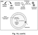
- MSNP cores were synthesized by sol-gel chemistry and soaked in a solution containing the protonating agent, TEA 8 SOS. These particles were coated with a LB, using a sonication procedure in the presence of a lipid biofilm of optimal composition for bilayer stability. This was followed by remote irinotecan loading by the proton gradient provided by TEA 8 SOS.
- Irinotecan is a weak basic and amphipathic molecule that can diffuse across the LB into the MSNP interior packaging space, where proton release by prior entrapped triethylammonium sucrose octasulfate (TEA 8 SOS) converts the drug into a hydrophilic derivative, incapable of back-diffusion across the LB.
- TAA 8 SOS triethylammonium sucrose octasulfate
- lipid bilayer nanoparticle drug carriers produced using the methods described herein offer numerous advantages.
- Such advantages include, but are not limited to ease of synthesis, improved drug loading and release profile, improved stability and improved biodistribution.
- this method of remote loading leads to a 4-fold or greater increase in loading capacity of irinotecan compared to passive drug encapsulation behind the LB in MSNP.
- non-encapsulated irinotecan gains entrance to MSNP pores by diffusing through the LB, whereupon protonation renders the irinotecan hydrophilic and incapable of escape.
- the increased loading capacity of the nanocarrier leads to increased drug delivery at the cancer site, with less of the free drug becoming available at sites such as the bone marrow and gastrointestinal tract (GIT), where irinotecan causes toxicity.
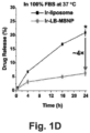
- the LB-MSNP has an improved loading capacity (83.5 weight% irinotecan loading) versus an in-house liposomal equivalent of MM-398 (42.5 weight% loading capacity).
- embodiments of the LB-MSNP formulation have demonstrated 3-5 times higher release capacity at an acidic pH (4.5) compared to the liposomal formulation.
- synthesis/loading methods described herein are easier to perform than either making conventional liposomes or the polymer-lipid technology used by Zhang et al. (2104) Biomaterials, 35(11): 3560-3665 ). This is advantageous for scale-up to GMP manufacturing, including cost savings by reducing the quantity of non-encapsulated drug.
- a LB-MSNP-based nanocarrier delivery system that allows for stable and protected loading of cargo at high loading levels, with the assistance of a cargo-trapping agent.
- the cargo can be a drug.
- Drug delivery by the modified LB-MSNP encapsulation and trapping allows for more frequent use of drug encapsulation, e.g ., chemotherapy drugs, because of enhanced efficacy, high drug loading capacity and reduced systemic toxicity.
- various embodiments of this system for example a polyanionic cargo-trapping agent within LB-MSNP, are employed as an effective design principle for delivering a range of additional weakly basic molecules and drugs, for the treatment of several different types of cancer and other disease processes.
- irinotecan delivery by the modified LB-MSNP encapsulation and trapping procedure allows for more frequent use of irinotecan and FOLFIRINOX in human PDAC patients because of enhanced efficacy, high drug loading capacity and reduced systemic toxicity of irinotecan. This also allows more PDAC patients to be treated with a more potent therapeutic regimen than gemcitabine, with promise of increased survival.
- an IV injectable, efficient, biocompatible, and translationally competitive irinotecan formulation (versus MM398 liposome) for PDAC treatment is provided.
- irinotecan and FOLFIRINOX therapy may also be available for the treatment of other cancers including, but not limited to colon, rectal, lung, and ovarian cancer.
- the silicasome delivery system disclosed herein is a multifunctional platform.
- Mesoporous silica drug carriers have been demonstrated to be able to deliver a wide range of cargoes to cancer cells as well as a variety of human cancer models in animals. These include gemcitabine and paclitaxil co-delivery to xenograft and orthotopic PDAC tumors in mice.
- MSNPs are biodegradable and proven to be safe in extensive animal testing ( Meng et al. (2013) ACS Nano, 7(2): 994-1005 ; Meng et al. (2011) ACS Nano, 5(5): 4131-4144 ; Meng et al. (2013) ACS Nano, 7(11): 10048-10065 ; Meng et al.
- the loading methods, lipid bilayer composition and stability of the resulting nanoparticle drug carriers described herein provide significant improvements and advantages over previous nanoparticle drug carriers.
- improved methods of loading lipid bilayer-coated porous nanoparticles are provided along with the drug delivery nanoparticles produced by such methods.
- the methods described herein achieve extremely high levels of drug loading e.g. , greater than 40 wt%, or greater than 45 wt%, or greater than 50 wt%, or greater than 60 wt%, or greater than 70 wt%, %, or greater than 80 wt%, etc .).
- the methods can involve:
- TEA 8 SOS polyanionic compound
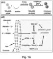
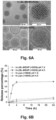
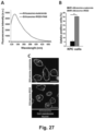
- TEA 8 SOS is a proton-generating agent that releases eight H+ ions and octavalent SOS 8- upon hydrolysis ( Figure 1A , panel A2).
- Ionexchange chromatography was used to generate TEA 8 SOS, which was soaked into MSNP, as described below in the Methods section ( Id .).
- the soaked particles were introduced to a round-bottom flask coated with a lipid biofilm, composed of DSPC/cholesterol/DSPE-PEG2000 at a molar ratio of 3:2:0.15. Sonication of the suspension yielded LB-coated particles, that contain the entrapment agent ( Figure 1A , panel A1).
- lipid bilayer coating disposed on the porous nanoparticle provides rapid and instantaneous pore sealing for drug entrapment. This simplifies synthesis and provides stable and high drug loading capacity.
- the methods described herein are effective to load porous particle with cargos at high levels and are compatible with this rapid and instantaneous pore sealing with the lipid bilayer.
- a biofilm technique has been developed for the LB-coated MSNP platform that can be used to rapidly encapsulate gemcitabine (GEM) (e.g. a water-soluble nucleoside) by a supported LB ( Meng et al. (2015) ACS Nano, 9(4): 3540-3557 ). Not only has this allowed the LB-MSNPs to achieve a loading capacity of up to 40 wt% GEM, but also enabled co-delivery of hydrophobic paclitaxel (PTX) that could be incorporated in the lipid bilayer (LB) ( Meng et al. (2015) ACS Nano, 9(4): 3540-3557 ). This has provided for a synergistic and ratiometric-designed carrier for PDAC treatment in an orthotopic human PDAC model in mice ( Id .).
- GEM gemcitabine
- lipid bilayer (LB) provided an improvement on previous drug delivery nanoparticles, prior to the utilization of the remote loading methods described herein, the bilayer still provided an impediment to achieving even higher drug loading levels.
- comparisons were made between the MSNP carrier and a liposome equivalent, in which a non-supported LB was used for irinotecan loading subsequent to encapsulation of triethylammonium sucrose octasulfate (TEA 8 SOS) ( Drummond et al. (2006) Cancer Res., 66(6): 3271-3277 ; Von Hoff et al. (2013) Br. J.
- the MSNP carrier achieve higher loading capacity for irinotecan and tumor killing than the liposomal formulation in a robust orthotopic PDAC model, but it also prevented drug toxicity due to increased carrier stability and reduced leakage compared to liposomes.
- the LB-MSNP platform exhibits the properties desirable for a first-line irinotecan carrier for PDAC (or other cancer) treatment.
- Zhang et al. for irinotecan drug loading of MSNPs to treat drug-resistant breast cancer tumors in Balb/c nude mice
- Zhang et al. (2014) Biomaterials, 35(11): 3650-3665 the methodology described in this reference is different, and does not achieve the same drug loading capacity compared to the methods and compositions described herein.
- Zhang et al. do not use a classical LB or a trapping agent, resulting in a carrier with only 1/5th the loading capacity ( ⁇ 15 %, w/w) of the carrier provided herein (-83.5%, w/w).
- a third major difference is the composition of the LB formulation.
- a mixture of commercially available lipids plus cholesterol e.g., DSPC/cholesterol/DSPE-PEG
- Zhang et al. use an in-house synthesized, pH-sensitive, Pluronic P123 grafted DOPE.
- the use of the Pluronic is apparently based on its ability to act as an inhibitor of drug efflux ( see, e.g., Batrakova et al. (2004) Pharm. Res. 21(12): 2226-2233 ), hence the consideration to treat drug-resistant breast cancer.
- the absence of cholesterol in the coated bilayer decreases the fluidity and stability of the platform.
- irinotecan encapsulation in the provided carrier shows ⁇ 5% leakage over 24 hours in a biological buffer with pH of 7.4 at 37 °C. This is 2.5x lower than the premature leakage (-16% leakage at pH 7.4 and 37°C over 24 hours) described by Zhang et al. (2014) Biomaterials, 35: 3650-3665 ).
- Zhang et al. Another difference from the drug deliver platform described by Zhang et al. is that various embodiments of the present invention use probe sonication for biofilm rehydration and pore sealing, followed by centrifugation purification or size exclusion chromatography.
- Zhang et al. utilizes a membrane extrusion method, as for liposomes.
- CTAC is used as a templating agent for MSNP synthesis, whereas Zhang et al. utilize CTAB.
- the present invention addresses the development of therapy for PDAC and other cancers and is not principally designed to overcome drug resistance, as are the drug delivery particles described by Zhang et al. for use in the breast cancer study.
- the lipid bilayer coated nanoparticle drug carriers described herein provide a unique design that outperforms the carriers of Zhang et al. based on drug loading capacity, colloidal stability, ease of production, and stable drug retention in the blood and body fluids.
- nanoparticle drug carriers described herein comprises a porous silica nanoparticle (e.g., a silica body having a surface and defining a plurality of pores that are suitable to receive molecules therein) coated with a lipid bilayer.
- the silica nanoparticle can be a mesoporous silica nanoparticle.
- the fact that the nanoparticle is referred to as a silica nanoparticle does not preclude materials other than silica from also being incorporated within the silica nanoparticle.
- the silica nanoparticle may be substantially spherical with a plurality of pore openings through the surface providing access to the pores.
- the silica nanoparticle can have shapes other than substantially spherical shapes.
- the silica nanoparticle can be substantially ovoid, rod-shaped, a substantially regular polygon, an irregular polygon, and the like.
- the silica nanoparticle comprises a silica body that defines an outer surface between the pore openings, as well as side walls within the pores.
- the pores can extend through the silica body to another pore opening, or a pore can extend only partially through the silica body such that that it has a bottom surface of defined by the silica body.
- the silica body is mesoporous. In other embodiments, the silica body is microporous.
- “mesoporous” means having pores with a diameter between about 2 nm and about 50 nm, while “microporous” means having pores with a diameter smaller than about 2 nm.
- the pores may be of any size, but in typical embodiments are large enough to contain one or more therapeutic compounds therein. In such embodiments, the pores allow small molecules, for example, therapeutic compounds such as anticancer compounds to adhere or bind to the inside surface of the pores, and to be released from the silica body when used for therapeutic purposes.
- the pores are substantially cylindrical.
- the nanoparticles comprise pores having pore diameters between about 1 nm and about 10 nm in diameter or between about 2 nm and about 8 nm. In certain embodiments the nanoparticles comprise pores having pore diameters between about 1 nm and about 6 nm, or between about 2 nm and about 5 nm. Other embodiments include particles having pore diameters less than 2.5 nm. In other embodiments, the pore diameters are between 1.5 and 2.5 nm. Silica nanoparticles having other pore sizes may be prepared, for example, by using different surfactants or swelling agents during the preparation of the silica nanoparticles.
- the nanoparticles can include particles as large (e.g. , average or median diameter (or other characteristic dimension) as about 1000 nm. However in various embodiments the nanoparticles are typically less than 500 nm or less than about 300 nm as, in general, particles larger than 300 nm may be less effective in entering living cells or blood vessel fenestrations. In certain embodiments the nanoparticles range in size from about 40 nm, or from about 50 nm, or from about 60 nm up to about 100 nm, or up to about 90 nm, or up to about 80 nm, or up to about 70 nm. In certain embodiments the nanoparticle range in size from about 60 nm to about 70 nm.
- Some embodiments include nanoparticles having an average maximum dimension between about 50 nm and about 1000 nm. Other embodiments include nanoparticles having an average maximum dimension between about 50 nm and about 500 nm. Other embodiments include nanoparticles having an average maximum dimension between about 50 nm and about 200 nm. In some embodiments, the average maximum dimension is greater than about 20nm, greater than about 30nm, greater than 40nm, or greater than about 50nm. Other embodiments include nanoparticles having an average maximum dimension less than about 500 nm, less than about 300nm, less than about 200nm, less than about 100nm or less than about 75 nm. As used herein, the size of the nanoparticle refers to the average or median size of the primary particles, as measured by transmission electron microscopy (TEM) or similar visualization technique.
- TEM transmission electron microscopy
- Illustrative mesoporous silica nanoparticles include, but are not limited to MCM-41, MCM-48, and SBA-15 ( see, e.g., Katiyaret al. (2006) J. Chromatog. 1122(1-2): 13-20 ).
- mesoporous silica nanoparticles are synthesized by reacting tetraethyl orthosilicate (TEOS) with a template made of micellar rods. The result is a collection of nano-sized spheres or rods that are filled with a regular arrangement of pores.
- TEOS tetraethyl orthosilicate
- the template can then be removed by washing with a solvent adjusted to the proper pH ( see, e.g., Trewyn et al. (2007) Chem. Eng. J. 137(1): 23-29 .
- mesoporous particles can also be synthesized using a simple sol-gel method (see, e.g., Nandiyanto, et al. (2009) Microporous andMesoporous Mat. 120(3): 447-453 , and the like).
- tetraethyl orthosilicate can also be used with an additional polymer monomer (as a template).
- 3-mercaptopropyl)trimethoxysilane (MPTMS) is used instead of TEOS.
- the mesoporous silica nanoparticles are cores were synthesized by a modification of the sol/gel procedure described by Meng et al. (2015) ACS Nano, 9(4): 3540-3557 .
- 50 mL of CTAC is mixed with 150 mL of H 2 O in a flask (e.g., a 500 mL conical flask), followed by stirring ( e.g., at 350 rpm for 15 min at 85°C). This us followed by the addition of 8 mL of 10% triethanolamine for 30 min at the same temperature.
- 7.5 mL of the silica precursor, TEOS is added dropwise at a rate of 1 mL/min using a peristaltic pump.
- the solution is stirred at 350 rpm at 85°C for 20 min, leading to the formation particles with a primary size of ⁇ 65 nm.
- the surfactant can be removed by washing the particles with a mixture of methanol/HCl (500:19 v/v) at room temperature for 24 h.
- the particles can be centrifuged at 10 000 rpm for 60 min and washed three times in methanol.
- mesoporous polymeric particle can be utilized.
- the syntheses of highly ordered mesoporous polymers and carbon frameworks from organic-organic assembly of triblock copolymers with soluble, low-molecular-weight phenolic resin precursors (resols) by an evaporation induced self-assembly strategy have been reported by Meng et al. (2006) Chem. Mat. 6(18): 4447-4464 and in the references cited therein.
- nanoparticles described herein are illustrative and non-limiting. Using the teachings provided herein numerous other lipid bilayer drug delivery nanoparticle will be available to one of skill in the art.
- the drug carrier nanoparticles described herein comprise a porous nanoparticle (e . g . a mesoporous silica nanoparticle (MSNP)) coated with a lipid bilayer.
- a porous nanoparticle e . g . a mesoporous silica nanoparticle (MSNP)
- MSNP mesoporous silica nanoparticle
- the bilayer composition is optimized to provide a rapid and uniform particle coating, to provide colloidal and circulatory stability, and to provide effective cargo retention, while also permitting a desirable cargo release profile.
- the lipid bilayer comprises a combination of a phospholipid, cholesterol, and in certain embodiments, a pegylated lipid (e . g ., DSPE-PEG 2000 ), or a factionalized pegylated lipid ( e . g ., DSPE-PEG 2000 -maleimide) to facilitate conjugation with targeting or other moieties.
- a pegylated lipid e . g ., DSPE-PEG 2000
- a factionalized pegylated lipid e . g ., DSPE-PEG 2000 -maleimide
- lipid film procedure To attach a surface LB coating, a coated lipid film procedure was developed in which drug- or TEO 8 SOS-soaked MSNP suspensions were added to a large lipid film surface, coated on, e . g ., a round-bottom flask.
- drug- or TEO 8 SOS-soaked MSNP suspensions were added to a large lipid film surface, coated on, e . g ., a round-bottom flask.
- lipid bilayer compositions Using different lipid bilayer compositions, a series of experiments can be performed to find a composition and optimal lipid/particle ratio that provides rapid and uniform particle wrapping, coating and effective cargo retention and/or release upon sonication. It is believed that this lipid composition and wrapping cannot be achieved by liposomal fusion to the particle surface under low energy vortexing conditions.
- 500 mg MSNPs are soaked in a 20 mL TEA 8 SOS (80 mM solution), which is added on top of the lipid biofilm, comprised of a 550 mg mixture of DSPC/Chol/DSPE-PEG 2000 (molar ratio 3:2:0.15), coated at the bottom of a round bottom flask ( see Example 1, and Liu et al. (2016) ACS Nano. 10(2): 2702-2715 ).
- the ratio of "3:2:0.15" equals to "58.3 mol% : 38.8 mol% : 3.9 mol%" if one uses mol% to present the ratio. This provides a lipid:particle ratio of ⁇ 1.1:1.
- TEA 8 SOS is removed by size exclusion chromatography over a Sepharose CL-4B column.
- the TEA 8 SOS loaded silicasomes are incubated in a 10 mg/mL irinotecan solution for drug loading in a water bath at 65 °C. The loading is stopped after 30 min by quenching in and ice water bath, following which the drug-loaded silicasomes were washed 3 times by centrifugation and re-suspended in PBS.
- lipid bilayer formulation described above and in Example 1 is illustrative and non-limiting. Depending on the drug(s) being loaded into the silicasome and the desired release provide, in various embodiments different lipid bilayer formulaitosn can be used and an optimal formulation can be determined.
- the lipid bilayer can compirese: 1) one or more saturated fatty acids with C14-C20 carbon chain, such as dimyristoylphosphatidylcholine (DMPC), dipalmitoylphosphatidylcholine (DPPC), distearoylphosphatidylcholine (DSPC), and diactylphosphatidylcholine (DAPC); and/or 2) One or more unsaturated fatty acids with a C14-C20 carbon chain, such as 1,2-dimyristoleoyl-sn-glycero-3-phosphocholine, 1,2-dipalmitoleoyl-sn-glycero-3-phosphocholine,1,2-dioleoyl-sn-glycero-3-phosphocholine (DOPC), 1,2-dieicosenoyl-sn-glycero-3-phosphocholine; and/or 3) Natural lipids comprising a mixture of fatty acids with C12-C20 carbon chain, such as Egg
- the silicasome contains a lipid (e . g ., a phospholipid), cholesterol, and a PEG functionalized lipid (e . g ., a mPEG phospholipid).
- a lipid e . g ., a phospholipid
- a PEG functionalized lipid e . g ., a mPEG phospholipid
- the mPEG phospholipids comprises a C14-C18 phospholipid carbon chain from, and a PEG molecular weight from 350-5000 ( e . g ., MPEG 5000, MPEG 3000, MPEG 2000, MPEG 1000, MPEG 750, MPEG 550, MPEG 350, and the like).
- the mPEG phospholipid comprises DSPE-PEG5000, DSPE-PEG3000, DSPE-PEG2000, DSPE-PEG1000, DSPE-PEG750, DSPE-PEG550, or DSPE-PEG350.
- MPEGs are commercially available ( see, e.g., //avantilipids.com/product-category/products/polymers-polymerizable-lipids/mpeg-phospholipids/).
- the ratio of phospholipid: CHOL:PEG is about phospholipid (50-90 mol%): CHOL (10-50 mol%) : PEG (1-10 mol%).
- the trapping agent can be altered, the lipid composition and molar ratios can be altered, and the drug or drugs can be altered to identify other silicasomes optimized for their particular cargo(s).
- an effective lipid formulation for a gemcitabine-containing silicasome comprises DPPC/cholesterol/DSPE-PEG at a molar ratio of 77.5:20:2.5
- an effective lipid formulation for an irinotecan containing silicasome comprises DSPC/Chol/DSPE-PEG 2000 (molar ratio 3:2:0.15, which equals 58.3 mol% : 38.8 mol% : 3.9 mol%).
- these methods can be varied to improve drug-loading capacity (weight of drug/total weight of carrier).
- the drug loading capacity is at least about 30%, or at least about 40%, at least about 50%, at least about 60%, at least about 70%, or at least 80% w/w.
- drug loading is greater than 40% w/w, or greater than 45% w/w, or greater than 50% w/w, or greater than 60% w/w, or greater than 70% w/w, %, or greater than 80% w/w.
- the protocols described herein provide a nanoparticle drug carrier (nanocarrier) outperforms nanocarriers made by the liposomal method of coating by fusion with the MSNP surface, as illustrated by the descriptions of the ease of synthesis and improved loading capacity and release profile provided herein.
- the LB coating procedure is used to rapidly encapsulate the protonation agent, e . g ., TEA 8 SOS, which subsequently provides irinotecan loading and entrapment by protonating the incoming drug diffusing across the LB. This leads to high drug loading in the particles pores.
- the rapid and effective pore sealing to retain the trapping agent (e . g ., TEA 8 SOS), without leakage, contributes to the effectiveness and stability of the carrier.
- the cargo-trapping reagent can be selected to interact with a desired cargo. In some embodiments, this interaction can be an ionic or protonation reaction, although other modes of interaction are contemplated.
- the cargo-trapping agent can have one or more ionic sites, i.e., can be mono-ionic or poly-ionic.
- the ionic moiety can be cationic, anionic, or in some cases, the cargo-trapping agent can include both cationic and anionic moieties.
- the ionic sites can be in equilibrium with corresponding uncharged forms; for example, an anionic carboxylate (-COO - ) can be in equilibrium with its corresponding carboxylic acid (-COOH); or in another example, an amine (-NH 2 ) can be in equilibrium with its corresponding protonated ammonium form (-NH 3 + ). These equilibriums are influenced by the pH of the local environment.
- the cargo can include one or more ionic sites.
- the cargo-trapping agent and cargo can be selected to interact inside the nanoparticle (e.g., the mesoporous silica nanoparticle). This interaction can help retain the cargo within the nanoparticle until release of the cargo is desired.
- the cargo can exist in a pH-dependent equilibrium between non-ionic and ionic forms. The non-ionic form can diffuse across the lipid bilayer and enter pores of the MSNP.
- the cargo-trapping agent e.g., a polyionic cargo-trapping agent
- the cargo-trapping agent can interact with the ionic form of the cargo and thereby retain the cargo within the nanocarrier, e .
- the interaction can be an ionic interaction, and can include formation of a precipitate.
- Trapping of cargo within the nanocarrier can provide higher levels of cargo loading compared to similar systems, e . g ., nanocarriers that omit the cargo-trapping agent, or liposomes that do include a trapping agent.
- Release of the cargo can be achieved by an appropriate change in pH to disrupt the interaction between the cargo and cargo-trapping agent, for example, by returning the cargo to its non-ionic state which can more readily diffuse across the lipid bilayer.
- the cargo is irinotecan and the cargo-trapping agent is TEA 8 SOS.
- the cargo trapping agent need not be limited to TEA8SOS.
- the cargo trapping comprises small molecules like (NH 4 ) 2 SO 4 , and the like.
- Other trapping agents include, but are not limited to, ammonium salts ( e .
- ammonium sulfate ammonium sucrose octasulfate, ammonium ⁇ -cyclodextrin sulfate, ammonium ⁇ -cyclodextrin sulfate, , ammonium ⁇ -cyclodextrin sulfate, ammonium phosphate, ammonium ⁇ -cyclodextrin phosphate, ammonium ⁇ -cyclodextrin phosphate, ammonium citrate, ammonium acetate, and the like), trimethylammonium salts ( e .
- transmembrane pH gradients can also be generated by acidic buffers (e.g. citrate) ( Chou et al. (2003) J. Biosci. Bioengineer., 95(4): 405-408 ; Nichols et al. (1976) Biochimica et Biophysica Acta (BBA)-Biomembranes, 455(1): 269-271 ), proton-generating dissociable salts (e.g. (NH 4 ) 2 SO 4 ) ( Haran et al.
- acidic buffers e.g. citrate
- dissociable salts e.g. (NH 4 ) 2 SO 4
- the cargo comprises an organic compound that includes at least one primary amine group, or at least one secondary amine group, or at least one tertiary amine group, or at least one quaternary amine group, or any combination thereof, capable of being protonated.
- the general characteristics of these cargo molecules include the following chemical properties:
- a list of potential chemotherapy agents can include irinotecan derivatives and metabolites such as SN38 together with other alkaloids (e . g . topotecan, 10-hydroxycamptothecin, belotecan, rubitecan, vinorelbine, LAQ824, vinblastine, vincristine, homoharringtonine, trabectedin), anthracyclines ( e . g . doxorubicin, epirubicin, pirarubicin, daunorubicin, rubidomycin, valrubicin, amrubicin), alkaline anthracenediones ( e . g .
- mitoxantrone alkaline alkylating agents (e . g . cyclophosphamide, mechlorethamine, temozolomide), purine or pyrimidine derivatives (e . g . 5-fluorouracil, 5'-deoxy-5-fluorouridine, gemcitabine, capecitabine), and protein kinase inhibitors ( e . g . pazopanib, enzastaurin, vandetanib erlotinib, dasatinib, nilotinib, sunitinib).
- alkaline alkylating agents e . g cyclophosphamide, mechlorethamine, temozolomide
- purine or pyrimidine derivatives e . g . 5-fluorouracil, 5'-deoxy-5-fluorouridine, gemcitabine, capecitabine
- protein kinase inhibitors e . g .
- the ability to package and deliver one or a combination of the above agents will enhance the wider utility of the multifunctional LB-MSNP platform, including treatment consideration of additional cancer types such as colon, breast, lung, liver, glioma, melanoma, etc.
- the trapping reagent facilitated LB-MSNP platform is useful in efficient drug loading and delivery.
- the trapping reagent will provide limited help, the provided one-step biofilm technique for MSNP pore healing is still valid for an even larger spectrum of drug molecules, such as anticancer drugs, anti-viral drugs, antifungal drugs, and antibiotics.
- efficient drug encapsulation includes, but is not limited to, everolimus, trabectedin, paclitaxel, TLK 286, AV-299, DN-101, pazopanib, GSK690693, RTA 744, ON 0910.Na, AZD 6244 (ARRY-142886), AMN-107, TKI-258, GSK461364, AZD 1152, enzastaurin, vandetanib, ARQ-197, MK-0457, MLN8054, PHA-739358, R-763, AT-9263, a FLT-3 inhibitor, a VEGFR inhibitor, an EGFR TK inhibitor, an aurora kinase inhibitor, a PIK-1 modulator, a Bcl-2 inhibitor, an HDAC inhibitor, a c-MET inhibitor, a PARP inhibitor, a Cdk inhibitor, an EGFR TK inhibitor, an IGFR-TK
- the cargo comprise an antifungal agent.
- antifungal agents include, but are not limited to Amphotericin B ( e . g ., for Most fungal infections except Pseudallescheria sp., and the like), Anidulafungin ( e . g ., for candidiasis, including candidemia, and the like), Caspofungin ( e . g ., for aspergillosis, candidiasis, including candidemia, and the like), Fluconazole ( e .
- Posaconazole e . g ., for prophylaxis for invasive aspergillosis and candidiasis, oral candidiasis, oral candidiasis refractory to itraconazole, and the like
- Voriconazole e . g ., for Invasive aspergillosis, Fusariosis, Scedosporiosis, and the like
- the nanoparticle drug carriers (silicasomes) described herein can comprise two or more therapeutic agents.
- the pores in the silicasome can be loaded with two, or with three, or with four, or more different therapeutic agents. This can, in certain embodiments, permit ratiometric delivery of these therapeutic agents.
- numerous multi-agent therapeutic regimen are known for the treatment of cancer.
- COMP metalhotrexate, prednisone
- LSA 2 -L 2 cyclophosphamide, vincristine, prednisone, daunomycin, methotrexate, cytarabine, thioguanine, asparaginase, and carmustine
- FOLFIRINOX irinotecan, oxaliplatin, 5-fluorouracil, leucovorin
- two or more agents that meet the requirements described herein for drugs to be loaded into silicasomes using the methods described herein can be provided in the silicasomes.
- multidrug regimen include agents that are not compatible with the loading methods described herein, some agents (e.g., irinotecan) can be provided in the silicasome to afford improved tolerance and other components of the treatment regimen can be administered by traditional modalities.
- agents e.g., irinotecan
- hydrophobic drugs e . g ., lipophilic drugs, and other agents
- hydrophobic drugs include, but are not limited to paclitaxel, ellipticine, camptothecan, L-asparaginase, doxorubicin, SN-38 and the like.
- the lipid bilayer component of the silicasome can contain one or more phospholipid prodrugs ( e . g ., drugs conjugated to a lipid).
- Illustrative lipid prodrugs include, but are not limited to acyclovir diphosphate dimyristoylglycerol (see, e.g., Hostetler, et al. (1993) Proc. Natl. Acad. Sci. USA, 90(24): 11835-11839 ), doxorubicin conjugated phospholipid prodrugs ( see, e.g., Wang et al. (2015) J. Mater. Chem.
- Phospholipid Derivatives of Nucleoside Analogs e.g., 5'-diphosphate-L-1,2-dipalmitin derivatives of 1- ⁇ -D-arabinofuranosylcytosine (ara-C), 9- ⁇ -D-arabinofuranosyladenine (ara-A), tubercidin, and the like ( see, e.g., Matsushita et al. (1981) Cancer Res., 41: 2707-2713 )), phospholipid linked chlorambucil ( see, e.g., Pederson et al. (2010) J. Med. Chem., 53: 3782-3792 ), and the like.
- Nucleoside Analogs e.g., 5'-diphosphate-L-1,2-dipalmitin derivatives of 1- ⁇ -D-arabinofuranosylcytosine (ara-C), 9- ⁇ -D-arabinofuranosyladenine (ara-A), tubercidin, and the like (
- the LB-coated nanoparticle can be conjugated to one or more targeting ligands, e.g., to facilitate specific delivery in endothelial cells, to cancer cells, to fusogenic ligands, e.g., to facilitate endosomal escape, ligands to promote transport across the blood-brain barrier, and the like.
- targeting ligands e.g., to facilitate specific delivery in endothelial cells, to cancer cells, to fusogenic ligands, e.g., to facilitate endosomal escape, ligands to promote transport across the blood-brain barrier, and the like.
- the silicasome is conjugated to a fusogenic peptides such as histidine-rich H5WYG (H 2 N-GLFHAIAHFIHGGWHGLIHGWYG-COOH, (SEQ ID NO:1)) ( see, e.g., Midoux et al., (1998) Bioconjug. Chem. 9: 260-267 ).
- a fusogenic peptides such as histidine-rich H5WYG (H 2 N-GLFHAIAHFIHGGWHGLIHGWYG-COOH, (SEQ ID NO:1)) (see, e.g., Midoux et al., (1998) Bioconjug. Chem. 9: 260-267 ).
- the silicasome is conjugated to targeting ligands which include antibodies as well as targeting peptides.
- Targeting antibodies include, but are not limited to intact immunoglobulins, immunoglobulin fragments (e.g., F(ab)' 2 , Fab, etc. ) single chain antibodies, diabodies, affibodies, unibodies, nanobodies, and the like.
- antibodies will be used that specifically bind a cancer marker (e.g., a tumor associated antigen).
- a cancer marker e.g., a tumor associated antigen
- the markers need not be unique to cancer cells, but can also be effective where the expression of the marker is elevated in a cancer cell (as compared to normal healthy cells) or where the marker is not present at comparable levels in surrounding tissues (especially where the chimeric moiety is delivered locally).
- Illustrative cancer markers include, for example, the tumor marker recognized by the ND4 monoclonal antibody. This marker is found on poorly differentiated colorectal cancer, as well as gastrointestinal neuroendocrine tumors ( see, e.g., Tobi et al. (1998) Cancer Detection and Prevention, 22(2): 147-152 ).
- Other important targets for cancer immunotherapy are membrane bound complement regulatory glycoproteins CD46, CD55 and CD59, which have been found to be expressed on most tumor cells in vivo and in vitro.
- Human mucins e.g. MUC1
- MUC1 are known tumor markers as are gp100, tyrosinase, and MAGE, which are found in melanoma. Wild-type Wilms' tumor gene WT1 is expressed at high levels not only in most of acute myelocytic, acute lymphocytic, and chronic myelocytic leukemia, but also in various types of solid tumors including lung cancer.
- Acute lymphocytic leukemia has been characterized by the TAAs HLA-Dr, CD1, CD2, CD5, CD7, CD19, and CD20.
- Acute myelogenous leukemia has been characterized by the TAAs HLA-Dr, CD7, CD13, CD14, CD15, CD33, and CD34.
- Breast cancer has been characterized by the markers EGFR, HER2, MUC1, Tag-72.
- Various carcinomas have been characterized by the markers MUC1, TAG-72, and CEA.
- Chronic lymphocytic leukemia has been characterized by the markers CD3, CD19, CD20, CD21, CD25, and HLA-DR.
- Hairy cell leukemia has been characterized by the markers CD19, CD20, CD21, CD25.
- Hodgkin's disease has been characterized by the Leu-M1 marker.
- Various melanomas have been characterized by the HMB 45 marker.
- Non-hodgkins lymphomas have been characterized by the CD20, CD19, and Ia marker.
- various prostate cancers have been characterized by the PSMA and SE10 markers.
- tumor cells display unusual antigens that are either inappropriate for the cell type and/or its environment, or are only normally present during the organisms' development (e . g ., fetal antigens).
- antigens include the glycosphingolipid GD2, a disialoganglioside that is normally only expressed at a significant level on the outer surface membranes of neuronal cells, where its exposure to the immune system is limited by the blood-brain barrier.
- GD2 is expressed on the surfaces of a wide range of tumor cells including neuroblastoma, medulloblastomas, astrocytomas, melanomas, small-cell lung cancer, osteosarcomas and other soft tissue sarcomas. GD2 is thus a convenient tumor-specific target for immunotherapies.
- tumor cells display cell surface receptors that are rare or absent on the surfaces of healthy cells, and which are responsible for activating cellular signaling pathways that cause the unregulated growth and division of the tumor cell.
- Examples include (ErbB2) HER2/ neu , a constitutively active cell surface receptor that is produced at abnormally high levels on the surface of breast cancer tumor cells.
- CD20 CD52
- CD33 epidermal growth factor receptor
- the target markers include, but are not limited to members of the epidermal growth factor family (e.g., HER2, HER3, EGF, HER4), CD1, CD2, CD3, CD5, CD7, CD13, CD14, CD15, CD19, CD20, CD21, CD23, CD25, CD33, CD34, CD38, 5E10, CEA, HLA-DR, HM 1.24, HMB 45, 1a, Leu-M1, MUC1, PMSA, TAG-72, phosphatidyl serine antigen, and the like.
- members of the epidermal growth factor family e.g., HER2, HER3, EGF, HER4
- tumor associated antigens are intended to be illustrative and not limiting.
- Other tumor associated antigens will be known to those of skill in the art.
- ligand to that receptor can function as targeting moieties.
- mimetics of such ligands can also be used as targeting moieties.
- peptide ligands can be used in addition to or in place of various antibodies.
- An illustrative, but non-limiting list of suitable targeting peptides is shown in Table 2. In certain embodiments any one or more of these peptides can be conjugated to a silicasome described herein. Table 2. Illustrative, but non-limiting peptides that target membrane receptors expressed or overexpressed by various cancer cells.
- Target Membrane Receptor Targeting Peptide SEQ ID NO Integrin receptor A v ⁇ 3 c(RGDfK) 2 c(RGDfC) 3 c(RGDyC) 4 RGD GFR GE11 (YHWYGYTPQNVI) 5 GFR GSG-KCCYSL 6 SSTR2 Ostreotide GRP QWAVGHML 7 CCK DYMGWMDF 8 NT RRPYIL 9 RRPYILQLYENKPRRPYIL 10 LHRH Gondaorelin GPRC family members Antagonist G c() indicates cyclopeptide. Lower case indicates "D" amino acid.
- the silicasomes can be conjugated to moieties that facilitate stability in circulation and/or that hide the silicasome from the reticuloendothelial system (REC) and/or that facilitate transport across a barrier (e . g ., a stromal barrier, the blood brain barrier, etc.), and/or into a tissue.
- a barrier e . g ., a stromal barrier, the blood brain barrier, etc.
- the silicasomes are conjugated to transferrin or ApoE to facilitate transport across the blood brain barrier.
- the silicasomes are conjugated to folate.
- silicasomes to targeting (or other) agents are well known to those of skill in the art. Examples include, but are not limited to the use of biotin and avidin or streptavidin (see, e.g., U.S.
- Patent No: US 4,885,172 A by traditional chemical reactions using, for example, bifunctional coupling agents such as glutaraldehyde, diimide esters, aromatic and aliphatic diisocyanates, bis-p-nitrophenyl esters of dicarboxylic acids, aromatic disulfonyl chlorides and bifunctional arylhalides such as 1,5-difluoro-2,4-dinitrobenzene; p,p'-difluoro m,m'-dinitrodiphenyl sulfone, sulfhydryl-reactive maleimides, and the like.
- bifunctional coupling agents such as glutaraldehyde, diimide esters, aromatic and aliphatic diisocyanates, bis-p-nitrophenyl esters of dicarboxylic acids, aromatic disulfonyl chlorides and bifunctional arylhalides such as 1,5-difluoro-2,4-dinitrobenzene; p,p'-difluor
- a peptide in this example iRGD is coupled to the silicasome by substituting DSPE-PEG 2000 with DSPE-PEG 2000 -maleimide (see methods section in Example 3), allowing thiol-maleimide coupling to the cysteine-modified peptide.
- the targeting (and other) moieties can be conjugated to a lipid comprising the lipid bilayer.
- the nanoparticle drug carriers described herein are administered alone or in a mixture with a physiologically-acceptable carrier (such as physiological saline or phosphate buffer) selected in accordance with the route of administration and standard pharmaceutical practice.
- a physiologically-acceptable carrier such as physiological saline or phosphate buffer
- the silicasomes can be formulated as a sterile suspension, dispersion, or emulsion with a pharmaceutically acceptable carrier.
- normal saline can be employed as the pharmaceutically acceptable carrier.
- suitable carriers include, e . g ., water, buffered water, 0.4% saline, 0.3% glycine, and the like, including glycoproteins for enhanced stability, such as albumin, lipoprotein, globulin, etc.
- the carrier is preferably added following silicasome formation.
- the silicasome can be diluted into pharmaceutically acceptable carriers such as normal saline.
- pharmaceutically acceptable carriers such as normal saline.
- These compositions may be sterilized by conventional, well-known sterilization techniques.
- the resulting aqueous solutions, suspensions, dispersions, emulsions, etc. may be packaged for use or filtered under aseptic conditions.
- the silicasomes are lyophilized, the lyophilized preparation being combined with a sterile aqueous solution prior to administration.
- compositions may also contain pharmaceutically acceptable auxiliary substances as required to approximate physiological conditions, such as pH-adjusting and buffering agents, tonicity adjusting agents and the like, for example, sodium acetate, sodium lactate, sodium chloride, potassium chloride, calcium chloride, etc.
- auxiliary substances such as pH-adjusting and buffering agents, tonicity adjusting agents and the like, for example, sodium acetate, sodium lactate, sodium chloride, potassium chloride, calcium chloride, etc.
- the pharmaceutical formulation may include lipid-protective agents that protect lipids against free-radical and lipid-peroxidative damages on storage.
- Lipophilic free-radical quenchers such as alpha-tocopherol and watersoluble iron-specific chelators, such as ferrioxamine, are suitable.
- the concentration of silicasomes in the pharmaceutical formulations can vary widely, e . g ., from less than approximately 0.05%, usually at least approximately 2 to 5% to as much as 10 to 50%, or to 40%, or to 30% by weight and are selected primarily by fluid volumes, viscosities, etc., in accordance with the particular mode of administration selected. For example, the concentration may be increased to lower the fluid load associated with treatment. This may be particularly desirable in patients having atherosclerosis-associated congestive heart failure or severe hypertension. Alternatively, silicasomes composed of irritating lipids may be diluted to low concentrations to lessen inflammation at the site of administration. The amount of silicasomes administered will depend upon the particular drug used, the disease state being treated and the judgment of the clinician but will generally be between approximately 0.01 and approximately 50 mg per kilogram of body weight, preferably between approximately 0.1 and approximately 5 mg per kg of body weight.
- PEG-modified phospholipids in the silicasomes.
- PEG-ceramide, or ganglioside G MI -modified lipids can be incorporated in the silicasomes. Addition of such components helps prevent silicasome aggregation and provides for increasing circulation lifetime and increasing the delivery of the loaded silicasomes to the target tissues.
- concentration of the PEG-modified phospholipids, PEG-ceramide, or G MI -modified lipids in the silicasome will be approximately 1 to 15%.
- silicasome charge is an important determinant in silicasome clearance from the blood. It is believed that charged silicasomes will be typically taken up more rapidly by the reticuloendothelial system ( see, e.g., Juliano (1975), Biochem. Biophys. Res. Commun. 63: 651-658 discussing liposome clearance by the RES) and thus have shorter half-lives in the bloodstream. Silicasomes with prolonged circulation half-lives are typically desirable for therapeutic uses. For instance, in certain embodiments, silicasomes that are maintained from 8 hrs, or 12 hrs, or 24 hrs, or greater are desirable.
- drug-loaded silicasomes can be incorporated into a broad range of topical dosage forms including but not limited to gels, oils, emulsions, and the like, e.g., for the treatment of a topical cancer.
- the suspension containing the drug-loaded silicasomes is formulated and administered as a topical cream, paste, ointment, gel, lotion, and the like.
- buffering agent may be any pharmaceutically acceptable buffering agent.
- Buffer systems include, but are not limited to citrate buffers, acetate buffers, borate buffers, and phosphate buffers.
- buffers include, but are not limited to citric acid, sodium citrate, sodium acetate, acetic acid, sodium phosphate and phosphoric acid, sodium ascorbate, tartaric acid, maleic acid, glycine, sodium lactate, lactic acid, ascorbic acid, imidazole, sodium bicarbonate and carbonic acid, sodium succinate and succinic acid, histidine, and sodium benzoate, benzoic acid, and the like.
- pharmaceutical formulations comprising silicasomes described herein additionally incorporate a chelating agent.
- the chelating agent may be any pharmaceutically acceptable chelating agent.
- Chelating agents include, but are not limited to ethylene diaminetetraacetic acid (also synonymous with EDTA, edetic acid, versene acid, and sequestrene), and EDTA derivatives, such as dipotassium edetate, disodium edetate, edetate calcium disodium, sodium edetate, trisodium edetate, and potassium edetate.
- Other chelating agents include citric acid ( e . g ., citric acid monohydrate) and derivatives thereof.
- citric acid examples include anhydrous citric acid, trisodiumcitrate-dihydrate, and the like.
- Still other chelating agents include, but are not limited to, niacinamide and derivatives thereof and sodium deoxycholate and derivatives thereof.
- pharmaceutical formulations comprising silicasomes described herein additionally incorporate a bioactive agent contain an antioxidant.
- the antioxidant may be any pharmaceutically acceptable antioxidant.
- Antioxidants are well known to those of ordinary skill in the art and include, but are not limited to, materials such as ascorbic acid, ascorbic acid derivatives ( e . g ., ascorbylpalmitate, ascorbylstearate, sodium ascorbate, calcium ascorbate, etc.
- cryoprotecting agent may be any pharmaceutically acceptable cryoprotecting agent.
- Common cryoprotecting agents include, but are not limited to, histidine, polyethylene glycol, polyvinyl pyrrolidine, lactose, sucrose, mannitol, polyols, and the like.
- pharmaceutical formulations comprising silicasomes described herein are formulated with an isotonic agent.
- the isotonic agent can be any pharmaceutically acceptable isotonic agent. This term is used in the art interchangeably with iso-osmotic agent, and is known as a compound that is added to the pharmaceutical preparation to increase the osmotic pressure, e . g ., in some embodiments to that of 0.9% sodium chloride solution, which is iso-osmotic with human extracellular fluids, such as plasma.
- Illustrative isotonicity agents include, but are not limited to, sodium chloride, mannitol, sorbitol, lactose, dextrose and glycerol.
- pharmaceutical formulations of the silicasomes may optionally comprise a preservative.
- preservatives include, but are not limited to, those selected from the group consisting of chlorobutanol, parabens, thimerosol, benzyl alcohol, and phenol.
- Suitable preservatives include but are not limited to: chlorobutanol ( e.g., 0.3-0.9% w/v), parabens ( e.g., 0.01-5.0%), thimerosal ( e . g ., 0.004-0.2%), benzyl alcohol ( e.g., 0.5-5%), phenol ( e.g., 0.1-1.0%), and the like.
- pharmaceutical formulations comprising silicasomes are formulated with a humectant, e . g ., to provide a pleasant mouth-feel in oral applications.
- Humectants known in the art include, but are not limited to, cholesterol, fatty acids, glycerin, lauric acid, magnesium stearate, pentaerythritol, and propylene glycol.
- an emulsifying agent is included in the formulations, for example, to ensure complete dissolution of all excipients, especially hydrophobic components such as benzyl alcohol.
- emulsifiers are known in the art, e.g., polysorbate 60.
- a pharmaceutically acceptable flavoring agent and/or sweetener For some embodiments related to oral administration, it may be desirable to add a pharmaceutically acceptable flavoring agent and/or sweetener.
- Compounds such as saccharin, glycerin, simple syrup, and sorbitol are useful as sweeteners.
- the cargo ( e . g. , drug) loaded silicasomes can be administered to a subject ( e . g ., patient) by any of a variety of techniques.
- the pharmaceutical formulations are administered parenterally, e . g ., intraarticularly, intravenously, intraperitoneally, subcutaneously, or intramuscularly.
- the pharmaceutical compositions are administered intravenously, intraarteraly, or intraperitoneally by a bolus injection (see, e.g., U.S. Pat. Nos. 3,993,754 ; 4,145,410 ; 4,235,871 ; 4,224,179 ; 4,522,803 ; and 4,588,578 describing administration of liposomes).
- a bolus injection see, e.g., U.S. Pat. Nos. 3,993,754 ; 4,145,410 ; 4,235,871 ; 4,224,179 ; 4,522,803 ; and 4,588,578 describing administration of liposomes.
- Particular pharmaceutical formulations suitable for this administration are found in Remington's Pharmaceutical Sciences, Mack Publishing Company, Philadelphia, Pa., 17th ed. (1985 ).
- the formulations comprise a solution of the silicasomes suspended in an acceptable carrier, preferably an aqueous carrier.
- suitable aqueous solutions include, but are not limited to physiologically compatible buffers such as Hanks solution, Ringer's solution, or physiological ( e . g ., 0.9% isotonic) saline buffer and/or in certain emulsion formulations.
- the solution(s) can contain formulatory agents such as suspending, stabilizing and/or dispersing agents.
- the active agent(s) can be provided in powder form for constitution with a suitable vehicle, e . g ., sterile pyrogen-free water, before use.
- penetrants appropriate to the barrier to be permeated can be used in the formulation.
- These compositions may be sterilized by conventional, well-known sterilization techniques, or may be sterile filtered.
- the resulting aqueous solutions may be packaged for use as is, or lyophilized, the lyophilized preparation being combined with a sterile aqueous solution prior to administration.
- compositions may contain pharmaceutically acceptable auxiliary substances as required to approximate physiological conditions, such as pH adjusting and buffering agents, tonicity adjusting agents, wetting agents and the like, for example, sodium acetate, sodium lactate, sodium chloride, potassium chloride, calcium chloride, sorbitan monolaurate, triethanolamine oleate, etc., e.g., as described above.
- auxiliary substances such as pH adjusting and buffering agents, tonicity adjusting agents, wetting agents and the like, for example, sodium acetate, sodium lactate, sodium chloride, potassium chloride, calcium chloride, sorbitan monolaurate, triethanolamine oleate, etc., e.g., as described above.
- the pharmaceutical formulations containing silicsasomes described herein may be contacted with the target tissue by direct application of the preparation to the tissue.
- the application may be made by topical, "open” or “closed” procedures.
- topical it is meant the direct application of the pharmaceutical preparation to a tissue exposed to the environment, such as the skin, oropharynx, external auditory canal, and the like.
- Open procedures are those procedures that include incising the skin of a patient and directly visualizing the underlying tissue to which the pharmaceutical formulations are applied. This is generally accomplished by a surgical procedure, such as a thoracotomy to access the lungs, abdominal laparotomy to access abdominal viscera, or other direct surgical approaches to the target tissue.
- Closed procedures are invasive procedures in which the internal target tissues are not directly visualized, but accessed via inserting instruments through small wounds in the skin.
- the preparations may be administered to the peritoneum by needle lavage.
- the pharmaceutical preparations may be administered to the meninges or spinal cord by infusion during a lumbar puncture followed by appropriate positioning of the patient as commonly practiced for spinal anesthesia or metrizamide imaging of the spinal cord.
- the preparations may be administered through endoscopic devices.
- the pharmaceutical formulations are introduced via a cannula.
- the pharmaceutical formulations comprising silicasomes described herein are administered via inhalation (e . g ., as an aerosol).
- Inhalation can be a particularly effective delivery rout for administration to the lungs and/or to the brain.
- the silicasomes are conveniently delivered in the form of an aerosol spray from pressurized packs or a nebulizer, with the use of a suitable propellant, e . g ., dichlorodifluoromethane, trichlorofluoromethane, dichlorotetrafluoroethane, carbon dioxide or other suitable gas.
- the dosage unit can be determined by providing a valve to deliver a metered amount.
- Capsules and cartridges of e.g. gelatin for use in an inhaler or insufflator may be formulated containing a powder mix of the compound and a suitable powder base such as lactose or starch.
- the silicasomes described herein are formulated for oral administration.
- suitable formulations can be readily formulated by combining the silicasome(s) with pharmaceutically acceptable carriers suitable for oral delivery well known in the art.
- Such carriers enable the active agent(s) described herein to be formulated as tablets, pills, dragees, caplets, lozenges, gelcaps, capsules, liquids, gels, syrups, slurries, suspensions and the like, for oral ingestion by a patient to be treated.
- suitable excipients can include fillers such as sugars ( e .
- cellulose preparations e . g ., maize starch, wheat starch, rice starch, potato starch, gelatin, gum tragacanth, methyl cellulose, hydroxypropylmethyl-cellulose, sodium carboxymethylcellulose
- synthetic polymers e . g ., polyvinylpyrrolidone (PVP)
- PVP polyvinylpyrrolidone
- granulating agents e. g ., polyvinylpyrrolidone (PVP)
- binding agents e. g ., disintegrating agents may be added, such as the cross-linked polyvinylpyrrolidone, agar, or alginic acid or a salt thereof such as sodium alginate.
- solid dosage forms may be sugar-coated or enteric-coated using standard techniques. The preparation of enteric-coated particles is disclosed for example in U.S. Pat. Nos. 4,786,505 and 4,853,230 .
- the silicasome(s) can be formulated in rectal or vaginal compositions such as suppositories or retention enemas, e . g ., containing conventional suppository bases such as cocoa butter or other glycerides.
- rectal or vaginal compositions such as suppositories or retention enemas, e . g ., containing conventional suppository bases such as cocoa butter or other glycerides.
- Methods of formulating active agents for rectal or vaginal delivery are well known to those of skill in the art ( see, e.g., Allen (2007) Suppositories, Pharmaceutical Press ) and typically involve combining the active agents with a suitable base ( e .
- the base is selected and compounded for a desired melting/delivery profile.
- the route of delivery of silicasomes can also affect their distribution in the body. Passive delivery of silicasomes involves the use of various routes of administration e . g ., parenterally, although other effective administration forms, such as intraarticular injection, inhalant mists, orally active formulations, transdermal iontophoresis, or suppositories are also envisioned. Each route produces differences in localization of the silicasomes.
- the amount of the liposomal pharmaceutical agent formulations that is effective or therapeutic for the treatment of a disease or condition in mammals and particularly in humans will be apparent to those skilled in the art.
- the optimal quantity and spacing of individual dosages of the formulations herein will be determined by the nature and extent of the condition being treated, the form, route and site of administration, and the particular patient being treated, and such optima can be determined by conventional techniques. It will also be appreciated by one of skill in the art that the optimal course of treatment, e . g ., the number of doses given per day for a defined number of days, can be ascertained by those skilled in the art using conventional course of treatment determination tests.
- the silicasomes and/or pharmaceutical formations thereof can be used therapeutically in animals (including man) in the treatment of various cancers, or various infections, and the like including conditions that require: (1) repeated administrations, (2) the sustained delivery of the drug in its bioactive form, or (3) the decreased toxicity with suitable efficacy compared with the free drug in question.
- the silicasomes and/or pharmaceutical formations thereof are administered in a therapeutically effective dose.
- therapeutically effective as it pertains to the silicasomes described herein and formulations thereof means that a biologically active substance present or and/or in the silicasome provided/released in a manner sufficient to achieve a particular medical effect for which the biologically active substance (therapeutic agent) is intended.
- a therapeutically effective dose for cancer chemotherapy may be a dose (and/or dosage regimen) effective to slow the growth and/or proliferation of cancer cells, and/or to slow, stop the growth of a solid tumor or shrink or eliminate a solid tumor, and/or slow, stop the proliferation of metastatic cells, and the like.
- a therapeutically effective dose for treating an infection can be a dose (and/or dosage regimen) sufficient to inhibit the growth and/or proliferation of a pathogen, and/or to kill a pathogen, and/or to mitigate one or more symptoms produced by the pathogen.
- Exact dosages will vary depending upon such factors as the particular therapeutic agent and desirable medical effect, as well as patient factors such as age, sex, general condition, and the like. Those of skill in the art can readily take these factors into account and use them to establish effective therapeutic concentrations without resort to undue experimentation.
- the prescribing physician will ultimately determine the appropriate dosage of the drug for a given human (or non-human) subject, and this can be expected to vary according to the age, weight, and response of the individual as well as the nature and severity of the patient's disease.
- the dosage of the drug provided by the silicasome(s) can be approximately equal to that employed for the free drug.
- the silicasomes described herein can significantly reduce the toxicity of the drug(s) administered thereby and significantly increase a therapeutic window. Accordingly, in some cases dosages in excess of those prescribed for the free drug will be utilized.
- the dose of encapsulated drug administered at a particular time point will be in the range from about 1 to about 1,000 mg/m 2 /day, or to about 800 mg/m 2 /day, or to about 600 mg/m 2 /day, or to about 400 mg/m 2 /day.
- a dosage is utilized that provides a range from about 1 to about 350 mg/m 2 /day, 1 to about 300 mg/m 2 /day, 1 to about 250 mg/m 2 /day, 1 to about 200 mg/m 2 /day, 1 to about 150 mg/m 2 /day, 1 to about 100 mg/m 2 /day, from about 5 to about 80 mg/m 2 /day, from about 5 to about 70 mg/m 2 /day, from about 5 to about 60 mg/m 2 /day, from about 5 to about 50 mg/m 2 /day, from about 5 to about 40 mg/m 2 /day, from about 5 to about 20 mg/m 2 /day, from about 10 to about 80 mg/m 2 /day, from about 10 to about 70 mg/m 2 /day, from about 10 to about 60 mg/m 2 /day, from about 10 to about 50 mg/m 2 /day, from about 10 to about 40 mg/m 2 /day, from about
- the does administered at a particular time point may also be about 130 mg/m 2 /day, about 120 mg/m 2 /day, about 100 mg/m 2 /day, about 90 mg/m 2 /day, about 85 mg/m 2 /day, about 80 mg/m 2 /day, about 70 mg/m 2 /day, about 60 mg/m 2 /day, about 50 mg/m 2 /day, about 40 mg/m 2 /day, about 30 mg/m 2 /day, about 20 mg/m 2 /day, about 15 mg/m 2 /day, or about 10 mg/m 2 /day.
- Dosages may also be estimated using in vivo animal models, as will be appreciated by those skill in the art.
- the effective therapeutic dose of the Ir-silicasome in a KPC-derived orthotopic animal model is about 40 mg/kg, which is equivalent to 120 mg/m 2 in a 70 Kg human subject ( Liu, et al. (2016) ACS Nano, 10: 2702-2715 ). Fibonacci analysis indicates this dose can be achieved by starting and intermediary doses of 40 and 80 mg/m 2 .
- the dose administered may be higher or lower than the dose ranges described herein, depending upon, among other factors, the bioavailability of the composition, the tolerance of the individual to adverse side effects, the mode of administration and various factors discussed above. Dosage amount and interval may be adjusted individually to provide plasma levels of the composition that are sufficient to maintain therapeutic effect, according to the judgment of the prescribing physician. Skilled artisans will be able to optimize effective local dosages without undue experimentation in view of the teaching provided herein.
- Multiple doses e . g ., continuous or bolus
- of the compositions as described herein may also be administered to individuals in need thereof of the course of hours, days, weeks, or months. For example, but not limited to, 1, 2, 3, 4, 5, or 6 times daily, every other day, every 10 days, weekly, monthly, twice weekly, three times a week, twice monthly, three times a month, four times a month, five times a month, every other month, every third month, every fourth month, etc.
- the method(s) comprise a method of treating a cancer.
- the method can comprise administering to a subject in need thereof an effective amount of a nanoparticle drug carrier, and/or a pharmaceutical formulation comprising a nanoparticle drug carrier as described herein, where the drug in said nanoparticle drug carrier and/or said pharmaceutical formulation comprises an anti-cancer drug.
- the nanoparticle drug carrier and/or pharmaceutical formulation is a primary therapy in a chemotherapeutic regimen.
- the nanoparticle drug carrier and/or pharmaceutical formulation is a component in a multi-drug chemotherapeutic regimen.
- the multi-drug chemotherapeutic regimen comprises at least two drugs selected from the group consisting of irinotecan (IRIN), oxaliplatin (OX), 5-fluorouracil (5-FU), and leucovorin (LV).
- the multi-drug chemotherapeutic regimen comprises at least three drugs selected from the group consisting of irinotecan (IRIN), oxaliplatin (OX), 5-fluorouracil (5-FU), and leucovorin (LV).
- the multi-drug chemotherapeutic regimen comprises at least irinotecan (IRIN), oxaliplatin (OX), 5-fluorouracil (5-FU), and leucovorin (LV).
- the nanoparticle drug carrier(s) and/or pharmaceutical formulation(s) comprising nanoparticle drug carriers described herein are effective for treating any of a variety of cancers.
- the cancer is pancreatic ductal adenocarcinoma (PDAC).
- PDAC pancreatic ductal adenocarcinoma
- the cancer is a cancer selected from the group consisting of acute lymphoblastic leukemia (ALL), acute myeloid leukemia (AML), adrenocortical carcinoma, AIDS-related cancers ( e .
- Kaposi sarcoma lymphoma
- anal cancer appendix cancer, astrocytomas, atypical teratoid/rhabdoid tumor, bile duct cancer, extrahepatic cancer, bladder cancer, bone cancer ( e . g ., Ewing sarcoma, osteosarcoma, malignant fibrous histiocytoma), brain stem glioma, brain tumors ( e .
- astrocytomas glioblastoma, brain and spinal cord tumors, brain stem glioma, central nervous system atypical teratoid/rhabdoid tumor, central nervous system embryonal tumors, central nervous system germ cell tumors, craniopharyngioma, ependymoma, breast cancer, bronchial tumors, burkitt lymphoma, carcinoid tumors ( e .
- CLL chronic lymphocytic leukemia
- CML chronic myelogenous leukemia
- colon cancer colorectal cancer
- craniopharyngioma cutaneous t-cell lymphoma
- duct cancers e . g . (bile, extrahepatic), ductal carcinoma in situ (DCIS), embryonal tumors, endometrial cancer, ependymoma, esophageal cancer, esthesioneuroblastoma, extracranial germ cell tumor, extragonadal germ cell tumor, extrahepatic bile duct cancer, eye cancer ( e .
- intraocular melanoma g., intraocular melanoma, retinoblastoma), fibrous histiocytoma of bone, malignant, and osteosarcoma, gallbladder cancer, gastric (stomach) cancer, gastrointestinal carcinoid tumor, gastrointestinal stromal tumors (GIST), germ cell tumors ( e .
- ovarian cancer testicular cancer, extracranial cancers, extragonadal cancers, central nervous system), gestational trophoblastic tumor, brain stem cancer, hairy cell leukemia, head and neck cancer, heart cancer, hepatocellular (liver) cancer, histiocytosis, langerhans cell cancer, Hodgkin lymphoma, hypopharyngeal cancer, intraocular melanoma, islet cell tumors, pancreatic neuroendocrine tumors, kaposi sarcoma, kidney cancer ( e .
- ALL acute lymphoblastic
- AML acute myeloid
- CLL chronic lymphocytic
- CML chronic myelogenous
- hairy cell lip and oral cavity cancer
- liver cancer primary
- lung cancer e e g
- mycosis fungoides Sézary syndrome
- Hodgkin non-Hodgkin
- primary central nervous system CNS
- macroglobulinemia Waldenström, male breast cancer, malignant fibrous histiocytoma of bone and osteosarcoma, melanoma ( e.g., childhood, intraocular (eye)), merkel cell carcinoma, mesothelioma, metastatic squamous neck cancer, midline tract carcinoma, mouth cancer, multiple endocrine neoplasia syndromes, multiple myeloma/plasma cell neoplasm, mycosis fungoides, myelodysplastic syndromes, chronic myeloid leukemia (CML), multiple myeloma, nasal cavity and paranasal sinus cancer, nasopharyngeal cancer, neuroblastoma, oral cavity cancer, lip and oropharyngeal cancer, osteosarcoma, ovarian cancer , pancreatic cancer,
- the nanoparticle drug carrier is not conjugated to an iRGD peptide and the nanoparticle drug carrier is administered in conjunction with an iRGD peptide (e.g., the nanoparticle drug carrier and the iRGD peptide are co-administered as separate formulations).
- the method(s) comprise a method of treating an infection.
- the method can comprise administering to a subject in need thereof an effective amount of a nanoparticle drug carrier, and/or a pharmaceutical formulation comprising a nanoparticle drug carrier as described herein, where the drug in said nanoparticle drug carrier and/or said pharmaceutical formulation comprises an antimicrobial or anti-viral agent.
- the infection comprises a nosocomial infection.
- the infection is caused by viral, bacterial, or fungal pathogens.
- the infection comprises a bloodstream infection (BSI), pneumonia (e.g., ventilator-associated pneumonia (VAP)), a gastrointestinal infection, a urinary tract infection (UTI), a surgical site infection (SSI), or a skin infection.
- the infection is caused by a pathogen such as Staphylococcus aureus (e.g., blood infection), Escherichia coli ( e.g., UTI), Enterococci (e.g., blood, UTI, wound), Pseudomonas aeruginosa ( e.g., kidney or respiratory infection), Mycobacterium tuberculosis ( e.g., lung), and the like.
- the infection is a viral infection (e.g., HIV, hepatitis B, hepatitis C, etc. ) .
- the infection is caused by a drug-resistant pathogen.
- drug-resistant pathogens include, but are not limited to methicillin-resistant Staphylococcus aureus (MRSA), vancomycin-resistant Enterococcus (VRE) and multi-drug-resistant Mycobacterium tuberculosis (MDR-TB), and Klebsiella pneumoniae carbapenemase-producing bacteria (KPC).
- the nanoparticle drug carrier and/or pharmaceutical formulation is administered via a route selected from the group consisting of intravenous administration, intraarterial administration, intracerebral administration, intrathecal administration, oral administration, aerosol administration, administration via inhalation (including intranasal and intratracheal delivery, intracranial administration via a cannula, and subcutaneous or intramuscular depot deposition.
- the nanoparticle drug carrier and/or pharmaceutical formulation is administered as an injection, from an IV drip bag, or via a drug-delivery cannula.
- the subject is a human and in other embodiments the subject is a non-human mammal.
- kits are containing the lipid bilayer coated nanoparticle drug carriers described herein for the treatment of a pathology (e.g., a cancer, a microbial infection, a viral infection, etc. ) .
- the kits typically comprise a drug-loaded silicasome as described herein and/or an immunoconjugate comprising a drug-loaded silicasome described herein.
- the silicasome contains irinotecan.
- the silicasome has attached thereto an iRGD peptide while in other embodiments the kit contains a separate iRGD peptide formulated for co-administration with the drug (e.g., irinotecan) loaded silicasome or silicasome immunoconjugate.
- the drug e.g., irinotecan
- kits can include instructional materials disclosing means of use of the drug-loaded silicasome or silicasome immunoconjugate (e.g. as a therapeutic for a pancreatic cancer, gastric cancer, cervical cancer, ovarian cancer, etc.).
- kits optionally include labeling and/or instructional materials providing directions (e.g., protocols) for the use of the nanoparticle drug carriers described herein, e.g., alone or in combination for the treatment of various cancers.
- instructional materials can also include recommended dosages, description(s) of counterindications, and the like.
- instructional materials in the various kits typically comprise written or printed materials they are not limited to such. Any medium capable of storing such instructions and communicating them to an end user is contemplated by this invention. Such media include, but are not limited to electronic storage media (e.g., magnetic discs, tapes, cartridges, chips), optical media (e.g., CD ROM), and the like. Such media may include addresses to internet sites that provide such instructional materials.
- electronic storage media e.g., magnetic discs, tapes, cartridges, chips
- optical media e.g., CD ROM
- Such media may include addresses to internet sites that provide such instructional materials.
- MSNPs mesoporous silica nanoparticles Due to the large internal surface area that can be used for drug packaging, tunable pore sizes, carrier stability, and controlled drug release abilities, MSNPs have been demonstrated to constitute a versatile and multifunctional nanocarrier platform for cancer therapy ( Lu et al. (2007) Small, 3: 1341-1346 ; Slowing et al. (2008) Adv. Drug Deliv. Rev. 60: 1278-1288 ; Meng et al. (2010) J.
- TEA 8 SOS is a proton- generating agent that releases eight H+ ions and octavalent SOS 8- upon hydrolysis ( Figure 1A , panel A2). Ion-exchange chromatography was used to generate TEA 8 SOS, which was soaked into MSNP, as described below in the Methods section (Id.). The soaked particles were introduced to a round-bottom flask coated with a lipid biofilm, composed of DSPC/cholesterol/DSPE-PEG2000 at a molar ratio of 3:2:0.15. The optimal lipid:particle ratio was determined to be 1.1:1.0.
- Cryo-electron microscopy revealed particle sizes of ⁇ 75 and ⁇ 80 nm for liposomes and LB-MSNPs, respectively ( Figure 1C ).
- High-magnification cryoEM images confirmed that there was uniform coating of Ir-LB-MSNPs by an intact ⁇ 7 nm thick bilayer.
- Low-magnification cyroEM images showed that the LB was intact in >99% of the particles ( Figure 11 ). This confirms the reproducibility of the one-step encapsulation protocol, which holds significant advances over other methods of MSNP bilayer coating ( Meng et al. (2015) ACS Nano, 9(4): 3540-3557 ).
- Biodistribution and PK studies were performed in immuno- competent B6/129 mice, orthotopically implanted with a luciferase-expressing cell line derived from a spontaneous PDAC tumor in a transgenic KrasLSL-G12D/+; Trp53LSL-R172H/+; Pdx-1-Cre (KPC) animal ( Hingorani et al. (2005) Cancer Cell, 7: 469-483 ).
- the KPC-derived orthotopic model mimics human PDAC in the Kras mutation, expression of a relatively abundant stroma, local tissue invasion, and development of metastases ( Tseng et al. (2010) Clin. Cancer Res. 16: 3684- 3695 ; Provenzano et al. (2013) Br. J.
- IVIS imaging was obtained prior to (column 2) and following IV injection of 100 mg/kg labeled LB-MSNPs or liposomes at the indicated time points (columns 3-6, Figure 2A ). This was coupled with bioluminescence IVIS imaging to detect luciferase expression in the developing tumors in animals receiving intraperitoneal (IP) injection of D-Luciferin ( Figure 2A , first column). Robust fluorescence intensity was observed at the tumor sites within 2 h of LB-MSNP injection, following which the signal was sustained for at least 48 h. In contrast, the NIR signal intensity was dimmer and disappeared more rapidly in mice injected with liposomes (with similar labeling efficiency).
- Ir-LB-MSNP Provides More Efficacious Killing than Free Drug or Ir-Liposomes in an Orthotopic PDAC Model.
- IV injection commenced on day 14 after orthotopic tumor implantation of 2 ⁇ 10 6 KPC cells; at this time, the mean primary tumor size was ⁇ 5 mm, without macro-metastases. IV injection was repeated every 4 days, for up to 8 repeat repetitions ( Figure 3B ).
- the control group included animals receiving IV saline only. Orthotopic tumor growth was monitored at set time points by IVIS bioluminescence imaging ( Figure 3B ).
- IHC staining for cleaved caspase-3 demonstrated the presence of apoptotic cells and blunting of the intestinal villi by the free drug ( Figure 4C ). While treatment with Ir-liposomes protected against blunting of the villi, IHC staining revealed the presence of apoptosis in apical enterocytes. This is in clear contrast to treatment with Ir-LB-MSNPs, which did not result in any apoptosis or damage to the villi ( Figure 4C ).
- Bone marrow toxicity was studied in a separate experiment, in which animals received three injections of a 40 mg/kg irinotecan dose equivalent, as described above. After sacrifice, sternal bone marrow was collected to evaluate myelotoxicity by H&E staining ( Figure 4D ) ( Iusuf et al. (2014) Mol. Cancer Ther. 13: 492-503 ). While animals treated with free drug or Ir-liposomes showed extensive marrow damage, evidenced by ⁇ 30% of the marrow space being filled by ematopoietic cells ( Travlos (2006) Toxicol. Pathol.
- the LB-MSNP carrier did not induce pronounced systemic toxicity, which is in stark contrast to liposomes that had an adverse impact on the bone marrow, GI tract, and the liver.
- the LB-MSNP carrier provides an innovative approach for the introduction of irinotecan to the treatment of PDAC, with the possibility to promote the use of this drug, as a first-line consideration rather than being reserved for patients with failure of GEM treatment, as is currently the case for the recent FDA-improved liposomal carrier.
- the LB-MSNPs also allow dense drug packaging against the porous silica sidewalls as a result of bonding and/or electrostatic interactions ( Tarn et al. (2013) Acc. Chem. Res. 46(3): 792-801 ; Meng et al. (2010) ACS Nano, 4: 4539-4550 ; Ashley et al. (2011) Nat. Mater., 10: 389-397 ). This allowed us to achieve improved irinotecan loading capacity, which provides superior killing efficiency at the orthotopic tumor site, compared to liposomes.
- the improved loading capacity is further assisted by the supported LB stability, which improves cargo protection compared to the liposomal bilayer during blood circulation and biodistribution to the tumor site.
- the decreased fluctuation of the supported LB presumably as a result of the robust electrostatic and van der Waals interactions with the particle surface, reduces the lipid loss due to serum protein interactions with a nonsupported lipid bilayer (liposome).
- a supported LB may also protect the carrier against complement-mediated lysis and the shear forces generated during blood flow ( Liu et al. (2000) In: Colloids and Surfaces A: Physicochemical and Engineering Aspects, 172(1-3): 57-67 ; Heurtault et al.
- GEM frequently serves as first-line therapy for PDAC, with a survival outcome of 6.8 months ( Burris et al. (1997) J. Clin. Oncol., 15: 2403-2413 ; Teague et al. (2014) Ther. Adv. Med. Oncol. 7: 68-84 ).
- FOLFIRINOX can improve the survival to ⁇ 11 months, the frequent occurrence of serious drug toxicity (mostly due to irinotecan and oxaliplatin) precludes its use as first-line therapy.
- FOLFIRINOX is often reserved for patients with good performance status. Unfortunately, this position has not changed with the introduction of Onivyde, which is not approved as first-line therapy and includes black box warnings of severe diarrhea and neutropenia.
- the Ir-LB-MSNP platform was more efficacious and biocompatible with lower systemic toxicity than our equivalent in-house Ir-liposomal formulation.
- Each treatment approach should carefully consider the design complexity, cost, and impact on good manufacturing practice before implementation ( Sugahara et al. (2010) Science, 328: 1031-1035 ).
- the list of potential chemotherapy agents include the topoisomerase I inhibitor, topotecan; the antitumor anthracycline antibiotics, doxorubicin and mitoxantrone; the mitotic inhibitors, vinblastine and vinorelbine; the tyrosine kinase inhibitors, imatinib, osimerti- nib, and sunitinib, etc.
- the ability to package and deliver one or a combination of the above agents will enhance the wider utility of our multifunctional LB-MSNP platform, including treatment consideration of additional cancer types such as colon, breast, lung, liver, glioma, melanoma, etc.
- transmembrane pH gradients can also be generated by acidic buffers (e.g., citrate) ( Chou et al. (2003) J. Biosci. Bioeng., 95(4): 405-408 ; Nichols et al. (1976) Biochim. Biophys. Acta, Biomembr.
- MSNP delivery platform for irinotecan, which, in spite of its morphological resemblance to a liposome, provides biocompatibility and therapeutic efficacy superior to an equivalent liposomal formulation or free drug.
- the new carrier could be used to advance irinotecan delivery and drug combinations in the FOLFIRINOX regimen to first- line treatment status for PDAC.
- Tetraethylorthosilicate triethanolamine, cetyltrimethylammonium chloride solution (CTAC, 25 wt % in water), triethylamine (TEA), Dowex 50WX8 resin, Sepharose CL-4B, and Sephadex G-25 were purchased from Sigma-Aldrich, USA.
- 1,2-Distearoyl- sn -glycero-3-phosphocholine DSPC
- 1,2-distearoyl- sn -glycero-3-phosphoethanolamine-A-[methoxy(polyethylene glycol)-2000] (ammonium salt) DSPE-PEG2000
- cholesterol cholesterol
- Sucrose octasulfate (SOS) sodium salt was purchased from Toronto Research Chemicals, Inc., Canada.
- Irinotecan hydrochloride trihydrate was purchased from LC Laboratories, USA. Penicillin, streptomycin, and Dulbecco's modified Eagle medium (DMEM) were obtained from Invitrogen.
- Fetal bovine serum (FBS) was purchased from Gemini Bio Products.
- a rabbit mAb antibody (catalog #9664) that detects activated (cleaved) caspase-3 was purchased from Cell Signaling.
- Matrigel Matrix Basement Membrane was purchased from BD Bioscience.
- Centrifugal filter units cutoff size: 10K and 30K were purchased from EMD Millipore. All chemicals were directly used without further purification.
- MSNP cores were synthesized by slight modification of our previous sol/gel procedure ( Meng et al. (2015) ACS Nano, 9(4): 3540-3557 ).
- 50 mL of CTAC was mixed with 150 mL of H 2 O in a 500 mL conical flask, followed by stirring at 350 rpm for 15 min at 85°C. This was followed by the addition of 8 mL of 10% triethanolamine for 30 min at the same temperature. Then, 7.5 mL of the silica precursor, TEOS, was added dropwise at a rate of 1 mL/min using a peristaltic pump.
- the solution was stirred at 350 rpm at 85°C for 20 min, leading to the formation particles with a primary size of ⁇ 65 nm.
- the surfactant was removed by washing the particles with a mixture of methanol/HCl (500:19 v/v) at room temperature for 24 h.
- the particles were centrifuged at 10 000 rpm for 60 min and washed three times in methanol.
- TEA 8 SOS was synthesized from a commercially available SOS sodium salt by ion-exchange chromatography ( Drummond et al. (2006) Cancer Res., 66(6): 3271-3277 ). Briefly, 500 mg of the salt was dissolved in 1 mL of deionized (DI) water, yielding an aqueous solution of 500 mg/mL. The solution was passed through an 8 cm ⁇ 1.5 cm cationic ion- exchange resin (Dowex 50WX8) column, prepared in DI water. The SOS salt was added to the column and eluted with the water for conversion to sucrose octasulfonic (SOS) acid.
- DI deionized
- SOS sucrose octasulfonic
- the acidic SOS eluate was titrated immediately with neat triethylamine to reach a final pH of 5.8. This resulted in the formation of the TEA8SOS.
- the salt concentration was adjusted to ⁇ 80 mM and stored at 4°C in a refrigerator before use.
- the particle suspension was added immediately to a coated lipid biofilm, obtained by adding a mixture of 110 mg of DSPC/Chol/DSPE-PEG2000 (molar ratio 3:2:0.15), suspended in chloroform at 10 mg/mL, to the bottom of a 6 cm round-bottom flask. This is equivalent to a particle:lipid ratio of 1.0:1.1. Following solvent evaporation in a rotary evaporator at room temperature, we obtained a biofilm with ⁇ 30 cm 2 surface area. Following the addition of the particle suspension to the biofilm, probe sonication was used for 30 min with a 15/15 s on/off working cycle at a power output of 32.5 W.
- Unentrapped TEA 8 SOS was removed by size-exclusion chromatography over a Sepharose CL-4B column (1.5 ⁇ 15 cm), using a HEPES-buffered dextrose solution (5 mM HEPES, 5% dextrose, pH 6.5) for elusion.
- Irinotecan was dissolved in HEPES-buffered dextrose (5 mM HEPES, 5% dextrose, pH 6.5) at 10 mg/mL, which was mixed with the TEA 8 SOS-loaded particles to achieve an irinotecan/MSNP ratio of 1:1 (w/w). The mixture was incubated at 65°C in a water bath for 30 min and then quenched in ice water for 10 min. The drug-loaded particles were washed and purified three times by centrifugation at 15 000 rpm for 10 min. The washed particles were resuspended in water, saline, or PBS as indicated.
- Irinotecan Encapsulation by LB-MSNP in the Absence of a Trapping Agent Irinotecan Encapsulation by LB-MSNP in the Absence of a Trapping Agent.
- MSNPs 40 mg were added to 4 mL of a 10 mg/mL irinotecan solution (in water) with sonication, followed by stirring for 4 h at room temperature.
- the drug was then encapsulated by a LB, using sonication of lipid biofilm of similar composition as described above. After probe sonication, the particles were purified by centrifugation and washing and resuspended as described above.
- the lipid suspension was extruded 15 times through a polycarbonate membrane with a 0.1 ⁇ m pore size at 65°C. Unentrapped TEA 8 SOS was removed by a size-exclusion chromatography column (Sepharose CL-4B), eluted with a HEPES-buffered dextrose solution (5 mM HEPES, 5% dextrose, pH 6.5).
- lipids 53 mg lipids, DSPC/ Chol/DSPE-PEG2000 at molar ratio of 3:2:0.015
- irinotecan solution 10 mg/mL in water
- the lipid suspension was extruded 15 times through a polycarbonate membrane with a 0.1 ⁇ m pore size at 65°C.
- the drug-laden liposome was purified using the same method as described above.
- All particles used in this example were extensively characterized for morphology, size, size distribution, shape, and surface charge.
- the uniformity of LB coating and its intactness were characterized by TEM (JEOL 1200-EX) and cryoEM (TF20 FEI Tecnai-G2).
- Particle size and ⁇ -potential were measured by a ZETAPALS instrument (Brookhaven Instruments Corporation). These measurements were performed with the nanoparticles suspended in water, PBS, and cell culture media plus 10% FBS at a particle concentration of 100 ⁇ g/mL.
- the drug retention stability was tested in 100% FBS at 37°C for 24 h. Briefly, 40 ⁇ L of drug-loaded NPs (irinotecan: 10 mg/mL) was added to 1 mL of serum at 37°C, with continuous shaking. The mixture was centrifuged at set time intervals, using a centrifugal filter separation device (MW cutoff of 10K). The released irinotecan was quantified in a microplate reader or by HPLC. A serum filtrate without NPs was used as a control. The stability of both carriers was also tested through a lyophilization procedure. Briefly, Ir-MSNPs were suspended in 5% dextrose.
- the sample was lyophilized at -60 °C overnight and stored in a -80°C freezer.
- the stored samples were resuspended in water by gentle vortexing, following which the suspended solutions were characterized for size, ⁇ -potential, and drug release as described previously.
- PANC-1 cells were purchased from American Type Culture Collection (ATCC). Both cell types were cultured in DMEM, containing 10% FBS, 100 U/mL penicillin, 100 ⁇ g/mL streptomycin, and 2 mM L-glutamine. To allow bioluminescence tumor imaging in an IVIS system, both cells were permanently transfected with a luciferase-based lentiviral vector in the UCLA vector core facility, as previously described ( Meng et al. (2015) ACS Nano, 9(4): 3540-3557 ). Following a limiting dilution protocol to select single cell clones, the KPC-luc and PANC-1-luc cell populations were used to develop PDAC models.
- the MTDs of free and encapsulated irinotecan formulations were determined using a protocol from the Toxicology and Pharmacology Branch (dtp.nci.nih. gov/branches/tpb/default.htm) at the National Cancer Institute ( Drummond et al. (2006) Cancer Res., 66(6): 3271-3277 ).
- Two healthy male BALB/c mice were IV injected with a 50 mg/kg (C1 dose) equivalent of free or encapsulated irinotecan. This was followed by a dose escalation factor of 1.8 to obtain the Cn dose, which results in animal death within 24 h after the last administration.
- the MTD was validated by injecting six mice, which were followed for 15 days to determine the absence of morbidity, mortality, or >15% weight loss.
- mice Female B6/129 mice ( ⁇ 8 weeks) were purchased from The Jackson Laboratory and maintained under pathogen-free conditions. All animal experiments were performed with protocols approved by the UCLA Animal Research Committee. To grow orthotopic xenografts, the mice were anesthetized with isoflurane, followed by IP injection of 50 mg/kg ketamine and 10 mg/kg xylazine . The surgical site was shaved to leave a margin of 1 cm around the incision site and sterilized by scrubbing with betadine and 70% ethanol. The mice were positioned for surgery on a heating pad, and the incision site in the left flank was draped with sterile gauze.
- a surgical incision of 0.5-0.7 cm was made to expose the pancreatic injection site, followed by an injection of 50 ⁇ L of DMEM/Matrigel (1:1 v/v) containing 2 ⁇ 10 6 KPC-luc cells into the pancreatic tail by a 27 gauge needle.
- the fascial layers were closed with absorbable sutures (PDS II, Ethicon) and the skin with nonabsorbable sutures (PROLENE, Ethicon).
- the mice were kept on the warming pads until full recovery from the anesthesia and then transferred to clean cages.
- the efficacy study was performed in tumor-bearing mice approximately 2 weeks after implantation, at which point the primary tumors had grown to ⁇ 0.5 cm, without evidence of macroscopic tumor metastasis.
- the tumor- bearing mice were used 3 weeks after tumor implantation, at which point the primary tumors had grown to a size of ⁇ 0.8 cm.
- mice were injected with 60 mg/kg free or encapsulated irinotecan (particle dose, 120 mg/kg for MSNPs and 150 mg/kg for liposomes), followed by sacrifice after 24 h. HPLC analysis was performed on harvested tumor tissue, liver, spleen, kidney, intestines, and blood to determine irinotecan content.
- the harvested tumor and organ samples were weighed and homogenized on ice. Following the extraction of 0.1 mL of plasma or tissue homogenate with 0.4 mL of an acidic solution (0.1 mol/L phosphoric acid/methanol, 1:4 v/v), the extracts were vortexed twice for 10 s and centrifuged at 13 000 rpm for 10 min. The irinotecan-containing supernatants were filtrated through 0.22 ⁇ m filters for HPLC analysis in a system containing a Knauer Smartline pneumatic pump, C18 column, K-2600 spectrophotometer, and Gina data acquisition software.
- the mobile phase delivered at a flow rate of 1.00 mL/min, comprised a 3% triethylammonium acetate aqueous buffer (pH 5.5) and acetonitrile (73:27 v/v) ( Noble et al. (2006) Cancer Res., 66(5): 2801-2806 ). Twenty microliters of irinotecan-containing sample was injected to measure the drug absorption at 254 nm, typically eluted in 4.4 min. An irinotecan standard curve was generated over the concentration range of 0.05-100 ⁇ g/mL. Irinotecan content was expressed as % of injected dose per gram of tissue (%ID/g).
- Tumor-bearing B6/129 mice were randomly assigned to four groups, including eight animals per group.
- the mice were IV injected with free or encapsulated irinotecan to receive a dose of 40 mg/kg (i.e., a liposomal dose of 100 or 80 mg/kg MSNP) every 4 days.
- a maximum of eight injections were administered over a 28 day observation period.
- Saline injections served as a negative control.
- Tumor burden was monitored by IVIS imaging every 8 days and quantitatively expressed as bioluminescence imaging intensity in the operator-defined ROI. The statistical analysis of the differences between the groups was performed using a t-test (Excel software, Microsoft).
- mice All surviving mice were sacrificed at day 47, at which point all animals in the saline and drug-free group had died.
- the tumor tissue and major organs (GI tract, liver, spleen, heart, lung, and kidneys) were harvested for quantification of ex vivo bioluminescence image intensity.
- mice receiving a single injection of the dose equivalent of 60 mg/kg irinotecan were sacrificed after 24 h.
- the liver tissues were cryo-embedded for immunofluorescence staining of KCs and the expression of activated caspase-3, using a FITC-labeled anti-F4/80 antibody or a RITC-conjugated secondary antibody to identify KC and cleaved caspase-3, respectively.
- Hoechst 33342 dye was used to localize the cellular nuclei in the same sections.
- the stained slides were examined under a confocal microscope (Observer D1, Zeiss).
- Pancreatic ductal adenocarcinoma is an incurable disease with a 5-year survival rate less than 5%.
- first line chemotherapy for PDAC includes treatment by gemcitabine (GEM) (single agent therapy) or use of a four-drug regimen, which includes leucovorin, 5-FU, oxaliplatin, and irinotecan, a.k.a. FOLFIRINOX. While FOLFIRINOX has an increased PDAC response rate compared to GEM, i.e. 31.6% versus 9.4%, this regimen is far more toxic and therefore restricted to a minority of advanced stage PDAC patients with good functional status ( Conroy et al. (2011) N. Engl. J. Med.
- Irinotecan contributes in a major way to FOLFIRINOX toxicity due to adverse effects on the bone marrow (e.g., ⁇ 60% incidence of neutropenia), as well as the G.I.T. ( e.g., vomiting, diarrhea, nausea, anorexia in -50% of patients) ( Ueno et al. (2007) Cancer Chemother. Pharmacol. 59(4): 447-454 ). There is an unmet need to develop a FOLFIRINOX regimen that is less toxic, including the reducing the toxicity of irinotecan.
- nanoparticle carriers to deliver irinotecan provides a potential solution to reducing the toxicity of this drug, as illustrated by the delivery of irinotecan by liposomal ( Messerer et al. (2004) Clin. Cancer Res, 10(19): 6638-6649 ; Drummond et al. (2006) Cancer Res., 66(6): 3271-3277 ) or polymer-based nanocarriers ( Onishi et al. (2003) Biol. Pharmaceut. Bull. 26(1): 116-119 ). At pre-clinical level, the benefits of using these nanocarriers include toxicity reduction, increased antitumor efficacy, and enhanced survival rates of various murine PDAC models.
- irinotecan carriers have been tested in clinical trials, including liposomal formulations that promote drug loading by an ionophore, A23187, (Irinophore C) or a protonating entrapment agent, triethylammonium sucrose octasulfate (TEA 8 SOS).
- TM-398 The therapeutic efficacy of MM-398 is dependent on the encapsulation and trapping of iriotecan in the liposome through the use of a multivalent anionic trapping agent, TEA 8 SOS.
- TEA 8 SOS leads to the protonation of irinotecan diffusing across the lipid bilayer, converting it into a hydrophilic component that cannot exit the lipid bilayer.
- MM-398 is currently in Phase 3 trials by Merrimack Pharmaceuticals(www.merrimackpharma.com) (Poster presentation by Hoff et al., Society for Medical Oncology, 2014). This poster can be viewed at //merrimackpharma.com/sites/default/files/documents/ESMOGI2014MM398.pdf).
- MM-398 induces tumor regression in multiple mouse models of pancreatic cancer, including an orthotopic metastatic model.
- the subcutaneous models included BxPC3, AsPC-1, Panc-1 and MiaPaCa.
- the orthotopic implant in the pancreas was carried out with BxPC3. No detailed data were presented. IV administration of MM-398 has been shown to induce complete tumor regression in various PDAC tumor models in mice, including inhibition on metastatic tumor foci formation ( Drummond et al. (2006) Cancer Res. 15(66): 3271-3277 ).
- MSNP mesoporous silica nanoparticle
- LB-MSNP mediated nano-encapsulation and targeted delivery of irinotecan will improve the clinical use of FOLFIRINOX by decreasing toxicity, including reduced toxicity of irinotecan in the bone marrow and GIT.
- This discovery permits an IV injectable, efficient, biocompatible, and commercially competitive irinotecan formulation capable of outperforming liposomal carriers because of loading capacity and sufficient cancer site drug release.
- this invention could also be employed as an effective design principle for delivering other weak basic molecules (pKa >7.0) that can be used for cancer treatment as well as treatment of other diseases.
- the following description illustrates certain embodiments of a carrier design for irinotecan loading as well as the comparative analysis of the LB-MSNP carrier with an in-house-synthesized liposomal formulation similar to MM-398.
- Irinotecan encapsulation by a LB-MSNP that also includes a trapping agent to achieve additional active loading
- the synthesis procedure is comprised of four parts, namely, MSNP core synthesis, synthesis of TEA 8 SOS loaded LB-MSNP, active loading of irinotecan into the particles through the action of TEA 8 SOS, and finally purification of the carrier.
- Tetraethyl orthosicate (TEOS, ⁇ 99.0%, Lot #BCBK9540V), triethanolamine ( ⁇ 99.0%, Lot #BCBH9036V), cetyltrimethylammonium chloride solution (CTAC, 25 wt.% in water, Lot #STBC7888V), triethylamine (TEA, ⁇ 99.0%, Lot #SHBD9006V) were purchased from Sigma-Aldrich, USA.
- Distearoylphosphatidylcholine (DSPC, >99.0% Lot #180PC-147), methoxypoly(ethylene)glycol (PEG2000)-derivatized distearoylphosphatidylethanolamine (DSPE-PEG2000, >99.0%, Lot #180PEG2PE-122), and cholesterol (Chol, >98.0%, Lot #CH-94) were purchased from Avanti Polar Lipids, USA.
- Sucrose octasulfate sodium salt (Lot #1-TMH-175-4) was purchased from Toronto Research Chemicals, Inc, Canada.
- Irinotecan hydrochloride trihydrate (IRIN, ⁇ 99.0%, Lot #RCN-104) was purchased LC Laboratories, USA. All chemicals were directly used without further purification.
- MSNP cores were synthesized by a minor modification of our sol/gel procedure.
- the reaction was carried out in a 500 mL conical flask. 50 mL CTAC (25%) was mixed with 150 mL H 2 O, while stirring at 350 rpm for 15 min at 85 °C. This was followed by the addition of 8 mL 10% triethanolamine at 85°C for 30 min.
- the silica precursor, TEOS (7.5 mL) was added drop-wise to the mixture at a rate of 1 mL/min controlled by a peristaltic pump. To achieve the synthesis of particles with primary size of ⁇ 60 nm, the solution was stirred at 350 rpm at 85°C for 20 min.
- the particles were washed by menthol and HCl (500:19, v/v) at room temperature for 24 hours. The particles were centrifuged at 10,000 rpm for 60 min and washed 3 times in menthol. In order to obtain high quality MSNP cores, frequent quality checks were performed, including DLS measurements to monitor particle aggregation and contamination, TEM for visualizing particle morphology, and infrared spectroscopy & cytotoxicity assays to check whether the detergent was thoroughly removed.
- the collected sucrose octasulfonic acid was immediately titrated with trimethylamine (TEA) to a final pH of 5.5 to 6.0, resulting in the formation of TEA 8 SOS.
- TEA 8 SOS concentration was adjusted to ⁇ 80 mM, based on titration of the amount of TEA.
- the multivalent anionic TEA 8 SOS was stored in a 4°C refrigerator before use.
- TEA 8 SOS LB-MSNP was purified using a chromatography procedure, in which a Sepharose CL-4B column (15 mm in diameter and 15 cm in length, purchased from Sigma, Lot #MKBP0885V ) was eluted with a HEPES-buffer (5 mM HEPES, 5% dextrose, pH 6.5).
- Irinotecan was dissolved in a HEPES buffered solution (5 mM HEPES, 5% dextrose, pH 6.5) at concentration of 10 mg/mL.
- the drug solution was mixed with a TEA 8 SOS loaded LB-MSNP suspension at an irinotecan/particle ratio of 1:1 (w/w), and incubated in a water bath at 60 °C for 0.5 h. The reaction was terminated by placing the container in ice water for 10 min.
- the Ir-LB-MSNP(+SOS) was purified from the free liposomes and free irinotecan by particle centrifugation at 15,000 rpm for 10 minutes, followed by resuspension in saline or 5% dextrose. The washing procedure was repeated 3 times.
- MSNPs (40 mg) were placed in 4 mL irinotecan solution (in water, 10 mg/mL) with sonication for 5 min, and then stirred for another 4 h at room temperature.
- the irinotecan-soaked MSNP suspension was added to the dry lipid membrane (91 mg of a lipid mixture containing DSPC/Cholesterol/PE-PEG2000 at molar ratio of 3:2:0.15), followed by probe sonication for 30 minutes, using a 15/15 seconds on/off working cycle at a power output of 32.5 W.
- the particles were purified from free liposomes and free drug using centrifugation at 15,000 rpm for 10 minutes, followed by particle washing and resuspension as described above. We refer to the particles without the trapping agent as "Ir-LB-MSNP (-SOS)".
- lipids 212 mg lipid, DSPC/Chol/DSPE-PEG at a molar ratio of 3:2:0.015
- a 4 mL TEA 8 SOS solution 80 mM was mixed with the lipid ethanol solution at the same temperature, followed by sonication in a water bath for 2 minutes.
- the lipid suspension was extruded 15 times through a polycarbonate membrane with 0.1 ⁇ m pore size at 60-65°C.
- Non-entrapped TEA 8 SOS was removed by chromatography over a Sepharose CL-4B column, eluted with a HEPES-buffer (5 mM HEPES, 5% dextrose, pH 6.5).
- the irinotecan-loaded liposomes were then purified by removing the free irinotecan over a Sephadex G-25 column (15 mm in diameter and 15 cm in length, Sigma, Lot #019K1077), eluted by HEPES-buffered saline (5 mmol/L HEPES, 145 mmol/L NaCl, pH 6.5).
- HEPES-buffered saline 5 mmol/L HEPES, 145 mmol/L NaCl, pH 6.5.
- lipids 53 mg lipid, DSPC/Chol/DSPE-PEG at molar ratio of 3:2:0.015
- irinotecan solution in water, 10 mg/mL
- the lipid suspension was extruded 15 times through a polycarbonate membrane with 0.1 um pore size at 60 °C to 65 °C.
- the drug-laden liposomes were purified by removing the free irinotecan using a Sephadex G-25 column, eluted with HEPES-buffered saline (5 mmol/L HEPES, 145 mmol/L NaCl, pH 6.5). We refer to this liposomal preparation as "Ir-Lipo(-SOS)".
- Table 3 shows the hydrodynamic sizes, size distribution and surface charge of Ir-LB-MSNP(+SOS) compared to the control formulations.
- Ir-LB-MSNP(+SOS) has a hydrodynamic size of 110-130 nm with a polydispersity index (PDI) of ⁇ 0.1. This particle showed a negative zeta potential value in pure water, i.e. -24.7 mV. The zeta potential changed to -2.95 mV in PBS, and -3.27 mV in complete DMEM culture medium.
- PDI polydispersity index
- NP DLS hydrodynamic size (nm) (PDI) Zeta Potential Water PBS DMEM+10% FBS Water PBS DMEM+10% FBS Ir-Lipo(-SOS) 101.7 (0.113) 95.5 (0.014) 76.5 (0.275) -11.7 -4.95 -4.63 Ir-Lipo(+SOS) 102.0 (0.112) 104.7 (0.067) 98.6 (0.172) -13.4 -2.26 -4.38 IF-LBL-MSNP(-SOS) 129.4 (0.140) 116.1 (0.087) 110.2 (0.17) -22.4 -5.87 -3.57 Ir-LBL-MSNP(+SOS) 128.0 (0.107 125.7 (0.112) 121.1 (0.200 -24.7 -2.95 -3.27 Hydrodynamic size, size distribution and surface charge of different irinotecan-loaded particles.
- the particles were dispersed in water, PBS and DMEM media supplemented with 10% FBS at a MSNP or lipid concentrations of ⁇ 0.1 mg/mL. Size and zeta potential were measured by ZETAPALS particle sizer and zeta potential analyzer (Brookhaven instruments Corp.). The results represent the mean of three measurements
- Table 4 shows the loading capacity and loading efficiency of the LB-MSNP with and without the use of the trapping agent.
- the loading capacity (Total irinotecan - irinotecan in supernatant) / (the mass of MSNP or liposome) ⁇ 100%.
- the loading efficiency is defined as [(Total irinotecan - irinotecan in supernatant) / total irinotecan] ⁇ 100%.
- the loading capacities were 4.12% w/w and 45.2% w/w in the absence or presence of 80 mM trapping reagent.
- the MTD was further validated by injecting 5 mice, and following them for 15 days to ascertain that there was no morbidity, mortality or >20% weight loss.
- Our results demonstrated that the MTD dose values of Ir-LB-MSNP(+SOS), Ir-Lipo(+SOS), and free irinotecan were 295 mg/kg, 350 mg/kg, and 60 mg/kg, respectively.
- nano formulations showed approximately 5-fold higher MTD doses, clearly suggesting the ability of toxicity reduction in use of both LB-MSNP and liposomal formulations.
- mice received single IV administration of Ir-LB-MSNP(+SOS), Ir-Lipo(+SOS), and free irinotecan at a drug dose of 60 mg/kg IV.
- 24 hours post-treatment the mice were sacrificed for organ harvest.
- Appropriate size sections of the sternum, gastrointestinal (GI, stomach and intestine) tract, thymus, liver, kidney, spleen, and lung were fixed in 10% formalin and then embedded into paraffin. Tissue sections of 4 ⁇ m thickness were mounted on glass slides. The sections were stained with hematoxylin-eosin (H&E) and examined by light microscopy. The slides were read by an experienced veterinary pathologist.
- H&E hematoxylin-eosin
- the goal of this project is to develop an IV injectable irinotecan nano delivery platform for effective PDAC killing and toxicity reduction.
- the LB-MSNP platform has shown promising results in PDAC cells and acute animal toxicity studies.
- pancreatic ductal adenocarcinoma PDAC
- PDAC pancreatic ductal adenocarcinoma
- a transcytosis transport pathway that is regulated by the neuropilin-1 (NRP-1) receptor has recently been reported.
- This unique transport pathway can be triggered by the cyclical iRGD peptide, which binds to tumor-associated integrins, where the peptide is processed for subsequent binding to NRP-1.
- Co-administration of iRGD enhanced the uptake of irinotecan-silicasomes 3-4 fold in a robust Kras orthotopic PDAC model, leading to a survival benefit and significant decrease in metastasis.
- Silicasomes with imbedded gold-nanoparticle cores were used for ultrastructural viewing of the transcytosis pathway in vivo, demonstrating that iRGD co-administration induces a vesicular transport pathway that transports the electron-dense carriers from the blood vessel lumen to a perinuclear site in cancer cells.
- iRGD-mediated enhancement of silicasome uptake was also observed in patient-derived xenografts, commensurate with the level of NRP-1 expression on tumor blood vessels.
- silicasome for irinotecan delivery that outperforms liposomes in a robust pancreatic ductal adenocarcinoma (PDAC) animal model.
- PDAC pancreatic ductal adenocarcinoma
- transcytosis pathway presents the possibility of enhancing the benefit of the irinotecan-silicasome carrier in PDAC patients.
- MSNP multifunctional mesoporous silica nanoparticle
- LB supported lipid bilayer
- This carrier has also been designated as a "silicasome" to distinguish it from morphologically similar liposomal carriers, which contain a non-supported LB.
- silicasomes described herein have been shown to exhibit a significantly higher loading capacity for this drug, improved circulatory stability (as a result of the supported LB), and reduced drug leakage.
- silicasomes also provide major toxicity reduction in the gastrointestinal tract, liver, and bone marrow compared to the liposomal equivalent (Id.).
- the silicasome platform has also been adapted for synergistic delivery of paclitaxel and gemcitabine, allowing it to significantly (>10-fold) outperform the combination of free gemcitabine with Abraxane ® in an orthotopic PDAC model. Noteworthy, above results with silicasome carriers could be achieved by "passive" delivery, without the need to resort to the use of targeting ligands.
- Irinotecan is a weak basic and amphipathic molecule that can diffuse across the LB into the MSNP interior packaging space, where proton release by prior entrapped triethylammonium sucrose octasulfate (TEA 8 SOS) converts the drug into a hydrophilic derivative, incapable of back-diffusion across the LB ( Fig.
- MSNP surface area BET ⁇ 850 m 2 /g MSNP pore volume (BET) ⁇ 0.75 cm 3 /g MSNP pore size (BET, TEM) ⁇ 3 nm MSNP core size (TEM) ⁇ 65 nm LB thickness (cryo-EM) ⁇ 7 nm Silicasome size (cryl-EM) ⁇ 80 nm Hydrodynamic size (DLS) ⁇ 130 nm Zeta potential ⁇ -10 mV Drug loading capacity (irinotecan) ⁇ 50% (w/w, drug/MSNP)
- Luciferase-expressing KPC cells derived from a spontaneous PDAC tumor from a transgenic Kras LSL-G12D/+ ; Trp53 LSL-R172H/+ ; Pdx-1-Cre animal were orthotopically implanted in the pancreas tails in immunocompetent B6/129 mice (see, e.g., Fig. 21B , left panel, and Liu et al. (2016) ACS Nano. 10(2): 2702-2715 ; Tseng et al. (2010) Clin. Cancer Res. 16(14): 3684-3695 ).
- Fig. 21B summarizes the details of the tumor growth characteristics and metastasis as seen during animal autopsy and IVIS imaging ( Fig. 21B ) ( Tseng et al. (2010) Clin. Cancer Res. 16(14): 3684-3695 ; Torres et al. (2013) PloS One 8: e80580 ).
- Silicasome biodistribution to the orthotopic tumor site was assessed by a one-time intravenous (IV) injection of 50 mg/kg near-infrared (NIR) labeled (DyLight 680) particles that were either non-conjugated (i.e., "IRGD free” status) or peptide-conjugated (“silicasome-iRGD”) ( Fig. 22A ).
- IV intravenous
- NIR near-infrared
- IRGD free peptide-conjugated
- Fig. 22A Fig. 22A
- a third group of animals received co-administration of 8 ⁇ mol/kg free peptide plus non-conjugated particles ("silicasome + iRGD").
- IVIS imaging of the explanted organs demonstrated a prominent increase in the NIR signaling intensity at the tumor site for the "silicasome + iRGD" group compared to the signaling intensity in the silicasome-iRGD or silicasome only groups ( Fig. 22A ).
- Imaging intensity was quantified by IVIS Lumina Living Image software.
- the peptide-conjugated silicasomes could be seen to enhance carrier uptake in KPC cells ( Figure 27 , panels B and C).
- iRGD co-administration enhances the efficacy of irinotecan delivery by the silicasome
- Fig. 23A The control groups consisted of animals receiving IV PBS, the Ir-silicasome alone (same dose), or free iRGD alone (same dose). Kaplan-Meier plots were used to express animal survival (Id.) and animal autopsy was used to assess the local tumor spread and the presence of metastasis. Numerous metastatic foci could be seen in the spleens, intestines, stomachs, livers, and kidneys of animals treated with saline or free iRGD peptide ( Fig. 23B ).
- iRGD co-administration further enhanced the shrinking of the primary tumor as well as inhibiting the spread to the liver, stomach and intestines ( Fig. 23B ).
- the heat map in Fig. 23C provides a quantitative display of the impact of the co-administered peptide on metastatic disease.
- iRGD-mediated silicasome uptake requires NRP-1 expression on the tumor vasculature
- iRGD in mediating drug uptake is dependent on homing to ⁇ v ⁇ 3 or ⁇ v ⁇ 5 integrins, which are preferentially expressed on cancer blood vessels ( Ruoslahti and Pierschbacher (1987) Science, 238(4826): 491-497 ; Hanahan and Weinberg (2000) Cell, 100(1): 57-70 ). Binding of the cyclical peptide to the integrins is followed by cleavage and release of the C-terminal end of the peptide, which mediates the interaction with NRP-1 (this is also known as C-end rule).
- NRP-1 binding leads to the triggering of a system of vesicles that can assist drug and nanoparticle transport ( Sugahara et al. (2009) Cancer Cell, 16(6): 510-520 ; Pang et al. (2014) Nat. Commun. 5: 4904 ).
- IHC staining was performed by Alexa Fluor ® 488-labeled anti-NRP-1, while endothelial cells and nuclei were localized by staining with Alexa Fluor ® 594-labeled anti-CD31 and DAPI, respectively.
- Electron microscopy which could provide visual enhancement demonstration, was used to attempt for ultrastructural analysis of the iRGD-mediated transport pathway in the KPC model.
- EM Electron microscopy
- iRGD co-administration could clearly be seen to induce the formation of grape-like vesicles, 110-370 nm in diameter, that are spread across the endothelial cells, from the luminal to the abluminal side of the blood vessel ( Fig. 25A , regions "3" and "4").
- silicasomes were synthesized to include a ⁇ 10 nm Au-nanoparticle that can be readily visualized by TEM ( Fig. 21A ) ( Liu et al. 92013) ACS Nano. 7(7): 6244-6257 ).
- Mice expressing orthotopic KPC tumors were IV injected with 50 mg/kg Au-silicasomes, in the absence or presence of 8 ⁇ mol/kg iRGD. Tumor tissue was harvested 24 h post-injection and fixed to perform TEM analysis.
- FIG. 25B A representative electron micrograph displaying, in one image, (i) electron-dense silicasomes in the blood vessel lumen, (ii) vesicular transport in endothelial cells, and (iii) particle deposition in the tumor matrix of an animal receiving iRGD co-administration is shown in Fig. 25B . Higher magnifications of regions 1-3 of the image confirm the presence of Au-containing particles. It was also possible during iRGD co-administration to demonstrate the appearance of silicasomes in a perinuclear distribution in tumor cells undergoing apoptosis ( Fig. 25C ). This site is several hundred ⁇ m away from the nearest tumor blood vessel ( Fig. 25C ).
- NRP-1 pathway can be functionally engaged by iRGD in the KPC tumor model
- TD time difference analysis
- Transferred tumor tissues were phenotypically characterized for cancer features that are distinctive of the corresponding human PDAC tumors, including stromal abundance and the expression of oncogenes and signal pathway components that are characteristic of PDAC ( Id .).
- stromal abundance and the expression of oncogenes and signal pathway components that are characteristic of PDAC ( Id .).
- IHC staining was performed on tumor tissue harvested from NSG mice at the 3 rd or 4 th tumor passage, using image J software to compare the density of NRP-1 expression, as well as its co-localization with CD31 and the cellular nucleus (DAPI) ( Fig. 26A ).
- NRP-1 neuropeptide-1
- the vesicular system can also be used for the transport of nanoparticles, as demonstrated by the reduction of particle transport after the injection of receptor-blocking antibodies.
- EM imaging provided ultrastructural evidence that iRGD could induce the appearance of grouped vesicles in endothelial cells, with the ability to carry Au-labeled silicasomes from the blood vessel lumen to the tumor matrix.
- NRP-1 regulates a transcytosis pathway in human pancreatic tumors, which have been implanted in NSG mice.
- the utility of a transcytosis pathway to enhance irinotecan delivery in PDAC is significant for a number of reasons.
- the first is the display of a dysplastic stroma, which contributes to drug resistance at the tumor site, in addition to augmenting tumor growth and metastasis ( Feig et al. (2012) Clin. Cancer Res. 18(16): 4266-4276 ; Dimou et al. (2012) Ther. Adv. Med. Oncol. 1758834012446008 ). While it is often understood that abnormal vascular permeability is the reason for the nanoparticle extravasation, a concept referred to as the EPR effect, we know that the pancreatic cancer stroma actively interfere in vascular permeability ( Meng et al. (2013) ACS Nano.
- Ruoslahti et al. described an endocytic pathway, which plays a role in tumor nutrition, and can also be therapeutically engaged by tumor-penetrating iRGD peptides ( Pang et al. (2014) Nat. Commun. 5: 4904 ).
- vascular growth factors such as VEGF, VEGF-A, VEGF-A165, TGF- ⁇ and semaphorin 3A
- proteolytic cleavage and release of the CendR motif could trigger NRP-1 mediated transcytosis ( Pang et al. (2014) Nat. Commun.
- VEGF can provide a temporary increase in the perfusion and blood vessel leakiness at the PDAC site to augment nanoparticle uptake ( Olive et al. (2009) Science, 324(5933): 1457-1461 ; Jacobetz et al. (2013) Gut, 62(1): 112-120 ).
- agents that promote vascular permeability at the tumor site include bradykinin, nitric oxide, angiotensin-converting enzyme inhibitors, tumor necrosis factor ⁇ , heme oxygenase-1, collagenase and hyaluronidase ( Kobayashi et al.(2014) Theranostics, 4(1): 81-89 ; Eikenes et al. (2004) Cancer Res. 64(14): 4768-4773 ; Seynhaeve et al. (2007) Cancer Res. 67(19): 9455-9462 ; Fang et al. (2011) Adv. Drug Deliv. Rev. 63(3): 136-151 ; Fang et al. (2012) Cancer Sci.
- silicasome carrier can achieve stromal reduction by co-delivery of paclitaxel with gemcitabine ( Meng et al. (2015) ACS Nano. 9(4): 3540-3557 ), to the extent that our carrier could outperform the administration of Abraxane plus free gemcitabine during treatment of orthotopic PDAC tumors in mice ( Frese et al. (2012) Cancer Discov. 2(3): 260-269 ).
- the 65 nm MSNP core was synthesized using a sol-gel procedure as shown above ( see also, Liu et al. (2016) ACS Nano. 10(2): 2702-2715 ).
- a lipid biofilm was used to produce the silicasomes as previously reported ( Meng et al. (2015) ACS Nano. 9(4): 3540-3557 ; see Example 1 and Liu et al. (2016) ACS Nano. 10(2): 2702-2715 ).
- TEA 8 SOS 80 mM solution, which was added on top of the lipid biofilm, comprised of a 550 mg mixture of DSPC/Chol/DSPE-PEG 2000 (molar ratio 3:2:0.15), coated at the bottom of a round bottom flask (see Example 1, and Liu et al. (2016) ACS Nano. 10(2): 2702-2715 ).
- free TEA 8 SOS was removed by size exclusion chromatography over a Sepharose CL-4B column.
- the TEA 8 SOS loaded silicasomes were incubated in a 10 mg/mL irinotecan solution for drug loading in a water bath at 65 °C. The loading was stopped after 30 min by quenching in and ice water bath, following which the drug-loaded silicasomes were washed 3 times by centrifugation and re-suspended in PBS.
- iRGD-conjugated silicasomes were synthesized by linking the peptide to a PEG chain included in the LB. This was accomplished by using commercially available DSPE-PEG 2000 -maleimide in place of DSPE-PEG 2000 , while maintaining the molar ratio of the lipids as described above. An excess (0.15 mL, 5 mg/mL) of the cysteine-modified iRGD peptide was conjugated to the DSPE-PEG 2000 -maleimide, using a thiol-maleimide reaction, carried out at room temperature for 4 h ( Sugahara et al. (2009) Cancer Cell, 16(6): 510-520 ).
- the particles were washed to remove the non-reacted iRGD.
- the success of the conjugation reaction was confirmed by also preparing a batch of particles conjugated to a fluorescein (FAM) labeled iRGD peptide, followed by extensive washing (Id.) ( Fig. 27 , panel A).
- FAM fluorescein
- the solution was stirred at 350 rpm for 20 min, leading to the creation of Au core/MSNP shell particles with an average size of ⁇ 65 nm.
- the particles were purified by sequential washing in 1% NaCl in methanol (w/v) and pure methanol.
- the Au-labeled MSNPs were then coated with a LB, as described before.
- Animals were IV injected with 50 mg/kg of the conjugated and non-conjugated silicasomes, with or without the co-administration of 8 ⁇ mol/kg iRGD. Animals were sacrificed after 24 h followed by ex vivo imaging of the excised tumors and major organs. Tumor biodistribution was also confirmed by assessing the Si content, using an ICP-OES protocol (Id.).
- Tumor-bearing B6/129 mice were randomly assigned into 4 groups, with 6 animals each.
- the first group was IV injected with silicasomes containing an irinotecan dose of 40 mg/kg (80 mg/kg MSNP) every 3 days, for a total of 4 administrations.
- the second group received the same dose of the Ir-silicasome plus co-administration of 8 ⁇ mol/kg iRGD.
- the third and fourth groups were treated with PBS or iRGD alone.
- the mice were monitored daily up to the point of spontaneous animal death or approaching moribund status (see Example 1 and Liu et al. (2016) ACS Nano. 10(2): 2702-2715 ; Olive et al. (2009) Science, 324(5933): 1457-1461 ).
- Bioluminescence imaging of the primary tumor and metastasis sites were performed by injecting the animals intraperitoneally with 75 mg/kg D-Luciferin, 10 min before sacrifice.
- the tumor tissue and major organs were harvested for quantitative assessment of the bioluminescence image intensity.
- KPC orthotopic tumor mice were treated by IV injection of 50 mg/kg of the Au-encapsulated silicasomes, with or without the co-administration of 8 ⁇ mol/kg iRGD.
- Tumor biopsies were collected after 24 h, washed in PBS, and immediately fixed at 4 °C with 2.5% glutaraldehyde. Further sample preparation and sectioning were performed by the Electron Microscopy Services Center at UCLA. After fixation in 1% OsO 4 , the samples were dehydrated in propylene oxide and embedded in resin. Tissue slices of 60-80 nm thick were placed on copper grids, and viewed under a JEOL 1200-EX electron microscope.
- Tetraethylorthosicate triethanolamine. cetyltrimethylammonium chloride solution (CTAC. 25 wt% in water). (3-aminopropyl)triethoxysilane (APTES). triethylamine (TEA). gold (III) chloride hydrate. trisodium citrate dehydrate. Dowex 50WX8 resin. and Sepharose CL-4B were purchased from Sigma-Aldrich. USA. 1. 2-Distearoyl-sn-glycero-3-phosphocholine (DSPC). 1.
- 2-distearoyl-sn-glycero-3-phosphoethanolamine-N-[maleimide(polyethylene glycol)-2000] (ammonium salt) (DSPE-PEG 2000 -maleimide) and cholesterol (Chol) were purchased from Avanti Polar Lipids. USA.
- Sucrose octasulfate (SOS) sodium salt was purchased from Toronto Research Chemicals. Canada.
- Irinotecan hydrochloride trihydrate was purchased from LC Laboratories. USA. Penicillin. streptomycin.
- DMEM Dulbecco's modified Eagle medium
- Fetal bovine serum (FBS) was purchased from Gemini Bio Products. USA.
- iRGD (CRGDKGPDC, SEQ ID NO: 11) was purchased from Biomatik. USA.
- Reactive iRGD modified with cysteine residue and blocking anti-NPR-1 antibody (against recombinant b1b2 domain of NRP-1) were kindly provided by Dr. Ruoslahti.
- Cyclo (-RGDfK) SEQ ID NO:12
- Anti-CD31 antibody was purchased from BD Pharmingen TM . USA.
- Anti-NPR-1 antibody (ab81321) was purchased from Abcam. USA. Control normal goat IgG (sc-2028) was purchased from Santa Cruz Biotechnology. USA. Alexa Fluor ® 488 conjugated goat anti-rabbit IgG (H+L) secondary antibody (A11008). Alexa Fluor ® 594 conjugated goat anti-rat IgG (H+L) secondary antibody (A11007) and DyLight 680 NHS ester were purchased from Thermo Fisher Scientific Inc. USA. Matrigel TM Matrix Basement Membrane was purchased from BD Bioscience. USA. All chemicals were directly used without further purification.
- DyLight 680 NHS ester was used for MSNP labeling.
- the MSNP surface was functionalized with NH 2 groups for conjugation of the NHS ester.
- 10 mg MSNPs were suspended in 1 mL of ethanol and mixed with 1 ⁇ L of APTES. The reaction took place under an inert N 2 atmosphere. while stirring at 80°C overnight. Subsequently. the mixture was centrifuged and washed 3 times with ethanol.
- the NH 2 -conjugated MSNPs were suspended in 1 mL of DMF, mixed with 0.01 mg of DyLight 680 NHS ester and stirred at room temperature for 2 h.
- the labeled MSNPs were washed with ethanol and deionized water.
- NIR-labeled MSNPs were used to make the silicasomes.
- Gold nanoparticles of ⁇ 10 nm were made by adding 5 mL HAuCl 4 (10 mM) and 45 mL Milli-Q water to a 100 mL round-bottom flask equipped with a condenser. After reaching boiling temperature. while being stirred vigorously. 5.8 mL of sodium citrate (38.8 mM) was added into the boiling solution. This was accompanied by in a color change from pale yellow to burgundy. The boiling solution was stirred for 10 min at 160 °C and then stirred for an additional 15 min without heating. The gold particles were used as a core for synthesis of silicasomes. as described in the methods section of the manuscript.
- An immortalized cell line was derived from a spontaneously tumor in a transgenic Kras LSL-G12D/+ ; Trp53 LSL-R172H/+ ; Pd x- 1 - C re mouse.
- the cells were permanently transfected with a luciferase-based lentiviral vector in the UCLA vector core facility.
- a representative cell clone was obtained after use of a limiting dilution protocol.
- mice Female B6/129 mice ( ⁇ 8 weeks) were purchased from The Jackson Laboratory. To grow orthotopic xenografts. the mice were anesthetized with isoflurane. followed by IP injection of 50 mg/kg ketamine and 10 mg/kg xylazine. The surgical site was shaved to leave a margin of 1 cm around the incision site and sterilized by scrubbing with betadine and 70% ethanol. The mice were positioned for surgery on a heating pad. and the incision site in the left flank was draped with sterile gauze. A surgical incision of ⁇ 0.7 cm was made to expose the injection site.
- a blocking anti-NRP-1 antibody or a control IgG was injected 15 min before the KPC-derived orthotopic tumor-bearing mice received IV injection of 50 mg/kg NIR-labeled silicasome plus 8 ⁇ mol/kg free iRGD. Animals receiving the silicasome alone were used as controls. Animals were sacrificed at 24 h post-injection. and ex vivo NIR imaging was used to study the biodistribution of NIR labeled silicasomes. The ex vivo imaging data was quantified by NIR intensity analysis using IVIS software. followed by Si content analysis using ICP-OES.
- Dual color immunofluorescence staining was used to determine the NPR-1 positive blood vessels in KPC tumor tissue.
- the tumor tissues were cryo-embedded. using the OCT reagent. and used to prepare tumor sections.
- the sections were first treated with anti-NRP1 monoclonal antibody (1:250) at 4 °C overnight. After removal of the primary antibody and washing in PBS 3 times. the Alexa Fluor ® 488 secondary antibody (1:500) was added and incubated for 1 h at room temperature.
- the same sections were also stained with anti-CD31 antibody. followed by Alexa Fluor ® 594-conjugated secondary antibody treatment to identify CD31 expression.
- DAPI was used to localize the cellular nuclei.
- the stained slides were examined under a fluorescence microscope (Observer D1. Zeiss). The colocalization ratio of NRP-1 + /CD31 + blood vessels were determined by Imaging J software.
- the "protocell can be formed by mixing the cargo components and the porous particle with liposomes or lipids, followed by fusing the lipid bilayer on the porous particle and synergistically loading the cargo components into one or more pores of the porous particle to form the protocell compound".
- An example protocell shown in Figure 1C of the 8,992,984 patent depicts "positively charged porous particles [that] can be fused with negatively charged lipid bilayers, such as a DOPS lipid bilayer, wherein the positively charged porous particles can absorb negatively charge cargo components (e.g., calcein or DNA or siRNA)".
- Figure 32 depicts data generated during our attempt to produce a "Protocell” that is equivalent to the protocells described by Ashley et al. (2011) Nat. Mat., 10:389-397 . Briefly, 100 ⁇ L 25 mg/mL MSNPs were added to 100 ⁇ L of a liposome at 2.5 mg/mL, followed by the sequence of steps described in the Ashley et al. publication (Id.). The final product is shown in the upper panel. This product was subjected to assessment of hydrodynamic size, size distribution and colloidal stability. The picture insert on the left shows a phase separation of the "coated" product and the DLS panel in the middle shows a small peak of uncoated particles plus a large speak of an agglomerated particle mass.
- the TEM image at the right confirms particle aggregation for the procells and colloidal stability for the silicasomes.
- the lack of colloidal stability of the protocells disqualifies the product for use by IV administration.
- the silicasome unlike the protocell, the silicasome exhibits excellent colloidal stability, low PDI, and a narrow size distribution.
- Table 6 illustrates various differences between the silicasome and protocell technologies. Table 6. Comparison of certain features of the silicasome versus the protocell technologies. Composition, method, or outcome Silicasome Protocell MSNP synthesis method Sol-gel method, yielding uniform particle size distribution with characteristics shown in Examples 1-3 Aerosol-assisted self-assembly, yielding particles with a heterogeneous size distribution and characteristics shown above LB composition
- the lipid composition of the biofilm of illustrative, but non-limiting silicasomes is: The actual lipid composition of the coated particles is unknown, but is made from liposomes with the following compositions: DPPC/Chol/DSPE-PEG (PTX/GEM co-delivery silicasome) DOPC/Chol/DOTAP DOPC/Chol/DOPS DOPC/Chol/DSPE-PEG DSPC/Chol/DSPE-PEG (irinotecan silicasome) DPPC /Chol/DSPE-PEG DOPC/Chol
- MSNP core size ranges from 60-70 nm.
- the MSNP core is not made in a sol-gel procedure, therefore, the core particles are not uniform but a mixture of large and small particles.
- MSNP core size ranges between 50 to 300 nm. Therefore, the PDI of the sample is too heterogeneous to be calculated.
- this includes about 360 payload structures, such as chemoagents, kinase inhibitors, antibodies and protein.
- payload structures such as chemoagents, kinase inhibitors, antibodies and protein.
- the encapsulation of various DNAs and siRNAs is also claimed. While electrostatic interaction may play a role in certain cases, most of these APIs are encapsulated via a passive encapsulation process.
- the general characteristics of these cargo molecules include the following chemical properties: (i) organic molecular compounds that include primary, secondary, tertiary, or quaternary amine(s); (ii) a p K a ⁇ 11 to allow protonation and entrapment behind the LB; (iii) a water solubility ranging from around 5 to 25 mg/mL and amphipathic characteristics that allow diffusion across the LB; (iv) an octanol/water partition coefficient or log P value of - 3.0 to 3.0; (v) suitable molecular weight with a geometric size less than MSNP pore size (2-8 nm) to allow entry into the MSNP pores.
Landscapes
- Health & Medical Sciences (AREA)
- Chemical & Material Sciences (AREA)
- Life Sciences & Earth Sciences (AREA)
- Medicinal Chemistry (AREA)
- Pharmacology & Pharmacy (AREA)
- Animal Behavior & Ethology (AREA)
- General Health & Medical Sciences (AREA)
- Public Health (AREA)
- Veterinary Medicine (AREA)
- Engineering & Computer Science (AREA)
- Epidemiology (AREA)
- Bioinformatics & Cheminformatics (AREA)
- Nanotechnology (AREA)
- Physics & Mathematics (AREA)
- Biomedical Technology (AREA)
- Optics & Photonics (AREA)
- Chemical Kinetics & Catalysis (AREA)
- Organic Chemistry (AREA)
- General Chemical & Material Sciences (AREA)
- Nuclear Medicine, Radiotherapy & Molecular Imaging (AREA)
- Inorganic Chemistry (AREA)
- Oncology (AREA)
- Communicable Diseases (AREA)
- Ceramic Engineering (AREA)
- Proteomics, Peptides & Aminoacids (AREA)
- Immunology (AREA)
- Dispersion Chemistry (AREA)
- Dermatology (AREA)
- Hematology (AREA)
- Virology (AREA)
- Medicinal Preparation (AREA)
- Pharmaceuticals Containing Other Organic And Inorganic Compounds (AREA)
- Medicines Containing Antibodies Or Antigens For Use As Internal Diagnostic Agents (AREA)
- Acyclic And Carbocyclic Compounds In Medicinal Compositions (AREA)
- Paints Or Removers (AREA)
Applications Claiming Priority (3)
| Application Number | Priority Date | Filing Date | Title |
|---|---|---|---|
| US201662276634P | 2016-01-08 | 2016-01-08 | |
| EP17736481.7A EP3399966B1 (de) | 2016-01-08 | 2017-01-06 | Mesoporöse siliciumdioxidnanoteilchen mit lipiddoppelbeschichtung zur frachtlieferung |
| PCT/US2017/012625 WO2017120537A1 (en) | 2016-01-08 | 2017-01-06 | Mesoporous silica nanoparticles with lipid bilayer coating for cargo delivery |
Related Parent Applications (1)
| Application Number | Title | Priority Date | Filing Date |
|---|---|---|---|
| EP17736481.7A Division EP3399966B1 (de) | 2016-01-08 | 2017-01-06 | Mesoporöse siliciumdioxidnanoteilchen mit lipiddoppelbeschichtung zur frachtlieferung |
Publications (2)
| Publication Number | Publication Date |
|---|---|
| EP4218739A2 true EP4218739A2 (de) | 2023-08-02 |
| EP4218739A3 EP4218739A3 (de) | 2023-08-09 |
Family
ID=59274072
Family Applications (2)
| Application Number | Title | Priority Date | Filing Date |
|---|---|---|---|
| EP17736481.7A Active EP3399966B1 (de) | 2016-01-08 | 2017-01-06 | Mesoporöse siliciumdioxidnanoteilchen mit lipiddoppelbeschichtung zur frachtlieferung |
| EP23158990.4A Pending EP4218739A3 (de) | 2016-01-08 | 2017-01-06 | Mesoporöse siliciumdioxidnanopartikel mit lipiddoppelschichtbeschichtung zur frachtlieferung |
Family Applications Before (1)
| Application Number | Title | Priority Date | Filing Date |
|---|---|---|---|
| EP17736481.7A Active EP3399966B1 (de) | 2016-01-08 | 2017-01-06 | Mesoporöse siliciumdioxidnanoteilchen mit lipiddoppelbeschichtung zur frachtlieferung |
Country Status (8)
| Country | Link |
|---|---|
| US (5) | US10143660B2 (de) |
| EP (2) | EP3399966B1 (de) |
| JP (2) | JP7068173B2 (de) |
| KR (3) | KR102408525B1 (de) |
| CN (1) | CN108601746B (de) |
| AU (3) | AU2017206077B2 (de) |
| CA (1) | CA3010711A1 (de) |
| WO (1) | WO2017120537A1 (de) |
Families Citing this family (57)
| Publication number | Priority date | Publication date | Assignee | Title |
|---|---|---|---|---|
| EP4378461A3 (de) | 2013-03-05 | 2024-09-11 | The Regents of University of California | Mit einer lipiddoppelschicht beschichtete mesoporöse siliciumdioxidnanoteilchen mit hoher ladekapazität für ein oder mehrere antikrebsmittel |
| EP3270932B1 (de) | 2015-03-16 | 2024-08-21 | PDX Pharmaceuticals, Inc. | Vernetzte polymermodifizierte nanopartikel |
| CN108601746B (zh) | 2016-01-08 | 2024-09-13 | 加利福尼亚大学董事会 | 用于运载物递送的具有脂质双层包衣的中孔二氧化硅纳米颗粒 |
| AU2017269335B2 (en) | 2016-05-26 | 2021-07-01 | Recurium Ip Holdings, Llc | EGFR inhibitor compounds |
| CN110446501A (zh) | 2017-03-09 | 2019-11-12 | 代阿麦迪卡股份有限公司 | 组织激肽释放酶1的剂型 |
| US11306111B2 (en) | 2017-07-31 | 2022-04-19 | January Therapeutics, Inc. | Organophosphate derivatives |
| US12383499B2 (en) | 2018-01-01 | 2025-08-12 | The Regents Of The University Of California | Scale up synthesis of silicasome nanocarriers |
| CN108562737A (zh) * | 2018-01-12 | 2018-09-21 | 天津大学 | 用阳离子脂质体包裹负载钙黄绿素的介孔二氧化硅纳米颗粒的制备方法 |
| US12208138B2 (en) * | 2018-02-19 | 2025-01-28 | Boise State University | Radiation-triggered liposomes |
| US20210007999A1 (en) * | 2018-03-02 | 2021-01-14 | January Therapeutics, Inc. | Nanoparticle compositions |
| CN112004527B (zh) * | 2018-04-23 | 2024-09-24 | 盈擘生技股份有限公司 | 用于治疗肺部疾病的可吸入脂质体缓释组合物 |
| WO2020055713A1 (en) * | 2018-09-10 | 2020-03-19 | Taiwan Liposome Co., Ltd. | Sustained-release ophthalmic pharmaceutical compositions and uses thereof |
| CA3115355A1 (en) | 2018-10-16 | 2020-04-23 | Silicycle Inc. | Tunable process for silica capsules/spheres preparation and their use |
| TWI767149B (zh) * | 2018-10-17 | 2022-06-11 | 台灣微脂體股份有限公司 | 含有免疫調節劑的緩釋藥物組合物及其用途 |
| CN109557158B (zh) * | 2018-12-12 | 2020-12-08 | 郑州大学 | 酶固定基质在有机磷杀虫剂生物检测领域中的应用 |
| US12351653B1 (en) | 2019-03-08 | 2025-07-08 | Lisata Therapeutics, Inc. | Pharmaceutical compositions comprising novel cyclic peptides |
| EP3714879A1 (de) | 2019-03-28 | 2020-09-30 | Sisaf Ltd | Strukturierte verkapselte siliciumhaltige partikel |
| GB201904334D0 (en) | 2019-03-28 | 2019-05-15 | Sisaf Ltd | Carrier system for preparing herbaceous extracts |
| GB201904336D0 (en) | 2019-03-28 | 2019-05-15 | Sisaf Ltd | A delivery system |
| GB201904337D0 (en) | 2019-03-28 | 2019-05-15 | Sisaf Ltd | A delivery system |
| GB201904338D0 (en) * | 2019-03-28 | 2019-05-15 | Sisaf Ltd | Fluorouracil-containing formulations |
| CN113874029B (zh) | 2019-04-01 | 2025-02-11 | 汉阳大学校产学协力团 | 基于cp2c靶向肽的抗癌剂 |
| WO2020236305A1 (en) * | 2019-05-20 | 2020-11-26 | Board Of Regents Of The University Of Texas System | Formulations of cyclic macromolecule-based nanoparticles encapsulating small molecules |
| WO2021011501A1 (en) * | 2019-07-12 | 2021-01-21 | Oregon Health & Science University | Therapeutic constructs for co-delivery of mitotic kinase inhibitor and immune checkpoint inhibitor |
| US20220249389A1 (en) | 2019-07-12 | 2022-08-11 | Oregon Health & Science University | Immunotherapeutic constructs and methods of their use |
| US11708637B2 (en) | 2019-08-13 | 2023-07-25 | The Regents Of The University Of California | Methods of supporting a graphene sheet disposed on a frame support |
| CN110548029B (zh) * | 2019-10-09 | 2022-08-16 | 上海交通大学医学院 | 甲磺酸奥希替尼在制备治疗真菌感染药物中的应用 |
| CN111074282B (zh) * | 2019-10-15 | 2021-11-05 | 东营施普瑞石油工程技术有限公司 | 一种pH响应型可降解智能缓蚀剂、制备方法及其应用 |
| CN110559280B (zh) * | 2019-10-16 | 2021-06-25 | 中国人民解放军军事科学院军事医学研究院 | 治疗肺癌的载紫杉醇脂质体细菌吸入制剂 |
| AU2021266721A1 (en) * | 2020-05-04 | 2022-12-08 | Lisata Therapeutics Australia Pty Ltd. | Methods for treating pancreatic cancer and other solid tumors |
| WO2021226368A1 (en) * | 2020-05-06 | 2021-11-11 | Nanotech Pharma Inc. | Liposome formulations for treatment of cancers and drug resistance of cancers |
| EP4171605A4 (de) * | 2020-06-30 | 2024-11-27 | The Regents Of The University Of California | Mit gsk3-inhibitor beladene nanoformulierungen als krebsimmuntherapeutikum |
| WO2022115075A1 (en) * | 2020-11-30 | 2022-06-02 | Ege Üni̇versi̇tesi̇ | Targeted nanoparticles carrying dual drugs in the treatment of melanoma |
| WO2022132748A1 (en) * | 2020-12-14 | 2022-06-23 | Bps Just Energy Technology, Llc | Antimicrobial compositions with supramolecular structures |
| US20220378711A1 (en) * | 2021-05-11 | 2022-12-01 | United States Government As Represented By The Department Of Veterans Affairs | PH-Responsive Nanoparticles for Treating Cancer |
| CN113521035B (zh) * | 2021-08-05 | 2023-04-25 | 河南大学 | 化学免疫联合治疗纳米药物的制备方法及应用 |
| CN115960160B (zh) * | 2021-10-12 | 2024-07-16 | 南京大学 | 活细胞膜整合素αvβ3聚糖的原位糖链延长方法 |
| WO2023064887A1 (en) | 2021-10-15 | 2023-04-20 | The Board Of Trustees Of The University Of Illinois | Mechanochemical dynamic therapy for focal cancer treatment |
| CN114487083B (zh) * | 2022-01-19 | 2024-05-07 | 中国地质大学(北京) | 一种磁性羟基纳米材料Fe3O4@COFs及其在磺胺质谱检测领域的应用 |
| US20250177303A1 (en) * | 2022-03-10 | 2025-06-05 | The Regents Of The University Of California | A drug nanocarrier system to deliver a combination of tlr agonists and/or a lipoxin plus immunogenic cell death inducing chemotherapeutic agents for cancer immunotherapy |
| CN114796055B (zh) * | 2022-05-19 | 2023-08-15 | 广东艾粒生物科技有限责任公司 | 富含脂质体纳米粒的美白敷料及其制备方法 |
| WO2024035784A1 (en) * | 2022-08-10 | 2024-02-15 | Auburn University | Shrinking nanomaterial for biomedical applications |
| US20260069713A1 (en) * | 2022-08-29 | 2026-03-12 | Vivasor, Inc. | Lipid-Coated Nanoparticles |
| CN115636885B (zh) * | 2022-10-26 | 2023-08-11 | 湖北中医药大学 | 一种季铵环糊精及其制备方法和应用、银纳米粒子环糊精复合物及其制备方法和应用 |
| EP4364726A1 (de) * | 2022-11-02 | 2024-05-08 | Nano Targeting & Therapy Biopharma Inc. | Verfahren zur behandlung von krebs und/oder krebsmetastasen |
| JP2026501821A (ja) * | 2023-01-08 | 2026-01-16 | アティヴァーレ セラピューティクス インコーポレイテッド | 免疫調節系としてメソポーラスシリカロッドを使用したサイトカインの腫瘍間送達および腫瘍内送達 |
| CN121194787A (zh) * | 2023-04-13 | 2025-12-23 | 伊迪亚生物科学公司 | 包含mat2a抑制剂和拓扑异构酶抑制剂、剪接抑制剂磺酰胺或kif18抑制剂的组合疗法 |
| KR102666470B1 (ko) * | 2023-05-04 | 2024-05-17 | 주식회사 아리바이오 | 텔로머라제 활성화제와 나노 입자를 포함하는 약물 전달용 조성물 및 이를 포함하는 탈모 예방, 개선 또는 치료용 조성물 |
| KR102666469B1 (ko) * | 2023-05-04 | 2024-05-17 | 주식회사 아리바이오 | 텔로머라제 활성화제와 나노 입자를 포함하는 약물 전달용 조성물 및 이를 포함하는 탈모 예방, 개선 또는 치료용 조성물 |
| IT202300015396A1 (it) * | 2023-07-21 | 2025-01-21 | Torino Politecnico | Nanoparticelle rivestite e loro uso per il trasporto di agenti terapeutici e diagnostici |
| US12102693B1 (en) * | 2023-08-25 | 2024-10-01 | Nanomedtrix, Llc | Pharmaceutical composition with nanoparticle-based drug delivery combined with non-invasive radiographic monitoring model |
| KR102647264B1 (ko) * | 2023-11-01 | 2024-03-14 | 주식회사 아리바이오 | 텔로머라제 활성화제와 나노 입자를 포함하는 약물 전달용 조성물 및 이를 포함하는 탈모 예방, 개선 또는 치료용 조성물 |
| WO2025100777A1 (ko) * | 2023-11-08 | 2025-05-15 | 주식회사 엔테라퓨틱스 | 표면이 개질된 나노입자 및 이의 약물 전달 용도 |
| CN117797833A (zh) * | 2023-12-14 | 2024-04-02 | 苏州大学 | 含钼纳米材料作为脂氧合酶的应用 |
| WO2025179264A1 (en) * | 2024-02-22 | 2025-08-28 | President And Fellows Of Harvard College | Functionalized silica nanoparticles and methods of making and using the same |
| CN119236105B (zh) * | 2024-09-29 | 2025-09-05 | 中南大学 | 一种基于铜死亡的多功能抗肿瘤纳米药物递送系统及其制备方法与应用 |
| US12390420B1 (en) | 2024-10-22 | 2025-08-19 | Bryet Us, Inc. | Compositions for targeted delivery of therapeutic agents and methods for the synthesis and use thereof |
Citations (14)
| Publication number | Priority date | Publication date | Assignee | Title |
|---|---|---|---|---|
| US3993754A (en) | 1974-10-09 | 1976-11-23 | The United States Of America As Represented By The United States Energy Research And Development Administration | Liposome-encapsulated actinomycin for cancer chemotherapy |
| US4145410A (en) | 1976-10-12 | 1979-03-20 | Sears Barry D | Method of preparing a controlled-release pharmaceutical preparation, and resulting composition |
| US4224179A (en) | 1977-08-05 | 1980-09-23 | Battelle Memorial Institute | Process for the preparation of liposomes in aqueous solution |
| US4235871A (en) | 1978-02-24 | 1980-11-25 | Papahadjopoulos Demetrios P | Method of encapsulating biologically active materials in lipid vesicles |
| US4522803A (en) | 1983-02-04 | 1985-06-11 | The Liposome Company, Inc. | Stable plurilamellar vesicles, their preparation and use |
| US4588578A (en) | 1983-08-08 | 1986-05-13 | The Liposome Company, Inc. | Lipid vesicles prepared in a monophase |
| US4786505A (en) | 1986-04-30 | 1988-11-22 | Aktiebolaget Hassle | Pharmaceutical preparation for oral use |
| US4853230A (en) | 1986-04-30 | 1989-08-01 | Aktiebolaget Hassle | Pharmaceutical formulations of acid labile substances for oral use |
| US4885172A (en) | 1985-06-26 | 1989-12-05 | The Liposome Company, Inc. | Composition for targeting, storing and loading of liposomes |
| US4956778A (en) | 1987-07-02 | 1990-09-11 | Mitsubishi Denki Kabushiki Kaisha | Constant speed holding device |
| US5091513A (en) | 1987-05-21 | 1992-02-25 | Creative Biomolecules, Inc. | Biosynthetic antibody binding sites |
| US5132405A (en) | 1987-05-21 | 1992-07-21 | Creative Biomolecules, Inc. | Biosynthetic antibody binding sites |
| US5733743A (en) | 1992-03-24 | 1998-03-31 | Cambridge Antibody Technology Limited | Methods for producing members of specific binding pairs |
| US8992984B1 (en) | 2009-10-21 | 2015-03-31 | Stc.Unm | Protocells and their use for targeted delivery of multicomponent cargos to cancer cells |
Family Cites Families (31)
| Publication number | Priority date | Publication date | Assignee | Title |
|---|---|---|---|---|
| US4737323A (en) | 1986-02-13 | 1988-04-12 | Liposome Technology, Inc. | Liposome extrusion method |
| DE4217353B4 (de) | 1992-05-26 | 2008-02-21 | Nimbus Biotechnologie Gmbh | Verfahren zur säulenchromatographischen Trennung von Proteinen mittels Lipidbilayer-beschichteter Silicagele |
| US5827533A (en) | 1997-02-06 | 1998-10-27 | Duke University | Liposomes containing active agents aggregated with lipid surfactants |
| DE19814775C2 (de) | 1998-04-02 | 2001-04-19 | Nimbus Biotechnologie Gmbh | Verfahren zur Bestimmung der Lipid-Bindungskonstanten von Substanzen in wässriger Lösung an Oberflächen aus amphiphilen Molekülen |
| US7618565B2 (en) | 2001-08-16 | 2009-11-17 | Polytechnic Institute Of New York University | Lipobeads and their production |
| US20040005352A1 (en) | 2002-04-16 | 2004-01-08 | Lopez Gabriel P. | Biologically functionalized porous microspheres |
| AU2003268087A1 (en) | 2002-08-23 | 2004-03-11 | Ian Ma | Liposomal gemcitabine compositions for better drug delivery |
| US7563451B2 (en) | 2003-07-22 | 2009-07-21 | Iowa State University Research Foundation, Inc. | Capped mesoporous silicates |
| CA2928387A1 (en) * | 2004-05-03 | 2005-11-17 | Merrimack Pharmaceuticals, Inc. | Drug delivery liposomes containing anionic polyols or anionic sugars |
| MX2007001362A (es) | 2004-08-03 | 2007-04-16 | Chemetall Gmbh | Procedimiento para revestir superficies metalicas con un revestimiento anticorrosivo. |
| EP1796689A4 (de) | 2004-09-20 | 2009-01-14 | British Columbia Cancer Agency | Freies oder liposomales gemcitabin allein oder in kombination mit freiem oder liposomalem idarubicin |
| US20110104073A1 (en) | 2007-01-18 | 2011-05-05 | Qi Zeng | Iron/Iron Oxide Nanoparticle and Use Thereof |
| US9993437B2 (en) | 2007-12-06 | 2018-06-12 | The Regents Of The University Of California | Mesoporous silica nanoparticles for biomedical applications |
| US20100284924A1 (en) | 2008-01-23 | 2010-11-11 | The Regents Of The University Of California | Nano-devices having impellers for capture and release of molecules |
| US20100310465A1 (en) | 2008-01-23 | 2010-12-09 | The Regents Of The University Of California | Nano-devices having releasable seals for controlled release of molecules |
| US20120021034A1 (en) | 2008-12-19 | 2012-01-26 | The Regents Of The University Of California | Structured silver-mesoporous silica nanoparticles having antimicrobial activity |
| WO2010078569A2 (en) | 2009-01-05 | 2010-07-08 | Stc.Unm | Porous nanoparticle supported lipid bilayer nanostructures |
| TWI383808B (zh) | 2009-11-23 | 2013-02-01 | 國立清華大學 | 磷脂質包覆中孔洞二氧化矽奈米球的合成與其生醫應用 |
| WO2012009448A2 (en) | 2010-07-13 | 2012-01-19 | The Regents Of The University Of California | Cationic polymer coated mesoporous silica nanoparticles and uses thereof |
| US20120207795A1 (en) | 2010-07-13 | 2012-08-16 | The Regents Of The University Of California | Cationic polymer coated mesoporous silica nanoparticles and uses thereof |
| AU2012249474A1 (en) | 2011-04-28 | 2013-11-07 | Stc.Unm | Porous nanoparticle-supported lipid bilayers (protocells) for targeted delivery and methods of using same |
| US10220004B2 (en) | 2011-07-14 | 2019-03-05 | The Regents Of The University Of California | Method of controlled delivery using sub-micron-scale machines |
| BR112014001346A8 (pt) | 2011-07-19 | 2018-05-08 | Stc Unm | composto nanotransportador, método, composto farmacêutico e uso de um composto |
| JP2014532071A (ja) * | 2011-10-14 | 2014-12-04 | エスティーシー. ユーエヌエムStc.Unm | カーゴの経皮送達を含む標的送達用の多孔性ナノ粒子に担持された脂質二重層(プロトセル)及びその方法 |
| US8926994B2 (en) | 2011-12-07 | 2015-01-06 | The Methodist Hospital Research Institute | Mesoporous silicon particles for the presentation of tumor antigens and adjuvant for anti-cancer immunity |
| WO2014081827A1 (en) | 2012-11-20 | 2014-05-30 | Mpt, Inc. | Advertising media and method and system for applying advertising media |
| EP4378461A3 (de) * | 2013-03-05 | 2024-09-11 | The Regents of University of California | Mit einer lipiddoppelschicht beschichtete mesoporöse siliciumdioxidnanoteilchen mit hoher ladekapazität für ein oder mehrere antikrebsmittel |
| EP3270932B1 (de) | 2015-03-16 | 2024-08-21 | PDX Pharmaceuticals, Inc. | Vernetzte polymermodifizierte nanopartikel |
| LU92784B1 (en) | 2015-07-22 | 2017-01-31 | Luxembourg Inst Of Science And Tech (List) | Negatively charged self-assembling supported lipid bilayer on mesoporous silica nanoparticles, method of synthesis and use as a nanovector |
| CN108601746B (zh) | 2016-01-08 | 2024-09-13 | 加利福尼亚大学董事会 | 用于运载物递送的具有脂质双层包衣的中孔二氧化硅纳米颗粒 |
| US12383499B2 (en) | 2018-01-01 | 2025-08-12 | The Regents Of The University Of California | Scale up synthesis of silicasome nanocarriers |
-
2017
- 2017-01-06 CN CN201780010248.8A patent/CN108601746B/zh active Active
- 2017-01-06 JP JP2018535362A patent/JP7068173B2/ja active Active
- 2017-01-06 KR KR1020187022622A patent/KR102408525B1/ko active Active
- 2017-01-06 AU AU2017206077A patent/AU2017206077B2/en active Active
- 2017-01-06 EP EP17736481.7A patent/EP3399966B1/de active Active
- 2017-01-06 WO PCT/US2017/012625 patent/WO2017120537A1/en not_active Ceased
- 2017-01-06 KR KR1020247013856A patent/KR20240064009A/ko not_active Ceased
- 2017-01-06 KR KR1020227019372A patent/KR20220085840A/ko not_active Ceased
- 2017-01-06 CA CA3010711A patent/CA3010711A1/en active Pending
- 2017-01-06 EP EP23158990.4A patent/EP4218739A3/de active Pending
- 2017-10-30 US US15/798,287 patent/US10143660B2/en active Active
-
2018
- 2018-10-18 US US16/164,030 patent/US10765636B2/en active Active
-
2020
- 2020-08-05 US US16/947,539 patent/US11096900B2/en active Active
-
2021
- 2021-07-23 US US17/384,214 patent/US12171884B2/en active Active
-
2022
- 2022-02-10 AU AU2022200881A patent/AU2022200881B2/en active Active
- 2022-04-26 JP JP2022072344A patent/JP7636805B2/ja active Active
-
2023
- 2023-09-18 AU AU2023233055A patent/AU2023233055B2/en not_active Expired - Fee Related
-
2024
- 2024-11-01 US US18/935,112 patent/US20250281417A1/en active Pending
Patent Citations (14)
| Publication number | Priority date | Publication date | Assignee | Title |
|---|---|---|---|---|
| US3993754A (en) | 1974-10-09 | 1976-11-23 | The United States Of America As Represented By The United States Energy Research And Development Administration | Liposome-encapsulated actinomycin for cancer chemotherapy |
| US4145410A (en) | 1976-10-12 | 1979-03-20 | Sears Barry D | Method of preparing a controlled-release pharmaceutical preparation, and resulting composition |
| US4224179A (en) | 1977-08-05 | 1980-09-23 | Battelle Memorial Institute | Process for the preparation of liposomes in aqueous solution |
| US4235871A (en) | 1978-02-24 | 1980-11-25 | Papahadjopoulos Demetrios P | Method of encapsulating biologically active materials in lipid vesicles |
| US4522803A (en) | 1983-02-04 | 1985-06-11 | The Liposome Company, Inc. | Stable plurilamellar vesicles, their preparation and use |
| US4588578A (en) | 1983-08-08 | 1986-05-13 | The Liposome Company, Inc. | Lipid vesicles prepared in a monophase |
| US4885172A (en) | 1985-06-26 | 1989-12-05 | The Liposome Company, Inc. | Composition for targeting, storing and loading of liposomes |
| US4786505A (en) | 1986-04-30 | 1988-11-22 | Aktiebolaget Hassle | Pharmaceutical preparation for oral use |
| US4853230A (en) | 1986-04-30 | 1989-08-01 | Aktiebolaget Hassle | Pharmaceutical formulations of acid labile substances for oral use |
| US5091513A (en) | 1987-05-21 | 1992-02-25 | Creative Biomolecules, Inc. | Biosynthetic antibody binding sites |
| US5132405A (en) | 1987-05-21 | 1992-07-21 | Creative Biomolecules, Inc. | Biosynthetic antibody binding sites |
| US4956778A (en) | 1987-07-02 | 1990-09-11 | Mitsubishi Denki Kabushiki Kaisha | Constant speed holding device |
| US5733743A (en) | 1992-03-24 | 1998-03-31 | Cambridge Antibody Technology Limited | Methods for producing members of specific binding pairs |
| US8992984B1 (en) | 2009-10-21 | 2015-03-31 | Stc.Unm | Protocells and their use for targeted delivery of multicomponent cargos to cancer cells |
Non-Patent Citations (169)
| Title |
|---|
| "Remington's Pharmaceutical Sciences", 1985, MACK PUBLISHING COMPANY |
| ANDERSON ET AL., LANGMUIR, vol. 25, 2009, pages 6997 - 7005 |
| ARGYO ET AL., CHEM. MATER, vol. 26, 2014, pages 435 - 451 |
| ASHLEY ET AL., ACS NANO, vol. 6, 2012, pages 2174 - 2188 |
| ASHLEY ET AL., NAT. MAT., vol. 10, 2011, pages 389 - 397 |
| ASHLEY ET AL., NAT. MATER, vol. 10, 2011, pages 389 - 397 |
| ASHLEY ET AL., NAT. MATER., vol. 10, 2011, pages 389 - 397 |
| AVNIR ET AL., ARTHRITIS & RHEUMATISM, vol. 58, no. 1, 2008, pages 119 - 129 |
| AVNIR ET AL., ARTHRITIS RHEUM, vol. 58, 2008, pages 119 - 129 |
| BATRAKOVA ET AL., PHARM. RES, vol. 21, no. 12, 2004, pages 2226 - 2233 |
| BURRIS ET AL., J. CLIN. ONCOL, vol. 15, 1997, pages 2403 - 2413 |
| CABRAL ET AL., NAT. NANOTECHNOL, vol. 6, no. 12, 2011, pages 815 - 823 |
| CABRAL ET AL., NAT. NANOTECHNOL., vol. 6, no. 12, 2011, pages 815 - 823 |
| CAUDA ET AL., NANO LETT, vol. 10, 2010, pages 2484 - 2492 |
| CERN CERN ET AL., J. CONTROL. RELEASE, vol. 160, no. 2, 2012, pages 147 - 157 |
| CHOU ET AL., J. BIOSCI. BIOENG, vol. 95, no. 4, 2003, pages 405 - 408 |
| CHOU ET AL., J. BIOSCI. BIOENG., vol. 95, no. 4, 2003, pages 405 - 408 |
| CHOU ET AL., J. BIOSCI. BIOENGINEER, vol. 95, no. 4, 2003, pages 405 - 408 |
| COLBERN ET AL., CLIN. CANCER RES., vol. 4, no. 12, 1998, pages 3077 - 3082 |
| CONROY ET AL., N. ENGL. J. MED, vol. 364, no. 19, 2011, pages 1817 - 1825 |
| CONROY ET AL., N. ENGL. J. MED., vol. 364, 2011, pages 1817 - 1825 |
| DENGLER ET AL., J. CONTROLLED RELEASE, vol. 168, 2013, pages 209 - 224 |
| DERKSEN ET AL., BIOCHIM. BIOPHYS. ACTA, BIOENERG., vol. 971, 1988, pages 127 - 136 |
| DIMOU ET AL., THER. ADV. MED. ONCOL, 2012, pages 1758834012446008 |
| DRUMMOND ET AL., CANCER RES, vol. 66, no. 5, 2006, pages 2801 - 2806 |
| DRUMMOND ET AL., CANCER RES., vol. 66, no. 66, 2006, pages 3271 - 3277 |
| EIKENES ET AL., CANCER RES., vol. 64, no. 14, 2004, pages 4768 - 4773 |
| EIKENES, BR. J. CANCER, vol. 93, no. 1, 2005, pages 81 - 88 |
| ELLIS, MOL. CANCER THER., vol. 5, no. 5, 2006, pages 1099 - 1107 |
| ESTRELLA ET AL., CANCER RES., vol. 73, no. 5, 2013, pages 1524 - 1535 |
| FANG ET AL., ADV. DRUG DELIV. REV, vol. 63, no. 3, 2011, pages 136 - 151 |
| FANG ET AL., CANCER SCI, vol. 103, no. 3, 2012, pages 535 - 541 |
| FEIG ET AL., CLIN. CANCER RES, vol. 18, no. 16, 2012, pages 4266 - 4276 |
| FEIG ET AL., CLIN. CANCER RES., vol. 18, no. 16, 2012, pages 4266 - 4276 |
| FENG ET AL., J. EXP. MED., vol. 183, no. 5, 1996, pages 1981 - 1986 |
| FENSKE ET AL., BIOCHIM. BIOPHYS. ACTA, BIOMEMBR., vol. 1414, 1998, pages 188 - 204 |
| FENSKE ET AL., BIOCHIMICA ET BIOPHYSICA ACTA (BBA)-BIOMEMBRANES, vol. 1414, no. 1, 1998, pages 188 - 204 |
| FRESE ET AL., CANCER DISCOV, vol. 2, no. 3, 2012, pages 260 - 269 |
| FRITZE ET AL., BIOCHIM. BIOPHYS. ACTA, BIOMEMBR., vol. 1758, 2006, pages 1633 - 1640 |
| FRITZE ET AL., BIOCHIMICA ET BIOPHYSICA ACTA (BBA)-BIOMEMBRANES, vol. 1758, no. 10, 2006, pages 1633 - 1640 |
| GLINKAPRUD'HOMME, J. LEUKOC. BIOL., vol. 84, no. 1, 2008, pages 302 - 310 |
| GUO ET AL., BIOMATERIALS, vol. 34, 2013, pages 8323 - 8332 |
| HANAHA,WEINBERG, CELL, vol. 100, no. 1, 2000, pages 57 - 70 |
| HARAN ET AL., BIOCHIM. BIOPHYS. ACTA, BIOMEMBR, vol. 1151, 1993, pages 201 - 215 |
| HARAN ET AL., BIOCHIM. BIOPHYS. ACTA, BIOMEMBR., vol. 1151, 1993, pages 201 - 215 |
| HARAN ET AL., BIOCHIMICA ET BIOPHYSICA ACTA (BBA)-BIOMEMBRANES, vol. 1151, no. 2, 1993, pages 201 - 215 |
| HESHI, J. MATER. CHEM, vol. 21, 2011, pages 5845 - 5855 |
| HEURTAULT ET AL., BIOMATERIALS, vol. 24, no. 23, 2003, pages 4283 - 4300 |
| HINGORANI ET AL., CANCER CELL, vol. 7, 2005, pages 469 - 483 |
| HOFF ET AL., ESMO POSTER, Retrieved from the Internet <URL:www.merrimackpharma.com> |
| HOFF ET AL., SOCIETY FOR MEDICAL ONCOLOGY, 2014 |
| HOSTETLER ET AL., PROC. NATL. ACAD. SCI. USA, vol. 90, no. 24, 1993, pages 11835 - 11839 |
| HUANG ET AL., CARBON, vol. 101, 2016, pages 135 - 142 |
| HUSSAIN ET AL., SCI REP, vol. 4, 2014, pages 5232 |
| HUSTON ET AL., PROC. NAT. ACAD. SCI. USA, vol. 85, 1988, pages 5879 - 5883 |
| HWANG ET AL., PROC. NATL. ACAD. SCI. USA, vol. 77, 1980, pages 4030 - 4034 |
| IUSUF ET AL., MOL. CANCER THER, vol. 13, 2014, pages 492 - 503 |
| JACOBETZ ET AL., GUT, vol. 62, no. 1, 2013, pages 112 - 120 |
| JAHDE ET AL., CANCER RES., vol. 52, 1992, pages 6209 - 6215 |
| JAINSTYLIANOPOULOS, NAT. REV. CLIN. ONCOL, vol. 7, no. 11, 2010, pages 653 - 664 |
| JULIANO, BIOCHEM. BIOPHYS. RES. COMMUN., vol. 63, 1975, pages 651 - 658 |
| KALRA, AACR MEETING, 2012 |
| KANO ET AL., PROC. NATL. ACAD. SCI. USA, vol. 104, no. 9, 2007, pages 3460 - 3465 |
| KATIYARET, J. CHROMATOG, vol. 1122, no. 1-2, 2006, pages 13 - 20 |
| KINGPERRY, ONCOLOGIST, vol. 6, 2001, pages 162 - 176 |
| KOBAYASHI ET AL., THERANOSTICS, vol. 4, no. 1, 2014, pages 81 - 89 |
| KOLODKIN ET AL., CELL, vol. 90, no. 4, 1997, pages 753 - 762 |
| KOLTERSANDHOFF, FEBSLETT, vol. 584, 2010, pages 1700 - 1712 |
| LEE ET AL., ANGEW. CHEM., vol. 122, 2010, pages 8390 - 8395 |
| LI ET AL., ADV. FUNC. MAT, vol. 25, no. 5, 2015, pages 788 - 798 |
| LI ET AL., CHEM. SOC. REV, vol. 41, no. 7, 2012, pages 2590 - 2605 |
| LIANG ET AL., J. COLLOID INTERFACE SCI, vol. 278, 2004, pages 53 - 62 |
| LIHUANG, MOL. PHARM, vol. 5, no. 4, 2008, pages 496 - 504 |
| LIHUANG, MOL. PHARM., vol. 5, no. 4, 2008, pages 496 - 504 |
| LIU ET AL., ACS NANA, vol. 10, no. 2, 2016, pages 2702 - 2715 |
| LIU ET AL., ACS NANO, vol. 10, no. 2, 2016, pages 2702 - 2715 |
| LIU ET AL., IN: COLLOIDS AND SURFACES A: PHYSICOCHEMICAL AND ENGINEERING ASPECTS, vol. 172, no. 1-3, 2000, pages 57 - 67 |
| LIU ET AL., J. AM. CHEM. SOC, vol. 131, 2009, pages 7567 - 7569 |
| LIU ET AL., J. AM. CHEM. SOC., vol. 131, no. 22, 2009, pages 1354 - 1355 |
| LIU ET AL., PROC. NATL. ACAD. SCI. USA, vol. 109, no. 41, 2012, pages 16618 - 16623 |
| LIU, J. AM. CHEM. SOC., vol. 131, 2009, pages 7567 - 7569 |
| LU ET AL., SMALL, vol. 3, 2007, pages 1341 - 1346 |
| LU ET AL., SMALL, vol. 6, no. 16, 2010, pages 1794 - 18050 |
| MAEDA ET AL., INT. IMMUNOPHARMACOL., vol. 3, no. 3, 2003, pages 319 - 328 |
| MAEDA ET AL., J. CONTROL. RELEASE, vol. 65, no. 1, 2000, pages 271 - 284 |
| MAEDA, CANCER SCI, vol. 104, no. 7, 2013, pages 779 - 789 |
| MAORMALNICK, INT. J. HEPATOL, 2013, pages 815105 |
| MATHIJSSEN ET AL., CLIN. CANCER RES., vol. 7, 2001, pages 2182 - 2194 |
| MATSUSHITA, CANCER RES., vol. 41, 1981, pages 2707 - 2713 |
| MAURER-SPUREJ ET AL., BIOCHIM. BIOPHYS. ACTA, BIOMEMBR., vol. 1416, 1999, pages 1 - 10 |
| MAURER-SPUREJ ET AL., BIOCHIMICA ETBIOPHYSICAACTA (BBA)-BIOMEMBRANES, vol. 1416, no. 1, 1999, pages 1 - 10 |
| MENG ET AL., ACS NANO, vol. 4, 2010, pages 4539 - 4550 |
| MENG ET AL., ACS NANO, vol. 5, no. 5, 2011, pages 4131 - 4144 |
| MENG ET AL., ACS NANO, vol. 7, no. 11, 2013, pages 10048 - 10065 |
| MENG ET AL., ACS NANO, vol. 9, no. 4, 2015, pages 3540 - 3557 |
| MENG ET AL., ACSNANO, vol. 5, no. 5, 2011, pages 9788 - 9798 |
| MENG ET AL., ACSNANO, vol. 7, no. 11, 2013, pages 10048 - 10065 |
| MENG ET AL., ACSNANO, vol. 9, no. 4, 2015, pages 3540 - 3557 |
| MENG ET AL., CHEM. MAT, vol. 6, no. 18, 2006, pages 4447 - 4464 |
| MENG ET AL., J. AM. CHEM. SOC., vol. 132, no. 36, 2010, pages 12690 - 12697 |
| MENG, AM. CHEM. SOC., vol. 132, no. 36, 2010, pages 12690 - 12697 |
| MENG, NANO, vol. 7, no. 11, 2013, pages 10048 - 10065 |
| MESSERER ET AL., CLIN. CANCER RES, vol. 10, no. 19, 2004, pages 6638 - 6649 |
| MESSERER ET AL., CLINICAL CANCER RES, vol. 10, no. 19, 2004, pages 6638 - 6649 |
| MICHEL ET AL., INT. J. MOL. SCI, vol. 13, 2012, pages 11610 - 11642 |
| MIDOUX ET AL., BIOCONJUG. CHEM, vol. 9, 1998, pages 260 - 267 |
| MINDELL, ANNU REV PHYSIOL, vol. 74, 2012, pages 69 - 86 |
| NANDIYANTO ET AL., MICROPOROUS ANDMESOPOROUS MAT, vol. 120, no. 3, 2009, pages 447 - 453 |
| NICHOLS ET AL., BIOCHIM. BIOPHYS. ACTA, BIOMEMBR., vol. 455, 1976, pages 269 - 271 |
| NICHOLS ET AL., BIOCHIMICA ETBIOPHYSICA ACTA (BBA)-BIOMEMBRANES, vol. 455, no. 1, 1976, pages 269 - 271 |
| OLIVE ET AL., SCIENCE, vol. 324, no. 5933, 2009, pages 1457 - 1461 |
| ONISHI ET AL., BIOL. PHARM. BULL, vol. 26, no. 1, 2003, pages 116 - 119 |
| ONISHI ET AL., BIOL. PHARMACEUT. BULL, vol. 26, no. 1, 2003, pages 116 - 119 |
| ONISHI ET AL., BIOL. PHARMACEUT. BULL., vol. 26, no. 1, 2003, pages 116 - 119 |
| PANG ET AL., NAT. COMMUN, vol. 5, 2014, pages 4904 |
| PANG ET AL., NAT. COMMUN., vol. 5, 2014, pages 4904 |
| PEDERSON ET AL., J. MED. CHEM., vol. 53, 2010, pages 3782 - 3792 |
| PROVENZANO ET AL., BR. J. CANCER, vol. 109, no. 12, 2013, pages 2549 - 2556 |
| RAMSAY ET AL., CLIN. CANCER RES., vol. 14, no. 4, 2008, pages 1208 - 1217 |
| RAMSAY ET AL., EUR. J. PHARM. BIOPHARM, vol. 68, no. 3, 2008, pages 607 - 617 |
| RAMSAY ET AL., EUR. J. PHARM. BIOPHARM., vol. 68, no. 3, 2008, pages 607 - 617 |
| RAMSAY ET AL., EUR. J. PHARMACEUT. BIOPHARMACEUT., vol. 68, no. 3, 2008, pages 607 - 617 |
| RAMSAY, J. PHARM. BIOPHARM, vol. 68, no. 3, 2008, pages 607 - 617 |
| REITER ET AL., PROTEIN ENG, vol. 8, 1995, pages 1323 - 1331 |
| RUCKERT ET AL., J. SURG. RES, vol. 172, no. 1, 2012, pages 29 - 39 |
| RUOSLAHTI ET AL., J. CELL. BIOL., vol. 188, no. 6, 2010, pages 759 - 768 |
| RUOSLAHTI, ADV. DRUG DELIV. REV, vol. 30094, no. 16, 2016, pages S0169 - 409X |
| RUOSLAHTI, ADV. MATER, vol. 24, no. 28, 2012, pages 3747 - 3756 |
| RUOSLAHTIPIERSCHBACHER, SCIENCE, vol. 238, no. 4826, 1987, pages 491 - 497 |
| RUOSLAHTISUGAHARA ET AL., CANCER CELL, vol. 16, no. 6, 2009, pages 510 - 520 |
| SABIN, EUR. PHYS. J. E, vol. 20, no. 4, 2006, pages 401 - 408 |
| SADZUKA ET AL., CANCER LETT., vol. 127, no. 1, 1998, pages 99 - 106 |
| SEYNHAEVE ET AL., CANCER RES., vol. 67, no. 19, 2007, pages 9455 - 9462 |
| SIEGEL ET AL., CA CANCER J. CLIN, vol. 64, no. 1, 2014, pages 9 - 29 |
| SLOWING ET AL., ADV. DRUG DELIV. REV, vol. 60, 2008, pages 1278 - 1288 |
| STRYER: "Biochemistry", 1981, W. H. FREEMAN & CO, pages: 213 |
| SUGAHARA ET AL., MOL. CANCER THER, vol. 14, no. 1, 2015, pages 120 - 128 |
| SUGAHARA ET AL., SCIENCE, vol. 328, no. 5981, 2010, pages 1031 - 1035 |
| TANG ET AL., ADV. MAT, vol. 24, no. 12, 2012, pages 1504 - 1534 |
| TANG ET AL., ADV. MAT., vol. 24, no. 12, 2012, pages 1504 - 1534 |
| TARN ET AL., ACC. CHEM. RES, vol. 46, no. 3, 2013, pages 792 - 801 |
| TARN ET AL., ACC. CHEM. RES., vol. 46, no. 3, 2013, pages 792 - 801 |
| TEAGUE, THER. ADV. MED. ONCOL, vol. 7, 2014, pages 68 - 84 |
| TEESALU ET AL., PROC. NATL. ACAD. SCI. USA, vol. 106, no. 38, 2009, pages 16157 - 16162 |
| TOBI, CANCER DETECTION AND PREVENTION, vol. 22, no. 2, 1998, pages 147 - 152 |
| TORRES ET AL., PLOS ONE, vol. 8, 2013, pages e80580 |
| TRAVLOS, TOXICOL. PATHOL, vol. 34, 2006, pages 566 - 598 |
| TREWYN ET AL., CHEM. ENG. J., vol. 137, no. 1, 2007, pages 23 - 29 |
| TSENG ET AL., CLIN. CANCER RES, vol. 16, no. 14, 2010, pages 3684 - 3695 |
| TSENG ET AL., CLIN. CANCER RES., vol. 16, no. 14, 2010, pages 3684 - 3695 |
| UENO ET AL., CANCER CHEMOTHER. PHARMACOL, vol. 59, no. 4, 2007, pages 447 - 454 |
| UENO ET AL., CANCER CHEMOTHER. PHARMACOL., vol. 59, no. 4, 2007, pages 447 - 454 |
| VALENCIA ET AL., NANOMED, vol. 8, no. 5, 2013, pages 687 - 698 |
| WANG ET AL., J. MATER. CHEM. B., vol. 3, 2015, pages 3297 - 3305 |
| WANG ET AL., SMALL, vol. 10, 2014, pages 3927 - 3931 |
| WILLIAMS ET AL.: "Methods in Immunology and Immunochemistry", vol. 1, 1967, ACADEMIC PRESS |
| WOJTKOWIAK ET AL., CANCER RES., vol. 72, no. 16, 2012, pages 3938 - 3947 |
| XU ET AL., MOL. PHARMACEUTICS, vol. 10, 2013, pages 2031 - 2044 |
| XU ET AL., PHARMACEUT. RES, vol. 31, no. 10, 2014, pages 2583 - 2592 |
| XU ET AL., PHARMACEUT. RES., vol. 31, no. 10, 2014, pages 2583 - 2592 |
| YOSHIDA ET AL., PLOS ONE, vol. 7, 2012, pages e39545 |
| YU ET AL., THERANOSTICS, vol. 5, 2015, pages 931 - 945 |
| ZHANG ET AL., BIOMATERIALS, vol. 31, no. 12, 2010, pages 3335 - 3346 |
| ZHANG ET AL., BIOMATERIALS, vol. 35, no. 11, 2014, pages 3650 - 36651 |
| ZHANG ET AL., J. AM. CHEM. SOC., vol. 134, no. 38, 2012, pages 15790 - 15804 |
| ZHAO ET AL., IN VIVO. SCI. BULL, vol. 60, 2015, pages 3 - 20 |
| ZHU ET AL., ASIAN J. PHARM. SCI, vol. 9, no. 2, 2014, pages 82 - 91 |
| ZIEGLER ET AL., NEWS PHYSIOL. SCI, vol. 19, 2004, pages 124 - 128 |
| ZUCKER ET AL., J. CONTROL. RELEASE, vol. 139, no. 1, 2009, pages 73 - 80 |
Also Published As
| Publication number | Publication date |
|---|---|
| JP2022105089A (ja) | 2022-07-12 |
| AU2022200881A1 (en) | 2022-03-10 |
| EP3399966A4 (de) | 2019-09-04 |
| KR20220085840A (ko) | 2022-06-22 |
| KR20180094112A (ko) | 2018-08-22 |
| AU2023233055A1 (en) | 2023-10-05 |
| US20200383929A1 (en) | 2020-12-10 |
| JP2019501198A (ja) | 2019-01-17 |
| CA3010711A1 (en) | 2017-07-13 |
| AU2022200881B2 (en) | 2023-10-12 |
| US11096900B2 (en) | 2021-08-24 |
| WO2017120537A1 (en) | 2017-07-13 |
| EP4218739A3 (de) | 2023-08-09 |
| US10765636B2 (en) | 2020-09-08 |
| US20220160644A1 (en) | 2022-05-26 |
| EP3399966A1 (de) | 2018-11-14 |
| AU2023233055B2 (en) | 2025-07-03 |
| KR20240064009A (ko) | 2024-05-10 |
| US20180098945A1 (en) | 2018-04-12 |
| JP7068173B2 (ja) | 2022-05-16 |
| CN108601746B (zh) | 2024-09-13 |
| EP3399966B1 (de) | 2023-03-29 |
| CN108601746A (zh) | 2018-09-28 |
| US12171884B2 (en) | 2024-12-24 |
| AU2017206077B2 (en) | 2021-11-18 |
| US10143660B2 (en) | 2018-12-04 |
| AU2017206077A1 (en) | 2018-07-05 |
| JP7636805B2 (ja) | 2025-02-27 |
| US20250281417A1 (en) | 2025-09-11 |
| KR102408525B1 (ko) | 2022-06-13 |
| US20190160015A1 (en) | 2019-05-30 |
Similar Documents
| Publication | Publication Date | Title |
|---|---|---|
| US12171884B2 (en) | Mesoporous silica nanoparticles with lipid bilayer coating for cargo delivery | |
| US12383499B2 (en) | Scale up synthesis of silicasome nanocarriers | |
| US10022327B2 (en) | Porous nanoparticle-supported lipid bilayers (protocells) for targeted delivery and methods of using same | |
| US20180110831A1 (en) | Cd 47 containing porous nanoparticle supported lipid bilayers (protocells) field of the invention | |
| US20230241000A1 (en) | Gsk3 inhibitor-loaded nano formulations as a cancer immunotherapeutic | |
| US20230338363A1 (en) | Combination chemo-immunotherapy for pancreatic cancer using the immunogenic effects of an irinotecan silicasome nanocarrier plus anti-pd-1 | |
| US20230398077A1 (en) | Silicasome nanocarrier for metal-based drug delivery | |
| Hajimolaali et al. | Review of recent preclinical and clinical research on ligand-targeted liposomes as delivery systems in triple negative breast cancer therapy | |
| Wang et al. | Active targeting liposomes: promising approach for tumor-targeted therapy | |
| US20260069538A1 (en) | Cxcr4 antagonist loaded liposomes and silicasomes | |
| HK1261293B (zh) | 用於运载物递送的具有脂质双层包衣的中孔二氧化硅纳米颗粒 | |
| HK1261293A1 (en) | Mesoporous silica nanoparticles with lipid bilayer coating for cargo delivery | |
| Divi | Development and evaluation of brain tumor targeted liposome delivery system for paclitaxel | |
| Bellini | Development of apoferritin nanoparticles for chemotherapeutic delivery and drug resistance overcoming in breast cancer models | |
| Detampel | Targeting of hepatocytes Using vector-conjugated liposomes: evaluation of targeting strategies | |
| HK1190632A (en) | Porous nanoparticle-supported lipid bilayers (protocells) for targeted delivery and methods of using same |
Legal Events
| Date | Code | Title | Description |
|---|---|---|---|
| PUAI | Public reference made under article 153(3) epc to a published international application that has entered the european phase |
Free format text: ORIGINAL CODE: 0009012 |
|
| STAA | Information on the status of an ep patent application or granted ep patent |
Free format text: STATUS: THE APPLICATION HAS BEEN PUBLISHED |
|
| PUAL | Search report despatched |
Free format text: ORIGINAL CODE: 0009013 |
|
| AC | Divisional application: reference to earlier application |
Ref document number: 3399966 Country of ref document: EP Kind code of ref document: P |
|
| AK | Designated contracting states |
Kind code of ref document: A2 Designated state(s): AL AT BE BG CH CY CZ DE DK EE ES FI FR GB GR HR HU IE IS IT LI LT LU LV MC MK MT NL NO PL PT RO RS SE SI SK SM TR |
|
| AK | Designated contracting states |
Kind code of ref document: A3 Designated state(s): AL AT BE BG CH CY CZ DE DK EE ES FI FR GB GR HR HU IE IS IT LI LT LU LV MC MK MT NL NO PL PT RO RS SE SI SK SM TR |
|
| RIC1 | Information provided on ipc code assigned before grant |
Ipc: A61K 31/337 20060101ALI20230705BHEP Ipc: A61K 47/50 20170101ALI20230705BHEP Ipc: A61K 9/51 20060101AFI20230705BHEP |
|
| STAA | Information on the status of an ep patent application or granted ep patent |
Free format text: STATUS: REQUEST FOR EXAMINATION WAS MADE |
|
| 17P | Request for examination filed |
Effective date: 20240208 |
|
| RBV | Designated contracting states (corrected) |
Designated state(s): AL AT BE BG CH CY CZ DE DK EE ES FI FR GB GR HR HU IE IS IT LI LT LU LV MC MK MT NL NO PL PT RO RS SE SI SK SM TR |