EP4301874B1 - Marqueurs d'interaction chromosomique - Google Patents
Marqueurs d'interaction chromosomique Download PDFInfo
- Publication number
- EP4301874B1 EP4301874B1 EP22709783.9A EP22709783A EP4301874B1 EP 4301874 B1 EP4301874 B1 EP 4301874B1 EP 22709783 A EP22709783 A EP 22709783A EP 4301874 B1 EP4301874 B1 EP 4301874B1
- Authority
- EP
- European Patent Office
- Prior art keywords
- cancer
- chromosome
- individual
- obd189
- probe
- Prior art date
- Legal status (The legal status is an assumption and is not a legal conclusion. Google has not performed a legal analysis and makes no representation as to the accuracy of the status listed.)
- Active
Links
Images













Classifications
-
- C—CHEMISTRY; METALLURGY
- C12—BIOCHEMISTRY; BEER; SPIRITS; WINE; VINEGAR; MICROBIOLOGY; ENZYMOLOGY; MUTATION OR GENETIC ENGINEERING
- C12Q—MEASURING OR TESTING PROCESSES INVOLVING ENZYMES, NUCLEIC ACIDS OR MICROORGANISMS; COMPOSITIONS OR TEST PAPERS THEREFOR; PROCESSES OF PREPARING SUCH COMPOSITIONS; CONDITION-RESPONSIVE CONTROL IN MICROBIOLOGICAL OR ENZYMOLOGICAL PROCESSES
- C12Q1/00—Measuring or testing processes involving enzymes, nucleic acids or microorganisms; Compositions therefor; Processes of preparing such compositions
- C12Q1/68—Measuring or testing processes involving enzymes, nucleic acids or microorganisms; Compositions therefor; Processes of preparing such compositions involving nucleic acids
- C12Q1/6876—Nucleic acid products used in the analysis of nucleic acids, e.g. primers or probes
- C12Q1/6883—Nucleic acid products used in the analysis of nucleic acids, e.g. primers or probes for diseases caused by alterations of genetic material
- C12Q1/6886—Nucleic acid products used in the analysis of nucleic acids, e.g. primers or probes for diseases caused by alterations of genetic material for cancer
-
- C—CHEMISTRY; METALLURGY
- C12—BIOCHEMISTRY; BEER; SPIRITS; WINE; VINEGAR; MICROBIOLOGY; ENZYMOLOGY; MUTATION OR GENETIC ENGINEERING
- C12Q—MEASURING OR TESTING PROCESSES INVOLVING ENZYMES, NUCLEIC ACIDS OR MICROORGANISMS; COMPOSITIONS OR TEST PAPERS THEREFOR; PROCESSES OF PREPARING SUCH COMPOSITIONS; CONDITION-RESPONSIVE CONTROL IN MICROBIOLOGICAL OR ENZYMOLOGICAL PROCESSES
- C12Q1/00—Measuring or testing processes involving enzymes, nucleic acids or microorganisms; Compositions therefor; Processes of preparing such compositions
- C12Q1/68—Measuring or testing processes involving enzymes, nucleic acids or microorganisms; Compositions therefor; Processes of preparing such compositions involving nucleic acids
- C12Q1/6876—Nucleic acid products used in the analysis of nucleic acids, e.g. primers or probes
-
- C—CHEMISTRY; METALLURGY
- C12—BIOCHEMISTRY; BEER; SPIRITS; WINE; VINEGAR; MICROBIOLOGY; ENZYMOLOGY; MUTATION OR GENETIC ENGINEERING
- C12Q—MEASURING OR TESTING PROCESSES INVOLVING ENZYMES, NUCLEIC ACIDS OR MICROORGANISMS; COMPOSITIONS OR TEST PAPERS THEREFOR; PROCESSES OF PREPARING SUCH COMPOSITIONS; CONDITION-RESPONSIVE CONTROL IN MICROBIOLOGICAL OR ENZYMOLOGICAL PROCESSES
- C12Q1/00—Measuring or testing processes involving enzymes, nucleic acids or microorganisms; Compositions therefor; Processes of preparing such compositions
- C12Q1/68—Measuring or testing processes involving enzymes, nucleic acids or microorganisms; Compositions therefor; Processes of preparing such compositions involving nucleic acids
- C12Q1/6844—Nucleic acid amplification reactions
- C12Q1/686—Polymerase chain reaction [PCR]
-
- C—CHEMISTRY; METALLURGY
- C12—BIOCHEMISTRY; BEER; SPIRITS; WINE; VINEGAR; MICROBIOLOGY; ENZYMOLOGY; MUTATION OR GENETIC ENGINEERING
- C12Q—MEASURING OR TESTING PROCESSES INVOLVING ENZYMES, NUCLEIC ACIDS OR MICROORGANISMS; COMPOSITIONS OR TEST PAPERS THEREFOR; PROCESSES OF PREPARING SUCH COMPOSITIONS; CONDITION-RESPONSIVE CONTROL IN MICROBIOLOGICAL OR ENZYMOLOGICAL PROCESSES
- C12Q2531/00—Reactions of nucleic acids characterised by
- C12Q2531/10—Reactions of nucleic acids characterised by the purpose being amplify/increase the copy number of target nucleic acid
- C12Q2531/113—PCR
-
- C—CHEMISTRY; METALLURGY
- C12—BIOCHEMISTRY; BEER; SPIRITS; WINE; VINEGAR; MICROBIOLOGY; ENZYMOLOGY; MUTATION OR GENETIC ENGINEERING
- C12Q—MEASURING OR TESTING PROCESSES INVOLVING ENZYMES, NUCLEIC ACIDS OR MICROORGANISMS; COMPOSITIONS OR TEST PAPERS THEREFOR; PROCESSES OF PREPARING SUCH COMPOSITIONS; CONDITION-RESPONSIVE CONTROL IN MICROBIOLOGICAL OR ENZYMOLOGICAL PROCESSES
- C12Q2600/00—Oligonucleotides characterized by their use
- C12Q2600/106—Pharmacogenomics, i.e. genetic variability in individual responses to drugs and drug metabolism
-
- G—PHYSICS
- G01—MEASURING; TESTING
- G01N—INVESTIGATING OR ANALYSING MATERIALS BY DETERMINING THEIR CHEMICAL OR PHYSICAL PROPERTIES
- G01N2800/00—Detection or diagnosis of diseases
- G01N2800/52—Predicting or monitoring the response to treatment, e.g. for selection of therapy based on assay results in personalised medicine; Prognosis
Definitions
- the invention relates to immunotherapy.
- Cancer is a major burden of disease worldwide. Each year, tens of millions of people are diagnosed with cancer around the world, and more than half of the patients eventually die from it. In many countries, cancer ranks the second most common cause of death following cardiovascular diseases. With significant improvement in treatment and prevention of cardiovascular diseases, cancer has or will soon become the number one killer in many parts of the world. As elderly people are most susceptible to cancer and population aging continues in many countries, cancer will remain a major health problem around the globe.
- Immunotherapy of cancer usually works by assisting the immune system in some way to fight cancer cells.
- WO2019/086898 discloses a process for analysing chromosome regions and interactions relating to immunoresponsiveness.
- the inventors have identified chromosome conformation signatures that define states of the immune system that are relevant to therapy of cancer. This elucidates the role of this modality in regulation of the immune system and allows a 'readout' of in respect of how a patient's immune system will respond to immunotherapy. It has also allowed identification of certain types of responder population for which immunotherapy is not appropriate, and in fact be very harmful. This analysis at the level of the 3D architecture of the genome defined by chromosome interactions offers very early readouts of patient response to immunotherapy allowing decisions to be made at early disease stages as to the most appropriate therapies. Detection of the relevant chromosome interactions has according to the invention has been found to be robust, working across different immunotherapies and cancers.
- the identified markers are consistent with deregulations in T cells, NK (natural killer) cells, macrophages, B cells and dendritic cells (DC) showing the role played by the specific set up at cellular level of the adaptive and innate immune system in individual patients as part of the cancer-host interaction which defines disease progression (hyper-progressors) and responsiveness to immunotherapy.
- the invention provides a method of determining how an individual responds to immunotherapy for cancer comprising detecting the presence or absence in the individual of:
- the method of the invention may be referred to as the 'process' of the invention herein.
- the word 'type' will be interpreted as per the context, but will usually refer to detection of whether a specific chromosome interaction is present or absent.
- the typing will generally be by physical determination of whether the chromosome interaction is present.
- the word 'responder' is used to refer to refer to response to immunotherapy, and covers both aspects relating to responsiveness to immunotherapy (the universal marker set) and detection of hyper-progressors.
- the term 'responder group' covers all four of the different groups discussed herein:
- the chromosome interactions which are typed in the method of the invention are defined in Tables 2 and 8.
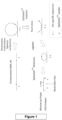
- the chromosome interactions which are typed in the method of the invention are further defined in Table 1. They are defined by means of the probe sequences which detect the ligated product made by an EpiSwitch method (see Figure 1 ). They are also defined by the position numbers of the interaction which are within the probe name and they are also defined by the primer sequences which allow detection of the ligated sequence.
- the chromosome interactions which are typed in the invention are typically interactions between distal regions of a chromosome, said interactions being dynamic and altering, forming or breaking depending upon the state of the region of the chromosome. That state will reflect how the immune system interacts with immunotherapy which is given which responder group the individual falls into.
- the chromosome interaction may, for example, reflect if it is being transcribed or repressed. Chromosome interactions which are specific to responder 'groups' as defined herein have been found to be stable, thus providing a reliable means of measuring the differences between groups (for example reflecting different responses to immunotherapy).
- Chromosome interactions specific to responder groups will normally be present before or in the early stages of a disease process, for example compared to other epigenetic markers such as methylation or changes to binding of histone proteins.
- the process of the invention is able to provide valuable information about the way the immune system will react at an early stage. This allows early intervention (for example treatment) which as a consequence will be more effective and also allows early choices to be made of the type of treatment which is appropriate for the patient, and which treatments should not be used.
- Chromosome interactions also reflect the current state of the individual and therefore can be used to assess changes to disease status. Furthermore there is little variation in the relevant chromosome interactions between individuals within the same group.
- chromosome interactions which are detected in the invention could be impacted by changes to the underlying DNA sequence, by environmental factors, DNA methylation, non-coding antisense RNA transcripts, non-mutagenic carcinogens, histone modifications, chromatin remodelling and specific local DNA interactions.
- chromosome interactions as defined herein are a regulatory modality in their own right and do not have a one to one correspondence with any genetic marker (DNA sequence change) or any other epigenetic marker.
- the changes which lead to the chromosome interactions may be impacted by changes to the underlying nucleic acid sequence which themselves do not directly affect a gene product or the mode of gene expression.
- Such changes may be for example, SNPs within and/or outside of the genes, gene fusions and/or deletions of intergenic DNA, microRNA, and non-coding RNA.
- SNPs SNPs within and/or outside of the genes
- gene fusions and/or deletions of intergenic DNA, microRNA, and non-coding RNA For example, it is known that roughly 20% of SNPs are in non-coding regions, and therefore the process as described is also informative in non-coding situation.
- regions of the chromosome which come together to form the interaction are less than 5 kb, 3 kb, 1 kb, 500 base pairs or 200 base pairs apart on the same chromosome.
- the process of the invention comprises a typing system for detecting chromosome interactions relating to responder status.
- Any suitable typing method can be used, for example a method in which the proximity of the chromosomes in the interaction is detected and/or in which a marker that reflects chromosome interaction status is detected.
- the typing method may be performed using the EpiSwitch TM system mentioned herein, which for example may be carried out by a method comprising the following steps (for example on DNA and/or a sample from the subject):
- Detection of this ligated nucleic acid allows determination of the presence or absence of a particular chromosome interaction.
- the ligated nucleic acid therefore acts as a marker for the presence of the chromosome interaction.
- the ligated nucleic acid is detected by PCR or a probe based method, including a qPCR method.
- the chromosomes can be cross-linked by any suitable means, for example by a cross-linking agent, which is typically a chemical compound.
- the interactions are cross-linked using formaldehyde, but may also be cross-linked by any aldehyde, or D-Biotinoyl-e-aminocaproic acid-N-hydroxysuccinimide ester or Digoxigenin-3-O-methylcarbonyl-e-aminocaproic acid-N-hydroxysuccinimide ester.
- Para-formaldehyde can cross link DNA chains which are 4 Angstroms apart.
- the chromosome interactions are on the same chromosome. Typically the chromosome interactions are 2 to 10 Angstroms apart.
- the cross-linking is preferably in vitro.
- the cleaving is preferably by restriction digestion with an enzyme, such as Taql.
- the ligating may form DNA loops.
- PCR polymerase chain reaction
- the size of the PCR product produced may be indicative of the specific chromosome interaction which is present, and may therefore be used to identify the status of the locus.
- the primers shown in any table herein are used, for example the primer pairs shown in Table 2 or 8 are used (corresponding to the chromosome interaction which is being detected).
- the primers shown in Table 1 may be used. Homologues of such primers or primer pairs may also be used, which can have at least 70% identity to the original sequence.
- probe sequences as shown in any table herein may be used, for example the probe sequences shown in Table 2 or 8 (corresponding to the chromosome interaction which is being detected). Probe sequences as shown in Table 1 may be used. Homologues of such probe sequences may also be used, which can have at least 70% identity to the original sequence.
- Typing according to the process of the invention may be carried out at multiple time points, for example to monitor the progression of the disease. This may be at one or more defined time points, for example at at least 1, 2, 5, 8 or 10 different time points. The durations between at least 1, 2, 5 or 8 of the time points may be at least 5, 10, 20, 50, 80 or 100 days. Typically there are 3 time points at least 50 days apart.
- the individual who is tested in the method of the invention is preferably a eukaryote, animal, bird or mammal. Most preferably the individual is a human. The individual may be male or female. In the case of a human individual they are typically aged 65 or above.
- the invention includes detecting and treating particular groups in a population, typically differing in their responder status, for example their response to immunotherapy.
- the inventors have discovered that chromosome interactions differ between these groups, and identifying these differences will allow physicians to categorize their patients as a part of a particular group of the population.
- the invention therefore provides physicians with a process of personalizing medicine for an individual based on their epigenetic chromosome interactions. Such testing may be used to select how to subsequently treat the patient, for example the type of drug that will be administered.
- the process of the invention may be carried out to select treatment for an individual, for example whether or not to give any specific treatment mentioned herein is administered to the individual.
- the individual that is tested in the process of the invention may have been selected in some way, for example based on a risk factor, symptom or physical characteristic.
- the individual may have been selected based on having a symptom of cancer and/or or being in the early stages of cancer.
- the individual may be susceptible to any cancer mentioned herein and/or may be in need of any therapy mentioned in.
- the individual may be receiving any therapy mentioned herein.
- the individual may have, or be suspected of having, cancer, for example any specific cancer mentioned herein.
- the cancer which is relevant to the invention can include any cancer mentioned herein, and preferably is melanoma, lung cancer, non-small cell lung carcinoma (NSCLC), diffuse large B-cell lymphoma, liver cancer, hepatocellular carcinoma, prostate cancer, breast cancer, leukaemia, acute myeloid leukaemia, pancreatic cancer, thyroid cancer, nasal cancer, brain cancer, bladder cancer, cervical cancer, non-Hodgkin lymphoma, ovarian cancer, colorectal cancer or kidney cancer.
- the cancer may be one which can be treated by immunotherapy, for example any specific immunotherapy mentioned herein.
- the invention relates to determining response to immunotherapy, and in particular whether the individual is responsive to immunotherapy and/or whether they are hyper-progressors in whom immunotherapy will cause acceleration of disease.
- the response which is determined is to a therapy which comprises a molecule or cell that is relevant to the immune system, such as a composition that comprises an antibody or immune cell (for example a T cell or dendritic cell) or any therapeutic substance mentioned herein. It may be response to a substance that modulates or stimulates the immune system, such as a vaccine therapy.
- the immunotherapy may modulate, block or stimulate an immune checkpoint, and thus may target or modulate PD-L1, PD-L2 or CTLA4 or any other immune checkpoint molecule disclosed herein, and thus is preferably immunocheckpoint therapy.
- the response is responsiveness to an antibody therapy, or to any specific therapy disclosed herein.
- the therapy may be a combination therapy, for example any specific combination therapy disclosed herein.
- the response is to a PD-1 inhibitor or PD-L1 inhibitor, including an antibody specific for PD-1 or PD-L1.
- PD-1 is 'programmed cell death protein' and PD-L1 is 'programmed death-ligand 1'.
- 'antibody' includes all fragments and derivatives of an antibody that retain the ability to bind the antigen target, for example single chain scFV's or Fab's.
- the therapy may be mono or combination therapy, for example with immunocheckpoint modulators (preferably inhibitors) for PD-1 and or its ligand, PD-L1.
- the therapy could comprise administering at least one immunocheckpoint modulator, for example as disclosed herein, such as in any table, figure or example.
- the therapy could be a combination of an anti-PD-1 or anti-PD-L1 combined with another drug that targets a checkpoint like CTLA4 (Ipilimumab/Yervoy) or small molecules.
- CTLA4 Ipilimumab/Yervoy
- the PD-1 inhibitors could be pembrolizumab (Keytruda) or nivolumab (Opdivo).
- the modulator of PD-L1 or therapeutic agent could be Atezolizumab (Tecentriq), Avelumab (Bavencio), Durvalumab (Imfinzi), CA-170, Ipilimumab, Tremelimumab, Nivolumab, Pembrolizumab, Pidilizumab, BMS935559, GVAXMPDL3280A, MEDI4736, MSB0010718C, MDX-1105/BMS-936559, AMP-224, MEDI0680.
- the therapy may comprise administering agents that target and/or modulate interferon gamma or the JAK-START pathway.
- the therapeutic agent may be any such agent disclosed in any table herein or may target any 'target' disclosed herein, including any protein disclosed herein. It is understood that any agent that is disclosed in a combination should be seen as also disclosed for administration individually.
- Hyper-progression in cancer can be recognised in a straightforward manner by the skilled person, and it is preferably an increase in disease progression in cancer upon administration of immunotherapy and/or an adverse response to immunotherapy in an individual with cancer. It be measured using any suitable parameter for disease, such as a 2-fold increase in tumour size. It is typically more than a 50% increase in tumour burden within 60 days of administration of immunotherapy. In one aspect hyper-progressors can be defined as having less than 60 days progression-free after administration of immunotherapy and/or overall survival of less than 150 days after immunotherapy.
- a person is found to be responsive to immunotherapy they could be given any immunotherapy mentioned herein.
- a combination therapy such as any combination therapy listed herein.
- a combination therapy comprises an antibody and a small molecule.
- Tables 1, 2 and 8 show specific markers which can be used to detect responder status. Their presence or absence can be used in such a detection (i.e. they are 'disseminating' markers).
- Tables 1 and 8 show markers which detect responsiveness to immunotherapy, and the table shows which are linked to responsiveness and which are linked to non-responsiveness.
- Table 2 shows markers which detect hyper-progressors, and the tables shows which are linked to being a hyper-progressor and which are linked to stable disease.
- the markers are defined using probe sequences (which detect a ligated product as defined herein).
- the first two sets of Start-End positions show probe positions, and the second two sets of Start-End positions show the relevant 4kb region.
- Simple permutation-based estimation is used to determine how likely a given RP value or better is observed in a random experiment. This has the following steps:
- the rank product statistic ranks chromosome interactions according to intensities within each microarray and calculates the product of these ranks across multiple microarrays.
- This technique can identify chromosome interactions that are consistently detected among the most differential chromosome interactions in a number of replicated microarrays. Where the p-value is 0 this indicates that there is very little variation in the Rank Product of the CCS across the samples, this is a good example of the signal to noise and effect size of CCS. Where p value is 0 and pfp is 0 this means that permutated Rank Product doesn't differ from the actual observed Rank Product. These methods are described Breitling R and Herzyk P (2005) Rank-based methods as a non-parametric alternative of the t-test for the analysis of biological microarray data. J Bioinf Comp Biol 3, 1171-1189 .
- FC indicates prevalence of marker in each comparison, 2 means twice over average test, 1.5 means 1.5 over the average test, etc., and so FC indicates the weight of a marker to phenotype/group.
- the FC value can be used to give an indication of how many markers are needed for a highly effective test.
- probes When detection is performed using a probe, typically sequence from both regions of the probe (i.e. from both sites of the chromosome interaction) could be detected.
- probes are used in the process which comprise or consist of the same or complementary sequence to a probe shown in any table. In some aspects probes are used which comprise sequence which is homologous to any of the probe sequences shown in the tables.
- the invention described herein relates to chromosome conformation profile and 3D architecture as a regulatory modality in its own right, closely linked to the phenotype.
- the discovery of biomarkers was based on annotations through pattern recognition and screening on representative cohorts of clinical samples representing the differences in phenotypes. We annotated and screened significant parts of the genome, across coding and non-coding parts and over large sways of non-coding 5' and 3' of known genes for identification of statistically disseminating consistent conditional disseminating chromosome conformations, which for example anchor in the non-coding sites within (intronic) or outside of open reading frames.
- Selection of markers for panels may be done by cross-validation statistical performance (and not for example by the functional relevance of the neighbouring genes, used for the reference name).
- a panel of markers (with names of adjacent genes) is a product of clustered selection from the screening across significant parts of the genome, in non-biased way analysing statistical disseminating powers over 14,000-60,000 annotated EpiSwitch sites across significant parts of the genome. It should not be perceived as a tailored capture of a chromosome conformation on the gene of know functional value for the question of stratification.
- the total number of sites for chromosome interaction are 1.2 million, and so the potential number of combinations is 1.2 million to the power 1.2 million. The approach that we have followed nevertheless allows the identifying of the relevant chromosome interactions.
- each marker can be seen as representing an event of biological epigenetic as part of network deregulation that is manifested in the relevant condition. In practical terms it means that these markers are prevalent across groups of patients when compared to controls. On average, as an example, an individual marker may typically be present in 80% of the relevant responder group and in 10% of controls, and therefore the results of the testing by the method of the invention is straightforward to interpret and essentially amounts to a 'binary readout'.
- GLMNET multivariate biomarker analysis
- the tables herein show the reference names for the array probes (60-mer) for array analysis that overlaps the juncture between the long range interaction sites, the chromosome number and the start and end of two chromosomal fragments that come into juxtaposition.
- all 11 of the markers of Table 1 are typed.
- all 11 of the markers of Table 2 are typed.
- all 8 of the markers of Table 8 are typed.
- the process of the invention will normally be carried out on a sample.
- the sample may be obtained at a defined time point, for example at any time point defined herein.
- the sample will normally contain DNA from the individual. It will normally contain cells.
- a sample is obtained by minimally invasive means, and may for example be a blood sample. DNA may be extracted and cut up with a standard restriction enzyme. This can pre-determine which chromosome conformations are retained and will be detected with the EpiSwitch TM platforms. Due to the synchronisation of chromosome interactions between tissues and blood, including horizontal transfer, a blood sample can be used to detect the chromosome interactions in tissues, such as tissues relevant to disease.
- the sample will contain at least 2 ⁇ 10 5 cells.
- the sample may contain up to 5 ⁇ 10 5 cells. In one aspect, the sample will contain 2 ⁇ 10 5 to 5.5 ⁇ 10 5 cells.
- Crosslinking of epigenetic chromosomal interactions present at the chromosomal locus is described herein. This may be performed before cell lysis takes place. Cell lysis may be performed for 3 to 7 minutes, such as 4 to 6 or about 5 minutes. In some aspects, cell lysis is performed for at least 5 minutes and for less than 10 minutes.
- DNA restriction is performed at about 55°C to about 70°C, such as for about 65°C, for a period of about 10 to 30 minutes, such as about 20 minutes.
- a frequent cutter restriction enzyme is used which results in fragments of ligated DNA with an average fragment size up to 4000 base pair.
- the restriction enzyme results in fragments of ligated DNA have an average fragment size of about 200 to 300 base pairs, such as about 256 base pairs.
- the typical fragment size is from 200 base pairs to 4,000 base pairs, such as 400 to 2,000 or 500 to 1,000 base pairs.
- a DNA precipitation step is not performed between the DNA restriction digest step and the DNA ligation step.
- DNA ligation is described herein. Typically the DNA ligation is performed for 5 to 30 minutes, such as about 10 minutes.
- the protein in the sample may be digested enzymatically, for example using a proteinase, optionally Proteinase K.
- the protein may be enzymatically digested for a period of about 30 minutes to 1 hour, for example for about 45 minutes.
- PCR detection is capable of detecting a single copy of the ligated nucleic acid, preferably with a binary read-out for presence/absence of the ligated nucleic acid.
- Figure 1 shows a preferred process of detecting chromosome interactions.
- the process of the invention can be described in different ways. It can be described as a process of making one or more ligated nucleic acids comprising (i) in vitro cross-linking of chromosome regions which have come together in a chromosome interaction; (ii) subjecting said cross-linked DNA to cutting or restriction digestion cleavage; and (iii) ligating said cross-linked cleaved DNA ends to form one or more ligated nucleic acids, wherein optionally detection of the ligated nucleic acid may be used to determine the chromosome state at a locus, and wherein preferably the chromosomal interactions may be 1, 3, 5, 8 or all the chromosome interactions of Table 1 or 2. In this process the chromosomal interactions may be 1, 3, 5 or 8 of the chromosome interactions of Table 8.
- homologues of polynucleotide / nucleic acid (e.g. DNA) sequences are referred to herein.
- Such homologues typically have at least 70% homology, preferably at least 80%, at least 85%, at least 90%, at least 95%, at least 97%, at least 98% or at least 99% homology, for example over a region of at least 10, 15, 20, 30, 100 or more contiguous nucleotides, or across the portion of the nucleic acid which is from the region of the chromosome involved in the chromosome interaction.
- the homology may be calculated on the basis of nucleotide identity (sometimes referred to as "hard homology").
- homologues of polynucleotide / nucleic acid (e.g. DNA) sequences are referred to herein by reference to percentage sequence identity.
- such homologues have at least 70% sequence identity, preferably at least 80%, at least 85%, at least 90%, at least 95%, at least 97%, at least 98% or at least 99% sequence identity, for example over a region of at least 10, 15, 20, 30, 100 or more contiguous nucleotides, or across the portion of the nucleic acid which is from the region of the chromosome involved in the chromosome interaction.
- the homologues may have at least 70% sequence identity, preferably at least 80%, at least 85%, at least 90%, at least 95%, at least 97%, at least 98% or at least 99% sequence identity across the entire probe, primer or primer pair.
- the UWGCG Package provides the BESTFIT program which can be used to calculate homology and/or % sequence identity (for example used on its default settings) ( Devereux et al (1984) Nucleic Acids Research 12, p387-395 ).
- the PILEUP and BLAST algorithms can be used to calculate homology and/or % sequence identity and/or line up sequences (such as identifying equivalent or corresponding sequences (typically on their default settings)), for example as described in Altschul S. F. (1993) J Mol Evol 36:290-300 ; Altschul, S, F et al (1990) J Mol Biol 215:403-10 .
- HSPs high scoring sequence pair
- Extensions for the word hits in each direction are halted when: the cumulative alignment score falls off by the quantity X from its maximum achieved value; the cumulative score goes to zero or below, due to the accumulation of one or more negative-scoring residue alignments; or the end of either sequence is reached.
- the BLAST algorithm parameters W5 T and X determine the sensitivity and speed of the alignment.
- the BLAST algorithm performs a statistical analysis of the similarity between two sequences; see e.g., Karlin and Altschul (1993) Proc. Natl. Acad. Sci. USA 90: 5873-5787 .
- One measure of similarity provided by the BLAST algorithm is the smallest sum probability (P(N)), which provides an indication of the probability by which a match between two polynucleotide sequences would occur by chance.
- P(N) the smallest sum probability
- a sequence is considered similar to another sequence if the smallest sum probability in comparison of the first sequence to the second sequence is less than about 1, preferably less than about 0.1, more preferably less than about 0.01, and most preferably less than about 0.001.
- the homologous sequence typically differs by 1, 2, 3, 4 or more bases, such as less than 10, 15 or 20 bases (which may be substitutions, deletions or insertions of nucleotides). These changes may be measured across any of the regions mentioned above in relation to calculating homology and/or % percentage sequence identity.
- Homology of a 'pair of primers' can be calculated, for example, by considering the two sequences as a single sequence (as if the two sequences are joined together) for the purpose of then comparing against the another primer pair which again is considered as a single sequence.
- markers which are disclosed herein have been found to be 'disseminating markers' capable of determining responder status and tables 1 and 2 show which responder group each marker is present in (responder/non-responder to immunotherapy, or hyper-progressor/stable disease).
- markers are prevalent across the relevant responder group when compared to controls (as is shown by the FC value, for example).
- an individual marker may typically be present in 80% of the relevant responder group and in 10% of controls.
- the result will be a combination of 'present' and 'absent' chromosome interactions for each of the markers shown in Table 1 or 2 allowing determination of the responder group for the individual.
- presence/absence of at least 8 markers out of 11 compared to the 'ideal' result shown in the table can be used to assign the individual to a responder group.
- This section is relevant both to immunotherapies which define the responder group of the individual and also to therapy which may be given to individuals based on the results of the testing method of the invention.
- therapeutic agents for use in preventing or treating any condition mentioned herein. This may comprise administering to an individual in need a therapeutically effective amount of the agent.
- agent in the manufacture of a medicament to prevent or treat the condition, for example in individuals tested by the method of the invention.
- the formulation of the agent will depend upon the nature of the agent.
- the agent will be provided in the form of a pharmaceutical composition containing the agent and a pharmaceutically acceptable carrier or diluent. Suitable carriers and diluents include isotonic saline solutions, for example phosphate-buffered saline. Typical oral dosage compositions include tablets, capsules, liquid solutions and liquid suspensions.
- the agent may be formulated for parenteral, intravenous, intramuscular, subcutaneous, transdermal or oral administration.
- the dose of an agent may be determined according to various parameters, especially according to the substance used; the age, weight and condition of the individual to be treated; the route of administration; and the required regimen. A physician will be able to determine the required route of administration and dosage for any particular agent.
- a suitable dose may however be from 0.1 to 100 mg/kg body weight such as 1 to 40 mg/kg body weight, for example, to be taken from 1 to 3 times daily.
- an immunotherapeutic agent preferably selected from any of tables 4 to 6, for use in a method of treating an individual identified as being responsive to immunotherapy, optionally said method comprising:
- a screening method to identify therapeutic agents for cancer comprising determining whether a candidate agent is able to cause a change to all of the chromosome interactions shown in Table 1 and/or Table 2.
- This screening method may comprise determining whether a candidate agent is able to cause a change to all of the chromosome interactions shown in Table 8.
- nucleic acids including probes and primers.
- nucleic acids are DNA. It is understood that where a specific sequence is provided the invention may use the complementary sequence as required in the particular aspect.
- the primers or probes shown in Table 1 or 2 may be used in the invention. In one aspect probes or primers are used which comprise any of: the sequences shown in Table 1 or 2; or fragments and/or homologues of any sequence shown in Table 1 or 2.
- the primers or probes shown in Table 8 may be used in the invention. In one aspect probes or primers are used which comprise any of: the sequences shown in Table 8; or fragments and/or homologues of any sequence shown in Table 8.
- nucleic acids mentioned herein may be labelled, preferably using an independent label such as a fluorophore (fluorescent molecule) or radioactive label which assists detection of successful hybridisation. Certain labels can be detected under UV light.
- an independent label such as a fluorophore (fluorescent molecule) or radioactive label which assists detection of successful hybridisation.
- Certain labels can be detected under UV light.
- nucleic acids or therapeutic agents may be in purified or isolated form. They may be in a form which is different from that found in nature, for example they may be present in combination with other substance with which they do not occur in nature.
- the nucleic acids (including portions of sequences defined herein) may have sequences which are different to those found in nature, for example having at least 1, 2, 3, 4 or more nucleotide changes in the sequence as described in the section on homology.
- the nucleic acids may have heterologous sequence at the 5' or 3' end.
- the nucleic acids may be chemically different from those found in nature, for example they may be modified in some way, but preferably are still capable of Watson-Crick base pairing.
- nucleic acids will be provided in double stranded or single stranded form.
- the invention provides all of the specific nucleic acid sequences mentioned herein in single or double stranded form, and thus includes the complementary strand to any sequence which is disclosed.
- kits for carrying out any process of the invention including detection of a chromosomal interaction relating to prognosis.
- a kit can include a specific binding agent capable of detecting the relevant chromosomal interaction, such as agents capable of detecting a ligated nucleic acid generated by processes of the invention.
- Preferred agents present in the kit include probes capable of hybridising to the ligated nucleic acid or primer pairs, for example as described herein, capable of amplifying the ligated nucleic acid in a PCR reaction.
- Preferred agents include any of the specific primers and probes disclosed herein and/or homologues of such primers and probes.
- the device preferably comprises any specific binding agents, probe or primer pair capable of detecting the chromosome interaction, such as any such agent, probe or primer pair described herein.
- quantitative detection of the ligated sequence which is relevant to a chromosome interaction is carried out using a probe which is detectable upon activation during a PCR reaction, wherein said ligated sequence comprises sequences from two chromosome regions that come together in an epigenetic chromosome interaction, wherein said process comprises contacting the ligated sequence with the probe during a PCR reaction, and detecting the extent of activation of the probe, and wherein said probe binds the ligation site.
- the process typically allows particular interactions to be detected in a MIQE compliant manner using a dual labelled fluorescent hydrolysis probe.
- the probe is generally labelled with a detectable label which has an inactive and active state, so that it is only detected when activated.
- the extent of activation will be related to the extent of template (ligation product) present in the PCR reaction. Detection may be carried out during all or some of the PCR, for example for at least 50% or 80% of the cycles of the PCR.
- the probe can comprise a fluorophore covalently attached to one end of the oligonucleotide, and a quencher attached to the other end of the nucleotide, so that the fluorescence of the fluorophore is quenched by the quencher.
- the fluorophore is attached to the 5'end of the oligonucleotide, and the quencher is covalently attached to the 3' end of the oligonucleotide.
- Fluorophores that can be used in the process of the invention include FAM, TET, JOE, Yakima Yellow, HEX, Cyanine3, ATTO 550, TAMRA, ROX, Texas Red, Cyanine 3.5, LC610, LC 640, ATTO 647N, Cyanine 5, Cyanine 5.5 and ATTO 680.
- Quenchers that can be used with the appropriate fluorophore include TAM, BHQ1, DAB, Eclip, BHQ2 and BBQ650, optionally wherein said fluorophore is selected from HEX, Texas Red and FAM.
- Preferred combinations of fluorophore and quencher include FAM with BHQ1 and Texas Red with BHQ2.
- Hydrolysis probes of the invention are typically temperature gradient optimised with concentration matched negative controls. Preferably single-step PCR reactions are optimized. More preferably a standard curve is calculated.
- An advantage of using a specific probe that binds across the junction of the ligated sequence is that specificity for the ligated sequence can be achieved without using a nested PCR approach.
- the processes described herein allow accurate and precise quantification of low copy number targets.
- the target ligated sequence can be purified, for example gel-purified, prior to temperature gradient optimization.
- the target ligated sequence can be sequenced.
- PCR reactions are performed using about 10ng, or 5 to 15 ng, or 10 to 20ng, or 10 to 50ng, or 10 to 200ng template DNA.
- Forward and reverse primers are designed such that one primer binds to the sequence of one of the chromosome regions represented in the ligated DNA sequence, and the other primer binds to other chromosome region represented in the ligated DNA sequence, for example, by being complementary to the sequence.
- the invention includes selecting primers and a probe for use in a PCR process as defined herein comprising selecting primers based on their ability to bind and amplify the ligated sequence and selecting the probe sequence based properties of the target sequence to which it will bind, in particular the curvature of the target sequence.
- Probes are typically designed/chosen to bind to ligated sequences which are juxtaposed restriction fragments spanning the restriction site.
- the predicted curvature of possible ligated sequences relevant to a particular chromosome interaction is calculated, for example using a specific algorithm referenced herein.
- the curvature can be expressed as degrees per helical turn, e.g. 10.5° per helical turn.
- Ligated sequences are selected for targeting where the ligated sequence has a curvature propensity peak score of at least 5° per helical turn, typically at least 10°, 15° or 20° per helical turn, for example 5° to 20° per helical turn.
- the curvature propensity score per helical turn is calculated for at least 20, 50, 100, 200 or 400 bases, such as for 20 to 400 bases upstream and/or downstream of the ligation site.
- the target sequence in the ligated product has any of these levels of curvature.
- Target sequences can also be chosen based on lowest thermodynamic structure free energy.
- certain chromosome interactions are not typed, for example any specific interaction mentioned not mentioned herein.
- the markers of Table 1 or Table 2 are typed and no other markers are typed.
- the markers of Table 2 or Table 8 are typed and no other markers are typed.
- the markers of Table 1 and Table 2 are typed and no other markers are typed.
- the markers of Table 2 and Table 8 are typed and no other markers are typed.
- the EpiSwitch TM platform technology detects epigenetic regulatory signatures of regulatory changes between normal and abnormal conditions at loci.
- the EpiSwitch TM platform identifies and monitors the fundamental epigenetic level of gene regulation associated with regulatory high order structures of human chromosomes also known as chromosome conformation signatures.
- Chromosome signatures are a distinct primary step in a cascade of gene deregulation. They are high order biomarkers with a unique set of advantages against biomarker platforms that utilize late epigenetic and gene expression biomarkers, such as DNA methylation and RNA profiling.
- the custom EpiSwitch TM array-screening platforms come in 4 densities of, 15K, 45K, 100K, and 250K unique chromosome conformations, each chimeric fragment is repeated on the arrays 4 times, making the effective densities 60K, 180K, 400K and 1 million respectively.
- the 15K EpiSwitch TM array can screen the whole genome including around 300 loci interrogated with the EpiSwitch TM Biomarker discovery technology.
- the EpiSwitch TM array is built on the Agilent SurePrint G3 Custom CGH microarray platform; this technology offers 4 densities, 60K, 180K, 400K and 1 million probes.
- the density per array is reduced to 15K, 45K, 100K and 250K as each EpiSwitch TM probe is presented as a quadruplicate, thus allowing for statistical evaluation of the reproducibility.
- the average number of potential EpiSwitch TM markers interrogated per genetic loci is 50, as such the numbers of loci that can be investigated are 300, 900, 2000, and 5000.
- the EpiSwitch TM array is a dual colour system with one set of samples, after EpiSwitch TM library generation, labelled in Cy5 and the other of sample (controls) to be compared/ analyzed labelled in Cy3.
- the arrays are scanned using the Agilent SureScan Scanner and the resultant features extracted using the Agilent Feature Extraction software.
- the data is then processed using the EpiSwitch TM array processing scripts in R.
- the arrays are processed using standard dual colour packages in Bioconductor in R: Limma*.
- the normalisation of the arrays is done using the normalisedWithinArrays function in Limma* and this is done to the on chip Agilent positive controls and EpiSwitch TM positive controls.
- the data is filtered based on the Agilent Flag calls, the Agilent control probes are removed and the technical replicate probes are averaged, in order for them to be analysed using Limma*.
- the pool of probes is initially selected based on adjusted p-value, FC and CV ⁇ 30% (arbitrary cut off point) parameters for final picking. Further analyses and the final list are drawn based only on the first two parameters (adj. p-value; FC).
- EpiSwitch TM screening arrays are processed using the EpiSwitch TM Analytical Package in R in order to select high value EpiSwitch TM markers for translation on to the EpiSwitch TM PCR platform.
- FDR Fealse Discovery Rate
- the top 40 markers from the statistical lists are selected based on their ER for selection as markers for PCR translation.
- the top 20 markers with the highest negative ER load and the top 20 markers with the highest positive ER load form the list.
- the resultant markers from step 1 the statistically significant probes form the bases of enrichment analysis using hypergeometric enrichment (HE).
- HE hypergeometric enrichment
- the statistical probes are processed by HE to determine which genetic locations have an enrichment of statistically significant probes, indicating which genetic locations are hubs of epigenetic difference.
- the most significant enriched loci based on a corrected p-value are selected for probe list generation. Genetic locations below p-value of 0.3 or 0.2 are selected. The statistical probes mapping to these genetic locations, with the markers from step 2, form the high value markers for EpiSwitch TM PCR translation.
- EpiSwitch TM biomarker signatures demonstrate high robustness, sensitivity and specificity in the stratification of complex disease phenotypes. This technology takes advantage of the latest breakthroughs in the science of epigenetics, monitoring and evaluation of chromosome conformation signatures as a highly informative class of epigenetic biomarkers.
- Current research methods deployed in academic environment require from 3 to 7 days for biochemical processing of cellular material in order to detect CCSs. Those procedures have limited sensitivity, and reproducibility; and furthermore, do not have the benefit of the targeted insight provided by the EpiSwitch TM Analytical Package at the design stage.
- EpiSwitch TM Array CCS sites across the genome are directly evaluated by the EpiSwitch TM Array on clinical samples from testing cohorts for identification of all relevant stratifying lead biomarkers.
- the EpiSwitch TM Array platform is used for marker identification due to its high-throughput capacity, and its ability to screen large numbers of loci rapidly.
- the array used was the Agilent custom-CGH array, which allows markers identified through the in silico software to be interrogated.
- EpiSwitch TM Array Potential markers identified by EpiSwitch TM Array are then validated either by EpiSwitch TM PCR or DNA sequencers (i.e. Roche 454, Nanopore MiniON, etc.). The top PCR markers which are statistically significant and display the best reproducibility are selected for further reduction into the final EpiSwitch TM Signature Set, and validated on an independent cohort of samples.
- EpiSwitch TM PCR can be performed by a trained technician following a standardised operating procedure protocol established. All protocols and manufacture of reagents are performed under ISO 13485 and 9001 accreditation to ensure the quality of the work and the ability to transfer the protocols.
- EpiSwitch TM PCR and EpiSwitch TM Array biomarker platforms are compatible with analysis of both whole blood and cell lines. The tests are sensitive enough to detect abnormalities in very low copy numbers using small volumes of blood.
- the method of the invention may include analysis of the chromosome interactions identified in the individual, for example using a classifier, which may increase performance, such as sensitivity or specificity.
- the classifier is typically one that has been 'trained' on samples from the population and such training may assist the classifier to detect any responder group mentioned herein.
- the invention is illustrated by the following:
- one is a universal marker set that allows responsiveness to therapy to be detected across a range of cancers and specific therapies, and the second is a marker set that detects hyper-progressors who should never be treated with particular types of immunotherapy.
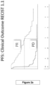
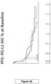
- Figure 1 shows performance of baseline prediction of response /non-response for avelumab (PD-L1) in NSCLC for based on a large set of markers tested.
- - PD-1 and PD-L1 assets such as pembrolizumab, durvalumab, avelumab, atezolizumab, in melanoma, NSCLC, Lung, HCC, Bladder, Prostate, NPC, Parotid Gland, Alveolar Soft Part Sarcoma.
- the universal 11 marker classifier works well across all those cohorts and identifies universal profile that delivers robust baseline classification for response/non-response, irrespective of which exactly PD-1 or PD-L1 treatment and what exactly type of cancer was tested.
- We capture with 11 markers a very specific conducive/non-conducive epigenetic systemic network set up of features which define outcomes in immune-checkpoint therapies.
- a very serious issue in cancer immunotherapy is the consistent presence of a subgroup of patients who should never be treated with PD-1/PD-L1 therapies. They are called hyper-progressors (or super-progressors), where progressor means progression into disease. These patients upon treatment react very differently - their rate of tumour growth shoots up and they die very quickly, essentially in a matter of weeks.
- Hyper-progressors can be defined as patients who respond adversely to immuno-checkpoint immuno-oncology treatment by demonstrating significant reduction in either progression-free survival (as a measure of survival in response to drug treatment) (PFS ⁇ 60 days) or overall survival (OS ⁇ 150 days).
- markers are specifically selected to identify and exclude patients prior to treatment on the basis of a predicted severe reduction in their survival as a consequence of immunotherapy. This could be seen as a subgroup of the bigger cohort of non-responders that could be identified and predicted by the universal 11 marker set.
- melanoma Whilst the present work has been carried out on patients with melanoma, lung cancer, hepatocellular carcinoma (liver cancer), bladder , prostate, nasal cancer, parotid gland (salivary gland cancer), alveolar soft part sarcoma (soft tissue cancer), it also applies to other cancers where immune-checkpoint inhibitors PD-1/PD-L1 are used for therapy, such as breast cancer, cervical cancer, colon cancer, head and neck cancer, Hodgkin lymphoma, kidney cancer, stomach cancer, rectal cancer, and any solid tumour.
- the marker sets that have been identified capture a network of deregulations at the level of cellular 3D genomics, which reflects a network of deregulated cell types acting in conjunction to sustain and advance the pathological or physiological phenotype of cancer. So far, observing statistically significant chromosome conformations as evidence of deregulation in conjunction with cell sub-typing CD loci, we can state that observed universal signatures contain and represent deregulations in T cells, NK (natural killer) cells, macrophages, B cells and dendritic cells (DC).
- T cells NK (natural killer) cells
- macrophages macrophages
- B cells dendritic cells
- the marker sets were developed using the following patients:
- a blind cohort was collected to test the feature of Non-Response. This cohort was collected in Malaysia and consisted of 21 patients who all provided blood samples at baseline prior to immunotherapy. All patients had previous lines of therapy. 3 check-points were used: Atezolizumab (anti-PD-L1), Durvalumab (anti-PD-L1) and Pembrolizumab (anti-PD-1). There were 7 disease indications: Lung, HCC, Bladder, Prostate, NPC, Parotid Gland and Alveolar soft part sarcoma. 11 of the patients had multiple collections: 2-4. 3 patients had up to 4 collections. The ethnicity of the Patients was either Han Chinese or Indonesian.
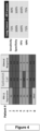
- Figure 4 shows the high concordance between baseline EpiSwitch calls, PD-L1 expression and observed clinical response.
- Figure 5 shows data for a training set for an 11 marker model based on 80 NSCLC patients who are a mixture of 1L, 2L Avelumab (54) and 2L Pembrolizumab (36).
- Figure 6 shows data for a test set of the 11 marker model based on 38 NSCLC patients who are a mixture of 1L, 2L Avelumab (27) and 2L Pembrolizumab (11).
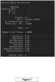
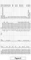
- Figures 7 and 8 show data for a second test set for the Malaysian Observational Study looking a mixture of CPI and tumours.
- Figure 9 shows the calls for the patients who had multiple collections. The calls over the time points are largely consistent and concordant: Patient 12 on Durvalumab shows the profile of Responder over 4 collections, and a score over the second collection entering into grey zone R/NR. Overall profile of probabilities actually get stronger over time for Responder.
- Patient 1 on Atezolizumab shows an interesting initial non-response, but becomes a late responder.
- Patient 17 on Pembrolizumab is an NPC patient, and shows Response profile over both samplings.
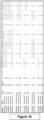
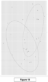
- FIG 10 shows EpiSwitch calls for patients sampled over multiple time points, with OBD.261.263 and OBD.301.303 representing universal response markers and OBD.029.031, OBD.045.047, OBD.645.647, OBD.753.755 and OBD.8.69.871 representing universal non-response markers.
- Figure 13 shows 11 EpiSwitch CCSs selected from a total of 60, markers selected based on binary difference, OS and PFS rank log analysis. The associated genetic locations are also shown. Figures 10 and 11 show a pathway analysis of the genetic locations.
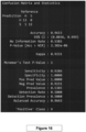
- Figure 16 shows data for a training set for hyper-progressors: an XGBoost 11 marker model.
- Figure 17 shows data for a test test for hyper-progressors with 6 samples excluded from marker selection.
- Figure 18 shows a logistic principal component analysis (PCA) of a training set.
- Figure 19 shows a logistic PCA of the training set with predicted test samples. This is a second classification approach and is in concordance with XGBoost.
- Figure 20 shows the logistic PCA of the training set with PFS as label.
- Figure 21 shows the logistic PCA of the training set with OS as label.
- the EpiSwitch TM nested PCR platform data output was analysed with multiple statistical techniques, including, but not limited to, established univariate (Fishers Exact test) and multivariant (permutated GLMNET, Random Forest with SHapley Additive exPlanations values (SHAP)), procedures.
- XGBoost A gradient boosted decision tree algorithm. An ensemble of weak decision tree models is generated and combined to produce one strong classification model (level wise tree growth);
- PCA Logistic Principal Component Analysis
- GLMNET Generalized linear model fitted via penalized maximum likelihood technique (iv)
- LightGBM gradient boosting framework that uses tree based learning algorithms (vertical leaf tree growth).
- SHAP SHapley Additive exPlanations
- the first one is global interpretability - the collective SHAP values can show how much each predictor contributes, either positively or negatively, to the target variable. This is like the variable importance plot but it is able to show the positive or negative relationship for each variable with the target.
- the second benefit is local interpretability - each observation gets its own set of SHAP values This greatly increases its transparency. We can explain why a case receives its prediction and the contributions of the predictors. Traditional variable importance algorithms only show the results across the entire population but not on each individual case. The local interpretability enables us to pinpoint and contrast the impacts of the factors.
- the SHAP values can be calculated for any tree-based model (our model is an XGboost, boosted tree based model), while other methods use linear regression or logistic regression models as the surrogate models.
- Immune checkpoint inhibitors are a class of drugs targeting a narrow set of proteins in a specific regulatory network present in immune cells, like T cells, and some cancer cells of the patients.
- the checkpoint protein targets and the network they control help keep immune responses from being too strong and provide additional protection from the autoimmune conditions, but in the case of cancer can keep T cells from killing cancer cells.
- Use of immune checkpoint inhibitors helps to reactivate the immune response in cancer patients, with efficacious outcome and improved survival in patients.
- Immune checkpoint inhibitors act by resetting and activating immune response by targeting either: 1) PD-1 (Nivolumab, Pembrolizumab, Cemiplimab, Camrelizumab, Tislelizumab, Sasanlimab); or 2) PD-1 ligand, called PD-L1 (Avelumab, Atezolizumab and Durvalumab).
- PD-1 Nivolumab, Pembrolizumab, Cemiplimab, Camrelizumab, Tislelizumab, Sasanlimab
- PD-L1 PD-1 ligand
- the present work has asked several questions, including if, taking into account the role played by immune system of the patient in successful response to immune checkpoint inhibitors and the limited number of targets for the therapy, particularly PD-1 receptor and its ligand PD-L1, one can discover and validate EpiSwitch biomarkers in a qPCR format of detection for baseline patients that would universally predict response/non-response to treatment in advance, irrespective of the type of the checkpoint inhibitor used and across the spectrum oncological conditions.
- MIQE compliant qPCR format is the standard for clinical PCR based tests. This format is very different from nested PCR or array format, due to its limitations on primer and probes sequence designs and continuous range of detection, traditionally measured though Cq cycle numbers.
- Stage 1 From previous markers identified by arrays, the top 24 markers were identified that satisfied initial theoretical restrictions and requirements for sequence designs on qPCR primers and probes, i.e. unique sequences for detection, correct annealing temperature of annealing for primers and probes, overlapping 3C juncture, for the detection of the chromosome conformations.
- the designs for 20 markers passed quality control and satisfactory optimization on temperature gradient.
- qPCR formatted marker leads were used to identify if there is a minimal number of biomarkers which as a signature will have strong stratification power for predicting response and non-response across an extensive cohort for patients treated with immune checkpoint inhibitors - Pembrolizumab, Avelumab, Atezolizumab, Durvalumab against each of three targets (PD-1, PD-L1) from a broad selection of oncological conditions: melanoma, non-small cell lung cancer, urethral, HCC, Bladder, Prostate, NPC, Parotid Gland, Alveolar Soft Part Sarcoma, Nasopharyngeal carcinoma, vulval carcinoma, colon cancer, Breast Cancer, Bone Cancer, Brain Cancer, sagittal sinus Carcinoma, lymphoma, larynx cancer, Cervix Cancer, Oral Cavity Carcinoma.
- Stage 3 In screen 1 all 20 markers were first evaluated on pooled DNA templates from 3 patients clinical outcome categories: responders, non-responders and stable diseases.
- the sample cohorts represented patients with various type of cancers (see annotations attached) who received various immuno-checkpoints inhibitors as monotherapies: Avelumab, Pembrolizumab, Atezolizumab and Durvalumab.
- Stage 4 In screen 2, the top 13 markers, shortlisted from screen 1 were evaluated on individual samples from the same patients used in screen 1: 24 patients/samples in total (see Table 9, patients shown with an asterisk).
- the selection of best markers at stage 2 and 3 was carried out using a linear model.
- the linear model is fitted to the Cq values for each marker, comparing PR v PD, PR v SD and PD v SD.
- the coefficients of the fitted models describe the differences between the CCSs in each of the comparisons.
- the linear models are then used to compute moderated t-statistics log-odds of differential CCSs by empirical Bayes moderation of the standard errors towards a global value (0 log or 1 linear).
- the markers are then ranked by the adjusted p value and their CCSs abundance difference between the groups. Markers between PR v PD are given more weight.
- Stage 5 In screen 3 the top 8 markers (shortlisted from screen 2) were validated on IO samples consisting of patients with the following clinical outcomes: 55% NR (non-responders), 24% SD (stable disease, according to the regulatory ruling and clinical practice, SD should be subject to IO therapy just as the group of potential responders), 20% R (responders, also referred as PR for patrial responders) and 1% CR (complete responders).
- NR non-responders
- SD stable disease, according to the regulatory ruling and clinical practice, SD should be subject to IO therapy just as the group of potential responders
- 20% R responders, also referred as PR for patrial responders
- 1% CR complete responders.
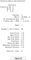
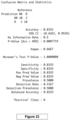
- the efficacy of stratification by the model based on 8 markers is shown in Figures 22 to 24 .
- Predictive Value (PPV) 100xTP/(TP+FP).
- Negative Predictive Value (NPV) 100xTN/(FN+TN).
- the immunotherapy checkpoint classifier is built using CatBoost.
- Catboost is a member of the Gradient Boosted Decision Trees (GBDT's) machine learning ensemble techniques (see Hancock and Khoshgoftaar; J. Big Data (2020) 7:94 ).
- ICI Immune Checkpoint Inhibitor
- Stratification based on 8 marker classifier places patients into groups of likely responders or non-responders to ICI monotherapy, prior to the application of the therapy.
- the classification is applied across all ICI monotherapies against PD-L1 and its ligand PD-L1, in the context of all oncological indications used in ICI monotherapy treatments.
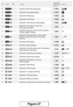
- Figures 25 to 27 describe characteristics of the identified chromosome interactions markers.
- Figure 25 shows the importance of markers in terms of their power in the model.
- Figure 26 shows the genetic locations.
- Figure 27 shows the pathways the genes are associated with. These are pathways implicated in checkpoint response. This indicates the model predictive, and it is noteworthy the markers within the model have biological relevance. In fact, one of the markers is located between PD-L1 and PD-L2 (q057_q059).
- Combinations in cancer immunotherapy biologicals, immunocytokines (L19-IL2 and L19-TNF), cytotoxics (Paxlitaxel) Drug targets Preferred Disease Ipilimumab &Nivolumab PD-1 and CTLA-4 metastatic melanoma Paclitaxel, ipilimumab & carboplatin CTLA4 non-small-cell lung cancer Ipilimumab & GVAX CTLA-4 pancreatic cancer Pidilizumab & rituximab PD-1 hematologic malignancies L19-IL2 & L19-TNF STAT Melanoma MEDI0680 & Durvalumab PD1/PDL1 Advanced solid malignancies Table 5.
- CD52 Ofatumumab (Second generation human IgG1 antibody).
- CD20 Pegylated liposomal doxorubicin (PLD) plus motolimod (VTX2337).
- Sipuleucel-T Approved Cancer Vaccine.
- Rituximab (monoclonal antibody).
- CD20 Interferon gamma Combinatorial ablation and immunotherapy.
- Polysaccharide-K Adoptive cell therapy Anti-CD47 antibodies.
- CD47 Polypurine reverse Hoogsteen oligonucleotides (PPRHs).
- GD2 BGB-A317 (monoclonal antibody).
- PD-1 inhibitor Affimer biotherapeutic.
- PD-L1 inhibitor Polysaccharides Neoantigens Table 7a Probe RP/Rsum FC 1 PDCD1LG2_S_5495992_5498009_5563479_5572586_RR 6499 -1.13 2 MYC_8_127691489 127694045_127738939_127740424_FR 6220 1.15 3 IKBKB_8_42264241_42271203_42331044_42332799_FR 1585 1.3 4 ORF712_9_120888366_120893320_120913546_120919710_RR 8471 1.11 5 ITK_5_157178319_157181048_157266725_157271762_RR 4605 1.15 6 IL17D_13_20664875_20671757_20688261_20691044_FF 6848 -1.1 7 IKBKB_8_42264241_42271203_42290979_42292124_
Landscapes
- Chemical & Material Sciences (AREA)
- Life Sciences & Earth Sciences (AREA)
- Health & Medical Sciences (AREA)
- Organic Chemistry (AREA)
- Proteomics, Peptides & Aminoacids (AREA)
- Engineering & Computer Science (AREA)
- Zoology (AREA)
- Wood Science & Technology (AREA)
- Analytical Chemistry (AREA)
- Immunology (AREA)
- Genetics & Genomics (AREA)
- Pathology (AREA)
- Microbiology (AREA)
- General Engineering & Computer Science (AREA)
- Biophysics (AREA)
- Molecular Biology (AREA)
- Physics & Mathematics (AREA)
- Biotechnology (AREA)
- General Health & Medical Sciences (AREA)
- Biochemistry (AREA)
- Bioinformatics & Cheminformatics (AREA)
- Oncology (AREA)
- Hospice & Palliative Care (AREA)
- Chemical Kinetics & Catalysis (AREA)
- Measuring Or Testing Involving Enzymes Or Micro-Organisms (AREA)
- Investigating Or Analysing Biological Materials (AREA)
- Saccharide Compounds (AREA)
- Medicines Containing Antibodies Or Antigens For Use As Internal Diagnostic Agents (AREA)
- Medicines Containing Material From Animals Or Micro-Organisms (AREA)
Claims (15)
- Procédé de détermination de la façon dont un individu répond à une immunothérapie contre le cancer, comprenant la détection de la présence ou de l'absence chez l'individu de :- toutes les interactions chromosomiques représentées par les séquences de sondes indiquées dans le tableau 8c afin de déterminer si l'individu sera sensible à l'immunothérapie ; et/ou- toutes les interactions chromosomiques représentées par les séquences de sondes indiquées dans le tableau 2.a3 pour déterminer ainsi si l'individu est un hyperprogresseur chez qui l'immunothérapie accélérera la maladie.
- Procédé selon la revendication 1, comprenant en outre la détection de la présence ou de l'absence chez l'individu de toutes les interactions chromosomiques représentées par les séquences de sondes indiquées dans le tableau 1.a3 pour déterminer ainsi si l'individu sera sensible à l'immunothérapie.
- Procédé selon la revendication 1 ou 2, dans lequel la présence ou l'absence des interactions chromosomiques est déterminée :- dans un échantillon en provenance d'un individu, et/ou- dans l'ADN en provenance d'un individu, et/ou- par la détection de la présence ou de l'absence d'une boucle d'ADN au niveau du site des interactions chromosomiques, et/ou- la détection de la présence ou de l'absence de régions distales d'un chromosome réunies dans une conformation chromosomique, et/ou- par la détection de la présence d'un acide nucléique ligaturé qui est généré pendant ledit typage et dont la séquence comprend deux régions correspondant chacune aux régions du chromosome qui se rejoignent dans l'interaction chromosomique, et/ou- par un processus qui détecte la proximité des régions chromosomiques qui se sont réunies dans l'interaction chromosomique.
- Procédé selon l'une quelconque des revendications précédentes, dans lequel ladite détection de la présence ou de l'absence des interactions chromosomiques s'effectue par un processus comprenant :(i) la réticulation in vitro des interactions chromosomiques épigénétiques présentes ;(ii) éventuellement, l'isolation de l'ADN réticulé ;(iii) la soumission dudit ADN réticulé à un clivage ;(iv) la ligature desdites extrémités d'ADN clivées réticulées pour former de l'ADN ligaturé ; et(v) l'identification de la présence ou de l'absence dans ledit ADN ligaturé d'une séquence d'ADN qui correspond à chaque interaction chromosomique ;la détermination de la présence ou de l'absence de chaque interaction chromosomique.
- Procédé selon la revendication 3 ou 4, dans lequel ledit ADN ligaturé est détecté par PCR ou à l'aide d'une sonde.
- Procédé selon la revendication 5 dans lequel :(i) la détection s'effectue au moyen d'une sonde, dans lequel ladite sonde présente de préférence au moins 70 % d'identité avec l'une quelconque des sondes indiquées dans le tableau 1.a3, 2.a3, ou 8c ; ou(ii) la détection s'effectue à l'aide de la PCR, dans lequel la PCR utilise de préférence une paire d'amorces qui présentent au moins 70 % d'identité avec l'une des paires d'amorces indiquées dans :- les tableaux 1a6 et 1a7,- le tableau 2.a6, ou- les tableaux 8b et 8c.
- Procédé selon l'une quelconque des revendications précédentes, dans lequel(i) le procédé est mis en œuvre avant que l'individu reçoive une immunothérapie et/ou est mis en œuvre pour sélectionner quelle thérapie l'individu doit recevoir contre le cancer, et/ou(ii) le procédé est mis en œuvre sur un individu atteint d'un cancer ou suspecté d'être atteint d'un cancer, et/ou(iii) le procédé est mis en œuvre sur un individu présélectionné sur la base d'une caractéristique physique, d'un facteur de risque ou de la présence d'un symptôme du cancer.
- Procédé selon l'une quelconque des revendications précédentes, dans lequel l'individu :- est à un stade précoce du cancer ; et/ou- subit, ou est sur le point de subir, une thérapie anticancéreuse, par exemple, une immunothérapie anticancéreuse.
- Procédé selon l'une quelconque des revendications précédentes, dans lequel le cancer est :(i) un traitement dans lequel des inhibiteurs de point de contrôle immunitaire PD-1/PD-L1 sont utilisés pour la thérapie ; et/ou(ii) le mélanome, le cancer du poumon, le carcinome hépatocellulaire (cancer du foie), le cancer de la vessie, le cancer de la prostate, le cancer du nez, le cancer de la glande parotide (cancer des glandes salivaires), le sarcome alvéolaire des parties molles (cancer des tissus mous) ; et/ou(iii) le cancer du sein, le cancer du col de l'utérus, le cancer du côlon, le cancer de la tête et du cou, le lymphome hodgkinien, le cancer du rein, le cancer de l'estomac, le cancer du rectum ou une tumeur solide.
- Procédé selon l'une quelconque des revendications précédentes, dans lequel l'immunothérapie :(i) comprend un anticorps ou une cellule immunitaire, de préférence une cellule T ou une cellule dendritique ; et/ou(ii) comprend un vaccin, de préférence contre le cancer ; et/ou(iii) module, bloque ou stimule un point de contrôle immunitaire, et de préférence cible ou module PD-L1, PD-L2 ou CTLA4 ou toute autre molécule de point de contrôle immunitaire divulgué dans le tableau 3 ; et/ou(iv) comprend une thérapie indiquée dans l'un quelconque des tableaux 4 à 6 ; et(v) augmente la destruction des cellules cancéreuses par le système immunitaire, de préférence dans lequel une telle destruction est effectuée par une cellule T.
- Procédé selon l'une quelconque des revendications précédentes, dans lequel l'immunothérapie est :(i) un inhibiteur de PD-1 ou un inhibiteur de PD-L1, et est de préférence un anticorps spécifique de PD-1 ou PD-L1 ; et/ou(ii) un inhibiteur de PD-2 ou un inhibiteur de PD-L2, et est de préférence un anticorps spécifique de PD-2 ou PD-L2.
- Procédé selon la revendication 4, dans lequel la détection de la présence ou de l'absence des interactions chromosomiques comprend la détection spécifique de l'ADN ligaturé par PCR quantitative (qPCR) qui utilise des amorces pouvant amplifier l'ADN ligaturé et une sonde qui se lie au site de ligature pendant la réaction PCR, dans lequel ladite sonde comprend une séquence qui est complémentaire à la séquence de chacune des régions chromosomiques qui se sont réunies dans l'interaction chromosomique.
- Procédé selon la revendication 13, dans lequel la sonde comprend :- un fluorophore fixé de manière covalente à l'extrémité 5' de la sonde, et/ou- un extincteur fixé de manière covalente à l'extrémité 3' de la sonde
- Procédé selon la revendication 13, dans lequel :- ledit fluorophore est choisi parmi HEX, Texas Red et FAM ; et/ou- ladite sonde comprend une séquence d'acides nucléiques de longueur de 10 à 40 bases nucléotidiques, de préférence une longueur de 20 à 30 bases nucléotidiques.
- Immunothérapie contre le cancer qui est un anticorps spécifique de PD-1 ou PD-L1 destiné à être utilisé dans un procédé de traitement d'un cancer chez un individu, dans laquelle ledit procédé de traitement comprend :- l'identification, si oui ou non l'individu est sensible à l'anticorps spécifique de PD-1 ou PD-L1 par le procédé selon l'une quelconque des revendications précédentes, et- l'administration à un individu qui a été identifié sensible à un anticorps spécifique de PD-1 ou PD-L1 dudit anticorps spécifique de PD-1 ou PD-L1.
Applications Claiming Priority (3)
| Application Number | Priority Date | Filing Date | Title |
|---|---|---|---|
| US202163156659P | 2021-03-04 | 2021-03-04 | |
| US202163282284P | 2021-11-23 | 2021-11-23 | |
| PCT/GB2022/050561 WO2022185062A1 (fr) | 2021-03-04 | 2022-03-03 | Marqueurs d'interaction chromosomique |
Publications (3)
| Publication Number | Publication Date |
|---|---|
| EP4301874A1 EP4301874A1 (fr) | 2024-01-10 |
| EP4301874B1 true EP4301874B1 (fr) | 2025-04-23 |
| EP4301874C0 EP4301874C0 (fr) | 2025-04-23 |
Family
ID=80735500
Family Applications (1)
| Application Number | Title | Priority Date | Filing Date |
|---|---|---|---|
| EP22709783.9A Active EP4301874B1 (fr) | 2021-03-04 | 2022-03-03 | Marqueurs d'interaction chromosomique |
Country Status (15)
| Country | Link |
|---|---|
| US (1) | US20240229135A9 (fr) |
| EP (1) | EP4301874B1 (fr) |
| JP (1) | JP2024511292A (fr) |
| KR (1) | KR20240006020A (fr) |
| AU (1) | AU2022230780B2 (fr) |
| BR (1) | BR112023017802A2 (fr) |
| CA (1) | CA3209544A1 (fr) |
| ES (1) | ES3029416T3 (fr) |
| GB (1) | GB2618727B (fr) |
| HU (1) | HUE071359T2 (fr) |
| IL (1) | IL304883A (fr) |
| PL (1) | PL4301874T3 (fr) |
| TW (1) | TW202302862A (fr) |
| WO (1) | WO2022185062A1 (fr) |
| ZA (1) | ZA202309162B (fr) |
Families Citing this family (1)
| Publication number | Priority date | Publication date | Assignee | Title |
|---|---|---|---|---|
| GB202412610D0 (en) * | 2024-08-28 | 2024-10-09 | Oxford BioDynamics PLC | Screening method |
Citations (3)
| Publication number | Priority date | Publication date | Assignee | Title |
|---|---|---|---|---|
| WO2020198676A1 (fr) * | 2019-03-28 | 2020-10-01 | Bristol-Myers Squibb Company | Méthodes de traitement de tumeur |
| WO2020225551A1 (fr) * | 2019-05-08 | 2020-11-12 | Oxford BioDynamics PLC | Marqueurs de conformation chromosomique du cancer de la prostate et du lymphome |
| WO2021228888A1 (fr) * | 2020-05-12 | 2021-11-18 | Asylia Diagnostics | Biomarqueurs pour maladie hyperprogressive et réponse thérapeutique après immunothérapie |
Family Cites Families (1)
| Publication number | Priority date | Publication date | Assignee | Title |
|---|---|---|---|---|
| CA3078675A1 (fr) * | 2017-11-03 | 2019-05-09 | Oxford Biodynamics Limited | Regulation genetique |
-
2022
- 2022-03-02 TW TW111107496A patent/TW202302862A/zh unknown
- 2022-03-03 IL IL304883A patent/IL304883A/en unknown
- 2022-03-03 JP JP2023553436A patent/JP2024511292A/ja active Pending
- 2022-03-03 EP EP22709783.9A patent/EP4301874B1/fr active Active
- 2022-03-03 KR KR1020237033851A patent/KR20240006020A/ko active Pending
- 2022-03-03 PL PL22709783.9T patent/PL4301874T3/pl unknown
- 2022-03-03 GB GB2313067.7A patent/GB2618727B/en active Active
- 2022-03-03 HU HUE22709783A patent/HUE071359T2/hu unknown
- 2022-03-03 BR BR112023017802A patent/BR112023017802A2/pt unknown
- 2022-03-03 AU AU2022230780A patent/AU2022230780B2/en active Active
- 2022-03-03 WO PCT/GB2022/050561 patent/WO2022185062A1/fr not_active Ceased
- 2022-03-03 CA CA3209544A patent/CA3209544A1/fr active Pending
- 2022-03-03 US US18/547,579 patent/US20240229135A9/en active Pending
- 2022-03-03 ES ES22709783T patent/ES3029416T3/es active Active
-
2023
- 2023-09-28 ZA ZA2023/09162A patent/ZA202309162B/en unknown
Patent Citations (3)
| Publication number | Priority date | Publication date | Assignee | Title |
|---|---|---|---|---|
| WO2020198676A1 (fr) * | 2019-03-28 | 2020-10-01 | Bristol-Myers Squibb Company | Méthodes de traitement de tumeur |
| WO2020225551A1 (fr) * | 2019-05-08 | 2020-11-12 | Oxford BioDynamics PLC | Marqueurs de conformation chromosomique du cancer de la prostate et du lymphome |
| WO2021228888A1 (fr) * | 2020-05-12 | 2021-11-18 | Asylia Diagnostics | Biomarqueurs pour maladie hyperprogressive et réponse thérapeutique après immunothérapie |
Non-Patent Citations (3)
| Title |
|---|
| OXFORD BIODYNAMICS: "EpiSwitch Methodology A robust and reliable methodology for personalised medicine", 1 November 2018 (2018-11-01), XP055712853, Retrieved from the Internet <URL:https://www.oxfordbiodynamics.com/wp-content/uploads/2018/11/EpiSwitch_Methodology_v2_US.pdf> [retrieved on 20200708] * |
| REFAE SADAL ET AL: "Hyperprogression under Immune Checkpoint Inhibitor: a potential role for germinal immunogenetics", SCIENTIFIC REPORTS, vol. 10, no. 1, 1 December 2020 (2020-12-01), pages 3565, XP055916627, Retrieved from the Internet <URL:https://www.nature.com/articles/s41598-020-60437-0.pdf> DOI: 10.1038/s41598-020-60437-0 * |
| SHEN PAN ET AL: "Hyperprogressive Disease in Cancers Treated With Immune Checkpoint Inhibitors", FRONTIERS IN PHARMACOLOGY, vol. 12, 1 July 2021 (2021-07-01), XP055916632, Retrieved from the Internet <URL:https://www.ncbi.nlm.nih.gov/pmc/articles/PMC8287409/pdf/fphar-12-678409.pdf> DOI: 10.3389/fphar.2021.678409 * |
Also Published As
| Publication number | Publication date |
|---|---|
| AU2022230780B2 (en) | 2024-05-23 |
| JP2024511292A (ja) | 2024-03-13 |
| HUE071359T2 (hu) | 2025-08-28 |
| CA3209544A1 (fr) | 2022-09-09 |
| US20240132959A1 (en) | 2024-04-25 |
| TW202302862A (zh) | 2023-01-16 |
| IL304883A (en) | 2023-10-01 |
| EP4301874C0 (fr) | 2025-04-23 |
| GB202313067D0 (en) | 2023-10-11 |
| KR20240006020A (ko) | 2024-01-12 |
| EP4301874A1 (fr) | 2024-01-10 |
| WO2022185062A1 (fr) | 2022-09-09 |
| AU2022230780A1 (en) | 2023-10-12 |
| GB2618727A (en) | 2023-11-15 |
| GB2618727B (en) | 2024-07-17 |
| BR112023017802A2 (pt) | 2023-12-12 |
| US20240229135A9 (en) | 2024-07-11 |
| ES3029416T3 (en) | 2025-06-24 |
| PL4301874T3 (pl) | 2025-06-23 |
| ZA202309162B (en) | 2025-03-26 |
Similar Documents
| Publication | Publication Date | Title |
|---|---|---|
| US20170283884A1 (en) | Methods for predicting drug responsiveness | |
| JP7631400B2 (ja) | 遺伝子調節 | |
| EP4301874B1 (fr) | Marqueurs d'interaction chromosomique | |
| US20260002208A1 (en) | Chromosome interactions | |
| AU2021286283B2 (en) | Chromosome conformation markers of prostate cancer and lymphoma | |
| US20240068032A1 (en) | Disease marker | |
| US20220333199A1 (en) | Diagnostic Chromosome Marker | |
| WO2021245409A1 (fr) | Détection d'une conformation chromosomique comme marqueur de fibrose, par exemple une sclérodermie | |
| CN117795098A (zh) | 染色体相互作用标志物 | |
| WO2025062136A1 (fr) | Dosage pour la détection chromosomique | |
| HK40033107A (en) | Genetic regulation of immunoresponse by chromosome interactions | |
| HK40033107B (en) | Genetic regulation of immunoresponse by chromosome interactions |
Legal Events
| Date | Code | Title | Description |
|---|---|---|---|
| STAA | Information on the status of an ep patent application or granted ep patent |
Free format text: STATUS: UNKNOWN |
|
| STAA | Information on the status of an ep patent application or granted ep patent |
Free format text: STATUS: THE INTERNATIONAL PUBLICATION HAS BEEN MADE |
|
| PUAI | Public reference made under article 153(3) epc to a published international application that has entered the european phase |
Free format text: ORIGINAL CODE: 0009012 |
|
| STAA | Information on the status of an ep patent application or granted ep patent |
Free format text: STATUS: REQUEST FOR EXAMINATION WAS MADE |
|
| 17P | Request for examination filed |
Effective date: 20231002 |
|
| AK | Designated contracting states |
Kind code of ref document: A1 Designated state(s): AL AT BE BG CH CY CZ DE DK EE ES FI FR GB GR HR HU IE IS IT LI LT LU LV MC MK MT NL NO PL PT RO RS SE SI SK SM TR |
|
| DAV | Request for validation of the european patent (deleted) | ||
| DAX | Request for extension of the european patent (deleted) | ||
| STAA | Information on the status of an ep patent application or granted ep patent |
Free format text: STATUS: EXAMINATION IS IN PROGRESS |
|
| 17Q | First examination report despatched |
Effective date: 20240613 |
|
| 17Q | First examination report despatched |
Effective date: 20240626 |
|
| GRAP | Despatch of communication of intention to grant a patent |
Free format text: ORIGINAL CODE: EPIDOSNIGR1 |
|
| STAA | Information on the status of an ep patent application or granted ep patent |
Free format text: STATUS: GRANT OF PATENT IS INTENDED |
|
| INTG | Intention to grant announced |
Effective date: 20250205 |
|
| GRAS | Grant fee paid |
Free format text: ORIGINAL CODE: EPIDOSNIGR3 |
|
| GRAA | (expected) grant |
Free format text: ORIGINAL CODE: 0009210 |
|
| STAA | Information on the status of an ep patent application or granted ep patent |
Free format text: STATUS: THE PATENT HAS BEEN GRANTED |
|
| AK | Designated contracting states |
Kind code of ref document: B1 Designated state(s): AL AT BE BG CH CY CZ DE DK EE ES FI FR GB GR HR HU IE IS IT LI LT LU LV MC MK MT NL NO PL PT RO RS SE SI SK SM TR |
|
| REG | Reference to a national code |
Ref country code: GB Ref legal event code: FG4D |
|
| REG | Reference to a national code |
Ref country code: CH Ref legal event code: EP |
|
| REG | Reference to a national code |
Ref country code: DE Ref legal event code: R096 Ref document number: 602022013541 Country of ref document: DE |
|
| REG | Reference to a national code |
Ref country code: IE Ref legal event code: FG4D |
|
| REG | Reference to a national code |
Ref country code: ES Ref legal event code: FG2A Ref document number: 3029416 Country of ref document: ES Kind code of ref document: T3 Effective date: 20250624 |
|
| U01 | Request for unitary effect filed |
Effective date: 20250519 |
|
| U07 | Unitary effect registered |
Designated state(s): AT BE BG DE DK EE FI FR IT LT LU LV MT NL PT RO SE SI Effective date: 20250523 |
|
| REG | Reference to a national code |
Ref country code: HU Ref legal event code: AG4A Ref document number: E071359 Country of ref document: HU |
|
| PG25 | Lapsed in a contracting state [announced via postgrant information from national office to epo] |
Ref country code: NO Free format text: LAPSE BECAUSE OF FAILURE TO SUBMIT A TRANSLATION OF THE DESCRIPTION OR TO PAY THE FEE WITHIN THE PRESCRIBED TIME-LIMIT Effective date: 20250723 Ref country code: GR Free format text: LAPSE BECAUSE OF FAILURE TO SUBMIT A TRANSLATION OF THE DESCRIPTION OR TO PAY THE FEE WITHIN THE PRESCRIBED TIME-LIMIT Effective date: 20250724 |
|
| PG25 | Lapsed in a contracting state [announced via postgrant information from national office to epo] |
Ref country code: HR Free format text: LAPSE BECAUSE OF FAILURE TO SUBMIT A TRANSLATION OF THE DESCRIPTION OR TO PAY THE FEE WITHIN THE PRESCRIBED TIME-LIMIT Effective date: 20250423 |
|
| PG25 | Lapsed in a contracting state [announced via postgrant information from national office to epo] |
Ref country code: RS Free format text: LAPSE BECAUSE OF FAILURE TO SUBMIT A TRANSLATION OF THE DESCRIPTION OR TO PAY THE FEE WITHIN THE PRESCRIBED TIME-LIMIT Effective date: 20250723 |
|
| PG25 | Lapsed in a contracting state [announced via postgrant information from national office to epo] |
Ref country code: IS Free format text: LAPSE BECAUSE OF FAILURE TO SUBMIT A TRANSLATION OF THE DESCRIPTION OR TO PAY THE FEE WITHIN THE PRESCRIBED TIME-LIMIT Effective date: 20250823 |
|
| PG25 | Lapsed in a contracting state [announced via postgrant information from national office to epo] |
Ref country code: SM Free format text: LAPSE BECAUSE OF FAILURE TO SUBMIT A TRANSLATION OF THE DESCRIPTION OR TO PAY THE FEE WITHIN THE PRESCRIBED TIME-LIMIT Effective date: 20250423 |
|
| PG25 | Lapsed in a contracting state [announced via postgrant information from national office to epo] |
Ref country code: SK Free format text: LAPSE BECAUSE OF FAILURE TO SUBMIT A TRANSLATION OF THE DESCRIPTION OR TO PAY THE FEE WITHIN THE PRESCRIBED TIME-LIMIT Effective date: 20250423 |
|
| U1N | Appointed representative for the unitary patent procedure changed after the registration of the unitary effect |
Representative=s name: AVIDITY IP; GB |
|
| PLBE | No opposition filed within time limit |
Free format text: ORIGINAL CODE: 0009261 |
|
| STAA | Information on the status of an ep patent application or granted ep patent |
Free format text: STATUS: NO OPPOSITION FILED WITHIN TIME LIMIT |
|
| REG | Reference to a national code |
Ref country code: CH Ref legal event code: L10 Free format text: ST27 STATUS EVENT CODE: U-0-0-L10-L00 (AS PROVIDED BY THE NATIONAL OFFICE) Effective date: 20260304 |
|
| 26N | No opposition filed |
Effective date: 20260126 |
|
| REG | Reference to a national code |
Ref country code: CH Ref legal event code: U11 Free format text: ST27 STATUS EVENT CODE: U-0-0-U10-U11 (AS PROVIDED BY THE NATIONAL OFFICE) Effective date: 20260401 |
|
| PGFP | Annual fee paid to national office [announced via postgrant information from national office to epo] |
Ref country code: GB Payment date: 20260325 Year of fee payment: 5 |
|
| PGFP | Annual fee paid to national office [announced via postgrant information from national office to epo] |
Ref country code: HU Payment date: 20260303 Year of fee payment: 5 |
|
| PGFP | Annual fee paid to national office [announced via postgrant information from national office to epo] |
Ref country code: TR Payment date: 20260226 Year of fee payment: 5 |
|
| PGFP | Annual fee paid to national office [announced via postgrant information from national office to epo] |
Ref country code: CZ Payment date: 20260226 Year of fee payment: 5 |
|
| PGFP | Annual fee paid to national office [announced via postgrant information from national office to epo] |
Ref country code: PL Payment date: 20260224 Year of fee payment: 5 |
|
| U20 | Renewal fee for the european patent with unitary effect paid |
Year of fee payment: 5 Effective date: 20260320 |