EP4309571B1 - Unité de surveillance et système de chirurgie haute fréquence comprenant une telle unité de surveillance - Google Patents
Unité de surveillance et système de chirurgie haute fréquence comprenant une telle unité de surveillance Download PDFInfo
- Publication number
- EP4309571B1 EP4309571B1 EP23214347.9A EP23214347A EP4309571B1 EP 4309571 B1 EP4309571 B1 EP 4309571B1 EP 23214347 A EP23214347 A EP 23214347A EP 4309571 B1 EP4309571 B1 EP 4309571B1
- Authority
- EP
- European Patent Office
- Prior art keywords
- frequency
- patient
- monitoring unit
- measuring
- impedance
- Prior art date
- Legal status (The legal status is an assumption and is not a legal conclusion. Google has not performed a legal analysis and makes no representation as to the accuracy of the status listed.)
- Active
Links
Images



Classifications
-
- A—HUMAN NECESSITIES
- A61—MEDICAL OR VETERINARY SCIENCE; HYGIENE
- A61B—DIAGNOSIS; SURGERY; IDENTIFICATION
- A61B18/00—Surgical instruments, devices or methods for transferring non-mechanical forms of energy to or from the body
- A61B18/04—Surgical instruments, devices or methods for transferring non-mechanical forms of energy to or from the body by heating
- A61B18/12—Surgical instruments, devices or methods for transferring non-mechanical forms of energy to or from the body by heating by passing a current through the tissue to be heated, e.g. high-frequency current
- A61B18/1206—Generators therefor
- A61B18/1233—Generators therefor with circuits for assuring patient safety
-
- A—HUMAN NECESSITIES
- A61—MEDICAL OR VETERINARY SCIENCE; HYGIENE
- A61B—DIAGNOSIS; SURGERY; IDENTIFICATION
- A61B18/00—Surgical instruments, devices or methods for transferring non-mechanical forms of energy to or from the body
- A61B18/04—Surgical instruments, devices or methods for transferring non-mechanical forms of energy to or from the body by heating
- A61B18/12—Surgical instruments, devices or methods for transferring non-mechanical forms of energy to or from the body by heating by passing a current through the tissue to be heated, e.g. high-frequency current
- A61B18/1206—Generators therefor
-
- A—HUMAN NECESSITIES
- A61—MEDICAL OR VETERINARY SCIENCE; HYGIENE
- A61B—DIAGNOSIS; SURGERY; IDENTIFICATION
- A61B18/00—Surgical instruments, devices or methods for transferring non-mechanical forms of energy to or from the body
- A61B18/04—Surgical instruments, devices or methods for transferring non-mechanical forms of energy to or from the body by heating
- A61B18/12—Surgical instruments, devices or methods for transferring non-mechanical forms of energy to or from the body by heating by passing a current through the tissue to be heated, e.g. high-frequency current
- A61B18/14—Probes or electrodes therefor
-
- A—HUMAN NECESSITIES
- A61—MEDICAL OR VETERINARY SCIENCE; HYGIENE
- A61B—DIAGNOSIS; SURGERY; IDENTIFICATION
- A61B18/00—Surgical instruments, devices or methods for transferring non-mechanical forms of energy to or from the body
- A61B18/04—Surgical instruments, devices or methods for transferring non-mechanical forms of energy to or from the body by heating
- A61B18/12—Surgical instruments, devices or methods for transferring non-mechanical forms of energy to or from the body by heating by passing a current through the tissue to be heated, e.g. high-frequency current
- A61B18/14—Probes or electrodes therefor
- A61B18/16—Indifferent or passive electrodes for grounding
-
- A—HUMAN NECESSITIES
- A61—MEDICAL OR VETERINARY SCIENCE; HYGIENE
- A61B—DIAGNOSIS; SURGERY; IDENTIFICATION
- A61B5/00—Measuring for diagnostic purposes; Identification of persons
- A61B5/05—Detecting, measuring or recording for diagnosis by means of electric currents or magnetic fields; Measuring using microwaves or radio waves
- A61B5/053—Measuring electrical impedance or conductance of a portion of the body
-
- A—HUMAN NECESSITIES
- A61—MEDICAL OR VETERINARY SCIENCE; HYGIENE
- A61B—DIAGNOSIS; SURGERY; IDENTIFICATION
- A61B5/00—Measuring for diagnostic purposes; Identification of persons
- A61B5/68—Arrangements of detecting, measuring or recording means, e.g. sensors, in relation to patient
- A61B5/6887—Arrangements of detecting, measuring or recording means, e.g. sensors, in relation to patient mounted on external non-worn devices, e.g. non-medical devices
- A61B5/6892—Mats
-
- A—HUMAN NECESSITIES
- A61—MEDICAL OR VETERINARY SCIENCE; HYGIENE
- A61B—DIAGNOSIS; SURGERY; IDENTIFICATION
- A61B17/00—Surgical instruments, devices or methods
- A61B2017/00017—Electrical control of surgical instruments
- A61B2017/00199—Electrical control of surgical instruments with a console, e.g. a control panel with a display
-
- A—HUMAN NECESSITIES
- A61—MEDICAL OR VETERINARY SCIENCE; HYGIENE
- A61B—DIAGNOSIS; SURGERY; IDENTIFICATION
- A61B18/00—Surgical instruments, devices or methods for transferring non-mechanical forms of energy to or from the body
- A61B2018/00571—Surgical instruments, devices or methods for transferring non-mechanical forms of energy to or from the body for achieving a particular surgical effect
- A61B2018/00577—Ablation
-
- A—HUMAN NECESSITIES
- A61—MEDICAL OR VETERINARY SCIENCE; HYGIENE
- A61B—DIAGNOSIS; SURGERY; IDENTIFICATION
- A61B18/00—Surgical instruments, devices or methods for transferring non-mechanical forms of energy to or from the body
- A61B2018/00571—Surgical instruments, devices or methods for transferring non-mechanical forms of energy to or from the body for achieving a particular surgical effect
- A61B2018/00589—Coagulation
-
- A—HUMAN NECESSITIES
- A61—MEDICAL OR VETERINARY SCIENCE; HYGIENE
- A61B—DIAGNOSIS; SURGERY; IDENTIFICATION
- A61B18/00—Surgical instruments, devices or methods for transferring non-mechanical forms of energy to or from the body
- A61B2018/00571—Surgical instruments, devices or methods for transferring non-mechanical forms of energy to or from the body for achieving a particular surgical effect
- A61B2018/00601—Cutting
-
- A—HUMAN NECESSITIES
- A61—MEDICAL OR VETERINARY SCIENCE; HYGIENE
- A61B—DIAGNOSIS; SURGERY; IDENTIFICATION
- A61B18/00—Surgical instruments, devices or methods for transferring non-mechanical forms of energy to or from the body
- A61B2018/00571—Surgical instruments, devices or methods for transferring non-mechanical forms of energy to or from the body for achieving a particular surgical effect
- A61B2018/00607—Coagulation and cutting with the same instrument
-
- A—HUMAN NECESSITIES
- A61—MEDICAL OR VETERINARY SCIENCE; HYGIENE
- A61B—DIAGNOSIS; SURGERY; IDENTIFICATION
- A61B18/00—Surgical instruments, devices or methods for transferring non-mechanical forms of energy to or from the body
- A61B2018/00636—Sensing and controlling the application of energy
- A61B2018/00666—Sensing and controlling the application of energy using a threshold value
-
- A—HUMAN NECESSITIES
- A61—MEDICAL OR VETERINARY SCIENCE; HYGIENE
- A61B—DIAGNOSIS; SURGERY; IDENTIFICATION
- A61B18/00—Surgical instruments, devices or methods for transferring non-mechanical forms of energy to or from the body
- A61B2018/00636—Sensing and controlling the application of energy
- A61B2018/00773—Sensed parameters
- A61B2018/00791—Temperature
-
- A—HUMAN NECESSITIES
- A61—MEDICAL OR VETERINARY SCIENCE; HYGIENE
- A61B—DIAGNOSIS; SURGERY; IDENTIFICATION
- A61B18/00—Surgical instruments, devices or methods for transferring non-mechanical forms of energy to or from the body
- A61B2018/00636—Sensing and controlling the application of energy
- A61B2018/00773—Sensed parameters
- A61B2018/00845—Frequency
-
- A—HUMAN NECESSITIES
- A61—MEDICAL OR VETERINARY SCIENCE; HYGIENE
- A61B—DIAGNOSIS; SURGERY; IDENTIFICATION
- A61B18/00—Surgical instruments, devices or methods for transferring non-mechanical forms of energy to or from the body
- A61B2018/00636—Sensing and controlling the application of energy
- A61B2018/00773—Sensed parameters
- A61B2018/00869—Phase
-
- A—HUMAN NECESSITIES
- A61—MEDICAL OR VETERINARY SCIENCE; HYGIENE
- A61B—DIAGNOSIS; SURGERY; IDENTIFICATION
- A61B18/00—Surgical instruments, devices or methods for transferring non-mechanical forms of energy to or from the body
- A61B2018/00636—Sensing and controlling the application of energy
- A61B2018/00773—Sensed parameters
- A61B2018/00875—Resistance or impedance
-
- A—HUMAN NECESSITIES
- A61—MEDICAL OR VETERINARY SCIENCE; HYGIENE
- A61B—DIAGNOSIS; SURGERY; IDENTIFICATION
- A61B18/00—Surgical instruments, devices or methods for transferring non-mechanical forms of energy to or from the body
- A61B2018/00636—Sensing and controlling the application of energy
- A61B2018/00773—Sensed parameters
- A61B2018/00892—Voltage
-
- A—HUMAN NECESSITIES
- A61—MEDICAL OR VETERINARY SCIENCE; HYGIENE
- A61B—DIAGNOSIS; SURGERY; IDENTIFICATION
- A61B18/00—Surgical instruments, devices or methods for transferring non-mechanical forms of energy to or from the body
- A61B2018/00636—Sensing and controlling the application of energy
- A61B2018/00898—Alarms or notifications created in response to an abnormal condition
-
- A—HUMAN NECESSITIES
- A61—MEDICAL OR VETERINARY SCIENCE; HYGIENE
- A61B—DIAGNOSIS; SURGERY; IDENTIFICATION
- A61B18/00—Surgical instruments, devices or methods for transferring non-mechanical forms of energy to or from the body
- A61B18/04—Surgical instruments, devices or methods for transferring non-mechanical forms of energy to or from the body by heating
- A61B18/12—Surgical instruments, devices or methods for transferring non-mechanical forms of energy to or from the body by heating by passing a current through the tissue to be heated, e.g. high-frequency current
- A61B18/14—Probes or electrodes therefor
- A61B18/16—Indifferent or passive electrodes for grounding
- A61B2018/165—Multiple indifferent electrodes
-
- A—HUMAN NECESSITIES
- A61—MEDICAL OR VETERINARY SCIENCE; HYGIENE
- A61B—DIAGNOSIS; SURGERY; IDENTIFICATION
- A61B18/00—Surgical instruments, devices or methods for transferring non-mechanical forms of energy to or from the body
- A61B18/04—Surgical instruments, devices or methods for transferring non-mechanical forms of energy to or from the body by heating
- A61B18/12—Surgical instruments, devices or methods for transferring non-mechanical forms of energy to or from the body by heating by passing a current through the tissue to be heated, e.g. high-frequency current
- A61B18/14—Probes or electrodes therefor
- A61B18/16—Indifferent or passive electrodes for grounding
- A61B2018/167—Passive electrodes capacitively coupled to the skin
-
- A—HUMAN NECESSITIES
- A61—MEDICAL OR VETERINARY SCIENCE; HYGIENE
- A61B—DIAGNOSIS; SURGERY; IDENTIFICATION
- A61B2560/00—Constructional details of operational features of apparatus; Accessories for medical measuring apparatus
- A61B2560/02—Operational features
- A61B2560/0266—Operational features for monitoring or limiting apparatus function
-
- A—HUMAN NECESSITIES
- A61—MEDICAL OR VETERINARY SCIENCE; HYGIENE
- A61B—DIAGNOSIS; SURGERY; IDENTIFICATION
- A61B2560/00—Constructional details of operational features of apparatus; Accessories for medical measuring apparatus
- A61B2560/04—Constructional details of apparatus
- A61B2560/0462—Apparatus with built-in sensors
- A61B2560/0468—Built-in electrodes
-
- A—HUMAN NECESSITIES
- A61—MEDICAL OR VETERINARY SCIENCE; HYGIENE
- A61B—DIAGNOSIS; SURGERY; IDENTIFICATION
- A61B2562/00—Details of sensors; Constructional details of sensor housings or probes; Accessories for sensors
- A61B2562/02—Details of sensors specially adapted for in-vivo measurements
- A61B2562/0271—Thermal or temperature sensors
-
- A—HUMAN NECESSITIES
- A61—MEDICAL OR VETERINARY SCIENCE; HYGIENE
- A61B—DIAGNOSIS; SURGERY; IDENTIFICATION
- A61B5/00—Measuring for diagnostic purposes; Identification of persons
- A61B5/01—Measuring temperature of body parts ; Diagnostic temperature sensing, e.g. for malignant or inflamed tissue
-
- A—HUMAN NECESSITIES
- A61—MEDICAL OR VETERINARY SCIENCE; HYGIENE
- A61B—DIAGNOSIS; SURGERY; IDENTIFICATION
- A61B5/00—Measuring for diagnostic purposes; Identification of persons
- A61B5/48—Other medical applications
- A61B5/4848—Monitoring or testing the effects of treatment, e.g. of medication
-
- A—HUMAN NECESSITIES
- A61—MEDICAL OR VETERINARY SCIENCE; HYGIENE
- A61B—DIAGNOSIS; SURGERY; IDENTIFICATION
- A61B5/00—Measuring for diagnostic purposes; Identification of persons
- A61B5/74—Details of notification to user or communication with user or patient; User input means
- A61B5/746—Alarms related to a physiological condition, e.g. details of setting alarm thresholds or avoiding false alarms
Definitions
- the present invention relates to a monitoring unit which is designed to monitor a patient during operation of a high-frequency surgical device and to a high-frequency surgical system with such a monitoring unit.
- High frequency surgery is sometimes a dominant form of electrosurgery. It involves the (assisted) use of electrical energy for the (targeted) thermally induced change and/or destruction of tissue cells in order to achieve so-called hemostasis (stopping bleeding), tissue transection and/or sealing (coagulation).
- High frequency alternating current e.g. 0.3 to 4 MHz
- electrodes or applicators
- the electrical tissue resistance present in the tissue leads to a thermal tissue interaction, which will be discussed again below.
- Other, non-exhaustive, advantages of this technology include the prevention of the spread of germs, mechanical tissue protection and the possibility of endoscopic use of the HF surgical technique.
- the so-called current density (physical unit: ampere per cm 2 ) can be assigned a key role in HF surgery technology, whereby the electrical current density colloquially describes how densely packed an electrical current flows.
- the current density thus also describes, for example, the load on an electrical conductor (in the case of HF surgery represented by animal or human tissue) by an electrical (alternating) current.
- the current density is defined as the ratio of the current intensity I to the cross-sectional area A available to the current.
- coagulation precipitation, flocculation or clotting of a substance
- coagulation precipitation, flocculation or clotting of a substance
- high current densities can be generated locally in the tissue (section) to be cut, preferably using small-area, knife- or needle-shaped electrodes.
- the tissue treated with the high-frequency alternating current heats up rapidly to over 100 °C, so that the resulting steam pressure tears the cell membranes of the tissue cells explosively.
- the electrically insulating steam generated between the electrode and the tissue subsequently prevents the unhindered flow of ohmic current into the tissue, so that a voltage (so-called cutting voltage) builds up between the electrode and the tissue, which causes sparks to form between the electrode and the tissue. A further energy input then occurs via sparks.
- a sinusoidal alternating voltage with an effective value (U eff ) of 230 V eff (mains voltage) has a peak value (U p ) of approx. 325 V p , so that the crest factor (C F ) here is 1.41 ( ⁇ 2).
- the "heart" of HF surgery technology is a so-called high-frequency generator, whereby the underlying technology has been constantly improved over the past decades, particularly driven by a variety of microprocessor and control technology developments.
- a generator-supplying mains current is converted into a high-frequency current (necessary for HF surgery).
- the high-frequency output power of the HF surgery devices is constantly being reduced, driven by safety-related standardization and technical development (e.g. through the standardization (IEC 60601-2-2)) and is also limited to an upper limit of 400 W.
- IEC 60601-2-2 also recommends using working frequencies above 300 kHz and below 5 MHz.
- HF surgical applications still pose the problem of (sometimes very severe) burns and/or chemical burns to the patient in tissue areas that are often located away from the actual application center of the HF surgical device (i.e. the operating environment), e.g. on the feet, hands, parts of the upper and lower back, etc.
- burns and/or chemical burns are due to side effects of the introduction of high-frequency electrical energy into the patient's tissue, which will be discussed again below.
- a potential "source” of unwanted burns and/or chemical burns is often improper handling of the patient's preparation prior to the operation, for example by improperly positioning (or placing) the patient on an operating table or chair. Attempts are being made to counteract this incorrect operation or handling (human error) by defining and standardizing a large number of safety-relevant application rules. Depending on the operating environment, this may be successful, but it does not completely prevent burns and chemical burns from occurring in patients.
- the monopolar HF surgery technique has an active electrode and a neutral surface electrode, each of which is connected to the HF generator.
- the physical effects required for the HF surgery are generated at the active electrode.
- the neutral electrode which has a larger surface area than the active electrode, is placed on the patient's thigh or upper arm, for example.
- the high-frequency alternating current introduced by the active electrode is ideally completely discharged via the neutral electrode.
- the rest of the patient's body is ideally placed in an electrically insulated position.
- both the active and the neutral electrode are integrated in one instrument, e.g. a pair of bipolar forceps with arms (branches) that are insulated from each other.
- the high-frequency alternating current flows into the tissue via the active electrode and back to the HF generator via the neutral electrode.
- the bipolar technique ensures a higher level of safety compared to the monopolar technique, particularly in neurological fine preparations.
- the risk of unwanted burns and/or chemical burns to the patient e.g. through touching conductive objects during the operation, is rather low, but cannot be ruled out.
- a solution to this problem, not claimed here, can be achieved by a monitoring unit that is set up to monitor a patient during operation of a high-frequency surgical device.
- the high-frequency surgical device is designed to separate and/or coagulate biological tissue (of the patient) using electrical high-frequency energy.
- the monitoring unit has measuring electrodes that are arranged in a periphery of the patient, and an evaluation and control unit that is set up to impress a predetermined measuring alternating voltage or a predetermined measuring alternating current into the measuring electrodes and to measure an impedance falling between the measuring electrodes and to monitor a temporal progression of the impedance and/or a temporal change in the same.
- the evaluation and control unit is set up to generate a warning signal when a relative change in the impedance in the progression of the impedance falls below or exceeds a predetermined first limit value and/or the impedance falls below a predetermined second limit value.
- the monitoring unit has the advantage that, by means of impedance measurement and monitoring of the patient's periphery, the patient can be protected from unwanted burns and/or chemical burns (e.g. caused by uncontrolled leakage currents and/or creepage currents) on parts of the tissue not actually affected by the surgical procedure (e.g. on a foot, a hand and/or parts of the patient's back).
- unwanted burns and/or chemical burns e.g. caused by uncontrolled leakage currents and/or creepage currents
- the impedance measurement makes it possible to predict an impending burn and/or chemical burn, which can be indicated by issuing a warning signal so that the surgeon or assistant staff can intervene manually.
- the high-frequency surgical device can be switched off in a controlled manner using the evaluation and control unit or using a high-frequency generator.
- the high-frequency surgical device preferably has an active electrode and a neutral electrode, wherein at least the active electrode is connected to a preferably present high-frequency generator.
- the active electrode is designed to separate and/or coagulate biological tissue using the electrical high-frequency energy
- the neutral electrode is designed to divert the electrical high-frequency energy from the biological tissue.
- the neutral electrode is also connected to the high-frequency generator.
- An uncontrolled high-frequency current flow through a section of tissue or a part of tissue in the periphery of the operating zone can be caused, for example, by improper positioning of the patient on the operating table.
- This can lead, for example, to the formation of a so-called conduction bridge through (accidental) contact with a piece of surgical equipment that is conductive in the high frequency range (ventilation or suction hose, part of the operating table), through which at least part or (in the worst case) all of the alternating current introduced via the active electrode can flow away.
- the cutting current introduced via the active electrode should flow away exclusively via the neutral electrode.
- these conduction bridges can be determined in real time.
- the phrase "in a periphery of the patient” means that the measuring electrodes (preferably a plurality of measuring electrodes, but at least more than one measuring electrode) are arranged in the immediate and/or indirect vicinity of the operating center and/or on or around the patient.
- the measuring electrodes can be arranged (regularly) distributed over a (preferably entire) support surface, for example on a surgical support on which the patient is placed during the operation.
- the measuring electrodes can be arranged on surgical equipment (ventilation and suction tubes, cabling, etc.) in the periphery of the patient. It is preferred that the measuring electrodes are arranged on surfaces that are in direct contact with a body part or the skin of the patient (e.g. the back area by being placed directly on the operating table) and/or that can potentially come into contact with a section of tissue (or a body part) of the patient (e.g. if the patient is not properly positioned, the patient's hand could slip off the operating table and then come into contact with a ventilation tube, for example).
- surgical equipment ventilation and suction tubes, cabling, etc.
- the measuring electrodes are arranged on surfaces that are in direct contact with a body part or the skin of the patient (e.g. the back area by being placed directly on the operating table) and/or that can potentially come into contact with a section of tissue (or a body part) of the patient (e.g. if the patient is not properly positioned, the patient's hand could slip off
- the impedance measured at the measuring electrodes in the passive state decreases suddenly (impedance
- U eff / l eff ). If the relative change in the impedance in the measured impedance curve exceeds or falls below the predetermined first limit value (which was determined, for example, by means of a large number of test measurements) and/or the impedance falls below a predetermined second limit value (for example, an impedance so low that immediate burning of the tissue in question is to be expected), then preferably the warning signal is generated and/or the high-frequency surgical device is switched off by means of the evaluation and control unit.
- the predetermined first limit value which was determined, for example, by means of a large number of test measurements
- a predetermined second limit value for example, an impedance so low that immediate burning of the tissue in question is to be expected
- the term "relative change in impedance” refers to the respective gradient (increase or decrease) of the impedance over time.
- the measuring electrodes are designed such that they act together as the neutral electrodes.
- the "measurement alternating voltage” or “measurement alternating current” is understood to mean the current or voltage introduced into the measuring electrodes for impedance measurement.
- a preferably continuous impedance measurement can be carried out between the measuring electrodes, i.e., for example, a measurement alternating current is impressed and a voltage is measured, or a measurement alternating voltage is impressed and a corresponding current is measured. If the impedance between two measuring electrodes either drops sharply ( ⁇ Z/ ⁇ t) (first limit value) or falls below a threshold (second limit value), this indicates, for example, the leakage of conductive liquids or the touching of a part of the skin with an electrically conductive object and allows a reaction.
- tissue types e.g. muscle, fat, etc.
- conditions bleeding, dry, intimate or loose electrode contact, etc.
- broad impedance spectrum approximately 50-1000 ⁇
- the impedance measurement using the measuring electrodes is fundamentally different from existing safety devices, such as neutral electrode monitoring, overdose protection circuits, optical and acoustic activation displays and HF leakage current compensation, all of which can be integrated in a high-frequency generator.
- the distinction consists in particular in that the monitoring device is decoupled from the safety concepts of the high-frequency generator, preferably as an independent or separate monitoring and/or safety unit. This means that potential danger points for impending burns can be identified, preferably on the entire contact surface of the patient's body, and can be remedied at an early stage, for example by means of a warning signal. This also means that faults that are due to human error can be monitored, which is not possible or only possible to a limited extent with the safety systems integrated in the high-frequency generator.
- a further solution to this problem, not claimed here, can be achieved by a monitoring unit that is designed to monitor a patient during operation of a high-frequency surgical device, wherein the high-frequency surgical device is designed to separate and/or coagulate biological tissue (of the patient) using high-frequency energy.
- the monitoring unit has measuring sensors and an evaluation and control unit, wherein the measuring sensors are designed to detect a parameter that is caused during the separation and/or coagulation of the biological tissue due to the high-frequency energy.
- the evaluation and control unit is designed to generate a warning signal based on the parameter.
- the measurement or detection of an unintentional and/or uncontrolled flow of current through a part of the patient's body is carried out using measuring sensors, i.e. not directly by measuring impedance values. Rather, it is possible to monitor impending burns and/or chemical burns using other technical methods, if possible for the entire surface of the patient's body, and to detect impending burns at an early stage.
- the evaluation and control unit records a parameter that can be used to indirectly determine an undesirable flow of current through a part of the patient's body.
- a high-frequency surgery system which has a high-frequency generator which is designed to generate a to generate high-frequency energy.
- the system also has a high-frequency surgical device with an active electrode and a neutral electrode, wherein at least the active electrode is connected to the high-frequency generator.
- the active electrode is designed to separate and/or coagulate biological tissue using the electrical high-frequency energy
- the neutral electrode is designed to divert the electrical high-frequency energy from the biological tissue.
- the high-frequency surgical system also has an embodiment of the monitoring unit according to the invention.
- the monitoring unit with one or more optical sensors (e.g. cameras) in order to obtain a live image of the operation.
- the (3D) images captured by the camera(s) can preferably be superimposed with the measured variables (impedance or parameters) in such a way that a visual localization of the undesired current flow or the location at which an (additional) current bridge occurs is possible, for example by means of a display.
- first and second aspects allow for a simple retrofit, meaning that all existing systems can be upgraded to be safer.
- the monitoring system can provide a significantly higher level of safety for the surgeon, but especially for the patient. Incorrect behavior or failure to comply with regulations when preparing for the operation, e.g. when positioning the patient, can be at least partially compensated for by the monitoring unit. This means that there are fewer burns and less burden on insurers, resulting in direct and indirect cost savings.
- the high-frequency surgical device is preferably a high-frequency electroscalpel.
- the high-frequency surgical device can be designed in a monopolar or bipolar design.
- the high-frequency surgical device can generally be a device in which high-frequency electrical energy is applied for surgical purposes.
- the measuring electrodes are arranged on several components in the periphery of the patient, so that the evaluation and control unit is configured to determine a spatial impedance distribution in the periphery of the patient.
- One advantage of this design is that preferably all equipment that is located near the patient or his body can be monitored for the potential formation of a line bridge or current bridge. This makes it possible to indirectly monitor preferably all parts of the patient's body that are susceptible to burns by means of impedance measurements on preferably all components in the operating area.
- the monitoring unit further comprises a display and the evaluation and control unit is configured to display the preferred warning signal on the display in the form of a spatial position and location of a location in the periphery of the patient at which the first limit value is exceeded or undershot and/or the second limit value is undershot.
- One advantage of this design is that the surgeon or an assistant can locate the location of the uncontrolled current flow, which does not occur via the neutral electrode provided for this purpose, as precisely as possible. This enables targeted intervention and an impending burn can be prevented or at least stopped early, thus preventing collateral damage to the affected skin tissue.
- the evaluation and control unit is further configured to switch off the high-frequency surgical device if the first limit value is exceeded or undershot and/or if the second limit value is undershot.
- An advantage of this design is that the surgeon or assistant staff does not have to take manual action if another line bridge is detected, but the high-frequency surgical device is automatically switched off in order to prevent burns When switched off, the flow of current through the active electrode and thus the introduction of high-frequency electrical energy is abruptly stopped, so that burns are avoided.
- the switch-off mechanism can preferably be switched off manually, as situations can arise, particularly during emergency operations, in which electrically conductive (body) fluid(s) (blood, excrement, etc.) escape uncontrollably and, when they come into contact with material surfaces such as the operating table or the operating couch, the conductivity between these materials and the skin lying on them suddenly increases, thus creating conductive bridges. Although this factor causes unwanted burns, in these exceptional situations it is more important to ensure that the high-frequency surgical device can operate due to the acute danger to human life.
- electrically conductive (body) fluid(s) blood, excrement, etc.
- the measuring electrodes form a pattern of spatially distributed, adjacent and preferably electrically insulated surface electrodes, and the evaluation and control unit is designed to measure the impedance between two adjacent surface electrodes.
- One advantage of this design is that it is possible to localize the area where a line bridge has occurred (as precisely as possible) and can be marked on a screen, for example (for example a 3D image of the operating couch or the operating table, on which the corresponding areas are each marked with a red cloud of dots).
- a screen for example (for example a 3D image of the operating couch or the operating table, on which the corresponding areas are each marked with a red cloud of dots).
- an acoustic alarm and/or an interruption of the HF surgical device can occur at the same time.
- the measuring electrodes are designed in the form of an interdigital structure.
- Exemplary interdigital structure can be found in the following scientific literature, for example Fig. 2 out of Tsai, Chiu, Chou (2015). Optimal Design of SAW Gas Sensing Device by Using Improved Adaptive Neuro-Fuzzy Inference System. IEEE Access, Issue 3. pp. 420-429 be taken.
- Interdigital structures have finger-like structures. These structures are also called interdigital electrodes and usually interlock like a comb without touching each other.
- the finger-like structures are made of metal, for example.
- the predetermined measuring alternating voltage or the predetermined measuring alternating current has a frequency of 1 kHz to 10 kHz and/or does not correspond to a frequency at which the high-frequency surgical device is operated.
- the measuring frequency is therefore preferably AC, and particularly preferably not a frequency component of the cutting current that is fed into the active electrode. This is advantageous for the signal-to-noise ratio and enables a simplified, interference-free analysis or evaluation of the measuring impedance.
- the preferred frequency of 1 kHz to 10 kHz enables simplified detection of "leaks".
- frequencies can also be selected that are at least close to the frequency of the cutting current.
- appropriate noise filtering or synchronous demodulation should be carried out to filter out measurement signal interference.
- the measuring electrodes are arranged such that they are in electrically conductive contact with one or more parts of the body and/or clothing of the patient.
- the impedance measured between the measuring electrodes already includes the electrical resistance of one or more parts of the body, and thus in the case of impending burns, e.g. if fluids suddenly leak out during an operation, a changing conductivity of the skin can be determined indirectly via the falling impedance.
- the measuring electrodes are in direct contact with the surface of the skin on the operating table.
- the measuring electrodes can also be in contact with clothing that the patient wears during the operation. Such clothing, e.g. a surgical gown, can absorb moisture if (body) fluids or disinfectants unexpectedly leak out, even becoming soaked, which causes a sudden drop in impedance.
- the measuring electrodes are adhesively connected to one or more parts of the patient's body in an electrically conductive manner.
- One advantage of this design is that it ensures direct contact between the skin surface and the measuring electrodes. This increases the quality of the impedance measurement because interference signals (e.g. caused by air bridges) are avoided.
- a number of measuring electrodes is at least greater than a number of one or more body parts of the patient to be monitored.
- An advantage of this design is the possibility of more precise localization, correction of false signals and redundancy in the case where some of the measuring electrodes have poor electrical contact with the patient.
- the measuring electrodes are arranged on a surface of a patient support or integrated into the surface of the patient support.
- the measuring electrodes can be arranged directly on the surface of the operating table or even integrated into the surface. Alternatively, the measuring electrodes can also be integrated into a net-like cover that is pulled over the operating table before the operation. It is also possible that the measuring electrodes are, for example, a tube-like mesh cover that can be pulled over the ventilation and/or suction tubes.
- the patient support can be, for example, an operating chair or an operating table.
- An advantage of this design is that immediate, i.e. direct, contact between the measuring electrodes and the patient's skin surface can be ensured.
- the measuring electrodes are incorporated into a removable cover of a patient support or a mat.
- the cover can also be easily cleaned.
- the cover can be made of a fabric and/or plastic, for example.
- incorporated means that the measuring electrodes or measuring sensors are preferably woven, sewn, glued, stuck on or attached in some other way to the cover.
- the patient's comfort should preferably be taken into account in order to avoid bruising or pressure points, especially during operations lasting several hours.
- the measuring electrodes each have electrically conductive threads, which are preferably woven into the coating.
- the measuring electrodes are differentially connected in series with one another or in parallel with one another or are inductively or capacitively coupled with one another, and preferably function as a neutral electrode of the high-frequency surgical device, which derives the electrical high-frequency energy from the biological tissue.
- the measuring electrodes can be electrically connected to one another in any way, depending on the type of electrode used.
- the specific type of connection is then always decisive in relation to the evaluation using the evaluation and control unit, since the calculation of the measured variables changes depending on the type of connection.
- the measuring electrodes as a whole serve as the neutral electrode, which has the advantage that the use of an additional neutral electrode is not necessary.
- the high-frequency current introduced into the tissue using the active electrode then flows away in a regulated, controlled manner via the neutral electrode.
- the predetermined measuring alternating voltage or the predetermined measuring alternating current is impressed into the measuring electrodes on the basis of a four-wire measurement.
- the four-wire measurement is used to measure electrical resistances with a four-wire connection when line and connection resistances can distort the measurement.
- a known electrical measuring current flows through the resistance (the tissue between the measuring electrodes) via two of the lines.
- the voltage drop across the resistance is tapped at high impedance via two further lines and measured with a voltmeter; the resistance to be measured is calculated according to Ohm's law.
- the measuring electrodes are each electrically connected to one another as electrode pairs, so that the impression of the predetermined measuring alternating voltage or the predetermined measuring alternating current and the measurement of the impedance are carried out in pairs, with each individual measuring electrode of the measuring electrodes can be dynamically paired with any other measuring electrode of the measuring electrodes, and wherein the measurement of the impedance per measuring electrode pair is preferably carried out sequentially or simultaneously.
- Another advantage of this design is that it is possible to measure the impedance segmentally or in sections. Another advantage is the possibility of localizing possible burns inexpensively. The most likely location(s) of possible burns can be shown and/or marked visually, e.g. on a screen.
- measuring channels can be used at several locations that are far apart from one another and connected to other neighboring channels. Since a leak is very likely to affect neighboring electrode pairs over a larger area, the location of the combustion can still be determined relatively precisely in this way by "hitting” (i.e. measuring a sharply falling impedance) together with neighboring electrodes. At the same time, a higher resolution is achieved than if only fewer channels were used.
- the evaluation and control unit is configured to measure a real part and/or imaginary part of the impedance falling across the measuring electrodes and/or to measure an amplitude and phase of the measuring alternating voltage or the measuring alternating current.
- the measurement of the amplitude and phase of the measuring alternating voltage or the measuring alternating current corresponds to a determination of the imaginary and real parts of the impedance.
- the measuring sensors are temperature sensors, preferably thermoactive elements, which are arranged in the periphery of the patient and are designed to detect a temperature as the parameter.
- the evaluation and control unit is designed to to generate the preferred warning signal when a relative change in temperature exceeds a predetermined first threshold.
- the first threshold value is, for example, a limit temperature above which burns can potentially occur.
- this measuring technique assumes a certain inertia in the formation of a combustion, i.e. that it takes at least a few seconds for a combustion to develop.
- thermoelectric sensor types e.g. thermocouples, positive temperature coefficient (PTC) thermistor, negative temperature coefficient thermistor (NTC)
- PTC positive temperature coefficient
- NTC negative temperature coefficient thermistor
- the temperature sensors are arranged on several components in the periphery of the patient, so that the evaluation and control unit is configured to determine a spatial temperature distribution in the periphery of the patient.
- the monitoring unit additionally has one or more thermally active measuring cameras, e.g. in the form of infrared cameras, by means of which the temperature distribution of the visible operating area can be recorded.
- thermally active measuring cameras e.g. in the form of infrared cameras
- the actual operating center in which the cutting takes place using the high-frequency surgical device, is preferably hidden from the temperature distribution, since temperatures critical for combustion are generated in this area anyway.
- the measuring sensors are each magnetic and/or electrical antennas, which are designed to detect electromagnetic measurement signals from the periphery of the patient as the parameter, which are generated by the high-frequency energy during operation of the high-frequency surgical device.
- the evaluation and control unit is designed to calculate a current density distribution in space (i.e. of the patient and his (preferably immediate) surroundings) from the electromagnetic measurement signals by solving a mathematical inverse problem.
- the current (density) distribution in space can be calculated from the measurement signals picked up by the antennas by solving an inverse problem.
- the question is therefore what the current density distribution in the operating area, which is caused by the high-frequency operating device due to the cutting current, would have to look like in order to generate the measurement signals picked up by the antennas.
- the term "antennas” refers to, for example, magnetic antennas (e.g. ferrite core antennas), electrical antennas (e.g. dipole antennas, e.g. ⁇ /4, ⁇ /2, sub- ⁇ /2 antennas and/or short antennas with extended capacities (i.e. a virtual extension) and/or magnetic field sensors (e.g. Hall sensors, SQUID sensors, magnetoresistive sensors and/or fluxgate magnetometers).
- magnetic antennas e.g. ferrite core antennas
- electrical antennas e.g. dipole antennas, e.g. ⁇ /4, ⁇ /2, sub- ⁇ /2 antennas and/or short antennas with extended capacities (i.e. a virtual extension) and/or magnetic field sensors (e.g. Hall sensors, SQUID sensors, magnetoresistive sensors and/or fluxgate magnetometers).
- warning signal can be generated.
- the warning signal can be issued in acoustic, optical and/or tactile form, for example. Alternatively or in addition, it is possible to interrupt or switch off the high-frequency surgical device.
- the calculated three-dimensional current density distribution is shown graphically and preferably overlaid with images from a 3D stereo camera (e.g. from the manufacturer Kinect, Microsoft, Intel, NDI) in such a way that this current density information is visualized, for example, on a partially transmissive 3D image of the patient.
- a 3D stereo camera e.g. from the manufacturer Kinect, Microsoft, Intel, NDI
- This visualization is possible in real time thanks to today's graphics cards (GPUs) and programmable logic gates or integrated circuits (FPGAs).
- Another advantage of this design is that a retrofit can be implemented in a relatively simple and cost-effective manner.
- several electrical or magnetic antennas can be mounted in the respective corners of the room.
- the measurement signals captured in each case are fed in their entirety to the evaluation and control unit.
- the evaluation and control unit can, for example, be integrated into the high-frequency generator or arranged separately from it as a separate computing unit.
- inverse problem refers to a mathematical problem in which one deduces the cause underlying the effect (in this case the current density distribution) from an observed or desired effect (in this case the measured signals of the antennas) of a system. It is used for further Information on Frank Natterer: The Mathematics of Computerized Tomography. Society for Industrial and Applied Mathematics, Philadelphia 2001. ISBN 0-89871-493-1 referred to.
- the transurethral resection of the hypertrophic Prostate (TUR-P) and resections in the bladder (TUR-B) in urology or transcervical resection of the endometrium in gynecology are standard procedures.
- Bladder or prostate tissue is resected transurethrally using a (rigid) resectoscope using HF surgery monopolarly under an electrically insulating sugar solution or bipolarly under an electrically conductive NaCl solution with a loop electrode.
- H 2 O molecules can dissociate into H 2 and O 2 and accumulate on the roof of the bladder. If resected in this gas mixture, an explosion can occur. Using the three-dimensional current density distribution, an impending dissociation can be detected as early as possible.
- the determination of the current density distribution is also advantageous for other high-frequency surgical procedures in which the operation is performed using an endoscope.
- the evaluation and control unit is configured to generate the warning signal and/or switch off the high-frequency surgical device when the current density of one or more predetermined positions in the room exceeds a predetermined second threshold value.
- An advantage of this design is that the surgeon does not have to switch off the HF surgical device manually, but such switching off is automated.
- the "second threshold” in this case is understood to mean, for example, a critical current density in an area other than the operating center, where one would not expect high current densities.
- the "one or more positions" may, for example, represent one or more parts of a patient's back on which the patient lies on the operating table and/or one or more parts of a foot or other limb of the patient.
- a monitoring unit is also particularly preferred, wherein the monitoring unit is designed to monitor a patient during operation of a high-frequency surgical device, wherein the high-frequency surgical device is designed to separate and/or coagulate biological tissue using high-frequency energy, wherein the monitoring unit has magnetic and/or electrical antennas which are each designed to detect electromagnetic measurement signals from a periphery of the patient which are generated by the high-frequency energy during operation of the high-frequency surgical device. Furthermore, the monitoring unit has an evaluation and control unit which is designed to calculate a current density distribution in space from the electromagnetic measurement signals by solving a mathematical inverse problem.
- the monitoring unit according to the invention and the system according to the invention can basically be used in all medical-technical devices, methods and/or treatment techniques in which high-frequency electrical energy is applied to the patient.
- the monitoring unit can also be used advantageously.
- This can, for example, offer safety advantages (due to fail-safe features through redundancies) as well as measurement advantages, since physically different measurement results can be combined for cross-checking, which can increase the precision of the monitoring result.
- the monitoring unit according to the invention can also be used in the veterinary field, i.e. when operating on animals, and that the aforementioned patient may not necessarily be human, but may also be animal.
- Fig. 1 shows a first embodiment of a high-frequency surgery system in a schematic view.
- the high-frequency surgery system is identified in its entirety by the reference number 100.
- the high-frequency surgery system has an embodiment of a monitoring unit 10.
- the monitoring unit 10 is designed to monitor a patient 12 during operation of a high-frequency surgical device 14.
- the high-frequency surgical device 14 is in the present case an electroscalpel in a monopolar design.
- the high-frequency surgical device 14 has an active electrode 16 and a neutral electrode 18.
- the active electrode 16 is designed to cut through and/or coagulate biological tissue of the patient 12 by means of electrical high-frequency energy or by feeding in a high-frequency alternating current.
- the neutral electrode 18 is designed to divert the electrical high-frequency energy from the biological tissue. In the present case, the neutral electrode 18 is adhesively applied as a surface electrode to a thigh of the patient 12.
- the active electrode 16 and the neutral electrode 18 are electrically coupled to a high-frequency generator 20 via one or more cables.
- the high-frequency generator 20 is designed to convert an electrical current obtained from the mains (e.g. via a 230 V, 16 A; or a 380 V, 32 A connection) into a high-frequency alternating current required for operating the high-frequency surgical device using power electronics (e.g. by means of frequency converters).
- the frequency of this cutting current is preferably above 300 kHz and below 5 MHz.
- the neutral electrode 18 can also be attached to other parts of the patient's body 12, or alternatively be formed by a ground connection of an operating table 22.
- the operating table 22 is designed as a horizontal patient support that stands on a floor via a monolithic base 24.
- the operating table is preferably completely electrically isolated from the environment. In this case, the current fed into the tissue of the patient 12 by the active electrode 16 flows back to the grounded high-frequency generator (as ground in the electrical sense) via the neutral electrode 18.
- the high-frequency surgery system 100 has an embodiment of the monitoring unit 10
- the monitoring unit 10 has measuring electrodes 30. In the present case, only three of the large number of measuring electrodes 30 are labeled in a simplified manner.
- the measuring electrodes 30 are arranged in the immediate periphery of the patient 12.
- the peripheral arrangement of the measuring electrodes 30 is limited in the present case to the measuring electrodes 30 being arranged directly on a support surface 32 of the patient bed 22.
- the measuring electrodes 30 are preferably electrically insulated from one another. A falling impedance between the measuring electrodes is created by the flow of the measuring alternating current through the tissue of the patient 12 lying between two measuring electrodes.
- the measuring electrodes 30 are in direct contact with the skin of the patient 12, who is lying on the patient bed 22.
- the monitoring unit 10 has an evaluation and control unit 34.
- the evaluation and control unit 34 is designed to impress a predetermined measuring alternating voltage or a predetermined measuring alternating current into the measuring electrodes 30.
- the evaluation and control unit 34 is designed to measure an impedance Z (or an imaginary part X and/or real part R) falling between the measuring electrodes 30 or to determine it via current or voltage measurement.
- the evaluation and control unit 34 is designed to determine a temporal progression of the impedance 36 (see Fig.
- the warning signal is shown on a display 42 or a screen that is connected to the evaluation and control unit 34 via one or more cables.
- the display is arranged on an operating terminal 44, via which the patient 12 is ventilated during the operation using a ventilation tube 46.
- a second embodiment of the high frequency surgery system 100 is shown with a second embodiment of the monitoring unit 10.
- the high frequency surgery device 14 is in Fig. 2 designed as a bipolar design of an electric scalpel.
- the active electrode 16 and neutral electrode 18 are arranged, for example, in a type of tweezers with two legs that are insulated from each other, whereby the active-neutral assignment can be swapped between the legs during operation.
- the monitoring unit 10 has measuring sensors 48.
- the measuring sensors 48 are arranged in the periphery of the patient 12. In the present case, the measuring sensors 48 are arranged at a distance from one another over the entire surface 32 of the patient support 22. Furthermore, further measuring sensors are arranged on the ventilation tube 46.
- the measuring sensors 48 are designed to record a parameter that is measured during the separation and/or coagulation of the biological tissue due to the high-frequency energy.
- the evaluation and control unit 34 is designed to generate a warning signal based on the parameter.
- the measuring sensors are 48 temperature sensors, in particular thermoelectric elements, such as thermocouples.
- the measured parameter is a temperature difference in Kelvin. If a parasitic current flow occurs via another power bridge (see Fig. 1 ), heating can be measured in the form of a temperature difference via the temperature sensors 48. If the temperature difference measured over time exceeds a first threshold value, in the form of a maximum temperature T Max, the warning signal is triggered.
- FIG. 4 shows a checkerboard arrangement of measuring electrodes 30 shown in black and white.
- the impedance can be measured between two adjacent electrodes, but in principle also between any two positive-negative electrode pairs in the checkerboard arrangement, in which the measuring electrodes 30 form a pattern of spatially distributed, adjacent and preferably electrically insulated surface electrodes.
- Fig. 5 a so-called interdigital structure is shown.
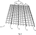
- Fig. 6 shows an embodiment of measuring electrodes 30, which are incorporated, e.g. woven, in the form of metallic threads in a fabric.
- the fabric 49 shown can be made, for example, from non-conductive material or polyester and have a large number of interwoven and/or braided threads. Some of these threads serve as measuring electrodes 30 in metallic or electrically conductive form (e.g. carbon-based).
- a seat or bed cover for the patient support 22 can be made from such a fabric, for example.
- a (support) mat can also be made from such a fabric.
- a checkerboard pattern arrangement of the electrodes can also be produced.
- Fig. 6 Schematic impedance measurements are shown as arrows between the respective measuring electrodes 30.
- the impedances Z 1 to Z 4 are measured indirectly via a current or voltage measurement by the evaluation and control unit 20.
- the impedance Z 4 is not measured directly between two adjacent measuring electrodes 30, but a measuring electrode was skipped between the measuring electrodes 30.
- Fig. 7 shows a third embodiment of the high-frequency surgery system 100 with a third embodiment of the monitoring unit 10.
- the monitoring unit 10 has magnetic and/or electrical antennas 50 as measuring sensors 48.
- the antennas 50 are designed to detect electromagnetic signals 52 from the periphery of the patient 12 as the parameter, which are generated by the high-frequency energy during operation of the high-frequency surgery device 14.
- the evaluation and control unit 34 is set up to calculate a current density distribution in space from the measurement signals by solving a mathematical inverse problem, whereby the recorded measurement signals are proportional to 1/distance • ⁇ l/ ⁇ t for magnetic antennas 50 and proportional to 1/distance • ⁇ V/ ⁇ t for electrical antennas 50.
- the antennas 50 are preferably arranged in an immediate operating environment, for example on the patient support 22 and/or on a ceiling above the operating area and/or in corners of the operating room. Six antennas 50 are shown schematically here, of which only one has been marked for reasons of clarity.
- a method for monitoring a patient 12 during operation of a high-frequency surgical device 14 is also particularly preferred, wherein the high-frequency surgical device 14 is designed to separate and/or coagulate biological tissue using high-frequency electrical energy.
- the method comprises the steps of: impressing a predetermined measuring alternating voltage or a predetermined measuring alternating current into measuring electrodes 30, which are arranged in a periphery of the patient 12, and measuring a voltage between the measuring electrodes 30 falling impedance, as well as monitoring a temporal course of the impedance 36 and/or a temporal change thereof.
- Also preferred as an alternative solution is a method for monitoring a patient 12 during operation of a high-frequency surgical device 14, wherein the high-frequency surgical device 14 is designed to separate and/or coagulate biological tissue using electrical high-frequency energy.
- the method comprises the steps of: detecting electromagnetic measurement signals from a periphery of the patient using magnetic and/or electrical antennas, wherein the electromagnetic measurement signals are generated by the high-frequency energy during operation of the high-frequency surgical device; and calculating a current density distribution in space (or of the patient and his periphery) from the electromagnetic measurement signals by solving a mathematical inverse problem.
- the preferred methods for monitoring a patient 12 during operation of a high-frequency surgical device 14 can be designed in accordance with the above-disclosed embodiments and embodiments in a respective method-specific modification without being mentioned in redundant form at this point.
Landscapes
- Health & Medical Sciences (AREA)
- Life Sciences & Earth Sciences (AREA)
- Surgery (AREA)
- Engineering & Computer Science (AREA)
- Veterinary Medicine (AREA)
- Molecular Biology (AREA)
- Physics & Mathematics (AREA)
- Public Health (AREA)
- Biomedical Technology (AREA)
- Heart & Thoracic Surgery (AREA)
- Medical Informatics (AREA)
- General Health & Medical Sciences (AREA)
- Animal Behavior & Ethology (AREA)
- Nuclear Medicine, Radiotherapy & Molecular Imaging (AREA)
- Otolaryngology (AREA)
- Plasma & Fusion (AREA)
- Biophysics (AREA)
- Pathology (AREA)
- Radiology & Medical Imaging (AREA)
- Surgical Instruments (AREA)
Claims (8)
- Unité de surveillance, qui est configurée pour surveiller un patient (12) pendant le fonctionnement d'un dispositif (14) de chirurgie à haute fréquence, dans laquelle le dispositif (14) de chirurgie à haute fréquence est configuré pour séparer et/ou coaguler le tissu biologique au moyen d'une énergie à haute fréquence, dans laquelle l'unité de surveillance (10) a :- des capteurs de mesure (48), et- une unité (34) d'évaluation et de contrôle,dans laquelle les capteurs de mesure (48) sont configurés pour détecter un paramètre qui est causé pendant la séparation et/ou la coagulation du tissu biologique à cause de l'énergie à haute fréquence caractérisée en ce que les capteurs de mesure (48) sont chacun des antennes (50) magnétiques et/ou électriques qui sont configurées pour détecter des signaux de mesure électromagnétiques provenant de la périphérie du patient (12) en tant que paramètre, les signaux de mesure étant générés par l'énergie à haute fréquence pendant le fonctionnement du dispositif (14) de chirurgie à haute fréquence, etdans laquelle l'unité (34) d'évaluation et de contrôle est configurée pour calculer une distribution spatiale de la densité de courant électrique à partir des signaux de mesure électromagnétiques en résolvant un problème mathématique inverse.
- Unité de surveillance selon la revendication 1, qui est en outre configurée pour générer un signal d'avertissement en cas de dépassement d'une valeur de seuil prédéterminée.
- Unité de surveillance selon la revendication 2, dans laquelle le signal d'avertissement est émis sous forme acoustique, optique et/ou tactile.
- Unité de surveillance selon l'une quelconque des revendications précédentes, dans laquelle la distribution spatiale tridimensionnelle calculée de la densité de courant électrique peut être représentée visuellement.
- Unité de surveillance selon la revendication 4, dans laquelle la distribution spatiale tridimensionnelle de la densité du courant électrique, représentée visuellement, peut être superposée aux images d'une caméra stéréo 3D, de sorte que les informations sur la densité du courant électrique sont visualisables sur une représentation 3D partiellement transmissive du patient.
- Unité de surveillance selon la revendication 2, dans laquelle l'unité d'évaluation et de contrôle est configurée pour générer le signal d'avertissement et/ou pour arrêter le dispositif de chirurgie à haute fréquence si la densité de courant électrique d'une ou plusieurs positions prédéterminées dans l'espace dépasse une deuxième valeur de seuil prédéterminée.
- Unité de surveillance selon la revendication 6, dans laquelle la ou les positions représentent une ou plusieurs parties du dos du patient, sur lequel le patient est allongé sur la table d'opération, et/ou une ou plusieurs parties du pied ou d'autres extrémités du patient.
- Système de chirurgie à haute fréquence qui a :- un générateur (20) à haute fréquence configuré pour générer une énergie à haute fréquence,- un dispositif (14) de chirurgie à haute fréquence ayant une électrode active (16) et une électrode dispersive (18), dans lequel au moins l'électrode active (16) est connectée au générateur (20) à haute fréquence,dans laquelle l'électrode active (16) est configurée pour séparer et/ou coaguler le tissu biologique au moyen de l'énergie électrique à haute fréquence, etdans laquelle l'électrode dispersive (18) est configurée pour décharger l'énergie électrique à haute fréquence du tissu biologique, et- une unité de surveillance (10) selon l'une quelconque des revendications 1 à 7.
Applications Claiming Priority (3)
| Application Number | Priority Date | Filing Date | Title |
|---|---|---|---|
| DE102020003524.8A DE102020003524B4 (de) | 2020-06-12 | 2020-06-12 | Überwachungseinheit und Hochfrequenz-Chirurgiesystem mit einer solchen Überwachungseinheit |
| PCT/EP2021/065506 WO2021250116A1 (fr) | 2020-06-12 | 2021-06-09 | Unité de surveillance et système de chirurgie haute fréquence comprenant une telle unité de surveillance |
| EP21734288.0A EP4164531B1 (fr) | 2020-06-12 | 2021-06-09 | Unité de surveillance et système de chirurgie haute fréquence comprenant une telle unité de surveillance |
Related Parent Applications (1)
| Application Number | Title | Priority Date | Filing Date |
|---|---|---|---|
| EP21734288.0A Division EP4164531B1 (fr) | 2020-06-12 | 2021-06-09 | Unité de surveillance et système de chirurgie haute fréquence comprenant une telle unité de surveillance |
Publications (3)
| Publication Number | Publication Date |
|---|---|
| EP4309571A2 EP4309571A2 (fr) | 2024-01-24 |
| EP4309571A3 EP4309571A3 (fr) | 2024-03-13 |
| EP4309571B1 true EP4309571B1 (fr) | 2024-11-20 |
Family
ID=76601186
Family Applications (2)
| Application Number | Title | Priority Date | Filing Date |
|---|---|---|---|
| EP21734288.0A Active EP4164531B1 (fr) | 2020-06-12 | 2021-06-09 | Unité de surveillance et système de chirurgie haute fréquence comprenant une telle unité de surveillance |
| EP23214347.9A Active EP4309571B1 (fr) | 2020-06-12 | 2021-06-09 | Unité de surveillance et système de chirurgie haute fréquence comprenant une telle unité de surveillance |
Family Applications Before (1)
| Application Number | Title | Priority Date | Filing Date |
|---|---|---|---|
| EP21734288.0A Active EP4164531B1 (fr) | 2020-06-12 | 2021-06-09 | Unité de surveillance et système de chirurgie haute fréquence comprenant une telle unité de surveillance |
Country Status (9)
| Country | Link |
|---|---|
| US (1) | US12672908B2 (fr) |
| EP (2) | EP4164531B1 (fr) |
| KR (1) | KR102884814B1 (fr) |
| CN (1) | CN115916082A (fr) |
| DE (1) | DE102020003524B4 (fr) |
| DK (2) | DK4309571T3 (fr) |
| ES (2) | ES3012783T3 (fr) |
| PL (2) | PL4309571T3 (fr) |
| WO (1) | WO2021250116A1 (fr) |
Families Citing this family (4)
| Publication number | Priority date | Publication date | Assignee | Title |
|---|---|---|---|---|
| DE102020003524B4 (de) | 2020-06-12 | 2023-03-23 | Coco Beteiligungsgesellschaft mbH | Überwachungseinheit und Hochfrequenz-Chirurgiesystem mit einer solchen Überwachungseinheit |
| DE102021203843A1 (de) | 2021-04-19 | 2022-10-20 | Robert Bosch Gesellschaft mit beschränkter Haftung | Elektrochirurgisches Instrument und Teil des elektrochirurgischen Instruments |
| KR102718603B1 (ko) * | 2022-02-22 | 2024-10-16 | 한양대학교 에리카산학협력단 | 전기 수술 장치, 임피던스 측정 장치, 전기 수술 장치의 제어 방법 및 임피던스 측정 방법 |
| CN115192186B (zh) * | 2022-08-10 | 2023-09-15 | 浙江舒友仪器设备股份有限公司 | 一种高频电极自动激活输出系统 |
Family Cites Families (23)
| Publication number | Priority date | Publication date | Assignee | Title |
|---|---|---|---|---|
| DE4232255C1 (de) | 1992-09-25 | 1994-02-17 | Siemens Ag | Neutralelektrode und Verfahren zu deren Wiederverwendung für ein Hochfrequenz-Chirurgiegerät mit Applikationsüberwachungseinrichtung |
| DE4329898A1 (de) * | 1993-09-04 | 1995-04-06 | Marcus Dr Besson | Kabelloses medizinisches Diagnose- und Überwachungsgerät |
| NL1001282C2 (nl) * | 1995-09-26 | 1997-03-28 | A J Van Liebergen Holding B V | Inrichting voor slagvolumebepaling van een menselijk hart. |
| EP1006904A4 (fr) * | 1996-10-30 | 2000-06-14 | Megadyne Med Prod Inc | Electrode electrochirurgicale de retour reutilisable |
| US6332891B1 (en) * | 1999-02-16 | 2001-12-25 | Stryker Corporation | System and method for performing image guided surgery |
| US7169145B2 (en) * | 2003-11-21 | 2007-01-30 | Megadyne Medical Products, Inc. | Tuned return electrode with matching inductor |
| CA2541037A1 (fr) | 2005-03-31 | 2006-09-30 | Sherwood Services Ag | Electrode de retour pour la regulation de la temperature et systeme de surveillance de l'electrode de retour |
| US20070049914A1 (en) | 2005-09-01 | 2007-03-01 | Sherwood Services Ag | Return electrode pad with conductive element grid and method |
| US20070167942A1 (en) * | 2006-01-18 | 2007-07-19 | Sherwood Services Ag | RF return pad current distribution system |
| DE102007020583B4 (de) * | 2006-07-19 | 2012-10-11 | Erbe Elektromedizin Gmbh | Elektrodeneinrichtung mit einerImnpedanz-Messeinrichtung und Verfahren zum Herstellen einer derartigen Elektrodeneinrichtung |
| US20080051777A1 (en) * | 2006-08-28 | 2008-02-28 | Dieter Haemmerich | Radiofrequency ablation device for reducing the incidence of skin burns |
| US8080007B2 (en) * | 2007-05-07 | 2011-12-20 | Tyco Healthcare Group Lp | Capacitive electrosurgical return pad with contact quality monitoring |
| DE102007051097A1 (de) | 2007-10-24 | 2009-04-30 | Celon Ag Medical Instruments | HF-Chirurgiegerät und Verfahren für ein HF-Chirurgiegerät |
| US20090234352A1 (en) | 2008-03-17 | 2009-09-17 | Tyco Healthcare Group Lp | Variable Capacitive Electrode Pad |
| DE102009013917A1 (de) * | 2008-10-30 | 2010-05-12 | Erbe Elektromedizin Gmbh | Elektrochirurgisches Gerät mit einer Temperaturmesseinrichtung, Verfahren zur Bestimmung einer Temperatur und/oder einer Temperaturänderung an einer Neutralelektrode |
| WO2013139945A1 (fr) | 2012-03-21 | 2013-09-26 | Farin Guenter | Dispositif et procédé destinés à mesurer et réguler des effets thermiques dans le domaine de la chirurgie haute fréquence |
| US9867650B2 (en) * | 2013-12-26 | 2018-01-16 | Megadyne Medical Products, Inc. | Universal self-limiting electrosurgical return electrode |
| US10085791B2 (en) * | 2013-12-26 | 2018-10-02 | Megadyne Medical Products, Inc. | Universal self-limiting electrosurgical return electrode |
| US10111703B2 (en) * | 2014-05-06 | 2018-10-30 | Cosman Instruments, Llc | Electrosurgical generator |
| US10646266B2 (en) * | 2015-11-13 | 2020-05-12 | Covidien Lp | System and method for return electrode monitoring |
| US20200188055A1 (en) * | 2017-10-01 | 2020-06-18 | James Keith Bleiler | Surgical Safety Devices and Methods |
| DE102018114482A1 (de) | 2018-06-16 | 2019-12-19 | Olympus Winter & Ibe Gmbh | Elektrochirurgische Vorrichtung |
| DE102020003524B4 (de) | 2020-06-12 | 2023-03-23 | Coco Beteiligungsgesellschaft mbH | Überwachungseinheit und Hochfrequenz-Chirurgiesystem mit einer solchen Überwachungseinheit |
-
2020
- 2020-06-12 DE DE102020003524.8A patent/DE102020003524B4/de active Active
-
2021
- 2021-06-09 ES ES23214347T patent/ES3012783T3/es active Active
- 2021-06-09 KR KR1020237000030A patent/KR102884814B1/ko active Active
- 2021-06-09 CN CN202180041994.XA patent/CN115916082A/zh active Pending
- 2021-06-09 EP EP21734288.0A patent/EP4164531B1/fr active Active
- 2021-06-09 DK DK23214347.9T patent/DK4309571T3/da active
- 2021-06-09 PL PL23214347.9T patent/PL4309571T3/pl unknown
- 2021-06-09 DK DK21734288.0T patent/DK4164531T3/da active
- 2021-06-09 EP EP23214347.9A patent/EP4309571B1/fr active Active
- 2021-06-09 US US18/009,613 patent/US12672908B2/en active Active
- 2021-06-09 WO PCT/EP2021/065506 patent/WO2021250116A1/fr not_active Ceased
- 2021-06-09 PL PL21734288.0T patent/PL4164531T3/pl unknown
- 2021-06-09 ES ES21734288T patent/ES2974209T3/es active Active
Also Published As
| Publication number | Publication date |
|---|---|
| EP4164531B1 (fr) | 2023-12-13 |
| US20230218333A1 (en) | 2023-07-13 |
| PL4309571T3 (pl) | 2025-03-31 |
| DK4164531T3 (da) | 2024-03-11 |
| ES3012783T3 (en) | 2025-04-10 |
| US12672908B2 (en) | 2026-07-07 |
| WO2021250116A1 (fr) | 2021-12-16 |
| CN115916082A (zh) | 2023-04-04 |
| PL4164531T3 (pl) | 2024-05-27 |
| KR102884814B1 (ko) | 2025-11-11 |
| EP4309571A2 (fr) | 2024-01-24 |
| EP4164531A1 (fr) | 2023-04-19 |
| DE102020003524A1 (de) | 2021-12-16 |
| DE102020003524B4 (de) | 2023-03-23 |
| EP4309571A3 (fr) | 2024-03-13 |
| KR20230023705A (ko) | 2023-02-17 |
| ES2974209T3 (es) | 2024-06-26 |
| DK4309571T3 (da) | 2025-02-17 |
Similar Documents
| Publication | Publication Date | Title |
|---|---|---|
| EP4164531B1 (fr) | Unité de surveillance et système de chirurgie haute fréquence comprenant une telle unité de surveillance | |
| EP2367493B1 (fr) | Appareil électrochirurgical doté d'un dispositif de mesure de température pour la détermination de la température et/ou d'une modification de la température d'une électrode neutre | |
| DE69232587T2 (de) | Elektrochirurgische vorrichtung für die laproskopie und ähnliche verfahren | |
| DE102007020583B4 (de) | Elektrodeneinrichtung mit einerImnpedanz-Messeinrichtung und Verfahren zum Herstellen einer derartigen Elektrodeneinrichtung | |
| AU2003232023B8 (en) | Self-limiting electrosurgical return electrode | |
| DE60316052T2 (de) | Elektrochirurgisches Instrument mit Lichtmonitor auf Greifarm | |
| EP0598780B1 (fr) | Generateur de haute frequence utile en chirurgie pour inciser des tissus | |
| DE102007056974B4 (de) | HF-chirurgische Prüfeinrichtung und Verfahren zum Identifizieren einer am Patienten angelegten HF-Neutralelektrode | |
| EP0390937B1 (fr) | Dispositif pour surveiller l'adhésion des électrodes neutres utilisées en chirurgie à haute fréquence | |
| DE202009001333U1 (de) | Ablationsleistungsindikator für elektrochirurgische Geräte | |
| DE112004002125T5 (de) | Hochfrequenzenergieversorgung | |
| WO2015055797A1 (fr) | Ensemble bobine de champ et système de détection de positions | |
| EP0998216B1 (fr) | Agencement pour la detection, la caracterisation par diagnostic differentiel et le traitement de tumeurs | |
| AT503188A1 (de) | Vorrichtung zur prüfung einer neutralelektrode | |
| Golpaygani et al. | A study on performance and safety tests of electrosurgical equipment | |
| EP2337516B1 (fr) | Générateur électrochirurgical haute fréquence | |
| MacDonald et al. | Safety issues during surgical monitoring | |
| Bifulco et al. | Investigating the role of capacitive coupling between the operating table and the return electrode of an electrosurgery unit in the modification of the current density distribution within the patients’ body | |
| HK40104818A (en) | Monitoring unit and high-frequency surgical system comprising such a monitoring unit | |
| HK40097185A (en) | Monitoring unit and high-frequency surgery system comprising such a monitoring unit | |
| HK40097185B (en) | Monitoring unit and high-frequency surgery system comprising such a monitoring unit | |
| DD241550A1 (de) | Hochfrequenzchirugiegeraet | |
| AT527092A1 (de) | Messvorrichtung zur Messung einer Temperatur an einer medizinischen Elektrode | |
| DE102022111745A1 (de) | Vorrichtungen, systeme und verfahren zur energiebasierten behandlung von synovialgelenken und anderen fluidgefüllten räumen | |
| Gerhard et al. | Reducing electrosurgical hazards for transurethral resections |
Legal Events
| Date | Code | Title | Description |
|---|---|---|---|
| PUAI | Public reference made under article 153(3) epc to a published international application that has entered the european phase |
Free format text: ORIGINAL CODE: 0009012 |
|
| STAA | Information on the status of an ep patent application or granted ep patent |
Free format text: STATUS: THE APPLICATION HAS BEEN PUBLISHED |
|
| AC | Divisional application: reference to earlier application |
Ref document number: 4164531 Country of ref document: EP Kind code of ref document: P |
|
| AK | Designated contracting states |
Kind code of ref document: A2 Designated state(s): AL AT BE BG CH CY CZ DE DK EE ES FI FR GB GR HR HU IE IS IT LI LT LU LV MC MK MT NL NO PL PT RO RS SE SI SK SM TR |
|
| REG | Reference to a national code |
Ref country code: DE Ref legal event code: R079 Free format text: PREVIOUS MAIN CLASS: A61B0005000000 Ipc: A61B0018120000 Ref country code: DE Ref legal event code: R079 Ref document number: 502021005911 Country of ref document: DE Free format text: PREVIOUS MAIN CLASS: A61B0005000000 Ipc: A61B0018120000 |
|
| PUAL | Search report despatched |
Free format text: ORIGINAL CODE: 0009013 |
|
| STAA | Information on the status of an ep patent application or granted ep patent |
Free format text: STATUS: REQUEST FOR EXAMINATION WAS MADE |
|
| AK | Designated contracting states |
Kind code of ref document: A3 Designated state(s): AL AT BE BG CH CY CZ DE DK EE ES FI FR GB GR HR HU IE IS IT LI LT LU LV MC MK MT NL NO PL PT RO RS SE SI SK SM TR |
|
| RIC1 | Information provided on ipc code assigned before grant |
Ipc: A61B 5/00 20060101ALI20240208BHEP Ipc: A61B 5/24 20210101ALI20240208BHEP Ipc: A61B 5/053 20210101ALI20240208BHEP Ipc: A61B 18/16 20060101ALI20240208BHEP Ipc: A61B 18/14 20060101ALI20240208BHEP Ipc: A61B 18/12 20060101AFI20240208BHEP |
|
| 17P | Request for examination filed |
Effective date: 20240220 |
|
| RBV | Designated contracting states (corrected) |
Designated state(s): AL AT BE BG CH CY CZ DE DK EE ES FI FR GB GR HR HU IE IS IT LI LT LU LV MC MK MT NL NO PL PT RO RS SE SI SK SM TR |
|
| GRAP | Despatch of communication of intention to grant a patent |
Free format text: ORIGINAL CODE: EPIDOSNIGR1 |
|
| STAA | Information on the status of an ep patent application or granted ep patent |
Free format text: STATUS: GRANT OF PATENT IS INTENDED |
|
| RIC1 | Information provided on ipc code assigned before grant |
Ipc: A61B 5/00 20060101ALI20240621BHEP Ipc: A61B 5/24 20210101ALI20240621BHEP Ipc: A61B 5/053 20210101ALI20240621BHEP Ipc: A61B 18/16 20060101ALI20240621BHEP Ipc: A61B 18/14 20060101ALI20240621BHEP Ipc: A61B 18/12 20060101AFI20240621BHEP |
|
| REG | Reference to a national code |
Ref country code: HK Ref legal event code: DE Ref document number: 40104818 Country of ref document: HK |
|
| INTG | Intention to grant announced |
Effective date: 20240710 |
|
| GRAS | Grant fee paid |
Free format text: ORIGINAL CODE: EPIDOSNIGR3 |
|
| GRAA | (expected) grant |
Free format text: ORIGINAL CODE: 0009210 |
|
| STAA | Information on the status of an ep patent application or granted ep patent |
Free format text: STATUS: THE PATENT HAS BEEN GRANTED |
|
| AC | Divisional application: reference to earlier application |
Ref document number: 4164531 Country of ref document: EP Kind code of ref document: P |
|
| AK | Designated contracting states |
Kind code of ref document: B1 Designated state(s): AL AT BE BG CH CY CZ DE DK EE ES FI FR GB GR HR HU IE IS IT LI LT LU LV MC MK MT NL NO PL PT RO RS SE SI SK SM TR |
|
| REG | Reference to a national code |
Ref country code: GB Ref legal event code: FG4D Free format text: NOT ENGLISH |
|
| REG | Reference to a national code |
Ref country code: CH Ref legal event code: EP |
|
| REG | Reference to a national code |
Ref country code: DE Ref legal event code: R096 Ref document number: 502021005911 Country of ref document: DE |
|
| REG | Reference to a national code |
Ref country code: IE Ref legal event code: FG4D Free format text: LANGUAGE OF EP DOCUMENT: GERMAN |
|
| REG | Reference to a national code |
Ref country code: DK Ref legal event code: T3 Effective date: 20250211 |
|
| REG | Reference to a national code |
Ref country code: SE Ref legal event code: TRGR |
|
| REG | Reference to a national code |
Ref country code: LT Ref legal event code: MG9D |
|
| REG | Reference to a national code |
Ref country code: NL Ref legal event code: MP Effective date: 20241120 |
|
| REG | Reference to a national code |
Ref country code: ES Ref legal event code: FG2A Ref document number: 3012783 Country of ref document: ES Kind code of ref document: T3 Effective date: 20250410 |
|
| PG25 | Lapsed in a contracting state [announced via postgrant information from national office to epo] |
Ref country code: HR Free format text: LAPSE BECAUSE OF FAILURE TO SUBMIT A TRANSLATION OF THE DESCRIPTION OR TO PAY THE FEE WITHIN THE PRESCRIBED TIME-LIMIT Effective date: 20241120 Ref country code: IS Free format text: LAPSE BECAUSE OF FAILURE TO SUBMIT A TRANSLATION OF THE DESCRIPTION OR TO PAY THE FEE WITHIN THE PRESCRIBED TIME-LIMIT Effective date: 20250320 Ref country code: PT Free format text: LAPSE BECAUSE OF FAILURE TO SUBMIT A TRANSLATION OF THE DESCRIPTION OR TO PAY THE FEE WITHIN THE PRESCRIBED TIME-LIMIT Effective date: 20250320 |
|
| PG25 | Lapsed in a contracting state [announced via postgrant information from national office to epo] |
Ref country code: FI Free format text: LAPSE BECAUSE OF FAILURE TO SUBMIT A TRANSLATION OF THE DESCRIPTION OR TO PAY THE FEE WITHIN THE PRESCRIBED TIME-LIMIT Effective date: 20241120 Ref country code: NL Free format text: LAPSE BECAUSE OF FAILURE TO SUBMIT A TRANSLATION OF THE DESCRIPTION OR TO PAY THE FEE WITHIN THE PRESCRIBED TIME-LIMIT Effective date: 20241120 |
|
| PG25 | Lapsed in a contracting state [announced via postgrant information from national office to epo] |
Ref country code: BG Free format text: LAPSE BECAUSE OF FAILURE TO SUBMIT A TRANSLATION OF THE DESCRIPTION OR TO PAY THE FEE WITHIN THE PRESCRIBED TIME-LIMIT Effective date: 20241120 |
|
| PG25 | Lapsed in a contracting state [announced via postgrant information from national office to epo] |
Ref country code: NO Free format text: LAPSE BECAUSE OF FAILURE TO SUBMIT A TRANSLATION OF THE DESCRIPTION OR TO PAY THE FEE WITHIN THE PRESCRIBED TIME-LIMIT Effective date: 20250220 |
|
| PG25 | Lapsed in a contracting state [announced via postgrant information from national office to epo] |
Ref country code: GR Free format text: LAPSE BECAUSE OF FAILURE TO SUBMIT A TRANSLATION OF THE DESCRIPTION OR TO PAY THE FEE WITHIN THE PRESCRIBED TIME-LIMIT Effective date: 20250221 Ref country code: LV Free format text: LAPSE BECAUSE OF FAILURE TO SUBMIT A TRANSLATION OF THE DESCRIPTION OR TO PAY THE FEE WITHIN THE PRESCRIBED TIME-LIMIT Effective date: 20241120 |
|
| PG25 | Lapsed in a contracting state [announced via postgrant information from national office to epo] |
Ref country code: RS Free format text: LAPSE BECAUSE OF FAILURE TO SUBMIT A TRANSLATION OF THE DESCRIPTION OR TO PAY THE FEE WITHIN THE PRESCRIBED TIME-LIMIT Effective date: 20250220 |
|
| PG25 | Lapsed in a contracting state [announced via postgrant information from national office to epo] |
Ref country code: SM Free format text: LAPSE BECAUSE OF FAILURE TO SUBMIT A TRANSLATION OF THE DESCRIPTION OR TO PAY THE FEE WITHIN THE PRESCRIBED TIME-LIMIT Effective date: 20241120 |
|
| PG25 | Lapsed in a contracting state [announced via postgrant information from national office to epo] |
Ref country code: EE Free format text: LAPSE BECAUSE OF FAILURE TO SUBMIT A TRANSLATION OF THE DESCRIPTION OR TO PAY THE FEE WITHIN THE PRESCRIBED TIME-LIMIT Effective date: 20241120 |
|
| PG25 | Lapsed in a contracting state [announced via postgrant information from national office to epo] |
Ref country code: RO Free format text: LAPSE BECAUSE OF FAILURE TO SUBMIT A TRANSLATION OF THE DESCRIPTION OR TO PAY THE FEE WITHIN THE PRESCRIBED TIME-LIMIT Effective date: 20241120 |
|
| PG25 | Lapsed in a contracting state [announced via postgrant information from national office to epo] |
Ref country code: SK Free format text: LAPSE BECAUSE OF FAILURE TO SUBMIT A TRANSLATION OF THE DESCRIPTION OR TO PAY THE FEE WITHIN THE PRESCRIBED TIME-LIMIT Effective date: 20241120 |
|
| PG25 | Lapsed in a contracting state [announced via postgrant information from national office to epo] |
Ref country code: CZ Free format text: LAPSE BECAUSE OF FAILURE TO SUBMIT A TRANSLATION OF THE DESCRIPTION OR TO PAY THE FEE WITHIN THE PRESCRIBED TIME-LIMIT Effective date: 20241120 |
|
| PGFP | Annual fee paid to national office [announced via postgrant information from national office to epo] |
Ref country code: SE Payment date: 20250618 Year of fee payment: 5 |
|
| REG | Reference to a national code |
Ref country code: DE Ref legal event code: R097 Ref document number: 502021005911 Country of ref document: DE |
|
| PLBE | No opposition filed within time limit |
Free format text: ORIGINAL CODE: 0009261 |
|
| STAA | Information on the status of an ep patent application or granted ep patent |
Free format text: STATUS: NO OPPOSITION FILED WITHIN TIME LIMIT |
|
| PGFP | Annual fee paid to national office [announced via postgrant information from national office to epo] |
Ref country code: ES Payment date: 20250718 Year of fee payment: 5 |
|
| PGFP | Annual fee paid to national office [announced via postgrant information from national office to epo] |
Ref country code: DE Payment date: 20250813 Year of fee payment: 5 |
|
| PGFP | Annual fee paid to national office [announced via postgrant information from national office to epo] |
Ref country code: IT Payment date: 20250630 Year of fee payment: 5 |
|
| PGFP | Annual fee paid to national office [announced via postgrant information from national office to epo] |
Ref country code: CH Payment date: 20250701 Year of fee payment: 5 |
|
| 26N | No opposition filed |
Effective date: 20250821 |
|
| REG | Reference to a national code |
Ref country code: DE Ref legal event code: R084 Ref document number: 502021005911 Country of ref document: DE |
|
| PG25 | Lapsed in a contracting state [announced via postgrant information from national office to epo] |
Ref country code: MC Free format text: LAPSE BECAUSE OF FAILURE TO SUBMIT A TRANSLATION OF THE DESCRIPTION OR TO PAY THE FEE WITHIN THE PRESCRIBED TIME-LIMIT Effective date: 20241120 |
|
| PG25 | Lapsed in a contracting state [announced via postgrant information from national office to epo] |
Ref country code: LU Free format text: LAPSE BECAUSE OF NON-PAYMENT OF DUE FEES Effective date: 20250609 |
|
| PG25 | Lapsed in a contracting state [announced via postgrant information from national office to epo] |
Ref country code: IE Free format text: LAPSE BECAUSE OF NON-PAYMENT OF DUE FEES Effective date: 20250609 |
|
| PG25 | Lapsed in a contracting state [announced via postgrant information from national office to epo] |
Ref country code: LT Free format text: LAPSE BECAUSE OF FAILURE TO SUBMIT A TRANSLATION OF THE DESCRIPTION OR TO PAY THE FEE WITHIN THE PRESCRIBED TIME-LIMIT Effective date: 20241120 |
|
| REG | Reference to a national code |
Ref country code: CH Ref legal event code: U11 Free format text: ST27 STATUS EVENT CODE: U-0-0-U10-U11 (AS PROVIDED BY THE NATIONAL OFFICE) Effective date: 20260701 |
|
| PGFP | Annual fee paid to national office [announced via postgrant information from national office to epo] |
Ref country code: GB Payment date: 20260623 Year of fee payment: 6 Ref country code: DK Payment date: 20260622 Year of fee payment: 6 Ref country code: PL Payment date: 20260511 Year of fee payment: 6 |
|
| PGFP | Annual fee paid to national office [announced via postgrant information from national office to epo] |
Ref country code: AT Payment date: 20260618 Year of fee payment: 6 |
|
| PGFP | Annual fee paid to national office [announced via postgrant information from national office to epo] |
Ref country code: BE Payment date: 20260622 Year of fee payment: 6 |
|
| PGFP | Annual fee paid to national office [announced via postgrant information from national office to epo] |
Ref country code: FR Payment date: 20260623 Year of fee payment: 6 |
|
| PG25 | Lapsed in a contracting state [announced via postgrant information from national office to epo] |
Ref country code: CY Free format text: LAPSE BECAUSE OF FAILURE TO SUBMIT A TRANSLATION OF THE DESCRIPTION OR TO PAY THE FEE WITHIN THE PRESCRIBED TIME-LIMIT Effective date: 20241120 |