EP4403099A2 - Verfahren und vorrichtung zur nichtinvasiven detektion von physiologischen und pathophysiologischen schlafzuständen - Google Patents
Verfahren und vorrichtung zur nichtinvasiven detektion von physiologischen und pathophysiologischen schlafzuständen Download PDFInfo
- Publication number
- EP4403099A2 EP4403099A2 EP24180528.2A EP24180528A EP4403099A2 EP 4403099 A2 EP4403099 A2 EP 4403099A2 EP 24180528 A EP24180528 A EP 24180528A EP 4403099 A2 EP4403099 A2 EP 4403099A2
- Authority
- EP
- European Patent Office
- Prior art keywords
- upstroke
- index
- person
- breathing
- sleep
- Prior art date
- Legal status (The legal status is an assumption and is not a legal conclusion. Google has not performed a legal analysis and makes no representation as to the accuracy of the status listed.)
- Granted
Links
Images


Classifications
-
- A—HUMAN NECESSITIES
- A61—MEDICAL OR VETERINARY SCIENCE; HYGIENE
- A61B—DIAGNOSIS; SURGERY; IDENTIFICATION
- A61B5/00—Measuring for diagnostic purposes; Identification of persons
- A61B5/48—Other medical applications
- A61B5/4806—Sleep evaluation
- A61B5/4818—Sleep apnoea
-
- A—HUMAN NECESSITIES
- A61—MEDICAL OR VETERINARY SCIENCE; HYGIENE
- A61B—DIAGNOSIS; SURGERY; IDENTIFICATION
- A61B5/00—Measuring for diagnostic purposes; Identification of persons
- A61B5/02—Detecting, measuring or recording for evaluating the cardiovascular system, e.g. pulse, heart rate, blood pressure or blood flow
- A61B5/02028—Determining haemodynamic parameters not otherwise provided for, e.g. cardiac contractility or left ventricular ejection fraction
-
- A—HUMAN NECESSITIES
- A61—MEDICAL OR VETERINARY SCIENCE; HYGIENE
- A61B—DIAGNOSIS; SURGERY; IDENTIFICATION
- A61B5/00—Measuring for diagnostic purposes; Identification of persons
- A61B5/02—Detecting, measuring or recording for evaluating the cardiovascular system, e.g. pulse, heart rate, blood pressure or blood flow
- A61B5/0205—Simultaneously evaluating both cardiovascular conditions and different types of body conditions, e.g. heart and respiratory condition
- A61B5/02055—Simultaneously evaluating both cardiovascular condition and temperature
-
- A—HUMAN NECESSITIES
- A61—MEDICAL OR VETERINARY SCIENCE; HYGIENE
- A61B—DIAGNOSIS; SURGERY; IDENTIFICATION
- A61B5/00—Measuring for diagnostic purposes; Identification of persons
- A61B5/02—Detecting, measuring or recording for evaluating the cardiovascular system, e.g. pulse, heart rate, blood pressure or blood flow
- A61B5/024—Measuring pulse rate or heart rate
-
- A—HUMAN NECESSITIES
- A61—MEDICAL OR VETERINARY SCIENCE; HYGIENE
- A61B—DIAGNOSIS; SURGERY; IDENTIFICATION
- A61B5/00—Measuring for diagnostic purposes; Identification of persons
- A61B5/03—Measuring fluid pressure within the body other than blood pressure, e.g. cerebral pressure ; Measuring pressure in body tissues or organs
-
- A—HUMAN NECESSITIES
- A61—MEDICAL OR VETERINARY SCIENCE; HYGIENE
- A61B—DIAGNOSIS; SURGERY; IDENTIFICATION
- A61B5/00—Measuring for diagnostic purposes; Identification of persons
- A61B5/08—Measuring devices for evaluating the respiratory organs
- A61B5/0816—Measuring devices for examining respiratory frequency
-
- A—HUMAN NECESSITIES
- A61—MEDICAL OR VETERINARY SCIENCE; HYGIENE
- A61B—DIAGNOSIS; SURGERY; IDENTIFICATION
- A61B5/00—Measuring for diagnostic purposes; Identification of persons
- A61B5/48—Other medical applications
- A61B5/4806—Sleep evaluation
- A61B5/4812—Detecting sleep stages or cycles
-
- A—HUMAN NECESSITIES
- A61—MEDICAL OR VETERINARY SCIENCE; HYGIENE
- A61B—DIAGNOSIS; SURGERY; IDENTIFICATION
- A61B5/00—Measuring for diagnostic purposes; Identification of persons
- A61B5/72—Signal processing specially adapted for physiological signals or for diagnostic purposes
-
- A—HUMAN NECESSITIES
- A61—MEDICAL OR VETERINARY SCIENCE; HYGIENE
- A61B—DIAGNOSIS; SURGERY; IDENTIFICATION
- A61B5/00—Measuring for diagnostic purposes; Identification of persons
- A61B5/72—Signal processing specially adapted for physiological signals or for diagnostic purposes
- A61B5/7235—Details of waveform analysis
-
- A—HUMAN NECESSITIES
- A61—MEDICAL OR VETERINARY SCIENCE; HYGIENE
- A61B—DIAGNOSIS; SURGERY; IDENTIFICATION
- A61B5/00—Measuring for diagnostic purposes; Identification of persons
- A61B5/72—Signal processing specially adapted for physiological signals or for diagnostic purposes
- A61B5/7271—Specific aspects of physiological measurement analysis
- A61B5/7275—Determining trends in physiological measurement data; Predicting development of a medical condition based on physiological measurements, e.g. determining a risk factor
-
- A—HUMAN NECESSITIES
- A61—MEDICAL OR VETERINARY SCIENCE; HYGIENE
- A61B—DIAGNOSIS; SURGERY; IDENTIFICATION
- A61B5/00—Measuring for diagnostic purposes; Identification of persons
- A61B5/72—Signal processing specially adapted for physiological signals or for diagnostic purposes
- A61B5/7271—Specific aspects of physiological measurement analysis
- A61B5/7278—Artificial waveform generation or derivation, e.g. synthesizing signals from measured signals
-
- A—HUMAN NECESSITIES
- A61—MEDICAL OR VETERINARY SCIENCE; HYGIENE
- A61B—DIAGNOSIS; SURGERY; IDENTIFICATION
- A61B5/00—Measuring for diagnostic purposes; Identification of persons
- A61B5/02—Detecting, measuring or recording for evaluating the cardiovascular system, e.g. pulse, heart rate, blood pressure or blood flow
- A61B5/024—Measuring pulse rate or heart rate
- A61B5/02416—Measuring pulse rate or heart rate using photoplethysmograph signals, e.g. generated by infrared radiation
-
- A—HUMAN NECESSITIES
- A61—MEDICAL OR VETERINARY SCIENCE; HYGIENE
- A61B—DIAGNOSIS; SURGERY; IDENTIFICATION
- A61B5/00—Measuring for diagnostic purposes; Identification of persons
- A61B5/02—Detecting, measuring or recording for evaluating the cardiovascular system, e.g. pulse, heart rate, blood pressure or blood flow
- A61B5/024—Measuring pulse rate or heart rate
- A61B5/02444—Details of sensor
-
- A—HUMAN NECESSITIES
- A61—MEDICAL OR VETERINARY SCIENCE; HYGIENE
- A61B—DIAGNOSIS; SURGERY; IDENTIFICATION
- A61B5/00—Measuring for diagnostic purposes; Identification of persons
- A61B5/02—Detecting, measuring or recording for evaluating the cardiovascular system, e.g. pulse, heart rate, blood pressure or blood flow
- A61B5/024—Measuring pulse rate or heart rate
- A61B5/0245—Measuring pulse rate or heart rate by using sensing means generating electric signals, i.e. ECG signals
-
- A—HUMAN NECESSITIES
- A61—MEDICAL OR VETERINARY SCIENCE; HYGIENE
- A61B—DIAGNOSIS; SURGERY; IDENTIFICATION
- A61B5/00—Measuring for diagnostic purposes; Identification of persons
- A61B5/08—Measuring devices for evaluating the respiratory organs
- A61B5/0826—Detecting or evaluating apnoea events
-
- A—HUMAN NECESSITIES
- A61—MEDICAL OR VETERINARY SCIENCE; HYGIENE
- A61B—DIAGNOSIS; SURGERY; IDENTIFICATION
- A61B5/00—Measuring for diagnostic purposes; Identification of persons
- A61B5/145—Measuring characteristics of blood in vivo, e.g. gas concentration or pH-value ; Measuring characteristics of body fluids or tissues, e.g. interstitial fluid or cerebral tissue
- A61B5/1455—Measuring characteristics of blood in vivo, e.g. gas concentration or pH-value ; Measuring characteristics of body fluids or tissues, e.g. interstitial fluid or cerebral tissue using optical sensors, e.g. spectral photometrical oximeters
- A61B5/14551—Measuring characteristics of blood in vivo, e.g. gas concentration or pH-value ; Measuring characteristics of body fluids or tissues, e.g. interstitial fluid or cerebral tissue using optical sensors, e.g. spectral photometrical oximeters for measuring blood gases
-
- A—HUMAN NECESSITIES
- A61—MEDICAL OR VETERINARY SCIENCE; HYGIENE
- A61B—DIAGNOSIS; SURGERY; IDENTIFICATION
- A61B8/00—Diagnosis using ultrasonic, sonic or infrasonic waves
- A61B8/02—Measuring pulse or heart rate
-
- A—HUMAN NECESSITIES
- A61—MEDICAL OR VETERINARY SCIENCE; HYGIENE
- A61B—DIAGNOSIS; SURGERY; IDENTIFICATION
- A61B8/00—Diagnosis using ultrasonic, sonic or infrasonic waves
- A61B8/48—Diagnostic techniques
- A61B8/488—Diagnostic techniques involving Doppler signals
Definitions
- the present invention and the present patent application relates to a method and apparatus for analyzing externally recorded physiological data of a patient for detecting and monitoring various breathing conditions of the patient.
- this patent application relates to the analysis of circulatory pulse waveforms recorded from the body's surface of a patient, for determining changes in the performance of the heart during its contraction phase, due to the intra-thoracic pressure changes caused by the breathing cycle of the patient, and inferring there from the presence of certain breathing conditions and sleep disordered breathing conditions of the patient.
- characteristic patterns described by the time course of changes of the pulse signal baseline values are used to distinguish between central and obstructive sleep apneas, when the presence of sleep disordered breathing has initially been found on the basis of oximetry based oxygen desaturation events.
- the modulation patterns are considered both in terms of the magnitude or amplitude of the modulation, or to characteristic patterns of the shape of the modulation envelope.
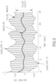
- 6,669,632 describes a method of determining pleural pressure based on determining the difference between a first envelope defined by connecting the peaks of consecutive pulse signals in a series of pulsewaves, (where the amplitude of the peaks of the pulses in the given time series of pulsewaves oscillates in time with the breathing cycle) as shown in Fig. 1A as dark-colored black lines (marked as "FIRST ENVELOPE”), and a second envelope defined by connecting consecutive peaks corresponding to the peaks of consecutive breathing cycles of the first envelope of pulsewaves, as also shown in Figure 1A as light-colored black lines (marked as "SECOND ENVELOPE").
- the patterns characterizing time-series of differences between individual pulsewave trough to peak signal amplitudes in a series of pulses are used to identify and categorize sleep related disordered breathing conditions.
- the area under the curves of the pulse signals can be used instead of trough to peak differences.
- the present invention overcomes these and other limitations which adversely affect accurate assessment of the physiological condition of the patient.
- WO2011057116A "Detection and differentiation of sleep disordered breathing" by Bauer PT. et al. describes a vector method using three-orthogonal-axis accelerometer(s) for monitoring a subject's sleep-disordered breathing and processing and analyzing collected data to detect associated, disordered breathing.
- the inventors describe a preferred and best-mode manner of describing the their invention according to which; " it proposes a method for monitoring sleep-disordered breathing including the steps of (a) collecting, simultaneously, multi-facet, three-axis data from a sleeping subject utilizing an anatomy-attached, three-orthogonal-axis accelerometer, and (b) following such collecting, processing and analyzing the collected data to detect associated, disordered breathing including assessing the presence of at least one of (a) sleep-disordered breathing generally, (b) sleep apnea specifically, (c) differentiation between central and obstructive sleep apnea, and (d) hypopnea.”
- the main signal-gathering device, the three-axis accelerometer will be located on the subject's chest in a position on the thorax, such as the one mentioned above, so it can easily pick up respiratory movement.”
- the medical literature teaches methods and apparatus applied to the body for directly measuring respiratory activity for diagnosing sleep disordered breathing conditions, such as the paper by P. Dehkordi et al, who in their paper entitled “Monitoring torso acceleration for estimating the respiratory flow and efforts for sleep apnea detection”.
- Conf Proc IEEE Eng Med Biol Soc 2012; 2012:6345-8 describe a method based on ensemble learning to estimate the respiratory flow, the thoracic respiratory effort and the abdominal respiratory effort from acceleration of suprasternal notch, the thorax and the abdomen respectively.
- the estimated flow can be used to detect the breathing cessations and the estimated efforts can be used to classify them into obstructive and central apneas.
- Results demonstrate the feasibility of using torso acceleration as a simple and inexpensive solution for long term measuring and monitoring of respiratory functions for sleep apnea detection.
- World J Cardiol. 2012 Apr 26;4(4):121-7 describes the utilization of multiple lead ECG, heart sound signals, body position, snoring, and respiration.
- Body position and respiration are determined from a triaxial accelerometer.
- the data are downloaded to a PC application with automated algorithms for detection of respiration, including Sleep Disordered Breathing events, snoring, body position, and activity level. Differentiation between obstructive, central and mixed apneas were determined using conventional thermistors.
- accelerometric based and PTT methods may be influenced by changes in intrathoracic pressure related to sleep related respiratory acts, there are some important limitations associated with these methods.
- accelerometric measurement is extremely sensitive to the degree of coupling to the body surface as well as to movement and posture changes of the patient which can adversely the accuracy of the assessments
- PTT measurement involves the use of multiple measurement modalities and requires a high degree of precision and complicated analysis for accurate assessments, as well as requiring careful instrumentation of the subject.
- the present invention overcomes these and other limitations which adversely affect accurate assessment of the physiological condition of the patient.
- the discrimination between obstructive sleep apnea, and central sleep apnea is a focal point of the present invention.
- obstructive sleep apnea the more common form, is caused when upper airway partial or complete obstruction results in insufficient or absent ventilation, whereas central sleep apnea is caused by abnormal or periodically absent respiratory drive due to faulty central nervous system control of breathing.
- This latter type of sleep apnea is far less common than obstructive sleep apnea and may well call for a far different treatment regimen.
- a method for the non-invasive detection of certain medical conditions and physiological states includes the steps of: monitoring circulatory pulse waveforms recorded from a person's body; monitoring circulatory pulse systolic upstroke waveforms recorded from a person's body; normalizing said circulatory pulse systolic upstroke waveforms such that the magnitude of the trough to peak amplitude of each such systolic upstroke is set to a predetermined value; determining at least one index of the upstroke to characterize the strength of the contraction of the heart during the systolic upstroke phase of a heart beat; determining changes of said at least one index of the upstroke to characterize the strength of the contraction of the heart during a series of heart beats related to a breathing cycle, or to an absent anticipated breathing cycle, of the patient; and, determining the certain medical conditions and physiological states based on changes of the at least one index of the upstroke related to a breathing cycle, or to an absent anticipated breathing cycle, of the patient.
- the medical conditions and physiological states
- said at least one index of the upstroke is determined utilizing any detected changes in said systolic upstroke, including one or more of: (a) changes in the dynamic signal time-course thereof, (b) the relative fraction of the entire systolic upstroke amplitude occurring within a prescribed period of time, (c) the relative fraction of the entire systolic upstroke amplitude occurring within a prescribed period of time commencing at a predetermined fraction of the entire systolic upstroke amplitude, (d) the time between predetermined fractions of the normalized upstroke, (e) as a rate of change moment of the systolic upstroke, or (f) as a moment of the systolic upstroke, and wherein the normalized systolic upstroke index is substantially unaffected by spontaneous changes in the pulse amplitude.
- said breathing conditions, said certain sleep disordered breathing conditions, and said assessment of the myocardial contractility, and its dynamic changes are determined by at least one of (a) analysis of time series patterns of derived upstroke index values; and, (b) sleep disordered breathing analysis based on monitoring at least circulatory pulse waveforms recorded from an person's body.
- Said monitoring circulatory pulse waveforms recorded from a person's body and said monitoring circulatory pulse systolic upstroke waveforms recorded from a person's body are made using one or more of: a peripheral arterial tone (PAT) measurement, volumetric change measurement, an optical density measurement, a surface-reflectivity measurement, a pulse oximetry measurement, an electrical resistivity measurement, a Doppler ultrasound measurement, a laser Doppler measurement, a flow meter device, a segmental plethysmograph, a circumferential strain gauge device, an optical plethysmograph, an optical plethysmograph signal derived from a pulse oximeter, an isotope washout device, a thermal washout device, a temperature measurement, a temperature change measurement, an electromagnetic measurement device, a sensor affected by a change in finger geometry or red blood cell alignment or flux associated with pulsatile volume changes, or Hall effect sensors.
- PAT peripheral arterial tone
- volumetric change measurement an optical density measurement
- said certain breathing conditions, certain sleep disordered breathing conditions, or assessment of the person's myocardial contractility, and its dynamic changes are selected from one or more of: sleep state, wake state, REM stage sleep, non-REM state sleep, non-REM light sleep state, non-REM deep sleep state, obstructive sleep apnea, central sleep apnea, mixed sleep apnea, obstructive sleep hypopnea, upper airway resistance syndrome, fixed or transient elevated respiratory resistance, respiratory effort related arousal (RERA), Chenye-Stokes breathing, Valsalva and Mueller maneuvers, or periodic limb movement syndrome.
- sleep state sleep state
- REM stage sleep non-REM state sleep
- non-REM light sleep state non-REM deep sleep state
- obstructive sleep apnea central sleep apnea
- mixed sleep apnea obstructive sleep hypopnea
- upper airway resistance syndrome fixed or transient elevated respiratory resistance
- said sleep disordered breathing condition is one or more of: an obstructive, a central or of a mixed nature sleep disordered breathing condition based on the magnitude of change of the said analysis of a time series pattern of derived upstroke index values.
- said determination of certain breathing conditions, certain sleep disordered breathing conditions, or assessment of the person's myocardial contractility, and its dynamic changes are made by one or more measuring devices selected from: a peripheral arterial tone (PAT) signal, a pulsewave signal, pulse oximetry determined arterial blood oxygen saturation level (SaOz), a sleep wake detection method such as an actigraph device, an accelerometric signal measurement device, and at least one body position sensor and an acoustic sensor.
- PAT peripheral arterial tone
- a pulsewave signal pulse oximetry determined arterial blood oxygen saturation level
- SaOz pulse oximetry determined arterial blood oxygen saturation level
- a sleep wake detection method such as an actigraph device, an accelerometric signal measurement device, and at least one body position sensor and an acoustic sensor.
- the method further includes the steps of: (a) detecting an accelerometric signal of the person, (b) analyzing the accelerometric signal, and (c) determining the person's certain breathing condition, or certain sleep disordered breathing condition based on one or more of: the upstroke changes analysis, the sleep disordered breathing analysis previously described in PAT technology related patents and patent applications and discussed in detail below, and the accelerometric signal analysis.
- the assessment of the person's myocardial contractility is determined as one of: an absolute value of said upstroke index or a mean value of a series of upstroke index values, during at least one breathing cycle or an absent anticipated breathing cycle, and wherein the change over time of an absolute value of said upstroke index or a mean value of a series of upstroke index values during at least one breathing cycle or at least one absent anticipated breathing cycle, is a function of changes in a person's myocardial contractility over time.
- a high level of variability in the time series of upstroke index during sleep indicates the presence of obstructive sleep apnea
- a low level or no change in variability in the time series of upstroke index in the upstroke index during sleep indicates the presence of central sleep apnea
- a combination of high and low, or high and no variability, in the time series of upstroke index during sleep indicates the presence of mixed obstructive and central sleep apnea.
- PTT pulse transit time
- the high and low variability of the at least one upstroke index is determined based on comparison to the cyclic variability of upstroke index of a subject during: (a) at least one normal breathing cycle while asleep, (b) during at least one normal breathing cycle while awake, or (c) to a predefined threshold value.
- the determination of said sleep disordered breathing condition as being one of an obstructive, central or mixed nature based on the magnitude of change of the said analysis of a time series pattern of derived upstroke index values is further based on a predefined decline in blood oxygen saturation of the patient.
- a system for the non-invasive detection of certain medical conditions and physiological states in a person includes a device that monitors circulatory pulse waveforms recorded from the person's body and monitors circulatory pulse systolic upstroke waveforms recorded from the person's body; the device normalizes said circulatory pulse systolic upstroke waveforms such that the magnitude of the trough to peak amplitude of each such systolic upstroke is set to a predetermined value; determines at least one index of the upstroke to characterize the strength of the contraction of the heart during the systolic upstroke phase of a heart beat; determines changes of said at least one index of the upstroke during a series of heart beats wherein said at least one upstroke index is related to the breathing cycle or to an absent anticipated breathing cycle of the person; and, determines that the certain medical conditions and physiological states based upon the at least one index of the upstroke has been detected.
- the medical conditions and physiological states include one or more of: breathing conditions, certain sleep disordered breathing conditions, or assessment of the person's myocardial contractility, and its dynamic changes.
- the inventive method of the present invention is essentially based on measuring the effectiveness of the heart's pumping action, and in particular, to the influence exerted upon the heart's pumping action caused by intra thoracic pressure changes brought about by the patient's breathing cycle.
- the interaction between the heart pumping action and the breathing cycle occurs in view of the heart's location in the thorax since it is situated in the midst of a constantly changing external pressure environment caused by the breathing cycle.
- the pressure applied to the exposed heart may directly affect its pumping action in relation to the magnitude of the imposed pressure level; for example, increased negative intra-thoracic pressure would tend to oppose ventricular contraction, thereby reducing the effective rate blood ejection from the ventricle.
- the effective influence of thoracic pressure fluctuations due to breathing on the pumping action of the heart may be conveniently determined non-invasively by measuring arterial pulsewave signals, and analyzing such signals to measure the effect on the contraction dynamics of the left ventricle to determine relative changes of the systolic upstroke (relative to the entire systolic upstroke).
- This type of analysis may be performed in a host of ways, for instance by determining the fractional change in upstroke amplitude occurring within a prescribed period of time, from some prescribed fraction of the systolic upstroke, or by determining the time interval between prescribed fractions of the upstroke amplitude, to determine an upstroke index, and in many other ways.
- pulsewave detection is most readily and conveniently performed using non- invasively recorded pulse wave forms from the body surface, it may also be applied to invasively determined arterial pulse waves.
- the method is of particular value in determining the acute influence of intra-thoracic pressure, and in particular short term changes of intra-thoracic pressure on the time-course of ejection of blood from the heart.
- Information based on the dynamic changes of the systemic pulse-waves, which such intra-thoracic pressure changes produce, can be particularly helpful in recognizing certain types of naturally occurring respiratory acts, which may be associated with various physiological and patho-physiological states.
- respiratory events include: obstructive sleep apnea or hypopnea events, fixed or transient elevated respiratory resistance, upper airways resistance, respiratory effort related arousals, central sleep apnea events, Valsalva and Mueller maneuvers, Chenye-Stokes breathing, and more.
- determining changes in the effectiveness of the heart's pumping action over time as reflected by the absolute value of an upstroke index, as well as its changes over time may also provide a way of assessing myocardial contractility, and its dynamic changes, without compromising the effectiveness of the respiratory event diagnosis.
- the techniques described may be used in conjunction with any known method of measuring pulsewaves from the body, such as commonly available pulse oximeters, optical plethysmographs, pneumatic measurements methods such as those commonly used in blood pressure measurement devices, and many other methods, but may be of optimal value when used in combination with measurement methods for measuring the peripheral arterial tone (PAT), of patients, such as described in the following US patents and currently pending patents applications in the USA:
- PAT peripheral arterial tone
- the method of the present invention may be used with particular benefit in conjunction with the above listed patents and patent applications, especially when these are directed to detecting the presence of sleep disordered breathing conditions, based on peripheral vascular changes.
- PAT measurements confer substantial advantages in terms of the quality of the recorded pulse signal. Such advantages include the elimination of venous blood related pulse activity, the prevention of venous blood pooling (and thus induced veno-arteriolar reflex vasoconstriction) at the measurement site, optimization of the signal dynamic gain due to vascular wall tension reduction, etc.
- the present invention possesses a number of important advantages which are designed to avoid the substantial shortcomings of the prior art methods.
- Such local vascular tone changes can result in very substantial degrees of vasoconstriction or vasodilation in the region of measurement.
- the potential degree of change in signal amplitude of, for example, the finger arterial bed spans a one hundredfold range. This may be far greater than any systemic changes brought about by variations in the contraction of the heart.
- local vascular tone changes can greatly affect the signal amplitude, and may very well obscure pulse signal changes due to the heart's pumping action, and changes thereof, due to the intra- thoracic pressure changes related to the sleep disordered breathing states.
- the method of the present invention is explicitly designed to avoid such confounding effects of vascular tone variations since:
- a major advantage of the currently described inventive method is that it facilitates quantification of the pulse signal changes due to the action of the heart as affected by the thoracic pressure, irrespective of vascular tone due to the contractile state musculature of the blood vessels themselves. This is a critical improvement over the prior art since, as mentioned, the above-mentioned vasomotor influences may be confounding, and thus obscure the true nature of the specific breathing related changes of interest.
- the present inventive method is essentially based on the steps of acquiring pulsatile arterial wave forms from the patient, transducing and digitizing signals, and, using a processor, conditioning the signal, determining the trough to peak of the systolic upstrokes of each such pulse, for example, based on identifying their respective turning points, calculating an upstroke index, and analyzing the pattern of changes of the upstroke index to determine the presence and nature of a particular breathing condition.
- FIG. 3A This sequence of steps is illustrated in Figure 3A .
- this analysis is ideally used in combination with the peripheral arterial tone (PAT) signal, together with pulse oximetry determined arterial blood oxygen saturation level (SaOz), (which may be derived from the same source as the PAT signal), and a sleep wake detection method such as an actigraph device, and sleep disordered breathing analysis, and their applications in the determination of various sleep related conditions as described in the above listed patents and patent applications.
- Figure 3B schematically illustrates the nature of this combination.
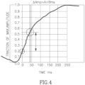
- Figure 4 depicts an illustrative example of an approach to determining a normalized upstroke index based on the detection of the systolic upstroke peak and trough values.
- Many other ways of determining an upstroke index may be employed, using conventional analytical methods such as determining the time interval between prescribed fractions of the upstroke amplitude, etc.
- Figure 5 shows the contrasting patterns of two series of such upstroke indices related to apnea and non-apnea states.
- the relative magnitude of the cyclic upstroke index changes in the apnea case are clearly much larger than for the non-apnea case, reflecting the greater fluctuations in intrathoracic pressure occurring during apneas due to greater fluctuations in respiratory effort during the course of a breathing cycle.
- the differences in the respective montages of serial upstrokes, normalized with respect to the trough to peak amplitude of each upstroke, and synchronized at the start of upstroke, is quite significant.
- the patient's individual level of breathing cycle related variability of the upstroke index values during non-apneic breathing may provide a functional threshold upon which to base the determination of a central apnea event, such that in the absence of breathing cycle related variability of the upstroke index values, or when such breathing cycle related pulse upstroke index variability is significantly less than during normal non-apneic breathing, absence of respiratory effort can be assumed to have occurred.
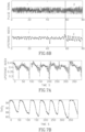
- Figure 6A illustrates the time-course of the upstroke index based on a series of consecutive pulse signals.
- the consistent pattern of breathing cycle related changes in the upstroke index can be clearly seen at 2, while the series of pulses appears to be of a generally consistent amplitude at 1.
- Figure 6B shows examples of similar pulse wave signals associated with noticeably different levels of upstroke index.
- the cyclic breathing related fluctuations of the upstroke index values are noticeably smaller than at 2, on the right side of Figure 6B .
- the pulsewave amplitudes in 1 and 2 are essentially the same, and importantly, no apparent differences can be discerned in the modulation of either series of pulsewaves. This helps to illustrate the advantage of the present method over the prior art.
- each upstroke index appears to be increasing towards the end of each apnea.
- the upstroke signal appears to not be fluctuating in several time periods during these measurements. This is especially noticeable as from the onset of the third and fourth apneic events, where the upstroke signal is not fluctuating but is gradually declining over time, and in relation to the level of blood oxygen saturation.
- This pattern of central and then obstructive apnea is characteristic of the so called mixed apnea.
- the progressive trend for the absolute value of the upstroke index to decline as the degree of saturation increases might also be useful to provide a relative index of declining myocardial contractility. This finding may have important clinical value in assessing the impact of blood oxygen desaturation on the heart.
- the pulse waves to be used in the inventive method are derived from any pulse detection methods known to the art, and may be derived from any part of body from which a pulse signal can be measured.
- Detection modalities include, but are not limited to, optical plethymographic signals, which may be derived from a pulse oximeter or any other kind of photo-optical signal device, and maybe obtained using either the known transmission or reflection modes, or any combination thereof, as well as any form of laser Doppler or Doppler system, volumetric plethysmographs, segmental plethysmographs, surface strain gauge and circumferential strain gauge devices, and any other devices which are affected by a change in the geometry of the body surface due to pulsewaves and blood volume changes, such as piezo and electromagnetic devices.
- the pulse waves to be used in the inventive method are derived from any one of the peripheral arterial tone (PAT) measurement devises mentioned above, including, for example: US 6319205 , US 6461305 , US 6916289 , US 6488633 , US6322515 , US7806831 , US 6939304 , US 7374540 , US 7621877 , US 7819811 , and corresponding foreign patents and patent applications, as well as the following currently pending applications in the USA and their respective corresponding foreign patents and patent applications; PCT/IL2012/937737 , PCT/IL2012/050466 , and PCT/IL/2011/662610 .
- PAT peripheral arterial tone
- the above PAT measurement devices confer substantial advantages to the measurement of the arterial pulse signals, including the prevention of blood pooling in the veins at the measuring site, the unloading of the tension in the arterial walls to promote optimal dynamic response of the signal, substantially prevent uncontrolled venous backflow at said measuring site, and prevent the occurrence of veno-arteriolar reflex vasoconstriction by preventing venous distension.
- the PAT probes may be applied to the digits of the hands or feet, or may be applied to any body surface region as described in US 7621877 "Body Surface Probe, Apparatus And Method For Non- Invasively Detecting Medical Conditions".
- the same PAT signal information from which the systolic upstroke index is derived may also be used in the sleep disordered breathing analysis for the clinically validated detection of a host of sleep related physiological and pathophysiological states and conditions which include sleep /wake determination, sleep apnea events, sleep hypopnea events, upper airway resistance syndrome (UARS) events, Cheyne-Stokes breathing events, REM (rapid eye movements) sleep stage, periodic leg movement syndrome (PLMS) and arousals related to disordered breathing events during sleep, as described in the above cited patents and patent applications.
- This embodiment is schematically illustrated in Figure 3B .
- the addition of the present inventive method to the previously described PAT related broad diagnostic spectrum of sleep related disorders may further enhance this diagnostic capability by facilitating the distinction between obstructive and central apneas, as well as providing a quantitative index of the degree of intra thoracic pressure changes associated with breathing, which may be of particular importance in the diagnosis and grading of subtle and hard to diagnose upper airway resistance syndrome (UARS) events.
- UARS upper airway resistance syndrome
- This may have far reaching clinical significance as it may provide a sensitive quantitative feedback index useful in optimizing the real time level of pressure application required by the patient from a therapeutic positive airway pressure (PAP) device.
- PAP therapeutic positive airway pressure
- inventive method of the present application may be particularly beneficially used in conjunction with the method and apparatus described in PCT/IL2009/000528 : "Method And Apparatus For Examining Subjects For Particular Physiological Conditions Utilizing Acoustic Information" since the apparatus used in that patent application includes both multi-axis accelerometric and sound recording apparatus which are coupled to the patient's torso.
- the accelerometric output can be used to provide a continuous signal which reflects the dynamic changes of the torso in relation to the patient's breathing.
- this type of information allows the nature of the events to be more accurately determined. For instance, an apneic event which is not associated with accelerometric changes can be identified as a central apnea, as opposed to an obstructive apnea event, and vice versa.
- the actual clinical performance of the combination of PAT related evaluation of sleep disordered breathing, and added accelerometric information has been found to accurately discriminate between obstructive and central apneas, with a sensitivity of 70.6%, and a specificity of 87.3%, relative to accepted clinical polysomnographic assessment.
- the analysis of such added chest wall motion information may be further improved with the addition of patient posture information, which may also be derived using multi-dimensional accelerometric data input, since the strength of the detected respiratory effort is affected by patient posture, for example, chest wall motion has weaker signal when the patient is in the prone position.
- patient posture information which may also be derived using multi-dimensional accelerometric data input, since the strength of the detected respiratory effort is affected by patient posture, for example, chest wall motion has weaker signal when the patient is in the prone position.
- the quantitative assessment of respiratory related sounds which as noted is a feature of PCT/IL2009/000528 : "Method And Apparatus For Examining Subjects For Particular Physiological Conditions Utilizing Acoustic Information" may help to clarify the sleep disordered diagnosis, particularly in distinguishing between central and obstructive apneas, since the presence of respiratory related sound will rule out the diagnosis of central apnea.
- a further way in which acoustic and/or accelerometric information can be used to differentiate between obstructive and central apneas, is related to their respective applications in the detection of cardiac cycle events, such as cardio-balistographic detection of ventricular contraction by accelerometry, and the acoustic detection of the heart sounds related to left ventricular systole, may be used in conjunction with the detection of peripheral pulsewaves to derive the PTT.
- characteristic dynamic changes in PTT may be used to recognize obstructive apneas and thereby allow the distinction between central and obstructive events to be made. (See, Argod J, Pépin JL, Lévy P; "Differentiating obstructive and central sleep respiratory events through pulse transit time”. Am J Respir Crit Care Med. 1998 Dec;158(6):1778-83 .).
- the incorporation of the accelerometric information into the present inventive method as depicted in Figure 9 may thus help in confirming the diagnosis based on the systolic upstroke analysis, and furthermore, may be helpful in cases in which cardiac arrhythmias are present which may affect the frequency of pulse waves available for systolic upstroke analysis, or in cases where the pulse signal data is absent or otherwise compromised, such as during severe peripheral vasoconstriction.
- the standalone embodiment of the invention without added apparatus applied to the torso as depicted in Figure 3B has the important advantages of requiring minimal patient instrumentation, a reduced possibility of device failure, allows for maximal patient comfort and freedom of movement, and greatly simplifies the patient instrumentation, which is most important in a home based ambulatory test which is self-applied by the patient.
- Figure 10 A further possible embodiment of the present invention is depicted in Figure 10 , which is essentially the same as that depicted in Figure 9 , but without analyzing the systolic upstroke.
- this embodiment provides a possible add-on means for distinguishing between central and obstructive sleep disordered events to an existing system in which pulse signals are not directly amenable for the described upstroke analysis. This might be helpful for example when the sampling rate of the pulsatile information is too low for accurately applying the upstroke analysis, or when the pulsatile information is presented as peak to trough amplitude differences only.
- pulsewave signal may also be applicable as a backup for the embodiment illustrated in Figure 9 , in the event that the pulsewave signal is temporarily unavailable, for example, when blood flow to the measurement site is absent or diminished due to occlusion during a blood pressure measurement, or due to physiologically mediated vasoconstriction, or due to failure of the signal.
Landscapes
- Health & Medical Sciences (AREA)
- Life Sciences & Earth Sciences (AREA)
- Engineering & Computer Science (AREA)
- Surgery (AREA)
- General Health & Medical Sciences (AREA)
- Biophysics (AREA)
- Pathology (AREA)
- Veterinary Medicine (AREA)
- Biomedical Technology (AREA)
- Heart & Thoracic Surgery (AREA)
- Medical Informatics (AREA)
- Molecular Biology (AREA)
- Public Health (AREA)
- Animal Behavior & Ethology (AREA)
- Physics & Mathematics (AREA)
- Physiology (AREA)
- Cardiology (AREA)
- Pulmonology (AREA)
- Artificial Intelligence (AREA)
- Computer Vision & Pattern Recognition (AREA)
- Psychiatry (AREA)
- Signal Processing (AREA)
- Hematology (AREA)
- Measuring Pulse, Heart Rate, Blood Pressure Or Blood Flow (AREA)
- Measurement Of The Respiration, Hearing Ability, Form, And Blood Characteristics Of Living Organisms (AREA)
Applications Claiming Priority (3)
| Application Number | Priority Date | Filing Date | Title |
|---|---|---|---|
| US201662347111P | 2016-06-08 | 2016-06-08 | |
| PCT/IB2017/053154 WO2017212370A1 (en) | 2016-06-08 | 2017-05-29 | Method and apparatus for non-invasive detection of physiological and patho-physiological sleep conditions |
| EP17809808.3A EP3468463B1 (de) | 2016-06-08 | 2017-05-29 | Verfahren und vorrichtung zur nichtinvasiven detektion von physiologischen und patho-physiologischen schlafzuständen |
Related Parent Applications (2)
| Application Number | Title | Priority Date | Filing Date |
|---|---|---|---|
| EP17809808.3A Division-Into EP3468463B1 (de) | 2016-06-08 | 2017-05-29 | Verfahren und vorrichtung zur nichtinvasiven detektion von physiologischen und patho-physiologischen schlafzuständen |
| EP17809808.3A Division EP3468463B1 (de) | 2016-06-08 | 2017-05-29 | Verfahren und vorrichtung zur nichtinvasiven detektion von physiologischen und patho-physiologischen schlafzuständen |
Publications (3)
| Publication Number | Publication Date |
|---|---|
| EP4403099A2 true EP4403099A2 (de) | 2024-07-24 |
| EP4403099A3 EP4403099A3 (de) | 2024-10-09 |
| EP4403099B1 EP4403099B1 (de) | 2026-03-18 |
Family
ID=60577640
Family Applications (2)
| Application Number | Title | Priority Date | Filing Date |
|---|---|---|---|
| EP24180528.2A Active EP4403099B1 (de) | 2016-06-08 | 2017-05-29 | System zur nichtinvasiven detektion von physiologischen und pathophysiologischen schlafzuständen |
| EP17809808.3A Active EP3468463B1 (de) | 2016-06-08 | 2017-05-29 | Verfahren und vorrichtung zur nichtinvasiven detektion von physiologischen und patho-physiologischen schlafzuständen |
Family Applications After (1)
| Application Number | Title | Priority Date | Filing Date |
|---|---|---|---|
| EP17809808.3A Active EP3468463B1 (de) | 2016-06-08 | 2017-05-29 | Verfahren und vorrichtung zur nichtinvasiven detektion von physiologischen und patho-physiologischen schlafzuständen |
Country Status (10)
| Country | Link |
|---|---|
| US (2) | US20190099125A1 (de) |
| EP (2) | EP4403099B1 (de) |
| JP (1) | JP6732052B2 (de) |
| CN (2) | CN109310347B (de) |
| AU (1) | AU2017278999B2 (de) |
| CA (1) | CA3025280A1 (de) |
| ES (1) | ES2986981T3 (de) |
| IL (1) | IL263182A (de) |
| RU (1) | RU2731311C2 (de) |
| WO (1) | WO2017212370A1 (de) |
Families Citing this family (24)
| Publication number | Priority date | Publication date | Assignee | Title |
|---|---|---|---|---|
| US8437843B1 (en) | 2006-06-16 | 2013-05-07 | Cleveland Medical Devices Inc. | EEG data acquisition system with novel features |
| US9202008B1 (en) | 2007-06-08 | 2015-12-01 | Cleveland Medical Devices Inc. | Method and device for sleep analysis |
| US10426399B1 (en) | 2007-06-08 | 2019-10-01 | Cleveland Medial Devices Inc. | Method and device for in-home sleep and signal analysis |
| WO2017184753A1 (en) | 2016-04-19 | 2017-10-26 | Inspire Medical Systems, Inc. | Accelerometer-based sensing for sleep disordered breathing (sdb) care |
| US10617302B2 (en) | 2016-07-07 | 2020-04-14 | Masimo Corporation | Wearable pulse oximeter and respiration monitor |
| EP4360546B1 (de) * | 2018-07-11 | 2025-02-19 | Ectosense NV | System zur schlafdiagnose |
| US12495968B2 (en) | 2018-10-12 | 2025-12-16 | Masimo Corporation | System for transmission of sensor data using dual communication protocol |
| AU2019357721B2 (en) | 2018-10-12 | 2025-05-08 | Masimo Corporation | System for transmission of sensor data using dual communication protocol |
| EP3943011B1 (de) * | 2019-03-22 | 2025-05-28 | Medical Optfellow Inc. | Computerprogramm und informationsverarbeitungsvorrichtung zur bestimmung der verschlimmerung der chronisch obstruktiven lungenerkrankung |
| JP7614171B2 (ja) | 2019-07-25 | 2025-01-15 | インスパイア・メディカル・システムズ・インコーポレイテッド | 検知された姿勢情報に基づいて植込み型医療デバイスを操作するためのシステムおよび方法 |
| AU2020319084B2 (en) * | 2019-07-25 | 2025-06-05 | Inspire Medical Systems, Inc. | Sleep detection for sleep disordered breathing (SDB) care |
| AU2020316105B2 (en) | 2019-07-25 | 2025-07-31 | Inspire Medical Systems, Inc. | Respiration detection |
| EP3928690B1 (de) * | 2020-06-26 | 2024-09-11 | Ectosense NV | Verfahren und vorrichtung zur beurteilung des peripheren arteriellen tonus |
| WO2022048871A1 (en) * | 2020-09-04 | 2022-03-10 | Ectosense NV | Method and apparatus for detecting sleep-disturbing events from a signal indicative of a peripheral arterial tone of an individual |
| CN112263242A (zh) * | 2020-10-26 | 2021-01-26 | 哈尔滨工业大学 | 基于fmcw雷达的呼吸检测及模式分类方法 |
| CN112656371A (zh) * | 2020-12-12 | 2021-04-16 | 深圳市苏仁智能科技有限公司 | 一种基于心率呼吸信号的人体睡眠体征检测方法及系统 |
| US12059266B2 (en) | 2020-12-30 | 2024-08-13 | Itamar Medical Ltd. | System and method for arrhythmia detection during an at home sleep test |
| EP4432919A1 (de) | 2022-01-05 | 2024-09-25 | Masimo Corporation | Am handgelenk und am finger getragenes pulsoximetriesystem |
| CN115089134A (zh) * | 2022-04-07 | 2022-09-23 | 深圳小梦智眠科技有限公司 | 用品组件及其工作方法 |
| CN115862877B (zh) * | 2023-03-03 | 2023-05-05 | 安徽星辰智跃科技有限责任公司 | 睡眠可持续性检测量化及辅助干预的方法、系统和装置 |
| WO2024217901A1 (en) * | 2023-04-21 | 2024-10-24 | Koninklijke Philips N.V. | Machine-learning model, detecting device, home sleep apnea test device, computer program product, ane computer-readable storage medium |
| EP4467068A1 (de) * | 2023-05-26 | 2024-11-27 | Koninklijke Philips N.V. | Verfahren, maschinenlernmodell, detektionsvorrichtung, heimschlafapnoetestvorrichtung, computerprogrammprodukt und computerlesbares speichermedium |
| US12605077B2 (en) | 2023-08-25 | 2026-04-21 | Pranaq Pte. Ltd. | Photoplethysmogram system and method |
| CN120436583B (zh) * | 2025-06-10 | 2026-02-13 | 南方医科大学第三附属医院(广东省骨科研究院) | 一种基于多模态数据的阻塞性睡眠呼吸暂停综合征检测方法、设备及程序产品 |
Citations (13)
| Publication number | Priority date | Publication date | Assignee | Title |
|---|---|---|---|---|
| US5385144A (en) | 1992-07-23 | 1995-01-31 | Minolta Co., Ltd. | Respiration diagnosis apparatus |
| US6319205B1 (en) | 1996-07-30 | 2001-11-20 | Itamar Medical (C.M.) 1997 Ltd. | Method and apparatus for the non-invasive detection of medical conditions by monitoring peripheral arterial tone |
| US6461305B1 (en) | 1998-06-07 | 2002-10-08 | Itamar Medical | Pressure applicator devices particularly useful for non-invasive detection of medical conditions |
| US6488633B1 (en) | 1999-07-14 | 2002-12-03 | Itamar Medical (C.M.) Ltd. | Probe devices particularly useful for non-invasive detection of medical conditions |
| US6669632B2 (en) | 2001-03-30 | 2003-12-30 | Denso Corporation | Apparatus and method for electronically predicting pleural pressure from pulse wave signals |
| US6856829B2 (en) | 2000-09-07 | 2005-02-15 | Denso Corporation | Method for detecting physiological condition of sleeping patient based on analysis of pulse waves |
| US6939304B2 (en) | 2000-10-23 | 2005-09-06 | Itamar Medical Ltd. | Method and apparatus for non-invasively evaluating endothelial activity in a patient |
| US7374540B2 (en) | 2001-04-05 | 2008-05-20 | Itamar Medical Ltd. | Non-invasive probe for detecting medical conditions |
| US7510531B2 (en) | 2003-09-18 | 2009-03-31 | Cardiac Pacemakers, Inc. | System and method for discrimination of central and obstructive disordered breathing events |
| US7621877B2 (en) | 2002-07-15 | 2009-11-24 | Itamar Medical Ltd. | Body surface probe, apparatus and method for non-invasively detecting medical conditions |
| US7806831B2 (en) | 2000-03-02 | 2010-10-05 | Itamar Medical Ltd. | Method and apparatus for the non-invasive detection of particular sleep-state conditions by monitoring the peripheral vascular system |
| US7819811B2 (en) | 2002-11-06 | 2010-10-26 | Itamar Medical Ltd. | Detecting medical conditions with noninvasive body probes |
| WO2011057116A1 (en) | 2009-11-05 | 2011-05-12 | Inovise Medical, Inc. | Detection and differentiation of sleep disordered breathing |
Family Cites Families (21)
| Publication number | Priority date | Publication date | Assignee | Title |
|---|---|---|---|---|
| US5178151A (en) * | 1988-04-20 | 1993-01-12 | Sackner Marvin A | System for non-invasive detection of changes of cardiac volumes and aortic pulses |
| SU1692548A1 (ru) * | 1988-10-25 | 1991-11-23 | Научно-Исследовательский Институт Нормальной Физиологии Им.П.К.Анохина | Способ контрол за функциональным состо нием человека |
| US7081095B2 (en) * | 2001-05-17 | 2006-07-25 | Lynn Lawrence A | Centralized hospital monitoring system for automatically detecting upper airway instability and for preventing and aborting adverse drug reactions |
| US6223064B1 (en) * | 1992-08-19 | 2001-04-24 | Lawrence A. Lynn | Microprocessor system for the simplified diagnosis of sleep apnea |
| JP3911843B2 (ja) * | 1998-04-28 | 2007-05-09 | オムロンヘルスケア株式会社 | 血圧監視装置 |
| US6129675A (en) * | 1998-09-11 | 2000-10-10 | Jay; Gregory D. | Device and method for measuring pulsus paradoxus |
| US6616613B1 (en) * | 2000-04-27 | 2003-09-09 | Vitalsines International, Inc. | Physiological signal monitoring system |
| US7024234B2 (en) * | 2002-09-20 | 2006-04-04 | Lyle Aaron Margulies | Method and apparatus for monitoring the autonomic nervous system |
| UA90651C2 (uk) * | 2002-10-09 | 2010-05-25 | Компьюмедикс Лимитед | Спосіб та пристрій для підтримування та контролювання якості сну при терапевтичному лікуванні |
| KR20050072435A (ko) * | 2002-10-09 | 2005-07-11 | 컴퓨메딕스 리미티드 | 치료 처리중 수면 품질을 유지하고 모니터하기 위한 방법및 장치 |
| US8467876B2 (en) * | 2003-10-15 | 2013-06-18 | Rmx, Llc | Breathing disorder detection and therapy delivery device and method |
| AU2005291858B2 (en) * | 2004-10-06 | 2011-07-28 | Resmed Limited | Method and apparatus for non-invasive monitoring of respiratory parameters in sleep disordered breathing |
| JP2007130182A (ja) * | 2005-11-09 | 2007-05-31 | Toshiba Corp | 照明制御装置、照明制御システム、照明制御方法および照明制御プログラム |
| NZ607280A (en) * | 2006-03-06 | 2014-06-27 | Resmed Ltd | Method and apparatus for improved flow limitation detection of obstructive sleep apnea |
| US8646447B2 (en) * | 2006-11-13 | 2014-02-11 | Resmed Limited | Systems, methods, and/or apparatuses for non-invasive monitoring of respiratory parameters in sleep disordered breathing |
| DE102007020038A1 (de) * | 2007-04-27 | 2008-10-30 | Fraunhofer-Gesellschaft zur Förderung der angewandten Forschung e.V. | Nachweis einer Apnoe mit blutdruckabhängig erfassten Signalen |
| WO2010036901A1 (en) * | 2008-09-26 | 2010-04-01 | University Of Louisville Research Foundation, Inc. | Methods and kits for diagnosing obstructive sleep apnea |
| ES2823307T3 (es) * | 2009-08-13 | 2021-05-06 | Hidetsugu Asanoi | Dispositivo para calcular información de forma de onda respiratoria y dispositivo médico que utiliza la información de forma de onda respiratoria |
| US20130006121A1 (en) * | 2010-03-16 | 2013-01-03 | The Johns Hopkins University | Device for Identifying Hemodynamic Changes |
| WO2012155257A1 (en) * | 2011-05-17 | 2012-11-22 | University Health Network | Osa/csa diagnosis using recorded breath sound amplitude profile and pitch contour |
| US8740806B2 (en) * | 2012-11-07 | 2014-06-03 | Somnarus Inc. | Methods for detection of respiratory effort and sleep apnea monitoring devices |
-
2017
- 2017-05-29 EP EP24180528.2A patent/EP4403099B1/de active Active
- 2017-05-29 ES ES17809808T patent/ES2986981T3/es active Active
- 2017-05-29 CN CN201780035115.6A patent/CN109310347B/zh active Active
- 2017-05-29 WO PCT/IB2017/053154 patent/WO2017212370A1/en not_active Ceased
- 2017-05-29 RU RU2018143323A patent/RU2731311C2/ru active
- 2017-05-29 JP JP2018563722A patent/JP6732052B2/ja active Active
- 2017-05-29 CN CN202211139291.3A patent/CN115517627A/zh active Pending
- 2017-05-29 EP EP17809808.3A patent/EP3468463B1/de active Active
- 2017-05-29 CA CA3025280A patent/CA3025280A1/en active Pending
- 2017-05-29 AU AU2017278999A patent/AU2017278999B2/en active Active
-
2018
- 2018-11-21 IL IL263182A patent/IL263182A/en unknown
- 2018-12-03 US US16/207,520 patent/US20190099125A1/en not_active Abandoned
-
2021
- 2021-10-25 US US17/509,226 patent/US20220211323A1/en active Pending
Patent Citations (15)
| Publication number | Priority date | Publication date | Assignee | Title |
|---|---|---|---|---|
| US5385144A (en) | 1992-07-23 | 1995-01-31 | Minolta Co., Ltd. | Respiration diagnosis apparatus |
| US6319205B1 (en) | 1996-07-30 | 2001-11-20 | Itamar Medical (C.M.) 1997 Ltd. | Method and apparatus for the non-invasive detection of medical conditions by monitoring peripheral arterial tone |
| US6322515B1 (en) | 1996-07-30 | 2001-11-27 | Itamar Medical | Method and apparatus for the non-invasive detection of medical conditions by monitoring peripheral arterial tone |
| US6916289B2 (en) | 1998-06-07 | 2005-07-12 | Itamar Medical Ltd. | Pressure applicator devices particularly useful for non-invasive detection of medical conditions |
| US6461305B1 (en) | 1998-06-07 | 2002-10-08 | Itamar Medical | Pressure applicator devices particularly useful for non-invasive detection of medical conditions |
| US6488633B1 (en) | 1999-07-14 | 2002-12-03 | Itamar Medical (C.M.) Ltd. | Probe devices particularly useful for non-invasive detection of medical conditions |
| US7806831B2 (en) | 2000-03-02 | 2010-10-05 | Itamar Medical Ltd. | Method and apparatus for the non-invasive detection of particular sleep-state conditions by monitoring the peripheral vascular system |
| US6856829B2 (en) | 2000-09-07 | 2005-02-15 | Denso Corporation | Method for detecting physiological condition of sleeping patient based on analysis of pulse waves |
| US6939304B2 (en) | 2000-10-23 | 2005-09-06 | Itamar Medical Ltd. | Method and apparatus for non-invasively evaluating endothelial activity in a patient |
| US6669632B2 (en) | 2001-03-30 | 2003-12-30 | Denso Corporation | Apparatus and method for electronically predicting pleural pressure from pulse wave signals |
| US7374540B2 (en) | 2001-04-05 | 2008-05-20 | Itamar Medical Ltd. | Non-invasive probe for detecting medical conditions |
| US7621877B2 (en) | 2002-07-15 | 2009-11-24 | Itamar Medical Ltd. | Body surface probe, apparatus and method for non-invasively detecting medical conditions |
| US7819811B2 (en) | 2002-11-06 | 2010-10-26 | Itamar Medical Ltd. | Detecting medical conditions with noninvasive body probes |
| US7510531B2 (en) | 2003-09-18 | 2009-03-31 | Cardiac Pacemakers, Inc. | System and method for discrimination of central and obstructive disordered breathing events |
| WO2011057116A1 (en) | 2009-11-05 | 2011-05-12 | Inovise Medical, Inc. | Detection and differentiation of sleep disordered breathing |
Non-Patent Citations (5)
| Title |
|---|
| ARGOD JPÉPIN JLLEVY P: "Differentiating obstructive and central sleep respiratory events through pulse transit time", AM J RESPIR CRIT CARE MED., vol. 158, no. 6, December 1998 (1998-12-01), pages 1778 - 83, XP002492057 |
| DILLIER R ET AL.: "Continuous respiratory monitoring for sleep apnea screening by ambulatory hemodynamic monitor", WORLD J CARDIOL., vol. 4, no. 4, 26 April 2012 (2012-04-26), pages 121 - 7 |
| MORILLO DS. ET AL.: "Monitoring and analysis of cardio respiratory and snoring signals by using an accelerometer", CONF PROC IEEE ENG MED BIOL SOC., vol. 2007, 2007, pages 3942 - 5 |
| MORILLO ET AL.: "An accelerometer-based device for sleep apnea screening", IEEE TRANS INF TECHNOL BIOMED., vol. 14, no. 2, March 2010 (2010-03-01), pages 491 - 9, XP011345650, DOI: 10.1109/TITB.2009.2027231 |
| P. DEHKORDI ET AL.: "Monitoring torso acceleration for estimating the respiratory flow and efforts for sleep apnea detection", CONF PROC IEEE ENG MED BIOL SOC, vol. 2012, 2012, pages 6345 - 8, XP032464385, DOI: 10.1109/EMBC.2012.6347445 |
Also Published As
| Publication number | Publication date |
|---|---|
| IL263182A (en) | 2018-12-31 |
| WO2017212370A1 (en) | 2017-12-14 |
| RU2018143323A (ru) | 2020-07-10 |
| JP2019520884A (ja) | 2019-07-25 |
| CN115517627A (zh) | 2022-12-27 |
| EP3468463B1 (de) | 2024-07-17 |
| US20220211323A1 (en) | 2022-07-07 |
| RU2731311C2 (ru) | 2020-09-01 |
| EP4403099B1 (de) | 2026-03-18 |
| ES2986981T3 (es) | 2024-11-13 |
| US20190099125A1 (en) | 2019-04-04 |
| EP3468463A4 (de) | 2020-03-04 |
| CN109310347B (zh) | 2022-09-20 |
| EP4403099A3 (de) | 2024-10-09 |
| RU2018143323A3 (de) | 2020-07-10 |
| EP3468463A1 (de) | 2019-04-17 |
| JP6732052B2 (ja) | 2020-07-29 |
| AU2017278999A1 (en) | 2018-12-06 |
| CA3025280A1 (en) | 2017-12-14 |
| AU2017278999B2 (en) | 2020-06-25 |
| CN109310347A (zh) | 2019-02-05 |
Similar Documents
| Publication | Publication Date | Title |
|---|---|---|
| US20220211323A1 (en) | Method and apparatus for non-invasive detection of physiological and patho-physiological sleep conditions | |
| Hung et al. | Estimation of respiratory waveform using an accelerometer | |
| EP3927234B1 (de) | Schlafüberwachungs- und positionstherapiesystem und -verfahren | |
| CN110035691B (zh) | 用于测量睡眠呼吸暂停的方法和设备 | |
| US10092268B2 (en) | Method and apparatus to monitor physiologic and biometric parameters using a non-invasive set of transducers | |
| EP2840962B1 (de) | Vorrichtung und computerprogramm zur erzeugung eines vorhofflimmern ausdrückenden signals | |
| JP6894451B2 (ja) | 被検者の睡眠段階を判定するための判定システム及び方法 | |
| US20080269583A1 (en) | Detection and Monitoring of Stress Events During Sleep | |
| CN112512419B (zh) | 提供对象的时间信息 | |
| JP6627112B2 (ja) | 生体機能検査装置、生体機能検査装置の作動方法及びプログラム | |
| KR101885981B1 (ko) | 무구속적으로 측정된 심폐신호를 이용한 수면효율의 예측방법 | |
| Shahshahani et al. | Motion artifact reduction for respiratory monitoring: A multichannel ultrasound sensor for diaphragm tracking | |
| US20160287092A1 (en) | Blood vessel mechanical signal analysis | |
| Heise et al. | Unobtrusively detecting apnea and hypopnea events via a hydraulic bed sensor | |
| JP7288334B2 (ja) | 睡眠状態推定装置、睡眠状態推定装置の作動方法及び睡眠状態推定プログラム | |
| Kamelia et al. | The Role of New Pulmonary Artery Wedge Pressure Formula to Predict Diastolic Dysfunction in Obstructive Sleep Apnea | |
| Park et al. | Ballistocardiography | |
| Sakai et al. | Development of lead system for ECG-derived respiration aimed at detection of obstructive sleep apnea syndrome | |
| EP3476275A1 (de) | Physiologische signalverarbeitungsvorrichtung und -verfahren | |
| JP7658924B2 (ja) | 心拍間隔検出システム | |
| Min | Technological Trends in Respiratory Effort Sensors for Sleep Testing | |
| Sleep | Microarousal Recognition |
Legal Events
| Date | Code | Title | Description |
|---|---|---|---|
| PUAI | Public reference made under article 153(3) epc to a published international application that has entered the european phase |
Free format text: ORIGINAL CODE: 0009012 |
|
| STAA | Information on the status of an ep patent application or granted ep patent |
Free format text: STATUS: THE APPLICATION HAS BEEN PUBLISHED |
|
| AC | Divisional application: reference to earlier application |
Ref document number: 3468463 Country of ref document: EP Kind code of ref document: P |
|
| AK | Designated contracting states |
Kind code of ref document: A2 Designated state(s): AL AT BE BG CH CY CZ DE DK EE ES FI FR GB GR HR HU IE IS IT LI LT LU LV MC MK MT NL NO PL PT RO RS SE SI SK SM TR |
|
| PUAL | Search report despatched |
Free format text: ORIGINAL CODE: 0009013 |
|
| AK | Designated contracting states |
Kind code of ref document: A3 Designated state(s): AL AT BE BG CH CY CZ DE DK EE ES FI FR GB GR HR HU IE IS IT LI LT LU LV MC MK MT NL NO PL PT RO RS SE SI SK SM TR |
|
| RIC1 | Information provided on ipc code assigned before grant |
Ipc: A61B 5/02 20060101AFI20240905BHEP |
|
| STAA | Information on the status of an ep patent application or granted ep patent |
Free format text: STATUS: REQUEST FOR EXAMINATION WAS MADE |
|
| 17P | Request for examination filed |
Effective date: 20250318 |
|
| GRAP | Despatch of communication of intention to grant a patent |
Free format text: ORIGINAL CODE: EPIDOSNIGR1 |
|
| STAA | Information on the status of an ep patent application or granted ep patent |
Free format text: STATUS: GRANT OF PATENT IS INTENDED |
|
| INTG | Intention to grant announced |
Effective date: 20250603 |
|
| GRAJ | Information related to disapproval of communication of intention to grant by the applicant or resumption of examination proceedings by the epo deleted |
Free format text: ORIGINAL CODE: EPIDOSDIGR1 |
|
| STAA | Information on the status of an ep patent application or granted ep patent |
Free format text: STATUS: REQUEST FOR EXAMINATION WAS MADE |
|
| INTC | Intention to grant announced (deleted) | ||
| GRAP | Despatch of communication of intention to grant a patent |
Free format text: ORIGINAL CODE: EPIDOSNIGR1 |
|
| STAA | Information on the status of an ep patent application or granted ep patent |
Free format text: STATUS: GRANT OF PATENT IS INTENDED |
|
| P01 | Opt-out of the competence of the unified patent court (upc) registered |
Free format text: CASE NUMBER: UPC_APP_7371_4403099/2025 Effective date: 20250917 |
|
| INTG | Intention to grant announced |
Effective date: 20251020 |
|
| GRAS | Grant fee paid |
Free format text: ORIGINAL CODE: EPIDOSNIGR3 |
|
| GRAA | (expected) grant |
Free format text: ORIGINAL CODE: 0009210 |
|
| STAA | Information on the status of an ep patent application or granted ep patent |
Free format text: STATUS: THE PATENT HAS BEEN GRANTED |
|
| AC | Divisional application: reference to earlier application |
Ref document number: 3468463 Country of ref document: EP Kind code of ref document: P |
|
| AK | Designated contracting states |
Kind code of ref document: B1 Designated state(s): AL AT BE BG CH CY CZ DE DK EE ES FI FR GB GR HR HU IE IS IT LI LT LU LV MC MK MT NL NO PL PT RO RS SE SI SK SM TR |
|
| REG | Reference to a national code |
Ref country code: CH Ref legal event code: F10 Free format text: ST27 STATUS EVENT CODE: U-0-0-F10-F00 (AS PROVIDED BY THE NATIONAL OFFICE) Effective date: 20260318 Ref country code: GB Ref legal event code: FG4D |
|
| REG | Reference to a national code |
Ref country code: IE Ref legal event code: FG4D |
|
| REG | Reference to a national code |
Ref country code: DE Ref legal event code: R096 Ref document number: 602017094429 Country of ref document: DE |