EP4414459A2 - Simultane räumlich-zeitliche messung der genexpression und der zellaktivität - Google Patents
Simultane räumlich-zeitliche messung der genexpression und der zellaktivität Download PDFInfo
- Publication number
- EP4414459A2 EP4414459A2 EP24182415.0A EP24182415A EP4414459A2 EP 4414459 A2 EP4414459 A2 EP 4414459A2 EP 24182415 A EP24182415 A EP 24182415A EP 4414459 A2 EP4414459 A2 EP 4414459A2
- Authority
- EP
- European Patent Office
- Prior art keywords
- analyte
- capture
- substrate
- gasket
- sample
- Prior art date
- Legal status (The legal status is an assumption and is not a legal conclusion. Google has not performed a legal analysis and makes no representation as to the accuracy of the status listed.)
- Granted
Links
Images













Classifications
-
- C—CHEMISTRY; METALLURGY
- C12—BIOCHEMISTRY; BEER; SPIRITS; WINE; VINEGAR; MICROBIOLOGY; ENZYMOLOGY; MUTATION OR GENETIC ENGINEERING
- C12Q—MEASURING OR TESTING PROCESSES INVOLVING ENZYMES, NUCLEIC ACIDS OR MICROORGANISMS; COMPOSITIONS OR TEST PAPERS THEREFOR; PROCESSES OF PREPARING SUCH COMPOSITIONS; CONDITION-RESPONSIVE CONTROL IN MICROBIOLOGICAL OR ENZYMOLOGICAL PROCESSES
- C12Q1/00—Measuring or testing processes involving enzymes, nucleic acids or microorganisms; Compositions therefor; Processes of preparing such compositions
- C12Q1/68—Measuring or testing processes involving enzymes, nucleic acids or microorganisms; Compositions therefor; Processes of preparing such compositions involving nucleic acids
- C12Q1/6806—Preparing nucleic acids for analysis, e.g. for polymerase chain reaction [PCR] assay
-
- C—CHEMISTRY; METALLURGY
- C12—BIOCHEMISTRY; BEER; SPIRITS; WINE; VINEGAR; MICROBIOLOGY; ENZYMOLOGY; MUTATION OR GENETIC ENGINEERING
- C12Q—MEASURING OR TESTING PROCESSES INVOLVING ENZYMES, NUCLEIC ACIDS OR MICROORGANISMS; COMPOSITIONS OR TEST PAPERS THEREFOR; PROCESSES OF PREPARING SUCH COMPOSITIONS; CONDITION-RESPONSIVE CONTROL IN MICROBIOLOGICAL OR ENZYMOLOGICAL PROCESSES
- C12Q1/00—Measuring or testing processes involving enzymes, nucleic acids or microorganisms; Compositions therefor; Processes of preparing such compositions
- C12Q1/68—Measuring or testing processes involving enzymes, nucleic acids or microorganisms; Compositions therefor; Processes of preparing such compositions involving nucleic acids
- C12Q1/6813—Hybridisation assays
- C12Q1/6816—Hybridisation assays characterised by the detection means
- C12Q1/6818—Hybridisation assays characterised by the detection means involving interaction of two or more labels, e.g. resonant energy transfer
-
- C—CHEMISTRY; METALLURGY
- C12—BIOCHEMISTRY; BEER; SPIRITS; WINE; VINEGAR; MICROBIOLOGY; ENZYMOLOGY; MUTATION OR GENETIC ENGINEERING
- C12N—MICROORGANISMS OR ENZYMES; COMPOSITIONS THEREOF; PROPAGATING, PRESERVING, OR MAINTAINING MICROORGANISMS; MUTATION OR GENETIC ENGINEERING; CULTURE MEDIA
- C12N5/00—Undifferentiated human, animal or plant cells, e.g. cell lines; Tissues; Cultivation or maintenance thereof; Culture media therefor
- C12N5/0062—General methods for three-dimensional culture
-
- C—CHEMISTRY; METALLURGY
- C12—BIOCHEMISTRY; BEER; SPIRITS; WINE; VINEGAR; MICROBIOLOGY; ENZYMOLOGY; MUTATION OR GENETIC ENGINEERING
- C12Q—MEASURING OR TESTING PROCESSES INVOLVING ENZYMES, NUCLEIC ACIDS OR MICROORGANISMS; COMPOSITIONS OR TEST PAPERS THEREFOR; PROCESSES OF PREPARING SUCH COMPOSITIONS; CONDITION-RESPONSIVE CONTROL IN MICROBIOLOGICAL OR ENZYMOLOGICAL PROCESSES
- C12Q1/00—Measuring or testing processes involving enzymes, nucleic acids or microorganisms; Compositions therefor; Processes of preparing such compositions
- C12Q1/68—Measuring or testing processes involving enzymes, nucleic acids or microorganisms; Compositions therefor; Processes of preparing such compositions involving nucleic acids
- C12Q1/6804—Nucleic acid analysis using immunogens
-
- C—CHEMISTRY; METALLURGY
- C12—BIOCHEMISTRY; BEER; SPIRITS; WINE; VINEGAR; MICROBIOLOGY; ENZYMOLOGY; MUTATION OR GENETIC ENGINEERING
- C12Q—MEASURING OR TESTING PROCESSES INVOLVING ENZYMES, NUCLEIC ACIDS OR MICROORGANISMS; COMPOSITIONS OR TEST PAPERS THEREFOR; PROCESSES OF PREPARING SUCH COMPOSITIONS; CONDITION-RESPONSIVE CONTROL IN MICROBIOLOGICAL OR ENZYMOLOGICAL PROCESSES
- C12Q1/00—Measuring or testing processes involving enzymes, nucleic acids or microorganisms; Compositions therefor; Processes of preparing such compositions
- C12Q1/68—Measuring or testing processes involving enzymes, nucleic acids or microorganisms; Compositions therefor; Processes of preparing such compositions involving nucleic acids
- C12Q1/6813—Hybridisation assays
- C12Q1/6834—Enzymatic or biochemical coupling of nucleic acids to a solid phase
- C12Q1/6837—Enzymatic or biochemical coupling of nucleic acids to a solid phase using probe arrays or probe chips
-
- C—CHEMISTRY; METALLURGY
- C12—BIOCHEMISTRY; BEER; SPIRITS; WINE; VINEGAR; MICROBIOLOGY; ENZYMOLOGY; MUTATION OR GENETIC ENGINEERING
- C12Q—MEASURING OR TESTING PROCESSES INVOLVING ENZYMES, NUCLEIC ACIDS OR MICROORGANISMS; COMPOSITIONS OR TEST PAPERS THEREFOR; PROCESSES OF PREPARING SUCH COMPOSITIONS; CONDITION-RESPONSIVE CONTROL IN MICROBIOLOGICAL OR ENZYMOLOGICAL PROCESSES
- C12Q1/00—Measuring or testing processes involving enzymes, nucleic acids or microorganisms; Compositions therefor; Processes of preparing such compositions
- C12Q1/68—Measuring or testing processes involving enzymes, nucleic acids or microorganisms; Compositions therefor; Processes of preparing such compositions involving nucleic acids
- C12Q1/6813—Hybridisation assays
- C12Q1/6841—In situ hybridisation
-
- C—CHEMISTRY; METALLURGY
- C12—BIOCHEMISTRY; BEER; SPIRITS; WINE; VINEGAR; MICROBIOLOGY; ENZYMOLOGY; MUTATION OR GENETIC ENGINEERING
- C12Q—MEASURING OR TESTING PROCESSES INVOLVING ENZYMES, NUCLEIC ACIDS OR MICROORGANISMS; COMPOSITIONS OR TEST PAPERS THEREFOR; PROCESSES OF PREPARING SUCH COMPOSITIONS; CONDITION-RESPONSIVE CONTROL IN MICROBIOLOGICAL OR ENZYMOLOGICAL PROCESSES
- C12Q1/00—Measuring or testing processes involving enzymes, nucleic acids or microorganisms; Compositions therefor; Processes of preparing such compositions
- C12Q1/68—Measuring or testing processes involving enzymes, nucleic acids or microorganisms; Compositions therefor; Processes of preparing such compositions involving nucleic acids
- C12Q1/6876—Nucleic acid products used in the analysis of nucleic acids, e.g. primers or probes
-
- G—PHYSICS
- G01—MEASURING; TESTING
- G01N—INVESTIGATING OR ANALYSING MATERIALS BY DETERMINING THEIR CHEMICAL OR PHYSICAL PROPERTIES
- G01N33/00—Investigating or analysing materials by specific methods not covered by groups G01N1/00 - G01N31/00
- G01N33/48—Biological material, e.g. blood, urine; Haemocytometers
- G01N33/50—Chemical analysis of biological material, e.g. blood, urine; Testing involving biospecific ligand binding methods; Immunological testing
- G01N33/5005—Chemical analysis of biological material, e.g. blood, urine; Testing involving biospecific ligand binding methods; Immunological testing involving human or animal cells
- G01N33/5008—Chemical analysis of biological material, e.g. blood, urine; Testing involving biospecific ligand binding methods; Immunological testing involving human or animal cells for testing or evaluating the effect of chemical or biological compounds, e.g. drugs, cosmetics
- G01N33/502—Chemical analysis of biological material, e.g. blood, urine; Testing involving biospecific ligand binding methods; Immunological testing involving human or animal cells for testing or evaluating the effect of chemical or biological compounds, e.g. drugs, cosmetics for testing non-proliferative effects
- G01N33/5023—Chemical analysis of biological material, e.g. blood, urine; Testing involving biospecific ligand binding methods; Immunological testing involving human or animal cells for testing or evaluating the effect of chemical or biological compounds, e.g. drugs, cosmetics for testing non-proliferative effects on expression patterns
-
- G—PHYSICS
- G01—MEASURING; TESTING
- G01N—INVESTIGATING OR ANALYSING MATERIALS BY DETERMINING THEIR CHEMICAL OR PHYSICAL PROPERTIES
- G01N33/00—Investigating or analysing materials by specific methods not covered by groups G01N1/00 - G01N31/00
- G01N33/48—Biological material, e.g. blood, urine; Haemocytometers
- G01N33/50—Chemical analysis of biological material, e.g. blood, urine; Testing involving biospecific ligand binding methods; Immunological testing
- G01N33/5005—Chemical analysis of biological material, e.g. blood, urine; Testing involving biospecific ligand binding methods; Immunological testing involving human or animal cells
- G01N33/5008—Chemical analysis of biological material, e.g. blood, urine; Testing involving biospecific ligand binding methods; Immunological testing involving human or animal cells for testing or evaluating the effect of chemical or biological compounds, e.g. drugs, cosmetics
- G01N33/5082—Supracellular entities, e.g. tissue, organisms
- G01N33/5088—Supracellular entities, e.g. tissue, organisms of vertebrates
-
- G—PHYSICS
- G01—MEASURING; TESTING
- G01N—INVESTIGATING OR ANALYSING MATERIALS BY DETERMINING THEIR CHEMICAL OR PHYSICAL PROPERTIES
- G01N33/00—Investigating or analysing materials by specific methods not covered by groups G01N1/00 - G01N31/00
- G01N33/48—Biological material, e.g. blood, urine; Haemocytometers
- G01N33/50—Chemical analysis of biological material, e.g. blood, urine; Testing involving biospecific ligand binding methods; Immunological testing
- G01N33/53—Immunoassay; Biospecific binding assay; Materials therefor
- G01N33/543—Immunoassay; Biospecific binding assay; Materials therefor with an insoluble carrier for immobilising immunochemicals
- G01N33/54306—Solid-phase reaction mechanisms
-
- C—CHEMISTRY; METALLURGY
- C12—BIOCHEMISTRY; BEER; SPIRITS; WINE; VINEGAR; MICROBIOLOGY; ENZYMOLOGY; MUTATION OR GENETIC ENGINEERING
- C12Q—MEASURING OR TESTING PROCESSES INVOLVING ENZYMES, NUCLEIC ACIDS OR MICROORGANISMS; COMPOSITIONS OR TEST PAPERS THEREFOR; PROCESSES OF PREPARING SUCH COMPOSITIONS; CONDITION-RESPONSIVE CONTROL IN MICROBIOLOGICAL OR ENZYMOLOGICAL PROCESSES
- C12Q2600/00—Oligonucleotides characterized by their use
- C12Q2600/158—Expression markers
-
- G—PHYSICS
- G01—MEASURING; TESTING
- G01N—INVESTIGATING OR ANALYSING MATERIALS BY DETERMINING THEIR CHEMICAL OR PHYSICAL PROPERTIES
- G01N2458/00—Labels used in chemical analysis of biological material
- G01N2458/10—Oligonucleotides as tagging agents for labelling antibodies
Definitions
- Cells within a tissue of a subject have differences in cell morphology and/or function due to varied analyte levels (e.g., gene and/or protein expression) within the different cells.
- the specific position of a cell within a tissue e.g., the cell's position relative to neighboring cells or the cell's position relative to the tissue microenvironment
- Spatial gene expression technology generally allows for the capture of gene transcripts from frozen or fixed tissues while maintaining the spatial positioning of the gene transcripts within the tissues.
- the frozen and/or fixed nature of the sample limits concurrent experiments associated with the temporal aspect of gene expression, particularly in live cells and during conditions in which the sample is manipulated, for example, using pharmacological compositions.
- spatial gene expression technology workflows generally are not amenable to the use of live tissues or cells; as such, the study or tracking of cellular activities is not available.
- live tissue sections e.g., generated through a vibratome
- cells e.g., cultured directly on the substrate
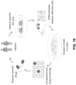
- the present disclosure provides methods for tracking temporal information, e.g., by recording cellular activity and gene expression, in combination with spatial gene expression arrays for simultaneous spatio-temporal measurements.
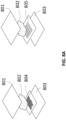
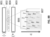
- the present disclosure also provides devices, e.g., a perfusion chamber system or flowcell mounted to the spatial array, and a multi-well plate system, for such measurements.
- devices e.g., a perfusion chamber system or flowcell mounted to the spatial array, and a multi-well plate system, for such measurements.
- drug screening methods using the spatio-temporal measurements to generate a comprehensive multiomic understanding of drug's impact on tissue or cell culture.
- a method for identifying location and/or abundance of an analyte in a biological sample comprising: (a) recording a cellular activity and/or an intracellular gene expression of a nucleic acid of the biological sample, and in some embodiments, the biological sample is located within a perfusion chamber in a plurality of perfusion chambers, and the perfusion chamber comprises a substrate; (b) contacting the biological sample with the substrate comprising a plurality of capture probes, and in some embodiments, a capture probe of the plurality of the capture probes comprises (i) a spatial barcode and (ii) a capture domain that binds specifically to a sequence present in the analyte; (c) extending the capture probe using the analyte that is specifically bound to the capture domain as a template, thereby generating an extended capture probe; (d) amplifying the extended capture probe to produce a plurality of extended capture probes; and (e) determining (i) all or a portion of
- the method for identifying a location of an analyte in a biological sample further comprising: mounting a gasket onto the substrate and mounting a cover onto the gasket to define a plurality of perfusion chambers.
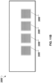
- the substrate comprises a plurality of substrate regions.
- a substrate region of the plurality comprises the capture probe comprising (i) the spatial barcode and (ii) the capture domain that binds specifically to the sequence present in the analyte.
- the gasket includes (i) a plurality of apertures corresponding to the plurality of substrate regions, respectively, (ii) a plurality of input channels being fluidly connected to the plurality of apertures, respectively, and (iii) a plurality of output channels being fluidly connected to the plurality of apertures, respectively.
- the plurality of apertures of the gasket are aligned with the plurality of substrate regions of the substrate when the gasket is mounted onto the substrate.
- the cover includes an inlet and an outlet, the inlet being fluidly connected to the plurality of input channels when the cover is mounted onto the gasket, the outlet being fluidly connected to the plurality of output channels when the cover is mounted onto the gasket.
- the plurality of perfusion chambers are defined by (i) the plurality of substrate regions of the substrate, (ii) the plurality of apertures of the gasket that is mounted onto the substrate, and (iii) the cover that is mounted onto the gasket.
- the mounting a gasket onto the substrate and the mounting a cover onto the gasket to define a plurality of perfusion chambers occur prior to the recording the cellular activity or the recording the intracellular gene expression.
- the method further comprises perfusing a test compound through the perfusion chamber before the recording step.
- the test compound is an agonist.
- the test compound is an antagonist.
- the test compound activates the cellular activity.
- the test compound inhibits the cellular activity.
- a method for identifying location and/or abundance of an analyte in a biological sample comprising: (a) recording a cellular activity and/or an intracellular gene expression of a nucleic acid of the biological sample, and in some embodiments, the biological sample is located within a well of a multi-well plate; (b) contacting the biological sample with a substrate comprising a plurality of capture probes, and in some embodiments, a capture probe of the plurality of the capture probes comprises (i) a spatial barcode and (ii) a capture domain that binds specifically to a sequence present in the analyte; (c) extending the capture probe using the analyte that is specifically bound to the capture domain as a template, thereby generating an extended capture probe; (d) amplifying the extended capture probe to produce a plurality of extended capture probes; and (e) determining (i) all or a portion of the sequence of the spatial barcode, or a complement thereof,
- provided herein is the method for identifying a location of an analyte in a biological sample, further comprising treating the biological sample with one or more drugs.
- a method for determining the effect of one or more drugs applied on a cellular activity and/or an intracellular gene expression of a nucleic acid in a biological sample comprising: (a) culturing the biological sample on a substrate, and in some embodiments, the substrate comprises a plurality of capture probes, and in some embodiments, a capture probe of the plurality of the capture probes comprises (i) a spatial barcode and (ii) a capture domain that is capable to bind specifically to a an analyte associated with the cellular activity and/or the intracellular gene expression in the biological sample; (b) treating the biological sample with the one or more drugs; (c) recording the cellular activity and/or the intracellular gene expression of the nucleic acid of the biological sample; (d) capturing the analyte from the biological sample, and in some embodiments, the analyte is captured by the capture domain of the capture probe; and (e) determining the effect of the one or more drugs applied on
- the multi-well plate is a 6-well plate, an 8-well plate, a 12-well plate, a 24-well plate, a 48-well plate, or a 96-well plate. In some embodiments, the multi-well plate is heat-resistant. In some embodiments, the multi-well plate is capable for automatic detecting of the cellular activity and/or the intracellular gene expression.
- the plurality of capture probes are directly attached to a surface of the well. In some embodiments, the plurality of capture probes are attached to the substrate. In some embodiments, the substrate is within the well. In some embodiments, the substrate is a coverslip. In some embodiments, the coverslip comprises plastic, metal, or glass. In some embodiments, the capture probe is attached to the substrate via a linkage group. In some embodiments, the linkage group is an amide group.
- the well or the substrate comprises a fiducial marker.
- the treating step occurs prior to the recording step. In some embodiments, the treating step and the recording step occur at substantially the same time.
- the biological sample is treated with the one or more drugs at substantially the same time. In some embodiments, the biological sample is treated with the one or more drugs at different times. In some embodiments, the biological sample is treated with one or more drugs about 6 hours, 12 hours, 18 hours, 24 hours, 30 hours, 36 hours, 42 hours, or 48 hours prior to fixing the biological sample.
- a drug of the one or more drugs is an agonist. In some embodiments, a drug of the one or more drugs is an antagonist.
- the biological sample is treated with a drug, and the drug is a small molecule.
- a drug of the one or more drugs activates the cellular activity and/or the intracellular gene expression. In some embodiments, a drug of the one or more drugs inhibits the cellular activity and/or the intracellular gene expression.
- a drug of the one or more drugs is conjugated with a fluorophore, and/or an oligonucleotide.
- the oligonucleotide comprises a sequence that uniquely identifies the drug.
- the biological sample is cultured in a culture medium to maintain its viability.
- the culture medium is replaced at an appropriate interval manually or automatically.
- the biological sample is cultured statically.
- the biological sample is cultured in the perfusion chamber, and the method further comprising perfusing the culture medium within the perfusion chamber.
- the culture medium is supplemented with oxygen.
- the culture medium comprises a blocking reagent.
- the biological sample is treated with the blocking reagent.
- the blocking reagent is bovine serum albumin (BSA), serum, gelatin (e.g., fish gelatin), milk (e.g., non-fat dry milk), casein, polyethylene glycol (PEG), polyvinyl alcohol (PVA), or polyvinylpyrrolidone (PVP), biotin blocking reagent, a peroxidase blocking reagent, levamisole, Carnoy's solution, glycine, lysine, sodium borohydride, pontamine sky blue, Sudan Black, trypan blue, FITC blocking agent, and/or acetic acid.
- BSA bovine serum albumin
- serum serum
- gelatin e.g., fish gelatin
- milk e.g., non-fat dry milk
- casein polyethylene glycol (PEG), polyvinyl alcohol (PVA), or polyvinylpyrrolidone (PVP)
- the cellular activity comprises protein activity, phosphorylation activity, G protein-coupled receptor related activity, ion channel activity, ligand-receptor binding activity, neural activity, protein synthesis, protein expression and localization, transient optical activity, cell-to-cell interactions, cellular morphology, or combinations thereof.
- the recording comprises optical recording.
- the optical recording comprises contacting the biological sample with a chemical dye.
- the chemical dye is a voltage-sensitive dye, a pH-sensitive dye, a temperature-sensitive dye, a light-sensitive dye, an oxygen-sensitive dye, or a metal sensitive dye.
- the chemical dye is a metal-sensitive dye (e.g., a calcium-sensitive dye).
- the optical recording comprises labelling the biological sample with an indicator.
- the indicator is a genetically-encoded indicator.
- the genetically-encoded indicator is a genetically-encoded neural activity indicator, a genetically-encoded voltage indicator (GEVI) or a genetically-encoded calcium indicator (GECI, or GCaMP).
- the chemical dye or the indicator further comprises a fluorophore.
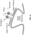
- the optical recording is achieved by in situ hybridization. In some embodiments, the optical recording is achieved by fluorescence resonance energy transfer (FRET).
- FRET fluorescence resonance energy transfer
- the optical recording comprises hybridization of a plurality of optically-labelled probes to (a) a protein, a lipid, a nucleic acid or a combination thereof associated with the cellular activity; or (b) the nucleic acid associated with the intracellular gene expression.
- an optically-labelled probe of the plurality is a peptide nucleic acid (PNA) probe labelled with a fluorophore.
- PNA peptide nucleic acid
- the PNA probe is at least 10 nucleic acid, at least 15 nucleic acids, at least 20 nucleic acids, at least 25 nucleic acids, at least 30 nucleic acids or more.
- the nucleic acid hybridized to the PNA probe is DNA.
- the nucleic acid hybridized to the PNA probe is RNA.
- the RNA is mRNA.
- the optical recording is conducted using fluorescent time-lapse microscopy.
- the recording step occurs prior to the contacting step. In some embodiments, the contacting step occurs prior to the recording step. In some embodiments, the recording step and the contacting step occur at substantially the same time.
- the biological sample is a tissue sample. In some embodiments, the tissue sample is a live tissue section.
- the tissue sample is an organoid sample or a spheroid culture sample.
- the tissue sample is an organoid sample.
- the organoid sample comprises normal organoids and/or cancer organoids.
- the organoid sample comprises cancer organoids.
- the organoid sample comprises intestinal organoids, liver organoids, pulmonary organoids, and/or neural organoids.
- the organoid sample is originated from disease-affected tissues (e.g., cancer tissues), normal tissue, disease-affected cells (e.g., cancer cells), normal cells, differentiated cells (e.g., somatic cells), and/or stem cells.
- the organoid sample is originated from stem cells.
- the stem cells are embryonic stem cells, induced pluripotent stem cells, and/or somatic stem cells.
- the tissue sample is embedded in hydrogels.
- the biological sample is a cell culture sample.
- the cell sample is a primary cell culture sample.
- the primary cell culture sample comprises individual cells that are isolated from a fresh tissue.
- the cell sample comprises a plurality of adherent cells.
- the cell sample comprises a plurality of suspension cells.
- the cell sample is transferred to the well from a separate cell culture.
- the cell sample comprises a plurality of disease-affected cells.
- one or more cells of the cell sample are transfected or infected.
- the biological sample is from a human patient or a model animal (e.g., a mouse).
- the analyte comprises a mutation. In some embodiments, the analyte comprises a single nucleotide polymorphism (SNP). In some embodiments, the analyte comprises a trinucleotide repeat. In some embodiments, the analyte is associated with a disease or condition.
- SNP single nucleotide polymorphism
- the analyte comprises a trinucleotide repeat. In some embodiments, the analyte is associated with a disease or condition.
- the analyte is a DNA molecule. In some embodiments, the analyte is a complementary DNA (cDNA). In some embodiments, the analyte is an RNA molecule. In some embodiments, the RNA molecule is an mRNA molecule.
- the capture domain of the capture probe is blocked prior to the contacting step. In some embodiments, the capture domain is blocked by a blocking probe.
- the biological sample comprises a plurality of live cells, and the live cells are stained by immunofluorescence before the recording step.
- the one or more live cells are treated with protease K and/or trypsin.
- the biological sample is stained using a detectable label.
- the detectable label is Can-Grunwald, Giemsa, hematoxylin and eosin (H&E), Jenner's, Leishman, Masson's trichrome, Papanicolaou, Romanowsky, silver, Sudan, Wright's, and/or Periodic Acid Schiff (PAS).
- the detectable label is H&E.
- the biological sample is imaged. In some embodiments, the biological samples is imaged using brightfield imaging.
- the biological sample is permeabilized after the recording step with a permeabilization agent selected from an organic solvent, a cross-linking agent, a detergent, and an enzyme, or a combination thereof.
- a permeabilization agent selected from an organic solvent, a cross-linking agent, a detergent, and an enzyme, or a combination thereof.
- the biological sample is fixed after contacting the biological sample with the substrate, or culturing the biological sample on the substrate.
- the biological sample is fixed with ethanol, methanol, acetone, formaldehyde (e.g., 2% formaldehyde), paraformaldehyde-Triton, glutaraldehyde, or combinations thereof.
- the capture probe further comprises a functional sequence.
- the functional sequence is a primer sequence or a complement thereof.
- the capture probe further comprises a unique molecular sequence or a complement thereof.
- the capture probe further comprises an additional primer binding sequence or a complement thereof.
- the capture domain comprises a sequence that is substantially complementary to the sequence of the analyte.
- the capture domain comprises a sequence that is partially complementary to the sequence of the analyte.
- the capture domain comprises an oligo d(T) sequence.
- the capture probe in the extending step, is extended at the 3' end. In some embodiments, the extending step utilizes a reverse transcriptase. In some embodiments, the extending step utilizes fluorescently labeled nucleotides.
- the biological sample is removed after the amplifying step. In some embodiments, the biological sample is enzymatically removed after the amplifying step.
- the amplifying step comprises amplifying (i) all or part of sequence of the analyte bound to the capture domain, or a complement thereof, and (ii) all or a part of the sequence of the spatial barcode, or a complement thereof.
- the amplifying step comprises rolling circle amplification.
- the amplifying step utilizes a DNA polymerase, a plurality of primers, and a plurality of nucleotides.
- the amplifying is not isothermal. In some embodiments, the amplifying is isothermal.
- the produced nucleic acid is released from the extended capture probe.
- the determining step comprises sequencing. In some embodiments, the determining step comprises sequencing (i) all or a portion of the sequence of the spatial barcode or the complement thereof, and (ii) all or a portion of the sequence of the analyte. In some embodiments, the sequencing is high throughput sequencing. In some embodiments, the sequencing step comprises in situ sequencing, Sanger sequencing methods, next-generation sequencing methods, and nanopore sequencing.
- the sequencing comprises ligating an adapter to the nucleic acid.
- a method for determining location and/or abundance of an analyte in a live tissue or cell sample comprising:
- a method for identifying location and/or abundance of an analyte in a live tissue or cell sample comprising:
- a method for identifying location and/or abundance of an analyte in a live tissue or cell sample comprising:
- kits comprising a) an array comprising a plurality of capture probes; b) a perfusion chamber defined by mounting a gasket on the array, and a cover mounted on the gasket, wherein the cover includes: (i) an inlet being fluidly connected to a plurality of input channels, and (ii) an outlet being fluidly connected to a plurality of output channels; and c) an instruction for using the kit.
- kits comprising a) a multi-well plate comprising a plurality of capture probes, wherein the plurality of capture probes are directly attached (e.g., printed) to a surface of a well of the multi-well plate; and b)an instruction for using the kit.
- kits comprising a) a coverslip comprising a plurality of capture probes; b) a multi-well plate, wherein the coverslip is attached to a surface of a well of the multi-well plate; and c) an instruction for using the kit.
- the multi-well plate is a 6-well plate, an 8-well plate, a 12-well plate, a 24-well plate, a 48-well plate, or a 96-well plate.
- kits comprising a) a slide comprising a plurality of arrays, wherein an array of the plurality of arrays comprises capture probes; b) a gasket comprising a plurality of apertures, wherein the gasket is configured to be mounted onto the slide such that the plurality of apertures are aligned with the plurality of arrays; and c) an instruction for using the kit.
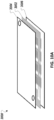
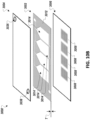
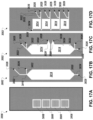
- an apparatus for measuring cellular activity and/or gene expression comprising: a gasket including a plurality of apertures, a plurality of input channels being fluidly connected to the plurality of apertures, respectively, and a plurality of output channels being fluidly connected to the plurality of apertures, respectively; and a cover configured to be mounted onto the gasket, the cover comprising: an inlet configured to be fluidly connected to the plurality of input channels of the gasket when the cover is mounted onto the gasket, and an outlet configured to be fluidly connected to the plurality of output channels of the gasket when the cover is mounted onto the gasket.
- the plurality of apertures includes: inlet ports that fluidly connect to the plurality of input channels, respectively; and outlet ports that fluidly connect to the plurality of output channels, respectively.
- the apparatus described herein further comprises a substrate having a plurality of substrate regions.
- the gasket is configured to be mounted onto the substrate such that the plurality of apertures are aligned with the plurality of substrate regions, respectively.
- the cover is configured to be mounted onto the gasket opposite to the substrate.
- a plurality of perfusion chambers are defined by (i) the plurality of substrate regions of the substrate, (ii) the plurality of apertures of the gasket that is mounted onto the substrate, and (iii) the cover that is mounted onto the gasket
- the gasket is made of silicone.
- each of the substrate regions is configured to attach a capture probe that includes (i) a spatial barcode and (ii) a capture domain that binds to a sequence present in an analyte.
- the gasket has a thickness that ranges between 0.6 mm and 1.0 mm.
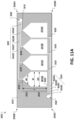
- the gasket includes an upstream bore and a downstream bore.
- the plurality of input channels extend between the upstream bore and the inlet ports of the plurality of apertures, respectively.
- the plurality of output channel extend between the downstream bore and the outlet ports of the plurality of apertures, respectively.
- the plurality of input channels have different lengths between the upstream bore and the inlet ports of the plurality of apertures, respectively.
- the plurality of output channels have different lengths between the downstream bore and the outlet ports of the plurality of apertures, respectively.
- the upstream bore is positioned to be opposite to the downstream bore with respect to a center of the gasket. In some embodiments, the upstream bore is configured to be aligned with the inlet of the cover. In some embodiments, the downstream bore is configured to be aligned with the outlet of the cover.
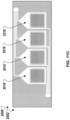
- each of the plurality of apertures includes at least a portion having a width that varies between the inlet port and the outlet port.
- each of the plurality of apertures includes: a first portion including the inlet port and having a first width; and a second portion including the outlet port and having a second width.
- the first width of the first portion varies between the inlet port and the second portion. In some embodiments, the first width of the first portion gradually increases from the inlet port to an interface between the first portion and the second portion. In some embodiments, the second width of the second portion is consistent. In some embodiments, the first width of the first portion is identical to the second width of the second portion at the interface between the first portion and the second portion.
- each when used in reference to a collection of items, is intended to identify an individual item in the collection but does not necessarily refer to every item in the collection, unless expressly stated otherwise, or unless the context of the usage clearly indicates otherwise.
- the perfusion chamber and the multi-well plate are two exemplary types of culturing systems that allow live tissue/cell samples to be recorded in situ, and culture medium can be perfused or replaced to maintain tissue/cell viability.
- One direct application of the methods described herein is to determine the effect of drugs to the cellular activity and/or gene expression, and correlate the determined effect with the spatial information as obtained using capture probes, thereby providing an in-depth understanding of the drug's effect to the biological sample.
- Spatial analysis methodologies and compositions described herein can provide a vast amount of analyte and/or expression data for a variety of analytes within a biological sample at high spatial resolution, while retaining native spatial context.
- Spatial analysis methods and compositions can include, e.g., the use of a capture probe including a spatial barcode (e.g., a nucleic acid sequence that provides information as to the location or position of an analyte within a cell or a tissue sample (e.g., mammalian cell or a mammalian tissue sample) and a capture domain that is capable of binding to an analyte (e.g., a protein and/or a nucleic acid) produced by and/or present in a cell.
- a spatial barcode e.g., a nucleic acid sequence that provides information as to the location or position of an analyte within a cell or a tissue sample
- a capture domain that is capable of binding to an analyte (
- Spatial analysis methods and compositions can also include the use of a capture probe having a capture domain that captures an intermediate agent for indirect detection of an analyte.
- the intermediate agent can include a nucleic acid sequence (e.g., a barcode) associated with the intermediate agent. Detection of the intermediate agent is therefore indicative of the analyte in the cell or tissue sample.
- Non-limiting aspects of spatial analysis methodologies and compositions are described in U.S. Patent Nos. 10,774,374 , 10,724,078 , 10,480,022 , 10,059,990 , 10,041,949 , 10,002,316 , 9,879,313 , 9,783,841 , 9,727,810 , 9,593,365 , 8,951,726 , 8,604,182 , 7,709,198 , U.S. Patent Application Publication Nos.
- a "barcode” is a label, or identifier, that conveys or is capable of conveying information (e.g., information about an analyte in a sample, a bead, and/or a capture probe).
- a barcode can be part of an analyte, or independent of an analyte.
- a barcode can be attached to an analyte.
- a particular barcode can be unique relative to other barcodes.
- an "analyte” can include any biological substance, structure, moiety, or component to be analyzed.
- target can similarly refer to an analyte of interest.
- Analytes can be broadly classified into one of two groups: nucleic acid analytes, and non-nucleic acid analytes.
- non-nucleic acid analytes include, but are not limited to, lipids, carbohydrates, peptides, proteins, glycoproteins (N-linked or O-linked), lipoproteins, phosphoproteins, specific phosphorylated or acetylated variants of proteins, amidation variants of proteins, hydroxylation variants of proteins, methylation variants of proteins, ubiquitylation variants of proteins, sulfation variants of proteins, viral proteins (e.g., viral capsid, viral envelope, viral coat, viral accessory, viral glycoproteins, viral spike, etc.), extracellular and intracellular proteins, antibodies, and antigen binding fragments.
- viral proteins e.g., viral capsid, viral envelope, viral coat, viral accessory, viral glycoproteins, viral spike, etc.
- the analyte(s) can be localized to subcellular location(s), including, for example, organelles, e.g., mitochondria, Golgi apparatus, endoplasmic reticulum, chloroplasts, endocytic vesicles, exocytic vesicles, vacuoles, lysosomes, etc.
- organelles e.g., mitochondria, Golgi apparatus, endoplasmic reticulum, chloroplasts, endocytic vesicles, exocytic vesicles, vacuoles, lysosomes, etc.
- analyte(s) can be peptides or proteins, including without limitation antibodies and enzymes. Additional examples of analytes can be found in Section (I)(c) of WO 2020/176788 and/or U.S. Patent Application Publication No. 2020/0277663 .
- an analyte can be detected indirectly, such as through detection of an intermediate agent, for example, a connected probe (e.g., a ligation product) or an analyte capture agent (e.g., an oligonucleotide-conjugated antibody), such as those described herein.
- an intermediate agent for example, a connected probe (e.g., a ligation product) or an analyte capture agent (e.g., an oligonucleotide-conjugated antibody), such as those described herein.
- a “biological sample” is typically obtained from the subject for analysis using any of a variety of techniques including, but not limited to, biopsy, surgery, and laser capture microscopy (LCM), and generally includes cells and/or other biological material from the subject.
- a biological sample can be a tissue section.
- a biological sample can be a fixed and/or stained biological sample (e.g., a fixed and/or stained tissue section).
- stains include histological stains (e.g., hematoxylin and/or eosin) and immunological stains (e.g., fluorescent stains).
- a biological sample e.g., a fixed and/or stained biological sample
- Biological samples are also described in Section (I)(d) of WO 2020/176788 and/or U.S. Patent Application Publication No. 2020/0277663 .
- a biological sample is permeabilized with one or more permeabilization reagents.
- permeabilization of a biological sample can facilitate analyte capture.
- Exemplary permeabilization agents and conditions are described in Section (I)(d)(ii)(13) or the Exemplary Embodiments Section of WO 2020/176788 and/or U.S. Patent Application Publication No. 2020/0277663 .
- Array-based spatial analysis methods involve the transfer of one or more analytes from a biological sample to an array of features on a substrate, where each feature is associated with a unique spatial location on the array. Subsequent analysis of the transferred analytes includes determining the identity of the analytes and the spatial location of the analytes within the biological sample. The spatial location of an analyte within the biological sample is determined based on the feature to which the analyte is bound (e.g., directly or indirectly) on the array, and the feature's relative spatial location within the array.
- a “capture probe” refers to any molecule capable of capturing (directly or indirectly) and/or labelling an analyte (e.g., an analyte of interest) in a biological sample.
- the capture probe is a nucleic acid or a polypeptide.
- the capture probe includes a barcode (e.g., a spatial barcode and/or a unique molecular identifier (UMI)) and a capture domain).
- UMI unique molecular identifier
- a capture probe can include a cleavage domain and/or a functional domain (e.g., a primer-binding site, such as for next-generation sequencing (NGS)).
- NGS next-generation sequencing
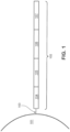
- FIG. 1 is a schematic diagram showing an exemplary capture probe, as described herein.
- the capture probe 102 is optionally coupled to a feature 101 by a cleavage domain 103, such as a disulfide linker.
- the capture probe can include a_functional sequence 104 that is useful for subsequent processing.
- the functional sequence 104 can include all or a part of sequencer specific flow cell attachment sequence (e.g., a P5 or P7 sequence), all or a part of a sequencing primer sequence, (e.g., a R1 primer binding site, a R2 primer binding site), or combinations thereof.
- the capture probe can also include a spatial barcode 105.
- the capture probe can also include a unique molecular identifier (UMI) sequence 106. While FIG.
- UMI unique molecular identifier
- the capture probe can also include a capture domain 107 to facilitate capture of a target analyte.
- the capture domain can have a sequence complementary to a sequence of a nucleic acid analyte.
- the capture domain can have a sequence complementary to a connected probe described herein.
- the capture domain can have a sequence complementary to a capture handle sequence present in an analyte capture agent.
- the capture domain can have a sequence complementary to a splint oligonucleotide.
- Such splint oligonucleotide in addition to having a sequence complementary to a capture domain of a capture probe, can have a sequence of a nucleic acid analyte, a sequence complementary to a portion of a connected probe described herein, and/or a capture handle sequence described herein.
- the functional sequences can generally be selected for compatibility with any of a variety of different sequencing systems, e.g., Ion Torrent Proton or PGM, Illumina sequencing instruments, PacBio, Oxford Nanopore, etc., and the requirements thereof.
- functional sequences can be selected for compatibility with non-commercialized sequencing systems. Examples of such sequencing systems and techniques, for which suitable functional sequences can be used, include (but are not limited to) Ion Torrent Proton or PGM sequencing, Illumina sequencing, PacBio SMRT sequencing, and Oxford Nanopore sequencing.
- functional sequences can be selected for compatibility with other sequencing systems, including non-commercialized sequencing systems.
- the spatial barcode 105 and functional sequences 104 are common to all of the probes attached to a given feature.
- the UMI sequence 106 of a capture probe attached to a given feature is different from the UMI sequence of a different capture probe attached to the given feature.
- FIG. 2 is a schematic illustrating a cleavable capture probe, wherein the cleaved capture probe can enter into a non-permeabilized cell and bind to analytes within the sample.
- the capture probe 201 contains a cleavage domain 202, a cell penetrating peptide 203, a reporter molecule 204, and a disulfide bond (-S-S-).
- 205 represents all other parts of a capture probe, for example a spatial barcode and a capture domain.
- FIG. 3 is a schematic diagram of an exemplary multiplexed spatially-barcoded feature.
- the feature 301 can be coupled to spatially-barcoded capture probes, wherein the spatially-barcoded probes of a particular feature can possess the same spatial barcode, but have different capture domains designed to associate the spatial barcode of the feature with more than one target analyte.
- a feature may be coupled to four different types of spatially-barcoded capture probes, each type of spatially-barcoded capture probe possessing the spatial barcode 302.
- One type of capture probe associated with the feature includes the spatial barcode 302 in combination with a poly(T) capture domain 303, designed to capture mRNA target analytes.
- a second type of capture probe associated with the feature includes the spatial barcode 302 in combination with a random N-mer capture domain 304 for gDNA analysis.
- a third type of capture probe associated with the feature includes the spatial barcode 302 in combination with a capture domain complementary to a capture handle sequence of an analyte capture agent of interest 305.
- a fourth type of capture probe associated with the feature includes the spatial barcode 302 in combination with a capture domain that can specifically bind a nucleic acid molecule 306 that can function in a CRISPR assay (e.g., CRISPR/Cas9). While only four different capture probe-barcoded constructs are shown in FIG.
- capture-probe barcoded constructs can be tailored for analyses of any given analyte associated with a nucleic acid and capable of binding with such a construct.
- the schemes shown in FIG. 3 can also be used for concurrent analysis of other analytes disclosed herein, including, but not limited to: (a) mRNA, a lineage tracing construct, cell surface or intracellular proteins and metabolites, and gDNA; (b) mRNA, accessible chromatin (e.g., ATAC-seq, DNase-seq, and/or MNase-seq) cell surface or intracellular proteins and metabolites, and a perturbation agent (e.g., a CRISPR crRNA/sgRNA, TALEN, zinc finger nuclease, and/or antisense oligonucleotide as described herein); (c) mRNA, cell surface or intracellular proteins and/or metabolites, a barcoded labelling agent (e.g., the MHC multimers
- a perturbation agent can be a small molecule, an antibody, a drug, an aptamer, a miRNA, a physical environmental (e.g., temperature change), or any other known perturbation agents. See, e.g., Section (II)(b) (e.g., subsections (i)-(vi)) of WO 2020/176788 and/or U.S. Patent Application Publication No. 2020/0277663 . Generation of capture probes can be achieved by any appropriate method, including those described in Section (II)(d)(ii) of WO 2020/176788 and/or U.S. Patent Application Publication No. 2020/0277663 .
- more than one analyte type e.g., nucleic acids and proteins
- a biological sample can be detected (e.g., simultaneously or sequentially) using any appropriate multiplexing technique, such as those described in Section (IV) of WO 2020/176788 and/or U.S. Patent Application Publication No. 2020/0277663 .
- an analyte capture agent refers to an agent that interacts with an analyte (e.g., an analyte in a biological sample) and with a capture probe (e.g., a capture probe attached to a substrate or a feature) to identify the analyte.
- the analyte capture agent includes: (i) an analyte binding moiety (e.g., that binds to an analyte), for example, an antibody or antigen-binding fragment thereof; (ii) analyte binding moiety barcode; and (iii) a capture handle sequence.
- an analyte binding moiety barcode refers to a barcode that is associated with or otherwise identifies the analyte binding moiety.
- the term “analyte capture sequence” or “capture handle sequence” refers to a region or moiety configured to hybridize to, bind to, couple to, or otherwise interact with a capture domain of a capture probe.
- a capture handle sequence is complementary to a capture domain of a capture probe.
- an analyte binding moiety barcode (or portion thereof) may be able to be removed (e.g., cleaved) from the analyte capture agent.
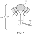
- FIG. 4 is a schematic diagram of an exemplary analyte capture agent 402 comprised of an analyte-binding moiety 404 and an analyte-binding moiety barcode domain 408.
- the exemplary analyte -binding moiety 404 is a molecule capable of binding to an analyte 406 and the analyte capture agent is capable of interacting with a spatially-barcoded capture probe.
- the analyte -binding moiety can bind to the analyte 406 with high affinity and/or with high specificity.
- the analyte capture agent can include an analyte -binding moiety barcode domain 408, a nucleotide sequence (e.g., an oligonucleotide), which can hybridize to at least a portion or an entirety of a capture domain of a capture probe.
- the analyte-binding moiety barcode domain 408 can comprise an analyte binding moiety barcode and a capture handle sequence described herein.
- the analyte -binding moiety 404 can include a polypeptide and/or an aptamer.
- the analyte -binding moiety 404 can include an antibody or antibody fragment (e.g., an antigen-binding fragment).
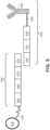
- FIG. 5 is a schematic diagram depicting an exemplary interaction between a feature-immobilized capture probe 524 and an analyte capture agent 526.
- the feature-immobilized capture probe 524 can include a spatial barcode 508 as well as functional sequences 506 and UMI 510, as described elsewhere herein.
- the capture probe can also include a capture domain 512 that is capable of binding to an analyte capture agent 526.
- the analyte capture agent 526 can include a functional sequence 518, analyte binding moiety barcode 516, and a capture handle sequence 514 that is capable of binding to the capture domain 512 of the capture probe 524.
- the analyte capture agent can also include a linker 520 that allows the capture agent barcode domain 516 to couple to the analyte binding moiety 522.
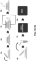
- FIGs. 6A , 6B , and 6C are schematics illustrating how streptavidin cell tags can be utilized in an array-based system to produce a spatially-barcoded cell or cellular contents.
- peptide-bound major histocompatibility complex MHC
- p2m biotin
- streptavidin moiety comprises multiple pMHC moieties.
- Each of these moieties can bind to a TCR such that the streptavidin binds to a target T-cell via multiple MHC/TCR binding interactions. Multiple interactions synergize and can substantially improve binding affinity.
- a capture agent barcode domain 601 can be modified with streptavidin 602 and contacted with multiple molecules of biotinylated MHC 603 such that the biotinylated MHC 603 molecules are coupled with the streptavidin conjugated capture agent barcode domain 601. The result is a barcoded MHC multimer complex 605.
- the capture agent barcode domain sequence 601 can identify the MHC as its associated label and also includes optional functional sequences such as sequences for hybridization with other oligonucleotides. As shown in FIG.
- one example oligonucleotide is capture probe 606 that comprises a complementary sequence (e.g., rGrGrG corresponding to C C C), a barcode sequence and other functional sequences, such as, for example, a UMI, an adapter sequence (e.g., comprising a sequencing primer sequence (e.g., R1 or a partial R1 ("pR1"), R2), a flow cell attachment sequence (e.g., P5 or P7 or partial sequences thereof)), etc.
- capture probe 606 may at first be associated with a feature (e.g., a gel bead) and released from the feature.
- capture probe 606 can hybridize with a capture agent barcode domain 601 of the MHC-oligonucleotide complex 605.
- the hybridized oligonucleotides (Spacer C C C and Spacer rGrGrG) can then be extended in primer extension reactions such that constructs comprising sequences that correspond to each of the two spatial barcode sequences (the spatial barcode associated with the capture probe, and the barcode associated with the MHC-oligonucleotide complex) are generated.
- one or both of the corresponding sequences may be a complement of the original sequence in capture probe 606 or capture agent barcode domain 601.
- the capture probe and the capture agent barcode domain are ligated together.
- the resulting constructs can be optionally further processed (e.g., to add any additional sequences and/or for clean-up) and subjected to sequencing.
- a sequence derived from the capture probe 606 spatial barcode sequence may be used to identify a feature and the sequence derived from spatial barcode sequence on the capture agent barcode domain 601 may be used to identify the particular peptide MHC complex 604 bound on the surface of the cell (e.g., when using MHC-peptide libraries for screening immune cells or immune cell populations).
- a spatial barcode with one or more neighboring cells, such that the spatial barcode identifies the one or more cells, and/or contents of the one or more cells, as associated with a particular spatial location.
- One method is to promote analytes or analyte proxies (e.g., intermediate agents) out of a cell and towards a spatially-barcoded array (e.g., including spatially-barcoded capture probes).
- Another method is to cleave spatially-barcoded capture probes from an array and promote the spatially-barcoded capture probes towards and/or into or onto the biological sample.
- capture probes may be configured to prime, replicate, and consequently yield optionally barcoded extension products from a template (e.g., a DNA or RNA template, such as an analyte or an intermediate agent (e.g., a connected probe (e.g., a ligation product) or an analyte capture agent), or a portion thereof), or derivatives thereof (see, e.g., Section (II)(b)(vii) of WO 2020/176788 and/or U.S. Patent Application Publication No. 2020/0277663 regarding extended capture probes).
- a template e.g., a DNA or RNA template, such as an analyte or an intermediate agent (e.g., a connected probe (e.g., a ligation product) or an analyte capture agent), or a portion thereof
- a template e.g., a DNA or RNA template, such as an analyte or an intermediate agent (e.g.,
- capture probes may be configured to form a connected probe (e.g., a ligation product) with a template (e.g., a DNA or RNA template, such as an analyte or an intermediate agent, or portion thereof), thereby creating ligations products that serve as proxies for a template.
- a connected probe e.g., a ligation product
- a template e.g., a DNA or RNA template, such as an analyte or an intermediate agent, or portion thereof
- an "extended capture probe” refers to a capture probe having additional nucleotides added to the terminus (e.g., 3' or 5' end) of the capture probe thereby extending the overall length of the capture probe.
- an "extended 3' end” indicates additional nucleotides were added to the most 3' nucleotide of the capture probe to extend the length of the capture probe, for example, by polymerization reactions used to extend nucleic acid molecules including templated polymerization catalyzed by a polymerase (e.g., a DNA polymerase or a reverse transcriptase).
- a polymerase e.g., a DNA polymerase or a reverse transcriptase
- extending the capture probe includes adding to a 3' end of a capture probe a nucleic acid sequence that is complementary to a nucleic acid sequence of an analyte or intermediate agent specifically bound to the capture domain of the capture probe.
- the capture probe is extended using reverse transcription.
- the capture probe is extended using one or more DNA polymerases. The extended capture probes include the sequence of the capture probe and the sequence of the spatial barcode of the capture probe.
- extended capture probes are amplified (e.g., in bulk solution or on the array) to yield quantities that are sufficient for downstream analysis, e.g., via DNA sequencing.
- extended capture probes e.g., DNA molecules
- an amplification reaction e.g., a polymerase chain reaction. Additional variants of spatial analysis methods, including in some embodiments, an imaging step, are described in Section (II)(a) of WO 2020/176788 and/or U.S. Patent Application Publication No. 2020/0277663 .
- Spatial information can provide information of biological and/or medical importance.
- the methods and compositions described herein can allow for: identification of one or more biomarkers (e.g., diagnostic, prognostic, and/or for determination of efficacy of a treatment) of a disease or disorder; identification of a candidate drug target for treatment of a disease or disorder; identification (e.g., diagnosis) of a subject as having a disease or disorder; identification of stage and/or prognosis of a disease or disorder in a subject; identification of a subject as having an increased likelihood of developing a disease or disorder; monitoring of progression of a disease or disorder in a subject; determination of efficacy of a treatment of a disease or disorder in a subject; identification of a patient subpopulation for which a treatment is effective for a disease or disorder; modification of a treatment of a subject with a disease or disorder; selection of a subject for participation in a clinical trial; and/or selection of a treatment for a subject with a disease or disorder.
- Spatial information can provide information of biological importance.
- the methods and compositions described herein can allow for: identification of transcriptome and/or proteome expression profiles (e.g., in healthy and/or diseased tissue); identification of multiple analyte types in close proximity (e.g., nearest neighbor analysis); determination of up- and/or down-regulated genes and/or proteins in diseased tissue; characterization of tumor microenvironments; characterization of tumor immune responses; characterization of cells types and their co-localization in tissue; and identification of genetic variants within tissues (e.g., based on gene and/or protein expression profiles associated with specific disease or disorder biomarkers).
- a substrate functions as a support for direct or indirect attachment of capture probes to features of the array.
- a "feature” is an entity that acts as a support or repository for various molecular entities used in spatial analysis.
- some or all of the features in an array are functionalized for analyte capture.
- Exemplary substrates are described in Section (II)(c) of WO 2020/176788 and/or U.S. Patent Application Publication No. 2020/0277663 .
- Exemplary features and geometric attributes of an array can be found in Sections (II)(d)(i), (II)(d)(iii), and (II)(d)(iv) of WO 2020/176788 and/or U.S. Patent Application Publication No. 2020/0277663 .
- analytes and/or intermediate agents can be captured when contacting a biological sample with a substrate including capture probes (e.g., a substrate with capture probes embedded, spotted, printed, fabricated on the substrate, or a substrate with features (e.g., beads, wells) comprising capture probes).
- capture probes e.g., a substrate with capture probes embedded, spotted, printed, fabricated on the substrate, or a substrate with features (e.g., beads, wells) comprising capture probes.
- contact contacted
- contacting a biological sample with a substrate refers to any contact (e.g., direct or indirect) such that capture probes can interact (e.g., bind covalently or non-covalently (e.g., hybridize)) with analytes from the biological sample.
- Capture can be achieved actively (e.g., using electrophoresis) or passively (e.g., using diffusion). Analyte capture is further described in Section (II)(e) of WO 2020/176788 and/or U.S. Patent Application Publication No. 2020/0277663 .
- spatial analysis can be performed by attaching and/or introducing a molecule (e.g., a peptide, a lipid, or a nucleic acid molecule) having a barcode (e.g., a spatial barcode) to a biological sample (e.g., to a cell in a biological sample).
- a plurality of molecules e.g., a plurality of nucleic acid molecules
- a plurality of barcodes e.g., a plurality of spatial barcodes
- a biological sample e.g., to a plurality of cells in a biological sample for use in spatial analysis.
- the biological sample after attaching and/or introducing a molecule having a barcode to a biological sample, the biological sample can be physically separated (e.g., dissociated) into single cells or cell groups for analysis.
- Some such methods of spatial analysis are described in Section (III) of WO 2020/176788 and/or U.S. Patent Application Publication No. 2020/0277663 .
- spatial analysis can be performed by detecting multiple oligonucleotides that hybridize to an analyte.
- spatial analysis can be performed using RNA-templated ligation (RTL).
- RTL RNA-templated ligation
- Methods of RTL have been described previously. See, e.g., Credle et al., Nucleic Acids Res. 2017 Aug 21;45(14):e128 .
- RTL includes hybridization of two oligonucleotides to adjacent sequences on an analyte (e.g., an RNA molecule, such as an mRNA molecule).
- the oligonucleotides are DNA molecules.
- one of the oligonucleotides includes at least two ribonucleic acid bases at the 3' end and/or the other oligonucleotide includes a phosphorylated nucleotide at the 5' end.
- one of the two oligonucleotides includes a capture domain (e.g., a poly(A) sequence, a non-homopolymeric sequence).
- a ligase e.g., SplintR ligase
- the two oligonucleotides hybridize to sequences that are not adjacent to one another. For example, hybridization of the two oligonucleotides creates a gap between the hybridized oligonucleotides.
- a polymerase e.g., a DNA polymerase
- the connected probe e.g., a ligation product
- the connected probe is released using an endonuclease (e.g., RNAse H).
- the released connected probe (e.g., a ligation product) can then be captured by capture probes (e.g., instead of direct capture of an analyte) on an array, optionally amplified, and sequenced, thus determining the location and optionally the abundance of the analyte in the biological sample.
- capture probes e.g., instead of direct capture of an analyte
- sequence information for a spatial barcode associated with an analyte is obtained, and the sequence information can be used to provide information about the spatial distribution of the analyte in the biological sample.
- Various methods can be used to obtain the spatial information.
- specific capture probes and the analytes they capture are associated with specific locations in an array of features on a substrate.
- specific spatial barcodes can be associated with specific array locations prior to array fabrication, and the sequences of the spatial barcodes can be stored (e.g., in a database) along with specific array location information, so that each spatial barcode uniquely maps to a particular array location.
- specific spatial barcodes can be deposited at predetermined locations in an array of features during fabrication such that at each location, only one type of spatial barcode is present so that spatial barcodes are uniquely associated with a single feature of the array.
- the arrays can be decoded using any of the methods described herein so that spatial barcodes are uniquely associated with array feature locations, and this mapping can be stored as described above.
- each array feature location represents a position relative to a coordinate reference point (e.g., an array location, a fiducial marker) for the array. Accordingly, each feature location has an "address" or location in the coordinate space of the array.
- Some exemplary spatial analysis workflows are described in the Exemplary Embodiments section of WO 2020/176788 and/or U.S. Patent Application Publication No. 2020/0277663 . See, for example, the Exemplary embodiment starting with "In some non-limiting examples of the workflows described herein, the sample can be immersed... " of WO 2020/176788 and/or U.S. Patent Application Publication No. 2020/0277663 . See also, e.g., the Visium Spatial Gene Expression Reagent Kits User Guide (e.g., Rev D, dated October 2020), and/or the Visium Spatial Tissue Optimization Reagent Kits User Guide (e.g., Rev D, dated October 2020).
- the Visium Spatial Gene Expression Reagent Kits User Guide e.g., Rev D, dated October 2020
- the Visium Spatial Tissue Optimization Reagent Kits User Guide e.g., Rev D, dated October 2020.
- spatial analysis can be performed using dedicated hardware and/or software, such as any of the systems described in Sections (II)(e)(ii) and/or (V) of WO 2020/176788 and/or U.S. Patent Application Publication No. 2020/0277663 , or any of one or more of the devices or methods described in Sections Control Slide for Imaging, Methods of Using Control Slides and Substrates for, Systems of Using Control Slides and Substrates for Imaging, and/or Sample and Array Alignment Devices and Methods, Informational labels of WO 2020/123320 .
- Suitable systems for performing spatial analysis can include components such as a chamber (e.g., a flow cell or sealable, fluid-tight chamber) for containing a biological sample.
- the biological sample can be mounted for example, in a biological sample holder.
- One or more fluid chambers can be connected to the chamber and/or the sample holder via fluid conduits, and fluids can be delivered into the chamber and/or sample holder via fluidic pumps, vacuum sources, or other devices coupled to the fluid conduits that create a pressure gradient to drive fluid flow.
- One or more valves can also be connected to fluid conduits to regulate the flow of reagents from reservoirs to the chamber and/or sample holder.
- the systems can optionally include a control unit that includes one or more electronic processors, an input interface, an output interface (such as a display), and a storage unit (e.g., a solid state storage medium such as, but not limited to, a magnetic, optical, or other solid state, persistent, writeable and/or re-writeable storage medium).
- the control unit can optionally be connected to one or more remote devices via a network.
- the control unit (and components thereof) can generally perform any of the steps and functions described herein. Where the system is connected to a remote device, the remote device (or devices) can perform any of the steps or features described herein.
- the systems can optionally include one or more detectors (e.g., CCD, CMOS) used to capture images.
- the systems can also optionally include one or more light sources (e.g., LED-based, diode-based, lasers) for illuminating a sample, a substrate with features, analytes from a biological sample captured on a substrate, and various control and calibration media.
- one or more light sources e.g., LED-based, diode-based, lasers
- the systems can optionally include software instructions encoded and/or implemented in one or more of tangible storage media and hardware components such as application specific integrated circuits.
- the software instructions when executed by a control unit (and in particular, an electronic processor) or an integrated circuit, can cause the control unit, integrated circuit, or other component executing the software instructions to perform any of the method steps or functions described herein.
- the systems described herein can detect (e.g., register an image) the biological sample on the array.
- Exemplary methods to detect the biological sample on an array are described in PCT Application No. 2020/061064 and/or U.S. Patent Application Serial No. 16/951,854 .
- the biological sample Prior to transferring analytes from the biological sample to the array of features on the substrate, the biological sample can be aligned with the array. Alignment of a biological sample and an array of features including capture probes can facilitate spatial analysis, which can be used to detect differences in analyte presence and/or level within different positions in the biological sample, for example, to generate a three-dimensional map of the analyte presence and/or level. Exemplary methods to generate a two- and/or three-dimensional map of the analyte presence and/or level are described in PCT Application No. 2020/053655 and spatial analysis methods are generally described in WO 2020/061108 and/or U.S. Patent Application Serial No. 16/951,864 .
- a map of analyte presence and/or level can be aligned to an image of a biological sample using one or more fiducial markers, e.g., objects placed in the field of view of an imaging system which appear in the image produced, as described in the Substrate Attributes Section, Control Slide for Imaging Section of WO 2020/123320 , PCT Application No. 2020/061066 , and/or U.S. Patent Application Serial No. 16/951,843 .
- fiducial markers e.g., objects placed in the field of view of an imaging system which appear in the image produced, as described in the Substrate Attributes Section, Control Slide for Imaging Section of WO 2020/123320 , PCT Application No. 2020/061066 , and/or U.S. Patent Application Serial No. 16/951,843 .
- Fiducial markers can be used as a point of reference or measurement scale for alignment (e.g., to align a sample and an array, to align two substrates, to determine a location of a sample or array on a substrate relative to a fiducial marker) and/or for quantitative measurements of sizes and/or distances.
- Spatial transcriptomics is performed on biological samples, for example tissue sections that are in some manner processed prior to assaying.
- Spatial workflows allow for the detection of, for example, gene expression from frozen or fixed tissues while maintaining the spatial position of the gene expression within the tissue.
- the disclosed methods and systems allow for the utilization of a spatial assay platform in conjunction with live tissue sections or cells for the study of, for example, pharmacological drug discovery, protein interactions, or other cellular activities that are best studied using live tissues or cells as a starting material.
- the biological sample is a tissue sample.
- the tissue sample is a live tissue section.
- the live tissue sample is sectioned (e.g., using a vibratome or any slicing instruments known in the art) from a fresh tissue.
- the live tissue section can be treated (e.g., an enzymatic dissociation) to release individual cells, such that the isolated individual cells can be analyzed using the methods described herein.
- the tissue sample is an intact tissue.
- the tissue sample is a semi-intact tissue.
- the live tissue sample is cultured (e.g., in a tissue culture medium) in the perfusion chamber or the multi-well plate described herein before a recording step.
- Recording as used herein includes but is not limited to determining or measuring the abundance of an analyte or biological activity in a sample.
- recording includes imaging the sample using any of the method steps disclosed herein.
- the recording includes determining the abundance of an analyte (e.g., protein, RNA, DNA) or biological activity in a sample.
- recording e.g., determining
- recording is performed using qualitative techniques.
- recording e.g., determining
- recording includes detecting the presence of and/or abundance of a reporter or detectable marker (e.g., fluorescent protein detection).
- Recording a cellular or biological activity includes measuring the presence or abundance of an activity in a cell. Activities include but are not limited to protein activity (e.g., kinase activity), phosphorylation activity, G protein-coupled receptor related activity, ion channel activity (e.g., switch between open and closed conformation), ligand-receptor binding activity, neural activity (e.g., neuronal action potentials), protein synthesis (e.g., transcription or translation) activity, protein expression and localization (e.g., sub-cellular organelle protein expression and trafficking), transient optical activity (e.g., optical reporter gene expression), cell-to-cell interaction, cellular morphology, vesicular trafficking (e.g., exocytosis or endocytosis), protein translocation and/or protein post-translational modifications (e.g., ubiquitination or glycosy
- the live tissue sample is cultured in a separate tissue culture or a substrate (e.g., a coverslip, sponge gels, etc.), then transferred to the perfusion chamber or the multi-well plate described herein.
- additional compounds e.g., artificial cerebrospinal fluid
- the live tissue sample is directly placed in the perfusion chamber or the multi-well plate, without culturing before the recording step.
- the live tissue sample can be sectioned from a fresh tissue and directly placed in the perfusion chamber or the multi-well plate before the recording step.
- the sectioned live tissue sample is placed in a suspension oxygenated medium to maintain tissue viability.
- the tissue sample is an organoid sample.
- An organoid is a miniaturized and simplified version of an organ produced in vitro in three dimensions that shows realistic micro-anatomy.
- some organoids are derived from one or a few cells from a tissue, embryonic stem cells or induced pluripotent stem cells, which can self-organize in three-dimensional culture owing to their self-renewal and differentiation capacities.
- the organoid sample is a cerebral organoid, gut organoid, intestinal organoid (e.g., small intestinal organoid), stomach (or gastric) organoid, lingual organoid, thyroid organoid, thymic organoid, testicular organoid, hepatic organoid, pancreatic organoid, epithelial organoid, liver organoid, pulmonary organoid, neural organoid, brain organoid, lung organoid, kidney organoid, embryonic organoid, blastocyst-like organoid, cardiac organoid, retinal organoid, or any combinations thereof.
- intestinal organoid e.g., small intestinal organoid
- stomach (or gastric) organoid e.g., lingual organoid
- thyroid organoid e.g., thymic organoid
- testicular organoid
- the organoid sample is originated from disease-affected tissues (e.g., cancer tissues) or normal tissues.
- the organoid sample is originated from disease-affected cells (e.g., cancer cells) or normal cells.
- the organoid is originated from stem cells (e.g., embryonic stem cells, induced pluripotent stem cells, and/or somatic stem cells), or differentiated cells (e.g., somatic cells).
- overall tissue viability of cells in the tissue sample is at least or about 60%, at least or about 65%, at least or about 70%, at least or about 75%, at least or about 80%, at least or about 85%, at least or about 90%, at least or about 95%, at least or about 96%, at least or about 97%, at least or about 98%, at least or about 99%, or 100%.
- the biological sample is a cell sample (e.g., cells present in a cell culture).
- the cell sample is cultured in the perfusion chamber or the multi-well plate before the recording step, e.g., for at least 1 hour, at least 2 hours, at least 3 hours, at least 4 hours, at least 6 hours, at least 12 hours, at least 1 day, at least 2 days, at least 3 days, at least 4 days, at least 5 days, at least a week, at least 2 weeks, at least 3 weeks, or longer.
- the cells of the cell sample are directly cultured in the perfusion chamber or the multi-well plate, e.g., by seeding the cells at an appropriate density known in the art.
- the perfusion chamber or the multi-well plate can serve as a cell culture container and a culture medium can be perfused (or added) within the perfusion chamber or the multi-well plate to maintain viability of the cells.
- the cell sample is transferred to the perfusion chamber or the multi-well plate from a separate cell culture.
- the cell sample is a cell line.
- At least one cell in the cell sample is transfected or infected by methods known in the art.
- the cell sample is infected by a viral vector, e.g., a virus that includes a nucleic acid that encodes at least one protein of interest.
- a viral vector e.g., a virus that includes a nucleic acid that encodes at least one protein of interest.
- Exemplary viral vectors include adenoviruses (reviewed in Altaras et al., 2005, Adv. Biochem. Eng. Biotechnol., 99:193-260 ), adeno-associated viruses (reviewed in Park et al., 2008, Front. Biosci., 13:2653-59 ; see also Williams, 2007, Mol.
- the cell sample is a primary cell culture sample.
- the primary cell culture sample comprises cells dissociated from fresh tissue samples. Additional nutrients may be supplemented to the culture medium according to specific cell types (e.g., neuron, epithelial cells, or endothelial cells) to maintain cell viability.
- the cell sample is an immortalized cell line (e.g., a human cell line) sample.
- the cell sample comprises adherent cells and/or suspension cells.
- overall viability of cells in the cell sample is at least or about 60%, at least or about 65%, at least or about 70%, at least or about 75%, at least or about 80%, at least or about 85%, at least or about 90%, at least or about 95%, at least or about 96%, at least or about 97%, at least or about 98%, at least or about 99%, or 100%.
- the biological sample is a three dimensional (3D) culture sample.
- the 3D culture sample is an organoid sample (e.g., comprising one or more types of organoids).
- the 3D culture sample is a spheroid sample (e.g., comprising one or more types of spheroids).
- the biological sample is embedded in hydrogels.
- the biological sample is from a human (e.g., human patients). In some embodiments, the biological sample is from an animal model (e.g., mice).
- the analyte can be any analyte disclosed herein, or multiples thereof.
- the analyte is a nucleic acid.
- the analyte is a protein.
- both a nucleic acid analyte and a protein analyte are measured from the same sample.
- the methods disclosed herein include measuring and/or determining the relative abundance of an analyte compared to a reference sample.
- the methods disclosed herein include determining the tertiary or quaternary structure of the analyte.
- the methods disclosed herein identify one or more post-translational modifications on an analyte. This includes, without limitation, the presence and abundance of one or more post-translational modifications.
- the methods disclosed herein determine the enzymatic activity of an analyte.
- the methods disclosed herein determine the location of an analyte.
- the location of the analyte is fluid over time. That is, the location of an analyte can vary depending on its function.
- the methods disclosed herein allow a user to determine analyte translocation or trafficking across any organelle in the cell (e.g., as described herein).
- translocation or trafficking occurs in or around an endoplasmic reticulum (ER) in a cell.
- ER endoplasmic reticulum
- translocation or trafficking occurs in or around a Golgi apparatus in a cell.
- an analyte can be detected as associating with a cell or nuclear membrane surface.
- the biological sample is treated with a blocking reagent described herein.
- the blocking reagent can prevent molecular (e.g., test compounds or drugs described herein) or cellular (e.g., live cells) adhesion to a capture probe.
- FIG. 10A is a schematic perspective view of the system 3000
- FIG. 10B is an exploded view of the system 3000 of FIG. 10A
- the system 3000 is configured to define one or more chambers that can be used for measuring cellular activity or gene expression in a biological sample.
- a chamber is an array area of a spatial transcriptomics slide, for example an array area of a Visium gene expression slide wherein the array area comprises a plurality of capture probes as described herein.
- the system 3000 can include a gasket 3002 and a cover 3004.
- the system 3000 may further include a substrate 3006.
- the gasket 3002 and the cover 3004 are configured to be reversibly mounted onto the substrate 3006 to define chambers that contain biological samples placed on the substrate 3006.
- Such chambers can be used as perfusion chambers into which fluids are introduced for various analyses, such as measuring cellular activities or gene expression.
- the system 3000 is configured to be able to achieve laminar flow so that multiple samples (e.g., cells, tissues, etc.) can be contacted with fluid at the same time.
- the substrate 3006 includes one or more substrate regions 3008, each of which is configured to receive a biological sample.
- the substrate 3006 includes a plurality of substrate regions 3008 for placing a plurality of samples.
- the substrate regions 3008 can comprise a plurality of capture probes, as described herein.
- the capture probes can include a spatial barcode and a capture domain that binds to a sequence present in an analyte.
- the substrate regions 3008 can be arranged on the substrate 3006 in various two-dimensional arrays. In the illustrated example, the substrate 3006 has four substrate regions 3008 arranged in line (i.e., a one by four array). However, other configurations are possible.
- eight substrate regions 3008 can be arranged in a two by four array.
- the substrate 3006 includes at least 1, 2, 3, 4, 5, 6, 7, 8, 9, 10, 12, 14, 16 or more substrate regions 3008.
- the substrate regions 3008 are arranged in one row (e.g., a one by two, one by three, one by four array, or an array of one by any suitable number).
- the substrate regions 3008 are arranged in two rows (e.g., a two by two, two by three, two by four array, or an array of two by any suitable number).
- the substrate regions 3008 can be arranged in a symmetrical array (e.g., an array of two by two, two by three, two by four, etc.) or a non-symmetrical array (e.g., an array that has a first number of substrate regions in a first column and a second number (different from the first number) of substrate regions in a second column).
- a symmetrical array e.g., an array of two by two, two by three, two by four, etc.
- a non-symmetrical array e.g., an array that has a first number of substrate regions in a first column and a second number (different from the first number) of substrate regions in a second column).
- the gasket 3002 includes a gasket body 3012 and one or more apertures 3014 defined in the gasket body 3012.
- the gasket 3002 is configured to be reversibly disposed onto the substrate 3006.
- the gasket 3002 includes the same number of apertures 3014 as the substrate regions 3008 of the substrate 3006.
- the gasket 3002 may have more or less apertures 3014 than number of the substrate regions 3008.
- the apertures 3014 are arranged to correspond to the substrate regions 3008 when the gasket 3002 is positioned on the substrate 3006.
- the apertures 3014 of the gasket 3002 are aligned with the substrate regions 3008 of the substrate 3006, respectively, thereby defining a plurality of chambers 3016, as shown in FIG. 11C .
- the plurality of chambers 3016 are used as perfusion chambers for biological samples on the substrate 3006.
- the plurality of chambers 3016 allow for independent experiments or replicates run on one substrate 3006.
- Each aperture 3014 can have at least two ports through which fluid can flow into and out from the aperture 3014.
- the aperture 3014 can include an inlet port 3018 and an outlet port 3020 as shown in FIG. 11A .
- the aperture 3014 can include an inlet port 3020 and an outlet port 3018.
- the inlet port 3018 is configured to permit fluid to enter the aperture 3014 (or the chamber 3016 defined at least by the aperture 3014)
- the outlet port 3020 is configured to permit fluid to exit the aperture 3014 (or the chamber 3016 defined at least by the aperture 3014).
- the gasket 3002 further includes one or more input channels 3022 and one or more output channels 3024.
- the input channels 3022 can be at least partially enclosed by the substrate 3006 and the cover 3004 when the gasket 3002 is sandwiched between the substrate 3006 and the cover 3004, so that fluid can flow along the input channels 3022.
- the output channels 3024 can be at least partially enclosed by the substrate 3006 and the cover 3004 when the gasket 3002 is disposed between the substrate 3006 and the cover 3004, so that fluid can flow along the output channels 3024.
- the inlet ports 3018 are fluidly connected to the input channels 3022, respectively. Accordingly, the input channels 3022 are fluidly connected to the apertures 3014 through the inlet ports 3018, respectively.
- the outlet ports 3020 are fluidly connected to the output channels 3024, respectively. Accordingly, the output channels 3024 are fluidly connected to the apertures 3014 through the outlet ports 3020, respectively.
- FIG. 11A shows the gasket 3002 can include an upstream bore 3026 and a downstream bore 3028, or vice versa.
- the upstream bore 3026 is fluidly connected to the input channels 3022 so that the input channels 3022 extend commonly from the upstream bore 3026. Accordingly, the input channels 3022 extend between the upstream bore 3026 and the inlet ports 3018 of the apertures 3014, respectively.
- the downstream bore 3028 is fluidly connected to the output channels 3024 so that the output channels 3024 extend commonly from the downstream bore 3028. Accordingly, the output channels 3024 extend between the downstream bore 3028 and the output ports 3020 of the apertures 3014
- the gasket 3002 is primarily described as having a single upstream bore 3026 for all the input channels 3022 and a single downstream bore 3028 for all the output channels 3024, alternative configurations are possible.
- the gasket 3002 can include multiple upstream bores 3026, and at least one of the multiple upstream bores 3026 is fluidly connected to multiple input channels 3022.
- the gasket 3002 can include multiple downstream bores 3028, and at least one of the multiple downstream bores 3028 is fluidly connected to multiple output channels 3024.
- the gasket 3002 can include multiple upstream bores 3026 for respective input channels 3022, and/or multiple downstream bores 3028 for respective output channels 3024.
- the input channels 3022 have different lengths between the upstream bore 3026 and the respective inlet ports 3018.
- the input channels 3022 includes first, second, third, and fourth input channels 3022A-3022D, and the first, second, third, and fourth input channels 3022A-3022D have different lengths L1-L4 that are determined as distances of routes or paths between the upstream bore 3026 and the respective inlet ports 3018 of the apertures 3014.
- at least two of the input channels 3022 can have the same length between the upstream bore 3026 and the respective inlet ports 3018.
- the upstream bore 3026 can be positioned between a group of first and second apertures 3014A-B and a group of third and fourth apertures 3014C-D (e.g., where the upstream bore 3026 is positioned along a hypothetical line that splits the two groups), and the first and second input channels 3022A-B can have the same lengths as the third and fourth input channels 3022C-D, respectively.
- the output channels 3024 have different lengths between the downstream bore 3028 and the respective outlet ports 3020.
- the output channels 3024 includes first, second, third, and fourth input channels 3024A-D, and the first, second, third, and fourth input channels 3024A-D have different lengths L5-L8 that are determined as distances of routes or paths between the downstream bore 3028 and the respective outlet ports 3020 of the apertures 3014.
- at least two of the outlet channels 3024 can have the same length between the downstream bore 3028 and the respective outlet ports 3020.
- the downstream bore 3028 can be positioned between the group of first and second apertures 3014A-B and the group of third and fourth apertures 3014C-D (e.g., where the downstream bore 3028 is positioned along a hypothetical line that splits the two groups), and the first and second output channels 3024A-B can have the same lengths as the third and fourth output channels 3024C-D, respectively.
- the upstream bore 3026 and the downstream bore 3028 can be arranged in various positions in the gasket 3002.
- the upstream bore 3026 can be positioned to be opposite to the downstream bore 3028 with respect to the group of the apertures 3014.
- the gasket 3004 has a rectangular shape of the gasket body 3012 with four corners 3030A-D.
- the upstream bore 3026 can be positioned at a first corner 3030A of the gasket body 3012, and the downstream bore 3028 can be positioned it a third corner 3030C that is opposite to the first corner 3030A.
- the upstream bore 3026 can be positioned to be opposite to the downstream bore 3028 relative to a center C of the gasket 3002.
- Other positions of the upstream bore 3026 and the downstream bore 3028 are also possible to provide suitable fluid stream into and out from the apertures 3014.
- the inlet/input features e.g., the inlet port 3018, the input channels 3022, the upstream bore 3026, etc.
- the outlet/output features e.g., the outlet port 3020, the output channels 3024, the downstream bore 3028, etc.
- the outlet/output features can be used for receiving fluid and the inlet/input features can be used for discharging the fluid.
- the gasket 3002 can be made of one or more various materials.
- the gasket 3002 can be made of a material that provides appropriate seals at the interface between the gasket 3002 and the substrate 3006, and at the interface between the gasket 3002 and the cover 3004, so that fluid paths are sealingly defined along the input channels 3022, the chambers 3016 (defined at least by the apertures 3014), and the output channels 3024 in the system 3000 without leakage between adjacent fluid paths or leakage from the system 3000 as a whole.
- the gasket 3002 can be made of silicone.
- the gasket 3002 can be made of natural rubber, nitrile rubber, polytetrafluoroethylene (PTFE), for example.
- PTFE polytetrafluoroethylene
- the gasket is reversibly applied and connected to the cover 3004 and/or the substrate 3006.
- the gasket 3002 can be configured in various dimensions.
- the gasket 3002 has a thickness T that ranges between 0.1 mm and 5.0 mm.
- the thickness T of the gasket 3002 can range between 0.6 mm and 1.0 mm.
- the thickness T of the gasket 3002 can be less than 0.1 mm.
- the thickness T of the gasket 3002 can be greater than 5.0 mm.
- the apertures 3014 of the gasket 3002 can be configured in various dimensions. Each aperture 3014 can have an area that ranges between 40 mm 2 and 90 mm 2 . Each aperture 3014 can have a volume that ranges between 2 mm 3 and 175 mm 3 . The apertures 3014 can have the same area and volume. Alternatively, at least one of the apertures 3014 have a different area or volume from the other apertures 3014.
- each aperture 3014 of the gasket 3002 can be configured in various shapes.
- each aperture 3014 has a length L and a width W.
- the aperture 3014 can have the width W that varies at least partially along the length L.
- the aperture 3014 includes a first portion 3032 and a second portion 3034.
- the first portion 3032 of the aperture 3014 includes the inlet port 3018, and the second portion 3034 of the aperture 3014 includes the outlet port 3020.
- the first portion 3032 has a first width W1, and the second portion has a second width W2.
- the first width W1 of the first portion 3032 can vary between the inlet port 3018 and the second portion 3034 (e.g., an end of the second portion 3034 that interfaces the inlet port 3018).
- the first width W1 of the first portion 3032 can gradually increase from the inlet port 3018 to the interface between the first portion 3032 and the second portion 3034, so that the first portion 3032 has generally a triangular shape.
- the second width W2 of the second portion 3034 can be consistent in the direction of the length L.
- the first width W1 of the first portion 3032 can be identical to the second width W2 of the second portion 3034 at the interface between the first portion 3032 and the second portion 3034.
- the aperture 3014 of the gasket 3002 can have different configurations.
- the aperture 3014 can be shaped as a square or rectangular that has a consistent width along the length.
- Other suitable shapes, such as circular, oval, hexagonal, etc. are also possible.
- the gasket 3002 can include the number of apertures 3014 that is identical to the number of the substrate regions 3008 of the substrate 3006, so that the apertures 3014 are aligned with the respective substrate regions 3008, as shown in FIGs. 11C and 17D .
- the gasket 3002 can have the apertures 3014 less than the substrate regions 3008 of the substrate 3006 so that at least one of the apertures 3014 is aligned with a plurality of substrate regions 3008.
- the gasket 3002 can have two apertures 3014 that is each configured to align with two of the substrate regions 3008.
- the gasket 3002 has a single aperture 3014 configured to align with four substrate regions 3008 of the substrate 3006, so that fluid that is introduced into the aperture 3014 is commonly supplied to all the four substrate regions 3008.
- the gasket 3002 can have the apertures 3014 more than the substrate regions 3008 of the substrate 3006 so that at least one of the substrate regions 3008 can be aligned with two or more apertures 3014.
- each aperture 3014 can have a single input channel 3022 and a single output channel 3024, as shown in FIG. 11A .
- each aperture 3014 can have multiple input channels 3022 and/or multiple output channels 3024.
- each aperture 3022 has two input channels 3022.
- the input channels 3022 and/or the output channels 3024 can have a consistent width along their lengths, as illustrated in FIGs. 11A and 17B -17D. In other implementations, the input channels 3022 and/or the output channels 3024 can have varied widths along their lengths. By way of example, as illustrated in FIG. 17C , the output channels 3024 can have a width that decreases from the output port 3020 to the downstream bore 3028.
- the cover 3004 is configured to be positioned on the gasket 3002.
- the cover 3004 can be mounted onto the gasket 3002 opposite to the substrate 3006, so that the plurality of chambers 3016 are defined in the system 3000.
- the cover 3004 can include an inlet 3036 and an outlet 3038.
- the inlet 3036 is positioned such that the inlet 3036 is fluidly connected to the plurality of input channels 3022 of the gasket 3002 when the cover 3004 is mounted onto the gasket 3002.
- the inlet 3036 can be used to introduce fluid into the system 3000 (e.g., into the chambers 3016 defined at least part by the apertures 3014 of the gasket 3002).
- the inlet 3036 can be aligned with the upstream bore 3026 of the gasket 3002 when the cover 3004 is disposed on the gasket 3002.
- the outlet 3038 is positioned such that the outlet 3038 is fluidly connected to the plurality of output channels 3024 of the gasket 3002 when the cover 3004 is mounted onto the gasket 3002.
- the outlet 3038 can be used to discharge fluid from the system 300 (e.g., from the chambers 3016 defined at least part by the apertures 3014 of the gasket 3002).
- the outlet 3038 can be aligned with the downstream bore 3028 of the gasket 3002 when the cover 3004 is disposed on the gasket 3002.
- the cover 3004 includes a single inlet 3036 and a single outlet 3038 for multiple chambers 3016 ( FIG. 11C ).
- the cover 3004 can have multiple inlets 3036 and/or multiple outlets 3038 for respective ones of at least some of the multiple chambers 3016.
- the cover 3004 can be made of one or more various materials. In some implementations, the cover 3004 is made of plastic. In other implementations, the cover 3004 can be made of Silica glass, Quartz, Polystyrene, PLA (Poly Lactic Acid), Acrylic or Polymethyl Methacrylate (PMMA), Polycarbonate (PC), Polyethylene (PE), Polypropylene (PP), Polyethylene Terephthalate (PETE or PET), Polyvinyl Chloride (PVC), and/or Acrylonitrile-Butadiene-Styrene (ABS), or other suitable materials.
- Silica glass Quartz, Polystyrene, PLA (Poly Lactic Acid), Acrylic or Polymethyl Methacrylate (PMMA), Polycarbonate (PC), Polyethylene (PE), Polypropylene (PP), Polyethylene Terephthalate (PETE or PET), Polyvinyl Chloride (PVC), and/or Acrylonitrile-Butadiene-Styrene (ABS), or other suitable materials.
- PLA Poly Lactic Acid
- the cover 3004 is separately made and disposed on the gasket 3002.
- the cover 3004 and the gasket 3002 can be made in a single piece that can be placed on a substrate in the same or similar way as described herein.
- the cover 3004 and the gasket 3002 are reversibly placed on the substrate, for example for removal during downstream processing of the sample located in one or more of the chambers on the substrate.
- a substrate described herein comprises a multi-well plate and includes one or more wells, each of which is configured to receive a biological sample.
- the multi-well plate includes a plurality of wells for placing a plurality of samples.
- the plurality of capture probes, as described herein are directly attached to the wells.
- the capture probes are directly printed at a surface (e.g., a bottom surface) in the wells.
- the plurality of capture probes are attached (e.g., printed) to a substrate, e.g., a coverslip, and the substrate is placed within the wells.
- the coverslips are custom made to fit within the multi-well plate.
- the coverslip comprises plastic (e.g., polystyrene plastic), metal, glass, or any materials compatible for attachment of the capture probes.
- the capture probes are attached to the wells or the substrate chemically, e.g., via one or more linkage groups.
- the linkage groups include amide groups, epoxides, thiol, Acrydite TM .
- the well or the substrate has additional surface chemistry to facilitate the growth and/or attachment of cells as known in the art.
- the well e.g., a polystyrene microplate well
- the substrate e.g., a polystyrene coverslip
- a plasma gas to order to modify the hydrophobic plastic surface to make it more hydrophilic.
- the resulting surface carries a net negative charge due to presence of oxygen-containing functional groups (e.g., hydroxyl and/or carboxyl groups).
- the well or the substrate is coated with poly-lysine and/or collagen. Details can be found, e.g., in Curtis et al., The Journal of Cell Biology 97.5 (1983): 1500-1506 , which is incorporated herein by reference in its entirety.
- the wells can be arranged in the multi-well plate in various two-dimensional ways.
- a multi-well plate can have a single well; two wells arranged in line (i.e., a one by two plate); or four wells arranged in line (i.e., a one by four plate).
- the multi-well plate includes at least 1, 2, 3, 4, 5, 6, 7, 8, 9, 10, 12, 14, 16, 24, 48, 96, 384, or more one or more wells.
- the wells are arranged in one row (e.g., a one by two, one by three, one by four plate, or a plate of one by any suitable number). In some instances, the wells are arranged in two rows (e.g., a two by two, two by three, two by four plate, or a plate of two by any suitable number).
- the wells can be arranged in a symmetrical plate (e.g., a plate of two by two, two by three, two by four, etc.) or a non-symmetrical array (e.g., a plate that has a first number of wells in a first column and a second number (different from the first number) of wells in a second column).
- the multi-well plate described herein is assembled by mounting a gasket comprising a plurality of apertures (e.g., any one of the gasket described herein) onto a substrate comprising a plurality of substrate regions (e.g., a spatial array slide).
- a well of the assembled multi-well plate is defined by the substrate region of the substrate, and the aperture of the gasket.
- the gasket is configured to be mounted onto the substrate such that the plurality of apertures are aligned with the plurality of substrate regions, respectively.
- the configuration of the substrate regions and apertures of the assembled multi-well plate can be any one of the configurations described herein.
- the assembled multi-well plate is disassembled after permeabilizing the biological sample (e.g., a live tissue sample or cell sample), and the substrate (e.g., the spatial array slide) is subjected to spatial analysis.
- tissue culture dishes useful in methods disclosed herein include, but are not limited to, 35mm, 60mm, 100mm, 150mm diameter dishes as well as flasks T-25, T-27, T-175, T-225.
- multi will plates includes multi-layer flasks.
- Methods disclosed herein can further include microfluidic culture chips (e.g., chips manufactured by Darwin Microfluidics, Paris, France).
- the multi-well plate is commercially available.
- the multi-well plate described herein is a 6-well plate, an 8-well plate, a 12-well plate, a 24-well plate, a 48-well plate, a 96-well plate or a 384-well plate.
- the multi-well plate is custom made, e.g., to accommodate specific needs (e.g., automatic detection of cellular activities or intracellular gene expressions).
- the multi-well plate is heat-resistant, such that the plate is compatible with one or more steps of the spatial analysis described here (e.g., reverse transcription and/or PCR amplification).
- the spatial gene expression of tissues or cells is obtained within each individual well of the multi-well plate.
- the multi-well plate is capable of automatic detection (e.g., plate-based scanning) of cellular activity and/or the intracellular gene expression as described herein.
- the well or the substrate described herein comprises one or more fiducial markers.
- the fiducial markers can allow for proper orientation, detection, and/or rotation of the sample on the substrate.
- the fiducial markers provide a visual reference of the biological sample (e.g., one or more cells in the biological sample) in respect to the substrate.
- cellular activity of a biological sample can be recorded using the methods as described herein.
- cellular activity can include, but are not limited to: protein activity (e.g., kinase activity), phosphorylation activity, G protein-coupled receptor related activity, ion channel activity (e.g., switch between open and closed conformation), ligand-receptor binding activity, neural activity (e.g., neuronal action potentials), protein synthesis (e.g., transcription or translation) activity, protein expression and localization (e.g., sub-cellular organelle protein expression and trafficking), transient optical activity (e.g., optical reporter gene expression), cell-to-cell interaction, cellular morphology, vesicular trafficking (e.g., exocytosis or endocytosis), protein translocation and/or protein post-translational modifications (e.g., ubiquitination or glycosylation).
- protein activity e.g., kinase activity
- phosphorylation activity e.g
- the cellular activity includes processes in cell signaling pathways or cascades. In some embodiments, the cellular activity includes conformational changes of biomolecules (e.g., proteins or nucleic acids). In some embodiments, the cellular activity is that which occurs upon contacting one or more cells with a pharmacological compound. For example, the detection of apoptosis of one or more cells is recorded pre and post addition of, for example, a drug that may or may not be useful in treating cancers or other aberrant cellular proliferation diseases. As such, the methods described herein can be used in drug discovery.
- biomolecules e.g., proteins or nucleic acids
- the cellular activity is that which occurs upon contacting one or more cells with a pharmacological compound. For example, the detection of apoptosis of one or more cells is recorded pre and post addition of, for example, a drug that may or may not be useful in treating cancers or other aberrant cellular proliferation diseases. As such, the methods described herein can be used in drug discovery.
- the present methods can be used to track or monitor the success or failure of a patient being treated with a drug or medicament, for example by tracking the presence or absence of the desired cellular activity that is expected to occur when a patient is treated with the desired drug or medicament. Therefore, the present methods can be used to help determine a treatment regimen for a patient, track or monitor the success of that treatment during therapy and track or monitor post therapeutic treatment success or relapse.
- the methods include a recording step of the biological sample.
- the biological sample is a live tissue section as described herein.
- the biological sample is a culture of cells as described herein.
- the recording detects one or more of the cellular activities disclosed herein.
- the recording step comprises optical recording.
- optical recording includes measuring of membrane potential activity.
- optical recording captures fast (approximately 1 msec) cellular electrical activity such as ion channel activity (e.g., switch between open and closed conformation) or neural activity (e.g., neuronal action potentials).
- optical recording includes using chemical dyes or indicators.
- the chemical dye or indicator comprises a fluorophore, such that the fluorescent signal, or lack thereof, can be recorded in order to detect one or more cellular activities.
- the recording step comprises electrical recording.
- the chemical dye is a voltage-sensitive dye, a pH-sensitive dye, a temperature-sensitive dye, a light-sensitive dye, an oxygen-sensitive dye, a metal sensitive dye, or any chemical dyes that can be used to track one or more cellular activities as described herein.
- the chemical dye is a voltage-sensitive dye.
- Voltage-sensitive dyes also known as potentiometric dyes, are dyes which change their spectral properties in response to voltage changes. They are able to provide linear measurements of firing activity of single neurons, large neuronal populations or activity of myocytes. Many physiological processes are accompanied by changes in cell membrane potential which can be detected with voltage sensitive dyes. Measurements may indicate the site of action potential origin, and measurements of action potential velocity and direction may be obtained.
- potentiometric dyes can be used to monitor the electrical activity inside cell organelles where it is not possible to insert an electrode, such as the mitochondria and dendritic spine.
- This technology is powerful for the study of patterns of activity in complex multicellular preparations. It also makes possible the measurement of spatial and temporal variations in membrane potential along the surface of single cells.
- the voltage-sensitive dye may be contacted with the biological sample using a contact method chosen from: intravenous injection; intramuscular injection; intraventricular injection; spinal tap; craniotomy with direct contact of dye to cortical surface of the tissue sample (e.g., a brain tissue sample).
- a contact method chosen from: intravenous injection; intramuscular injection; intraventricular injection; spinal tap; craniotomy with direct contact of dye to cortical surface of the tissue sample (e.g., a brain tissue sample).
- Non-limiting examples of suitable voltage-sensitive dyes include, but are not limited to: merocyanine-rhodamine dyes including NK 2761; minonaphthylethenylpyridinium dyes including Di-4-ANEPPS, di-8-ANEPPS, Di-2-ANEPEQ, Di-8-ANEPPQ and Di-12-ANEPPQ; dialkylaminophenylpolyenylpyridinium dyes including RH 160, RH 237, RH 414, RH 421, and RH 795; oxonol dyes including RH 155, RH 482, RH 1691, RH 1692, and RH 1838; and dipicrylamine (DPA).
- DPA dipicrylamine
- the voltage-sensitive dye includes a membrane-localized voltage-sensitive protein coupled to a capture protein.
- the capture protein is arranged and disposed to capture small-molecule fluorescent dyes.
- the voltage sensitive protein is an opsin, such as, but not limited to, a microbial opsin. Suitable microbial opsins include, but are not limited to, QuasAr2, Ace2N, or a combination thereof.
- the voltage sensitive protein includes at least one voltage-sensing domain selected from the group consisting of a Ciona intestinalis voltage-sensing domain (CiVSD), Danio rerio voltage-sensing domain (DrVSD), Gallus gallus voltage-sensing domain (GgVSD), or a combination thereof.
- a capture protein is a covalent capture protein.
- the covalent capture protein is selected from the group consisting of HaloTag, SNAP-tag, TMP-tag, pLac-tag, CLIP-tag, or a combination thereof.
- the capture protein is a non-covalent capture protein.
- the non-covalent capture protein is selected from the group consisting of TMP-tag, biotin-avidin, and a combination thereof.
- the chemical dye is a calcium-sensitive dye, e.g., a calcium-sensitive fluorescent dye (or indicator).
- Imaging Ca 2+ -sensitive fluorescent indicators provides a common approach for studying Ca 2+ signals in many contexts. Fluorescent indicators are particularly useful for measuring acute Ca 2+ changes in a relatively noninvasive manner. The availability of indicators that can be targeted to specific cellular domains, coupled with variations in affinity, brightness or spectral characteristics, provides tools for exploring spatially and temporally diverse Ca 2+ signals, and moreover, multiplexing the readout of Ca 2+ with other cellular functions.
- Non-limiting examples of fluorescent indicators to monitor intracellular Ca 2+ concentration include fluorescent protein reporters such as pericams, cameleons, modified yellow cameleons (YCs), and camgaroos.
- the Ca 2+ signals can also be measured with synthetic indicators such Fura-2, Indol-1, and Fluo-4.
- the procedures described can be applied to many imaging modalities, including wide-field, confocal, and total internal reflection (TIRF) microscopy. It is known in the art that the experimental details can vary depending on the cell type, imaging system, and characteristics of the Ca 2+ signals being studied. Using appropriate technology and suitable indicators, it is possible to monitor Ca 2+ signals spanning from subcellular to multicellular, at high speed or time lapse, within living cells. Details can be found in, e.g., Bootman, Martin D., et al. Cold Spring Harbor Protocols 2013.2 (2013): pdb-top066050 ; which is incorporated by reference in its entirety.
- the indicator is a genetically-encoded indicator, e.g., a genetically-encoded neural activity indicator, a genetically-encoded voltage indicator (GEVI) or a genetically-encoded calcium indicator (GCaMP).
- Genetically encoded voltage indicator is a protein that can sense membrane potential in a cell and relate the change in voltage to a form of output, often fluorescent level.
- a GEVI is an optogenetic recording tool that enables exporting electrophysiological signals from cultured cells, live animals, including the human brain. Examples of notable GEVIs include ArcLight, ASAP1, ASAP3, and Ace2N-mNeon.
- GEVI can have many configuration designs in order to realize voltage sensing function.
- An essential feature of GEVI structure is that it must situate on the cell membrane.
- the structure of a GEVI should permit the function of sensing the voltage difference and reporting it by change in fluorescence.
- VSD voltage-sensing domain
- GEVIs include at least four categories: (1) GEVIs that contain a fluorescent protein FRET pair, e.g., VSFP1, (2) single opsin GEVIs, e.g., Arch, (3) opsin-FP FRET pair GEVIs, e.g., MacQ-mCitrine, and (4) single FP with special types of voltage sensing domains, e.g. ASAP1.
- the GEVI contains a fluorescent protein FRET pair.
- the GEVI is a single opsin GEVI.
- the GEVI is an opsin-FP FRET pair GEVI.
- the GEVI is a single FP with special types of voltage sensing domains.
- a majority of GEVIs are based on the Ciona intestinalis voltage sensitive phosphatase (Ci-VSP or Ci-VSD (domain)).
- Ci-VSP Ciona intestinalis voltage sensitive phosphatase
- Some GEVIs might have similar components, but with different positioning of them.
- ASAP1 and ArcLight both use a VSD and one FP, but the FP of ASAP1 is on the outside of the cell whereas that of ArcLight is on the inside, and the two FPs of VSFP-Butterfly are separated by the VSD, while the two FPs of Mermaid are relatively close to each other.
- Non-limiting examples of GEVIs include FlaSh, VSFP1, SPARC, Flare, VSFP3.1, Mermaid, hVOS, Red-shifted VSFP's, PROPS, Zahra, Zahra 2, ArcLight, Arch, ElectricPk, VSFP-Butterfly, VSFP-CR, Mermaid2, Mac GEVIs, QuasAr1, QuasAr2, Archer, ASAP1, Ace GEVIs, ArcLightning, Pado, ASAP2f, FlicR1, Bongwoori, ASAP2s, ASAP-Y, (pa)QuasAr3(-s), Voltron(-ST), and/or ASAP3.
- the genetically-encoded indicator is a genetically-encoded calcium indicator (GECI, or GCaMP).
- GECI provides an alternative to synthetic indicators. GECIs can be easily targeted to specific cell types or sub-cellular compartments, and are compatible with long-term, repeated in vivo measurements.
- GECIs consist of a calcium-binding domain such as calmodulin or troponin C, fused to one or more (e.g., one, two, three, four, or more) fluorescent proteins (FPs). In single-FP GECIs, the fluorescence intensity of a circularly permuted FP (cpFP) is modulated by calcium binding-dependent changes in the chromophore environment.
- cpFP circularly permuted FP
- GECIs are useful for screening for agonists or antagonists of G-protein coupled receptor (GPCR) or ion channels and monitoring neural activity.
- GPCR G-protein coupled receptor
- intracellular calcium level changes induced by G-protein coupled receptor activation can be indicated by fluorescent signal changes emitted by GAcMP. Additional description can be found, e.g., in U.S. Patent No. 9,518,980 B2 , which is incorporated by reference in its entirety.
- the indicator discriminates between live and dead cells.
- vitality dyes such as LIVE-OR-DYE dead cell dyes are cell membrane impermeable and amine-reactive. Such vitality stains can enter cells with damaged cell membranes, label intracellular proteins, and exhibit high levels of fluorescence. These vitality dyes can also react to cell surface proteins, however because cell surface proteins are less abundant than intracellular proteins the fluorescence associated with live cells is very low.
- Additional indicators include non-fluorescent dyes that only fluorescence when they enter cells such as live cell labeling or tracking dyes and cell proliferation dyes, for example Cytopainter live cell dyes are hydrophobic compounds that permeate across cell membranes and become highly fluorescent once inside the cell.
- the step of recording one or more cellular activities includes imaging the biological sample.
- the sample is recorded before the sample is provided with one or more dyes or indicators.
- the sample is recorded at the same time as when the sample is provided with one or more dyes or indicators.
- the sample is recorded after the sample is provided with one or more dyes or indicators.
- the sample is recorded before, during and/or after the sample is provided with one or more dyes or indicators.
- temporal formation of cellular activities can be assessed using the methods described herein.
- temporal (e.g., real-time) formation of the target cellular activity can be recorded by detecting and measuring the cellular activity (e.g., using dyes) by a fluorescent time lapse microscopy.
- Imaging can be performed using any of the microscopy techniques described herein. In some instances, imaging is performed using fluorescence microscopy, fluorescent time lapse microscopy, confocal microscopy, multi-photon microscopy (e.g., two-photon excitation microscopy), or any known microscopy techniques known in the art.
- the sample is fixed.
- the analytes in the sample can hybridize to a plurality of probes on an array (e.g., substrate) as described herein.
- the poly-adenylation (poly(A)) sequence of an mRNA hybridizes to a poly-thymine (poly(T)) capture domain sequence on a capture probe.
- the capture probe is extended using the analyte that is specifically bound to the capture domain as a template to generate an extended capture probe.
- the extended capture probe is amplified to produce a plurality of extended capture probes.
- the plurality of extended capture probes, or libraries created therefrom, is sequenced.
- all or a portion of the sequence of the spatial barcode, or a complement thereof is determined. In some instances, all or a portion of the sequence of the analyte, or a complement thereof, is determined. In some instances, the determined sequences are used to identify the location of the analyte in the biological sample.
- intracellular gene expression of a nucleic acid of a biological sample can be recorded using the methods as described herein.
- the method involves multiple molecular dyes attached to a single probe (e.g., a probe that can hybridize to a target nucleic acid) for increased signal-to-noise ratio.
- dual probes containing split fluorescent proteins are used.
- FRET Forster resonance energy transfer
- fluorescent quenching can occur when the probe binds to the target nucleic acid to restrict signal.
- the probe contains fluorescent nucleotides.
- the nucleic acid described herein can be visualized in vivo.
- the nucleic acid described herein e.g., an RNA
- the nucleic acid described herein can be visualized in vitro.
- the intracellular gene expression can be recorded optically through the use of in situ hybridization (e.g., fluorescent in situ hybridization (FISH)).
- FISH fluorescent in situ hybridization
- the objective of in situ hybridization is to determine the presence or absence of one or more nucleic acid sequences of interest at particular spatial locations in a cell or chromosomal sites.
- Particular nucleic acid sequences are identified within cells by taking advantage of a property of nucleic acids (i.e., their ability to specifically anneal to each other to form hybrids). This process can be used to hybridize two complementary strands of DNA, one strand of RNA to one strand of DNA, and two complementary strands of RNA.
- strand hybridization occurs between natural and artificial nucleic acids.
- gene expression detection examines a particular transcript of interest.
- the transcript is associated with normal physiology. In some instances, the transcript is associated with a pathophysiology (e.g., cancer or aberrant development or other disease state).
- a FISH probe is designed to detect one or more mutations. For example, and without limitation, a FISH probe can be designed to detect a point mutation, a single nucleotide polymorphism, an insertion, a deletion, and/or a translocation. In some instances, the probe is designed to detect one or more exons in an mRNA analyte that is alternatively spliced.
- the probe is directly labeled with a detectable marker as disclosed herein.
- the detectable marker is a fluorescent, radioactive, chemiluminescent, or colorimetric detectable marker.
- the probe is not directly labeled with a detectable marker.
- a second moiety e.g., comprising a fluorophore
- the second moiety includes a fluorescent, radioactive, chemiluminescent, or colorimetric detectable marker.
- the hybridized probe is visualized.
- the annealed hybrids can be detected by various methods, including isotopic and nonisotopic (fluorescent and nonfluorescent) approaches. Additional description is found at e.g., Jensen, The Anatomical Record 297.8 (2014): 1349-1353 , which is incorporated by reference in its entirety.
- the intracellular gene expression can be assessed optically through the use of fluorescence resonance energy transfer (or Forster resonance energy transfer, FRET).
- FRET fluorescence resonance energy transfer
- the technique of FRET when applied to optical microscopy permits determination of the approach between two molecules within a distance (several nanometers (e.g., 10 nm)) sufficiently close for molecular interactions to occur.
- the mechanism of fluorescence resonance energy transfer involves a donor fluorophore in an excited electronic state, which may transfer its excitation energy to a nearby acceptor fluorophore in a non-radiative fashion through long-range dipole-dipole interactions.
- nucleotide incorporations can be detected through FRET, as described for example in Levene et al., Science (2003), 299, 682-686 , Lundquist et al., Opt. Lett. (2008), 33, 1026-1028 , and Korlach et al., Proc. Natl. Acad. Sci. USA (2008), 105, 1176-1181 , each of which is incorporated by reference in its entirety.
- the recording step comprises hybridizing a plurality of optically-labelled probes to a target nucleic acid.
- the optically-labelled probes comprise an optical label, e.g., a fluorophore.
- the optically-labelled probes are fluorescently-labelled probes, e.g., fluorescently labelled peptide nucleic acid (PNA) probes.
- PNAs are synthetic nucleic acid (e.g., DNA) analogs in which the phosphodiester backbone is replaced by repetitive units of N-(2-aminoethyl) glycine to which the purine and pyrimidine bases are attached via a methyl carbonyl linker.
- the procedures for PNA synthesis are similar to those employed for peptide synthesis, using standard solid-phase manual or automated synthesis.
- the PNA molecules are labelled with biotin or fluorophores.
- the PNA molecule disclosed herein includes a detection moiety such as a fluorescent, radioactive, chemiluminescent, or colorimetric detectable marker.
- the PNA molecule is associate with (e.g., conjugated to) a biotin molecule that can be detected using e.g., an avidin or streptavidin pulldown, as disclosed herein.
- a subsequent generation of PNAs involve modification of the N-(2-aminoethyl) glycine backbone (PNA analogs) or chimeric architecture, like PNA-peptide chimeras or PNA-DNA chimeras developed in order to improve the solubility and the cellular uptake of PNAs or to exhibit new biological properties.
- the synthetic backbone provides PNA with unique hybridization characteristics. Unlike DNA and RNA, the PNA backbone is not charged. Consequently, there is no electrostatic repulsion when PNAs hybridize to its target nucleic acid sequence, giving a higher stability to the PNA-DNA or PNA-RNA duplexes than the natural homo- or heteroduplexes.
- Tm thermal melting temperature
- the optically-labelled probes can bind to a target nucleic acid in the biological sample.
- the target nucleic acid is a DNA. In some instances, the target DNA has been denatured.
- the target nucleic acid is an RNA. In some embodiments, the target nucleic acid is a single-stranded DNA or RNA. In some embodiments, the target nucleic acid is a double-stranded DNA or double-stranded RNA. In some embodiments, the target nucleic acid is a DNA/RNA duplex. In some embodiments, the target nucleic acid is an mRNA, a siRNA, a microRNA, or a derivative thereof.
- the optically-labelled probe (e.g., a fluorescently labelled PNA probe) has at least 10, at least 11, at least 12, at least 13, at least 14, at least 15, at least 16, at least 17, at least 18, at least 19, at least 20, at least 21, at least 22, at least 23, at least 24, at least 25, at least 26, at least 27, at least 28, at least 29, or at least 30 nucleic acids.
- the optically-labelled probe (e.g., a fluorescently labelled PNA probe) can be used at a final concentration of at least or about 0.1 ⁇ 10 -6 M, at least or about 0.5 ⁇ 10 -6 M, at least or about 1 ⁇ 10 -6 M, at least or about 2 ⁇ 10 -6 M, at least or about 3 ⁇ 10 -6 M, at least or about 4 ⁇ 10 -6 M, at least or about 5 ⁇ 10 -6 M, at least or about 10 ⁇ 10 -6 M, at least or about 20 ⁇ 10 -6 M, at least or about 50 ⁇ 10 -6 M, at least or about 100 ⁇ 10 -6 M, or higher.
- one or more (e.g., 1, 2, 3, 4, or more) optically-labelled probes can hybridize to different portions of the target nucleic acid.
- one or more (e.g., 1, 2, 3, 4, or more) optically-labelled probes are used for the detection of the target nucleic acid with a duplex structure, in which case each probe hybridizes specifically to either the sense or antisense strand of the target nucleic acid.
- the optical label can be directly detectable by itself (e.g., radioisotope labels or fluorescent labels) or, in the case of an enzymatic label, can be indirectly detectable, e.g., by catalyzing chemical alterations of a chemical substrate compound or composition, which chemical substrate compound or composition is directly detectable.
- Optical labels can be suitable for small scale detection and/or suitable for high-throughput screening.
- suitable optical labels include, but are not limited to, radioisotopes, fluorophores, chemiluminescent compounds, bioluminescent compounds, and dyes.
- the optical label can be qualitatively detected (e.g., optically or spectrally), or it can be quantified.
- Qualitative detection generally includes a detection method in which the existence or presence of the optical label is confirmed, whereas quantifiable detection generally includes a detection method having a quantifiable (e.g., numerically reportable) value such as an intensity, duration, polarization, and/or other properties.
- the optically-labelled probes comprise one or more optical labels, 1, 2, 3, 4, or more.
- optical labels can be incorporated during nucleic acid polymerization or amplification (e.g., Cy5 ® -labelled nucleotides, such as Cy5 ® -dCTP). Any suitable optical label can be used.
- the optical label is a fluorophore.
- the fluorophore can be from a group that includes: 7-AAD (7-Aminoactinomycin D), Acridine Orange (+DNA), Acridine Orange (+RNA), Alexa Fluor ® 350, Alexa Fluor ® 430, Alexa Fluor ® 488, Alexa Fluor ® 532, Alexa Fluor ® 546, Alexa Fluor ® 555, Alexa Fluor ® 568, Alexa Fluor ® 594, Alexa Fluor ® 633, Alexa Fluor ® 647, Alexa Fluor ® 660, Alexa Fluor ® 680, Alexa Fluor ® 700, Alexa Fluor ® 750, Allophycocyanin (APC), AMCA / AMCA-X, 7-Aminoactinomycin D (7-AAD), 7- Amino-4-methylcoumarin, 6-Aminoquinoline, Aniline Blue, ANS, APC-Cy7, ATTO-TAG TM CBQCA, ATTO-TAG
- an optical label is or includes a luminescent or chemiluminescent moiety.
- luminescent/chemiluminescent moieties include, but are not limited to, peroxidases such as horseradish peroxidase (HRP), soybean peroxidase (SP), alkaline phosphatase, and luciferase. These protein moieties can catalyze chemiluminescent reactions given the appropriate chemical substrates (e.g., an oxidizing reagent plus a chemiluminescent compound). A number of compound families are known to provide chemiluminescence under a variety of conditions.
- Non-limiting examples of chemiluminescent compound families include 2,3-dihydro-1,4-phthalazinedione luminol, 5-amino-6,7,8-trimethoxy- and the dimethylamino[ca]benz analog. These compounds can luminesce in the presence of alkaline hydrogen peroxide or calcium hypochlorite and base.
- chemiluminescent compound families include, e.g., 2,4,5-triphenylimidazoles, para-dimethylamino and - methoxy substituents, oxalates such as oxalyl active esters, p-nitrophenyl, N-alkyl acridinum esters, luciferins, lucigenins, or acridinium esters.
- temporal formation of specific transcripts can be assessed using the methods described herein.
- one or more fluorescently labelled PNA probes e.g., a first probe labelled with a donor fluorophore and a second probe labelled with an acceptor fluorophore
- a first probe labelled with a donor fluorophore and a second probe labelled with an acceptor fluorophore can hybridize to different portions (e.g., two adjacent sequences) of a target transcript.
- Temporal (e.g., real-time) formation of the target transcript can be recorded by measuring the excitation status of the acceptor fluorophore during an appropriate time period, e.g., by fluorescent time lapse microscopy.
- the donor and acceptor fluorophores can be any FRET pairs known in the art, as described in Bajar et al., Sensors 16.9 (2016): 1488 , which is incorporated by reference in its entirety.
- the cellular activity as described above can be measured at the same time or using the same sample as a sample in which gene expression is recorded.
- a cellular activity is associated with one biomarker (e.g., one fluorescent color) and gene expression (e.g., detection of a particular gene) is associated with a second biomarker (e.g., a different fluorescent marker).
- both cellular activity and gene expression can be recorded optically using fluorescence microscopy, fluorescent time lapse microscopy, confocal microscopy, multi-photon microscopy (e.g., two-photon excitation microscopy), total internal reflection microscopy, super-resolution microscopy, or any known microscopy techniques known in the art.
- the cellular activity and gene expression can be recorded simultaneously, e.g., by using one or more optical labels (e.g., fluorophores) to tag the chemical dyes, indicators, or optically-labelled probes, such that the cellular activity and the gene expression can be recorded with minimal cross-interferences.
- optical labels e.g., fluorophores
- the sample is fixed.
- the analytes in the sample can hybridize to a plurality of probes on an array (e.g., substrate) as described herein.
- the poly-adenylation (poly(A)) sequence of an mRNA hybridizes to a poly-thymine (poly(T)) sequence of a capture domain on a capture probe.
- the capture probe is extended using the analyte that is specifically bound to the capture domain as a template to generate an extended capture probe.
- the extended capture probe is amplified to produce a plurality of extended capture probes (e.g., a plurality of nucleic acids).
- the plurality of extended capture probes, or libraries created therefrom is sequenced. In some instances, all or a portion of the sequence of the spatial barcode, or a complement thereof, is determined. In some instances, all or a portion of the sequence of the analyte, or a complement thereof, is determined. In some instances, the determined sequences are used to identify the location of the analyte in the biological sample.
- the methods described herein include culturing the biological sample in the perfusion chamber or the multi-well plate.
- the biological sample is cultured in a culture medium to maintain its viability.
- the culture medium is replaced at an appropriate interval (e.g., about every 12 hours, about every day, about every 2 days, about every 3 days, about every 4 days, about every 5 days, about every 6 days, or about every week).
- the culture medium is replaced manually (e.g., by pipetting) or automatically.
- the biological sample is cultured statically.
- the biological sample is cultured in a perfusion chamber with inlets and outlets as described herein.
- the culture medium is perfused to the perfusion chamber at a constant flow rate.
- a culture medium e.g., a tissue or cell culture medium, saline, artificial cerebral spinal fluid (ACSF)
- ACSF artificial cerebral spinal fluid
- the culture medium is oxygenated. In some embodiments, additional nutrients or compounds are supplemented to the culture medium to maintain viability of the biological sample.
- the culture medium includes the blocking reagent described herein. In some instances, the blocking reagent can prevent molecule (e.g., the test compounds or drugs described herein) or cell (e.g., live cells) adhesion to the capture probes.
- the blocking reagent is bovine serum albumin (BSA), serum, gelatin (e.g., fish gelatin), milk (e.g., non-fat dry milk), casein, polyethylene glycol (PEG), polyvinyl alcohol (PVA), or polyvinylpyrrolidone (PVP), biotin blocking reagent, a peroxidase blocking reagent, levamisole, Carnoy's solution, glycine, lysine, sodium borohydride, pontamine sky blue, Sudan Black, trypan blue, FITC blocking agent, acetic acid, and/or oligonucleotides including a complementary sequence to the capture oligos.
- BSA bovine serum albumin
- serum serum
- gelatin e.g., fish gelatin
- milk e.g., non-fat dry milk
- casein polyethylene glycol (PEG), polyvinyl alcohol (PVA), or polyvinylpyrrolidone
- PVP polyvinylpyrroli
- the live tissues or cells are cultured in the perfusion chamber or the multi-well plate described herein and grown under conditions to maintain cell viability.
- temperatures and CO 2 concentrations for growing tissues and cells are well known in the art and the methods disclosed herein practice those conditions to maintain sample viability.
- tissue culture incubators are utilized during growth and perfusion of the live tissues or cells, typical conditions to maintain cell viability being, for example, 37°C and 5% CO 2 .
- chemical dyes, indicators, or optically-labelled probes as described herein can be added to the culture medium, and perfused to the perfusion chamber (or added to the multi-well plate) to interact with the biological sample.
- the chemical dyes, indicators, or optically-labelled probes can be added to the biological sample before placing the biological sample into the perfusion chamber or the multi-well plate.
- the chemical dyes, indicators, or optically-labelled probes can be added to the biological sample after placing the biological sample to the perfusion chamber or the multi-well plate.
- the chemical dyes, indicators, or optically-labelled probes can be internalized to the cells (e.g., cells from a live tissue section) by electropermeabilization, endocytic internalization, or lipid-based permeabilization, while maintaining cell viability.
- the methods described herein can be used to measure dynamic transcriptional FRET imaging. In some embodiments, the methods described herein can be used for real-time transcript detection during pharmacological treatment.
- the biological sample e.g., cells from a cell culture
- the capture probes described herein are directly printed on the substrate (or the surface).
- the existence of capture probes on the substrate (or the surface) does not affect growth of the biological sample.
- growth or culturing of the biological sample on the substrate with attached capture probes does not affect data quality of the spatial analysis, e.g., spatial UMI and gene plots; sequencing saturation; median genes per cells; median counts per cell; or median UMIs per cell.
- the methods described herein comprise labelling a plurality of live cells of the biological sample.
- the live cells are labelled by staining.
- the live cells are stained by immunofluorescence (IF).
- the live cells are stained intracellularly (e.g., by staining a sub-cellular organelle including endoplasmic reticulum (ER), Golgi, lysosome, mitochondria, and/or nucleus).
- the live cells are stained extracellularly (e.g., plasma membrane staining).
- the live cells are labelled by a fluorescently labelled antibody or antibody fragments thereof. In some embodiments, the live cells are labelled by the chemical dyes, indicators, or optically-labelled probes described herein. In some embodiments, the live cells are labelled by fluorescent reporters (e.g., the live cells are transfected to include a reporter gene). In some embodiments, the stained live cells are detected (e.g., recorded) by time-lapse fluorescence microscopy.
- the live cells are treated with proteinase K and/or trypsin.
- the treatment can facilitate entry (e.g., by absorption or endocytosis) of the fluorescently labelled antibody or fragments thereof.
- capture probes are blocked prior to contacting the biological sample with the substrate. In some embodiments, capture domains of the capture probes are blocked by blocking probes. In some instances, the capture probes are blocked to prevent molecule (e.g., the test compounds or drugs described herein) or cell (e.g., live cells) adhesion.
- molecule e.g., the test compounds or drugs described herein
- cell e.g., live cells
- the blocking probe is used to block or modify the free 3' end of the capture domain.
- blocking probes can be hybridized to the capture probes to mask the free 3' end of the capture domain, e.g., hairpin probes, partially double stranded probes, or complementary sequences.
- the free 3' end of the capture domain can be blocked by chemical modification, e.g., addition of an azidomethyl group as a chemically reversible capping moiety such that the capture probes do not include a free 3' end.
- blocking the capture domain reduces non-specific background staining.
- the blocking probes are reversible, such that the blocking probes can be removed from the capture domains during or after the time that the capture domains are in contact with the biological sample.
- the blocking probe can be removing with RNAse treatment (e.g., RNAse H treatment).
- the biological sample (e.g., a live tissue sample or cell sample) can be fixed in any of a variety of fixatives to preserve the biological structure of the sample prior to analysis.
- a sample can be fixed via immersion in ethanol, methanol, acetone, formaldehyde (e.g., 2% formaldehyde), paraformaldehyde-Triton, glutaraldehyde, or combinations thereof.
- the biological sample is fixed after recording cellular activity.
- the biological sample is fixed after recording gene expression.
- the biological sample is fixed after recording cellular activity and gene expression.
- the perfusion chamber can be disassembled such that the substrate (e.g., a spatial array) can be subjected to the spatial analysis as described herein.
- the multi-well plate is directly subjected to the spatial analysis, or the individual substrate (e.g., one or more coverslips) can be transferred to a proper platform (e.g., a slide), either manually or automatically, for the spatial analysis.
- the biological sample can be fixed during the recording step, followed by the spatial analysis as described herein.
- two or more fractions of the biological sample, located in separate perfusion chambers can be fixed at different time points during the recording step.
- the biological sample is fixed during test compound (e.g. drug) treatment.
- test compound e.g. drug
- the biological sample is fixed after test compound (e.g., drug) treatment, then subjected to the spatial analysis as described herein.
- one or more steps e.g., extension of capture probes by reverse transcription
- the spatial analysis are carried out directly in the multi-well plate.
- the multiple-well plate described herein can be custom made (e.g., heat-resistant), thereby to be compatible with the spatial analysis steps.
- the methods described herein comprise perfusing one or more (e.g., 1, 2, 3, 4, 5, 6, 7, 8, 9, 10, or more) test compounds through the perfusion chamber.
- the test compounds comprises one or more drugs.
- one or more test compounds can be perfused through the perfusion chamber, before recording the cellular activity or gene expression as described herein.
- one or more test compounds can be perfused through the perfusion chamber, at substantially the same time (e.g., simultaneously) of recording the cellular activity or gene expression as described herein.
- the perfusion and recording are automatically performed for high-throughput screening of test compounds (e.g., drugs).
- real-time cellular activity or gene expression changes are recorded immediately following perfusion of the test compounds.
- culture medium e.g., without any test compounds
- perfused to remove a test compound from the perfusion chamber followed by perfusion of the same or a different test compound.
- the methods described herein comprise treating a biological sample with one or more (e.g., 1, 2, 3, 4, 5, 6, 7, 8, 9, 10, or more) test compounds within a multi-well plate or a chambered substrate.
- the test compounds comprises one or more drugs.
- the test compounds are added to the multi-well plate or chambered substrate manually (e.g., by pipetting) or automatically.
- one or more test compounds are added to the multi-well plate or chambered substrate before recording the cellular activity or gene expression as described herein.
- one or more test compounds are added to the multi-well plate or chambered substrate at substantially the same time (e.g., simultaneously) of recording the cellular activity or gene expression as described herein.
- test compound treatment and/or the recording of the cellular activity or gene expression are automatically performed for high-throughput screening of test compounds (e.g., drugs).
- test compounds e.g., drugs
- real-time cellular activity or gene expression changes are recorded immediately following treatment of the test compounds.
- a test compound is removed (e.g., by pipetting), followed by addition of the same or a different test compound.
- the test compounds are pre-mixed and perfused to the perfusion chamber (or added to the multi-well plate) at the same time. In some embodiments, the test compounds are sequentially perfused or added. In some embodiments, one or more test compounds are perfused (or added) repeatedly to induce a cellular activity or gene expression change.
- the biological sample is treated with one or more test compounds (e.g., drugs) at substantially the same time. In some embodiments, the biological sample is treated with one or more test compounds (e.g., drugs) at different times.
- the test compound (e.g., a drug) can be an agonist. In some embodiments, the test compound (e.g., a drug) can be an antagonist. In some embodiments, the test compound can be a drug, e.g., a small-molecule drug, an antibody or antigen-binding fragment thereof, a pharmacological agent, or any test compounds of interest. In some embodiments, the test compound (e.g., a drug) activates or stimulates one or more cellular activities. In some embodiments, the test compound (e.g., a drug) inhibits one or more cellular activities. In some embodiments, the test compound (e.g., a drug) increases intracellular gene expression of a nucleic acid.
- the test compound decreases intracellular gene expression of a nucleic acid. In some embodiments, the test compound (e.g., a drug) decreases the viability and thus gene expression or other cellular activity within a cell or tissue.
- the test compound is an anti-cancer drug.
- cancer refers to cells having the capacity for autonomous growth, i.e., an abnormal state or condition characterized by rapidly proliferating cell growth. The term is meant to include all types of cancerous growths or oncogenic processes, metastatic tissues or malignantly transformed cells, tissues, or organs, irrespective of histopathologic type or stage of invasiveness.
- Cancers described herein include malignancies of the various organ systems, such as affecting lung, breast, thyroid, lymphoid, gastrointestinal, and genito-urinary tract, as well as adenocarcinomas which include malignancies such as most colon cancers, renal-cell carcinoma, prostate cancer and/or testicular tumors, non-small cell carcinoma of the lung, cancer of the small intestine and cancer of the esophagus.
- the test compounds described herein are designed for treating or diagnosing a carcinoma in a subject.
- carcinoma is art recognized and refers to malignancies of epithelial or endocrine tissues including respiratory system carcinomas, gastrointestinal system carcinomas, genitourinary system carcinomas, testicular carcinomas, breast carcinomas, prostatic carcinomas, endocrine system carcinomas, and melanomas.
- the cancer is renal carcinoma or melanoma.
- Exemplary carcinomas include those forming from tissue of the cervix, lung, prostate, breast, head and neck, colon and ovary.
- carcinosarcomas e.g., which include malignant tumors composed of carcinomatous and sarcomatous tissues.
- an “adenocarcinoma” refers to a carcinoma derived from glandular tissue or in which the tumor cells form recognizable glandular structures.
- the term “sarcoma” is art recognized and refers to malignant tumors of mesenchymal derivation.
- the test compound is an antibody or antigen-binding fragment thereof, e.g., Muromonab- CD3, Efalizumab, Tositumomab-I131, Nebacumab, Edrecolomab, Catumaxomab, Daclizumab, Abciximab, Rituximab, Basiliximab, Palivizumab, Infliximab, Trastuzumab, Adalimumab, Ibritumomab tiuxetan, Omalizumab, Cetuximab, Bevacizumab, Natalizumab, Panitumumab, Ranibizumab, Eculizumab, Certolizumab pegol, Ustekinumab, Canakinumab, Golimumab, Ofatumumab, Tocilizumab, Denosumab, Belimumab, Ipilimumab, Brentuximab
- the antibody or antigen-binding fragment thereof is a multi-specific antibody (e.g., a bispecific antibody). In some embodiments, the antibody or antigen-binding fragment thereof is a single-chain variable fragment (scFv). In some embodiments, the antibody or antigen-binding fragment thereof is part of a chimeric antigen receptor (CAR).
- a multi-specific antibody e.g., a bispecific antibody.
- the antibody or antigen-binding fragment thereof is a single-chain variable fragment (scFv).
- scFv single-chain variable fragment
- the antibody or antigen-binding fragment thereof is part of a chimeric antigen receptor (CAR).
- the test compound is a small molecule drug.
- the small molecule drug is designed for treating cancer.
- the molecular target of the small molecule drug can be selected from tyrosine & serine / threonine kinases (e.g., Imanitib, Gefitinib, Erlotinib, Sunitinib, Lapatinib, Nilotinib, Sorafenib, Temsirolimus, Everolimus, Pazopanib, Crizotinib, Ruxolitinib, Axitinib, Bosutinib, Cabozantinib, Ponatinib, Regorafenib, Ibrutinib, Trametinib, and Perifosine); proteasomes (e.g., Bortezomib and Carfilzomib); matrix metalloproteinases and heat shock proteins (e.g., Batim
- the small molecule drug is selected from Afatinib, Axitinib, Bosutinib, Cabozantinib, Certinib, Crizotinib, Dasatinib, Erlotinib, Gefitinib, Ibrutinib, Imatinib, Lapatinib, Linsitinib, Lenvatinib, Osimertinib, Pazopanib, Ponatinib, Regorafenib, Rucaparib, Ruxolitinib, Sunitinib, and Vandetanib. Details can be found, e.g., in Pathak, Akshat, et al. Vivechan International Journal of Research 9.1 (2016): 36 , which is incorporated herein by reference in its entirety.
- the small molecule drug is used for treating breast cancer, e.g., Ribociclib (Kisqali), Alpelisib (Piqray), Abemaciclib (Verzenio), Talazoparib (Telzenna), Nertinib (Nerlynx), Palbociclib (Ibrance), Ixabepilone (Ixempra), Anastrazole (Arimidex), Lapatinib (Tykerb), Toremifene (Fareston), Letrozole (Femara), Raloxifene (Evista), Tamoxifen Oral Liquid (Soltamax), Exemestane (Aromasin), Testosterone Cypionate (Depo-Testosterone), Fluoxymesterone (Halotestine/Androxy), Fadrozole (Afema), Tamibarotene (Amnolake), and Testosterone Propionate.
- Ribociclib Kisqali
- the small molecule drug is used for treating leukemia cancer, e.g., Gilteritinib (Xospata), Venetoclax (Venclexta), Bosutinib (Bosulif), Nilotinib (Tasigna), Tretinoin (Vesanoid), Clofarabine (Clolar), Cytarabine/Daunorubicin (Vyxeos), Dasatinib (Sprycel), Ponatinib (Iclusig), Enasidenib (Idhifa), Ivosidenib (Tibsovo), Cladribine (Mavenclad/Leustatin), Mercaptopurine Oral Suspension (Purixan), Methotrexate Oral Solution (Xatmep), Pentostatin (Nipent), Arsenic Trioxide (Trisenox), Quizartinib (Vanflyta), Histamine Dihydrochlor
- the small molecule drug is used for treating lung cancer, e.g., Gefitinib (Irresa), Entrectinib (Rozlytrek), Osimertinib (Tagrisso), Erlotinib (Tarceva), Brigatinib (Alunbirg), Lorbrena (Lorlatinib), Vinorelbine (Navelbine), Zykadia (Certinib), Pemetrexed (Alimta), Alectinib (Alecensa), Xalkori (Crizotinib), Nintedanib (Ofev), Anlotinib (Focus V), Amrubicin (Calsed), and Icotinib (Conmana).
- Gefitinib Irresa
- Entrectinib Rozlytrek
- Osimertinib Tagrisso
- Erlotinib Tarceva
- Brigatinib Alunbirg
- the small molecule drug is used for treating lymphomas, e.g., Copanlisib (Aliqopa), Methoxsalen Solution (Uvadex), Pralatrexate (Folotyn), Bexarotene Topical (Targretin Gel), Pixantrone (Pixuvri), Belinostat (Beleodaq), Zanubrutinib (Brukinsa), Vorinostat (Zolinza), Romidepsin (Istodax), Bexarotene (Targretin), and Mechlorethamine (Valchlor/Ledaga).
- Copanlisib Aliqopa
- Methoxsalen Solution Uvadex
- Pralatrexate Pralatrexate
- Bexarotene Topical Targretin Gel
- Pixantrone Panuvri
- Belinostat Belinostat
- Zanubrutinib Brukinsa
- the small molecule drug is used for treating myeloma, e.g., Ixazomib (Ninlaro), Melphalan Intravenous (EvomelalChemostat), Thalidomide (Thalomid), Selinexor (Xpovio), Panobinostat (Fary dak), Bortezomib (Velcade), Pomalidomide (Pomalyst), and Dexamethasone High Dose (Neofordex).
- myeloma e.g., Ixazomib (Ninlaro), Melphalan Intravenous (EvomelalChemostat), Thalidomide (Thalomid), Selinexor (Xpovio), Panobinostat (Fary dak), Bortezomib (Velcade), Pomalidomide (Pomalyst), and Dexamethasone High Dose (Neofordex).
- the small molecule drug is used for treating prostate cancer, e.g., Xtandi (Enzalutamide), Apalutamide (Erleada), Ertafitinib (Balversa), Darolutamide (Nubeqa), Bicalutamide (Casodex), Nilutamide (Nilandron), Abiraterone (Zytiga), Xofigo (Radium Ra 223 dichloride), and Pedeliporfin (Tookad).
- Xtandi Enzalutamide
- Apalutamide Erleada
- Ertafitinib Balversa
- Darolutamide Nubeqa
- Bicalutamide Nilutamide
- Zytiga Abiraterone
- Zytiga Abiraterone
- Xofigo Radium Ra 223 dichloride
- Pedeliporfin Tookad
- the small molecule drug is used for treating gastric cancer, e.g., Avapritinib (Ayvakit), Rivoceranib (Aitan), Gimeracil/Oteracil/Tegafur (Teysuno/TS-1), and Eptaplatin/Heptaplatin.
- Avapritinib Ayvakit
- Rivoceranib Aitan
- Gimeracil/Oteracil/Tegafur Teysuno/TS-1
- Eptaplatin/Heptaplatin Eptaplatin/Heptaplatin.
- the small molecular drug is used for treating cancer diagnosis, e.g., Fluciclovine 18F (Axumin), Tc 99m Tilmanocept (Lymphoseek), Perflubutane (Sonazoid), Hexyl Aminolevulinate (Cysview), Fluorocholine 18F (IASOcholine/Pcolina), and Gadobuterol (Gadavist).
- Fluciclovine 18F Axumin
- Tc 99m Tilmanocept Limphoseek
- Perflubutane Nonazoid
- Hexyl Aminolevulinate Cysview
- Fluorocholine 18F IASOcholine/Pcolina
- Gadobuterol Gadobuterol
- the small molecule drug is used for treating skin cancer, e.g., Benimetinib (Mektovi), Cobimetinib (Cotellic), Sonidegib (Odomzo), Vismodegib (Erivedge), Imiquimod (AldaralZyclara), Amivolevulinic Acid (Ameluz), Methyl Aminolevulinate (MetvixialMetvix PDT), and Vemurafenib (Zelboraf).
- skin cancer e.g., Benimetinib (Mektovi), Cobimetinib (Cotellic), Sonidegib (Odomzo), Vismodegib (Erivedge), Imiquimod (AldaralZyclara), Amivolevulinic Acid (Ameluz), Methyl Aminolevulinate (MetvixialMetvix PDT), and Vemurafenib (Zelboraf).
- the small molecule drug is
- the small molecule drug is used for treating thyroid tumors, e.g., Vendetanib (Caprelsa).
- the small molecule drug is used for treating renal cancer, e.g., Axitinib (Inlyta).
- the small molecule drug is used for treating colorectal cancer, e.g., Tipiracil/Trifluridine (Lonsurf), Irinotecan (Camptosar), Oxaliplatin (Eloxatin), Raltitrexed (Tomudex), Irinotecan-Eluting Beads (Paragon Beads/Debiri), and Fruquintinib (Elunate).
- the small molecule drug is used for treating solid tumors, e.g., Larotectinib (Viktravi).
- the small molecule drug targets multiple cancers, e.g., Fludarabin (FludaralOforta), Gemcitabine (Gemzar), Sorafnib (Nexavar), Rucaparib (Rubraca), Doxorubicin (Aridamycin), Acalabrutinib (Calquence), Ibrutinib (Imbruvica), Azacitidine (Vidaza), Lenalidomide (Revlimid), Doxorubicin Liposomal (Doxil), Tazemetostat (Tazverik), Busulfan (Busulfex), Afatinib (Gilotrif), Gemcitabine (Infugem), Carmustine Polifeprosan 20 wafer (Gliadel), Eribulin (Halaven), Paclitaxel-
- Fludarabin Flu
- the anti-cancer drug is a chemotherapy, e.g., campothecin, doxorubicin, cisplatin, carboplatin, procarbazine, mechlorethamine, cyclophosphamide, adriamycin, ifosfamide, melphalan, chlorambucil, bisulfan, nitrosurea, dactinomycin, daunorubicin, bleomycin, plicomycin, mitomycin, etoposide, verampil, podophyllotoxin, tamoxifen, taxol, transplatinum, 5-flurouracil, vincristin, vinblastin, and/or methotrexate.
- a chemotherapy e.g., campothecin, doxorubicin, cisplatin, carboplatin, procarbazine, mechlorethamine, cyclophosphamide, adriamycin, ifosf
- the test compound is an anti-viral drug, e.g., Acyclovir, Brivudin, Cidofovir, Famciclovir, Fomivirsen, Foscarnet, Ganciclovir, Penciclovir, Valacyclovir, Valganciclovir, Vidarabine, Amantadine, Rimantadine, Oseltamivir, Zanamivir, Interferons, Ribavirin, Adefovir, Emtricitabine, Entecavir, Lamivudine, Telbivudine, Tenofovir, Boceprevir, or Telaprevir.
- Acyclovir e.g., Brivudin, Cidofovir, Famciclovir, Fomivirsen, Foscarnet, Ganciclovir, Penciclovir, Valacyclovir, Valganciclovir, Vidarabine, Amantadine, Rimantadine, Oseltamivir,
- anti-viral drugs can be found, e.g., in Razonable, R. R. Mayo Clinic Proceedings. Vol. 86. No. 10. Elsevier, 2011 ; De Clercq, E., et al., Clinical Microbiology Reviews 29.3 (2016): 695-747 ; and De Clercq, E. Annual Review of Pharmacology and Toxicology 51 (2011): 1-24 ; each of which is incorporated herein by reference in its entirety.
- test compound described herein can be any molecule having desired functions (e.g., anti-cancer functions) or functions to be determined.
- desired functions e.g., anti-cancer functions
- test compound when the test compound is applied to a biological sample, the cellular activity (e.g., any of the cellular activities described herein) and/or gene expression (e.g., any of the gene expression detections described herein) of the biological sample are changed.
- test compounds e.g., polypeptides, polynucleotides, inorganic or organic large or small molecule test compounds, to identify agents useful in the treatment of disorders or diseases.
- small molecules refer to small organic or inorganic molecules of molecular weight below about 3,000 Daltons.
- small molecules useful for the invention have a molecular weight of less than 3,000 Daltons (Da).
- the small molecules can be, e.g., from at least about 100 Da to about 3,000 Da (e.g., between about 100 to about 3,000 Da, about 100 to about 2500 Da, about 100 to about 2,000 Da, about 100 to about 1,750 Da, about 100 to about 1,500 Da, about 100 to about 1,250 Da, about 100 to about 1,000 Da, about 100 to about 750 Da, about 100 to about 500 Da, about 200 to about 1500, about 500 to about 1000, about 300 to about 1000 Da, or about 100 to about 250 Da).
- test compounds can be, e.g., natural products or members of a combinatorial chemistry library.
- a set of diverse molecules should be used to cover a variety of functions such as charge, aromaticity, hydrogen bonding, flexibility, size, length of side chain, hydrophobicity, and rigidity.
- Combinatorial techniques suitable for synthesizing small molecules are known in the art, e.g., as exemplified by Obrecht and Villalgordo, Solid-Supported Combinatorial and Parallel Synthesis of Small-Molecular-Weight Compound Libraries, Pergamon-Elsevier Science Limited (1998 ), and include those such as the "split and pool” or “parallel” synthesis techniques, solid-phase and solution-phase techniques, and encoding techniques (See, for example, Czamik, Curr. Opin. Chem. Bio. 1:60-6 (1997 )).
- a number of small molecule libraries are commercially available. A number of suitable small molecule test compounds are listed in U.S. Patent No. 6,503,713 , which is incorporated herein by reference in its entirety.
- Libraries screened using the methods of the present invention can comprise a variety of types of test compounds.
- a given library can comprise a set of structurally related or unrelated test compounds.
- the test compounds are peptide or peptidomimetic molecules.
- the test compounds are nucleic acids.
- test compounds and libraries thereof can be obtained by systematically altering the structure of a first test compound, e.g., a first test compound that is structurally similar to a known natural binding partner of the target polypeptide, or a first small molecule identified as capable of binding the target polypeptide, e.g., using methods known in the art or the methods described herein, and correlating that structure to a resulting biological activity, e.g., a structure-activity relationship study. As one of skill in the art will appreciate, there are a variety of standard methods for creating such a structure-activity relationship.
- the work may be largely empirical, and in others, the three-dimensional structure of an endogenous polypeptide or portion thereof can be used as a starting point for the rational design of a small molecule compound or compounds.
- a general library of small molecules is screened, e.g., using the methods described herein.
- a test compound is applied to a test sample, e.g., a cell or living tissue, and one or more effects of the test compound is evaluated.
- the test sample is, or is derived from (e.g., a sample taken from) an in vivo model of a disorder as described herein.
- an animal model e.g., a rodent such as a rat, can be used.
- a test compound that has been screened by a method described herein and determined to be effective can be considered a candidate compound.
- a candidate compound that has been screened e.g., in an in vivo model of a disorder, and determined to have a desirable effect on the disorder, e.g., on one or more symptoms of the disorder, can be considered a candidate therapeutic agent.
- Candidate therapeutic agents once screened in a clinical setting, are therapeutic agents.
- Candidate compounds, candidate therapeutic agents, and therapeutic agents can be optionally optimized and/or derivatized, and formulated with physiologically acceptable excipients to form pharmaceutical compositions.
- test compounds identified as "hits” e.g., test compounds that activate or inhibit one or more cellular activities; alternatively, test compounds that increase or decrease intracellular gene expression of a nucleic acid
- a first screen can be selected and systematically altered, e.g., using rational design, to optimize binding affinity, avidity, specificity, or other parameters. Such optimization can also be screened for using the methods described herein.
- the invention includes screening a first library of compounds using a method known in the art and/or described herein, identifying one or more hits in that library, subjecting those hits to systematic structural alteration to create a second library of compounds structurally related to the hit, and screening the second library using the methods described herein.
- Test compounds identified as hits can be considered candidate therapeutic compounds, useful in treating disorders or diseases described herein.
- a variety of techniques useful for determining the structures of "hits” can be used in the methods described herein, e.g., NMR, mass spectrometry, gas chromatography equipped with electron capture detectors, fluorescence and absorption spectroscopy.
- the disclosure also includes compounds identified as "hits” by the methods described herein, and methods for their administration and use in the treatment, prevention, or delay of development or progression of a disorder described herein.
- Test compounds identified as candidate therapeutic compounds can be further screened by administration to a subject (e.g., an animal model) of a disorder or disease as described herein.
- the animal can be monitored for a change in the disorder, e.g., for an improvement in a parameter of the disorder, e.g., a parameter related to clinical outcome.
- the subject is a human.
- the methods described herein can be used to examine real-time pharmacological and/or stimulus-dependent response of the biological sample. In some embodiments, the methods described herein can examine the pharmacological impact on gene expression. In some embodiments, the methods described herein can be used in conjunction with activity-based fluorescent reporters (e.g., calcium probes and voltage-gated probes).
- activity-based fluorescent reporters e.g., calcium probes and voltage-gated probes.
- the biological sample described herein is treated with one or more test compounds (e.g., drugs) before recording the cellular activity and/or the intracellular gene expression as described herein. In some embodiments, the biological sample described herein is treated with one or more test compounds (e.g., drugs) at substantially the same time as recording the cellular activity and/or the intracellular gene expression as described herein.
- test compounds e.g., drugs
- the test compound is conjugated with a fluorophore. In some embodiments, the test compound is conjugated with an oligonucleotide. In some embodiments, the oligonucleotide comprises a sequence that uniquely identifies the test compound. In some instances, the biological sample can be treated with two or more test compounds, and each test compound is conjugated with a barcode sequence that uniquely identifies the test compound. Details can be found, e.g., in U.S. Patent Application No. 62/963,897 , which is incorporated herein by reference in its entirety.
- the temporal measurement results e.g., the cellular activity or gene expression recordings
- the spatial gene expression analysis results can be combined with the spatial gene expression analysis results to provide spatio-temporal insights in the biological sample (e.g., in response to drug treatment).
- the spatio-temporal insights provide comprehensive assessment of drug effects.
- a diffusion-resistant medium can be used.
- molecular diffusion of biological analytes can occur in all directions, including toward the capture probes (i.e., toward the spatially-barcoded array), and away from the capture probes (i.e., into the bulk solution).
- Increasing analyte migration toward the spatially-barcoded array reduces analyte diffusion away from the spatially-barcoded array and increases the capturing efficiency of the capture probes, thereby increasing resolution of the spatial array.
- a diffusion-resistant medium is placed on top of a biological sample (e.g., the live tissue sample or cell sample described herein) that is placed or cultured on top of a spatially-barcoded substrate.
- the diffusion-resistant medium can be placed onto an array that a biological sample has been cultured on top.
- the diffusion-resistant medium and spatially-barcoded array are the same component.
- the diffusion-resistant medium can contain spatially-barcoded capture probes within or on the diffusion-resistant medium (e.g., coverslip, slide, hydrogel, or membrane).
- a biological sample is placed or cultured on a substrate and a diffusion-resistant medium is placed on top of the biological sample.
- a spatially-barcoded capture probe array can be placed in close proximity over a diffusion-resistant medium.
- a diffusion-resistant medium may be sandwiched between a spatially-barcoded array and a biological sample on a substrate.
- a diffusion-resistant medium is disposed or spotted onto a biological sample.
- a diffusion-resistant medium is placed in close proximity to a biological sample.
- a diffusion-resistant medium can be any material known to limit diffusivity of biological analytes.
- a diffusion-resistant medium can be a solid lid (e.g., coverslip or glass slide).
- a diffusion-resistant medium may be made of glass, silicon, paper, hydrogel polymer monoliths, or other material.
- the glass slide can be an acrylated glass slide.
- the diffusion-resistant medium is a porous membrane.
- the material may be naturally porous.
- the material may have pores or wells etched into solid material.
- the pore volume can be manipulated to minimize loss of target analytes.
- the membrane chemistry can be manipulated to minimize loss of target analytes.
- the diffusion-resistant medium e.g., hydrogel
- a diffusion-resistant medium can be any material known to limit diffusivity of poly(A) transcripts.
- a diffusion-resistant medium can be any material known to limit the diffusivity of proteins.
- a diffusion-resistant medium can be any material know to limit the diffusivity of macromolecular constituents.
- a diffusion-resistant medium includes one or more diffusion-resistant media.
- one or more diffusion-resistant media can be combined in a variety of ways prior to placing the media in contact with a biological sample including, without limitation, coating, layering, or spotting.
- a hydrogel can be placed onto a biological sample followed by placement of a lid (e.g., glass slide) on top of the hydrogel.
- a force e.g., hydrodynamic pressure, ultrasonic vibration, solute contrasts, microwave radiation, vascular circulation, or other electrical, mechanical, magnetic, centrifugal, and/or thermal forces
- a force e.g., hydrodynamic pressure, ultrasonic vibration, solute contrasts, microwave radiation, vascular circulation, or other electrical, mechanical, magnetic, centrifugal, and/or thermal forces
- one or more forces and one or more diffusion-resistant media are used to control diffusion and enhance capture.
- a centrifugal force and a glass slide can used contemporaneously. Any of a variety of combinations of a force and a diffusion-resistant medium can be used to control or mitigate diffusion and enhance analyte capture.
- a diffusion-resistant medium along with the spatially-barcoded array and biological sample, is submerged in a bulk solution.
- a bulk solution includes permeabilization reagents.
- a diffusion-resistant medium includes at least one permeabilization reagent.
- a diffusion-resistant medium i.e. hydrogel
- a diffusion-resistant medium is soaked in permeabilization reagents before contacting the diffusion-resistant medium to the sample.
- a diffusion-resistant medium can include wells (e.g., micro-, nano-, or picowells) containing a permeabilization buffer or reagents.
- a diffusion-resistant medium can include permeabilization reagents.
- a diffusion-resistant medium can contain dried reagents or monomers to deliver permeabilization reagents when the diffusion-resistant medium is applied to a biological sample.
- a diffusion-resistant medium is added to the spatially-barcoded array and sample assembly before the assembly is submerged in a bulk solution.
- a diffusion-resistant medium is added to the spatially-barcoded array and sample assembly after the sample has been exposed to permeabilization reagents.
- permeabilization reagents are flowed through a microfluidic chamber or channel over the diffusion-resistant medium. In some embodiments, the flow controls the sample's access to the permeabilization reagents.
- target analytes diffuse out of the sample and toward a bulk solution and get embedded in a spatially-barcoded capture probe-embedded diffusion-resistant medium.
- a free solution is sandwiched between the biological sample and a diffusion-resistant medium.
- FIG. 7 is an illustration of an exemplary use of a diffusion-resistant medium.
- a diffusion-resistant medium/lid 702 can be contacted with a sample 703.
- a glass slide 704 is populated with spatially-barcoded capture probes 706, and the sample 703, is contacted with the array 704 and spatially-barcoded capture probes 706.
- a diffusion-resistant medium/lid 702 can be applied to the sample 703, wherein the sample 703 is sandwiched between a diffusion-resistant medium 702 and a capture probe coated slide 704.
- the diffusion-resistant medium/lid 702 When a permeabilization solution 701 is applied to the sample, using the diffusion-resistant medium/lid 702 directs migration of the analytes 705 toward the capture probes 706 by reducing diffusion of the analytes out into the medium.
- the diffusion resistant medium/lid may contain permeabilization reagents.
- electrophoretic transfer of analytes can be performed while retaining the relative spatial locations of analytes in a biological sample while minimizing passive diffusion of an analyte away from its location in a biological sample.
- an analyte captured by a capture probe e.g., capture probes on a substrate
- retains the spatial location of the analyte present in the biological sample from which it was obtained e.g., the spatial location of the analyte that is captured by a capture probe on a substrate when the analyte is actively migrated to the capture probe by electrophoretic transfer can be more precise or representative of the spatial location of the analyte in the biological sample than when the analyte is not actively migrated to the capture probe).
- electrophoretic transport and binding process is described by the Damköhler number (Da), which is a ratio of reaction and mass transport rates.
- the fraction of analytes bound and the shape of the biological sample will depend on the parameters in the Da.
- There parameters include electromigration velocity U e (depending on analyte electrophoretic mobility ⁇ e and electric field strength E), density of capture probes (e.g., barcoded oligonucleotides) p 0 , the binding rate between probes (e.g., barcoded oligonucleotides) and analytes k on , and capture area thickness L.
- Fast migration e.g., electromigration
- electrophoretic transfer of analytes can be performed while retaining the relative spatial alignment of the analytes in the sample.
- an analyte captured by the capture probes e.g., capture probes on a substrate
- Applying an electrophoretic field to analytes can also result in an increase in temperature (e.g., heat).
- the increased temperature e.g., heat
- a spatially-addressable microelectrode array is used for spatially-constrained capture of at least one charged analyte of interest by a capture probe.
- a spatially-addressable microelectrode array can allow for discrete (e.g., localized) application of an electric field rather than a uniform electric field.
- the spatially-addressable microelectrode array can be independently addressable.
- the electric field can be applied to one or more regions of interest in a biological sample.
- the electrodes may be adjacent to each other or distant from each other.
- the microelectrode array can be configured to include a high density of discrete sites having a small area for applying an electric field to promote the migration of charged analyte(s) of interest.
- electrophoretic capture can be performed on a region of interest using a spatially-addressable microelectrode array.
- a high density of discrete sites on a microelectrode array can be used.
- the surface can include any suitable density of discrete sites (e.g., a density suitable for processing the sample on the conductive substrate in a given amount of time). In one embodiment, the surface has a density of discrete sites greater than or equal to about 500 sites per 1 mm 2 .
- the surface has a density of discrete sites of about 100, about 200, about 300, about 400, about 500, about 600, about 700, about 800, about 900, about 1,000, about 2,000, about 3,000, about 4,000, about 5,000, about 6,000, about 7,000, about 8,000, about 9,000, about 10,000, about 20,000, about 40,000, about 60,000, about 80,000, about 100,000, or about 500,000 sites per 1 mm 2 .
- the surface has a density of discrete sites of at least about 200, at least about 300, at least about 400, at least about 500, at least about 600, at least about 700, at least about 800, at least about 900, at least about 1,000, at least about 2,000, at least about 3,000, at least about 4,000, at least about 5,000, at least about 6,000, at least about 7,000, at least about 8,000, at least about 9,000, at least about 10,000, at least about 20,000, at least about 40,000, at least about 60,000, at least about 80,000, at least about 100,000, or at least about 500,000 sites per 1 mm 2 .
- FIG. 8A and FIG. 8B Schematics illustrating an electrophoretic transfer system configured to direct nucleic acid analytes (e.g., mRNA transcripts) toward a spatially-barcoded capture probe array are shown in FIG. 8A and FIG. 8B .
- a sample 802 is sandwiched between the cathode 801 and the spatially-barcoded capture probe array 804, 805, and the spatially-barcoded capture probe array 804, 805 is sandwiched between the sample 802 and the anode 803, such that the sample 802, 806 is in contact with the spatially-barcoded capture probes 807.
- nucleic acid analytes 806 When an electric field is applied to the electrophoretic transfer system, negatively charged nucleic acid analytes 806 will be pulled toward the positively charged anode 803 and into the spatially-barcoded array 804, 805 containing the spatially-barcoded capture probes 807.
- the spatially-barcoded capture probes 807 interact with the nucleic acid analytes (e.g., mRNA transcripts hybridize to spatially-barcoded nucleic acid capture probes forming DNA/RNA hybrids) 806, making the analyte capture more efficient.
- the electrophoretic system set-up may change depending on the target analyte.
- proteins may be positive, negative, neutral, or polar depending on the protein as well as other factors (e.g., isoelectric point, solubility, etc.).
- the skilled practitioner has the knowledge and experience to arrange the electrophoretic transfer system to facilitate capture of a particular target analyte.
- FIGs. 9A-9G is an illustration showing an exemplary workflow protocol utilizing an electrophoretic transfer system.
- FIG. 9A depicts a flexible spatially-barcoded feature array being contacted with a sample.
- the feature array can be a flexible array, wherein the sample is immobilized on a hydrogel, membrane, or other substrate.
- FIG. 9B depicts contact of the array with the sample and imaging of the array-sample assembly. The image of the sample/array assembly can be used to verify sample placement, choose a region of interest, or any other reason for imaging a sample on an array as described herein.
- FIG. 9A depicts a flexible spatially-barcoded feature array being contacted with a sample.
- the feature array can be a flexible array, wherein the sample is immobilized on a hydrogel, membrane, or other substrate.
- FIG. 9B depicts contact of the array with the sample and imaging of the array-sample assembly.
- the image of the sample/array assembly can be used to verify sample placement
- FIG. 9C depicts application of an electric field using an electrophoretic transfer system to aid in proximal capture of a target analyte by the capture probes on the array.
- negatively charged mRNA target analytes migrate toward the positively charged anode.
- FIG. 9D depicts application of reverse transcription reagents and first strand cDNA synthesis of the captured target analytes.
- FIG. 9E depicts array removal and preparation for library construction ( FIG. 9F ) and next-generation sequencing ( FIG. 9G ).
- kits that include one or more reagents to detect one or more analytes described herein.
- the kit includes a substrate comprising a plurality of capture probes comprising a spatial barcode and the capture domain.
- the kit includes any of the apparatus or components thereof as described herein.
- a non-limiting example of a kit used to perform any of the methods described herein includes: a) an array comprising a plurality of capture probes; b) a perfusion chamber defined by mounting a gasket on the array, and a cover mounted on the gasket, wherein the cover includes: (i) an inlet being fluidly connected to a plurality of input channels, and (ii) an outlet being fluidly connected to a plurality of output channels; and c) an instruction for using the kit.
- a non-limiting example of a kit used to perform any of the methods described herein includes: a) a multi-well plate comprising a plurality of capture probes, wherein the plurality of capture probes are directly attached (e.g., printed) to a surface of a well of the multi-well plate; and b) an instruction for using the kit.
- Another non-limiting example of a kit used to perform any of the methods described herein includes: a) a coverslip comprising a plurality of capture probes; b) a multi-well plate, wherein the coverslip is attached to a surface of a well of the multi-well plate; and c) an instruction for using the kit.
- the multi-well plate is a 6-well plate, an 8-well plate, a 12-well plate, a 24-well plate, a 48-well plate, or a 96-well plate.
- a non-limiting example of a kit used to perform any of the methods described herein includes: a) a slide comprising a plurality of arrays, wherein an array of the plurality of arrays comprises capture probes; b) a gasket comprising a plurality of apertures, wherein the gasket is configured to be mounted onto the slide such that the plurality of apertures are aligned with the plurality of arrays; and c) an instruction for using the kit.
- Live samples can be prepared for spatio-temporal measurements as shown in FIGs. 12A-12D .
- Live samples e.g., live tissue sections
- FIG. 12A a Vibratome
- FIG. 12B viability maintained
- Traditional chambers equipped with an inlet and outlet port for constant flow of media can be designed to promote laminar flow as exemplified in FIG. 12C .
- Such chambers can be adapted to assemble with the spatial array described herein.
- perfusion chamber assembly can be custom made, for example, as shown in FIGs. 10A-10B .
- Testing can occur by, for example, placing the perfusion chamber under a microscope or other device or apparatus for capturing cellular activity or gene expression upon testing of the live sample ( FIG. 12D ).
- Example 2 Measuring cellular activity using the perfusion chamber or the multi-well plate
- the perfusion chamber or the multi-well plate described herein can be used for measurement of cellular activity.
- live brain tissue sample can be sectioned from a fresh brain tissue by Vibratome and cultured in a perfusion chamber or a multi-well plate described here.
- Tissue culture medium specific for live brain samples can be perfused through (or added by pipetting) to maintain tissue viability.
- Voltage-sensitive dyes can be added to the culture medium and perfused to the perfusion chamber (or added to the multi-well plate by pipetting) to interact with the brain tissue sample.
- Fast membrane potential changes e.g., action potentials in single neurons, can be optically recorded by time-lapse fluorescent microscopy.
- the brain tissue sample can be fixed by 2% paraformaldehyde. Following fixation, the cover and gasket are disassembled. The tissue sample can then be subjected to a spatial analysis workflow as described herein.
- Example 3 Measuring intracellular gene expression using the perfusion chamber or the multi-well plate
- a perfusion chamber (or a multi-well plate) described herein can be used for measurement of intracellular gene expression, e.g., splicing of a transcript by FRET.
- cells expressing the gene transcript can be seeded directly in the perfusion chamber (or the multi-well plate) and cultured for at least 2 days when cell confluence reaches about 80%.
- Two fluorescently labelled PNA probes can be perfused to the perfusion chamber (or added to the multi-well plate by pipetting) to interact with the cells.
- the first probe targeting to an upstream exon can be labelled with Cyan Fluorescent Protein (CFP) and the second probe targeting to a downstream exon can be labelled with Venus Fluorescent Protein (VFP or Venus).
- CFP Cyan Fluorescent Protein
- VFP or Venus Venus Fluorescent Protein
- the ligated upstream and downstream exons are close enough (e.g., within 10 nm) such that emission of CFP can excite VFP, resulting in VFP-specific fluorescence emission signals.
- Drugs that interfere with splicing can also be perfused (or added by pipetting) to interact with the cells and VFP-specific emission signals can be recorded.
- the cells can be fixed by 2% paraformaldehyde. Following fixation, the cover and gasket are disassembled. The cell sample can then be subjected to a spatial analysis workflow as described herein.
- the perfusion chamber or the multi-well plate described herein can be used for measurement of intracellular gene expression, e.g., visualizing transcripts in live cells or tissues.
- Live cells expressing a target mRNA sequence can be seeded directly in the perfusion chamber (or the multi-well plate) and cultured when cell density reaches an appropriate confluence.
- two fluorescently labeled oligo probes oligo probe A and oligo probe B targeting neighboring sequences of the target mRNA can be added to the perfusion chamber (or the multi-well plate) to internalize into the live cells.
- Oligo probe A can be labelled with a CFP as a donor fluorophore and oligo probe B can be labelled with VFP (or Venus) as an acceptor fluorophore.
- FRET can occur when emission spectra from the donor fluorophore overlaps with the excitation spectra of the acceptor fluorophore.
- FRET can be detected when distance of the donor and acceptor fluorophores are less than 10 nm and both fluorophores are correctly orientated.
- CFP from oligo probe A can be excited at 405 nm and emits at 477 nm.
- FIG. 15 shows an exemplary recording result of the emission at 477 nm and 528 nm when the cells are excited at 405 nm for a period of time.
- the two oligo probes can hybridize to the neighboring sequences of the target mRNA that allows FRET to occur, which is indicated as a decrease of the 477 emission signal with a simultaneous increase of the 528 emission signal.
- the free diffusing oligo probes When the target mRNA is degraded, the free diffusing oligo probes would have a reduced rate of FRET occurrence, which is indicated as an increase of the 477 nm emission signal with a simultaneous decrease of the 528 nm emission signal.
- drugs that interfere with the target mRNA expression can also trigger the 477 nm and 528 nm emission signal response during drug treatment.
- the live cells can be washed and perfused (or added by pipetting) with fixatives (e.g., 2% paraformaldehyde).
- snap-shots of the live cells after drug treatments can be obtained by perfusing (or adding) the fixatives at different time points with proper controls (e.g., no treatment controls). Following fixation, the cover and gasket are disassembled. The cell sample can then be subjected to a spatial analysis workflow as described herein.
- the perfusion chamber or the multi-well plate described herein can be used for drug screening.
- skin or blood cells from human patients having a specific disease can be collected to generate patient-specific induced pluripotent stem cells (iPS cells).
- the iPS cells can be cultured in a dish to mimic disease-affected cells.
- the iPS cells can be stimulated to differentiate into an organoid sample, which can be used as a disease model.
- the organoid sample can be dissected and transferred into the perfusion chamber or the multi-well plate as described herein.
- the test compounds e.g., drugs
- the test compounds can be diluted and used to treat the organoid sample in each chamber (or well) with proper controls.
- Cellular activities and/or intracellular gene expressions can be detected (e.g., recorded) in response to test compounds (e.g., drugs) treatment.
- test compounds e.g., drugs
- the organoid sample can then be subjected to a spatial analysis workflow as described herein. Based on changes of the detected cellular activities and/or the intracellular gene expressions, together with the spatial analysis results, disease-specific drugs can be determined and used to treat the specific disease in the human patients.
- A549 human epithelial lung carcinoma cells were cultured in a T75 flask using F-12K media (ATCC ® 30-2004) supplemented with 10% fetal bovine serum (FBS; ATCC ® 30-2020). The cells were harvested when confluence reached 80-90%.
- the cells were washed 1 ⁇ with phosphate buffered saline (PBS; no Mg 2+ , no Ca 2+ ), and dissociated in3 ml of 0.25% (w/v) trypsin supplemented with 0.53 mM ethylenediaminetetraacetic acid (EDTA) for 5 minutes at 37°C. After dissociation, 3 ml of warmed complete media (F-12K, 10% FBS) was added and the cells were gently mixed by pipetting. The cells were transferred to a 15 ml Falcon tube, followed by centrifugation at 300 g for 15 minutes. After centrifugation, the supernatant was discarded and the cell pellet was resuspended with 1 ml of complete media.
- PBS phosphate buffered saline
- EDTA ethylenediaminetetraacetic acid
- the total number of cells was counted using a Countess TM II automated cell counter (Thermo Fisher Scientific) after staining with 0.04% trypan blue dye. After determination of cell concentration, the cells were diluted to 20,000 cells per 200 ⁇ l, and 200 ⁇ l of the diluted cell suspension was added to each well of an assembled cassette.
- the assembled cassette included a spatial array slide that was assembled into a slide cassette via a removable gasket (details can be found in the Visium Spatial Tissue Optimization Reagent Kits User Guide (e.g., Rev D, dated October 2020), which is available at the 10x Genomics Support Documentation website). The assembled cassette was placed in a secondary container, and the cells were cultured until confluence reached 80-90%.
- the culture media was aspirated, and the cells were fixed in 4% paraformaldehyde (PFA).
- PFA paraformaldehyde
- the fixed cells were stained and imaged. Analysis was performed according to the Visium Tissue Optimization Protocol. In particular, cDNA footprint images were obtained following tissue removal, with the cell outlines representing the collected mRNA from the A549 cells.
- FIGs. 20A-20C show the merged cell image and cDNA footprint image.
- the cDNA footprint image overlaid and aligned with the stained A549 cell image on the array slide.
- the cDNA footprint followed the shape and morphology of the individual cells.
- transcript mislocalization or transcript diffusion was also observed, which led to a visible halo of transcripts surrounding the individual cells. It is contemplated that the addition of agents, such as crowding agents, would minimize the transcripts diffusing from the cells.
- the cells were treated with small molecule drugs (i.e., Osimertinib or Linsitinib) before harvesting.
- small molecule drugs i.e., Osimertinib or Linsitinib
- A549 cells were prepared and added to each well of an assembled cassette as described above.
- the assembled cassette included a spatial array slide that was assembled into a slide cassette via a removable gasket (details can be found in the Visium Spatial Gene Expression Reagent Kits User Guide (e.g., Rev D, dated October 2020), which is available at the 10x Genomics Support Documentation website).
- the assembled cassette was placed in a secondary container, and the cells were cultured until confluence reached 80-90% (approx. 3 days).
- the cells were left untreated (as a control); treated with Osimertinib; treated with Linsitinib; or treated with both Osimertinib and Linsitinib.
- the culture media was aspirated, and the cells were fixed in 4% PFA.
- the fixed cells were imaged by brightfield microscopy, and spatial gene expression analysis was performed according to the Visium Gene Expression Protocol, which included steps of library preparation, sequencing, and data analysis. Following treatment and processing, experimental data was obtained from one replicate of untreated cultures, one replicate of Linsitinib-treated cultures, and two replicates of Osimertinib-treated cultures.
- FIG. 21A A549 cells were viable and proliferated on top of the spatial array slide. Cell images were taken by capturing the autofluorescence emitted from the cells, and the viable cells appeared morphologically normal. Growth on or surrounding the printed fiducials were also observed. FIG. 21B indicates that regions of higher cell density coincided with higher relative UMI counts.
- Spatial UMI counts and gene detection counts were determined in untreated and drug-treated cultures. As shown in FIGs. 22A-22D , transcript capture was detected as indicated by spatial UMI and gene plots, indicating that culturing cells on top of the spatial array slide did not affect data quality of the spatial analysis. However, it is contemplated that one or both of non-uniform cell growth pattern and cell detachment during processing may have resulted in areas of low UMI and gene counts.
- FIGs. 23A-23E showed that the sequencing metrics derived from each of cultured capture areas exhibited comparable results. Because two replicates of Osimertinib-treated cells were used in the analysis, it was contemplated that the Osimertinib-treated areas would have an increased variation of sequencing saturation, median genes per cells, median counts per cell, and median UMIs per cell as compared to untreated areas or Linsitinib-treated areas ( FIGS. 23A-23D ). In addition, the two replicates of Osimertinib-treated cells also accounted for the clustering variation observed within the UMAP plot ( FIG. 23E ). Nevertheless, distinct clustering patterns were detected between the untreated, Osimertinib-treated, and Linsitinib-treated cultures.
- FIGs. 24A-24B a differential analysis on gene expression based on the untreated and drug-treated cultures were determined and the results are shown in FIGs. 24A-24B .
- the results showed that multiple genes were differentially expressed (e.g., either upregulated or downregulated) after treatment of Osimertinib or Linsitinib to the live cells.
- Osimertinib-treated cells exhibited increased expression of BMF, IGFBP1, CEMIP, PRRG3, and COL6A3; as compared to the untreated cells, Linsitinib-treated cells exhibited increased expression of SLC6A12, PRSS35, RAB17, AKR1B1, and IGFL2-AS1 ( FIG.
- the top differentially expressed genes were also detected.
- the top 5 genes with increased expression level in Osimertinib-treated cells were PRRG3, CITED4, AHNAK2, NPDC1, and ITGA3; as compared to untreated cells, the top 5 genes with increased expression level in Linsitinib-treated cells were PRSS35, AKR1B1, IGFL2-AS1, CCL2, and CRYAB ( FIG. 24B ).
Landscapes
- Chemical & Material Sciences (AREA)
- Life Sciences & Earth Sciences (AREA)
- Health & Medical Sciences (AREA)
- Engineering & Computer Science (AREA)
- Organic Chemistry (AREA)
- Immunology (AREA)
- Zoology (AREA)
- Wood Science & Technology (AREA)
- Proteomics, Peptides & Aminoacids (AREA)
- Molecular Biology (AREA)
- Analytical Chemistry (AREA)
- Biotechnology (AREA)
- Bioinformatics & Cheminformatics (AREA)
- Microbiology (AREA)
- Biochemistry (AREA)
- General Health & Medical Sciences (AREA)
- Biomedical Technology (AREA)
- Genetics & Genomics (AREA)
- Physics & Mathematics (AREA)
- General Engineering & Computer Science (AREA)
- Biophysics (AREA)
- Urology & Nephrology (AREA)
- Hematology (AREA)
- Cell Biology (AREA)
- Pathology (AREA)
- Food Science & Technology (AREA)
- General Physics & Mathematics (AREA)
- Medicinal Chemistry (AREA)
- Chemical Kinetics & Catalysis (AREA)
- Tropical Medicine & Parasitology (AREA)
- Toxicology (AREA)
- Measuring Or Testing Involving Enzymes Or Micro-Organisms (AREA)
Applications Claiming Priority (4)
| Application Number | Priority Date | Filing Date | Title |
|---|---|---|---|
| US202063029121P | 2020-05-22 | 2020-05-22 | |
| US202063044028P | 2020-06-25 | 2020-06-25 | |
| PCT/US2021/033405 WO2021236929A1 (en) | 2020-05-22 | 2021-05-20 | Simultaneous spatio-temporal measurement of gene expression and cellular activity |
| EP21733274.1A EP4153775B1 (de) | 2020-05-22 | 2021-05-20 | Simultane räumlich-zeitliche messung der genexpression und der zellaktivität |
Related Parent Applications (2)
| Application Number | Title | Priority Date | Filing Date |
|---|---|---|---|
| EP21733274.1A Division EP4153775B1 (de) | 2020-05-22 | 2021-05-20 | Simultane räumlich-zeitliche messung der genexpression und der zellaktivität |
| EP21733274.1A Division-Into EP4153775B1 (de) | 2020-05-22 | 2021-05-20 | Simultane räumlich-zeitliche messung der genexpression und der zellaktivität |
Publications (3)
| Publication Number | Publication Date |
|---|---|
| EP4414459A2 true EP4414459A2 (de) | 2024-08-14 |
| EP4414459A3 EP4414459A3 (de) | 2024-09-18 |
| EP4414459B1 EP4414459B1 (de) | 2025-09-03 |
Family
ID=76502821
Family Applications (2)
| Application Number | Title | Priority Date | Filing Date |
|---|---|---|---|
| EP24182415.0A Active EP4414459B1 (de) | 2020-05-22 | 2021-05-20 | Simultane räumlich-zeitliche messung der genexpression und der zellaktivität |
| EP21733274.1A Active EP4153775B1 (de) | 2020-05-22 | 2021-05-20 | Simultane räumlich-zeitliche messung der genexpression und der zellaktivität |
Family Applications After (1)
| Application Number | Title | Priority Date | Filing Date |
|---|---|---|---|
| EP21733274.1A Active EP4153775B1 (de) | 2020-05-22 | 2021-05-20 | Simultane räumlich-zeitliche messung der genexpression und der zellaktivität |
Country Status (4)
| Country | Link |
|---|---|
| US (3) | US11624086B2 (de) |
| EP (2) | EP4414459B1 (de) |
| ES (1) | ES2989052T3 (de) |
| WO (1) | WO2021236929A1 (de) |
Families Citing this family (75)
| Publication number | Priority date | Publication date | Assignee | Title |
|---|---|---|---|---|
| US10787701B2 (en) | 2010-04-05 | 2020-09-29 | Prognosys Biosciences, Inc. | Spatially encoded biological assays |
| US20190300945A1 (en) | 2010-04-05 | 2019-10-03 | Prognosys Biosciences, Inc. | Spatially Encoded Biological Assays |
| GB201106254D0 (en) | 2011-04-13 | 2011-05-25 | Frisen Jonas | Method and product |
| EP4592400A3 (de) | 2012-10-17 | 2025-10-29 | 10x Genomics Sweden AB | Verfahren und produkt zur optimierung einer lokalisierten oder räumlichen detektion einer genexpression in einer gewebeprobe |
| CA2916662C (en) | 2013-06-25 | 2022-03-08 | Prognosys Biosciences, Inc. | Methods and systems for determining spatial patterns of biological targets in a sample |
| EP3901282B1 (de) | 2015-04-10 | 2023-06-28 | Spatial Transcriptomics AB | Räumlich getrennte multiplex-nukleinsäureanalyse von biologischen proben |
| EP3731959B1 (de) | 2017-12-29 | 2025-10-08 | Clear Labs, Inc. | Automatisierte priming- und bibliotheksladevorrichtung |
| US11519033B2 (en) | 2018-08-28 | 2022-12-06 | 10X Genomics, Inc. | Method for transposase-mediated spatial tagging and analyzing genomic DNA in a biological sample |
| US12529094B2 (en) | 2018-12-10 | 2026-01-20 | 10X Genomics, Inc. | Imaging system hardware |
| WO2020123316A2 (en) | 2018-12-10 | 2020-06-18 | 10X Genomics, Inc. | Methods for determining a location of a biological analyte in a biological sample |
| US11649485B2 (en) | 2019-01-06 | 2023-05-16 | 10X Genomics, Inc. | Generating capture probes for spatial analysis |
| US11926867B2 (en) | 2019-01-06 | 2024-03-12 | 10X Genomics, Inc. | Generating capture probes for spatial analysis |
| WO2020243579A1 (en) | 2019-05-30 | 2020-12-03 | 10X Genomics, Inc. | Methods of detecting spatial heterogeneity of a biological sample |
| WO2021092433A2 (en) | 2019-11-08 | 2021-05-14 | 10X Genomics, Inc. | Enhancing specificity of analyte binding |
| WO2021091611A1 (en) | 2019-11-08 | 2021-05-14 | 10X Genomics, Inc. | Spatially-tagged analyte capture agents for analyte multiplexing |
| ES2982420T3 (es) | 2019-12-23 | 2024-10-16 | 10X Genomics Inc | Métodos para el análisis espacial mediante el uso de la ligazón con plantilla de ARN |
| CN115135984B (zh) | 2019-12-23 | 2025-12-23 | 10X基因组学有限公司 | 可逆固定试剂及其使用方法 |
| EP4081656A1 (de) | 2019-12-23 | 2022-11-02 | 10X Genomics, Inc. | Zusammensetzungen und verfahren zur verwendung von fixierten biologischen proben bei partitionsbasierten tests |
| US12365942B2 (en) | 2020-01-13 | 2025-07-22 | 10X Genomics, Inc. | Methods of decreasing background on a spatial array |
| US12405264B2 (en) | 2020-01-17 | 2025-09-02 | 10X Genomics, Inc. | Electrophoretic system and method for analyte capture |
| US11732299B2 (en) | 2020-01-21 | 2023-08-22 | 10X Genomics, Inc. | Spatial assays with perturbed cells |
| US11702693B2 (en) | 2020-01-21 | 2023-07-18 | 10X Genomics, Inc. | Methods for printing cells and generating arrays of barcoded cells |
| US20210230681A1 (en) | 2020-01-24 | 2021-07-29 | 10X Genomics, Inc. | Methods for spatial analysis using proximity ligation |
| US11821035B1 (en) | 2020-01-29 | 2023-11-21 | 10X Genomics, Inc. | Compositions and methods of making gene expression libraries |
| US12076701B2 (en) | 2020-01-31 | 2024-09-03 | 10X Genomics, Inc. | Capturing oligonucleotides in spatial transcriptomics |
| US12110541B2 (en) | 2020-02-03 | 2024-10-08 | 10X Genomics, Inc. | Methods for preparing high-resolution spatial arrays |
| US11898205B2 (en) | 2020-02-03 | 2024-02-13 | 10X Genomics, Inc. | Increasing capture efficiency of spatial assays |
| US11732300B2 (en) | 2020-02-05 | 2023-08-22 | 10X Genomics, Inc. | Increasing efficiency of spatial analysis in a biological sample |
| WO2021158925A1 (en) | 2020-02-07 | 2021-08-12 | 10X Genomics, Inc. | Quantitative and automated permeabilization performance evaluation for spatial transcriptomics |
| US11835462B2 (en) | 2020-02-11 | 2023-12-05 | 10X Genomics, Inc. | Methods and compositions for partitioning a biological sample |
| US12399123B1 (en) | 2020-02-14 | 2025-08-26 | 10X Genomics, Inc. | Spatial targeting of analytes |
| US12281357B1 (en) | 2020-02-14 | 2025-04-22 | 10X Genomics, Inc. | In situ spatial barcoding |
| US11891654B2 (en) | 2020-02-24 | 2024-02-06 | 10X Genomics, Inc. | Methods of making gene expression libraries |
| US11926863B1 (en) | 2020-02-27 | 2024-03-12 | 10X Genomics, Inc. | Solid state single cell method for analyzing fixed biological cells |
| US11768175B1 (en) | 2020-03-04 | 2023-09-26 | 10X Genomics, Inc. | Electrophoretic methods for spatial analysis |
| EP4139485B1 (de) | 2020-04-22 | 2023-09-06 | 10X Genomics, Inc. | Verfahren zur räumlichen analyse unter verwendung von gezieltem rna-abbau |
| US12416603B2 (en) | 2020-05-19 | 2025-09-16 | 10X Genomics, Inc. | Electrophoresis cassettes and instrumentation |
| EP4153776B1 (de) | 2020-05-22 | 2025-03-05 | 10X Genomics, Inc. | Räumliche analyse zur erkennung von sequenzvarianten |
| EP4414459B1 (de) | 2020-05-22 | 2025-09-03 | 10X Genomics, Inc. | Simultane räumlich-zeitliche messung der genexpression und der zellaktivität |
| WO2021242834A1 (en) | 2020-05-26 | 2021-12-02 | 10X Genomics, Inc. | Method for resetting an array |
| EP4600376A3 (de) | 2020-06-02 | 2025-10-22 | 10X Genomics, Inc. | Räumliche transkriptomik für antigenrezeptoren |
| AU2021283174A1 (en) | 2020-06-02 | 2023-01-05 | 10X Genomics, Inc. | Nucleic acid library methods |
| US12265079B1 (en) | 2020-06-02 | 2025-04-01 | 10X Genomics, Inc. | Systems and methods for detecting analytes from captured single biological particles |
| US12031177B1 (en) | 2020-06-04 | 2024-07-09 | 10X Genomics, Inc. | Methods of enhancing spatial resolution of transcripts |
| EP4421186B1 (de) | 2020-06-08 | 2025-08-13 | 10X Genomics, Inc. | Verfahren zur bestimmung eines chirurgischen randes und verfahren zur verwendung davon |
| US12435363B1 (en) | 2020-06-10 | 2025-10-07 | 10X Genomics, Inc. | Materials and methods for spatial transcriptomics |
| WO2021252747A1 (en) | 2020-06-10 | 2021-12-16 | 1Ox Genomics, Inc. | Fluid delivery methods |
| EP4165207B1 (de) | 2020-06-10 | 2024-09-25 | 10X Genomics, Inc. | Verfahren zur bestimmung der position eines analyten in einer biologischen probe |
| ES2994976T3 (en) | 2020-06-25 | 2025-02-05 | 10X Genomics Inc | Spatial analysis of dna methylation |
| US12209280B1 (en) | 2020-07-06 | 2025-01-28 | 10X Genomics, Inc. | Methods of identifying abundance and location of an analyte in a biological sample using second strand synthesis |
| US11981960B1 (en) | 2020-07-06 | 2024-05-14 | 10X Genomics, Inc. | Spatial analysis utilizing degradable hydrogels |
| US11761038B1 (en) | 2020-07-06 | 2023-09-19 | 10X Genomics, Inc. | Methods for identifying a location of an RNA in a biological sample |
| US12553898B1 (en) | 2020-08-10 | 2026-02-17 | 10X Genomics, Inc. | Fluorescent hybridization of antibody-oligonucleotide for multiplexing and signal amplification |
| US11981958B1 (en) | 2020-08-20 | 2024-05-14 | 10X Genomics, Inc. | Methods for spatial analysis using DNA capture |
| WO2022061150A2 (en) | 2020-09-18 | 2022-03-24 | 10X Geonomics, Inc. | Sample handling apparatus and image registration methods |
| US11926822B1 (en) | 2020-09-23 | 2024-03-12 | 10X Genomics, Inc. | Three-dimensional spatial analysis |
| US11827935B1 (en) | 2020-11-19 | 2023-11-28 | 10X Genomics, Inc. | Methods for spatial analysis using rolling circle amplification and detection probes |
| EP4729631A2 (de) | 2020-12-21 | 2026-04-22 | 10X Genomics, Inc. | Verfahren, zusammensetzungen und systeme zur erfassung von sonden und/oder barcodes |
| EP4421491A3 (de) | 2021-02-19 | 2024-11-27 | 10X Genomics, Inc. | Verfahren zur verwendung einer modularen testträgervorrichtung |
| AU2022238446A1 (en) | 2021-03-18 | 2023-09-07 | 10X Genomics, Inc. | Multiplex capture of gene and protein expression from a biological sample |
| EP4305196B1 (de) | 2021-04-14 | 2025-04-02 | 10X Genomics, Inc. | Verfahren zur messung der fehllokalisierung eines analyten |
| EP4320271B1 (de) | 2021-05-06 | 2025-03-19 | 10X Genomics, Inc. | Verfahren zur erhöhung der auflösung von räumlicher analyse |
| WO2022256503A1 (en) | 2021-06-03 | 2022-12-08 | 10X Genomics, Inc. | Methods, compositions, kits, and systems for enhancing analyte capture for spatial analysis |
| US12553805B2 (en) | 2021-08-02 | 2026-02-17 | 10X Genomics, Inc. | Methods of preserving a biological sample |
| WO2023034489A1 (en) | 2021-09-01 | 2023-03-09 | 10X Genomics, Inc. | Methods, compositions, and kits for blocking a capture probe on a spatial array |
| CN113981004B (zh) * | 2021-11-01 | 2024-02-09 | 中国科学院苏州纳米技术与纳米仿生研究所 | 细胞膜电位检测的遗传编码纳米探针及其制备方法与应用 |
| WO2023086880A1 (en) | 2021-11-10 | 2023-05-19 | 10X Genomics, Inc. | Methods, compositions, and kits for determining the location of an analyte in a biological sample |
| WO2023102118A2 (en) | 2021-12-01 | 2023-06-08 | 10X Genomics, Inc. | Methods, compositions, and systems for improved in situ detection of analytes and spatial analysis |
| WO2023122033A1 (en) | 2021-12-20 | 2023-06-29 | 10X Genomics, Inc. | Self-test for pathology/histology slide imaging device |
| WO2024065646A1 (zh) * | 2022-09-30 | 2024-04-04 | 深圳华大生命科学研究院 | 生物芯片的自动化控制方法及其自动化控制系统 |
| EP4482979B1 (de) | 2022-11-09 | 2025-10-01 | 10X Genomics, Inc. | Verfahren, zusammensetzungen und kits zur bestimmung der position mehrerer analyten in einer biologischen probe |
| WO2024167929A1 (en) * | 2023-02-06 | 2024-08-15 | Applied Materials, Inc. | Target probes with landmark sequences for localizing gene transcripts by fluorescence in situ hybridization |
| EP4689169A2 (de) * | 2023-04-05 | 2026-02-11 | Siomyx, Inc. | Verfahren und vorrichtungen zum nachweis biologischer analyten mit zeitaufgelösten optischen reportern |
| CN116808214B (zh) * | 2023-04-20 | 2025-09-16 | 滨州医学院附属医院 | lncRNA IGFL2-AS1基因抑制剂在制备肺癌治疗药物中的应用 |
| WO2025054574A1 (en) * | 2023-09-08 | 2025-03-13 | Valo Health, Inc. | Systems and methods for event-based tissue activity quantification |
Citations (43)
| Publication number | Priority date | Publication date | Assignee | Title |
|---|---|---|---|---|
| US6503713B1 (en) | 1999-10-04 | 2003-01-07 | University Of Medicine And Dentistry Of New Jersey | Methods for identifying RNA binding compounds |
| US7709198B2 (en) | 2005-06-20 | 2010-05-04 | Advanced Cell Diagnostics, Inc. | Multiplex detection of nucleic acids |
| US20130171621A1 (en) | 2010-01-29 | 2013-07-04 | Advanced Cell Diagnostics Inc. | Methods of in situ detection of nucleic acids |
| US20150000854A1 (en) | 2013-06-27 | 2015-01-01 | The Procter & Gamble Company | Sheet products bearing designs that vary among successive sheets, and apparatus and methods for producing the same |
| US20160108458A1 (en) | 2014-10-06 | 2016-04-21 | The Board Of Trustees Of The Leland Stanford Junior University | Multiplexed detection and quantification of nucleic acids in single-cells |
| US9518980B2 (en) | 2012-10-10 | 2016-12-13 | Howard Hughes Medical Institute | Genetically encoded calcium indicators |
| US20170016053A1 (en) | 2015-07-17 | 2017-01-19 | Nanostring Technologies, Inc. | Simultaneous quantification of gene expression in a user-defined region of a cross-sectioned tissue |
| US20170029875A1 (en) | 2014-04-18 | 2017-02-02 | William Marsh Rice University | Competitive compositions of nucleic acid molecules for enrichment of rare-allele-bearing species |
| US20170067096A1 (en) | 2015-08-07 | 2017-03-09 | Massachusetts Institute Of Technology | Nanoscale Imaging of Proteins and Nucleic Acids via Expansion Microscopy |
| US9593365B2 (en) | 2012-10-17 | 2017-03-14 | Spatial Transcriptions Ab | Methods and product for optimising localised or spatial detection of gene expression in a tissue sample |
| US20170089811A1 (en) | 2015-08-07 | 2017-03-30 | Massachusetts Institute Of Technology | Protein Retention Expansion Microscopy |
| US9727810B2 (en) | 2015-02-27 | 2017-08-08 | Cellular Research, Inc. | Spatially addressable molecular barcoding |
| US20170241911A1 (en) | 2016-02-22 | 2017-08-24 | Miltenyi Biotec Gmbh | Automated analysis tool for biological specimens |
| US9783841B2 (en) | 2012-10-04 | 2017-10-10 | The Board Of Trustees Of The Leland Stanford Junior University | Detection of target nucleic acids in a cellular sample |
| US9879313B2 (en) | 2013-06-25 | 2018-01-30 | Prognosys Biosciences, Inc. | Methods and systems for determining spatial patterns of biological targets in a sample |
| US20180051322A1 (en) | 2013-03-12 | 2018-02-22 | President And Fellows Of Harvard College | Method for Generating A Three-Dimensional Nucleic Acid Containing Matrix |
| US20180102577A1 (en) | 2016-10-06 | 2018-04-12 | Ford Global Technologies, Llc | Metal-air battery for a vehicle |
| WO2018091676A1 (en) | 2016-11-17 | 2018-05-24 | Spatial Transcriptomics Ab | Method for spatial tagging and analysing nucleic acids in a biological specimen |
| US20180216161A1 (en) | 2017-01-23 | 2018-08-02 | Massachusetts Institute Of Technology | Multiplexed Signal Amplified FISH via Splinted Ligation Amplification and Sequencing |
| US10041949B2 (en) | 2013-09-13 | 2018-08-07 | The Board Of Trustees Of The Leland Stanford Junior University | Multiplexed imaging of tissues using mass tags and secondary ion mass spectrometry |
| US10059990B2 (en) | 2015-04-14 | 2018-08-28 | Massachusetts Institute Of Technology | In situ nucleic acid sequencing of expanded biological samples |
| US20180245142A1 (en) | 2015-07-27 | 2018-08-30 | Illumina, Inc. | Spatial mapping of nucleic acid sequence information |
| US20190055594A1 (en) | 2016-02-26 | 2019-02-21 | The Board Of Trustee Of The Leland Stanford Junior University | Multiplexed single molecule rna visualization with a two-probe proximity ligation system |
| US20190085383A1 (en) | 2014-07-11 | 2019-03-21 | President And Fellows Of Harvard College | Methods for High-Throughput Labelling and Detection of Biological Features In Situ Using Microscopy |
| US20190161796A1 (en) | 2016-06-21 | 2019-05-30 | Cartana Ab | Nucleic acid sequencing |
| US20190194709A1 (en) | 2016-08-31 | 2019-06-27 | President And Fellows Of Harvard College | Methods of Combining the Detection of Biomolecules Into a Single Assay Using Fluorescent In Situ Sequencing |
| US20190264268A1 (en) | 2011-04-13 | 2019-08-29 | Spatial Transcriptions Ab | Methods of Detecting Analytes |
| US10405750B2 (en) | 2014-09-30 | 2019-09-10 | Washington University | Nonradiative voltage-sensitive dye for imaging neuronal action potential |
| US20190330617A1 (en) | 2016-08-31 | 2019-10-31 | President And Fellows Of Harvard College | Methods of Generating Libraries of Nucleic Acid Sequences for Detection via Fluorescent in Situ Sequ |
| US10480022B2 (en) | 2010-04-05 | 2019-11-19 | Prognosys Biosciences, Inc. | Spatially encoded biological assays |
| US20200024641A1 (en) | 2016-07-27 | 2020-01-23 | The Board Of Trustees Of The Leland Stanford Junior University | Highly-multiplexed fluorescent imaging |
| US20200080136A1 (en) | 2016-09-22 | 2020-03-12 | William Marsh Rice University | Molecular hybridization probes for complex sequence capture and analysis |
| WO2020053655A1 (en) | 2018-09-13 | 2020-03-19 | Zenith Epigenetics Ltd. | Combination therapy for the treatment of triple-negative breast cancer |
| WO2020061066A1 (en) | 2018-09-17 | 2020-03-26 | Computer World Services Corp. dba LabSavvy | Systems and methods for automated reporting and education for laboratory test results |
| WO2020061064A1 (en) | 2018-09-17 | 2020-03-26 | Piggy Llc | Systems, methods, and computer programs for providing users maximum benefit in electronic commerce |
| WO2020061108A1 (en) | 2018-09-17 | 2020-03-26 | Schneider Electric Systems Usa, Inc. | Industrial system event detection and corresponding response |
| WO2020123320A2 (en) | 2018-12-10 | 2020-06-18 | 10X Genomics, Inc. | Imaging system hardware |
| US20200224244A1 (en) | 2017-10-06 | 2020-07-16 | Cartana Ab | Rna templated ligation |
| US10724078B2 (en) | 2015-04-14 | 2020-07-28 | Koninklijke Philips N.V. | Spatial mapping of molecular profiles of biological tissue samples |
| US20200239946A1 (en) | 2017-10-11 | 2020-07-30 | Expansion Technologies | Multiplexed in situ hybridization of tissue sections for spatially resolved transcriptomics with expansion microscopy |
| US20200256867A1 (en) | 2016-12-09 | 2020-08-13 | Ultivue, Inc. | Methods for Multiplex Imaging Using Labeled Nucleic Acid Imaging Agents |
| WO2020176788A1 (en) | 2019-02-28 | 2020-09-03 | 10X Genomics, Inc. | Profiling of biological analytes with spatially barcoded oligonucleotide arrays |
| US10774374B2 (en) | 2015-04-10 | 2020-09-15 | Spatial Transcriptomics AB and Illumina, Inc. | Spatially distinguished, multiplex nucleic acid analysis of biological specimens |
Family Cites Families (752)
| Publication number | Priority date | Publication date | Assignee | Title |
|---|---|---|---|---|
| US4574729A (en) | 1984-08-06 | 1986-03-11 | E. I. Du Pont De Nemours & Co. | Chamber block for a cytocentrifuge having centrifugal force responsive supernatant withdrawal means |
| US5061049A (en) | 1984-08-31 | 1991-10-29 | Texas Instruments Incorporated | Spatial light modulator and method |
| US4683202A (en) | 1985-03-28 | 1987-07-28 | Cetus Corporation | Process for amplifying nucleic acid sequences |
| US4683195A (en) | 1986-01-30 | 1987-07-28 | Cetus Corporation | Process for amplifying, detecting, and/or-cloning nucleic acid sequences |
| US4965188A (en) | 1986-08-22 | 1990-10-23 | Cetus Corporation | Process for amplifying, detecting, and/or cloning nucleic acid sequences using a thermostable enzyme |
| US4883867A (en) | 1985-11-01 | 1989-11-28 | Becton, Dickinson And Company | Detection of reticulocytes, RNA or DNA |
| US4800159A (en) | 1986-02-07 | 1989-01-24 | Cetus Corporation | Process for amplifying, detecting, and/or cloning nucleic acid sequences |
| US5525464A (en) | 1987-04-01 | 1996-06-11 | Hyseq, Inc. | Method of sequencing by hybridization of oligonucleotide probes |
| GB8810400D0 (en) | 1988-05-03 | 1988-06-08 | Southern E | Analysing polynucleotide sequences |
| US4988617A (en) | 1988-03-25 | 1991-01-29 | California Institute Of Technology | Method of detecting a nucleotide change in nucleic acids |
| US5130238A (en) | 1988-06-24 | 1992-07-14 | Cangene Corporation | Enhanced nucleic acid amplification process |
| AU2684488A (en) | 1988-06-27 | 1990-01-04 | Carter-Wallace, Inc. | Test device and method for colored particle immunoassay |
| US5512439A (en) | 1988-11-21 | 1996-04-30 | Dynal As | Oligonucleotide-linked magnetic particles and uses thereof |
| US5002882A (en) | 1989-04-27 | 1991-03-26 | New England Biolabs, Inc. | Method for producing the XmaI restriction endonuclease and methylase |
| WO1991006678A1 (en) | 1989-10-26 | 1991-05-16 | Sri International | Dna sequencing |
| US5494810A (en) | 1990-05-03 | 1996-02-27 | Cornell Research Foundation, Inc. | Thermostable ligase-mediated DNA amplifications system for the detection of genetic disease |
| US5559032A (en) | 1990-06-29 | 1996-09-24 | Pomeroy; Patrick C. | Method and apparatus for post-transfer assaying of material on solid support |
| US5455166A (en) | 1991-01-31 | 1995-10-03 | Becton, Dickinson And Company | Strand displacement amplification |
| US5183053A (en) | 1991-04-12 | 1993-02-02 | Acuderm, Inc. | Elliptical biopsy punch |
| WO1993004199A2 (en) | 1991-08-20 | 1993-03-04 | Scientific Generics Limited | Methods of detecting or quantitating nucleic acids and of producing labelled immobilised nucleic acids |
| US5474796A (en) | 1991-09-04 | 1995-12-12 | Protogene Laboratories, Inc. | Method and apparatus for conducting an array of chemical reactions on a support surface |
| US6872816B1 (en) | 1996-01-24 | 2005-03-29 | Third Wave Technologies, Inc. | Nucleic acid detection kits |
| US6759226B1 (en) | 2000-05-24 | 2004-07-06 | Third Wave Technologies, Inc. | Enzymes for the detection of specific nucleic acid sequences |
| CA2119126C (en) | 1991-09-16 | 1996-09-03 | Molecular Probes, Inc. | Dimers of unsymmetrical cyanine dyes |
| US5321130A (en) | 1992-02-10 | 1994-06-14 | Molecular Probes, Inc. | Unsymmetrical cyanine dyes with a cationic side chain |
| US5308751A (en) | 1992-03-23 | 1994-05-03 | General Atomics | Method for sequencing double-stranded DNA |
| US5503980A (en) | 1992-11-06 | 1996-04-02 | Trustees Of Boston University | Positional sequencing by hybridization |
| US5410030A (en) | 1993-04-05 | 1995-04-25 | Molecular Probes, Inc. | Dimers of unsymmetrical cyanine dyes containing pyridinium moieties |
| US5436134A (en) | 1993-04-13 | 1995-07-25 | Molecular Probes, Inc. | Cyclic-substituted unsymmetrical cyanine dyes |
| US5658751A (en) | 1993-04-13 | 1997-08-19 | Molecular Probes, Inc. | Substituted unsymmetrical cyanine dyes with selected permeability |
| US5837832A (en) | 1993-06-25 | 1998-11-17 | Affymetrix, Inc. | Arrays of nucleic acid probes on biological chips |
| US6401267B1 (en) | 1993-09-27 | 2002-06-11 | Radoje Drmanac | Methods and compositions for efficient nucleic acid sequencing |
| SE9400522D0 (sv) | 1994-02-16 | 1994-02-16 | Ulf Landegren | Method and reagent for detecting specific nucleotide sequences |
| US5512462A (en) | 1994-02-25 | 1996-04-30 | Hoffmann-La Roche Inc. | Methods and reagents for the polymerase chain reaction amplification of long DNA sequences |
| US5677170A (en) | 1994-03-02 | 1997-10-14 | The Johns Hopkins University | In vitro transposition of artificial transposons |
| US6015880A (en) | 1994-03-16 | 2000-01-18 | California Institute Of Technology | Method and substrate for performing multiple sequential reactions on a matrix |
| WO1995025180A1 (en) | 1994-03-16 | 1995-09-21 | Gen-Probe Incorporated | Isothermal strand displacement nucleic acid amplification |
| US5552278A (en) | 1994-04-04 | 1996-09-03 | Spectragen, Inc. | DNA sequencing by stepwise ligation and cleavage |
| US5807522A (en) | 1994-06-17 | 1998-09-15 | The Board Of Trustees Of The Leland Stanford Junior University | Methods for fabricating microarrays of biological samples |
| US5641658A (en) | 1994-08-03 | 1997-06-24 | Mosaic Technologies, Inc. | Method for performing amplification of nucleic acid with two primers bound to a single solid support |
| DE69528706T2 (de) | 1994-08-19 | 2003-06-12 | Pe Corp. (Ny), Foster City | Gekoppeltes ampflikation- und ligationverfahren |
| US5750341A (en) | 1995-04-17 | 1998-05-12 | Lynx Therapeutics, Inc. | DNA sequencing by parallel oligonucleotide extensions |
| US5648245A (en) | 1995-05-09 | 1997-07-15 | Carnegie Institution Of Washington | Method for constructing an oligonucleotide concatamer library by rolling circle replication |
| CA2232230C (en) | 1995-10-13 | 2005-06-28 | Ralph H. Lambalot | Phosphopantetheinyl transferases and uses thereof |
| US5716825A (en) | 1995-11-01 | 1998-02-10 | Hewlett Packard Company | Integrated nucleic acid analysis system for MALDI-TOF MS |
| US5763175A (en) | 1995-11-17 | 1998-06-09 | Lynx Therapeutics, Inc. | Simultaneous sequencing of tagged polynucleotides |
| US5854033A (en) | 1995-11-21 | 1998-12-29 | Yale University | Rolling circle replication reporter systems |
| US6300063B1 (en) | 1995-11-29 | 2001-10-09 | Affymetrix, Inc. | Polymorphism detection |
| EP0880598A4 (de) | 1996-01-23 | 2005-02-23 | Affymetrix Inc | Verfahren zur analyse von nukleinsäure |
| US6875572B2 (en) | 1996-01-24 | 2005-04-05 | Third Wave Technologies, Inc. | Nucleic acid detection assays |
| US6913881B1 (en) | 1996-01-24 | 2005-07-05 | Third Wave Technologies, Inc. | Methods and compositions for detecting target sequences |
| US5985557A (en) | 1996-01-24 | 1999-11-16 | Third Wave Technologies, Inc. | Invasive cleavage of nucleic acids |
| EP2332957B1 (de) | 1996-02-09 | 2015-04-08 | Cornell Research Foundation, Inc. | Detektion von Unterschieden in der Sequenz von Nukleinsäuren mittels der Ligasedetektionsreaktion und adressierbaren Arrays |
| US6852487B1 (en) | 1996-02-09 | 2005-02-08 | Cornell Research Foundation, Inc. | Detection of nucleic acid sequence differences using the ligase detection reaction with addressable arrays |
| US6013440A (en) | 1996-03-11 | 2000-01-11 | Affymetrix, Inc. | Nucleic acid affinity columns |
| US5928906A (en) | 1996-05-09 | 1999-07-27 | Sequenom, Inc. | Process for direct sequencing during template amplification |
| EP2369007B1 (de) | 1996-05-29 | 2015-07-29 | Cornell Research Foundation, Inc. | Erkennung von Nukleinsäuresequenz-Unterschieden mittels der gleichzeitigen Ligaseerkennungs- und Polymerasekettenreaktion |
| US20050003431A1 (en) | 1996-08-16 | 2005-01-06 | Wucherpfennig Kai W. | Monovalent, multivalent, and multimeric MHC binding domain fusion proteins and conjugates, and uses therefor |
| US5965443A (en) | 1996-09-09 | 1999-10-12 | Wisconsin Alumni Research Foundation | System for in vitro transposition |
| US5925545A (en) | 1996-09-09 | 1999-07-20 | Wisconsin Alumni Research Foundation | System for in vitro transposition |
| US6221654B1 (en) | 1996-09-25 | 2001-04-24 | California Institute Of Technology | Method and apparatus for analysis and sorting of polynucleotides based on size |
| GB9620209D0 (en) | 1996-09-27 | 1996-11-13 | Cemu Bioteknik Ab | Method of sequencing DNA |
| US6083761A (en) | 1996-12-02 | 2000-07-04 | Glaxo Wellcome Inc. | Method and apparatus for transferring and combining reagents |
| US6060240A (en) | 1996-12-13 | 2000-05-09 | Arcaris, Inc. | Methods for measuring relative amounts of nucleic acids in a complex mixture and retrieval of specific sequences therefrom |
| US5837466A (en) | 1996-12-16 | 1998-11-17 | Vysis, Inc. | Devices and methods for detecting nucleic acid analytes in samples |
| GB9626815D0 (en) | 1996-12-23 | 1997-02-12 | Cemu Bioteknik Ab | Method of sequencing DNA |
| EP2319854B1 (de) | 1997-01-08 | 2016-11-30 | Sigma-Aldrich Co. LLC | Biokonjugation von Makromolekülen |
| US6309824B1 (en) | 1997-01-16 | 2001-10-30 | Hyseq, Inc. | Methods for analyzing a target nucleic acid using immobilized heterogeneous mixtures of oligonucleotide probes |
| US6327410B1 (en) | 1997-03-14 | 2001-12-04 | The Trustees Of Tufts College | Target analyte sensors utilizing Microspheres |
| US6023540A (en) | 1997-03-14 | 2000-02-08 | Trustees Of Tufts College | Fiber optic sensor with encoded microspheres |
| IL131610A0 (en) | 1997-03-21 | 2001-01-28 | Greg Firth | Extraction and utilisation of vntr alleles |
| JP2002503954A (ja) | 1997-04-01 | 2002-02-05 | グラクソ、グループ、リミテッド | 核酸増幅法 |
| JP2001517948A (ja) | 1997-04-01 | 2001-10-09 | グラクソ、グループ、リミテッド | 核酸配列決定法 |
| US6143496A (en) | 1997-04-17 | 2000-11-07 | Cytonix Corporation | Method of sampling, amplifying and quantifying segment of nucleic acid, polymerase chain reaction assembly having nanoliter-sized sample chambers, and method of filling assembly |
| US6969488B2 (en) | 1998-05-22 | 2005-11-29 | Solexa, Inc. | System and apparatus for sequential processing of analytes |
| WO1999005295A1 (en) | 1997-07-25 | 1999-02-04 | Thomas Jefferson University | Composition and method for targeted integration into cells |
| EP1012335A4 (de) | 1997-08-15 | 2004-06-09 | Hyseq Inc | Erfahren und zusammensetzungen zur detektion oder quantifizierung von nukleinsäurespezies |
| JP2001519538A (ja) | 1997-10-10 | 2001-10-23 | プレジデント・アンド・フェローズ・オブ・ハーバード・カレッジ | 核酸アレイのレプリカ増幅 |
| US7456012B2 (en) | 1997-11-06 | 2008-11-25 | Cellectricon Ab | Method and apparatus for spatially confined electroporation |
| US6054274A (en) | 1997-11-12 | 2000-04-25 | Hewlett-Packard Company | Method of amplifying the signal of target nucleic acid sequence analyte |
| US6242246B1 (en) | 1997-12-15 | 2001-06-05 | Somalogic, Inc. | Nucleic acid ligand diagnostic Biochip |
| AU2003200718B2 (en) | 1997-12-15 | 2006-10-19 | Somalogic, Inc. | Nucleic acid ligand diagnostic biochip |
| US6844158B1 (en) | 1997-12-22 | 2005-01-18 | Hitachi Chemical Co., Ltd. | Direct RT-PCR on oligonucleotide-immobilized PCR microplates |
| US6358475B1 (en) | 1998-05-27 | 2002-03-19 | Becton, Dickinson And Company | Device for preparing thin liquid for microscopic analysis |
| WO1999063385A1 (en) | 1998-06-04 | 1999-12-09 | Board Of Regents, The University Of Texas System | Digital optical chemistry micromirror imager |
| JP2000010058A (ja) | 1998-06-18 | 2000-01-14 | Hamamatsu Photonics Kk | 空間光変調装置 |
| EP1090293B2 (de) | 1998-06-24 | 2019-01-23 | Illumina, Inc. | Dekodierung von matrixartig-angeordneten Sensoren durch Mikropartikel |
| WO2000000637A2 (en) | 1998-06-26 | 2000-01-06 | Visible Genetics Inc. | Method for sequencing nucleic acids with reduced errors |
| US20040106110A1 (en) | 1998-07-30 | 2004-06-03 | Solexa, Ltd. | Preparation of polynucleotide arrays |
| US20030022207A1 (en) | 1998-10-16 | 2003-01-30 | Solexa, Ltd. | Arrayed polynucleotides and their use in genome analysis |
| US6787308B2 (en) | 1998-07-30 | 2004-09-07 | Solexa Ltd. | Arrayed biomolecules and their use in sequencing |
| IL141749A0 (en) | 1998-09-18 | 2002-03-10 | Micromet Ag | Dna amplification of a single cell |
| US6159736A (en) | 1998-09-23 | 2000-12-12 | Wisconsin Alumni Research Foundation | Method for making insertional mutations using a Tn5 synaptic complex |
| AR021833A1 (es) | 1998-09-30 | 2002-08-07 | Applied Research Systems | Metodos de amplificacion y secuenciacion de acido nucleico |
| US6573043B1 (en) | 1998-10-07 | 2003-06-03 | Genentech, Inc. | Tissue analysis and kits therefor |
| US6337472B1 (en) | 1998-10-19 | 2002-01-08 | The University Of Texas System Board Of Regents | Light imaging microscope having spatially resolved images |
| WO2000024940A1 (en) | 1998-10-28 | 2000-05-04 | Vysis, Inc. | Cellular arrays and methods of detecting and using genetic disorder markers |
| WO2000036152A1 (en) | 1998-12-14 | 2000-06-22 | Li-Cor, Inc. | A system and methods for nucleic acid sequencing of single molecules by polymerase synthesis |
| US6830884B1 (en) | 1998-12-15 | 2004-12-14 | Molecular Staging Inc. | Method of amplification |
| US6864052B1 (en) | 1999-01-06 | 2005-03-08 | Callida Genomics, Inc. | Enhanced sequencing by hybridization using pools of probes |
| GB9901475D0 (en) | 1999-01-22 | 1999-03-17 | Pyrosequencing Ab | A method of DNA sequencing |
| US6565727B1 (en) | 1999-01-25 | 2003-05-20 | Nanolytics, Inc. | Actuators for microfluidics without moving parts |
| EP1024201B1 (de) | 1999-01-27 | 2003-11-26 | Commissariat A L'energie Atomique | Microassay zur Serienanalyse der Genexpression und Anwendungen davon |
| US6157432A (en) | 1999-01-29 | 2000-12-05 | Hewlett-Packard Company | Heated ferroelectric liquid crystal spatial light modulator with improved contrast, improved grayscale resolution, and decreased pixel sticking when operated in a non-DC balanced mode |
| US20020150909A1 (en) | 1999-02-09 | 2002-10-17 | Stuelpnagel John R. | Automated information processing in randomly ordered arrays |
| US6294063B1 (en) | 1999-02-12 | 2001-09-25 | Board Of Regents, The University Of Texas System | Method and apparatus for programmable fluidic processing |
| US6153389A (en) | 1999-02-22 | 2000-11-28 | Haarer; Brian K. | DNA additives as a mechanism for unambiguously marking biological samples |
| US6355431B1 (en) | 1999-04-20 | 2002-03-12 | Illumina, Inc. | Detection of nucleic acid amplification reactions using bead arrays |
| US20050244870A1 (en) | 1999-04-20 | 2005-11-03 | Illumina, Inc. | Nucleic acid sequencing using microsphere arrays |
| US20060275782A1 (en) | 1999-04-20 | 2006-12-07 | Illumina, Inc. | Detection of nucleic acid reactions on bead arrays |
| US6673620B1 (en) | 1999-04-20 | 2004-01-06 | Cytologix Corporation | Fluid exchange in a chamber on a microscope slide |
| WO2000063437A2 (en) | 1999-04-20 | 2000-10-26 | Illumina, Inc. | Detection of nucleic acid reactions on bead arrays |
| JP2002542793A (ja) | 1999-04-22 | 2002-12-17 | ザ アルバート アインシュタイン カレッジ オブ メディシン オブ イエシバ ユニバーシティ | 多蛍光fishによる遺伝子発現パターンのアッセイ法 |
| US7276336B1 (en) | 1999-07-22 | 2007-10-02 | Agilent Technologies, Inc. | Methods of fabricating an addressable array of biopolymer probes |
| US20010055764A1 (en) | 1999-05-07 | 2001-12-27 | Empedocles Stephen A. | Microarray methods utilizing semiconductor nanocrystals |
| EP1190100B1 (de) | 1999-05-20 | 2012-07-25 | Illumina, Inc. | Kombinatorische entschlüsselung von zufallsnukleinsäurearrays |
| US6544732B1 (en) | 1999-05-20 | 2003-04-08 | Illumina, Inc. | Encoding and decoding of array sensors utilizing nanocrystals |
| US6136592A (en) | 1999-06-25 | 2000-10-24 | Leighton; Stephen B. | Multiple micro-arrays |
| US7501245B2 (en) | 1999-06-28 | 2009-03-10 | Helicos Biosciences Corp. | Methods and apparatuses for analyzing polynucleotide sequences |
| US6818395B1 (en) | 1999-06-28 | 2004-11-16 | California Institute Of Technology | Methods and apparatus for analyzing polynucleotide sequences |
| CA2379132C (en) | 1999-07-14 | 2011-05-31 | Packard Bioscience Company | Method for multiplexed analysis of a plurality of target nucleic acid sequences in a sample |
| CA2380850A1 (en) | 1999-08-02 | 2001-02-08 | Wisconsin Alumni Research Foundation | Mutant tn5 transposase enzymes and method for their use |
| WO2001012855A2 (en) | 1999-08-13 | 2001-02-22 | Yale University | Binary encoded sequence tags |
| CA2382157C (en) | 1999-08-18 | 2012-04-03 | Illumina, Inc. | Compositions and methods for preparing oligonucleotide solutions |
| WO2001018524A2 (en) | 1999-08-30 | 2001-03-15 | Illumina, Inc. | Methods for improving signal detection from an array |
| DK1218542T3 (da) | 1999-09-13 | 2004-08-02 | Nugen Technologies Inc | Fremgangsmåder og sammensætninger til lineær isotermisk amplifikation af polynukleotidsekvenser |
| US6274320B1 (en) | 1999-09-16 | 2001-08-14 | Curagen Corporation | Method of sequencing a nucleic acid |
| US7244559B2 (en) | 1999-09-16 | 2007-07-17 | 454 Life Sciences Corporation | Method of sequencing a nucleic acid |
| US6291180B1 (en) | 1999-09-29 | 2001-09-18 | American Registry Of Pathology | Ultrasound-mediated high-speed biological reaction and tissue processing |
| AU7537200A (en) | 1999-09-29 | 2001-04-30 | Solexa Ltd. | Polynucleotide sequencing |
| EP1235932A2 (de) | 1999-10-08 | 2002-09-04 | Protogene Laboratories, Inc. | Verfahren und vorrichtung zur durchführung von hohen zahlen von reaktionen unter benutzung von arrays |
| AU8023500A (en) | 1999-10-13 | 2001-04-23 | Mds Sciex | System and method for detecting and identifying molecular events in a test sample |
| US6569674B1 (en) | 1999-12-15 | 2003-05-27 | Amersham Biosciences Ab | Method and apparatus for performing biological reactions on a substrate surface |
| WO2001042796A1 (en) | 1999-12-13 | 2001-06-14 | The Government Of The United States Of America, As Represented By The Secretary, Department Of Health & Human Services, The National Institutes Of Health | High-throughput tissue microarray technology and applications |
| US6248535B1 (en) | 1999-12-20 | 2001-06-19 | University Of Southern California | Method for isolation of RNA from formalin-fixed paraffin-embedded tissue specimens |
| ATE403145T1 (de) | 2000-01-31 | 2008-08-15 | Univ Texas | Tragbare vorrichtung mit einer sensor-array- anordnung |
| GB0002389D0 (en) | 2000-02-02 | 2000-03-22 | Solexa Ltd | Molecular arrays |
| DK1259643T3 (da) | 2000-02-07 | 2009-02-23 | Illumina Inc | Fremgangsmåde til detektion af nukleinsyre ved anvendelse af universel priming |
| US8076063B2 (en) | 2000-02-07 | 2011-12-13 | Illumina, Inc. | Multiplexed methylation detection methods |
| US7611869B2 (en) | 2000-02-07 | 2009-11-03 | Illumina, Inc. | Multiplexed methylation detection methods |
| WO2001057268A2 (en) | 2000-02-07 | 2001-08-09 | Illumina, Inc. | Nucleic acid detection methods using universal priming |
| US7582420B2 (en) | 2001-07-12 | 2009-09-01 | Illumina, Inc. | Multiplex nucleic acid reactions |
| US7955794B2 (en) | 2000-09-21 | 2011-06-07 | Illumina, Inc. | Multiplex nucleic acid reactions |
| US7361488B2 (en) | 2000-02-07 | 2008-04-22 | Illumina, Inc. | Nucleic acid detection methods using universal priming |
| US20010031468A1 (en) | 2000-02-08 | 2001-10-18 | Alex Chenchik | Analyte assays employing universal arrays |
| US6770441B2 (en) | 2000-02-10 | 2004-08-03 | Illumina, Inc. | Array compositions and methods of making same |
| WO2001061044A1 (en) | 2000-02-15 | 2001-08-23 | Lynx Therapeutics, Inc. | Data analysis and display system for ligation-based dna sequencing |
| CA2403296A1 (en) | 2000-04-10 | 2001-10-18 | Benjamin F. Cravatt | Proteomic analysis using activity-based probe libraries |
| US6368801B1 (en) | 2000-04-12 | 2002-04-09 | Molecular Staging, Inc. | Detection and amplification of RNA using target-mediated ligation of DNA by RNA ligase |
| US7001792B2 (en) | 2000-04-24 | 2006-02-21 | Eagle Research & Development, Llc | Ultra-fast nucleic acid sequencing device and a method for making and using the same |
| US6291187B1 (en) | 2000-05-12 | 2001-09-18 | Molecular Staging, Inc. | Poly-primed amplification of nucleic acid sequences |
| EP1290225A4 (de) | 2000-05-20 | 2004-09-15 | Univ Michigan | Methode zur herstellung einer dna-bank durch positionale vervielfältigung |
| US6511809B2 (en) | 2000-06-13 | 2003-01-28 | E. I. Du Pont De Nemours And Company | Method for the detection of an analyte by means of a nucleic acid reporter |
| US7439016B1 (en) | 2000-06-15 | 2008-10-21 | Digene Corporation | Detection of nucleic acids by type-specific hybrid capture method |
| EP1305595A2 (de) | 2000-06-22 | 2003-05-02 | Clinomics Laboratories, Inc. | Mikroarrays aus eingefrorenem gewebe und verfahren zu ihrer verwendung |
| US6323009B1 (en) | 2000-06-28 | 2001-11-27 | Molecular Staging, Inc. | Multiply-primed amplification of nucleic acid sequences |
| ATE377093T1 (de) | 2000-07-07 | 2007-11-15 | Visigen Biotechnologies Inc | Sequenzbestimmung in echtzeit |
| GB0018120D0 (en) | 2000-07-24 | 2000-09-13 | Fermentas Ab | Nuclease |
| AU2001280796A1 (en) | 2000-07-25 | 2002-02-05 | The Regents Of The University Of California | Electrowetting-driven micropumping |
| CA2419490C (en) | 2000-08-15 | 2010-01-26 | Discerna Limited | Functional protein arrays |
| CN102764425B (zh) | 2000-08-21 | 2015-11-25 | 阿皮托普技术(布里斯托尔)有限公司 | 肽选择方法 |
| US6773566B2 (en) | 2000-08-31 | 2004-08-10 | Nanolytics, Inc. | Electrostatic actuators for microfluidics and methods for using same |
| US20020168639A1 (en) | 2000-09-22 | 2002-11-14 | Muraca Patrick J. | Profile array substrates |
| US6897023B2 (en) | 2000-09-27 | 2005-05-24 | The Molecular Sciences Institute, Inc. | Method for determining relative abundance of nucleic acid sequences |
| JP4516273B2 (ja) | 2000-11-15 | 2010-08-04 | ミナーヴァ・バイオテクノロジーズ・コーポレーション | オリゴヌクレオチド識別子 |
| EP1343973B2 (de) | 2000-11-16 | 2020-09-16 | California Institute Of Technology | Vorrichtung und verfahren zur durchführung von assays und screening mit hohem durchsatz |
| AU2002227156A1 (en) | 2000-12-01 | 2002-06-11 | Visigen Biotechnologies, Inc. | Enzymatic nucleic acid synthesis: compositions and methods for altering monomer incorporation fidelity |
| AR031640A1 (es) | 2000-12-08 | 2003-09-24 | Applied Research Systems | Amplificacion isotermica de acidos nucleicos en un soporte solido |
| US20030215936A1 (en) | 2000-12-13 | 2003-11-20 | Olli Kallioniemi | High-throughput tissue microarray technology and applications |
| US20030017451A1 (en) | 2000-12-21 | 2003-01-23 | Hui Wang | Methods for detecting transcripts |
| JP4061043B2 (ja) | 2000-12-28 | 2008-03-12 | 株式会社ポストゲノム研究所 | invitro転写/翻訳系によるペプチド等の製造方法 |
| US7135296B2 (en) | 2000-12-28 | 2006-11-14 | Mds Inc. | Elemental analysis of tagged biologically active materials |
| US6800453B2 (en) | 2001-01-23 | 2004-10-05 | President And Fellows Of Harvard College | Nucleic-acid programmable protein arrays |
| US20030087232A1 (en) | 2001-01-25 | 2003-05-08 | Fred Christians | Methods for screening polypeptides |
| ES2382542T3 (es) | 2001-01-25 | 2012-06-11 | Luminex Molecular Diagnostics, Inc. | Polinucleótidos para su utilización como etiquetas y complementos de etiqueta, fabricación y utilización de los mismos |
| KR20020063359A (ko) | 2001-01-27 | 2002-08-03 | 일렉트론 바이오 (주) | 핵산 및 올리거뉴클레오티드의 상보적 이중결합의 특정서열에 특이적으로 반응하는 절단기법을 이용한 핵산 혼성분석 방법 및 장치 |
| US6573051B2 (en) | 2001-03-09 | 2003-06-03 | Molecular Staging, Inc. | Open circle probes with intramolecular stem structures |
| JP2004523243A (ja) | 2001-03-12 | 2004-08-05 | カリフォルニア インスティチュート オブ テクノロジー | 非同期性塩基伸長によってポリヌクレオチド配列を分析するための方法および装置 |
| WO2003106973A2 (en) | 2002-06-18 | 2003-12-24 | Invitrogen Corporation | Methods and apparatus for low resistance electrophoresis of prior-cast, hydratable separation media |
| WO2003002979A2 (en) | 2001-06-28 | 2003-01-09 | Illumina, Inc. | Multiplex decoding of array sensors with microspheres |
| US7473767B2 (en) | 2001-07-03 | 2009-01-06 | The Institute For Systems Biology | Methods for detection and quantification of analytes in complex mixtures |
| US20040241688A1 (en) | 2001-07-19 | 2004-12-02 | Cuneyt Bukusoglu | Human tissue specific drug screening procedure |
| US20040091857A1 (en) | 2001-07-20 | 2004-05-13 | Nallur Girish N. | Gene expression profiling |
| GB0118031D0 (en) | 2001-07-24 | 2001-09-19 | Oxford Glycosciences Uk Ltd | Self assembled protein nucleic acid complexes and self assembled protein arrays |
| AU2002341621A1 (en) | 2001-09-10 | 2003-03-24 | Meso Scale Technologies, Llc | Methods, reagents, kits and apparatus for protein function analysis |
| US20030148335A1 (en) | 2001-10-10 | 2003-08-07 | Li Shen | Detecting targets by unique identifier nucleotide tags |
| US6942972B2 (en) | 2001-10-24 | 2005-09-13 | Beckman Coulter, Inc. | Efficient synthesis of protein-oligonucleotide conjugates |
| US20030175947A1 (en) | 2001-11-05 | 2003-09-18 | Liu Robin Hui | Enhanced mixing in microfluidic devices |
| JP2005535283A (ja) | 2001-11-13 | 2005-11-24 | ルビコン ゲノミクス インコーポレイテッド | ランダムフラグメント化により生成されたdna分子を用いたdna増幅および配列決定 |
| GB0127564D0 (en) | 2001-11-16 | 2002-01-09 | Medical Res Council | Emulsion compositions |
| US7163612B2 (en) | 2001-11-26 | 2007-01-16 | Keck Graduate Institute | Method, apparatus and article for microfluidic control via electrowetting, for chemical, biochemical and biological assays and the like |
| US7057026B2 (en) | 2001-12-04 | 2006-06-06 | Solexa Limited | Labelled nucleotides |
| US7098041B2 (en) | 2001-12-11 | 2006-08-29 | Kimberly-Clark Worldwide, Inc. | Methods to view and analyze the results from diffraction-based diagnostics |
| US7731909B1 (en) | 2002-01-22 | 2010-06-08 | Grace Bio-Labs, Inc. | Reaction surface array diagnostic apparatus |
| AU2003215240A1 (en) | 2002-02-14 | 2003-09-04 | Illumina, Inc. | Automated information processing in randomly ordered arrays |
| US7223371B2 (en) | 2002-03-14 | 2007-05-29 | Micronics, Inc. | Microfluidic channel network device |
| EP1540017A4 (de) | 2002-05-09 | 2006-05-24 | Us Genomics Inc | Verfahren zur analyse einer nukleinsäure |
| AU2003242605A1 (en) | 2002-06-03 | 2003-12-19 | Pamgene B.V. | Novel high density arrays and methods for analyte analysis |
| US7108976B2 (en) | 2002-06-17 | 2006-09-19 | Affymetrix, Inc. | Complexity management of genomic DNA by locus specific amplification |
| FR2841063B1 (fr) | 2002-06-18 | 2004-09-17 | Commissariat Energie Atomique | Dispositif de deplacement de petits volumes de liquide le long d'un micro-catenaire par des forces electrostatiques |
| US7223592B2 (en) | 2002-06-21 | 2007-05-29 | Agilent Technologies, Inc. | Devices and methods for performing array based assays |
| US7205128B2 (en) | 2002-08-16 | 2007-04-17 | Agilent Technologies, Inc. | Method for synthesis of the second strand of cDNA |
| US20050118616A1 (en) | 2002-08-16 | 2005-06-02 | Kawashima Tadashi R. | Amplification of target nucleotide sequence without polymerase chain reaction |
| WO2004018497A2 (en) | 2002-08-23 | 2004-03-04 | Solexa Limited | Modified nucleotides for polynucleotide sequencing |
| US20040038385A1 (en) | 2002-08-26 | 2004-02-26 | Langlois Richard G. | System for autonomous monitoring of bioagents |
| US20070166725A1 (en) | 2006-01-18 | 2007-07-19 | The Regents Of The University Of California | Multiplexed diagnostic platform for point-of care pathogen detection |
| JP4554365B2 (ja) | 2002-09-11 | 2010-09-29 | テンプル・ユニバーシティ−オブ・ザ・コモンウェルス・システム・オブ・ハイアー・エデュケイション | 高処理能力電気泳動分離のための自動化システム |
| US7595883B1 (en) | 2002-09-16 | 2009-09-29 | The Board Of Trustees Of The Leland Stanford Junior University | Biological analysis arrangement and approach therefor |
| US7662594B2 (en) | 2002-09-20 | 2010-02-16 | New England Biolabs, Inc. | Helicase-dependent amplification of RNA |
| CA2498764C (en) | 2002-09-20 | 2015-11-10 | New England Biolabs, Inc. | Helicase dependent amplification of nucleic acids |
| US6911132B2 (en) | 2002-09-24 | 2005-06-28 | Duke University | Apparatus for manipulating droplets by electrowetting-based techniques |
| JP2006501056A (ja) | 2002-09-25 | 2006-01-12 | カリフォルニア インスティテュート オブ テクノロジー | ミクロ流体大規模集積 |
| US20040259105A1 (en) | 2002-10-03 | 2004-12-23 | Jian-Bing Fan | Multiplex nucleic acid analysis using archived or fixed samples |
| US20040067492A1 (en) | 2002-10-04 | 2004-04-08 | Allan Peng | Reverse transcription on microarrays |
| WO2004041994A1 (en) | 2002-10-31 | 2004-05-21 | University Of Massachusetts | Rapid cell block embedding method and apparatus |
| US7547380B2 (en) | 2003-01-13 | 2009-06-16 | North Carolina State University | Droplet transportation devices and methods having a fluid surface |
| CA2513899C (en) | 2003-01-29 | 2013-03-26 | 454 Corporation | Methods of amplifying and sequencing nucleic acids |
| GB0302058D0 (en) | 2003-01-29 | 2003-02-26 | Univ Cranfield | Replication of nucleic acid arrays |
| CN103396933B (zh) | 2003-02-26 | 2016-04-20 | 凯利达基因组股份有限公司 | 通过杂交进行的随机阵列dna分析 |
| EP1604040B1 (de) | 2003-03-07 | 2010-10-13 | Rubicon Genomics, Inc. | Amplifikation und analyse von gesamtgenom- und gesamttranskriptom- bibliotheken, die durch ein dns-polymerisierungsverfahren produziert wurden |
| DK1601450T3 (da) | 2003-03-10 | 2013-09-08 | Expression Pathology Inc | Flydende vævspræparat ud fra histopatologisk bearbejdede biologiske prøver, væv og celler |
| FR2852317B1 (fr) | 2003-03-13 | 2006-08-04 | Biopuces a sondes et leurs methodes d'utilisation | |
| WO2004093645A2 (en) | 2003-04-17 | 2004-11-04 | Wisconsin Alumni Research Foundation | Tn5 transposase mutants and the use thereof |
| CN1300333C (zh) | 2003-04-17 | 2007-02-14 | 中国人民解放军军事医学科学院放射与辐射医学研究所 | 炭疽菌诊断基因芯片的制备及应用 |
| US20040219588A1 (en) | 2003-04-30 | 2004-11-04 | Masaru Furuta | Method for dispensing reagents onto biological samples and method for analyzing biological samples |
| JP2007509608A (ja) | 2003-05-23 | 2007-04-19 | ウペエフエル・エコル・ポリテクニック・フェデラル・ドゥ・ローザンヌ | アシル担体タンパク質に基づくタンパク質をラベルするための方法 |
| EP1628753A1 (de) | 2003-05-30 | 2006-03-01 | Applera Corporation | Apparatur und verfahren zur hybridisierung und spr detektion |
| WO2005047543A2 (en) | 2003-06-10 | 2005-05-26 | Applera Corporation | Ligation assay |
| WO2005003307A2 (en) | 2003-06-17 | 2005-01-13 | The Regents Of The University Of California | Compositions and methods for analysis and manipulation of enzymes in biosynthetic proteomes |
| WO2005003304A2 (en) | 2003-06-20 | 2005-01-13 | Illumina, Inc. | Methods and compositions for whole genome amplification and genotyping |
| US20070128656A1 (en) | 2003-06-26 | 2007-06-07 | University Of South Florida | Direct Fluorescent Label Incorporation Via 1st Strand cDNA Synthesis |
| NZ545000A (en) | 2003-07-03 | 2008-08-29 | Univ California | Genome mapping of functional DNA elements and cellular proteins |
| US8048627B2 (en) | 2003-07-05 | 2011-11-01 | The Johns Hopkins University | Method and compositions for detection and enumeration of genetic variations |
| US8808991B2 (en) | 2003-09-02 | 2014-08-19 | Keygene N.V. | Ola-based methods for the detection of target nucleic avid sequences |
| WO2005074417A2 (en) | 2003-09-03 | 2005-08-18 | Salk Institute For Biological Studies | Multiple antigen detection assays and reagents |
| AU2004286201B2 (en) | 2003-09-10 | 2010-09-09 | Altheadx, Inc. | Expression profiling using microarrays |
| GB0321306D0 (en) | 2003-09-11 | 2003-10-15 | Solexa Ltd | Modified polymerases for improved incorporation of nucleotide analogues |
| US20050266417A1 (en) | 2003-09-12 | 2005-12-01 | Francis Barany | Methods for identifying target nucleic acid molecules |
| US11118215B2 (en) | 2003-09-18 | 2021-09-14 | Nuevolution A/S | Method for obtaining structural information concerning an encoded molecule and method for selecting compounds |
| US7541166B2 (en) | 2003-09-19 | 2009-06-02 | Microfluidic Systems, Inc. | Sonication to selectively lyse different cell types |
| US20050226780A1 (en) | 2003-09-19 | 2005-10-13 | Donald Sandell | Manual seal applicator |
| US20050064435A1 (en) | 2003-09-24 | 2005-03-24 | Xing Su | Programmable molecular barcodes |
| JP4773360B2 (ja) | 2003-11-17 | 2011-09-14 | コーニンクレッカ フィリップス エレクトロニクス エヌ ヴィ | 流体を操作するためのシステム |
| US20050136414A1 (en) | 2003-12-23 | 2005-06-23 | Kevin Gunderson | Methods and compositions for making locus-specific arrays |
| US20050147976A1 (en) | 2003-12-29 | 2005-07-07 | Xing Su | Methods for determining nucleotide sequence information |
| WO2005071110A2 (en) | 2004-01-23 | 2005-08-04 | Lingvitae As | Improving polynucleotide ligation reactions |
| FR2866493B1 (fr) | 2004-02-16 | 2010-08-20 | Commissariat Energie Atomique | Dispositif de controle du deplacement d'une goutte entre deux ou plusieurs substrats solides |
| WO2005089508A2 (en) | 2004-03-18 | 2005-09-29 | Atom Sciences, Inc. | Dna sequence detection by limited primer extension |
| KR100624420B1 (ko) | 2004-04-10 | 2006-09-19 | 삼성전자주식회사 | 마이크로어레이에 대한 정보가 스팟의 형태로 저장되어있는 마이크로어레이, 그를 제조하는 방법 및 그를이용하는 방법 |
| AU2005241003B2 (en) | 2004-04-14 | 2011-05-19 | President And Fellows Of Harvard College | Nucleic-acid programmable protein arrays |
| JP4592060B2 (ja) | 2004-04-26 | 2010-12-01 | キヤノン株式会社 | Pcr増幅反応装置、ならびに、該装置を利用するpcr増幅反応方法 |
| RU2270254C2 (ru) | 2004-04-30 | 2006-02-20 | Институт Молекулярной Биологии Им. В.А. Энгельгардта Российской Академии Наук | Способ идентификации трансгенных последовательностей днк в растительном материале и продуктах на его основе, набор олигонуклеотидов и биочип для осуществления этого способа |
| DE102004022263A1 (de) | 2004-05-06 | 2005-12-15 | Clondiag Chip Technologies Gmbh | Vorrichtung und Verfahren zum Nachweis von molekularen Wechselwirkungen |
| EP1756307A1 (de) | 2004-05-20 | 2007-02-28 | Trillion Genomics Limited | Verwendung massenmarkierter sonden zum nachweis von zielnukleinsäuren mittels massenspektrometrie |
| JP4751650B2 (ja) | 2004-06-11 | 2011-08-17 | 株式会社リコー | 微小光学素子、この微小光学素子を用いた空間光変調装置及びプロジェクタ装置 |
| FR2872809B1 (fr) | 2004-07-09 | 2006-09-15 | Commissariat Energie Atomique | Methode d'adressage d'electrodes |
| WO2006073504A2 (en) | 2004-08-04 | 2006-07-13 | President And Fellows Of Harvard College | Wobble sequencing |
| US7608434B2 (en) | 2004-08-04 | 2009-10-27 | Wisconsin Alumni Research Foundation | Mutated Tn5 transposase proteins and the use thereof |
| JP2006058031A (ja) | 2004-08-17 | 2006-03-02 | Hitachi High-Technologies Corp | 化学分析装置 |
| US7776547B2 (en) | 2004-08-26 | 2010-08-17 | Intel Corporation | Cellular analysis using Raman surface scanning |
| CN100396790C (zh) | 2004-09-17 | 2008-06-25 | 北京大学 | 溶液识别、表面寻址蛋白质芯片及其制备和检测方法 |
| US7524672B2 (en) | 2004-09-22 | 2009-04-28 | Sandia Corporation | Microfluidic microarray systems and methods thereof |
| US7527970B2 (en) | 2004-10-15 | 2009-05-05 | The United States Of America As Represented By The Department Of Health And Human Services | Method of identifying active chromatin domains |
| EP2302055B1 (de) | 2004-11-12 | 2014-08-27 | Asuragen, Inc. | Verfahren und Zusammensetzungen, die miRNAs und miRNAa-inhibitorischen Molekülen verbunden sind |
| US7183119B2 (en) | 2004-11-15 | 2007-02-27 | Eastman Kodak Company | Method for sensitive detection of multiple biological analytes |
| US7745143B2 (en) | 2004-11-19 | 2010-06-29 | Plexera, Llc | Plasmon resonance biosensor and method |
| ATE420170T1 (de) | 2004-11-22 | 2009-01-15 | Peter Birk Rasmussen | Matrizengerichtete split-and-mix-synthese von bibliotheken kleiner moleküle |
| ITPD20040301A1 (it) | 2004-11-26 | 2005-02-26 | Dimensional Srl P | Metodo ed apparato per la separazione simultanea di molecole biologiche mediante elettroforesi bidimensionale |
| WO2006081222A2 (en) | 2005-01-25 | 2006-08-03 | Compass Genetics, Llc. | Isothermal dna amplification |
| US7458661B2 (en) | 2005-01-25 | 2008-12-02 | The Regents Of The University Of California | Method and apparatus for promoting the complete transfer of liquid drops from a nozzle |
| DK1859330T3 (da) | 2005-01-28 | 2012-10-15 | Univ Duke | Apparater og fremgangsmåder til håndtering af små dråber på et trykt kredsløbskort |
| EP2230316A1 (de) | 2005-02-01 | 2010-09-22 | AB Advanced Genetic Analysis Corporation | Nukleinsäuresequenzierung durch schrittweise Duplexverlängerung |
| US7393665B2 (en) | 2005-02-10 | 2008-07-01 | Population Genetics Technologies Ltd | Methods and compositions for tagging and identifying polynucleotides |
| US7407757B2 (en) | 2005-02-10 | 2008-08-05 | Population Genetics Technologies | Genetic analysis by sequence-specific sorting |
| US7727721B2 (en) | 2005-03-08 | 2010-06-01 | California Institute Of Technology | Hybridization chain reaction amplification for in situ imaging |
| GB0504774D0 (en) | 2005-03-08 | 2005-04-13 | Lingvitae As | Method |
| US7601498B2 (en) | 2005-03-17 | 2009-10-13 | Biotium, Inc. | Methods of using dyes in association with nucleic acid staining or detection and associated technology |
| US7776567B2 (en) | 2005-03-17 | 2010-08-17 | Biotium, Inc. | Dimeric and trimeric nucleic acid dyes, and associated systems and methods |
| US7303880B2 (en) | 2005-03-18 | 2007-12-04 | Wisconsin Alumni Research Foundation | Microdissection-based methods for determining genomic features of single chromosomes |
| GB0508983D0 (en) | 2005-05-03 | 2005-06-08 | Oxford Gene Tech Ip Ltd | Cell analyser |
| EP1888743B1 (de) | 2005-05-10 | 2011-08-03 | Illumina Cambridge Limited | Verbesserte polymerasen |
| US7803541B2 (en) | 2005-05-12 | 2010-09-28 | Panomics, Inc. | Multiplex branched-chain DNA assays |
| GB0509833D0 (en) | 2005-05-16 | 2005-06-22 | Isis Innovation | Cell analysis |
| AU2006247067B2 (en) | 2005-05-18 | 2012-06-07 | Novartis Ag | Methods for diagnosis and treatment of proliferative disorders mediated by CD40 signaling |
| US20060263789A1 (en) | 2005-05-19 | 2006-11-23 | Robert Kincaid | Unique identifiers for indicating properties associated with entities to which they are attached, and methods for using |
| CA2615323A1 (en) | 2005-06-06 | 2007-12-21 | 454 Life Sciences Corporation | Paired end sequencing |
| EP3257949A1 (de) | 2005-06-15 | 2017-12-20 | Complete Genomics Inc. | Nukleinsäureanalyse durch zufällige mischungen von nichtüberlappenden fragmenten |
| US20070087360A1 (en) | 2005-06-20 | 2007-04-19 | Boyd Victoria L | Methods and compositions for detecting nucleotides |
| DK2302070T3 (da) | 2005-06-23 | 2012-11-26 | Keygene Nv | Strategier til identifikation og detektion af polymorfismer med højt gennemløb |
| US20070026430A1 (en) | 2005-06-30 | 2007-02-01 | Applera Corporation | Proximity probing of target proteins comprising restriction and/or extension |
| US7883848B2 (en) | 2005-07-08 | 2011-02-08 | Olink Ab | Regulation analysis by cis reactivity, RACR |
| JP4822753B2 (ja) | 2005-07-11 | 2011-11-24 | 一般社団法人オンチップ・セロミクス・コンソーシアム | 細胞構成物質分取チップならびに細胞構成物質解析システム及びこれらを用いる細胞構成物質解析法 |
| US20070020640A1 (en) | 2005-07-21 | 2007-01-25 | Mccloskey Megan L | Molecular encoding of nucleic acid templates for PCR and other forms of sequence analysis |
| US20070023292A1 (en) | 2005-07-26 | 2007-02-01 | The Regents Of The University Of California | Small object moving on printed circuit board |
| US7805081B2 (en) | 2005-08-11 | 2010-09-28 | Pacific Biosciences Of California, Inc. | Methods and systems for monitoring multiple optical signals from a single source |
| WO2007030373A2 (en) | 2005-09-07 | 2007-03-15 | St. Jude Children's Research Hospital | Method for in situ hybridization analysis |
| JP2007074967A (ja) | 2005-09-13 | 2007-03-29 | Canon Inc | 識別子プローブ及びそれを用いた核酸増幅方法 |
| US7741106B2 (en) | 2005-09-21 | 2010-06-22 | Moyle William R | Sensors for biomolecular detection and cell classification |
| US7405281B2 (en) | 2005-09-29 | 2008-07-29 | Pacific Biosciences Of California, Inc. | Fluorescent nucleotide analogs and uses therefor |
| WO2007037678A2 (en) | 2005-09-29 | 2007-04-05 | Keygene N.V. | High throughput screening of mutagenized populations |
| WO2007041689A2 (en) | 2005-10-04 | 2007-04-12 | President And Fellows Of Harvard College | Methods of site-specific labeling of molecules and molecules produced thereby |
| GB0522310D0 (en) | 2005-11-01 | 2005-12-07 | Solexa Ltd | Methods of preparing libraries of template polynucleotides |
| US20070116612A1 (en) | 2005-11-02 | 2007-05-24 | Biopath Automation, L.L.C. | Prefix tissue cassette |
| WO2007058898A2 (en) | 2005-11-10 | 2007-05-24 | Panomics, Inc. | Detection of nucleic acids through amplification of surrogate nucleic acids |
| WO2007120208A2 (en) | 2005-11-14 | 2007-10-25 | President And Fellows Of Harvard College | Nanogrid rolling circle dna sequencing |
| EP1969137B1 (de) | 2005-11-22 | 2011-10-05 | Stichting Dienst Landbouwkundig Onderzoek | Multiplex-nukleinsäurenachweis |
| US20080311598A1 (en) | 2005-11-25 | 2008-12-18 | Koninklijke Philips Electronics, N.V. | Magnetic Biosensor For Determination of Enzymic Activity |
| WO2007100392A2 (en) | 2005-11-30 | 2007-09-07 | Biotium, Inc. | Enzyme substrate comprising a functional dye and associated technology and methods |
| US7803751B2 (en) | 2005-12-09 | 2010-09-28 | Illumina, Inc. | Compositions and methods for detecting phosphomonoester |
| DE102005060738A1 (de) | 2005-12-16 | 2007-06-21 | Qiagen Gmbh | Verfahren zur Extraktion von Biomolekülen aus fixierten Geweben |
| CA3179247A1 (en) | 2005-12-21 | 2007-07-05 | Meso Scale Technologies, Llc | Assay modules having assay reagents and methods of making and using same |
| CN101365803B (zh) | 2005-12-22 | 2013-03-20 | 关键基因股份有限公司 | 采用高通量测序技术的改进的转录谱描述策略 |
| CN103937899B (zh) | 2005-12-22 | 2017-09-08 | 凯津公司 | 用于基于aflp的高通量多态性检测的方法 |
| JP5700911B2 (ja) | 2005-12-23 | 2015-04-15 | ナノストリング テクノロジーズ,インコーポレーテッド | 配向され、固定化された巨大分子を含む組成物とその製造法 |
| WO2007076128A2 (en) | 2005-12-23 | 2007-07-05 | Nanostring Technologies, Inc. | Nanoreporters and methods of manufacturing and use thereof |
| ATE483804T1 (de) | 2006-01-04 | 2010-10-15 | Si Lok | Verfahren zur zuordnung von nukleinsäuren und zur identifikation fein strukturierter variationen in nukleinsäuren sowie hilfsmittel dafür |
| WO2007087312A2 (en) | 2006-01-23 | 2007-08-02 | Population Genetics Technologies Ltd. | Molecular counting |
| WO2007092538A2 (en) | 2006-02-07 | 2007-08-16 | President And Fellows Of Harvard College | Methods for making nucleotide probes for sequencing and synthesis |
| CN101432439B (zh) | 2006-02-24 | 2013-07-24 | 考利达基因组股份有限公司 | Dna阵列上的高通量基因组测序 |
| SG170028A1 (en) | 2006-02-24 | 2011-04-29 | Callida Genomics Inc | High throughput genome sequencing on dna arrays |
| WO2007107710A1 (en) | 2006-03-17 | 2007-09-27 | Solexa Limited | Isothermal methods for creating clonal single molecule arrays |
| JP2007248396A (ja) | 2006-03-17 | 2007-09-27 | Toshiba Corp | 核酸検出用デバイスおよび核酸検出装置 |
| GB0605584D0 (en) | 2006-03-20 | 2006-04-26 | Olink Ab | Method for analyte detection using proximity probes |
| CA2648149A1 (en) | 2006-03-31 | 2007-11-01 | Solexa, Inc. | Systems and devices for sequence by synthesis analysis |
| EP3239304B1 (de) | 2006-04-04 | 2020-08-19 | Keygene N.V. | Nachweis von molekularmarkern mit hohem durchsatz auf der basis von aflp und mit hoher durchsatzsequenzierung |
| ATE542921T1 (de) | 2006-04-18 | 2012-02-15 | Advanced Liquid Logic Inc | Pyrosequenzierung auf tröpfchenbasis |
| US7439014B2 (en) | 2006-04-18 | 2008-10-21 | Advanced Liquid Logic, Inc. | Droplet-based surface modification and washing |
| US8383338B2 (en) | 2006-04-24 | 2013-02-26 | Roche Nimblegen, Inc. | Methods and systems for uniform enrichment of genomic regions |
| US20070254305A1 (en) | 2006-04-28 | 2007-11-01 | Nsabp Foundation, Inc. | Methods of whole genome or microarray expression profiling using nucleic acids prepared from formalin fixed paraffin embedded tissue |
| CA2651815A1 (en) | 2006-05-10 | 2007-11-22 | Dxterity Diagnostics | Detection of nucleic acid targets using chemically reactive oligonucleotide probes |
| CA2653095C (en) | 2006-05-22 | 2013-07-16 | Nanostring Technologies, Inc. | Systems and methods for analyzing nanoreporters |
| US20080132429A1 (en) | 2006-05-23 | 2008-06-05 | Uchicago Argonne | Biological microarrays with enhanced signal yield |
| EP2032686B1 (de) | 2006-06-23 | 2022-01-12 | Illumina, Inc. | Vorrichtung und verfahren zur erzeugung von dna-cluster-anordnungen |
| JP2009542222A (ja) | 2006-06-29 | 2009-12-03 | ジーイー・ヘルスケア・バイオサイエンス・アクチボラグ | チャンバー装置 |
| US7312029B1 (en) | 2006-06-30 | 2007-12-25 | Searete Llc | Method of combing an elongated molecule |
| US8362242B2 (en) | 2006-06-30 | 2013-01-29 | Dh Technologies Development Pte. Ltd. | Analyte determination utilizing mass tagging reagents comprising a non-encoded detectable label |
| AT503862B1 (de) | 2006-07-05 | 2010-11-15 | Arc Austrian Res Centers Gmbh | Pathogen-identifizierung anhand eines 16s oder 18s-rrna mikroarray |
| JP4881950B2 (ja) | 2006-07-10 | 2012-02-22 | 株式会社日立ハイテクノロジーズ | 液体搬送デバイス |
| EP1878502A1 (de) | 2006-07-14 | 2008-01-16 | Roche Diagnostics GmbH | Temperiervorrichtung |
| CN101522915A (zh) | 2006-08-02 | 2009-09-02 | 加州理工学院 | 用于检测和/或分选靶标的方法和系统 |
| WO2008045158A1 (en) | 2006-10-10 | 2008-04-17 | Illumina, Inc. | Compositions and methods for representational selection of nucleic acids fro complex mixtures using hybridization |
| US8343746B2 (en) | 2006-10-23 | 2013-01-01 | Pacific Biosciences Of California, Inc. | Polymerase enzymes and reagents for enhanced nucleic acid sequencing |
| WO2008055256A2 (en) | 2006-11-02 | 2008-05-08 | The Regents Of The University Of California | Method and apparatus for real-time feedback control of electrical manipulation of droplets on chip |
| US20080108804A1 (en) | 2006-11-02 | 2008-05-08 | Kabushiki Kaisha Dnaform | Method for modifying RNAS and preparing DNAS from RNAS |
| WO2008069906A2 (en) | 2006-11-14 | 2008-06-12 | The Regents Of The University Of California | Digital expression of gene analysis |
| US9201063B2 (en) | 2006-11-16 | 2015-12-01 | General Electric Company | Sequential analysis of biological samples |
| CN102851369B (zh) | 2006-12-13 | 2015-01-21 | 卢米耐克斯公司 | 用于实时pcr多重分析的系统和方法 |
| US8262900B2 (en) | 2006-12-14 | 2012-09-11 | Life Technologies Corporation | Methods and apparatus for measuring analytes using large scale FET arrays |
| US8349167B2 (en) | 2006-12-14 | 2013-01-08 | Life Technologies Corporation | Methods and apparatus for detecting molecular interactions using FET arrays |
| GB2457851B (en) | 2006-12-14 | 2011-01-05 | Ion Torrent Systems Inc | Methods and apparatus for measuring analytes using large scale fet arrays |
| CN101221182A (zh) | 2007-01-08 | 2008-07-16 | 山东司马特生物芯片有限公司 | 一种荧光蛋白质芯片血清肿瘤系列诊断新方法 |
| WO2008093098A2 (en) | 2007-02-02 | 2008-08-07 | Illumina Cambridge Limited | Methods for indexing samples and sequencing multiple nucleotide templates |
| WO2008098100A2 (en) | 2007-02-07 | 2008-08-14 | Perscitus Biosciences, Llc | Detection of molecule proximity |
| US20090006002A1 (en) | 2007-04-13 | 2009-01-01 | Sequenom, Inc. | Comparative sequence analysis processes and systems |
| AU2008256851A1 (en) | 2007-05-23 | 2008-12-04 | Oregon Health & Science University | Microarray systems and methods for identifying DNA-binding proteins |
| JP2010528608A (ja) | 2007-06-01 | 2010-08-26 | 454 ライフ サイエンシーズ コーポレイション | 複合的な混合物から個々の試料を特定するためのシステムおよび方法 |
| WO2008151127A1 (en) | 2007-06-04 | 2008-12-11 | President And Fellows Of Harvard College | Compounds and methods for chemical ligation |
| US9458451B2 (en) | 2007-06-21 | 2016-10-04 | Gen-Probe Incorporated | Multi-channel optical measurement instrument |
| US8926811B2 (en) | 2007-06-27 | 2015-01-06 | Digital Biosystems | Digital microfluidics based apparatus for heat-exchanging chemical processes |
| EP2171097A2 (de) | 2007-06-29 | 2010-04-07 | Population Genetics Technologies LTD. | Verfahren und zusammensetzungen zur isolierung der varianten von nukleinsäuresequenzen |
| JP2009036694A (ja) | 2007-08-03 | 2009-02-19 | Tokyo Medical & Dental Univ | 空間分布を保った細胞内生体物質の解析方法 |
| US20090093378A1 (en) | 2007-08-29 | 2009-04-09 | Helen Bignell | Method for sequencing a polynucleotide template |
| ITBO20070627A1 (it) | 2007-09-14 | 2009-03-15 | Twof Inc | Metodo per la preparazione di dna microarray con sonde ad alta densita' lineare |
| US9388457B2 (en) | 2007-09-14 | 2016-07-12 | Affymetrix, Inc. | Locus specific amplification using array probes |
| EP2201143B2 (de) | 2007-09-21 | 2016-08-24 | Katholieke Universiteit Leuven | Werkzeuge und verfahren für genetische tests unter verwendung von sequenzierung der nächsten generation |
| EP2051051B1 (de) | 2007-10-16 | 2020-06-03 | Cambridge Research & Instrumentation, Inc. | Spektralbildgebungssystem mit dynamischer optischer Korrektur |
| EP2053132A1 (de) | 2007-10-23 | 2009-04-29 | Roche Diagnostics GmbH | Anreicherung und Sequenzanalyse von Genombereichen |
| US8518640B2 (en) | 2007-10-29 | 2013-08-27 | Complete Genomics, Inc. | Nucleic acid sequencing and process |
| US8592150B2 (en) | 2007-12-05 | 2013-11-26 | Complete Genomics, Inc. | Methods and compositions for long fragment read sequencing |
| WO2009091934A1 (en) | 2008-01-17 | 2009-07-23 | Sequenom, Inc. | Single molecule nucleic acid sequence analysis processes and compositions |
| KR20090081260A (ko) | 2008-01-23 | 2009-07-28 | 삼성전자주식회사 | 마이크로 어레이 혼성화 검출 방법 |
| CA2715292C (en) | 2008-02-15 | 2015-11-24 | Bio-Rad Laboratories, Inc. | Thermal cycler with self-adjusting lid |
| WO2009117698A2 (en) | 2008-03-21 | 2009-09-24 | Nugen Technologies, Inc. | Methods of rna amplification in the presence of dna |
| US20090253163A1 (en) | 2008-04-02 | 2009-10-08 | General Electric Company | Iterative staining of biological samples |
| DE102008023438B4 (de) | 2008-05-14 | 2011-06-30 | Bruker Daltonik GmbH, 28359 | Verfahren zur Analyse von Gewebeschnitten |
| US8093064B2 (en) | 2008-05-15 | 2012-01-10 | The Regents Of The University Of California | Method for using magnetic particles in droplet microfluidics |
| DE102008025656B4 (de) | 2008-05-28 | 2016-07-28 | Genxpro Gmbh | Verfahren zur quantitativen Analyse von Nukleinsäuren, Marker dafür und deren Verwendung |
| WO2009148560A2 (en) | 2008-05-30 | 2009-12-10 | Board Of Regents, The Universtiy Of Texas System | Methods and compositions for nucleic acid sequencing |
| US20100035249A1 (en) | 2008-08-05 | 2010-02-11 | Kabushiki Kaisha Dnaform | Rna sequencing and analysis using solid support |
| US8519115B2 (en) | 2008-08-14 | 2013-08-27 | Nanostring Technologies, Inc. | Stable nanoreporters |
| EP2326732A4 (de) | 2008-08-26 | 2012-11-14 | Fluidigm Corp | Testverfahren für erhöhten durchsatz von proben und/oder targets |
| EP2163900A1 (de) | 2008-09-04 | 2010-03-17 | Commissariat A L'energie Atomique | Neues Verfahren zur Abbildung durch Massenspektrometrie und neue mit Massenetiketten assoziierte Trityl-Derivate |
| US8586310B2 (en) | 2008-09-05 | 2013-11-19 | Washington University | Method for multiplexed nucleic acid patch polymerase chain reaction |
| WO2010026950A1 (ja) | 2008-09-08 | 2010-03-11 | 株式会社日立製作所 | Dna検出装置、及びdna検出デバイス、並びにdna検出方法 |
| US8383345B2 (en) | 2008-09-12 | 2013-02-26 | University Of Washington | Sequence tag directed subassembly of short sequencing reads into long sequencing reads |
| AU2009302248B2 (en) | 2008-10-08 | 2014-03-06 | Sage Science, Inc. | Multichannel preparative electrophoresis system |
| US9080211B2 (en) | 2008-10-24 | 2015-07-14 | Epicentre Technologies Corporation | Transposon end compositions and methods for modifying nucleic acids |
| US9751084B2 (en) | 2008-10-28 | 2017-09-05 | Emd Millipore Corporation | Biological culture assembly |
| CA2742272C (en) | 2008-10-30 | 2018-05-29 | Sequenom, Inc. | Products and processes for multiplex nucleic acid identification |
| US9394567B2 (en) | 2008-11-07 | 2016-07-19 | Adaptive Biotechnologies Corporation | Detection and quantification of sample contamination in immune repertoire analysis |
| EP2370803A2 (de) | 2008-12-03 | 2011-10-05 | The United States Government As Represented By The Department Of Veterans Affairs | Druckunterstützte molekulare wiederherstellung wie von biomolekülen, druckunterstützte antigenrückgewinnung und druckunterstützte gewebehistologie |
| US8790873B2 (en) | 2009-01-16 | 2014-07-29 | Affymetrix, Inc. | DNA ligation on RNA template |
| KR101059565B1 (ko) | 2009-02-11 | 2011-08-26 | 어플라이드 프레시젼, 인코포레이티드 | 밝은 기준점 표지를 갖는 마이크로어레이 및 그로부터 광 데이터를 수집하는 방법 |
| US8481698B2 (en) | 2009-03-19 | 2013-07-09 | The President And Fellows Of Harvard College | Parallel proximity ligation event analysis |
| EP2414548B1 (de) | 2009-03-30 | 2015-10-21 | Illumina, Inc. | Genexpressionsanalyse in einzelzellen |
| SG174617A1 (en) | 2009-04-02 | 2011-10-28 | Fluidigm Corp | Multi-primer amplification method for barcoding of target nucleic acids |
| CN102625850B (zh) | 2009-04-03 | 2014-11-26 | 蒂莫西·Z·刘 | 多重核酸检测方法和系统 |
| EP2425240A4 (de) | 2009-04-30 | 2012-12-12 | Good Start Genetics Inc | Verfahren und zusammensetzung zur bewertung genetischer marker |
| US9085798B2 (en) | 2009-04-30 | 2015-07-21 | Prognosys Biosciences, Inc. | Nucleic acid constructs and methods of use |
| US8497071B2 (en) | 2009-06-29 | 2013-07-30 | California Institute Of Technology | Isolation of unknown rearranged T-cell receptors from single cells |
| GB0912909D0 (en) | 2009-07-23 | 2009-08-26 | Olink Genomics Ab | Probes for specific analysis of nucleic acids |
| WO2011014811A1 (en) | 2009-07-31 | 2011-02-03 | Ibis Biosciences, Inc. | Capture primers and capture sequence linked solid supports for molecular diagnostic tests |
| CA2772606A1 (en) | 2009-09-01 | 2011-03-10 | Oregon Health & Science University | Reversible current gel electrophoresis device for separating biological macromolecules |
| SG169918A1 (en) | 2009-10-02 | 2011-04-29 | Fluidigm Corp | Microfluidic devices with removable cover and methods of fabrication and application |
| MX2012004105A (es) | 2009-10-09 | 2012-09-07 | Invisible Sentinel Inc | Dispositivo para deteccion de antigenos y usos del mismo. |
| WO2011047087A2 (en) | 2009-10-13 | 2011-04-21 | Nanostring Technologies, Inc. | Protein detection via nanoreporters |
| US9005891B2 (en) | 2009-11-10 | 2015-04-14 | Genomic Health, Inc. | Methods for depleting RNA from nucleic acid samples |
| US9498791B2 (en) | 2009-11-13 | 2016-11-22 | Ventana Medical Systems, Inc. | Opposables and automated specimen processing systems with opposables |
| US20120277113A1 (en) | 2009-11-18 | 2012-11-01 | Ruo-Pan Huang | Array-based proximity ligation association assays |
| WO2011068088A1 (ja) | 2009-12-04 | 2011-06-09 | 株式会社日立製作所 | 2次元cDNAライブラリを用いた遺伝子発現解析方法 |
| CN102648295B (zh) | 2009-12-07 | 2017-08-08 | 伊鲁米那股份有限公司 | 用于多重基因分型的多样品索引 |
| SG10201408300PA (en) | 2009-12-15 | 2015-01-29 | Agency Science Tech & Res | Processing of amplified dna fragments for sequencing |
| US8835358B2 (en) | 2009-12-15 | 2014-09-16 | Cellular Research, Inc. | Digital counting of individual molecules by stochastic attachment of diverse labels |
| US8889416B2 (en) | 2010-01-21 | 2014-11-18 | California Institute Of Technology | Methods and devices for micro-isolation, extraction, and/or analysis of microscale components |
| EP2354242A1 (de) | 2010-02-03 | 2011-08-10 | Epiontis GmbH | Assay zur Bestimmung des Typs und/oder des Zustands eines zellbasierten epigenetischen Musters und der chromatischen Struktur |
| EP2516681B1 (de) | 2010-02-11 | 2017-10-18 | Nanostring Technologies, Inc | Zusammensetzungen und verfahren zur erkennung von vorzugsweise kleiner rnas mittels brücken hybridisierung und ligation |
| US9945786B2 (en) | 2010-02-18 | 2018-04-17 | Bima Limited | Immobilised-bead immunomultiplex assay |
| WO2011112634A2 (en) | 2010-03-08 | 2011-09-15 | California Institute Of Technology | Molecular indicia of cellular constituents and resolving the same by super-resolution technologies in single cells |
| US10266876B2 (en) | 2010-03-08 | 2019-04-23 | California Institute Of Technology | Multiplex detection of molecular species in cells by super-resolution imaging and combinatorial labeling |
| US10787701B2 (en) | 2010-04-05 | 2020-09-29 | Prognosys Biosciences, Inc. | Spatially encoded biological assays |
| US20190300945A1 (en) | 2010-04-05 | 2019-10-03 | Prognosys Biosciences, Inc. | Spatially Encoded Biological Assays |
| WO2011127006A1 (en) | 2010-04-05 | 2011-10-13 | Prognosys Biosciences, Inc. | Co-localization affinity assays |
| US10240194B2 (en) | 2010-05-13 | 2019-03-26 | Gen9, Inc. | Methods for nucleotide sequencing and high fidelity polynucleotide synthesis |
| US8828688B2 (en) | 2010-05-27 | 2014-09-09 | Affymetrix, Inc. | Multiplex amplification methods |
| US9080210B2 (en) | 2010-06-09 | 2015-07-14 | Keygene N.V. | High throughput screening using combinatorial sequence barcodes |
| US11203786B2 (en) | 2010-08-06 | 2021-12-21 | Ariosa Diagnostics, Inc. | Detection of target nucleic acids using hybridization |
| ES2523140T3 (es) | 2010-09-21 | 2014-11-21 | Population Genetics Technologies Ltd. | Aumento de la confianza en las identificaciones de alelos con el recuento molecular |
| GB2497912B (en) | 2010-10-08 | 2014-06-04 | Harvard College | High-throughput single cell barcoding |
| US10669569B2 (en) | 2010-10-15 | 2020-06-02 | Navinci Diagnostics Ab | Dynamic range methods |
| CA3210003A1 (en) | 2010-10-22 | 2012-04-26 | Cold Spring Harbor Laboratory | Varietal counting of nucleic acids for obtaining genomic copy number information |
| EP2632593B1 (de) | 2010-10-27 | 2021-09-29 | Illumina, Inc. | Durchflusszellen zur biologischen oder chemischen analyse |
| WO2012058638A2 (en) | 2010-10-29 | 2012-05-03 | President And Fellows Of Harvard College | Nucleic acid nanostructure barcode probes |
| WO2012061832A1 (en) | 2010-11-05 | 2012-05-10 | Illumina, Inc. | Linking sequence reads using paired code tags |
| US20120258870A1 (en) | 2010-11-22 | 2012-10-11 | The University Of Chicago | Methods, Systems, and/or Use of Oligonucleotide Conjugates to Develop Panels for Use in Assays and Detections |
| US20140121118A1 (en) | 2010-11-23 | 2014-05-01 | Opx Biotechnologies, Inc. | Methods, systems and compositions regarding multiplex construction protein amino-acid substitutions and identification of sequence-activity relationships, to provide gene replacement such as with tagged mutant genes, such as via efficient homologous recombination |
| WO2012083225A2 (en) | 2010-12-16 | 2012-06-21 | Gigagen, Inc. | System and methods for massively parallel analysis of nycleic acids in single cells |
| US8951781B2 (en) | 2011-01-10 | 2015-02-10 | Illumina, Inc. | Systems, methods, and apparatuses to image a sample for biological or chemical analysis |
| CN103649331A (zh) | 2011-02-15 | 2014-03-19 | 莱卡生物系统纽卡斯尔有限责任公司 | 用于mrna的定位的原位检测的方法 |
| EP2689028B1 (de) | 2011-03-23 | 2017-08-30 | Pacific Biosciences Of California, Inc. | Isolierung von polymerase-nukleinsäurekomplexen und deren ladung auf substrate |
| EP2694709B1 (de) | 2011-04-08 | 2016-09-14 | Prognosys Biosciences, Inc. | Peptidkonstrukte und testsysteme dafür |
| US8946389B2 (en) | 2011-04-25 | 2015-02-03 | University Of Washington | Compositions and methods for multiplex biomarker profiling |
| ES2597032T3 (es) | 2011-05-04 | 2017-01-13 | HTG Molecular Diagnostics, Inc | Mejoras en un ensayo cuantitativo de protección contra la nucleasa (qNPA) y secuenciación (qNPS) |
| US8809238B2 (en) | 2011-05-09 | 2014-08-19 | Fluidigm Corporation | Probe based nucleic acid detection |
| US9745616B2 (en) | 2011-05-17 | 2017-08-29 | Dxterity Diagnostics Incorporated | Methods and compositions for detecting target nucleic acids |
| JP6040227B2 (ja) | 2011-05-19 | 2016-12-07 | アジーナ バイオサイエンス, インコーポレイテッド | マルチプレックス核酸同定のための製品およびプロセス |
| US9005935B2 (en) | 2011-05-23 | 2015-04-14 | Agilent Technologies, Inc. | Methods and compositions for DNA fragmentation and tagging by transposases |
| GB201108678D0 (en) | 2011-05-24 | 2011-07-06 | Olink Ab | Multiplexed proximity ligation assay |
| KR20140023326A (ko) | 2011-06-06 | 2014-02-26 | 비오까르띠 에스아 | 이온성 계면활성제에 의한 세포의 선택적 용해 |
| US8728987B2 (en) | 2011-08-03 | 2014-05-20 | Bio-Rad Laboratories, Inc. | Filtering small nucleic acids using permeabilized cells |
| US10385475B2 (en) | 2011-09-12 | 2019-08-20 | Adaptive Biotechnologies Corp. | Random array sequencing of low-complexity libraries |
| CA2859660C (en) | 2011-09-23 | 2021-02-09 | Illumina, Inc. | Methods and compositions for nucleic acid sequencing |
| EP3604555B1 (de) | 2011-10-14 | 2024-12-25 | President and Fellows of Harvard College | Sequenzierung durch strukturanordnung |
| EP3305400A3 (de) | 2011-10-28 | 2018-06-06 | Illumina, Inc. | System und verfahren zur herstellung von mikroarrays |
| US8637242B2 (en) | 2011-11-07 | 2014-01-28 | Illumina, Inc. | Integrated sequencing apparatuses and methods of use |
| US9200274B2 (en) | 2011-12-09 | 2015-12-01 | Illumina, Inc. | Expanded radix for polymeric tags |
| EP4249605B1 (de) | 2011-12-22 | 2024-08-28 | President And Fellows Of Harvard College | Verfahren zum nachweis von analyten |
| SG11201404899VA (en) | 2012-02-14 | 2014-10-30 | Univ Cornell | Method for relative quantification of nucleic acid sequence, expression, or copy changes, using combined nuclease, ligation, and polymerase reactions |
| WO2013123442A1 (en) | 2012-02-17 | 2013-08-22 | Fred Hutchinson Cancer Research Center | Compositions and methods for accurately identifying mutations |
| NO2694769T3 (de) | 2012-03-06 | 2018-03-03 | ||
| WO2013138510A1 (en) | 2012-03-13 | 2013-09-19 | Patel Abhijit Ajit | Measurement of nucleic acid variants using highly-multiplexed error-suppressed deep sequencing |
| PT2828218T (pt) | 2012-03-20 | 2020-11-11 | Univ Washington Through Its Center For Commercialization | Métodos para baixar a taxa de erro da sequenciação paralela massiva de adn utilizando sequenciação duplex de consensus |
| EP2647426A1 (de) | 2012-04-03 | 2013-10-09 | Max-Planck-Gesellschaft zur Förderung der Wissenschaften e.V. | Replikation von verteilten Nukleinsäuremolekülen mit Aufrechterhaltung ihrer relativen Verteilung durch hybridisierungsbasierte Bindung |
| CN204832037U (zh) | 2012-04-03 | 2015-12-02 | 伊鲁米那股份有限公司 | 检测设备 |
| EP2659977B1 (de) | 2012-05-02 | 2019-04-24 | IMEC vzw | Mikrofluidisches System zur Sequenzierung |
| US9914967B2 (en) | 2012-06-05 | 2018-03-13 | President And Fellows Of Harvard College | Spatial sequencing of nucleic acids using DNA origami probes |
| US8895249B2 (en) | 2012-06-15 | 2014-11-25 | Illumina, Inc. | Kinetic exclusion amplification of nucleic acid libraries |
| CN110079585A (zh) | 2012-07-30 | 2019-08-02 | 株式会社日立制作所 | 基因表达解析方法、基因表达解析用设备及基因表达解析装置 |
| US10221442B2 (en) | 2012-08-14 | 2019-03-05 | 10X Genomics, Inc. | Compositions and methods for sample processing |
| US10323279B2 (en) | 2012-08-14 | 2019-06-18 | 10X Genomics, Inc. | Methods and systems for processing polynucleotides |
| AU2013302756C1 (en) | 2012-08-14 | 2018-05-17 | 10X Genomics, Inc. | Microcapsule compositions and methods |
| US10273541B2 (en) | 2012-08-14 | 2019-04-30 | 10X Genomics, Inc. | Methods and systems for processing polynucleotides |
| US10032064B2 (en) | 2012-08-21 | 2018-07-24 | Cambridge Research & Instrumentation, Inc. | Visualization and measurement of cell compartments |
| WO2014044724A1 (en) | 2012-09-18 | 2014-03-27 | Qiagen Gmbh | Method and kit for preparing a target rna depleted sample |
| WO2014071361A1 (en) | 2012-11-05 | 2014-05-08 | Rubicon Genomics | Barcoding nucleic acids |
| EP2920324B1 (de) | 2012-11-14 | 2017-12-27 | Olink Bioscience AB | Lokalisiertes rca-basiertes verstärkungsverfahren |
| CN111254500B (zh) | 2012-12-10 | 2024-01-23 | 分析生物科学有限公司 | 靶向基因组分析的方法 |
| KR20200140929A (ko) | 2013-02-08 | 2020-12-16 | 10엑스 제노믹스, 인크. | 폴리뉴클레오티드 바코드 생성 |
| WO2014130576A1 (en) | 2013-02-19 | 2014-08-28 | Biodot, Inc. | Automated fish analysis of tissue and cell samples using an isolating barrier for precise dispensing of probe and other reagents on regions of interest |
| WO2014128129A1 (en) | 2013-02-25 | 2014-08-28 | Biocartis N.V. | Isolation of nucleic acids |
| US9512422B2 (en) | 2013-02-26 | 2016-12-06 | Illumina, Inc. | Gel patterned surfaces |
| DK2969479T3 (da) | 2013-03-13 | 2021-08-02 | Illumina Inc | Væskeanordninger i flere lag og fremgangsmåder til deres fremstilling |
| US9273349B2 (en) | 2013-03-14 | 2016-03-01 | Affymetrix, Inc. | Detection of nucleic acids |
| WO2014152397A2 (en) | 2013-03-14 | 2014-09-25 | The Broad Institute, Inc. | Selective purification of rna and rna-bound molecular complexes |
| WO2014149629A1 (en) | 2013-03-15 | 2014-09-25 | Htg Molecular Diagnostics, Inc. | Subtyping lung cancers |
| US9330295B2 (en) | 2013-03-15 | 2016-05-03 | Brown University | Spatial sequencing/gene expression camera |
| GB2525568B (en) | 2013-03-15 | 2020-10-14 | Abvitro Llc | Single cell barcoding for antibody discovery |
| EP3611262B1 (de) | 2013-03-15 | 2020-11-11 | Lineage Biosciences, Inc. | Verfahren zur sequenzierung des immunrepertoirs |
| US10656149B2 (en) | 2013-03-15 | 2020-05-19 | The Trustees Of Princeton University | Analyte detection enhancement by targeted immobilization, surface amplification, and pixelated reading and analysis |
| US9618520B2 (en) | 2013-04-25 | 2017-04-11 | Vladislav B. Bergo | Microarray compositions and methods of their use |
| US10510435B2 (en) | 2013-04-30 | 2019-12-17 | California Institute Of Technology | Error correction of multiplex imaging analysis by sequential hybridization |
| SG11201508985VA (en) | 2013-05-23 | 2015-12-30 | Univ Leland Stanford Junior | Transposition into native chromatin for personal epigenomics |
| AU2014302277A1 (en) | 2013-06-27 | 2015-12-24 | 10X Genomics, Inc. | Compositions and methods for sample processing |
| KR102758333B1 (ko) | 2013-08-28 | 2025-01-23 | 벡톤 디킨슨 앤드 컴퍼니 | 대량의 동시 단일 세포 분석 |
| US9582877B2 (en) | 2013-10-07 | 2017-02-28 | Cellular Research, Inc. | Methods and systems for digitally counting features on arrays |
| EP3066114B1 (de) | 2013-11-07 | 2019-11-13 | Agilent Technologies, Inc. | Mehrere transposaseadapter für dna-manipulationen |
| US9834814B2 (en) | 2013-11-22 | 2017-12-05 | Agilent Technologies, Inc. | Spatial molecular barcoding of in situ nucleic acids |
| GB2520765A (en) | 2013-12-02 | 2015-06-03 | Vanadis Diagnostics Ab | Multiplex detection of nucleic acids |
| GB201401885D0 (en) | 2014-02-04 | 2014-03-19 | Olink Ab | Proximity assay with detection based on hybridisation chain reaction (HCR) |
| US10858698B2 (en) | 2014-03-25 | 2020-12-08 | President And Fellows Of Harvard College | Barcoded protein array for multiplex single-molecule interaction profiling |
| BR112016023625A2 (pt) | 2014-04-10 | 2018-06-26 | 10X Genomics, Inc. | dispositivos fluídicos, sistemas e métodos para encapsular e particionar reagentes, e aplicações dos mesmos |
| WO2015154334A1 (zh) | 2014-04-11 | 2015-10-15 | 珠海美路得企业发展有限公司 | 一种生物芯片检测系统 |
| WO2015160895A2 (en) | 2014-04-15 | 2015-10-22 | Illumina, Inc. | Modified transposases for improved insertion sequence bias and increased dna input tolerance |
| US10408842B2 (en) | 2014-05-30 | 2019-09-10 | The Regents Of The University Of California | Subcellular western blotting of single cells |
| SG10202005892SA (en) | 2014-06-06 | 2020-07-29 | Herlev Hospital | Determining antigen recognition through barcoding of mhc multimers |
| WO2015188839A2 (en) | 2014-06-13 | 2015-12-17 | Immudex Aps | General detection and isolation of specific cells by binding of labeled molecules |
| US9909167B2 (en) | 2014-06-23 | 2018-03-06 | The Board Of Trustees Of The Leland Stanford Junior University | On-slide staining by primer extension |
| CN112430641A (zh) | 2014-06-30 | 2021-03-02 | 亿明达股份有限公司 | 使用单侧转座的方法和组合物 |
| EP3169810B1 (de) | 2014-07-17 | 2024-09-04 | California Institute of Technology | Multiplex-analyse von molekülen in einzelnen zellen durch bildkorrelation |
| US20170212101A1 (en) | 2014-07-18 | 2017-07-27 | Cdi Laboratories, Inc. | Methods and compositions to identify, quantify, and characterize target analytes and binding moieties |
| EP4328322A3 (de) | 2014-07-30 | 2024-05-22 | President and Fellows of Harvard College | Sondenbibliothekkonstruktion |
| EP3183358B1 (de) | 2014-08-19 | 2020-10-07 | President and Fellows of Harvard College | Rna-geführte systeme zur sondierung und abbildung von nukleinsäuren |
| US9957550B2 (en) | 2014-09-08 | 2018-05-01 | BioSpyder Technologies, Inc. | Attenuators |
| US9938566B2 (en) | 2014-09-08 | 2018-04-10 | BioSpyder Technologies, Inc. | Profiling expression at transcriptome scale |
| US9856521B2 (en) | 2015-01-27 | 2018-01-02 | BioSpyder Technologies, Inc. | Ligation assays in liquid phase |
| US11091810B2 (en) | 2015-01-27 | 2021-08-17 | BioSpyder Technologies, Inc. | Focal gene expression profiling of stained FFPE tissues with spatial correlation to morphology |
| CN107873054B (zh) | 2014-09-09 | 2022-07-12 | 博德研究所 | 用于复合单细胞核酸分析的基于微滴的方法和设备 |
| US20170283860A1 (en) | 2014-09-16 | 2017-10-05 | The Board Of Trustees Of The Leland Stanford Junio University | Methods and compositions for the removal of aldehyde adducts and crosslinks from biomolecules |
| KR20170064540A (ko) | 2014-09-26 | 2017-06-09 | 투 포어 가이즈, 인코포레이티드 | 합성 프로브의 나노포어 감지에 의한 표적 서열 검출 |
| BR112017008877A2 (pt) | 2014-10-29 | 2018-07-03 | 10X Genomics Inc | métodos e composições para sequenciamento de ácido nucleico-alvo |
| US9975122B2 (en) | 2014-11-05 | 2018-05-22 | 10X Genomics, Inc. | Instrument systems for integrated sample processing |
| EP3218467A4 (de) | 2014-11-13 | 2018-04-11 | The Board of Trustees of the University of Illinois | Biologisch manipulierte hyperfunktionale »super -helikasen |
| WO2016081740A1 (en) | 2014-11-21 | 2016-05-26 | Nanostring Technologies, Inc. | Enzyme- and amplification-free sequencing |
| KR102602507B1 (ko) | 2014-11-24 | 2023-11-14 | 나노스트링 테크놀로지스, 인크. | 유전자 정제 및 이미징을 위한 방법 및 장치 |
| CN104513785B (zh) | 2014-12-17 | 2017-07-18 | 华中科技大学 | 一种细胞寻址微流控芯片、细胞分析装置及方法 |
| EP3234192B1 (de) | 2014-12-19 | 2021-07-14 | The Broad Institute, Inc. | Unbeeinflusste identifizierung von doppelstrangbrüchen und genomischer umordnung durch genomweite einsatzerfassungssequenzierung |
| WO2016112073A1 (en) | 2015-01-06 | 2016-07-14 | Good Start Genetics, Inc. | Screening for structural variants |
| EP3248018B1 (de) | 2015-01-22 | 2020-01-08 | Becton, Dickinson and Company | Vorrichtungen und systeme zum molekularen barcoding von nukleinsäuretargets in einzelzellen |
| CA2974659C (en) | 2015-01-23 | 2021-04-13 | Mestek, Inc. | Airfoil blade and method of assembly |
| EP3253479B1 (de) | 2015-02-04 | 2022-09-21 | The Regents of The University of California | Sequenzierung von nukleinsäuren durch barcodierung bei diskreten einheiten |
| WO2016138500A1 (en) | 2015-02-27 | 2016-09-01 | Cellular Research, Inc. | Methods and compositions for barcoding nucleic acids for sequencing |
| WO2016149422A1 (en) | 2015-03-16 | 2016-09-22 | The Broad Institute, Inc. | Encoding of dna vector identity via iterative hybridization detection of a barcode transcript |
| WO2016154302A1 (en) | 2015-03-23 | 2016-09-29 | The University Of North Carolina At Chapel Hill | Universal molecular processor for precision medicine |
| WO2016160844A2 (en) | 2015-03-30 | 2016-10-06 | Cellular Research, Inc. | Methods and compositions for combinatorial barcoding |
| US20180095067A1 (en) | 2015-04-03 | 2018-04-05 | Abbott Laboratories | Devices and methods for sample analysis |
| MX2017012742A (es) | 2015-04-03 | 2017-11-15 | Abbott Lab | Dispositivos y metodos para el analisis de muestras. |
| US11408890B2 (en) | 2015-04-14 | 2022-08-09 | Massachusetts Institute Of Technology | Iterative expansion microscopy |
| WO2016168825A1 (en) | 2015-04-17 | 2016-10-20 | Centrillion Technology Holdings Corporation | Methods for performing spatial profiling of biological molecules |
| CN107810277B (zh) * | 2015-04-21 | 2021-11-02 | 通用自动化实验技术公司 | 用于高通量微生物学应用的高分辨率系统、试剂盒、设备和方法 |
| WO2016187224A1 (en) | 2015-05-21 | 2016-11-24 | Becton, Dickinson And Company | Methods of amplifying nucleic acids and compositions for practicing the same |
| WO2017015097A1 (en) | 2015-07-17 | 2017-01-26 | Nanostring Technologies, Inc. | Simultaneous quantification of a plurality of proteins in a user-defined region of a cross-sectioned tissue |
| EP3325646B1 (de) | 2015-07-22 | 2020-08-19 | F.Hoffmann-La Roche Ag | Identifizierung von antigen-epitopen und immunsequenzen zur erkennung der antigene |
| WO2017019481A1 (en) | 2015-07-24 | 2017-02-02 | The Johns Hopkins University | Compositions and methods of rna analysis |
| US11118216B2 (en) | 2015-09-08 | 2021-09-14 | Affymetrix, Inc. | Nucleic acid analysis by joining barcoded polynucleotide probes |
| WO2017044614A1 (en) | 2015-09-11 | 2017-03-16 | The Regents Of The University Of California | Isoelectric focusing arrays and methods of use thereof |
| WO2017048871A1 (en) | 2015-09-14 | 2017-03-23 | Essenlix Corp. | Device and system for analyzing a sample, particularly blood, as well as methods of using the same |
| WO2017075293A1 (en) | 2015-10-28 | 2017-05-04 | Silicon Valley Scientific, Inc. | Method and apparatus for encoding cellular spatial position information |
| WO2017075294A1 (en) | 2015-10-28 | 2017-05-04 | The Board Institute Inc. | Assays for massively combinatorial perturbation profiling and cellular circuit reconstruction |
| US11092607B2 (en) | 2015-10-28 | 2021-08-17 | The Board Institute, Inc. | Multiplex analysis of single cell constituents |
| US20190155835A1 (en) | 2015-11-03 | 2019-05-23 | President And Fellows Of Harvard College | Systems and Methods for Processing Spatially Related Sequence Data Received from a Sequencing Device |
| AU2016349288A1 (en) | 2015-11-03 | 2018-05-31 | President And Fellows Of Harvard College | Method and apparatus for volumetric imaging of a three-dimensional nucleic acid containing matrix |
| EP3371594A1 (de) | 2015-11-06 | 2018-09-12 | Ventana Medical Systems, Inc. | Repräsentative diagnose |
| JP6954899B2 (ja) | 2015-12-04 | 2021-10-27 | 10エックス ゲノミクス,インコーポレイテッド | 核酸の解析のための方法及び組成物 |
| US10281369B2 (en) | 2015-12-23 | 2019-05-07 | VOR, Inc. | Dual-image based bioimaging devices and techniques |
| US12071663B2 (en) | 2016-01-15 | 2024-08-27 | Massachusetts Institute Of Technology | Semi-permeable arrays for analyzing biological systems and methods of using same |
| WO2017139501A1 (en) | 2016-02-10 | 2017-08-17 | The Board Of Trustees Of The Leland Stanford Junior University | Rna fixation and detection in clarity-based hydrogel tissue |
| US10633648B2 (en) | 2016-02-12 | 2020-04-28 | University Of Washington | Combinatorial photo-controlled spatial sequencing and labeling |
| US20210207131A1 (en) | 2016-02-18 | 2021-07-08 | President And Fellows Of Harvard College | Multiplex Alteration of Cells Using a Pooled Nucleic Acid Library and Analysis Thereof |
| US11360025B2 (en) | 2016-02-22 | 2022-06-14 | Miltenyi Biotec B.V. & Co. KG | Automated analysis tool for biological specimens |
| WO2017156336A1 (en) | 2016-03-10 | 2017-09-14 | The Board Of Trustees Of The Leland Stanford Junior University | Transposase-mediated imaging of the accessible genome |
| WO2017161251A1 (en) | 2016-03-17 | 2017-09-21 | President And Fellows Of Harvard College | Methods for detecting and identifying genomic nucleic acids |
| US12060412B2 (en) | 2016-03-21 | 2024-08-13 | The Broad Institute, Inc. | Methods for determining spatial and temporal gene expression dynamics in single cells |
| EP3222351A1 (de) | 2016-03-23 | 2017-09-27 | Ecole Polytechnique Fédérale de Lausanne (EPFL) | Mikrofluidische netzwerkvorrichtung |
| WO2017184984A1 (en) | 2016-04-21 | 2017-10-26 | Cell Data Sciences, Inc. | Biomolecule processing from fixed biological samples |
| EP4613756A3 (de) | 2016-04-25 | 2025-11-12 | President And Fellows Of Harvard College | Hybridisierungskettenreaktionsverfahren für den in-situ-molekularnachweis |
| EP3452591B1 (de) | 2016-05-02 | 2023-08-16 | Encodia, Inc. | Makromolekülanalyse mit verwendung von nukleinsäurecodierung |
| CN116200462B (zh) | 2016-05-16 | 2025-10-03 | 布鲁克空间生物学公司 | 用于检测样品中目标核酸的方法 |
| US10894990B2 (en) | 2016-05-17 | 2021-01-19 | Shoreline Biome, Llc | High throughput method for identification and sequencing of unknown microbial and eukaryotic genomes from complex mixtures |
| EP3252452A1 (de) | 2016-05-25 | 2017-12-06 | The Board of Trustees of the Leland Stanford Junior University | Verfahren zur abbildung und analyse einer biologischen probe |
| US10640763B2 (en) | 2016-05-31 | 2020-05-05 | Cellular Research, Inc. | Molecular indexing of internal sequences |
| IL290679B2 (en) | 2016-07-05 | 2023-10-01 | California Inst Of Techn | Hybridization chain reaction based on a minimal initiator |
| WO2018023068A1 (en) | 2016-07-29 | 2018-02-01 | New England Biolabs, Inc. | Methods and compositions for preventing concatemerization during template- switching |
| US12421540B2 (en) | 2016-08-01 | 2025-09-23 | California Institute Of Technology | Sequential probing of molecular targets based on pseudo-color barcodes with embedded error correction mechanism |
| WO2018026873A1 (en) | 2016-08-01 | 2018-02-08 | California Institute Of Technology | Sequential probing of molecular targets based on pseudo-color barcodes with embedded error correction mechanism |
| CA3034617A1 (en) | 2016-08-30 | 2018-03-08 | California Institute Of Technology | Immunohistochemistry via hybridization chain reaction |
| CA3038535A1 (en) * | 2016-10-01 | 2018-04-05 | Berkeley Lights, Inc. | Dna barcode compositions and methods of in situ identification in a microfluidic device |
| JP2018062131A (ja) | 2016-10-13 | 2018-04-19 | コニカミノルタ株式会社 | 光書き込み装置及び画像形成装置 |
| WO2018075436A1 (en) | 2016-10-17 | 2018-04-26 | West Jason A A | High resolution spatial genomic analysis of tissues and cell aggregates |
| DK3529357T3 (da) | 2016-10-19 | 2022-04-25 | 10X Genomics Inc | Fremgangsmåder til stregkodning af nukleinsyremolekyler fra individuelle celler |
| US10515450B2 (en) | 2016-10-20 | 2019-12-24 | University Of Florida Research Foundation, Inc. | Systems and methods for using a single-cell to create chromosomal spreads |
| ES2940620T3 (es) | 2016-11-02 | 2023-05-09 | Archerdx Llc | Métodos de preparación de muestras de ácido nucleico para la secuenciación del repertorio inmunitario |
| EP4556575A3 (de) | 2016-11-21 | 2025-10-08 | Bruker Spatial Biology, Inc. | Verfahren zur sequenzierung von nukleinsäuren |
| WO2018102577A1 (en) | 2016-11-30 | 2018-06-07 | Howard Hughes Medical Institute | Chemigenetic voltage indicators |
| US20190177800A1 (en) | 2017-12-08 | 2019-06-13 | 10X Genomics, Inc. | Methods and compositions for labeling cells |
| US10815525B2 (en) | 2016-12-22 | 2020-10-27 | 10X Genomics, Inc. | Methods and systems for processing polynucleotides |
| US10011872B1 (en) | 2016-12-22 | 2018-07-03 | 10X Genomics, Inc. | Methods and systems for processing polynucleotides |
| US10550429B2 (en) | 2016-12-22 | 2020-02-04 | 10X Genomics, Inc. | Methods and systems for processing polynucleotides |
| JP7116062B2 (ja) | 2017-01-10 | 2022-08-09 | プレジデント アンド フェローズ オブ ハーバード カレッジ | 多重化シグナル増幅 |
| US10711269B2 (en) | 2017-01-18 | 2020-07-14 | Agilent Technologies, Inc. | Method for making an asymmetrically-tagged sequencing library |
| EP4029939B1 (de) | 2017-01-30 | 2023-06-28 | 10X Genomics, Inc. | Verfahren und systeme für eine einzelzellbarcodierung auf tröpfchenbasis |
| GB201701691D0 (en) | 2017-02-01 | 2017-03-15 | Illumina Inc | System and method with reflective fiducials |
| CN117887804A (zh) | 2017-02-02 | 2024-04-16 | 纽约基因组研究中心公司 | 用于识别或量化在生物样品中的靶标的方法和组合物 |
| WO2018148471A2 (en) | 2017-02-08 | 2018-08-16 | Essenlix Corporation | Optics, device, and system for assaying |
| JP7248368B2 (ja) | 2017-03-01 | 2023-03-29 | ザ ボード オブ トラスティーズ オブ ザ レランド スタンフォード ジュニア ユニバーシティー | 高度に特異的な環状近接ライゲーションアッセイ |
| US20180312822A1 (en) | 2017-04-26 | 2018-11-01 | 10X Genomics, Inc. | Mmlv reverse transcriptase variants |
| JP7377544B2 (ja) | 2017-05-17 | 2023-11-10 | マイクロバイオ・プロプライエタリー・リミテッド | バイオマーカーおよびその使用 |
| CN110719959B (zh) | 2017-06-05 | 2021-08-06 | 贝克顿迪金森公司 | 针对单细胞的样品索引 |
| US11442059B2 (en) | 2017-06-16 | 2022-09-13 | The Johns Hopkins University | Method for treating a chronic itch condition by administering small molecule MrgprX4 antagonists |
| US11180804B2 (en) | 2017-07-25 | 2021-11-23 | Massachusetts Institute Of Technology | In situ ATAC sequencing |
| WO2019032760A1 (en) | 2017-08-10 | 2019-02-14 | Rootpath Genomics, Inc. | IMPROVED METHOD OF ANALYZING NUCLEIC ACID CONTENT FROM MULTIPLE BIOLOGICAL PARTICLES |
| US10610865B2 (en) | 2017-08-22 | 2020-04-07 | 10X Genomics, Inc. | Droplet forming devices and system with differential surface properties |
| US10590244B2 (en) | 2017-10-04 | 2020-03-17 | 10X Genomics, Inc. | Compositions, methods, and systems for bead formation using improved polymers |
| US10837047B2 (en) | 2017-10-04 | 2020-11-17 | 10X Genomics, Inc. | Compositions, methods, and systems for bead formation using improved polymers |
| US11185862B2 (en) | 2017-10-31 | 2021-11-30 | National Technology & Engineering Solutions Of Sandia, Llc | Digital microfluidic systems with electrode bus and methods for droplet manipulation |
| SG11201913654QA (en) | 2017-11-15 | 2020-01-30 | 10X Genomics Inc | Functionalized gel beads |
| EP3717661A1 (de) | 2017-11-27 | 2020-10-07 | The Trustees of Columbia University in the City of New York | Vorrichtungen, verfahren und systeme zum rna-drucken und -sequenzieren |
| SG11202004649SA (en) | 2017-11-29 | 2020-06-29 | Xgenomes Corp | Sequencing of nucleic acids by emergence |
| JP2021505157A (ja) * | 2017-12-07 | 2021-02-18 | マサチューセッツ インスティテュート オブ テクノロジー | 単一細胞分析 |
| EP3568494B1 (de) | 2017-12-08 | 2021-05-05 | 10X Genomics, Inc. | Verfahren und zusammensetzungen zur markierung von zellen |
| EP3720972A1 (de) | 2017-12-08 | 2020-10-14 | California Institute of Technology | Multiplexmarkierung von molekülen durch barcodierung mit sequenzieller hybridisierung mit schnellem wechsel und rehybridisierung von sonden |
| WO2019126313A1 (en) | 2017-12-22 | 2019-06-27 | The University Of Chicago | Multiplex 5mc marker barcode counting for methylation detection in cell-free dna |
| WO2019140334A1 (en) | 2018-01-11 | 2019-07-18 | Essenlix Corporation | Homogeneous assay (ii) |
| AU2019207900B2 (en) | 2018-01-12 | 2025-07-10 | Claret Bioscience, Llc | Methods and compositions for analyzing nucleic acid |
| SG11202007686VA (en) | 2018-02-12 | 2020-09-29 | 10X Genomics Inc | Methods characterizing multiple analytes from individual cells or cell populations |
| SG11202008080RA (en) | 2018-02-22 | 2020-09-29 | 10X Genomics Inc | Ligation mediated analysis of nucleic acids |
| CN112262218B (zh) | 2018-04-06 | 2024-11-08 | 10X基因组学有限公司 | 用于单细胞处理中的质量控制的系统和方法 |
| WO2019213254A1 (en) | 2018-05-02 | 2019-11-07 | The General Hospital Corporation | High-resolution spatial macromolecule abundance assessment |
| CN112272710A (zh) | 2018-05-03 | 2021-01-26 | 贝克顿迪金森公司 | 高通量多组学样品分析 |
| US11414699B2 (en) | 2018-05-15 | 2022-08-16 | Mantra Bio, Inc. | Barcode-free single vesicle multiplexed protein and RNA analysis |
| WO2019226659A1 (en) | 2018-05-23 | 2019-11-28 | Pacific Biosciences Of California, Inc. | Enrichment of dna comprising target sequence of interest |
| WO2019241290A1 (en) | 2018-06-12 | 2019-12-19 | Accuragen Holdings Limited | Methods and compositions for forming ligation products |
| CN108949924B (zh) | 2018-06-27 | 2021-10-08 | 中国科学院宁波工业技术研究院慈溪生物医学工程研究所 | 基因缺失突变的荧光检测试剂盒和荧光检测方法 |
| CN112770776B (zh) | 2018-07-30 | 2025-08-19 | 瑞德库尔有限责任公司 | 用于样品处理或分析的方法和系统 |
| KR101981301B1 (ko) | 2018-08-10 | 2019-05-22 | 대신아이브(주) | 화재진압용 비행기 |
| US20210324457A1 (en) | 2018-08-28 | 2021-10-21 | Eswar Prasad Ramachandran Iyer | Methods for Generating Spatially Barcoded Arrays |
| US20210332424A1 (en) | 2018-08-28 | 2021-10-28 | 10X Genomics, Inc. | Methods of generating an array |
| US11519033B2 (en) | 2018-08-28 | 2022-12-06 | 10X Genomics, Inc. | Method for transposase-mediated spatial tagging and analyzing genomic DNA in a biological sample |
| WO2020047005A1 (en) | 2018-08-28 | 2020-03-05 | 10X Genomics, Inc. | Resolving spatial arrays |
| US10953408B2 (en) | 2018-09-04 | 2021-03-23 | The Governing Council Of The University Of Toronto Banting Institute | Patterned optoelectronic tweezers |
| WO2020056381A1 (en) | 2018-09-14 | 2020-03-19 | Cold Spring Harbor Laboratory | PROGRAMMABLE RNA-TEMPLATED SEQUENCING BY LIGATION (rSBL) |
| EP4345453B1 (de) | 2018-09-28 | 2025-11-12 | 10X Genomics, Inc. | Epitopidentifizierung mit hohem durchsatz und t-zell-rezeptor-spezifitätsbestimmung mithilfe beladbarer detektionsmoleküle |
| EP3864173B1 (de) | 2018-10-10 | 2025-08-27 | 10x Genomics, Inc. | Oberflächenerfassung von zielnukleinsäuremolekülen |
| US20220411783A1 (en) | 2018-10-12 | 2022-12-29 | The Broad Institute, Inc. | Method for extracting nuclei or whole cells from formalin-fixed paraffin-embedded tissues |
| EP4219753A3 (de) | 2018-10-19 | 2023-09-06 | Akoya Biosciences, Inc. | Detektion von gleichzeitig auftretenden rezeptorcodierenden nukleinsäuresegmenten |
| GB201818742D0 (en) | 2018-11-16 | 2019-01-02 | Cartana Ab | Method for detection of RNA |
| CN112867801B (zh) | 2018-11-30 | 2024-07-12 | Illumina公司 | 使用单一测定分析多种分析物 |
| JP7332694B2 (ja) | 2018-12-04 | 2023-08-23 | エフ. ホフマン-ラ ロシュ アーゲー | 細胞標的の空間指向性量子バーコード化 |
| US20230242976A1 (en) | 2018-12-10 | 2023-08-03 | 10X Genomics, Inc. | Imaging system hardware |
| US20210189475A1 (en) | 2018-12-10 | 2021-06-24 | 10X Genomics, Inc. | Imaging system hardware |
| US20220049293A1 (en) | 2018-12-10 | 2022-02-17 | 10X Genomics, Inc. | Methods for determining a location of a biological analyte in a biological sample |
| US10633644B1 (en) | 2018-12-20 | 2020-04-28 | New England Biolabs, Inc. | Proteinases with improved properties |
| US11421216B2 (en) | 2018-12-21 | 2022-08-23 | Illumina, Inc. | Nuclease-based RNA depletion |
| US11649485B2 (en) | 2019-01-06 | 2023-05-16 | 10X Genomics, Inc. | Generating capture probes for spatial analysis |
| US20220267844A1 (en) | 2019-11-27 | 2022-08-25 | 10X Genomics, Inc. | Methods for determining a location of a biological analyte in a biological sample |
| US12529092B2 (en) | 2019-01-28 | 2026-01-20 | The Broad Institute, Inc. | In-situ spatial transcriptomics |
| WO2020167862A1 (en) | 2019-02-12 | 2020-08-20 | 10X Genomics, Inc. | Systems and methods for transfer of reagents between droplets |
| US20230159995A1 (en) | 2019-02-28 | 2023-05-25 | 10X Genomics, Inc. | Profiling of biological analytes with spatially barcoded oligonucleotide arrays |
| CN113747974A (zh) | 2019-02-28 | 2021-12-03 | 10X基因组学有限公司 | 用于提高液滴形成效率的装置、系统和方法 |
| EP3938538A1 (de) | 2019-03-15 | 2022-01-19 | 10X Genomics, Inc. | Verfahren zur verwendung von räumlichen anordnungen für einzelzellsequenzierung |
| US20220145361A1 (en) | 2019-03-15 | 2022-05-12 | 10X Genomics, Inc. | Methods for using spatial arrays for single cell sequencing |
| US11633741B2 (en) | 2019-03-19 | 2023-04-25 | Miltenyi Biotec B.V. & Co. KG | Slide chamber |
| EP3887542A1 (de) | 2019-03-22 | 2021-10-06 | 10X Genomics, Inc. | Dreidimensionale räumliche analyse |
| US20220017951A1 (en) | 2019-03-22 | 2022-01-20 | 10X Genomics, Inc. | Three-dimensional spatial analysis |
| EP3947727A4 (de) | 2019-04-05 | 2023-01-04 | Board of Regents, The University of Texas System | Verfahren und anwendungen zur zellen-barcodierung |
| WO2020219901A1 (en) | 2019-04-26 | 2020-10-29 | 10X Genomics, Inc. | Imaging support devices |
| AU2020267743B2 (en) | 2019-05-07 | 2023-04-06 | Bio-Rad Laboratories, Inc. | System and method for automated single cell processing |
| US20200370095A1 (en) | 2019-05-24 | 2020-11-26 | Takara Bio Usa, Inc. | Spatial Analysis |
| WO2020243579A1 (en) | 2019-05-30 | 2020-12-03 | 10X Genomics, Inc. | Methods of detecting spatial heterogeneity of a biological sample |
| CN119220641A (zh) | 2019-05-31 | 2024-12-31 | 10X基因组学有限公司 | 检测目标核酸分子的方法 |
| WO2020252488A2 (en) | 2019-06-14 | 2020-12-17 | Akoya Biosciences, Inc. | Multiplexed tissue imaging |
| EP3754028A1 (de) | 2019-06-18 | 2020-12-23 | Apollo Life Sciences GmbH | Verfahren zur signalcodierung von analyten in einer probe |
| GB201909325D0 (en) | 2019-06-28 | 2019-08-14 | Cs Genetics Ltd | Reagents and methods for analysis for microparticles |
| US20220403374A1 (en) | 2019-09-03 | 2022-12-22 | Flexomics, Llc | Optically readable barcodes and systems and methods for characterizing molecular interactions |
| AU2020357801A1 (en) | 2019-09-30 | 2022-04-14 | Yale University | Deterministic Barcoding for spatial omics sequencing |
| EP4468252A3 (de) | 2019-10-01 | 2025-02-19 | 10x Genomics, Inc. | Systeme und methoden zur identifizierung morphologischer muster in gewebeproben |
| US20210140982A1 (en) | 2019-10-18 | 2021-05-13 | 10X Genomics, Inc. | Identification of spatial biomarkers of brain disorders and methods of using the same |
| US12157124B2 (en) | 2019-11-06 | 2024-12-03 | 10X Genomics, Inc. | Imaging system hardware |
| WO2021092433A2 (en) | 2019-11-08 | 2021-05-14 | 10X Genomics, Inc. | Enhancing specificity of analyte binding |
| WO2021091611A1 (en) | 2019-11-08 | 2021-05-14 | 10X Genomics, Inc. | Spatially-tagged analyte capture agents for analyte multiplexing |
| ES3024383T3 (en) | 2019-11-13 | 2025-06-04 | 10X Genomics Inc | Generating capture probes for spatial analysis |
| WO2021102003A1 (en) | 2019-11-18 | 2021-05-27 | 10X Genomics, Inc. | Systems and methods for tissue classification |
| AU2020388573A1 (en) | 2019-11-21 | 2022-06-23 | 10X Genomics, Inc, | Spatial analysis of analytes |
| EP4062372B1 (de) | 2019-11-22 | 2024-05-08 | 10X Genomics, Inc. | Systeme und verfahren zur räumlichen analyse von analyten unter verwendung von referenzmarkerausrichtung |
| US20210199660A1 (en) | 2019-11-22 | 2021-07-01 | 10X Genomics, Inc. | Biomarkers of breast cancer |
| EP4073241A2 (de) | 2019-12-11 | 2022-10-19 | 10X Genomics, Inc. | Varianten der reversen transcriptase |
| GB201918340D0 (en) | 2019-12-12 | 2020-01-29 | Cambridge Entpr Ltd | Spatial barcoding |
| EP4081656A1 (de) | 2019-12-23 | 2022-11-02 | 10X Genomics, Inc. | Zusammensetzungen und verfahren zur verwendung von fixierten biologischen proben bei partitionsbasierten tests |
| CN115135984B (zh) | 2019-12-23 | 2025-12-23 | 10X基因组学有限公司 | 可逆固定试剂及其使用方法 |
| ES2982420T3 (es) | 2019-12-23 | 2024-10-16 | 10X Genomics Inc | Métodos para el análisis espacial mediante el uso de la ligazón con plantilla de ARN |
| US20210198741A1 (en) | 2019-12-30 | 2021-07-01 | 10X Genomics, Inc. | Identification of spatial biomarkers of heart disorders and methods of using the same |
| WO2021142233A1 (en) | 2020-01-10 | 2021-07-15 | 10X Genomics, Inc. | Methods for determining a location of a target nucleic acid in a biological sample |
| US20220348992A1 (en) | 2020-01-10 | 2022-11-03 | 10X Genomics, Inc. | Methods for determining a location of a target nucleic acid in a biological sample |
| US12365942B2 (en) | 2020-01-13 | 2025-07-22 | 10X Genomics, Inc. | Methods of decreasing background on a spatial array |
| US12405264B2 (en) | 2020-01-17 | 2025-09-02 | 10X Genomics, Inc. | Electrophoretic system and method for analyte capture |
| US11702693B2 (en) | 2020-01-21 | 2023-07-18 | 10X Genomics, Inc. | Methods for printing cells and generating arrays of barcoded cells |
| US11732299B2 (en) | 2020-01-21 | 2023-08-22 | 10X Genomics, Inc. | Spatial assays with perturbed cells |
| US20210222253A1 (en) | 2020-01-21 | 2021-07-22 | 10X Genomics, Inc. | Identification of biomarkers of glioblastoma and methods of using the same |
| US20210230681A1 (en) | 2020-01-24 | 2021-07-29 | 10X Genomics, Inc. | Methods for spatial analysis using proximity ligation |
| US12076701B2 (en) | 2020-01-31 | 2024-09-03 | 10X Genomics, Inc. | Capturing oligonucleotides in spatial transcriptomics |
| US12110541B2 (en) | 2020-02-03 | 2024-10-08 | 10X Genomics, Inc. | Methods for preparing high-resolution spatial arrays |
| US11898205B2 (en) | 2020-02-03 | 2024-02-13 | 10X Genomics, Inc. | Increasing capture efficiency of spatial assays |
| US11732300B2 (en) | 2020-02-05 | 2023-08-22 | 10X Genomics, Inc. | Increasing efficiency of spatial analysis in a biological sample |
| WO2021158925A1 (en) | 2020-02-07 | 2021-08-12 | 10X Genomics, Inc. | Quantitative and automated permeabilization performance evaluation for spatial transcriptomics |
| US11835462B2 (en) | 2020-02-11 | 2023-12-05 | 10X Genomics, Inc. | Methods and compositions for partitioning a biological sample |
| WO2021168278A1 (en) | 2020-02-20 | 2021-08-26 | 10X Genomics, Inc. | METHODS TO COMBINE FIRST AND SECOND STRAND cDNA SYNTHESIS FOR SPATIAL ANALYSIS |
| AU2021224760A1 (en) | 2020-02-21 | 2022-09-15 | 10X Genomics, Inc. | Capturing genetic targets using a hybridization approach |
| EP4484571A3 (de) | 2020-02-21 | 2025-03-19 | 10X Genomics, Inc. | Verfahren und zusammensetzungen für integrierten räumlichen in-situ-assay |
| US11891654B2 (en) | 2020-02-24 | 2024-02-06 | 10X Genomics, Inc. | Methods of making gene expression libraries |
| US11768175B1 (en) | 2020-03-04 | 2023-09-26 | 10X Genomics, Inc. | Electrophoretic methods for spatial analysis |
| WO2021207610A1 (en) | 2020-04-10 | 2021-10-14 | 10X Genomics, Inc. | Cold protease treatment method for preparing biological samples |
| CN115916972A (zh) | 2020-04-16 | 2023-04-04 | 10X基因组学有限公司 | 用于被固定样品的组合物和方法 |
| EP4139485B1 (de) | 2020-04-22 | 2023-09-06 | 10X Genomics, Inc. | Verfahren zur räumlichen analyse unter verwendung von gezieltem rna-abbau |
| WO2021225900A1 (en) | 2020-05-04 | 2021-11-11 | 10X Genomics, Inc. | Spatial transcriptomic transfer modes |
| US12416603B2 (en) | 2020-05-19 | 2025-09-16 | 10X Genomics, Inc. | Electrophoresis cassettes and instrumentation |
| WO2021237056A1 (en) | 2020-05-22 | 2021-11-25 | 10X Genomics, Inc. | Rna integrity analysis in a biological sample |
| EP4414459B1 (de) | 2020-05-22 | 2025-09-03 | 10X Genomics, Inc. | Simultane räumlich-zeitliche messung der genexpression und der zellaktivität |
| EP4153776B1 (de) | 2020-05-22 | 2025-03-05 | 10X Genomics, Inc. | Räumliche analyse zur erkennung von sequenzvarianten |
| WO2021242834A1 (en) | 2020-05-26 | 2021-12-02 | 10X Genomics, Inc. | Method for resetting an array |
| AU2021283174A1 (en) | 2020-06-02 | 2023-01-05 | 10X Genomics, Inc. | Nucleic acid library methods |
| EP4600376A3 (de) | 2020-06-02 | 2025-10-22 | 10X Genomics, Inc. | Räumliche transkriptomik für antigenrezeptoren |
| EP4421186B1 (de) | 2020-06-08 | 2025-08-13 | 10X Genomics, Inc. | Verfahren zur bestimmung eines chirurgischen randes und verfahren zur verwendung davon |
| EP4165207B1 (de) | 2020-06-10 | 2024-09-25 | 10X Genomics, Inc. | Verfahren zur bestimmung der position eines analyten in einer biologischen probe |
| US20230279474A1 (en) | 2020-06-10 | 2023-09-07 | 10X Genomics, Inc. | Methods for spatial analysis using blocker oligonucleotides |
| WO2021252747A1 (en) | 2020-06-10 | 2021-12-16 | 1Ox Genomics, Inc. | Fluid delivery methods |
| ES2994976T3 (en) | 2020-06-25 | 2025-02-05 | 10X Genomics Inc | Spatial analysis of dna methylation |
| CN116323968A (zh) | 2020-07-31 | 2023-06-23 | 10X基因组学有限公司 | 用于空间分析的去交联化合物和使用方法 |
| WO2022051152A1 (en) | 2020-09-03 | 2022-03-10 | The Regents Of The University Of California | Pressure sensor device with organic electrochemical transistors |
| EP4530358A3 (de) | 2020-09-15 | 2025-06-18 | 10x Genomics, Inc. | Verfahren zur freisetzung einer erweiterten erfassungssonde aus einem substrat und verwendungen davon |
| CN116507739A (zh) | 2020-09-16 | 2023-07-28 | 10X基因组学有限公司 | 使用多个孔确定生物样品中分析物位置的方法 |
| EP4213993A2 (de) | 2020-09-18 | 2023-07-26 | 10X Genomics, Inc. | Probenhandhabungsvorrichtung und flüssigkeitsausgabeverfahren |
| US20240018572A1 (en) | 2020-10-22 | 2024-01-18 | 10X Genomics, Inc. | Methods for spatial analysis using rolling circle amplification |
| CN116829733A (zh) | 2020-11-06 | 2023-09-29 | 10X基因组学有限公司 | 用于将分析物结合至捕获探针的组合物和方法 |
| WO2022098810A1 (en) | 2020-11-06 | 2022-05-12 | 10X Genomics, Inc. | Assay support devices |
| WO2022103712A1 (en) | 2020-11-13 | 2022-05-19 | 10X Genomics, Inc. | Nano-partitions (encapsulated nucleic acid processing enzymes) for cell-lysis and multiple reactions in partition-based assays |
| AU2021385065A1 (en) | 2020-11-18 | 2023-06-15 | 10X Genomics, Inc. | Methods and compositions for analyzing immune infiltration in cancer stroma to predict clinical outcome |
| EP4729631A2 (de) | 2020-12-21 | 2026-04-22 | 10X Genomics, Inc. | Verfahren, zusammensetzungen und systeme zur erfassung von sonden und/oder barcodes |
| WO2022147296A1 (en) | 2020-12-30 | 2022-07-07 | 10X Genomics, Inc. | Cleavage of capture probes for spatial analysis |
| WO2022147005A1 (en) | 2020-12-30 | 2022-07-07 | 10X Genomics, Inc. | Methods for analyte capture determination |
| AU2022212231A1 (en) | 2021-01-29 | 2023-08-03 | 10X Genomics, Inc. | Method for transposase mediated spatial tagging and analyzing genomic dna in a biological sample |
| EP4421491A3 (de) | 2021-02-19 | 2024-11-27 | 10X Genomics, Inc. | Verfahren zur verwendung einer modularen testträgervorrichtung |
| AU2022238446A1 (en) | 2021-03-18 | 2023-09-07 | 10X Genomics, Inc. | Multiplex capture of gene and protein expression from a biological sample |
| WO2022212269A1 (en) | 2021-03-29 | 2022-10-06 | Illumina, Inc. | Improved methods of library preparation |
| EP4305196B1 (de) | 2021-04-14 | 2025-04-02 | 10X Genomics, Inc. | Verfahren zur messung der fehllokalisierung eines analyten |
| WO2022226057A1 (en) | 2021-04-20 | 2022-10-27 | 10X Genomics, Inc. | Methods for assessing sample quality prior to spatial analysis using templated ligation |
| US20220333192A1 (en) | 2021-04-20 | 2022-10-20 | 10X Genomics, Inc. | Methods and devices for spatial assessment of rna quality |
| EP4320271B1 (de) | 2021-05-06 | 2025-03-19 | 10X Genomics, Inc. | Verfahren zur erhöhung der auflösung von räumlicher analyse |
| WO2022256503A1 (en) | 2021-06-03 | 2022-12-08 | 10X Genomics, Inc. | Methods, compositions, kits, and systems for enhancing analyte capture for spatial analysis |
| WO2022271820A1 (en) | 2021-06-22 | 2022-12-29 | 10X Genomics, Inc. | Spatial detection of sars-cov-2 using templated ligation |
| US20230014008A1 (en) | 2021-07-13 | 2023-01-19 | 10X Genomics, Inc. | Methods for improving spatial performance |
| WO2023287765A1 (en) | 2021-07-13 | 2023-01-19 | 10X Genomics, Inc. | Methods for spatial analysis using targeted probe silencing |
| US20230034216A1 (en) | 2021-07-28 | 2023-02-02 | 10X Genomics, Inc. | Multiplexed spatial capture of analytes |
| US12553805B2 (en) | 2021-08-02 | 2026-02-17 | 10X Genomics, Inc. | Methods of preserving a biological sample |
| US20230042817A1 (en) | 2021-08-04 | 2023-02-09 | 10X Genomics, Inc. | Analyte capture from an embedded biological sample |
| EP4370675B1 (de) | 2021-08-12 | 2025-10-01 | 10X Genomics, Inc. | Verfahren, zusammensetzungen und systeme zur identifizierung von antigenbindenden molekülen |
| WO2023034489A1 (en) | 2021-09-01 | 2023-03-09 | 10X Genomics, Inc. | Methods, compositions, and kits for blocking a capture probe on a spatial array |
| WO2023076345A1 (en) | 2021-10-26 | 2023-05-04 | 10X Genomics, Inc. | Methods for spatial analysis using targeted rna capture |
| US20230135010A1 (en) | 2021-11-03 | 2023-05-04 | 10X Genomics, Inc. | Sequential analyte capture |
| WO2023086880A1 (en) | 2021-11-10 | 2023-05-19 | 10X Genomics, Inc. | Methods, compositions, and kits for determining the location of an analyte in a biological sample |
| WO2023102118A2 (en) | 2021-12-01 | 2023-06-08 | 10X Genomics, Inc. | Methods, compositions, and systems for improved in situ detection of analytes and spatial analysis |
| US20230175045A1 (en) | 2021-12-03 | 2023-06-08 | 10X Genomics, Inc. | Method for transposase mediated spatial tagging and analyzing genomic dna in a biological sample |
| WO2023150163A1 (en) | 2022-02-01 | 2023-08-10 | 10X Genomics, Inc. | Methods, compositions, and systems for capturing analytes from lymphatic tissue |
| WO2023150098A1 (en) | 2022-02-01 | 2023-08-10 | 10X Genomics, Inc. | Methods, kits, compositions, and systems for spatial analysis |
| WO2023150171A1 (en) | 2022-02-01 | 2023-08-10 | 10X Genomics, Inc. | Methods, compositions, and systems for capturing analytes from glioblastoma samples |
-
2021
- 2021-05-20 EP EP24182415.0A patent/EP4414459B1/de active Active
- 2021-05-20 WO PCT/US2021/033405 patent/WO2021236929A1/en not_active Ceased
- 2021-05-20 EP EP21733274.1A patent/EP4153775B1/de active Active
- 2021-05-20 ES ES21733274T patent/ES2989052T3/es active Active
- 2021-12-03 US US17/541,818 patent/US11624086B2/en active Active
-
2023
- 2023-03-07 US US18/179,814 patent/US11866767B2/en active Active
- 2023-11-07 US US18/503,872 patent/US20240110228A1/en active Pending
Patent Citations (47)
| Publication number | Priority date | Publication date | Assignee | Title |
|---|---|---|---|---|
| US6503713B1 (en) | 1999-10-04 | 2003-01-07 | University Of Medicine And Dentistry Of New Jersey | Methods for identifying RNA binding compounds |
| US8604182B2 (en) | 2005-06-20 | 2013-12-10 | Advanced Cell Diagnostics, Inc. | Multiplex detection of nucleic acids |
| US8951726B2 (en) | 2005-06-20 | 2015-02-10 | Advanced Cell Diagnostics, Inc. | Multiplex detection of nucleic acids |
| US7709198B2 (en) | 2005-06-20 | 2010-05-04 | Advanced Cell Diagnostics, Inc. | Multiplex detection of nucleic acids |
| US20130171621A1 (en) | 2010-01-29 | 2013-07-04 | Advanced Cell Diagnostics Inc. | Methods of in situ detection of nucleic acids |
| US10480022B2 (en) | 2010-04-05 | 2019-11-19 | Prognosys Biosciences, Inc. | Spatially encoded biological assays |
| US20190264268A1 (en) | 2011-04-13 | 2019-08-29 | Spatial Transcriptions Ab | Methods of Detecting Analytes |
| US9783841B2 (en) | 2012-10-04 | 2017-10-10 | The Board Of Trustees Of The Leland Stanford Junior University | Detection of target nucleic acids in a cellular sample |
| US9518980B2 (en) | 2012-10-10 | 2016-12-13 | Howard Hughes Medical Institute | Genetically encoded calcium indicators |
| US9593365B2 (en) | 2012-10-17 | 2017-03-14 | Spatial Transcriptions Ab | Methods and product for optimising localised or spatial detection of gene expression in a tissue sample |
| US20180051322A1 (en) | 2013-03-12 | 2018-02-22 | President And Fellows Of Harvard College | Method for Generating A Three-Dimensional Nucleic Acid Containing Matrix |
| US9879313B2 (en) | 2013-06-25 | 2018-01-30 | Prognosys Biosciences, Inc. | Methods and systems for determining spatial patterns of biological targets in a sample |
| US20150000854A1 (en) | 2013-06-27 | 2015-01-01 | The Procter & Gamble Company | Sheet products bearing designs that vary among successive sheets, and apparatus and methods for producing the same |
| US10041949B2 (en) | 2013-09-13 | 2018-08-07 | The Board Of Trustees Of The Leland Stanford Junior University | Multiplexed imaging of tissues using mass tags and secondary ion mass spectrometry |
| US20170029875A1 (en) | 2014-04-18 | 2017-02-02 | William Marsh Rice University | Competitive compositions of nucleic acid molecules for enrichment of rare-allele-bearing species |
| US20190085383A1 (en) | 2014-07-11 | 2019-03-21 | President And Fellows Of Harvard College | Methods for High-Throughput Labelling and Detection of Biological Features In Situ Using Microscopy |
| US10405750B2 (en) | 2014-09-30 | 2019-09-10 | Washington University | Nonradiative voltage-sensitive dye for imaging neuronal action potential |
| US20160108458A1 (en) | 2014-10-06 | 2016-04-21 | The Board Of Trustees Of The Leland Stanford Junior University | Multiplexed detection and quantification of nucleic acids in single-cells |
| US9727810B2 (en) | 2015-02-27 | 2017-08-08 | Cellular Research, Inc. | Spatially addressable molecular barcoding |
| US10002316B2 (en) | 2015-02-27 | 2018-06-19 | Cellular Research, Inc. | Spatially addressable molecular barcoding |
| US10774374B2 (en) | 2015-04-10 | 2020-09-15 | Spatial Transcriptomics AB and Illumina, Inc. | Spatially distinguished, multiplex nucleic acid analysis of biological specimens |
| US10724078B2 (en) | 2015-04-14 | 2020-07-28 | Koninklijke Philips N.V. | Spatial mapping of molecular profiles of biological tissue samples |
| US10059990B2 (en) | 2015-04-14 | 2018-08-28 | Massachusetts Institute Of Technology | In situ nucleic acid sequencing of expanded biological samples |
| US20170016053A1 (en) | 2015-07-17 | 2017-01-19 | Nanostring Technologies, Inc. | Simultaneous quantification of gene expression in a user-defined region of a cross-sectioned tissue |
| US20180245142A1 (en) | 2015-07-27 | 2018-08-30 | Illumina, Inc. | Spatial mapping of nucleic acid sequence information |
| US20170089811A1 (en) | 2015-08-07 | 2017-03-30 | Massachusetts Institute Of Technology | Protein Retention Expansion Microscopy |
| US20170067096A1 (en) | 2015-08-07 | 2017-03-09 | Massachusetts Institute Of Technology | Nanoscale Imaging of Proteins and Nucleic Acids via Expansion Microscopy |
| US20170241911A1 (en) | 2016-02-22 | 2017-08-24 | Miltenyi Biotec Gmbh | Automated analysis tool for biological specimens |
| US20190055594A1 (en) | 2016-02-26 | 2019-02-21 | The Board Of Trustee Of The Leland Stanford Junior University | Multiplexed single molecule rna visualization with a two-probe proximity ligation system |
| US20190161796A1 (en) | 2016-06-21 | 2019-05-30 | Cartana Ab | Nucleic acid sequencing |
| US20200024641A1 (en) | 2016-07-27 | 2020-01-23 | The Board Of Trustees Of The Leland Stanford Junior University | Highly-multiplexed fluorescent imaging |
| US20190330617A1 (en) | 2016-08-31 | 2019-10-31 | President And Fellows Of Harvard College | Methods of Generating Libraries of Nucleic Acid Sequences for Detection via Fluorescent in Situ Sequ |
| US20190194709A1 (en) | 2016-08-31 | 2019-06-27 | President And Fellows Of Harvard College | Methods of Combining the Detection of Biomolecules Into a Single Assay Using Fluorescent In Situ Sequencing |
| US20200080136A1 (en) | 2016-09-22 | 2020-03-12 | William Marsh Rice University | Molecular hybridization probes for complex sequence capture and analysis |
| US20180102577A1 (en) | 2016-10-06 | 2018-04-12 | Ford Global Technologies, Llc | Metal-air battery for a vehicle |
| WO2018091676A1 (en) | 2016-11-17 | 2018-05-24 | Spatial Transcriptomics Ab | Method for spatial tagging and analysing nucleic acids in a biological specimen |
| US20200256867A1 (en) | 2016-12-09 | 2020-08-13 | Ultivue, Inc. | Methods for Multiplex Imaging Using Labeled Nucleic Acid Imaging Agents |
| US20180216161A1 (en) | 2017-01-23 | 2018-08-02 | Massachusetts Institute Of Technology | Multiplexed Signal Amplified FISH via Splinted Ligation Amplification and Sequencing |
| US20200224244A1 (en) | 2017-10-06 | 2020-07-16 | Cartana Ab | Rna templated ligation |
| US20200239946A1 (en) | 2017-10-11 | 2020-07-30 | Expansion Technologies | Multiplexed in situ hybridization of tissue sections for spatially resolved transcriptomics with expansion microscopy |
| WO2020053655A1 (en) | 2018-09-13 | 2020-03-19 | Zenith Epigenetics Ltd. | Combination therapy for the treatment of triple-negative breast cancer |
| WO2020061108A1 (en) | 2018-09-17 | 2020-03-26 | Schneider Electric Systems Usa, Inc. | Industrial system event detection and corresponding response |
| WO2020061064A1 (en) | 2018-09-17 | 2020-03-26 | Piggy Llc | Systems, methods, and computer programs for providing users maximum benefit in electronic commerce |
| WO2020061066A1 (en) | 2018-09-17 | 2020-03-26 | Computer World Services Corp. dba LabSavvy | Systems and methods for automated reporting and education for laboratory test results |
| WO2020123320A2 (en) | 2018-12-10 | 2020-06-18 | 10X Genomics, Inc. | Imaging system hardware |
| US20200277663A1 (en) | 2018-12-10 | 2020-09-03 | 10X Genomics, Inc. | Methods for determining a location of a biological analyte in a biological sample |
| WO2020176788A1 (en) | 2019-02-28 | 2020-09-03 | 10X Genomics, Inc. | Profiling of biological analytes with spatially barcoded oligonucleotide arrays |
Non-Patent Citations (34)
| Title |
|---|
| ALTARAS ET AL., ADV. BIOCHEM. ENG. BIOTECHNOL., vol. 99, 2005, pages 193 - 260 |
| BAJAR ET AL., SENSORS, vol. 16, no. 9, 2016, pages 1488 |
| BANDO ET AL., CELL REPORTS, vol. 26, no. 3, 2019, pages 802 - 813 |
| BOOTMAN, MARTIN D. ET AL., COLD SPRING HARBOR PROTOCOLS, vol. 2, no. 2013, 2013 |
| CHEN ET AL., SCIENCE, vol. 348, no. 6233, 2015, pages 6090 |
| CLEVERSHANS, CELL, vol. 165, no. 7, 2016, pages 1586 - 1597 |
| CREDLE ET AL., NUCLEIC ACIDS RES., vol. 45, no. 14, 21 August 2017 (2017-08-21), pages 128 |
| CURTIS ET AL., THE JOURNAL OF CELL BIOLOGY, vol. 97, no. 5, 1983, pages 1500 - 1506 |
| CZARNIK, CURR. OPIN. CHEM. BIO., vol. 1, 1997, pages 60 - 6 |
| DE CLERCQ, E, ANNUAL REVIEW OF PHARMACOLOGY AND TOXICOLOGY, vol. 51, 2011, pages 1 - 24 |
| DE CLERCQ, E. ET AL., CLINICAL MICROBIOLOGY REVIEWS, vol. 29, no. 3, 2016, pages 695 - 747 |
| EKINSCHU, TRENDS IN BIOTECHNOLOGY, vol. 17, 1999, pages 217 - 218 |
| GAO ET AL., BMC BIOL., vol. 15, no. 50, 2017 |
| GUPTA ET AL., NATURE BIOTECHNOL., vol. 36, 2018, pages 1197 - 1202 |
| HARDIMAN: "Microarrays Methods and Applications: Nuts & Bolts", 2003, DNA PRESS |
| JENSEN, THE ANATOMICAL RECORD, vol. 297, no. 8, 2014, pages 1349 - 1353 |
| KORLACH ET AL., PROC. NATL. ACAD. SCI. USA, vol. 105, 2008, pages 1176 - 1181 |
| LEE ET AL., NAT. PROTOC, vol. 10, no. 3, 2015, pages 442 - 458 |
| LEVENE ET AL., SCIENCE, vol. 299, 2003, pages 682 - 686 |
| LUNDQUIST ET AL., OPT. LETT., vol. 33, 2008, pages 1026 - 1028 |
| MACBEATHSCHREIBER, SCIENCE, vol. 289, no. 5485, 2000, pages 1760 - 1763 |
| OBRECHTVILLALGORDO: "Pergamon-Elsevier Science Limited", 1998, article "Solid-Supported Combinatorial and Parallel Synthesis of Small-Molecular-Weight Compound Libraries" |
| PARK ET AL., FRONT. BIOSCI., vol. 13, 2008, pages 3083 - 95 |
| PATHAKAKSHAT ET AL., VIVECHAN INTERNATIONAL JOURNAL OF RESEARCH, vol. 9, no. 1, 2018, pages 36 |
| PATIL ET AL., AAPS J., vol. 7, 2005, pages E61 - 77 |
| PELLESTORPAULASOVA, EUROPEAN JOURNAL OF HUMAN GENETICS, vol. 12, no. 9, 2004, pages 694 - 700 |
| RODRIQUES ET AL., SCIENCE, vol. 363, no. 6434, 2019, pages 1463 - 1467 |
| SIMPSON: "Proteins and Proteomics: A Laboratory Manual", 2002, COLD SPRING HARBOR LABORATORY PRESS |
| SPILLEULRICH, JOURNAL OF CELL SCIENCE, vol. 128, no. 20, 2015, pages 3695 - 3706 |
| TREJO ET AL., PLOS ONE, vol. 14, no. 2, 2019, pages 212031 |
| WILLIAMS, MOL. THER., vol. 15, 2007, pages 2053 - 54 |
| WU ET AL., CHEMICAL SCIENCE, vol. 11, no. 1, 2020, pages 62 - 69 |
| XU ET AL., CURRENT OPINION IN CHEMICAL BIOLOGY, vol. 39, 2017, pages 1 - 10 |
| XU ET AL., JOURNAL OF HEMATOLOGY & ONCOLOGY, vol. 11, no. 1, 2018, pages 116 |
Also Published As
| Publication number | Publication date |
|---|---|
| EP4153775C0 (de) | 2024-07-24 |
| US20240110228A1 (en) | 2024-04-04 |
| US20220090175A1 (en) | 2022-03-24 |
| EP4153775A1 (de) | 2023-03-29 |
| US20230265489A1 (en) | 2023-08-24 |
| ES2989052T3 (es) | 2024-11-25 |
| EP4414459A3 (de) | 2024-09-18 |
| EP4153775B1 (de) | 2024-07-24 |
| US11866767B2 (en) | 2024-01-09 |
| WO2021236929A1 (en) | 2021-11-25 |
| EP4414459B1 (de) | 2025-09-03 |
| US11624086B2 (en) | 2023-04-11 |
Similar Documents
| Publication | Publication Date | Title |
|---|---|---|
| US11866767B2 (en) | Simultaneous spatio-temporal measurement of gene expression and cellular activity | |
| US20230313279A1 (en) | Methods of determining the location of an analyte in a biological sample using a plurality of wells | |
| US20230001413A1 (en) | Devices and methods for analyzing biological samples | |
| US11214796B2 (en) | Gene expression analysis ME1HOD using two dimensional cDNA library | |
| JP4822753B2 (ja) | 細胞構成物質分取チップならびに細胞構成物質解析システム及びこれらを用いる細胞構成物質解析法 | |
| EP3362795B1 (de) | Verfahren zur analyse einer zelle, chip zur zellanalyse, reagens für zellanalyse, kit für zellanalyse und vorrichtung für zellanalyse | |
| CN101978076B (zh) | 使用FISH技术检测循环肿瘤细胞中的IGF1R/Chr15的方法 | |
| US7741106B2 (en) | Sensors for biomolecular detection and cell classification | |
| JP7456637B2 (ja) | 脂質修飾オリゴヌクレオチドおよびその使用方法 | |
| JP6846832B2 (ja) | 一細胞の非破壊的計測情報とゲノム関連情報とを一体的に検出する方法 | |
| Liao et al. | Highly sensitive and multiplexed protein imaging with cleavable fluorescent tyramide reveals human neuronal heterogeneity | |
| US20060078984A1 (en) | Sensors for biomolecular detection and cell classification | |
| Olson et al. | Within the cell: analytical techniques for subcellular analysis | |
| WO2020096015A1 (ja) | 1種以上の被検物質と共存した細胞のゲノム関連情報を検出する方法 | |
| You et al. | Functional single cell selection and annotated profiling of dynamically changing cancer cells | |
| JP2004514885A (ja) | 細胞内のシグナル伝達経路の活性化状態を測定する方法 | |
| US12553898B1 (en) | Fluorescent hybridization of antibody-oligonucleotide for multiplexing and signal amplification | |
| US20240052404A1 (en) | Systems and methods for immunofluorescence quantification | |
| AU2003290523B2 (en) | Sensors for biomolecular detection and cell classification | |
| US12576400B2 (en) | Methods for incubating and analyzing a cell in a compartment of a fluidic device | |
| WO2026068813A1 (en) | Immuno-oncology gene panel compositions and methods of making and use thereof | |
| CN116829729A (zh) | 用于捕获探针和/或条形码的方法、组合物和系统 | |
| HK40048607A (en) | Lipid-modified oligonucleotides and methods of using the same |
Legal Events
| Date | Code | Title | Description |
|---|---|---|---|
| PUAI | Public reference made under article 153(3) epc to a published international application that has entered the european phase |
Free format text: ORIGINAL CODE: 0009012 |
|
| STAA | Information on the status of an ep patent application or granted ep patent |
Free format text: STATUS: REQUEST FOR EXAMINATION WAS MADE |
|
| REG | Reference to a national code |
Ref country code: DE Ref legal event code: R079 Ipc: C12Q0001680600 Ref country code: DE Ref legal event code: R079 Ref document number: 602021038105 Country of ref document: DE Free format text: PREVIOUS MAIN CLASS: C12Q0001684100 Ipc: C12Q0001680600 |
|
| 17P | Request for examination filed |
Effective date: 20240628 |
|
| AC | Divisional application: reference to earlier application |
Ref document number: 4153775 Country of ref document: EP Kind code of ref document: P |
|
| AK | Designated contracting states |
Kind code of ref document: A2 Designated state(s): AL AT BE BG CH CY CZ DE DK EE ES FI FR GB GR HR HU IE IS IT LI LT LU LV MC MK MT NL NO PL PT RO RS SE SI SK SM TR |
|
| PUAL | Search report despatched |
Free format text: ORIGINAL CODE: 0009013 |
|
| AK | Designated contracting states |
Kind code of ref document: A3 Designated state(s): AL AT BE BG CH CY CZ DE DK EE ES FI FR GB GR HR HU IE IS IT LI LT LU LV MC MK MT NL NO PL PT RO RS SE SI SK SM TR |
|
| RIC1 | Information provided on ipc code assigned before grant |
Ipc: C12Q 1/6841 20180101ALI20240809BHEP Ipc: C12Q 1/6806 20180101AFI20240809BHEP |
|
| GRAP | Despatch of communication of intention to grant a patent |
Free format text: ORIGINAL CODE: EPIDOSNIGR1 |
|
| STAA | Information on the status of an ep patent application or granted ep patent |
Free format text: STATUS: GRANT OF PATENT IS INTENDED |
|
| INTG | Intention to grant announced |
Effective date: 20250411 |
|
| GRAS | Grant fee paid |
Free format text: ORIGINAL CODE: EPIDOSNIGR3 |
|
| GRAA | (expected) grant |
Free format text: ORIGINAL CODE: 0009210 |
|
| STAA | Information on the status of an ep patent application or granted ep patent |
Free format text: STATUS: THE PATENT HAS BEEN GRANTED |
|
| AC | Divisional application: reference to earlier application |
Ref document number: 4153775 Country of ref document: EP Kind code of ref document: P |
|
| AK | Designated contracting states |
Kind code of ref document: B1 Designated state(s): AL AT BE BG CH CY CZ DE DK EE ES FI FR GB GR HR HU IE IS IT LI LT LU LV MC MK MT NL NO PL PT RO RS SE SI SK SM TR |
|
| REG | Reference to a national code |
Ref country code: CH Ref legal event code: EP |
|
| REG | Reference to a national code |
Ref country code: DE Ref legal event code: R096 Ref document number: 602021038105 Country of ref document: DE |
|
| REG | Reference to a national code |
Ref country code: IE Ref legal event code: FG4D |
|
| REG | Reference to a national code |
Ref country code: NL Ref legal event code: MP Effective date: 20250903 |
|
| PG25 | Lapsed in a contracting state [announced via postgrant information from national office to epo] |
Ref country code: NO Free format text: LAPSE BECAUSE OF FAILURE TO SUBMIT A TRANSLATION OF THE DESCRIPTION OR TO PAY THE FEE WITHIN THE PRESCRIBED TIME-LIMIT Effective date: 20251203 |
|
| REG | Reference to a national code |
Ref country code: LT Ref legal event code: MG9D |
|
| PG25 | Lapsed in a contracting state [announced via postgrant information from national office to epo] |
Ref country code: FI Free format text: LAPSE BECAUSE OF FAILURE TO SUBMIT A TRANSLATION OF THE DESCRIPTION OR TO PAY THE FEE WITHIN THE PRESCRIBED TIME-LIMIT Effective date: 20250903 |
|
| PG25 | Lapsed in a contracting state [announced via postgrant information from national office to epo] |
Ref country code: HR Free format text: LAPSE BECAUSE OF FAILURE TO SUBMIT A TRANSLATION OF THE DESCRIPTION OR TO PAY THE FEE WITHIN THE PRESCRIBED TIME-LIMIT Effective date: 20250903 |
|
| PG25 | Lapsed in a contracting state [announced via postgrant information from national office to epo] |
Ref country code: GR Free format text: LAPSE BECAUSE OF FAILURE TO SUBMIT A TRANSLATION OF THE DESCRIPTION OR TO PAY THE FEE WITHIN THE PRESCRIBED TIME-LIMIT Effective date: 20251204 |
|
| PG25 | Lapsed in a contracting state [announced via postgrant information from national office to epo] |
Ref country code: SE Free format text: LAPSE BECAUSE OF FAILURE TO SUBMIT A TRANSLATION OF THE DESCRIPTION OR TO PAY THE FEE WITHIN THE PRESCRIBED TIME-LIMIT Effective date: 20250903 |
|
| PG25 | Lapsed in a contracting state [announced via postgrant information from national office to epo] |
Ref country code: LV Free format text: LAPSE BECAUSE OF FAILURE TO SUBMIT A TRANSLATION OF THE DESCRIPTION OR TO PAY THE FEE WITHIN THE PRESCRIBED TIME-LIMIT Effective date: 20250903 |
|
| PG25 | Lapsed in a contracting state [announced via postgrant information from national office to epo] |
Ref country code: BG Free format text: LAPSE BECAUSE OF FAILURE TO SUBMIT A TRANSLATION OF THE DESCRIPTION OR TO PAY THE FEE WITHIN THE PRESCRIBED TIME-LIMIT Effective date: 20250903 Ref country code: PL Free format text: LAPSE BECAUSE OF FAILURE TO SUBMIT A TRANSLATION OF THE DESCRIPTION OR TO PAY THE FEE WITHIN THE PRESCRIBED TIME-LIMIT Effective date: 20250903 |
|
| PG25 | Lapsed in a contracting state [announced via postgrant information from national office to epo] |
Ref country code: RS Free format text: LAPSE BECAUSE OF FAILURE TO SUBMIT A TRANSLATION OF THE DESCRIPTION OR TO PAY THE FEE WITHIN THE PRESCRIBED TIME-LIMIT Effective date: 20251203 |
|
| PG25 | Lapsed in a contracting state [announced via postgrant information from national office to epo] |
Ref country code: ES Free format text: LAPSE BECAUSE OF FAILURE TO SUBMIT A TRANSLATION OF THE DESCRIPTION OR TO PAY THE FEE WITHIN THE PRESCRIBED TIME-LIMIT Effective date: 20250903 |
|
| REG | Reference to a national code |
Ref country code: AT Ref legal event code: MK05 Ref document number: 1833125 Country of ref document: AT Kind code of ref document: T Effective date: 20250903 |
|
| PG25 | Lapsed in a contracting state [announced via postgrant information from national office to epo] |
Ref country code: NL Free format text: LAPSE BECAUSE OF FAILURE TO SUBMIT A TRANSLATION OF THE DESCRIPTION OR TO PAY THE FEE WITHIN THE PRESCRIBED TIME-LIMIT Effective date: 20250903 |
|
| PG25 | Lapsed in a contracting state [announced via postgrant information from national office to epo] |
Ref country code: SM Free format text: LAPSE BECAUSE OF FAILURE TO SUBMIT A TRANSLATION OF THE DESCRIPTION OR TO PAY THE FEE WITHIN THE PRESCRIBED TIME-LIMIT Effective date: 20250903 |
|
| PG25 | Lapsed in a contracting state [announced via postgrant information from national office to epo] |
Ref country code: AT Free format text: LAPSE BECAUSE OF FAILURE TO SUBMIT A TRANSLATION OF THE DESCRIPTION OR TO PAY THE FEE WITHIN THE PRESCRIBED TIME-LIMIT Effective date: 20250903 |
|
| PG25 | Lapsed in a contracting state [announced via postgrant information from national office to epo] |
Ref country code: IT Free format text: LAPSE BECAUSE OF FAILURE TO SUBMIT A TRANSLATION OF THE DESCRIPTION OR TO PAY THE FEE WITHIN THE PRESCRIBED TIME-LIMIT Effective date: 20250903 |
|
| PG25 | Lapsed in a contracting state [announced via postgrant information from national office to epo] |
Ref country code: IS Free format text: LAPSE BECAUSE OF FAILURE TO SUBMIT A TRANSLATION OF THE DESCRIPTION OR TO PAY THE FEE WITHIN THE PRESCRIBED TIME-LIMIT Effective date: 20260103 |
|
| PG25 | Lapsed in a contracting state [announced via postgrant information from national office to epo] |
Ref country code: CZ Free format text: LAPSE BECAUSE OF FAILURE TO SUBMIT A TRANSLATION OF THE DESCRIPTION OR TO PAY THE FEE WITHIN THE PRESCRIBED TIME-LIMIT Effective date: 20250903 Ref country code: PT Free format text: LAPSE BECAUSE OF FAILURE TO SUBMIT A TRANSLATION OF THE DESCRIPTION OR TO PAY THE FEE WITHIN THE PRESCRIBED TIME-LIMIT Effective date: 20260105 |
|
| PG25 | Lapsed in a contracting state [announced via postgrant information from national office to epo] |
Ref country code: EE Free format text: LAPSE BECAUSE OF FAILURE TO SUBMIT A TRANSLATION OF THE DESCRIPTION OR TO PAY THE FEE WITHIN THE PRESCRIBED TIME-LIMIT Effective date: 20250903 Ref country code: SK Free format text: LAPSE BECAUSE OF FAILURE TO SUBMIT A TRANSLATION OF THE DESCRIPTION OR TO PAY THE FEE WITHIN THE PRESCRIBED TIME-LIMIT Effective date: 20250903 |