EP4414929A1 - Bildverarbeitungsvorrichtung, mrt-vorrichtung damit und bildverarbeitungsverfahren - Google Patents
Bildverarbeitungsvorrichtung, mrt-vorrichtung damit und bildverarbeitungsverfahren Download PDFInfo
- Publication number
- EP4414929A1 EP4414929A1 EP24155871.7A EP24155871A EP4414929A1 EP 4414929 A1 EP4414929 A1 EP 4414929A1 EP 24155871 A EP24155871 A EP 24155871A EP 4414929 A1 EP4414929 A1 EP 4414929A1
- Authority
- EP
- European Patent Office
- Prior art keywords
- image
- noise
- pixel
- difference
- region
- Prior art date
- Legal status (The legal status is an assumption and is not a legal conclusion. Google has not performed a legal analysis and makes no representation as to the accuracy of the status listed.)
- Pending
Links
Images






Classifications
-
- A—HUMAN NECESSITIES
- A61—MEDICAL OR VETERINARY SCIENCE; HYGIENE
- A61B—DIAGNOSIS; SURGERY; IDENTIFICATION
- A61B6/00—Apparatus or devices for radiation diagnosis; Apparatus or devices for radiation diagnosis combined with radiation therapy equipment
- A61B6/52—Devices using data or image processing specially adapted for radiation diagnosis
- A61B6/5258—Devices using data or image processing specially adapted for radiation diagnosis involving detection or reduction of artifacts or noise
-
- H—ELECTRICITY
- H04—ELECTRIC COMMUNICATION TECHNIQUE
- H04N—PICTORIAL COMMUNICATION, e.g. TELEVISION
- H04N23/00—Cameras or camera modules comprising electronic image sensors; Control thereof
- H04N23/80—Camera processing pipelines; Components thereof
- H04N23/81—Camera processing pipelines; Components thereof for suppressing or minimising disturbance in the image signal generation
-
- A—HUMAN NECESSITIES
- A61—MEDICAL OR VETERINARY SCIENCE; HYGIENE
- A61B—DIAGNOSIS; SURGERY; IDENTIFICATION
- A61B5/00—Measuring for diagnostic purposes; Identification of persons
- A61B5/0033—Features or image-related aspects of imaging apparatus, e.g. for MRI, optical tomography or impedance tomography apparatus; Arrangements of imaging apparatus in a room
-
- A—HUMAN NECESSITIES
- A61—MEDICAL OR VETERINARY SCIENCE; HYGIENE
- A61B—DIAGNOSIS; SURGERY; IDENTIFICATION
- A61B5/00—Measuring for diagnostic purposes; Identification of persons
- A61B5/05—Detecting, measuring or recording for diagnosis by means of electric currents or magnetic fields; Measuring using microwaves or radio waves
- A61B5/055—Detecting, measuring or recording for diagnosis by means of electric currents or magnetic fields; Measuring using microwaves or radio waves involving electronic [EMR] or nuclear [NMR] magnetic resonance, e.g. magnetic resonance imaging
-
- A—HUMAN NECESSITIES
- A61—MEDICAL OR VETERINARY SCIENCE; HYGIENE
- A61B—DIAGNOSIS; SURGERY; IDENTIFICATION
- A61B6/00—Apparatus or devices for radiation diagnosis; Apparatus or devices for radiation diagnosis combined with radiation therapy equipment
- A61B6/52—Devices using data or image processing specially adapted for radiation diagnosis
- A61B6/5211—Devices using data or image processing specially adapted for radiation diagnosis involving processing of medical diagnostic data
- A61B6/5229—Devices using data or image processing specially adapted for radiation diagnosis involving processing of medical diagnostic data combining image data of a patient, e.g. combining a functional image with an anatomical image
- A61B6/5235—Devices using data or image processing specially adapted for radiation diagnosis involving processing of medical diagnostic data combining image data of a patient, e.g. combining a functional image with an anatomical image combining images from the same or different ionising radiation imaging techniques, e.g. PET and CT
-
- G—PHYSICS
- G01—MEASURING; TESTING
- G01R—MEASURING ELECTRIC VARIABLES; MEASURING MAGNETIC VARIABLES
- G01R33/00—Arrangements or instruments for measuring magnetic variables
- G01R33/20—Arrangements or instruments for measuring magnetic variables involving magnetic resonance
- G01R33/44—Arrangements or instruments for measuring magnetic variables involving magnetic resonance using nuclear magnetic resonance [NMR]
- G01R33/48—NMR imaging systems
- G01R33/54—Signal processing systems, e.g. using pulse sequences ; Generation or control of pulse sequences; Operator console
- G01R33/56—Image enhancement or correction, e.g. subtraction or averaging techniques, e.g. improvement of signal-to-noise ratio and resolution
- G01R33/5608—Data processing and visualization specially adapted for MR, e.g. for feature analysis and pattern recognition on the basis of measured MR data, segmentation of measured MR data, edge contour detection on the basis of measured MR data, for enhancing measured MR data in terms of signal-to-noise ratio by means of noise filtering or apodization, for enhancing measured MR data in terms of resolution by means for deblurring, windowing, zero filling, or generation of gray-scaled images, colour-coded images or images displaying vectors instead of pixels
-
- G—PHYSICS
- G06—COMPUTING OR CALCULATING; COUNTING
- G06T—IMAGE DATA PROCESSING OR GENERATION, IN GENERAL
- G06T12/00—Tomographic reconstruction from projections
- G06T12/30—Image post-processing, e.g. metal artefact correction
-
- G—PHYSICS
- G06—COMPUTING OR CALCULATING; COUNTING
- G06T—IMAGE DATA PROCESSING OR GENERATION, IN GENERAL
- G06T5/00—Image enhancement or restoration
- G06T5/50—Image enhancement or restoration using two or more images, e.g. averaging or subtraction
-
- G—PHYSICS
- G06—COMPUTING OR CALCULATING; COUNTING
- G06T—IMAGE DATA PROCESSING OR GENERATION, IN GENERAL
- G06T5/00—Image enhancement or restoration
- G06T5/60—Image enhancement or restoration using machine learning, e.g. neural networks
-
- G—PHYSICS
- G06—COMPUTING OR CALCULATING; COUNTING
- G06T—IMAGE DATA PROCESSING OR GENERATION, IN GENERAL
- G06T5/00—Image enhancement or restoration
- G06T5/70—Denoising; Smoothing
-
- G—PHYSICS
- G06—COMPUTING OR CALCULATING; COUNTING
- G06V—IMAGE OR VIDEO RECOGNITION OR UNDERSTANDING
- G06V10/00—Arrangements for image or video recognition or understanding
- G06V10/20—Image preprocessing
- G06V10/25—Determination of region of interest [ROI] or a volume of interest [VOI]
-
- G—PHYSICS
- G06—COMPUTING OR CALCULATING; COUNTING
- G06V—IMAGE OR VIDEO RECOGNITION OR UNDERSTANDING
- G06V10/00—Arrangements for image or video recognition or understanding
- G06V10/40—Extraction of image or video features
- G06V10/56—Extraction of image or video features relating to colour
-
- H—ELECTRICITY
- H04—ELECTRIC COMMUNICATION TECHNIQUE
- H04N—PICTORIAL COMMUNICATION, e.g. TELEVISION
- H04N23/00—Cameras or camera modules comprising electronic image sensors; Control thereof
- H04N23/95—Computational photography systems, e.g. light-field imaging systems
- H04N23/951—Computational photography systems, e.g. light-field imaging systems by using two or more images to influence resolution, frame rate or aspect ratio
-
- G—PHYSICS
- G06—COMPUTING OR CALCULATING; COUNTING
- G06T—IMAGE DATA PROCESSING OR GENERATION, IN GENERAL
- G06T2207/00—Indexing scheme for image analysis or image enhancement
- G06T2207/10—Image acquisition modality
- G06T2207/10072—Tomographic images
- G06T2207/10088—Magnetic resonance imaging [MRI]
-
- G—PHYSICS
- G06—COMPUTING OR CALCULATING; COUNTING
- G06T—IMAGE DATA PROCESSING OR GENERATION, IN GENERAL
- G06T2207/00—Indexing scheme for image analysis or image enhancement
- G06T2207/20—Special algorithmic details
- G06T2207/20212—Image combination
- G06T2207/20216—Image averaging
-
- G—PHYSICS
- G06—COMPUTING OR CALCULATING; COUNTING
- G06T—IMAGE DATA PROCESSING OR GENERATION, IN GENERAL
- G06T2207/00—Indexing scheme for image analysis or image enhancement
- G06T2207/20—Special algorithmic details
- G06T2207/20212—Image combination
- G06T2207/20221—Image fusion; Image merging
-
- G—PHYSICS
- G06—COMPUTING OR CALCULATING; COUNTING
- G06T—IMAGE DATA PROCESSING OR GENERATION, IN GENERAL
- G06T2207/00—Indexing scheme for image analysis or image enhancement
- G06T2207/20—Special algorithmic details
- G06T2207/20212—Image combination
- G06T2207/20224—Image subtraction
Definitions
- the present invention relates to a technique for processing an image acquired by an MRI apparatus, and particularly, to an image processing technique for appropriately processing noise and artifacts according to how the noise or the artifacts occur and presenting the processed image.
- intraoperative imaging which allows for imaging using an image diagnostic apparatus during surgery and progressing with the surgery appropriately while checking a resection site or the like, has become widespread.
- An MRI apparatus is also utilized as a means of intraoperative imaging. For example, by repeatedly conducting MRI imaging during surgery and proceeding with the surgery while checking the tumor, it is expected to prevent a tumor from being left behind while preserving a normal tissue.
- the MRI apparatus induces nuclear magnetic resonance in the atomic nuclei (usually protons) within tissues constituting a subject and then uses the resulting nuclear magnetic resonance signals to form an image of the subject.
- the nuclear magnetic resonance signal which is a high-frequency signal at a predetermined frequency, is susceptible to an influence of radio wave noise emitted from various devices present in an examination room, which poses a problem of noise occurring in the image.
- the MRI apparatus is installed in a shielded room, which blocks external radio wave noise, to suppress the influence of the external radio wave noise.
- the MRI apparatus is installed not in the shielded room but in an open-space operating room.
- a device that is a source of noise is operated in a manner that power is turned off before conducting MRI imaging.
- the basic operation is to use the image as it is in a case where the tumor is visible, and to perform re-imaging in a case where the noise is severe and the tumor is difficult to discern. Since re-imaging leads to an extension in surgical time, noise suppression in the intraoperative MRI has become a significant problem.
- the above-described related arts all relate to noise suppression processing for the entire image. Meanwhile, occurrence causes of the noise and artifacts that pose particular problems in intraoperative MRI are characterized by their potential, occasional occurrence, or the unpredictability of their timing, such as when devices that should have been turned off are turned on. Therefore, it is difficult to appropriately address these noise and artifacts in the related arts, and it is not possible to solve problems such as reduced visibility of a site of interest even in a case where noise suppression for the entire image is achieved.
- An object of the present invention is to solve these problems and to present an image in which noise is effectively suppressed only at a location where noise occurs while preserving an original image as much as possible in a portion where potential or sudden noise does not occur.
- a weighting value in a case of using a difference between an original image, which is acquired by an MRI apparatus, and an image, which is obtained by performing general noise reduction processing on the original image, to perform weighted addition of both the images is decided on for each pixel, and both the images are combined.
- an image processing device that generates and presents a third MR image by using a first MR image acquired by an MRI apparatus and a second MR image obtained by performing processing of reducing noise and artifacts with respect to the first MR image
- the image processing device comprising one or more processors which includes: a difference image generation section that takes a difference for each pixel between the first MR image and the second MR image and generates a difference image; a weight calculation section that calculates a weighting value for each pixel by using the difference image; and a composite image generation section that uses the weighting value to combine the first MR image and the second MR image through weighted averaging for each pixel.
- an MRI apparatus comprising a function of the image processing device of the aspect of the present invention as a function of an image processing unit.
- an image processing method of generating and presenting a third MR image by using a first MR image acquired by an MRI apparatus and a second MR image obtained by performing processing of reducing noise and artifacts with respect to the first MR image comprising: taking a difference for each pixel between the first MR image and the second MR image and generating a difference image; calculating a weighting value for each pixel by using the difference image; and using the weighting value to combine the first MR image and the second MR image through weighted averaging for each pixel.
- a target of image processing includes noise and artifacts caused by various causes, but these are collectively referred to simply as noise in the present specification.
- noise a region where noise and artifacts have occurred on an image is simply referred to as a noise occurrence region.
- a weight corresponding to a degree of noise occurrence which is calculated for each pixel, makes it possible to obtain an image in which an influence of potential noise, unexpectedly occurring noise, or the like is eliminated without compromising information of the original image.
- side effects such as reduced visibility of an issue, which is a target of surgery, or a surrounding tissue can be minimized, and a probability of re-imaging or extensions in surgical time associated with the re-imaging can be reduced.
- the image processing device of the embodiment of the present invention is an image processing device that is used to process an MR image acquired by an MRI apparatus to present the processed image to a doctor, an imaging technician, or the like who is performing an MR examination (hereinafter, collectively referred to as a user) as an image useful for diagnosis, and is configured to present the image after processing to, for example, a display device 30 placed in an examination room (shielded room) in which an MRI apparatus 20 is installed, as shown in Fig. 1 .
- An image processing device 10 itself may be placed in a separate operation room or at a remote location from the examination room, may be located in the same examination room as the MRI apparatus 20, or may be an accessory device of the MRI apparatus itself.
- the MRI apparatus 20 is the same as a general MRI apparatus, and comprises a static magnetic field generation magnet 201 that generates a static magnetic field in a space (examination space) in which a subject 205 is placed, for example, a superconducting magnet, a normal conducting magnet, a permanent magnet, or the like, a gradient magnetic field coil 202 that is disposed in a static magnetic field space generated by the static magnetic field generation magnet 201, high-frequency coils (a transmission RF coil 203 and a reception RF coil 204), a gradient magnetic field power supply 212 and a transmitter 213 that drive the gradient magnetic field coil 202 and the high-frequency coil 203, a receiver 214 that receives a nuclear magnetic resonance signal detected by the reception RF coil 204, and a sequencer 215 that operates the gradient magnetic field power supply 212, the transmitter 213, and the receiver 214 in accordance with a predetermined pulse sequence.
- a static magnetic field generation magnet 201 that generates a static magnetic field in a space (ex
- the MRI apparatus 20 comprises a computer 220 that controls operations of the transmitter 213, the gradient magnetic field power supply 212, and the receiver 214 via the sequencer 215 and that processes the nuclear magnetic resonance signal received by the receiver 214 to perform computational operations (image processing) such as image reconstruction of an examination target.
- image processing image processing
- the image processing performed by the computer 220 may include, in addition to image reconstruction using general Fourier transformation and sequential reconstruction, known noise reduction processing on the reconstructed image, and the like.
- the display device 30 that displays a reconstructed image, an input device and an external storage device (not shown), and the like are connected to the computer 220, and the user can send instructions necessary for the operation of the MRI apparatus 20 or transmit images to the external storage device, via the display device and the input device.
- the display device 30 provided in the MRI apparatus 20 can also function as a display device that displays a processing result of the image processing device 10.
- the image processing device 10 can be configured by a general-purpose computer provided with a CPU and a memory, and comprises a third image generation unit 110 that performs processing on an image acquired by the MRI apparatus 20 and that generates an image useful for the user who is operating the MRI apparatus 20, and a display controller 130 that generates an image for presenting the image generated by the third image generation unit 110 and that displays the generated image on the display device 30, as functions related to the present invention.
- the image processing device 10 also includes a memory or a storage device that stores various kinds of data used by the third image generation unit 110, image data during calculation, and the like.
- a function other than the third image generation unit 110 need not be provided, a part of the processing performed by the image processing device 10 may be performed by a programmable IC such as ASIC, and the image processing device 10 may include an AI function such as CNN that has been trained on the computer inside or outside of the image processing device 10.
- the third image generation unit 110 uses an original image (first MR image), which is reconstructed by the MRI apparatus 20, and a noise-reduced image (second MR image), which is obtained by performing known noise reduction processing on the first MR image, to generate a third image in which noise is removed, with noise and artifacts occurring during imaging or during surgery performed during imaging as a target, and presents the third image to the user.
- first MR image original image
- second MR image noise-reduced image
- the image processed by the computer 220 may be used as the second MR image, or the image processing device 10 may import the original image from the MRI apparatus 20 and perform known noise reduction processing.
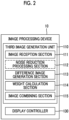
- Fig. 2 shows a functional block diagram of the third image generation unit 110.
- the third image generation unit 110 comprises an image reception section 111 that receives an image from the MRI apparatus 20, a noise reduction processing section 112, a difference image generation section 113 that generates a difference image between the first MR image and the second MR image, a weight calculation section 114 that uses a pixel value of each pixel in the difference image to calculate a weighting value for each pixel, and an image combining section 115 that uses the weighting value calculated by the weight calculation section 114 to combine the images.
- a dashed rectangle indicating the noise reduction processing section 112 means that this function may be either a function of the image processing device 10 or a function of the MRI apparatus, as described above.
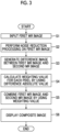
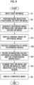
- Fig. 3 shows a flow of the operation of the third image generation unit 110 configured as described above. Here, it is assumed that intraoperative MRI is performed.
- the noise reduction processing section 112 performs noise reduction processing on the first MR image (S2).
- the noise reduction processing performed here is not particularly limited as long as it is generally known processing, and for example, any of processing of removing artifacts characteristically occurring depending on an imaging method through computational operations, processing using a filter, processing using CNN that has learned a noise image and a noise-free image as learning data, or the like can be employed, and a plurality of the processing may also be combined as necessary.
- the filtering strength may be adjusted according to the noise, and the sharpness of the image after noise reduction may be adjusted.
- the difference image generation section 113 takes a difference between the original image before the noise reduction and the second MR image after the noise reduction processing to generate a difference image and to calculate an absolute value of the difference of each pixel (S3).
- the weight calculation section 114 calculates a weighting value for each pixel by using the absolute value of the difference of each pixel calculated by the difference image generation section 113 (S4).
- the image combining section 115 combines the original image and the noise-reduced image for each pixel by using the weighting value for each pixel to generate a composite image which is the third image (S5).
- the processing of the third image generation unit 110 is completed with the above S1 to S5.
- the display controller 130 causes the display device 30 to display the third image generated by the third image generation unit 110 (S6).
- the form of the display is not particularly limited and will be described in detail in the embodiment to be described below. However, in order to make it easier for the user to check a region where processing is performed with respect to the original image, particularly a region with a high degree of noise reduction processing, it is preferable to display two images in parallel or superimposed. In a case of superimposing the two images, color coding or the like is performed to enhance visibility.
- the magnitude of the difference (absolute value) between the original image and the image after noise reduction is obtained for each pixel, and a weight for each pixel is set based on the magnitude of the difference to combine the images, whereby it is possible to present a third MR image in which noise reduction focused on noise that interferes with the checking of the original image is performed while preserving the maximum amount of information from the original image, such as, for a region where significant noise has occurred, that is, a region where the difference is large, increasing the weight of the noise-reduced image in the region and decreasing the weight of the original image, or for a region where little or no noise has occurred, increasing the weight of the original image in the region.
- the weight calculation section 114 of the third image generation unit 110 calculates a weight coefficient ⁇ for each pixel by using the absolute value of the difference of each pixel calculated by the difference image generation section 113 and calculates a weighting value using the weight coefficient ⁇ as an exponent, as the weighting value.
- the noise reduction processing section 112 performs noise reduction processing on the original image (first MR image) acquired by the MRI apparatus 20 and generates the second MR image (S1, S2).
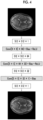
- noise removal is performed using a super resolution convolutional neural network (SRCNN) of all three layers as shown in Fig. 4 .
- This CNN is designed to remove random noise by using a random noise pattern as learning data, and the image after noise reduction (second MR image) tends to become blurred, which causes brain sulci or tumor boundaries to become indistinct, but the influence of the image processing cannot be grasped.
- the third image generation unit 110 combines the original image and the CNN-processed image to obtain the combined third image.
- a composite image in which information that has been compromised by the noise reduction processing is reproduced as faithfully as possible is generated such that the combined image tends to lean toward the image after CNN processing for a location where local noise or artifacts have occurred, while the combined image leans toward the original image for the other positions.
- the difference image generation section 113 calculates an absolute value d of the difference in signal intensity for each pixel between the original image and the image after CNN processing by using Equation (1) (S3).
- d i j I CNN i j ⁇ I org i j
- I CNN represents the image after CNN processing
- I org represents the original image
- (i,j) represents a pixel position (hereinafter, the same).
- the difference in signal intensity which is denoted by d, is large in a position where local noise or artifacts have occurred, but is small in the other region.
- the weight calculation section 114 calculates the coefficient ⁇ (referred to as a weight coefficient) in which the absolute value d of the difference is standardized by the average value of d by using Equation (2).
- the weight coefficient ⁇ is obtained for each pixel and is used to decide on the weighting value.
- a weight of an image in a case of performing weighted addition of the original image and the image after CNN processing is used as a fixed value (fixed weight), and the weight coefficient ⁇ is used as an exponent of the fixed weight. Therefore, a lower limit value and an upper limit value are set in advance for ⁇ . As an example, it is assumed that the lower limit value is 0.01 and the upper limit value is 100.
- ⁇ i j d ⁇ d i j
- the weight calculation section 114 decides on a weighting value W ⁇ in the image weighted addition represented by Equation (3) (S4), and the image combining section 115 uses Equation (3) to generate an image (third image) in which a degree of noise processing is adjusted according to a noise occurrence position.
- I adj i j I CNN i j ⁇ I org i j ⁇ w ⁇ i j + I org i j
- I adj represents an image after adjustment
- W represents the fixed weight.
- the fixed weight takes a value in a range of 0 to 1, and an appropriate value is set in advance as the weight in a case of combining the image after CNN processing and the original image separately from the adjustment in the present embodiment.
- an appropriate weight for obtaining an image in which random noise is suppressed while preserving the information of the original image can be determined to either be set as a default or be user-settable according to a noise pattern included in the original image, a method for processing of reducing noise and artifacts such as CNN processing, or the like.
- a relationship between such a fixed weight W and the weighting value W" using the weight coefficient for each pixel as an exponent is schematically shown on the upper side of Fig. 5 .
- W ⁇ takes the same value as W, but exponentially changes based on the value of ⁇ .
- Equation (3) although the weight coefficient is used as the exponent of the fixed weight W, the method of deciding on the weighting value using the weight coefficient ⁇ for each pixel is not limited to Equations (2) and (3), and for example, a method of setting a weight coefficient standardized by the maximum value of d to ⁇ ' and using a weighting value W ⁇ ' can also be employed.
- the image combining section 115 performs the weighted addition of the original image and the CNN-processed image in accordance with Equation (3) to generate a composite image which is the third image (S5).
- the composite image in a case where the fixed weight is used as a reference, in pixels in which noise has occurred and the signal intensity difference is large, the CNN-processed image has a larger weight, but in pixels in which no noise has occurred, the original image has a larger weight, which results in an image that reflects more information of the original image.
- Fig. 6 shows an example of an image generated through the image processing of the present embodiment.
- the left side is the original image (image before CNN processing)
- the center is the image after noise reduction processing (image after CNN processing)
- the right side is a composite image (adjusted image).
- the original image has noise occurring in regions 601 and 602 surrounded by rectangles in the figure, and the noise disappears in the image after CNN processing by performing the noise reduction processing on this image, but the overall sharpness has decreased, which results in a slight blurring of brain sulci, bleeding parts, or the like.
- noise is reduced in the noise occurrence region, and sharpness close to that of the original image is obtained in the other region.
- the image (adjusted image) combined by the image combining section 115 is immediately displayed on the display device 30 disposed close to the imaging unit of the MRI apparatus. Therefore, the display controller 130 receives the composite image from the image combining section 115 and generates a display image.
- the display image may be only the adjusted image as shown on the right side of Fig. 6 , but in order to make it easier for the user to check the adjusted region, the original image and the image after CNN processing may be displayed together, or the difference image may be displayed.
- the adjusted image may be displayed in one color such as black and white, and the difference image may be superimposed and displayed on the adjusted image in a color different from that of the adjusted image. In this case, it is preferable to make the display of the difference image transparent to maintain the visibility of the adjusted image.
- the display of the difference image may be made user-selectable between being displayed and hidden.
- the present embodiment it is possible to provide an image in which the influence of noise is reduced while ensuring the sharpness of the tissue that the user wants to observe even in a case where noise has occurred in an image due to potential noise or sudden radio wave noise.
- the weight is decided on by determining whether or not noise has occurred from the signal intensity difference for each pixel.
- the present embodiment is characterized by specifying a region where noise has occurred based on the signal intensity difference for each pixel and varying weighting rules between the specified region and the other region.
- the "noise occurrence region" in the present embodiment includes not only a region in an image space but also a region in a data space representing the magnitude of the difference.
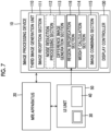
- the third image generation unit 110 comprises a region specification section 116.
- the region specification section 116 receives a user designation through the UI unit 50 provided with the display device 30 and the input device 40, which are attached to the MRI apparatus 20, and specifies the region.
- the same elements as those in Fig. 2 are designated by the same reference numerals, and the overlapping description will not be repeated.
- the original image reconstructed by the MRI apparatus 20 is input, the noise reduction processing is performed on the original image, and the difference for each pixel between the original image and the noise-reduced image is calculated (S1 to S3).
- the display controller 130 displays the difference image obtained using the difference for each pixel or the original image on the display device 30 of the UI unit 50 (S31).
- the user looks at the image displayed on the display device 30 and designates a region where noise or artifacts have occurred. For example, in a case where the original image as shown on the left side of Fig. 6 is displayed, the regions 601 and 602 determined by the user to be noise occurrence regions are selected and designated through the input device 40 such as a pointer or a mouse.
- the region specification section 116 receives the user designation (S32) and specifies the noise occurrence (the region of the image space) region.
- the noise occurrence region can be specified by the user through the designation of a region with high signal intensity in the difference image via the UI unit 50.
- a predetermined threshold value is set in advance for the weight coefficient ⁇ using the absolute value of the difference or Equation (2) mentioned above, and the region specification section 116 may specify a position where the weight coefficient ⁇ is equal to or less than the threshold value, that is, the absolute value of the difference increases, as the noise occurrence region (S33).
- steps S31 and S32 of Fig. 8 can be omitted.
- a region (a region on the data space) 603 having a threshold value TH or less is set as the noise occurrence region.
- the threshold value can be determined by using a discriminant analysis method or the like.
- the threshold value may be set in advance, but GUI that receives a numerical value input or an input by the operation on the histogram may be displayed on the display device 30 of the UI unit 50. This makes it possible to specify the noise region based on the user's perception.
- the region specification section 116 takes AND or OR on the regions designated by the two methods to specify the noise occurrence region.
- the weight calculation section 114 decides on the weighting values for the noise occurrence region and the other region in accordance with different weighting rules (S41), respectively.
- An example of the method of varying weighting rules is, as shown in Fig. 8 , to vary the fixed weight W used in Equation (3) between the noise occurrence region and the other region.
- the noise occurrence region is set to have a larger fixed weight than the other region.
- the calculation of the actually applied weighting value W" as a function of the weight coefficient ⁇ for each pixel is the same as that of Embodiment 1.
- the weighting value is greater than in the other region, and the combined image closely resembles the noise reduction processed image in the noise occurrence region.
- the weight coefficient need only be calculated only for that region, so that the computational load can be reduced and the time required for the presentation of the composite image can be accelerated.
- obtaining the composite image through the weighted addition (S51) after calculating the weighting value, and displaying the composite image (S6) are the same as in Embodiment 1.
- the aspect of display can also be made the same as the aspect described in Embodiment 1.
- the threshold value of the difference between images is set at the time of region specification or in a case where the threshold value of the difference is set by the user when superimposing and displaying the difference image on the original image in a different color
- the color need not be displayed for a region where the difference is lower than the threshold value.
- each region may be displayed with a different color.
- a predetermined range designated by the user, a dot-like noise position selected by using the threshold value, and the like are displayed in different colors. This makes it possible to check the difference in the pattern or in the occurrence position of the noise having different occurrence causes.
- the functional block diagram of the image processing device of the present embodiment is basically the same as the functional block diagram of Embodiment 1 shown in Fig. 2 or the functional block diagram of Embodiment 2 shown in Fig. 7 , and in the following description, reference is made to these drawings.
- the present embodiment is characterized in that the weight calculation section 114 calculates or decides on weighting values corresponding to various noise patterns.
- Fig. 11 shows some patterns of noise occurring in the MR image.
- the upper left shows a dot-like pattern with dot-like noise occurring at unspecified positions, with other patterns representing noise occurring within a limited range
- the upper right shows a zipper-like pattern with noise occurring in a dashed line shape
- the lower left shows a wavy pattern in which one or a plurality of regions become streaked
- the lower right shows a mosaic-like pattern resulting in a roughened image.
- noise pattern peculiar to the MR image there is a noise pattern peculiar to the MR image.
- the distribution of noise intensity (the pattern of the histogram) also differs depending on these noise patterns, and appropriate weighting values and fixed weights differ.
- the weight calculation section 114 applies a weighting method corresponding to the noise pattern by using a processor, such as machine learning, to decide on the weighting value for each pixel.
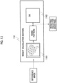
- a processor 1141 that receives the difference image as an input and outputs a noise pattern is provided, and various predetermined noise patterns and calculation algorithms for weighting values are stored in a database (DB 1142) in association with each other.
- the difference image represents a difference in signal intensity for each pixel between the original image and the image after noise reduction processing and includes information such as a magnitude of the signal intensity, a distribution in the image space, an intensity distribution, or the like.
- the processor is trained in advance using pairs of noise patterns and various difference images having these elements different from each other as training data and is trained to classify the difference images into the noise patterns.
- the relationship between the noise pattern and the calculation algorithm for the weighting value is specifically a calculation expression for calculating the weighting value from the weight coefficient ⁇ and the fixed weight W corresponding to the noise pattern.
- a relational expression is used in which the value of the fixed weight of the image after noise reduction processing is reduced in a case where the noise intensity obtained from the difference image is relatively small, a weighting algorithm that uses the weight coefficient ⁇ as the exponent of the fixed weight is used in a case where the noise intensity distribution exponentially changes, and for a pattern with a noise intensity distribution having a peak, heavier weights are set only in pixels near the peak.
- the DB 1142 that stores the relationship between such a noise pattern and the calculation algorithm for the weighting value is provided in the weight calculation section 114 in Fig. 12
- the DB 1142 may be provided in the storage device in the image processing device 10 or the external storage device.
- the weight calculation section 114 refers to the DB to select a calculation algorithm corresponding to the output noise pattern and calculates the weighting value for each pixel.
- a processor 1143 that receives the difference image created from the difference between the first MR image and the second MR image as an input and outputs the weighting value for each pixel is used.
- the processor 1143 is configured with, for example, a machine learning algorithm that has learned to output the optimal fixed weight and the relationship between the fixed weight and the weighting value by using, as learning data, various difference images or histograms thereof, the weighting value for each pixel, which is adjusted in advance, or the weighting value to be decided on according to the noise pattern, which is obtained by classifying the noise patterns through Method 1 and storing the noise patterns in the database.
- the processor 1141 first classifies the difference image into a noise pattern and then refers to the DB 1142 in which the noise pattern and the calculation algorithm for the weighting value are associated with each other, but in the present method, the processor 1143 receives the difference image as an input and outputs the optimal weighting value without outputting the noise pattern.
- the present embodiment it is possible to decide on the optimal weighting value according to the difference image through the trained CNN or the like, and it is possible to present the adjusted image with high accuracy corresponding to the noise pattern.
Landscapes
- Engineering & Computer Science (AREA)
- Physics & Mathematics (AREA)
- Health & Medical Sciences (AREA)
- Life Sciences & Earth Sciences (AREA)
- General Physics & Mathematics (AREA)
- Theoretical Computer Science (AREA)
- Medical Informatics (AREA)
- Nuclear Medicine, Radiotherapy & Molecular Imaging (AREA)
- Radiology & Medical Imaging (AREA)
- General Health & Medical Sciences (AREA)
- Surgery (AREA)
- High Energy & Nuclear Physics (AREA)
- Biomedical Technology (AREA)
- Molecular Biology (AREA)
- Pathology (AREA)
- Animal Behavior & Ethology (AREA)
- Biophysics (AREA)
- Public Health (AREA)
- Veterinary Medicine (AREA)
- Heart & Thoracic Surgery (AREA)
- Multimedia (AREA)
- Computer Vision & Pattern Recognition (AREA)
- Signal Processing (AREA)
- Optics & Photonics (AREA)
- Computing Systems (AREA)
- Artificial Intelligence (AREA)
- Condensed Matter Physics & Semiconductors (AREA)
- Magnetic Resonance Imaging Apparatus (AREA)
- Image Processing (AREA)
Applications Claiming Priority (1)
| Application Number | Priority Date | Filing Date | Title |
|---|---|---|---|
| JP2023020124A JP2024114385A (ja) | 2023-02-13 | 2023-02-13 | 画像処理装置及びそれを有するmri装置、及び画像処理方法 |
Publications (1)
| Publication Number | Publication Date |
|---|---|
| EP4414929A1 true EP4414929A1 (de) | 2024-08-14 |
Family
ID=89843348
Family Applications (1)
| Application Number | Title | Priority Date | Filing Date |
|---|---|---|---|
| EP24155871.7A Pending EP4414929A1 (de) | 2023-02-13 | 2024-02-05 | Bildverarbeitungsvorrichtung, mrt-vorrichtung damit und bildverarbeitungsverfahren |
Country Status (4)
| Country | Link |
|---|---|
| US (1) | US20240268778A1 (de) |
| EP (1) | EP4414929A1 (de) |
| JP (1) | JP2024114385A (de) |
| CN (1) | CN118488320A (de) |
Citations (8)
| Publication number | Priority date | Publication date | Assignee | Title |
|---|---|---|---|---|
| EP1447773A2 (de) * | 2003-02-13 | 2004-08-18 | Kabushiki Kaisha Toshiba | Bildverarbeitungsvorrichtung zur Reduktion von Pixelrauschen |
| WO2009128213A1 (ja) | 2008-04-14 | 2009-10-22 | 株式会社日立メディコ | 医用診断装置および医用診断装置の画質改善方法 |
| US20130216117A1 (en) * | 2012-02-22 | 2013-08-22 | Zakrytoe Akcionernoe Obshchestvo Impul's | Method of noise reduction in digital x-rayograms |
| US20140211034A1 (en) * | 2011-09-30 | 2014-07-31 | Fujifilm Corporation | Image processing apparatus and method |
| US20190378270A1 (en) * | 2018-06-07 | 2019-12-12 | Canon Medical Systems Corporation | Medical image diagnostic apparatus |
| JP2020119429A (ja) | 2019-01-28 | 2020-08-06 | キヤノンメディカルシステムズ株式会社 | 医用画像処理装置、医用画像処理方法、およびプログラム |
| US20220260659A1 (en) * | 2019-01-30 | 2022-08-18 | Canon Medical Systems Corporation | Medical information processing apparatus, magnetic resonance imaging apparatus, and medical information processing method |
| JP2023020124A (ja) | 2021-07-30 | 2023-02-09 | キヤノン株式会社 | ハンドヘルド型の記録装置 |
Family Cites Families (6)
| Publication number | Priority date | Publication date | Assignee | Title |
|---|---|---|---|---|
| JP5190944B2 (ja) * | 2008-06-26 | 2013-04-24 | 富士フイルム株式会社 | 内視鏡装置および内視鏡装置の作動方法 |
| JP6744764B2 (ja) * | 2016-05-26 | 2020-08-19 | 株式会社日立製作所 | 画像診断装置、及び画像取得方法 |
| JP7221981B2 (ja) * | 2018-09-18 | 2023-02-14 | 富士フイルム株式会社 | 画像処理装置、画像処理方法、及び画像処理プログラム |
| JP7204397B2 (ja) * | 2018-09-21 | 2023-01-16 | キヤノン株式会社 | 画像処理装置、画像処理方法、及びプログラム |
| JP2020096773A (ja) * | 2018-12-19 | 2020-06-25 | 富士フイルム株式会社 | 医用画像処理装置、方法およびプログラム |
| KR102769810B1 (ko) * | 2020-12-10 | 2025-02-20 | 주식회사 에어스메디컬 | K-space 도메인에 인공신경망을 적용한 자기 공명 영상 처리 장치 및 그 방법 |
-
2023
- 2023-02-13 JP JP2023020124A patent/JP2024114385A/ja active Pending
-
2024
- 2024-01-12 US US18/412,221 patent/US20240268778A1/en active Pending
- 2024-02-05 EP EP24155871.7A patent/EP4414929A1/de active Pending
- 2024-02-06 CN CN202410171591.2A patent/CN118488320A/zh active Pending
Patent Citations (8)
| Publication number | Priority date | Publication date | Assignee | Title |
|---|---|---|---|---|
| EP1447773A2 (de) * | 2003-02-13 | 2004-08-18 | Kabushiki Kaisha Toshiba | Bildverarbeitungsvorrichtung zur Reduktion von Pixelrauschen |
| WO2009128213A1 (ja) | 2008-04-14 | 2009-10-22 | 株式会社日立メディコ | 医用診断装置および医用診断装置の画質改善方法 |
| US20140211034A1 (en) * | 2011-09-30 | 2014-07-31 | Fujifilm Corporation | Image processing apparatus and method |
| US20130216117A1 (en) * | 2012-02-22 | 2013-08-22 | Zakrytoe Akcionernoe Obshchestvo Impul's | Method of noise reduction in digital x-rayograms |
| US20190378270A1 (en) * | 2018-06-07 | 2019-12-12 | Canon Medical Systems Corporation | Medical image diagnostic apparatus |
| JP2020119429A (ja) | 2019-01-28 | 2020-08-06 | キヤノンメディカルシステムズ株式会社 | 医用画像処理装置、医用画像処理方法、およびプログラム |
| US20220260659A1 (en) * | 2019-01-30 | 2022-08-18 | Canon Medical Systems Corporation | Medical information processing apparatus, magnetic resonance imaging apparatus, and medical information processing method |
| JP2023020124A (ja) | 2021-07-30 | 2023-02-09 | キヤノン株式会社 | ハンドヘルド型の記録装置 |
Also Published As
| Publication number | Publication date |
|---|---|
| US20240268778A1 (en) | 2024-08-15 |
| JP2024114385A (ja) | 2024-08-23 |
| CN118488320A (zh) | 2024-08-13 |
Similar Documents
| Publication | Publication Date | Title |
|---|---|---|
| US9245323B2 (en) | Medical diagnostic device and method of improving image quality of medical diagnostic device | |
| US10546367B2 (en) | Device and method for improving medical image quality | |
| JP5851301B2 (ja) | 画像処理装置、画像処理方法、及び磁気共鳴イメージング装置 | |
| EP2015249A2 (de) | Datenverarbeitungsgerät, medizinisches Diagnosegerät, Datenverarbeitungsverfahren und medizinisches Diagnoseverfahren | |
| WO2003042921A1 (en) | Method and apparatus for three-dimensional filtering of angiographic volume data | |
| JPH11155834A (ja) | 医用イメージング・システム及び医用画像を形成する方法 | |
| US12174281B2 (en) | System and method for control of motion in medical images using aggregation | |
| CN106530236B (zh) | 一种医学图像处理方法及系统 | |
| US11369331B2 (en) | Method for artifact reduction in a medical image data set, X-ray device, computer program and electronically readable data carrier | |
| US12148127B2 (en) | Medical image processing device, medical imaging apparatus, and noise reduction method for medical image | |
| WO2014080961A1 (ja) | 画像処理装置、画像処理方法およびx線診断装置 | |
| CN111353950B (zh) | 用于图像处理的方法、医学成像设备和电子可读数据载体 | |
| US6466700B1 (en) | Method of processing a noisy multidimensional image and medical imaging apparatus for carrying out this method | |
| EP4414928A1 (de) | Bildverarbeitungsvorrichtung, medizinische bildgebungsvorrichtung damit und bildverarbeitungsverfahren | |
| Firbank et al. | Measuring extraocular muscle volume using dynamic contours | |
| EP2757523A2 (de) | Verfahren und Vorrichtung zur Geräuschunterdrückung in Magnetresonanzbildern | |
| US20240268778A1 (en) | Image processing device, mri apparatus including the same, and image processing method | |
| US8150126B2 (en) | Method and system for scale-based vessel enhancement in X-ray angiography | |
| JP2025124553A (ja) | 核磁気共鳴撮像装置、及び体動情報処理方法 | |
| Yousefi et al. | A fully automated segmentation of radius bone based on active contour in wrist MRI data set | |
| JP5105823B2 (ja) | 磁気共鳴イメージング装置、医用画像処理方法、プログラム及び医用画像表示システム | |
| JP2021170174A (ja) | 情報処理装置、情報処理方法、及びコンピュータプログラム | |
| US12386004B2 (en) | Methods and systems for motion detection in magnetic resonance imaging | |
| JP3600656B2 (ja) | 画像処理方法及び磁気共鳴イメージング装置 | |
| US7609854B2 (en) | Method for displaying medical image information dependent on a detected position of the observer |
Legal Events
| Date | Code | Title | Description |
|---|---|---|---|
| PUAI | Public reference made under article 153(3) epc to a published international application that has entered the european phase |
Free format text: ORIGINAL CODE: 0009012 |
|
| STAA | Information on the status of an ep patent application or granted ep patent |
Free format text: STATUS: REQUEST FOR EXAMINATION WAS MADE |
|
| 17P | Request for examination filed |
Effective date: 20240205 |
|
| AK | Designated contracting states |
Kind code of ref document: A1 Designated state(s): AL AT BE BG CH CY CZ DE DK EE ES FI FR GB GR HR HU IE IS IT LI LT LU LV MC ME MK MT NL NO PL PT RO RS SE SI SK SM TR |
|
| RAP1 | Party data changed (applicant data changed or rights of an application transferred) |
Owner name: FUJIFILM CORPORATION |