EP4455265A2 - Procédés et matériaux pour utiliser des supports de fibrine pour une transplantation d'épithélium pigmentaire rétinien - Google Patents
Procédés et matériaux pour utiliser des supports de fibrine pour une transplantation d'épithélium pigmentaire rétinien Download PDFInfo
- Publication number
- EP4455265A2 EP4455265A2 EP24187425.4A EP24187425A EP4455265A2 EP 4455265 A2 EP4455265 A2 EP 4455265A2 EP 24187425 A EP24187425 A EP 24187425A EP 4455265 A2 EP4455265 A2 EP 4455265A2
- Authority
- EP
- European Patent Office
- Prior art keywords
- fibrin
- rpe
- retinal
- plasminogen
- cells
- Prior art date
- Legal status (The legal status is an assumption and is not a legal conclusion. Google has not performed a legal analysis and makes no representation as to the accuracy of the status listed.)
- Pending
Links
Images





















Classifications
-
- A—HUMAN NECESSITIES
- A61—MEDICAL OR VETERINARY SCIENCE; HYGIENE
- A61L—METHODS OR APPARATUS FOR STERILISING MATERIALS OR OBJECTS IN GENERAL; DISINFECTION, STERILISATION OR DEODORISATION OF AIR; CHEMICAL ASPECTS OF BANDAGES, DRESSINGS, ABSORBENT PADS OR SURGICAL ARTICLES; MATERIALS FOR BANDAGES, DRESSINGS, ABSORBENT PADS OR SURGICAL ARTICLES
- A61L27/00—Materials for grafts or prostheses or for coating grafts or prostheses
- A61L27/36—Materials for grafts or prostheses or for coating grafts or prostheses containing ingredients of undetermined constitution or reaction products thereof, e.g. transplant tissue, natural bone, extracellular matrix
- A61L27/38—Materials for grafts or prostheses or for coating grafts or prostheses containing ingredients of undetermined constitution or reaction products thereof, e.g. transplant tissue, natural bone, extracellular matrix containing added animal cells
- A61L27/3804—Materials for grafts or prostheses or for coating grafts or prostheses containing ingredients of undetermined constitution or reaction products thereof, e.g. transplant tissue, natural bone, extracellular matrix containing added animal cells characterised by specific cells or progenitors thereof, e.g. fibroblasts, connective tissue cells, kidney cells
- A61L27/3813—Epithelial cells, e.g. keratinocytes, urothelial cells
-
- A—HUMAN NECESSITIES
- A61—MEDICAL OR VETERINARY SCIENCE; HYGIENE
- A61K—PREPARATIONS FOR MEDICAL, DENTAL OR TOILETRY PURPOSES
- A61K31/00—Medicinal preparations containing organic active ingredients
- A61K31/185—Acids; Anhydrides, halides or salts thereof, e.g. sulfur acids, imidic, hydrazonic or hydroximic acids
- A61K31/19—Carboxylic acids, e.g. valproic acid
- A61K31/195—Carboxylic acids, e.g. valproic acid having an amino group
-
- A—HUMAN NECESSITIES
- A61—MEDICAL OR VETERINARY SCIENCE; HYGIENE
- A61K—PREPARATIONS FOR MEDICAL, DENTAL OR TOILETRY PURPOSES
- A61K31/00—Medicinal preparations containing organic active ingredients
- A61K31/185—Acids; Anhydrides, halides or salts thereof, e.g. sulfur acids, imidic, hydrazonic or hydroximic acids
- A61K31/19—Carboxylic acids, e.g. valproic acid
- A61K31/195—Carboxylic acids, e.g. valproic acid having an amino group
- A61K31/197—Carboxylic acids, e.g. valproic acid having an amino group the amino and the carboxyl groups being attached to the same acyclic carbon chain, e.g. gamma-aminobutyric acid [GABA], beta-alanine, epsilon-aminocaproic acid or pantothenic acid
-
- A—HUMAN NECESSITIES
- A61—MEDICAL OR VETERINARY SCIENCE; HYGIENE
- A61K—PREPARATIONS FOR MEDICAL, DENTAL OR TOILETRY PURPOSES
- A61K35/00—Medicinal preparations containing materials or reaction products thereof with undetermined constitution
- A61K35/12—Materials from mammals; Compositions comprising non-specified tissues or cells; Compositions comprising non-embryonic stem cells; Genetically modified cells
- A61K35/30—Nerves; Brain; Eyes; Corneal cells; Cerebrospinal fluid; Neuronal stem cells; Neuronal precursor cells; Glial cells; Oligodendrocytes; Schwann cells; Astroglia; Astrocytes; Choroid plexus; Spinal cord tissue
-
- A—HUMAN NECESSITIES
- A61—MEDICAL OR VETERINARY SCIENCE; HYGIENE
- A61K—PREPARATIONS FOR MEDICAL, DENTAL OR TOILETRY PURPOSES
- A61K35/00—Medicinal preparations containing materials or reaction products thereof with undetermined constitution
- A61K35/12—Materials from mammals; Compositions comprising non-specified tissues or cells; Compositions comprising non-embryonic stem cells; Genetically modified cells
- A61K35/36—Skin; Hair; Nails; Sebaceous glands; Cerumen; Epidermis; Epithelial cells; Keratinocytes; Langerhans cells; Ectodermal cells
-
- A—HUMAN NECESSITIES
- A61—MEDICAL OR VETERINARY SCIENCE; HYGIENE
- A61K—PREPARATIONS FOR MEDICAL, DENTAL OR TOILETRY PURPOSES
- A61K38/00—Medicinal preparations containing peptides
- A61K38/16—Peptides having more than 20 amino acids; Gastrins; Somatostatins; Melanotropins; Derivatives thereof
- A61K38/17—Peptides having more than 20 amino acids; Gastrins; Somatostatins; Melanotropins; Derivatives thereof from animals; from humans
- A61K38/36—Blood coagulation or fibrinolysis factors
- A61K38/363—Fibrinogen
-
- A—HUMAN NECESSITIES
- A61—MEDICAL OR VETERINARY SCIENCE; HYGIENE
- A61K—PREPARATIONS FOR MEDICAL, DENTAL OR TOILETRY PURPOSES
- A61K38/00—Medicinal preparations containing peptides
- A61K38/16—Peptides having more than 20 amino acids; Gastrins; Somatostatins; Melanotropins; Derivatives thereof
- A61K38/43—Enzymes; Proenzymes; Derivatives thereof
- A61K38/46—Hydrolases (3)
- A61K38/48—Hydrolases (3) acting on peptide bonds (3.4)
- A61K38/482—Serine endopeptidases (3.4.21)
- A61K38/484—Plasmin (3.4.21.7)
-
- A—HUMAN NECESSITIES
- A61—MEDICAL OR VETERINARY SCIENCE; HYGIENE
- A61K—PREPARATIONS FOR MEDICAL, DENTAL OR TOILETRY PURPOSES
- A61K38/00—Medicinal preparations containing peptides
- A61K38/16—Peptides having more than 20 amino acids; Gastrins; Somatostatins; Melanotropins; Derivatives thereof
- A61K38/55—Protease inhibitors
- A61K38/57—Protease inhibitors from animals; from humans
-
- A—HUMAN NECESSITIES
- A61—MEDICAL OR VETERINARY SCIENCE; HYGIENE
- A61L—METHODS OR APPARATUS FOR STERILISING MATERIALS OR OBJECTS IN GENERAL; DISINFECTION, STERILISATION OR DEODORISATION OF AIR; CHEMICAL ASPECTS OF BANDAGES, DRESSINGS, ABSORBENT PADS OR SURGICAL ARTICLES; MATERIALS FOR BANDAGES, DRESSINGS, ABSORBENT PADS OR SURGICAL ARTICLES
- A61L27/00—Materials for grafts or prostheses or for coating grafts or prostheses
- A61L27/14—Macromolecular materials
- A61L27/22—Polypeptides or derivatives thereof, e.g. degradation products
- A61L27/225—Fibrin; Fibrinogen
-
- A—HUMAN NECESSITIES
- A61—MEDICAL OR VETERINARY SCIENCE; HYGIENE
- A61L—METHODS OR APPARATUS FOR STERILISING MATERIALS OR OBJECTS IN GENERAL; DISINFECTION, STERILISATION OR DEODORISATION OF AIR; CHEMICAL ASPECTS OF BANDAGES, DRESSINGS, ABSORBENT PADS OR SURGICAL ARTICLES; MATERIALS FOR BANDAGES, DRESSINGS, ABSORBENT PADS OR SURGICAL ARTICLES
- A61L27/00—Materials for grafts or prostheses or for coating grafts or prostheses
- A61L27/28—Materials for coating prostheses
- A61L27/34—Macromolecular materials
-
- A—HUMAN NECESSITIES
- A61—MEDICAL OR VETERINARY SCIENCE; HYGIENE
- A61L—METHODS OR APPARATUS FOR STERILISING MATERIALS OR OBJECTS IN GENERAL; DISINFECTION, STERILISATION OR DEODORISATION OF AIR; CHEMICAL ASPECTS OF BANDAGES, DRESSINGS, ABSORBENT PADS OR SURGICAL ARTICLES; MATERIALS FOR BANDAGES, DRESSINGS, ABSORBENT PADS OR SURGICAL ARTICLES
- A61L27/00—Materials for grafts or prostheses or for coating grafts or prostheses
- A61L27/36—Materials for grafts or prostheses or for coating grafts or prostheses containing ingredients of undetermined constitution or reaction products thereof, e.g. transplant tissue, natural bone, extracellular matrix
- A61L27/3683—Materials for grafts or prostheses or for coating grafts or prostheses containing ingredients of undetermined constitution or reaction products thereof, e.g. transplant tissue, natural bone, extracellular matrix subjected to a specific treatment prior to implantation, e.g. decellularising, demineralising, grinding, cellular disruption/non-collagenous protein removal, anti-calcification, crosslinking, supercritical fluid extraction, enzyme treatment
- A61L27/3687—Materials for grafts or prostheses or for coating grafts or prostheses containing ingredients of undetermined constitution or reaction products thereof, e.g. transplant tissue, natural bone, extracellular matrix subjected to a specific treatment prior to implantation, e.g. decellularising, demineralising, grinding, cellular disruption/non-collagenous protein removal, anti-calcification, crosslinking, supercritical fluid extraction, enzyme treatment characterised by the use of chemical agents in the treatment, e.g. specific enzymes, detergents, capping agents, crosslinkers, anticalcification agents
-
- A—HUMAN NECESSITIES
- A61—MEDICAL OR VETERINARY SCIENCE; HYGIENE
- A61L—METHODS OR APPARATUS FOR STERILISING MATERIALS OR OBJECTS IN GENERAL; DISINFECTION, STERILISATION OR DEODORISATION OF AIR; CHEMICAL ASPECTS OF BANDAGES, DRESSINGS, ABSORBENT PADS OR SURGICAL ARTICLES; MATERIALS FOR BANDAGES, DRESSINGS, ABSORBENT PADS OR SURGICAL ARTICLES
- A61L27/00—Materials for grafts or prostheses or for coating grafts or prostheses
- A61L27/36—Materials for grafts or prostheses or for coating grafts or prostheses containing ingredients of undetermined constitution or reaction products thereof, e.g. transplant tissue, natural bone, extracellular matrix
- A61L27/38—Materials for grafts or prostheses or for coating grafts or prostheses containing ingredients of undetermined constitution or reaction products thereof, e.g. transplant tissue, natural bone, extracellular matrix containing added animal cells
- A61L27/3804—Materials for grafts or prostheses or for coating grafts or prostheses containing ingredients of undetermined constitution or reaction products thereof, e.g. transplant tissue, natural bone, extracellular matrix containing added animal cells characterised by specific cells or progenitors thereof, e.g. fibroblasts, connective tissue cells, kidney cells
- A61L27/3808—Endothelial cells
-
- A—HUMAN NECESSITIES
- A61—MEDICAL OR VETERINARY SCIENCE; HYGIENE
- A61L—METHODS OR APPARATUS FOR STERILISING MATERIALS OR OBJECTS IN GENERAL; DISINFECTION, STERILISATION OR DEODORISATION OF AIR; CHEMICAL ASPECTS OF BANDAGES, DRESSINGS, ABSORBENT PADS OR SURGICAL ARTICLES; MATERIALS FOR BANDAGES, DRESSINGS, ABSORBENT PADS OR SURGICAL ARTICLES
- A61L27/00—Materials for grafts or prostheses or for coating grafts or prostheses
- A61L27/36—Materials for grafts or prostheses or for coating grafts or prostheses containing ingredients of undetermined constitution or reaction products thereof, e.g. transplant tissue, natural bone, extracellular matrix
- A61L27/38—Materials for grafts or prostheses or for coating grafts or prostheses containing ingredients of undetermined constitution or reaction products thereof, e.g. transplant tissue, natural bone, extracellular matrix containing added animal cells
- A61L27/3839—Materials for grafts or prostheses or for coating grafts or prostheses containing ingredients of undetermined constitution or reaction products thereof, e.g. transplant tissue, natural bone, extracellular matrix containing added animal cells characterised by the site of application in the body
-
- A—HUMAN NECESSITIES
- A61—MEDICAL OR VETERINARY SCIENCE; HYGIENE
- A61L—METHODS OR APPARATUS FOR STERILISING MATERIALS OR OBJECTS IN GENERAL; DISINFECTION, STERILISATION OR DEODORISATION OF AIR; CHEMICAL ASPECTS OF BANDAGES, DRESSINGS, ABSORBENT PADS OR SURGICAL ARTICLES; MATERIALS FOR BANDAGES, DRESSINGS, ABSORBENT PADS OR SURGICAL ARTICLES
- A61L27/00—Materials for grafts or prostheses or for coating grafts or prostheses
- A61L27/50—Materials characterised by their function or physical properties, e.g. injectable or lubricating compositions, shape-memory materials, surface modified materials
- A61L27/52—Hydrogels or hydrocolloids
-
- A—HUMAN NECESSITIES
- A61—MEDICAL OR VETERINARY SCIENCE; HYGIENE
- A61L—METHODS OR APPARATUS FOR STERILISING MATERIALS OR OBJECTS IN GENERAL; DISINFECTION, STERILISATION OR DEODORISATION OF AIR; CHEMICAL ASPECTS OF BANDAGES, DRESSINGS, ABSORBENT PADS OR SURGICAL ARTICLES; MATERIALS FOR BANDAGES, DRESSINGS, ABSORBENT PADS OR SURGICAL ARTICLES
- A61L27/00—Materials for grafts or prostheses or for coating grafts or prostheses
- A61L27/50—Materials characterised by their function or physical properties, e.g. injectable or lubricating compositions, shape-memory materials, surface modified materials
- A61L27/54—Biologically active materials, e.g. therapeutic substances
-
- A—HUMAN NECESSITIES
- A61—MEDICAL OR VETERINARY SCIENCE; HYGIENE
- A61P—SPECIFIC THERAPEUTIC ACTIVITY OF CHEMICAL COMPOUNDS OR MEDICINAL PREPARATIONS
- A61P27/00—Drugs for disorders of the senses
- A61P27/02—Ophthalmic agents
-
- C—CHEMISTRY; METALLURGY
- C12—BIOCHEMISTRY; BEER; SPIRITS; WINE; VINEGAR; MICROBIOLOGY; ENZYMOLOGY; MUTATION OR GENETIC ENGINEERING
- C12N—MICROORGANISMS OR ENZYMES; COMPOSITIONS THEREOF; PROPAGATING, PRESERVING, OR MAINTAINING MICROORGANISMS; MUTATION OR GENETIC ENGINEERING; CULTURE MEDIA
- C12N5/00—Undifferentiated human, animal or plant cells, e.g. cell lines; Tissues; Cultivation or maintenance thereof; Culture media therefor
- C12N5/06—Animal cells or tissues; Human cells or tissues
- C12N5/0602—Vertebrate cells
- C12N5/0618—Cells of the nervous system
- C12N5/062—Sensory transducers, e.g. photoreceptors; Sensory neurons, e.g. for hearing, taste, smell, pH, touch, temperature, pain
-
- C—CHEMISTRY; METALLURGY
- C12—BIOCHEMISTRY; BEER; SPIRITS; WINE; VINEGAR; MICROBIOLOGY; ENZYMOLOGY; MUTATION OR GENETIC ENGINEERING
- C12N—MICROORGANISMS OR ENZYMES; COMPOSITIONS THEREOF; PROPAGATING, PRESERVING, OR MAINTAINING MICROORGANISMS; MUTATION OR GENETIC ENGINEERING; CULTURE MEDIA
- C12N5/00—Undifferentiated human, animal or plant cells, e.g. cell lines; Tissues; Cultivation or maintenance thereof; Culture media therefor
- C12N5/06—Animal cells or tissues; Human cells or tissues
- C12N5/0602—Vertebrate cells
- C12N5/0618—Cells of the nervous system
- C12N5/0621—Eye cells, e.g. cornea, iris pigmented cells
-
- C—CHEMISTRY; METALLURGY
- C12—BIOCHEMISTRY; BEER; SPIRITS; WINE; VINEGAR; MICROBIOLOGY; ENZYMOLOGY; MUTATION OR GENETIC ENGINEERING
- C12Y—ENZYMES
- C12Y304/00—Hydrolases acting on peptide bonds, i.e. peptidases (3.4)
- C12Y304/21—Serine endopeptidases (3.4.21)
- C12Y304/21007—Plasmin (3.4.21.7), i.e. fibrinolysin
-
- A—HUMAN NECESSITIES
- A61—MEDICAL OR VETERINARY SCIENCE; HYGIENE
- A61L—METHODS OR APPARATUS FOR STERILISING MATERIALS OR OBJECTS IN GENERAL; DISINFECTION, STERILISATION OR DEODORISATION OF AIR; CHEMICAL ASPECTS OF BANDAGES, DRESSINGS, ABSORBENT PADS OR SURGICAL ARTICLES; MATERIALS FOR BANDAGES, DRESSINGS, ABSORBENT PADS OR SURGICAL ARTICLES
- A61L2420/00—Materials or methods for coatings medical devices
- A61L2420/02—Methods for coating medical devices
-
- A—HUMAN NECESSITIES
- A61—MEDICAL OR VETERINARY SCIENCE; HYGIENE
- A61L—METHODS OR APPARATUS FOR STERILISING MATERIALS OR OBJECTS IN GENERAL; DISINFECTION, STERILISATION OR DEODORISATION OF AIR; CHEMICAL ASPECTS OF BANDAGES, DRESSINGS, ABSORBENT PADS OR SURGICAL ARTICLES; MATERIALS FOR BANDAGES, DRESSINGS, ABSORBENT PADS OR SURGICAL ARTICLES
- A61L2430/00—Materials or treatment for tissue regeneration
- A61L2430/16—Materials or treatment for tissue regeneration for reconstruction of eye parts, e.g. intraocular lens, cornea
-
- C—CHEMISTRY; METALLURGY
- C12—BIOCHEMISTRY; BEER; SPIRITS; WINE; VINEGAR; MICROBIOLOGY; ENZYMOLOGY; MUTATION OR GENETIC ENGINEERING
- C12N—MICROORGANISMS OR ENZYMES; COMPOSITIONS THEREOF; PROPAGATING, PRESERVING, OR MAINTAINING MICROORGANISMS; MUTATION OR GENETIC ENGINEERING; CULTURE MEDIA
- C12N2501/00—Active agents used in cell culture processes, e.g. differentation
- C12N2501/70—Enzymes
- C12N2501/73—Hydrolases (EC 3.)
- C12N2501/734—Proteases (EC 3.4.)
-
- C—CHEMISTRY; METALLURGY
- C12—BIOCHEMISTRY; BEER; SPIRITS; WINE; VINEGAR; MICROBIOLOGY; ENZYMOLOGY; MUTATION OR GENETIC ENGINEERING
- C12N—MICROORGANISMS OR ENZYMES; COMPOSITIONS THEREOF; PROPAGATING, PRESERVING, OR MAINTAINING MICROORGANISMS; MUTATION OR GENETIC ENGINEERING; CULTURE MEDIA
- C12N2501/00—Active agents used in cell culture processes, e.g. differentation
- C12N2501/999—Small molecules not provided for elsewhere
-
- C—CHEMISTRY; METALLURGY
- C12—BIOCHEMISTRY; BEER; SPIRITS; WINE; VINEGAR; MICROBIOLOGY; ENZYMOLOGY; MUTATION OR GENETIC ENGINEERING
- C12N—MICROORGANISMS OR ENZYMES; COMPOSITIONS THEREOF; PROPAGATING, PRESERVING, OR MAINTAINING MICROORGANISMS; MUTATION OR GENETIC ENGINEERING; CULTURE MEDIA
- C12N2506/00—Differentiation of animal cells from one lineage to another; Differentiation of pluripotent cells
- C12N2506/45—Differentiation of animal cells from one lineage to another; Differentiation of pluripotent cells from artificially induced pluripotent stem cells
-
- C—CHEMISTRY; METALLURGY
- C12—BIOCHEMISTRY; BEER; SPIRITS; WINE; VINEGAR; MICROBIOLOGY; ENZYMOLOGY; MUTATION OR GENETIC ENGINEERING
- C12N—MICROORGANISMS OR ENZYMES; COMPOSITIONS THEREOF; PROPAGATING, PRESERVING, OR MAINTAINING MICROORGANISMS; MUTATION OR GENETIC ENGINEERING; CULTURE MEDIA
- C12N2533/00—Supports or coatings for cell culture, characterised by material
- C12N2533/50—Proteins
- C12N2533/56—Fibrin; Thrombin
-
- C—CHEMISTRY; METALLURGY
- C12—BIOCHEMISTRY; BEER; SPIRITS; WINE; VINEGAR; MICROBIOLOGY; ENZYMOLOGY; MUTATION OR GENETIC ENGINEERING
- C12N—MICROORGANISMS OR ENZYMES; COMPOSITIONS THEREOF; PROPAGATING, PRESERVING, OR MAINTAINING MICROORGANISMS; MUTATION OR GENETIC ENGINEERING; CULTURE MEDIA
- C12N2533/00—Supports or coatings for cell culture, characterised by material
- C12N2533/90—Substrates of biological origin, e.g. extracellular matrix, decellularised tissue
Definitions
- This document relates to retinal pigment epithelium transplantation.
- this document relates to methods and materials for using fibrin supports for retinal pigment epithelium transplantation.
- Macular degeneration diseases represent a variety of diseases and etiology, but commonly stem from retinal pigment epithelium (RPE) dysfunction.
- RPE retinal pigment epithelium
- the bestrophinopathies e.g., Best's disease
- the prevalence has previously been reported as 1 in 16,000-21,500 ( Dalvin et al., Ophthalmic Genet., Epub: 1-5 (2016 )).
- AMD age-related macular degeneration
- RPE replacement as a treatment for macular degeneration has been a popular focus in recent history. Modern advances in stem cell technologies have made embryonic (ES) and induced pluripotent (IPS) stem cells attractive candidates for transplantation. Multiple reports show the ability to differentiate both stem cell sources towards an RPE lineage ( Sonoda et al., Nat. Protoc., 4:662-673 (2009 ); Johnson et al., Ophthalmol. Vis. Sci., 56:4619 (2015 ); Brandl et al., NeuroMolecular Med., 16:551-564 (2014 ); Idelson et al., Cell Stem Cell., 5:396-408 (2009 ); Carr et al., Mol. Vis., 15:283-295 (2009 )).
- ES embryonic
- IPS induced pluripotent
- ES-RPE and IPS-RPE have been shown to exhibit normal RPE function, including cell markers, phagocytosis, and pigmentation ( Singh et al., Ophthalmol. Vis. Sci., 54:6767-6778 (2013 )).
- a general tissue engineering strategy has been to utilize synthetic polymer substrates as a basal support to RPE during the differentiation process and subsequent implantation.
- Two materials that are currently in clinical trials include parylene ( Hu et al., Ophthalmic Res., 48:186-191 (2012 ); and Diniz et al., Invest. Ophthalmol. Vis. Sci., 54:5087-5096 (2013 )) and polyester ( Stanzel et al., Stem Cell Rep., 2:64-77 (2014 )). These materials can be modified to create micropores and improve cell attachment ( Lu et al., Biomed. Microdevices, 14:659-667 (2012 ); McHugh et al., Invest. Ophthalmol.
- Fibrin can be a cross-linking fibril network formed spontaneously after the activation of a precursor to its self-polymerizing monomers. Fibrin typically makes up the clot that forms physiologically during wound healing, and has a well characterized cascade of activation, formation, degradation, and clearance ( Undas et al., Arterioscler. Thromb. Vasc. Biol., 31:e88-e99 (2011 )). For example, fibrin gels can be rapidly degraded through the activation of plasminogen to plasmin, a process activated by enzymes like tissue plasminogen activator (tPA).
- tPA tissue plasminogen activator
- Fibrin often referred to as fibrin glue, is used in the clinic as a natural sealant during surgical incisions in soft tissues and is available commercially.
- the fibrin used herein can be highly adhesive, can have biomechanical rigidity, can be biocompatible, and can be degradable.
- fibrin hydrogels can be used as a temporary apically-apposed or basal support substrate for RPE transplantation.
- RPE transplantation can be performed using an RPE monolayer/fibrin implant provided herein.
- the fibrin scaffold can be on the apical side or basal side of the RPE monolayer for improved RPE attachment.
- RPE can be grown on the fibrin support to develop a monolayer with basal support. These cultures can be cut to develop individual units for implantation.
- the fibrin scaffold can be on the apical side of the RPE monolayer for improved RPE attachment.
- modular tiling of multiple (e.g., two, three, four, or more) RPE monolayer/fibrin implants can provide large area coverage, and laser tacking can be used to enable precision of delivery location.
- the fibrin scaffold can be degraded under controlled conditions during surgery using, for example via tPA.
- a retinal implant comprising, or consisting essentially of, (a) a retinal pigment epithelium monolayer having an apical surface and a basal surface, and (b) a fibrin hydrogel layer attached to the apical and/or basal surface of the monolayer.
- the fibrin hydrogel layer can be from about 20 ⁇ m to about 400 ⁇ m thick.
- the implant can comprise plasminogen.
- the implant can comprise from about 0.1 U of plasminogen per mL to about 40 U of plasminogen per mL.
- the implant can comprise from about 0.001 U of plasminogen per mL to about 40 U of plasminogen per mL.
- the fibrin hydrogel layer can be obtained autologously.
- this document features a method for making a retinal or sub-retinal implant.
- the method comprises, or consists essentially of, (a) obtaining a retinal pigment epithelium monolayer having an apical surface and a basal surface, and (b) depositing a coating of fibrinogen and thrombin onto the apical surface of the monolayer.
- the coating can be from about 20 ⁇ m to about 400 ⁇ m thick.
- the method coating can comprise from about 20 mg of fibrinogen per mL to about 80 mg of fibrinogen per mL.
- the method coating can comprise from about 2 U of thrombin per mL to about 1500 U of thrombin per mL.
- the method can comprise depositing plasminogen onto the apical surface of the monolayer.
- the method can comprise depositing plasminogen within the fibrin hydrogel onto the apical surface of the monolayer.
- the method coating can comprise from about 0.1 U of plasminogen per mL to about 40 U of plasminogen per mL.
- the implant can comprise from about 0.001 U of plasminogen per mL to about 40 U of plasminogen per mL.
- this document features a method for making a retinal or sub-retinal implant.
- the method comprises, or consists essentially of, culturing retinal epithelial cells on a fibrin basal support substrate in a medium comprising a protease inhibitor or an antifibrinolytic agent (e.g., a small molecule protease inhibitors).
- the medium can comprise the protease inhibitor, and the protease inhibitor can be aprotinin.
- the medium can comprise from about 5 U of aprotinin per mL to about 500 U of aprotinin per mL.
- the medium further can comprise plasminogen.
- the medium can comprise from about 0.1 U of plasminogen per mL to about 40 U of plasminogen per mL (e.g., 0.1 U of plasminogen per mL to about 30 U of plasminogen per mL).
- the implant can comprise from about 0.001 U of plasminogen per mL to about 40 U of plasminogen per mL.
- plasminogen can be added to the medium just prior to transplantation.
- the fibrin basal support substrate can comprise endothelial cells.
- the endothelial cells were obtained from a source selected from the group consisting of iPSC-derived endothelial cells, blood outgrowth endothelial cells (BOEC), endothelial colony-forming cells (ECFCs), endothelial progenitor cells (EPCs), and umbilical vein endothelial cells (UVEC).
- the fibrin basal support substrate can comprise sub-RPE tissue cell populations.
- the sub-RPE tissue cell populations can comprise melanocytes, pericytes, or fibroblasts.
- the fibrin basal support substrate can be obtained autologously.
- this document features a retinal implant comprising (a) a retinal pigment epithelium monolayer having an apical surface and a basal surface, and (b) a fibrin hydrogel layer attached to the basal surface of the monolayer.
- the fibrin hydrogel layer can be from about 20 ⁇ m to about 400 ⁇ m thick.
- the implant can comprise plasminogen.
- the implant can comprise from about 0.1 U of plasminogen per mL to about 40 U of plasminogen per mL or from about 0.001 U of plasminogen per mL to about 40 U of plasminogen per mL.
- the fibrin hydrogel layer can comprise a coating.
- the coating can comprise basement membrane proteins, matrigel, or geltrex. In some cases, the fibrin hydrogel monolayer can be obtained autologously.
- this document features a method for making a retinal implant.
- the method comprises (a) obtaining a fibrin hydrogel layer, (b) coating a surface of the fibrin hydrogel layer with an agent, and (c) forming a retinal pigment epithelium monolayer having an apical surface and a basal surface on the coating, wherein the basal surface is closer to the fibrin hydrogel layer than the apical surface.
- the fibrin hydrogel layer can be from about 20 ⁇ m to about 400 ⁇ m thick.
- the fibrin hydrogel layer can comprise from about 20 mg of fibrinogen per mL to about 80 mg of fibrinogen per mL.
- the fibrin hydrogel layer can comprise from about 2 U of thrombin per mL to about 1500 U of thrombin per mL.
- the fibrin hydrogel layer can comprise from about 0.1 U of plasminogen per mL to about 40 U of plasminogen per mL or from about 0.001 U of plasminogen per mL to about 40 U of plasminogen per mL.
- the fibrin hydrogel monolayer can be obtained autologously.
- this document features a method for making a retinal implant.
- the method comprises culturing retinal epithelial cells on a fibrin basal support substrate in a medium comprising a protease inhibitor or an anti-fibrinolytic agent.
- the medium can comprise the protease inhibitor, and the protease inhibitor can be aprotinin.
- the medium can comprise from about 5 U of aprotinin per mL to about 500 U of aprotinin per mL.
- the medium can comprise the anti-fibrinolytic agent, and the antifibrinolytic agent can be transexamic acid or aminocaproic acid.
- the medium can further comprise plasminogen.
- the medium can comprise from about 0.1 U of plasminogen per mL to about 40 U of plasminogen per mL or from about 0.001 U of plasminogen per mL to about 40 U of plasminogen per mL.
- the fibrin basal support substrate can comprise endothelial cells.
- the endothelial cells can be obtained from a source selected from the group consisting of iPSC-derived endothelial cells, blood outgrowth endothelial cells (BOEC), endothelial colony-forming cells (ECFCs), endothelial progenitor cells (EPCs), and umbilical vein endothelial cells (UVEC).
- the fibrin basal support substrate can comprise a coating.
- the coating can comprise basement membrane proteins, matrigel, or geltrex.
- the coating can be present prior to culturing the retinal epithelial cells on the fibrin basal support substrate.
- the fibrin basal support substrate can comprise sub-RPE tissue cell populations.
- the sub-RPE tissue cell populations can comprise melanocytes, pericytes, or fibroblasts.
- the fibrin basal support substrate can be obtained autologously.
- fibrin hydrogels can be used as a temporary substrate for RPE transplantation.
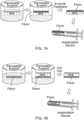
- the fibrin hydrogel can be a basal support substrate ( Figure 1B ) or an apically-apposed substrate ( Figure 1A ) for the RPE.
- the RPE can be sandwiched between two fibrin hydrogels; one basal support substrate and one apically-apposed substrate.
- the RPE monolayer/fibrin implants provided herein can maintain the RPE as a flat, wrinkle-free monolayer.
- the fibrin configuration can provide mechanical support and protection during the transplantation process and can ensure implantation of correct RPE polarity.
- the RPE monolayer/fibrin implants provided herein can reduce potential chronic inflammation, obstacles to RPE/Bruch's membrane attachment and can maintain diffusion permeability from choroid.
- any appropriate method can be used to produce a fibrin substrate for RPE monolayers.
- Gelation kinetics can be directly related to thrombin concentration, and fibrin hydrogel mechanical properties can be directly related to initial fibrinogen concentration.
- a higher fibrinogen concentration can result in increased cross links and a stiffer fibrin hydrogel.
- fibrin hydrogel can be formed as a thin sheet. In some cases, compaction of the fibrin hydrogel can further stiffen the hydrogel.
- fibrin thin film deposition can be achieved through either spray-coating or sandwich method.
- a mixture of fibrinogen and thrombin (and optionally plasminogen) can be sprayed onto the apical side of RPE monolayer and allowed to gel fully to achieve an apical fibrin coating.
- a droplet mixture of fibrinogen and thrombin (and optionally plasminogen) can be placed onto the apical side of RPE monolayer, and the droplet can be compressed or spread and allowed to gel fully to achieve an apical fibrin coating.
- a spray coating of a thin layer of fibrin can be used to form a fibrin hydrogel.
- Sprayer systems such as those used for general and laproscopic surgery can be repurposed to produce fibrin hydrogels as described herein. See, also, Chaurasia et al. (Transl. Vis. Sci. Technol., 1:2 (2012 )).
- the thickness of the fibrin hydrogels provided herein can be from about 10 ⁇ m to about 400 ⁇ m (e.g., from about 20 ⁇ m to about 400 ⁇ m, from about 50 ⁇ m to about 400 ⁇ m, from about 10 ⁇ m to about 200 ⁇ m, or from about 50 ⁇ m to about 200 ⁇ m).
- a fibrin hydrogel provided herein can be easily maneuvered with surgical tools for precise orientation and location.
- a fibrin hydrogel provided herein can be pliable, while maintaining its original shape and surface properties.
- adherent cells do not detach from the surface of a fibrin hydrogel provided herein.
- a fibrin hydrogel provided herein can be made by spraying a solution containing from about 0.01 mg/mL to about 80 mg/mL of fibrinogen (e.g., from about 20 mg/mL to about 80 mg/mL of fibrinogen). In some cases, greater than 30 mg/mL of fibrinogen (e.g., from about 30 mg/mL to about 80 mg/mL of fibrinogen) can be used to produce a fibrin hydrogel that can be manipulated with tweezers. In some cases, a fibrin hydrogel provided herein can be made by spraying a solution containing from about 40 mg/mL to about 60 mg/mL of fibrinogen.
- a fibrin hydrogel provided herein can be made using fibrinogen as described herein and from about 2 U/mL to about 1000 U/mL of thrombin (e.g., from about 10 U/mL to about 200 U/mL of thrombin). In some cases, greater than 5 U/mL of thrombin (e.g., from about 10 U/mL to about 100/mL of thrombin) can be used to produce a fibrin hydrogel that can be manipulated with tweezers. In some cases, a fibrin hydrogel provided herein can be made by spraying a solution containing from about 40 mg/mL to about 60 mg/mL of fibrinogen and from about 10 U/mL to about 100 U/mL of thrombin.
- a fibrin hydrogel provided herein can be preloaded with inactive plasminogen.
- inactive plasminogen can be pre-loaded into a fibrin hydrogel by binding it to the intact fibrin hydrogel.
- inclusion of plasminogen can be achieved through the incubation and diffusion of plasminogen into the fibrin gel prior to delivery of a fibrin supported RPE for implantation into an eye.
- an RPE/fibrin hydrogel implant provided herein that contains plasminogen can be exposed to tPA after the implant is positioned within an eye. In these cases, the tPA exposure activates the plasminogen into plasmin, which in turn degrades the fibrin hydrogel.
- a fibrin hydrogel provided herein can be made to contain from about 0.001 U/mL to about 40 U/mL of plasminogen (e.g., from about 0.5 U/mL to about 4 U/mL of plasminogen, from about 0.1 U/mL to about 30 U/mL of plasminogen, or from about 0.1 U/mL to about 40 U/mL of plasminogen).
- an RPE/fibrin hydrogel implant provided herein can be delivered as a suspension in solution with plasminogen and/or tissue plasminogen activator for implantation into an eye.
- an RPE/fibrin hydrogel implant provided herein can be produced over a collagen gel.
- the RPE/fibrin hydrogel implant can be harvested using collagenase (e.g., from about 200 U/mL to about 1500 U/mL of collagenase). Collagenase does not interfere with cell-cell interaction and allows the RPE monolayer to detach from the collagen gel. The RPE monolayer also remains adhered to the fibrin hydrogel following collagenase treatment.
- dispase e.g., from about 0.5 U/mL to about 10 U/mL of dispase
- an RPE/fibrin hydrogel implant provided herein can be produced in the presence of an antifibrinolytic agent, such as the protease inhibitor Aprotinin (e.g., from about 5 U/mL to about 500 U/mL of Aprotinin), to preserve the fibrin scaffold and prevent degradation of fibrin support throughout the culture period.
- an antifibrinolytic agent such as the protease inhibitor Aprotinin (e.g., from about 5 U/mL to about 500 U/mL of Aprotinin), to preserve the fibrin scaffold and prevent degradation of fibrin support throughout the culture period.
- anti-fibrinolytic agents that can be used as described herein include, without limitation, protease inhibitors (e.g., macroglobulin, thrombin, thrombin-activatable fibrinolysis inhibitor, and carboxypeptidases), members of the serine protease inhibitors (serpin) family (e.g., antitrypsin, alpha 2-antiplasmin, and plasminogen activator inhibitor 1 and 2), metalloprotease inhibitors (e.g., Tissue inhibitors of metalloproteinases 1-4, Batimastat, Cipemastat, and Ilorastat) and small molecules (e.g., aminocaproic acid (Amicar), tranexamic acid (Lysteda), heparin, alpha-N-acetyl-L-lysine methyl ester (NALME) , Vitamin K, and p-aminomethyl-benzoic acid).
- protease inhibitors e.g., macroglobulin,
- the fibrin hydrogel of an RPE/fibrin hydrogel implant provided herein can be a short-term (e.g., less than 72 hours, or less than 1 week), mechanical support for delivering the RPE monolayer.
- the fibrin hydrogel can be attached to the apical (top) side of RPE for delivery into subretinal space of eye, can be biocompatible, and can be rapidly degraded in a controllable manner using tPA as described herein.
- an RPE/fibrin hydrogel implant provided herein can be implanted into an eye to provide an effective RPE ( Figures 2A and 2B ).
- the document also provides methods for using an RPE/fibrin hydrogel implant provided herein to treat eye conditions such as high myopia, angioid streaks, and macular degeneration.
- Some of the diseases that classify as macular degeneration and that can be treated as described herein include, but are not limited to, age-related macular degeneration (AMD), central geographic atrophy, bestrophinopathies, Leber's congenital amaurosis, choroideremia, Gyrate atrophy, Sorsby's macular dystrophy, mitochondrial-inherited diabetes and deafness (MIDD), chloroquine-associated retinopathy, malattia leventinese, North Carolina dystrophy, hyperornithinemia, central serous chorioretinopathy, adult-onset foveomacular dystrophy and Stargardt's disease.
- AMD age-related macular degeneration
- bestrophinopathies Leber's congenital amaurosis
- choroideremia choroideremia
- a mammal e.g., a human
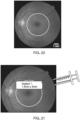
- a sub-retinal detachment is created to expose a damaged RPE region ( Figure 20 ).
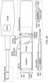
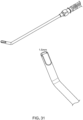
- an implantation device such as one shown in Figures 28 , 29 , or 31 can be used to deliver an RPE/fibrin hydrogel implant onto the region of interest ( Figure 21 ).
- a cannula see, e.g., Figures 32 and 33
- an air-phase bubble may be used to push the RPE/fibrin hydrogel implant into place.
- a laser tool (e.g., a laser tool used for diabetic retinopathy) can be used to tack the implant down via laser photocoagulation, preventing it from slipping ( Figure 22 ).
- an implantation device can be used to deliver a second RPE monolayer/fibrin implant onto the region of interest within an eye ( Figure 23 ).
- the second implant can be placed adjacent to the first, preferably through the original incision or cannula.
- a laser tool can be used to tack the second implant down, preventing it from slipping ( Figure 24 ).
- An implantation device can be used to deliver a third RPE monolayer/fibrin implant onto the region of interest within an eye ( Figure 25 ).
- the third implant can be placed adjacent to the second, preferably through the original incision or cannula.
- a laser tool can be used to tack the third implant down, preventing it from slipping ( Figure 26 ).
- any appropriate number can be used to cover the area to be treated. For example, one, two, three, four, five, six, or more RPE monolayer/fibrin implants can be implanted within a single eye being treated.
- this modular tiling approach can allow a clinician to personalize the implants to the patient's need, is scalable to large areas, is applicable to any region of the retina, and reduces the number of incisions required.
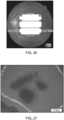
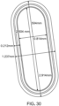
- a mechanical punch can be used to design an RPE monolayer/fibrin implant having a particular shape or size (see, e.g., Figure 27 ).
- Other methods to shape the fibrin implant can include gel casting with custom molds, laser microdissection microscopy, and 3D printing.
- An implantation device for implanting an RPE monolayer/fibrin implant provided herein into an eye can be a plunger style device with a mechanical control of ejection.
- an implantation device can have designed to deliver various sized RPE monolayer/fibrin implants and to have the ability to insert multiple implants rapidly using clip-style tips.
- an implantation device provided herein can have a liquid reservoir to maintain hydration of cells and hydrogel.
- an implantation device provided herein be designed for one hand manipulation and use.
- a pre-vascularization strategy can be combined with RPE culture to form choroid tissue.
- Fibrin can be vascularized by various methods, including through the use of a microfluidic device ( Moya et al., Methods Mol. Biol., 1202:21-7 (2014 )), 3D printing ( Pinnock et al., Methods, 99:20-7 (2016 )), and spontaneous vascularization of encapsulated endothelial cells within a matrix ( Mishra et al., Biomaterials, 77:255-66 (2016 )).
- These strategies can be combined with an RPE monolayer culture on top of the pre-vascularized fibrin to form RPE-choroid complex.
- RPE-choroid can be a therapeutic for macular degenerative diseases in which both the choroid and RPE are dysfunctional, including dry AMD.
- Endothelial cells can be obtained from various sources, such as iPSC-derived endothelial cells, blood outgrowth endothelial cells (BOEC), endothelial colony-forming cells (ECFCs), endothelial progenitor cells (EPCs), and umbilical vein endothelial cells (UVEC).
- a multi cell population tissue can be combined with RPE culture to form transplant tissue.
- the fibrin support can be loaded with other cell types found in the sub RPE tissue, including melanocytes, choroidal pericytes, and fibroblasts.
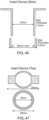
- This document also provides a fibrin hydrogel support device that can be used to grow the cells on a fibrin hydrogel that is suspended in cell culture medium.
- a RPE/fibrin hydrogel implant provided herein to be formed in a manner that avoids the need to detach the scaffold from a solid substrate and that allows access of culture media to the basal surface of the cells as they grow and differentiate.
- the device can include two separate pieces that can be easily attached to each other to hold the scaffold material in suspension ( Figures 46-48 ).
- a top piece can be a cylindrical inner tube that can be inserted into a bottom, base piece to press and secure a fibrin hydrogel layer sandwiched in between ( Figure 48 ).
- the bottom, base piece can include a side wall and an annular bottom with a central opening.
- the top cylindrical inner tube can be retained and supported by the bottom, base piece.
- the two components can be made of any appropriate material including, without limitation, Teflon, silicone, or other plastics via, for example, injection molding. In some cases, polystyrene can be used. The dimension of each component can be as shown in Figures 46-48 . In some cases, the top cylindrical inner tube can engage the bottom, base piece via threads, a snap-fit, or clipping mechanism.
- a fibrin hydrogel provided herein can be coated prior to apply cells.
- a fibrin hydrogel provided herein can be coated with basement membrane matrix and/or basement membrane protein (e.g., matrigel or other agent) prior to apply cells.
- basement membrane matrix and/or basement membrane protein e.g., matrigel or other agent
- agents that can be used to coat fibrin hydrogel provided herein include, without limitation, geltrex, laminin 511, laminin 521, victronectin, collagen, gelatin, and combinations thereof.
- matrigel and geltrex can be used interchangeably as they are both basement membrane matrix derived from mouse sarcoma cells.
- Fibrinogen was obtained from three sources: as Evicel from Ethicon (60 mg/mL), as Tisseel from Baxter (95 mg/mL), and as research grade material from Sigma-Aldrich (57 mg/mL). Thrombin also was obtained from three sources: part of Evicel from Ethicon, part of Tisseel from Baxter, and research grade material from Sigma-Aldrich. Plasminogen was obtained as research grade material from Sigma-Aldrich. Recombinant tissue plasminogen activator (tPA) was obtained as research grade material from Sigma-Aldrich.
- tPA tissue plasminogen activator
- IPSC-RPE cells were produced as described elsewhere with modification ( Johnson et al., Investig. Ophthalmol. Vis. Sci., 56:4619 (2015 )).
- a membrane support was utilized with apical and basal media, including either transwell or HA membrane.
- the membrane surface was coated with a collagen gel, per manufacturer's protocol, and, either subsequently or alternatively, coated with a geltrex or matrigel solution, up to 0.1 mg/mL for 2 hours at 37°C. Cells were then plated and allowed to form a monolayer for up to one month.
- IPSC-RPE was used from healthy control patients. Cells were used after diff stage 5. The trans epithelial resistance was measured above 100 ohms. Pigmentation was noted prior to use.
- Fibrin gels were formed by varying the fibrinogen concentration and thrombin concentration. Thin layer gels were formed initially by a plate sandwich method, in which a mixture of fibrinogen and thrombin solutions was sandwiched between two layers of parafilm within a plastic mold with a 200 ⁇ m gap thickness. The solution was allowed to gel up to 1 hour in a humid 37°C. The parafilm was removed, and the gels were hydrated and washed in PBS prior to use. Gels formed by this method had an average thickness of 196 ⁇ 90 ⁇ m.
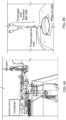
- a sprayer system was used to form thin layer fibrin gels.
- Dual microinjector systems (WPI) were connected to a pump controller, which was connected to a computer.
- the mixer was then connected to an atomizing nozzle (The Lee Co).
- a CO 2 gas regulator also was attached to the nozzle, to provide the air pressure for atomization.
- Figures 4A and 4B show the set up.
- the air pressure and amount of liquid dispensed was varied (0.3-1.5 bar).
- a custom MATLAB script was utilized to vary sprayer times and rates. The air pressure was varied on the regulator and controlled by a foot pedal. After spraying, the solution was allowed to gel up to 1 hour in a humid 37°C. The gels were hydrated and washed in PBS prior to use.
- the gels were stained with 0.01 mg/mL FITC isothioanate solution for 1 hour and shaken.
- the unlabeled FITC was removed through subsequent washes with PBS. Confocal z-series images were taken through the gel, and the measure of FITC stained slices was measured to obtain thickness.
- FDP fibrin degradation products
- RPE on membrane supports were washed with PBS. Once the PBS was removed, fibrin was sprayed on top and allowed to fully gel. After hydration, the cells were incubated in basal 750 U/mL purified collagenase or 1 U/mL dispase in DMEM up to 30 minutes. The membrane was carefully cut off and placed in a petri dish and submerged with PBS. The FRPE was then peeled off using forceps, or scraped off using a cell scraper.
- FRPE was stained for ZO-1, a cell-cell junctional protein found in epithelial cells. FRPE samples were punched and fixed in 10% formalin for 1 hour. Staining was done as described elsewhere (( Johnson et al., Invest. Ophthalmol. Vis. Sci., 56:4619-4630 (2015 )). Fixed cells were blocked with NGS, incubated overnight at 4°C in primary antibody, and incubated 2 hours in secondary antibody. Samples were mounted on glass slide using aquamount and imaged under Nikon Fluorescent microscope.
- TEM images were obtained to view the interaction of the fibrin and RPE.
- FRPE samples were fixed in 2.5% glutaraldehyde for 1 hour. Fixed samples were processed for resin embedment, and 0.5 ⁇ m sections were cut and mounted. Imaging was done on a TEM microscope.
- Punched FRPE implants were monitored over time using bright field microscopy.
- a live/dead kit was utilized on the FRPE, per manufacturer's protocol. Live cells were visualized under FITC spectrum (Absorbance: 495 nm; Emission: 520 nm), and dead cells were visualized under TRITC spectrum (Absorbance: 543 nm; Emission: 560 nm). Cell viability was calculated as a percent (live stained cells divided by total cells visualized).
- PCR was accomplished on cells to confirm their functionality 24 hours after detachment from culture support and 24 hours after degradation of fibrin support. Markers included PEDF, RPE65, Best1, and control.
- Figure 5 shows the success of attaching fibrin to the apical surface of the RPE and detachment from culture surface using dispase. After mechanical punching, the cells are still adherent to the fibrin.
- RPE attached to fibrin basal support remained viable after mechanical punching ( Figure 16 ). Further, the viability and adherence to the substrate was independent of the geltrex coating.
- the results provided herein demonstrate that the degradation kinetics of fibrin substrate can be varied from about 1.5 hours to about 20 hours by adjusting fibrinogen, plasminogen, and tPA concentrations.
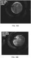
- iPSC-RPE cells were cultured for two weeks on a fibrin gel with ( Figure 14A ) and without ( Figure 14B ) geltrex coating, in media containing 50 U/mL Aprotinin. The inclusion of Aprotinin in the media appeared to prevent fibrin gel degradation.
- these studies indicated that attachment of the cells to the fibrin gel may occur independent of the presence of geltrex. The cells remained adherent after the gel was released from the plates ( Figure 15A ), and there was minimal cell removal after the gel was cut ( Figure 15B ).
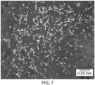
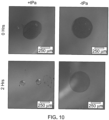
- a fibrin gel containing iPSC-RPE cells was degraded by digestion with 0.1 U/mL plasminogen and 22 U/mL tPA. Images were taken at 60 ( Figure 18A ) and 96 ( Figure 18B ) hours, showing that the cells detached from the plates as a monolayer. Cells in the monolayer were stained with calcein-AM (Live) and ethidium homodimer (Dead) to evaluate viability after 96 hours of gel digestion, showing most of the cells remained alive ( Figure 19 ).
- This protocol produces RPE monolayer supported by apical fibrin hydrogel.
- This protocol produces RPE monolayer with basal fibrin support.
- a clinic obtains a patient skin biopsy and sends it to a GMP facility to produce iPSC-RPE cells as described elsewhere ( Sonoda et al., Nat. Protoc., 4:662-673 (2009 ); Johnson et al., Ophthalmol. Vis. Sci., 56:4619 (2015 ); Brandl et al., NeuroMolecular Med., 16:551-564 (2014 ); Idelson et al., Cell Stem Cell., 5:396-408 (2009 ); and Carr et al., Mol. Vis., 15:283-295 (2009 )).
- the iPSC-RPE are cultured on fibrin hydrogels using the cell culture insert for up to 3 months.
- the RPE/fibrin gel is cut to specifications of the patient's need.
- the cut implants are loaded into the tip components of an implantation device, stored in culture media with or without plasminogen, and shipped to the clinic.
- the clip component pre-loaded with the RPE/fibrin gel is inserted into the implantation device.
- the patient is prepped for surgery.
- a standard 3 port vitrectomy is performed, followed by formation of a bleb using a fine cannula, followed by a retinotomy using retinal scissors.
- An incision e.g., a 3 mm or 1.5 mm incision
- the tip of the implantation device is inserted into the eye in position under the retinotomy, and the implant is deployed.
- a laser is used to tack the implant in place. This is repeated with additional implants to cover the area being treated.
- the retinal detachment is closed using silicone oil or perfluorocarbon liquid tampenade.
- the scleral incision is sutured closed.
- tPA is intravitreally injected.
- the patient is allowed to recover and heal. Once healed, visual testing is performed to confirm treatment.
- Example 6 Fibrin hydrogels as a xeno-free and rapidly degradable support for transplantation of RPE monolayers
- fibrin hydrogels were produced by varying the concentrations of fibrinogen and thrombin to form a thin rigid hydrogel with defined parameters for degradation in the scale of hours. Subsequently, the optimized conditions were utilized to produce fibrin gels on which iPSC-RPE were cultured, forming well-differentiated monolayers. Finally, the fibrin support was degraded in vitro and the effects of this degradation on the RPE monolayer were assessed.
- the results provided herein demonstrate that fibrin hydrogels can be used as a long-lived substrate for the differentiation of RPE from stem cells that can then be rapidly degraded under controlled circumstances following delivery to the subretinal space.
- Fibrinogen and thrombin were obtained from Ethicon (Somerville, NJ) (Evicel, fibrinogen at 60 mg/mL), Baxter (Deerfield, IL) (Tisseel, fibrinogen at 95 mg/mL), and Sigma-Aldrich (St Louis, MO) (fibrinogen at 57 mg/mL). Plasminogen and recombinant tissue plasminogen activator (tPA) were obtained from Sigma-Aldrich.
- Fibrin gels were formed by varying the fibrinogen concentration and thrombin concentration. Initial studies showed minimal variation with thrombin concentration, and all experiments utilized a final concentration of 100 U/mL thrombin.
- thin layer gels were formed by using a custom thickness mold, which consisted of two plates of polycarbonate and 2 layers of parafilm with a defined thickness spacer ranging from 0 to 200 ⁇ m. A mixture of fibrinogen and thrombin solutions was sandwiched between the two layers immediately after mixing, and the solution was allowed to gel for 1-2 hours at 37°C in a humidified incubator. After the top plate and parafilm were removed, the gels were hydrated and washed in PBS. A custom mechanical punch was used to cut out similar sized gels, in an oblong shape, 1.5 mm ⁇ 5 mm. Forceps were carefully used to manipulate the gels.
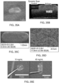
- Punched gels were imaged using OCT to determine thickness.
- An Envisu R4110 Leica; Wetzlar, Germany
- the gels Prior to imaging, the gels were placed in PBS in a clear 60 mm petri-dish. A and B scans were taken of the gels, and the thickness per gel was averaged across four random locations.
- SEM images were obtained of the fibrin hydrogel using a Hitachi S-4700 (Hitachi High Technologies; Schaumburg, IL) and Hitachi SEM software (V3.6). Gels were fixed in 2.5% paraformaldehyde and 1% glutaraldehyde in 0.1 M phosphate buffer pH 7 with divalent cations overnight. Gels were then critical-point dried using carbon dioxide, mounted on an aluminum stub, and sputter-coated for 60 seconds using gold-palladium.
- the testing was conducted on a custom-build z-stage driver.
- the force was measured using a 10 g miniature load cell (Honeywell; Morris Plains, NJ), and the data was collected using LabVIEW V12.0 (National Instruments; Austin, TX).
- LabVIEW V12.0 National Instruments; Austin, TX
- a static deflection test was conducted at 1 mm/second until fracture.
- the force and displacement curve was graphed and fit so that the linear region gave the mechanical stress values ( Figure 34B ). Maximum load was also obtained as the peak of the curve.
- Gel degradation kinetics were determined by varying the concentration of fibrinogen, plasminogen, or tissue plasminogen activator (tPA). Thrombin concentration did not appear to affect stiffness or degradation kinetics and was held constant at 10 U/mL. Identical sized gels (1.5 mm ⁇ 5.0 mm oblong) were generated using a custom punch. The punched gels were incubated in various concentrations of plasminogen (0.01-1 U/mL) and tPA solution (0.1-1,000 U/mL). To elucidate the effect of each variable (fibrinogen, plasminogen, and tPA concentrations), each was varied while holding the other two constant.
- tPA tissue plasminogen activator
- FDP fibrin degradation products
- iPSC line 006-BIOTR-001 generated from a 21-year old Caucasian female donor was used ( Johnson et al., Investig. Ophthalmol. Vis. Sci., 56:4619 (2015 )).
- iPSC-RPE cells were generated from this line by LAgen Laboratories (Rochester, MN) using a differentiation process described elsewhere ( Johnson et al., Investig. Ophthalmol. Vis. Sci., 56:4619 (2015 )).
- Fibrin gels were made by mixing fibrinogen (final: 30 mg/mL) and thrombin (final: 10 U/mL) solutions in the bottom of the culture surface (either 60 mm, 6 well plate, 12 well plate, or 12 well Transwell), and a custom Teflon weight was used to flatten and smooth the gel surface. The gels were then incubated for 1-2 hours in 5% CO 2 , 37°C humid incubator. Gels were washed with PBS prior to plating.
- RPE were passaged as described elsewhere ( Johnson et al., Investig. Ophthalmol. Vis. Sci., 56:4619 (2015 )). Suspended cells were plated onto the fibrin or matrigel-coated surfaces at a density of 0.4-0.5 ⁇ 10 6 cells/mL.
- RPE differentiation media RPEM (LAgen Laboratories), with 2% (v/v) B27 and 1% (v/v) antimycotic/antibiotic (Life Technologies) was supplemented with varying concentrations of aprotinin to preserve the fibrin gel. Media was changed every 2 days. RPE were cultured on the gels for 6-10 weeks prior to use. When appropriate, RPE cultured on matrigel-coated tissue culture polystyrene was used as a positive control.
- Immunofluorescence was used to visualize protein expression in the iPSC-RPE. Samples were fixed in 100% ice-cold methanol for 10 minutes at -20°C. Staining was as performed as described elsewhere ( Johnson et al., Investig. Ophthalmol. Vis. Sci., 56:4619 (2015 )), using the following primary antibodies at a 1:1000 dilution: polyclonal rabbit-anti Best1 (pAB125); polyclonal rabbit-anti Ezrin (Cell Signaling; Danvers, MA); and polyclonal rabbit-anti ZO1 (Life Technologies). Samples were mounted on glass slides using Fluoromount and imaged using a Nikon E600 fluorescence microscope (Nikon; Tokyo, Japan).
- a LIVE/DEAD Viability/Cytotoxicity kit (Live Technologies) was utilized per manufacturer's protocol to perform a live/dead assay. Live cells were visualized using a FITC filter (Absorbance: 495 nm/ Emission: 520 nm), and dead cells were visualized using a TRITC filter (Absorbance: 543 nm/ Emission: 560 nm). RPE monolayers cultured on fibrin were used before or after degradation. Degradation was achieved using 1 U/mL plasminogen with 100 U/mL tPA. Cell viability was calculated as the percent of live stained cells divided by total cells visualized. Results for experimental groups were normalized to control groups.
- RNA isolation kit Zymo; Irvine, CA
- Total RNA was treated with RNase-free DNAse I (Roche Bio; Basel, Switzerland).
- cDNA was synthesized from total RNA using Superscript III reverse transcriptase (Life Technologies).
- Total RNA was primed with oligo dT (Life Technologies). Primers were designed using Primer-BLAST software ( Ye et al., BMC Bioinformatics, 13:134 (2012 )).
- Sendai Viral Primer sequences were from the CytoTune TM -iPS 2.0 Sendai Reprogramming Kit. Primers were ordered desalted from IDT (Coralville, IA). Forty cycles of PCR using 10-100 ng of input cDNA and PowerUp Sybr Green Master Mix (Applied Biosystems; Foster City, CA) were done on an Applied Biosystems QuantStudio 5 qPCR instrument. PCR reactions were batched according to the annealing temperature of the primer sets. A gene was deemed present if the C T was less than 37 cycles.
- ELISA assay kit (RND Systems; Minneapolis, MN) was used to quantify VEGF and PEDF secretion using pre-coated plates, following the manufacturer's protocol from media collected after 48 hours. Total protein was determined using a standard curve.
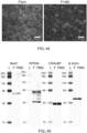
- RPE were scraped from fibrin in TPI buffer with 1% Triton-X, 20 mM Tris, 150 mM NaCl, and 5 mM EDTA, pH 8.0. Cells were lysed for 1 hour at 4°C. Samples were diluted and resolved on a capillary electrophoresis-based western blot instrument (Protein Simple Wes; San Jose, CA) using manufacturer's solution kits and protocol. Primary antibodies included RPE65 (401.8B11.3D9), Bestrophin 1 (pAB125), CRALBP (B2), and ⁇ -actin (AC-15).
- the materials provided herein for RPE transplant can be designed to be thin, laminar sheets with sufficient strength to maintain flatness while being manipulated with surgical instruments.
- a custom mold 50 mm ⁇ 50 mm square, 400 ⁇ m thick was used to produce identical thickness gels while varying concentrations of fibrinogen and thrombin. Thrombin concentration did not affect mechanical properties of fibrin hydrogels made at concentrations above 1 U*mL -1 *mg -2 fibrinogen as described elsewhere ( Rowe et al., Acta Biomater., 3:59-67 (2007 )). The following work was done with a fixed thrombin concentration (100 U/mL).
- Fibrin produced smooth, thin, and rigid gels that were opaque ( Figure 35A ). No swelling of the fibrin gel was noticeable after hydration. The edges of the gels were well defined. OCT imaging demonstrated that the gels formed by this method had an average thickness of 200 ⁇ 30 ⁇ m ( Figure 35B ), within the range expected based on the dimensions of the mold. SEM images of the fibrin gels indicated a smooth surface ( Figure 35C ), with a fibrilar microstructure similar to that described elsewhere for fibrin ( Filová et al., J. Biomed. Mater. Res. A., 90A:55-69 (2009 ); Figure 35D ).
- a custom oblong shaped punch with dimensions of 1.5 mm ⁇ 5 mm was used to generate similar sized hydrogels from a large sheet of fibrin formed in a custom mold.
- the 1.5 mm ⁇ 5 mm dimension was chosen to balance the need to cover the surface area of the macula (5 mm diameter) while maintaining a small incision to perform the implantation. With these dimensions, 3 implants could be lined up to cover >90% of the surface area of the macula while requiring a ⁇ 3 mm incision. Fibrin gels punched in this geometry appeared more rigid with increasing fibrinogen concentration. Gels of varying fibrinogen concentration were lifted with forceps out of the PBS to qualitatively observe an ability to retain its shape and support its hydrated weight ( Figure 35E ).
- the 10 mg/mL concentration gels exhibited immediate curling of the gel when removed from the PBS and folded onto itself.
- Gels made of 20 and 30 mg/mL fibrinogen concentrations exhibited reduced curling, and 40 mg/mL and above exhibited no curling. All gels exhibited sufficient plasticity to revert to a flat shape after being placed back in PBS.
- Gels made at or above 40 mg/mL fibrinogen concentration appeared pliable, durable, and maneuverable with a variety of surgical instruments. Obtaining gels from very high concentrations of fibrinogen solution can be difficult due to the high viscosity of the solution, and the highest concentration tested was 80 mg/mL. However, no observable differences on rigidity were made between gels made of 40 mg/mL and 80 mg/mL.
- the fibrin gels were punched out, imaged using OCT to quantify actual thickness, and mounted for mechanical testing.
- the fibrinogen concentration was fixed at 60 mg/mL, while varying thickness.
- OCT the 100 ⁇ m group had an actual thickness of 91 ⁇ 13 ⁇ m
- the 200 ⁇ m group was 198 ⁇ 10 ⁇ m
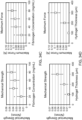
- a thickness of 100 ⁇ m yielded a mechanical strength of 0.004 ⁇ 0.003 N/mm and max force of 0.004 ⁇ 0.003 N, while a thickness of 200 ⁇ m yielded a mechanical strength of 0.020 ⁇ 0.013 N/mm and max force of 0.032 ⁇ 0.028 N.
- a thickness of 300 ⁇ m yielded a mechanical strength of 0.043 ⁇ 0.019 N/mm and max force of 0.094 ⁇ 0.031 N.
- the 100 ⁇ m gels were difficult to maneuver with surgical instruments compared to both the 200 ⁇ m and 300 ⁇ m gels, as they tore easily.
- the 200 ⁇ m thickness appeared to be the thinnest gel with sufficient mechanical strength for surgical manipulation.
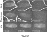
- Fibrin gels did not undergo noticeable degradation on their own when stored sterile in PBS at room temperature. To date, fibrin gels have been stored at room temperature for > 9 months. Fibrin gels in PBS did not undergo noticeable degradation when exposed to tPA ( Figure 36A ). However, when the combination of plasminogen and tPA was added, fibrin gels began to degrade rapidly. Degradation proceeded as an overall thinning of the gel with some gels breaking into smaller fragments. Degradation was considered complete when no visible remnants remained.
- plasminogen concentration variation had an effect on the degradation rate constant and total degradation time.
- the 1 U/mL group (P ⁇ 0.001) and the 0.5 U/mL group (p ⁇ 0.003) was different from all other groups.
- degradation rate constant increased with respect to tPA concentration until reaching a plateau at 100 U/mL.
- Total degradation time similarly approaches a plateau at 100 U/mL tPA concentration.
- fibrin gels were formed to fit various cell culture formats using a custom Teflon weight to flatten the meniscus. All cell culture was done using fibrin gels formed with 40 mg/mL fibrinogen concentration. RPE initially cultured on fibrin degraded the substrate within the first 48 hours ( Figure 37A ). To address this, the protease inhibitor aprotinin was used. Aprotinin is FDA approved for use in humans.
- aprotinin exhibited any toxicity toward iPSC-RPE.
- aprotinin was added to RPE culture media at concentrations varying from 0.5 U/mL to 50 U/mL, and the survival of the fibrin hydrogel supporting an RPE monolayer was monitored qualitatively over time. After 1 week, photomicrographs were taken of various groups over the course of 8 weeks ( Figure 37C ). At 0 U/mL, the majority of gel was degraded, and minimal cell attachment was observed within 2 days. Cells that attached to the surface did not form monolayers. In the 0.5 U/mL group, the majority of gel remained intact after 2 days in culture.
- iPSC-RPE cells grew over the patches in which fibrin gel remained but not in areas where the gel was degraded ( Figure 37C , asterisks). In the groups receiving 1-10U/mL, fewer patches of degraded gel were observed with increasing aprotinin concentration.
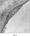
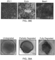
- iPSC-RPE cultured on fibrin are pigmented and form a cobblestone monolayer of cells ( Figure 38A ). Live/dead assay confirmed that the cells were viable ( Figure 38B ). Validation of the RPE phenotype was performed by qPCR using a panel of 20 key RPE markers ( Figure 40 ). A marker was considered present if a peak was observed prior to the 37 th cycle of PCR. Similar to what was observed for iPSC-RPE grown on matrigel coated tissue culture plastic, all RPE markers (notably, RPE65, CRALBP, and MITF ) were detected in iPSC-RPE grown on fibrin gels for 10 weeks. The pluripotency marker LIN28A and markers for sendai virus delivered "Yamanaka" factors ( KLF , KOS , c-myc ) were negative in all groups.
- Ezrin a marker of microvilli, was observed as puncta on the apical surface of the cells indicative of microvilli.
- ZO-1 was observed to outline the borders of all cells indicating the presence of junctional complexes and a single monolayer.
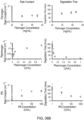
- RPE secretion of VEGF and PEDF was quantified by ELISA ( Figure 38C ).
- the media from the fibrin group had a VEGF concentration of 6.46 ⁇ 0.23 ng/mL.
- fibrin can be used as a material for RPE transplantation. Fibrin can be formed in a variety of shapes and sizes, with mechanical stiffness and degradation properties appropriate for RPE delivery.
- the results provided herein also demonstrate that a protease inhibitor such as aprotinin can be used to slow the ability of RPE to degrade the fibrin.
- a protease inhibitor such as aprotinin
- the cells can appear phenotypically similar to RPE. After the fibrin is degraded, the RPE can remain as a viable monolayer.
- Example 7 Implanting a fibrin hydrogel containing a RPE monolayer
- a fibrin hydrogel was implanted into the subretinal space of a rabbit eye ( Figure 41 ).
- the fibrin hydrogel was prepared by mixing fibrinogen (final: 40 mg/mL) and thrombin (final: 100 U/mL) solutions in a custom mold to generate a thin sheet (200 ⁇ m). A small volume of trypan blue was added to make the gel easier to visualize after implantation. Implants were punched to a 1.5 mm ⁇ 5 mm oblong geometry. Once prepared, the implant was loaded into an implantation device. To perform the surgical implantation, a white female New Zealand rabbit (3 kg) was anesthetized and prepped for surgery.
- a standard 3-port vitrectomy was performed, followed by bleb formation using a fine cannula, and a retinotomy was created using retinal scissors.
- a 3.2 mm slit knife was used to create an incision through the sclera.
- the implantation device was inserted into the eye and positioned under the retinotomy.
- the implant was deployed into place. After the implantation device removed, the scleral incision was sutured closed. Prior to the animal awakening, the implant could be seen as a flat sheet under a flattened retina.
- the animal was sacrificed after 48 hours, and the eye was harvested. Gross dissection examination revealed no remaining evidence of the fibrin hydrogel.
Landscapes
- Health & Medical Sciences (AREA)
- Life Sciences & Earth Sciences (AREA)
- Chemical & Material Sciences (AREA)
- Engineering & Computer Science (AREA)
- Biomedical Technology (AREA)
- General Health & Medical Sciences (AREA)
- Medicinal Chemistry (AREA)
- Animal Behavior & Ethology (AREA)
- Public Health (AREA)
- Veterinary Medicine (AREA)
- Epidemiology (AREA)
- Zoology (AREA)
- Cell Biology (AREA)
- Dermatology (AREA)
- Bioinformatics & Cheminformatics (AREA)
- Oral & Maxillofacial Surgery (AREA)
- Transplantation (AREA)
- Pharmacology & Pharmacy (AREA)
- Organic Chemistry (AREA)
- Chemical Kinetics & Catalysis (AREA)
- Biotechnology (AREA)
- Immunology (AREA)
- Genetics & Genomics (AREA)
- Wood Science & Technology (AREA)
- Botany (AREA)
- Gastroenterology & Hepatology (AREA)
- Neurology (AREA)
- Proteomics, Peptides & Aminoacids (AREA)
- Neurosurgery (AREA)
- Biochemistry (AREA)
- General Engineering & Computer Science (AREA)
- Urology & Nephrology (AREA)
- Developmental Biology & Embryology (AREA)
- Ophthalmology & Optometry (AREA)
- Microbiology (AREA)
- Virology (AREA)
- Dispersion Chemistry (AREA)
- General Chemical & Material Sciences (AREA)
- Molecular Biology (AREA)
- Acoustics & Sound (AREA)
Applications Claiming Priority (3)
| Application Number | Priority Date | Filing Date | Title |
|---|---|---|---|
| US201662431259P | 2016-12-07 | 2016-12-07 | |
| EP17879171.1A EP3551216B1 (fr) | 2016-12-07 | 2017-11-13 | Procédés et matériaux permettant d'utiliser des supports de fibrine pour une transplantation d'épithélium pigmentaire rétinien |
| PCT/US2017/061300 WO2018106414A1 (fr) | 2016-12-07 | 2017-11-13 | Procédés et matériaux permettant d'utiliser des supports de fibrine pour une transplantation d'épithélium pigmentaire rétinien |
Related Parent Applications (1)
| Application Number | Title | Priority Date | Filing Date |
|---|---|---|---|
| EP17879171.1A Division EP3551216B1 (fr) | 2016-12-07 | 2017-11-13 | Procédés et matériaux permettant d'utiliser des supports de fibrine pour une transplantation d'épithélium pigmentaire rétinien |
Publications (2)
| Publication Number | Publication Date |
|---|---|
| EP4455265A2 true EP4455265A2 (fr) | 2024-10-30 |
| EP4455265A3 EP4455265A3 (fr) | 2025-01-01 |
Family
ID=62492097
Family Applications (2)
| Application Number | Title | Priority Date | Filing Date |
|---|---|---|---|
| EP17879171.1A Active EP3551216B1 (fr) | 2016-12-07 | 2017-11-13 | Procédés et matériaux permettant d'utiliser des supports de fibrine pour une transplantation d'épithélium pigmentaire rétinien |
| EP24187425.4A Pending EP4455265A3 (fr) | 2016-12-07 | 2017-11-13 | Procédés et matériaux pour utiliser des supports de fibrine pour une transplantation d'épithélium pigmentaire rétinien |
Family Applications Before (1)
| Application Number | Title | Priority Date | Filing Date |
|---|---|---|---|
| EP17879171.1A Active EP3551216B1 (fr) | 2016-12-07 | 2017-11-13 | Procédés et matériaux permettant d'utiliser des supports de fibrine pour une transplantation d'épithélium pigmentaire rétinien |
Country Status (11)
| Country | Link |
|---|---|
| US (3) | US11679180B2 (fr) |
| EP (2) | EP3551216B1 (fr) |
| JP (3) | JP2020501543A (fr) |
| KR (2) | KR20230126745A (fr) |
| CN (1) | CN110225763B (fr) |
| AU (2) | AU2017372468B2 (fr) |
| CA (2) | CA3045464C (fr) |
| FI (1) | FI3551216T3 (fr) |
| MX (1) | MX2019006576A (fr) |
| SG (1) | SG11202008441YA (fr) |
| WO (1) | WO2018106414A1 (fr) |
Families Citing this family (9)
| Publication number | Priority date | Publication date | Assignee | Title |
|---|---|---|---|---|
| JP2020501543A (ja) | 2016-12-07 | 2020-01-23 | メイヨ・ファウンデーション・フォー・メディカル・エデュケーション・アンド・リサーチ | 網膜色素上皮移植のためのフィブリン支持体を用いた方法及び材料 |
| KR102624998B1 (ko) | 2017-06-05 | 2024-01-12 | 메이오 파운데이션 포 메디칼 에쥬케이션 앤드 리써치 | 줄기 세포의 배양, 증식 및 분화를 위한 방법 및 물질 |
| CN108893442A (zh) * | 2018-07-25 | 2018-11-27 | 广州赛莱拉干细胞科技股份有限公司 | 一种脂肪干细胞增殖培养基及其增殖培养方法 |
| CN108823160B (zh) * | 2018-07-25 | 2020-08-21 | 广州赛莱拉干细胞科技股份有限公司 | 一种脐带间充质干细胞原代培养基及其原代培养方法 |
| CA3129427A1 (fr) * | 2019-02-08 | 2020-08-13 | Mayo Foundation For Medical Education And Research | Dispositif d'implantation |
| AU2021377273A1 (en) * | 2020-11-13 | 2023-06-08 | Mayo Foundation For Medical Education And Research | Fibrin hydrogels |
| WO2023059565A2 (fr) * | 2021-10-04 | 2023-04-13 | The Regents Of The University Of Colorado, A Body Corporate | Dispositifs de support de transplantation de cellule et leurs procédés d'utilisation |
| JP2026502434A (ja) * | 2023-01-27 | 2026-01-23 | アルコン インコーポレイティド | 外網膜置換のための人工多能性幹細胞(ipsc)由来のマルチリボン生分解性ゲル |
| WO2025184466A1 (fr) * | 2024-03-01 | 2025-09-04 | Mayo Foundation For Medical Education And Research | Procédés et matériaux pour administrer des vecteurs à des zones localisées à l'intérieur d'un mammifère |
Family Cites Families (70)
| Publication number | Priority date | Publication date | Assignee | Title |
|---|---|---|---|---|
| US5487739A (en) | 1987-11-17 | 1996-01-30 | Brown University Research Foundation | Implantable therapy systems and methods |
| US6045791A (en) * | 1992-03-06 | 2000-04-04 | Photogenesis, Inc. | Retinal pigment epithelium transplantation |
| US5284476A (en) | 1992-03-20 | 1994-02-08 | Koch Paul S | Nuclear hydrolysis cannula |
| CA2161659C (fr) * | 1993-04-30 | 2004-09-14 | Yao Liu | Transplantation de l'epithelium pigmentaire retinien |
| DE69329556D1 (de) * | 1993-04-30 | 2000-11-16 | Photogenesis Inc | Transplantation des retinalen pigmentepithels |
| US5356395A (en) | 1993-12-14 | 1994-10-18 | Chen Shih Shuan | Safety syringe shield |
| AU771994B2 (en) | 1996-12-02 | 2004-04-08 | Ipxmedical, Llc | Compositions and methods for treating or preventing inflammatory diseases |
| US20110171185A1 (en) | 1999-06-30 | 2011-07-14 | Klimanskaya Irina V | Genetically intact induced pluripotent cells or transdifferentiated cells and methods for the production thereof |
| JP2003511204A (ja) | 1999-10-21 | 2003-03-25 | アルコン,インコーポレイティド | テノン下薬剤送出 |
| US6607522B1 (en) * | 2000-03-16 | 2003-08-19 | General Hospital Corporation | Methods for tissue welding using laser-activated protein solders |
| US20040039401A1 (en) | 2000-03-31 | 2004-02-26 | Chow Alan Y. | Implant instrument |
| US7867186B2 (en) | 2002-04-08 | 2011-01-11 | Glaukos Corporation | Devices and methods for treatment of ocular disorders |
| CN1720055A (zh) | 2002-10-04 | 2006-01-11 | 组织技术公司 | 视网膜上皮细胞在羊膜上的培养和移植 |
| JP4619789B2 (ja) | 2002-10-04 | 2011-01-26 | ナノマトリックス,インコーポレイテッド | 皮膚およびその他の組織のためのシーラント |
| US20040115176A1 (en) | 2002-10-23 | 2004-06-17 | Swartz Daniel D. | Fibrin-based tissue-engineered vasculature |
| US20040236343A1 (en) | 2003-05-23 | 2004-11-25 | Taylor Jon B. | Insertion tool for ocular implant and method for using same |
| US20050186672A1 (en) | 2004-01-27 | 2005-08-25 | Reliance Life Sciences Pvt. Ltd. | Tissue system with undifferentiated stem cells derived from corneal limbus |
| JPWO2005090550A1 (ja) | 2004-03-18 | 2008-02-07 | 株式会社バイオマスター | 幹細胞の増殖法 |
| WO2006088483A2 (fr) | 2004-06-16 | 2006-08-24 | Trustees Of Dartmouth College | Compositions et methodes permettant d'inhiber la synthese ou l'expression de mmp-1 |
| US20060047250A1 (en) | 2004-08-30 | 2006-03-02 | Hickingbotham Dyson W | Fluid delivery device |
| CH695412A5 (it) | 2004-10-19 | 2006-05-15 | Labo Cosprophar Ag | Metodo e kit particolarmente per applicare un prodotto cosmetico. |
| CA2594396A1 (fr) | 2005-01-14 | 2006-07-20 | Arblast Co., Ltd. | Composition en forme de feuille utilisant un amnios et procede de preparation de celle-ci |
| US8231908B2 (en) | 2005-07-25 | 2012-07-31 | Arblast Co., Ltd. | Sheet-like composition |
| US20070060887A1 (en) | 2005-08-22 | 2007-03-15 | Marsh David A | Ophthalmic injector |
| ITTO20060282A1 (it) | 2006-04-14 | 2007-10-15 | Univ Degli Studi Torino | Mezzo di coltura e composizione farmaceutica per la rigenerazione del tessuto cartilagineo relativo procedimento relativi usi e prodotti |
| WO2007150046A2 (fr) * | 2006-06-22 | 2007-12-27 | Sirion Therapeutics, Inc | Procédés et compositions pour traiter des conditions ophtalmiques par la modulation de l'activité de mégaline |
| US9039761B2 (en) | 2006-08-04 | 2015-05-26 | Allergan, Inc. | Ocular implant delivery assemblies with distal caps |
| EP2147094B2 (fr) | 2007-04-18 | 2018-02-21 | Hadasit Medical Research Services & Development Limited | Cellules de l'épithélium pigmentaire de la rétine dérivées de cellules souches |
| WO2009046194A2 (fr) | 2007-10-05 | 2009-04-09 | The Board Of Trustees Of The University Of Illinois | Produit d'étanchéité de fibrine |
| US20120219737A1 (en) | 2007-10-19 | 2012-08-30 | University Of Medicine And Dentistry Of New Jersey | Production of extracellular matrix, conditioned media and uses thereof |
| AU2009221859B2 (en) | 2008-03-05 | 2013-04-18 | Alcon Inc. | Methods and apparatus for treating glaucoma |
| US8425473B2 (en) | 2009-01-23 | 2013-04-23 | Iscience Interventional Corporation | Subretinal access device |
| EP2470645B1 (fr) | 2009-08-24 | 2019-06-26 | Wisconsin Alumni Research Foundation | Progéniteur rétinien humain sensiblement pur, progéniteur de cerveau antérieur, et cultures de cellules d'épithélium pigmentaire rétinien et leurs procédés de fabrication |
| ES2677945T3 (es) | 2009-08-25 | 2018-08-07 | Servicio Andaluz De Salud | Producción de tejidos artificiales mediante ingeniería tisular utilizando biomateriales de fibrina y agarosa |
| WO2011084550A2 (fr) | 2009-12-16 | 2011-07-14 | Eyevation, Llc | Dispositifs de délivrance et d'extraction |
| JP2013520239A (ja) | 2010-02-22 | 2013-06-06 | マッツォッキ,ルディ,エイ | 網膜を再構成するための最小限侵襲方法及び装置 |
| WO2011109712A2 (fr) | 2010-03-04 | 2011-09-09 | University Of Pittsburgh-Of The Commonwealth System Of Higher Education | Tissu du stroma cornéen humain mis au point par des techniques biologiques |
| US9267099B2 (en) * | 2010-03-15 | 2016-02-23 | Indiana University Research And Technology Corporation | Engineered lumenized vascular networks and support matrix |
| ES2542226T3 (es) | 2010-04-28 | 2015-08-03 | Technische Universität Dresden | Método para producir células progenitoras retinianas polarizadas a partir de células madre pluripotentes y su diferenciación para dar células del epitelio pigmentario retiniano |
| GB201011313D0 (en) | 2010-07-05 | 2010-08-18 | Ucl Business Plc | Implantation devices, methods and implants |
| JP5996544B2 (ja) | 2010-10-15 | 2016-09-21 | クリアサイド・バイオメディカル・インコーポレーテッドClearside Biomedical Incorporated | 眼球アクセス用装置 |
| US9132152B2 (en) | 2011-02-10 | 2015-09-15 | The Regents Of The University Of California | Compositions and methods for generating induced pluripotent stem cells |
| US9902933B2 (en) | 2011-02-25 | 2018-02-27 | Riken | Method of producing retinal pigment epithelial cell sheet |
| JP5816452B2 (ja) | 2011-03-31 | 2015-11-18 | 株式会社セルシード | 細胞シート移植治具及びその利用方法 |
| CA2835215C (fr) * | 2011-05-18 | 2023-10-10 | The Regents Of The University Of California | Compositions de cellule retienne humaine non-immortelle, leur methode depreparation et leur utilisation pour le traitement de maladies retiniennes |
| WO2012177968A1 (fr) | 2011-06-22 | 2012-12-27 | The Schepens Eye Research Institute, Inc. | Support pour transplantation de cellules sous-rétiniennes et administration de médicaments |
| CN103007355B (zh) | 2011-09-20 | 2014-08-13 | 同济大学 | 一种水凝胶-纳米纤维膜及其制备方法和用途 |
| EP3730103B1 (fr) | 2012-04-19 | 2024-12-04 | Alcon Inc. | Système d'administration pour implant oculaire |
| US9241832B2 (en) | 2012-04-24 | 2016-01-26 | Transcend Medical, Inc. | Delivery system for ocular implant |
| KR101389850B1 (ko) | 2012-05-04 | 2014-04-29 | 이화여자대학교 산학협력단 | 심장전구세포의 배양방법 및 그 용도 |
| CA3078098C (fr) | 2012-06-12 | 2022-03-15 | Alcon Inc. | Injecteur de gaz intra-oculaire |
| US10030221B2 (en) | 2012-09-20 | 2018-07-24 | Danisco Us Inc. | Microtiter plates for controlled release of culture components to cell cultures |
| CN105007841A (zh) | 2012-12-31 | 2015-10-28 | 乔治·D.·福卢什 | 用于大量出血的冻干纤维蛋白封闭剂 |
| EP2951290B1 (fr) | 2013-02-01 | 2017-11-29 | The United States of America, as represented by The Secretary, Department of Health and Human Services | Procédé de génération de cellules épithéliales pigmentaires rétiniennes (epr) à partir de cellules souches pluripotentes induites (cspi) |
| US9592151B2 (en) | 2013-03-15 | 2017-03-14 | Glaukos Corporation | Systems and methods for delivering an ocular implant to the suprachoroidal space within an eye |
| US9587221B2 (en) | 2013-11-11 | 2017-03-07 | Auburn University | Encapsulation and cardiac differentiation of hiPSCs in 3D PEG-fibrinogen hydrogels |
| WO2015077498A1 (fr) | 2013-11-20 | 2015-05-28 | The University Of North Carolina At Chapel Hill | Méthodes et compositions pour traiter d'affections de l'oeil |
| CN103656742B (zh) | 2013-11-29 | 2015-04-15 | 温州医科大学 | 功能化视网膜色素上皮细胞移植片的制备方法 |
| MX2016007671A (es) | 2013-12-11 | 2016-10-14 | Pfizer Ltd | Metodo para producir celulas del epitelio pigmentario retiniano. |
| JP6495603B2 (ja) | 2014-09-03 | 2019-04-03 | テルモ株式会社 | シート状細胞培養物とフィブリンゲルとの積層体の製造方法 |
| KR101613478B1 (ko) | 2014-09-22 | 2016-04-19 | (주)안트로젠 | 중간엽줄기세포-하이드로겔을 함유하는 조성물 및 이의 제조방법 |
| EP3011981A1 (fr) | 2014-10-24 | 2016-04-27 | Histocell, S.L. | Squelette de biomatériau pour régénérer la muqueuse orale |
| TWI711455B (zh) | 2015-05-13 | 2020-12-01 | 臺北榮民總醫院 | 多層視網膜細胞移植物 |
| AU2016318774B2 (en) | 2015-09-08 | 2022-08-18 | FUJIFILM Cellular Dynamics, Inc. | MACS-based purification of stem cell-derived retinal pigment epithelium |
| DK3347456T3 (da) | 2015-09-08 | 2024-02-19 | Us Health | Fremgangsmåde til reproducerbar differentiering af retinale pigmentepitelceller af klinisk kvalitet |
| US10307289B2 (en) | 2016-08-19 | 2019-06-04 | Drug Delivery Company, Llc | Sub-cutaneous medicinal dosage delivery implant system |
| JP2020501543A (ja) | 2016-12-07 | 2020-01-23 | メイヨ・ファウンデーション・フォー・メディカル・エデュケーション・アンド・リサーチ | 網膜色素上皮移植のためのフィブリン支持体を用いた方法及び材料 |
| CN110520139A (zh) | 2017-02-08 | 2019-11-29 | 哈达西特医疗研究服务和开发有限公司 | 用于治疗视网膜疾病的感光细胞 |
| KR102624998B1 (ko) | 2017-06-05 | 2024-01-12 | 메이오 파운데이션 포 메디칼 에쥬케이션 앤드 리써치 | 줄기 세포의 배양, 증식 및 분화를 위한 방법 및 물질 |
| CA3129427A1 (fr) | 2019-02-08 | 2020-08-13 | Mayo Foundation For Medical Education And Research | Dispositif d'implantation |
-
2017
- 2017-11-13 JP JP2019530497A patent/JP2020501543A/ja active Pending
- 2017-11-13 KR KR1020237028551A patent/KR20230126745A/ko not_active Ceased
- 2017-11-13 FI FIEP17879171.1T patent/FI3551216T3/fi active
- 2017-11-13 KR KR1020197017685A patent/KR20190093199A/ko not_active Ceased
- 2017-11-13 CA CA3045464A patent/CA3045464C/fr active Active
- 2017-11-13 AU AU2017372468A patent/AU2017372468B2/en active Active
- 2017-11-13 SG SG11202008441YA patent/SG11202008441YA/en unknown
- 2017-11-13 CN CN201780075836.XA patent/CN110225763B/zh active Active
- 2017-11-13 WO PCT/US2017/061300 patent/WO2018106414A1/fr not_active Ceased
- 2017-11-13 EP EP17879171.1A patent/EP3551216B1/fr active Active
- 2017-11-13 MX MX2019006576A patent/MX2019006576A/es unknown
- 2017-11-13 EP EP24187425.4A patent/EP4455265A3/fr active Pending
- 2017-11-13 CA CA3210578A patent/CA3210578A1/fr active Pending
- 2017-11-13 US US16/467,681 patent/US11679180B2/en active Active
-
2022
- 2022-01-28 JP JP2022011439A patent/JP2022068182A/ja active Pending
-
2023
- 2023-03-29 US US18/127,958 patent/US12303617B2/en active Active
-
2024
- 2024-05-09 JP JP2024076448A patent/JP2024112846A/ja active Pending
-
2025
- 2025-02-06 AU AU2025200808A patent/AU2025200808A1/en active Pending
- 2025-04-15 US US19/179,030 patent/US20250288724A1/en active Pending
Non-Patent Citations (32)
| Title |
|---|
| BRANDL ET AL., NEUROMOLECULAR MED, vol. 16, 2014, pages 551 - 564 |
| CARR ET AL., MOL. VIS, vol. 15, 2009, pages 283 - 295 |
| CHAURASIA: "1", TRANSL. VIS. SCI. TECHNOL, 2012, pages 2 |
| DALVIN ET AL., OPHTHALMIC GENET, 2016, pages 1 - 5 |
| DINIZ ET AL., INVEST. OPHTHALMOL. VIS. SCI, vol. 54, 2013, pages 5087 - 5096 |
| FILOVÁ ET AL., J. BIOMED. MATER. RES. A, vol. 90A, 2009, pages 55 - 69 |
| HAN BIN ET AL: "A fibrin-based bioengineered ocular surface with human corneal epithelial stem cells", CORNEA, MASSON PUB. USA, UNITED STATES, vol. 21, no. 5, 1 July 2002 (2002-07-01), pages 505 - 510, XP009117872, ISSN: 0277-3740, DOI: 10.1097/00003226-200207000-00013 * |
| HU ET AL., OPHTHALMIC RES, vol. 48, 2012, pages 186 - 191 |
| IDELSON ET AL., CELL STEM CELL, vol. 5, 2009, pages 396 - 408 |
| JOHNSON ET AL., INVEST. OPHTHALMOL. VIS. SCI, vol. 56, 2015, pages 4619 - 4630 |
| JOHNSON ET AL., INVESTIG. OPHTHALMOL. VIS. SCI, vol. 56, 2015, pages 4619 |
| JOHNSON ET AL., OPHTHALMOL. VIS. SCI, vol. 56, 2015, pages 4619 |
| LAI ET AL., PLOS ONE, vol. 8, 2013, pages e54058 |
| LU ET AL., BIOMED. MICRODEVICES, vol. 14, 2022, pages 659 - 667 |
| MANDAI ET AL., N ENG J MED, vol. 376, 2017, pages 1038 - 1046 |
| MCHUGH ET AL., INVEST. OPHTHALMOL. VIS. SCI, vol. 55, 2014, pages 1754 - 1762 |
| MISHRA ET AL., BIOMATERIALS, vol. 77, 2016, pages 255 - 66 |
| MOYA ET AL., METHODS MOL. BIOL, vol. 1202, 2014, pages 21 - 7 |
| PEYMAN ET AL., OPHTHALMIC SURG, vol. 22, 1991, pages 102 - 108 |
| PINNOCK ET AL., METHODS, vol. 99, 2016, pages 20 - 7 |
| ROWE ET AL., ACTA BIOMATER., vol. 3, 2007, pages 59 - 67 |
| SCHWARTZ ET AL., OPHTHALMOL. VIS. SCI, vol. 57, 2016, pages ORSFcl-9 |
| SCHWARTZ ET AL., THE LANCET, vol. 379, 2012, pages 713 - 720 |
| SCHWARTZ ET AL., THE LANCET, vol. 385, 2015, pages 509 - 516 |
| SINGH ET AL., OPHTHALMOL. VIS. SCI, vol. 54, 2013, pages 6767 - 6778 |
| SONODA ET AL., NAT. PROTOC, vol. 4, 2009, pages 662 - 673 |
| SONODA ET AL., NAT. PROTOC., vol. 4, 2009, pages 662 - 673 |
| STANZEL ET AL., STEM CELL REP, vol. 2, 2014, pages 205 - 218 |
| SUN ET AL., STEM CELLS, vol. 33, 2015, pages 1543 - 1553 |
| UEHARA ET AL., J. BONE JOINT SURG. AM, vol. 97, 2015, pages 1792 - 1798 |
| UNDAS ET AL., THROMB. VASC. BIOL, vol. 31, 2011, pages 88 - 99 |
| YE ET AL., BMC BIOINFORMATICS, vol. 13, 2012, pages 134 |
Also Published As
| Publication number | Publication date |
|---|---|
| JP2022068182A (ja) | 2022-05-09 |
| JP2020501543A (ja) | 2020-01-23 |
| US20250288724A1 (en) | 2025-09-18 |
| AU2025200808A1 (en) | 2025-03-13 |
| KR20190093199A (ko) | 2019-08-08 |
| US11679180B2 (en) | 2023-06-20 |
| CN110225763A (zh) | 2019-09-10 |
| SG11202008441YA (en) | 2020-10-29 |
| CA3045464A1 (fr) | 2018-06-14 |
| FI3551216T3 (fi) | 2024-10-14 |
| AU2017372468A1 (en) | 2019-06-13 |
| CA3210578A1 (fr) | 2018-06-14 |
| EP3551216B1 (fr) | 2024-07-10 |
| WO2018106414A1 (fr) | 2018-06-14 |
| CA3045464C (fr) | 2023-10-17 |
| US20200061246A1 (en) | 2020-02-27 |
| AU2017372468B2 (en) | 2024-11-07 |
| EP3551216A4 (fr) | 2020-08-19 |
| MX2019006576A (es) | 2019-08-21 |
| JP2024112846A (ja) | 2024-08-21 |
| US12303617B2 (en) | 2025-05-20 |
| KR20230126745A (ko) | 2023-08-30 |
| EP4455265A3 (fr) | 2025-01-01 |
| US20230277728A1 (en) | 2023-09-07 |
| EP3551216A1 (fr) | 2019-10-16 |
| CN110225763B (zh) | 2024-01-30 |
Similar Documents
| Publication | Publication Date | Title |
|---|---|---|
| US12303617B2 (en) | Methods and materials for using fibrin supports for retinal pigment epithelium transplantation | |
| Gandhi et al. | Fibrin hydrogels as a xenofree and rapidly degradable support for transplantation of retinal pigment epithelium monolayers | |
| Redenti et al. | Engineering retinal progenitor cell and scrollable poly (glycerol-sebacate) composites for expansion and subretinal transplantation | |
| US11253632B2 (en) | Biomaterial for suturing | |
| Jang et al. | Tailoring mechanical properties of decellularized extracellular matrix bioink by vitamin B2-induced photo-crosslinking | |
| San Choi et al. | Bioengineering endothelialized neo-corneas using donor-derived corneal endothelial cells and decellularized corneal stroma | |
| EP1489992B1 (fr) | Revetement de membrane amniotique pour surface tissulaire et dispositifs facilitant la fixation de membranes | |
| AU2009237462B2 (en) | Membrane | |
| US20090306772A1 (en) | Ocular Scaffolds and Methods for Subretinal Repair of Bruch's Membrane | |
| EP2468312A1 (fr) | Feuille pour kératoplasties | |
| WO2014186430A1 (fr) | Constructions cellulaires multicouches et leurs procédés d'utilisation et de production à l'aide de polymères naturels dégradables par des enzymes | |
| EP1859817A1 (fr) | Feuille de cellules cultivees, son procede de production et procede destine a restorer un tissu a l aide de ladite feuille | |
| US20220364049A1 (en) | Composition and methods for culturing retinal progenitor cells | |
| Shotorbani et al. | Cell sheet biofabrication by co-administration of mesenchymal stem cells secretome and vitamin C on thermoresponsive polymer | |
| Wei et al. | Transplantation of human embryonic stem cell-derived retinal pigment epithelial cells via injectable microfluidic-templated microgels for retinal regeneration | |
| Daniele et al. | Denuded Descemet’s membrane supports human embryonic stem cell-derived retinal pigment epithelial cell culture | |
| US20090246247A1 (en) | Composite enterocystoplasty | |
| EP4289937A1 (fr) | Culture cellulaire stratiforme pour recouvrir la surface coupée du pancréas | |
| JPWO2015046315A1 (ja) | 細胞の培養法 |
Legal Events
| Date | Code | Title | Description |
|---|---|---|---|
| PUAI | Public reference made under article 153(3) epc to a published international application that has entered the european phase |
Free format text: ORIGINAL CODE: 0009012 |
|
| STAA | Information on the status of an ep patent application or granted ep patent |
Free format text: STATUS: REQUEST FOR EXAMINATION WAS MADE |
|
| 17P | Request for examination filed |
Effective date: 20240709 |
|
| AC | Divisional application: reference to earlier application |
Ref document number: 3551216 Country of ref document: EP Kind code of ref document: P |
|
| AK | Designated contracting states |
Kind code of ref document: A2 Designated state(s): AL AT BE BG CH CY CZ DE DK EE ES FI FR GB GR HR HU IE IS IT LI LT LU LV MC MK MT NL NO PL PT RO RS SE SI SK SM TR |
|
| REG | Reference to a national code |
Ref country code: DE Ref legal event code: R079 Free format text: PREVIOUS MAIN CLASS: C12N0005000000 Ipc: A61K0038360000 |
|
| PUAL | Search report despatched |
Free format text: ORIGINAL CODE: 0009013 |
|
| AK | Designated contracting states |
Kind code of ref document: A3 Designated state(s): AL AT BE BG CH CY CZ DE DK EE ES FI FR GB GR HR HU IE IS IT LI LT LU LV MC MK MT NL NO PL PT RO RS SE SI SK SM TR |
|
| RIC1 | Information provided on ipc code assigned before grant |
Ipc: C12N 5/00 20060101ALI20241128BHEP Ipc: C12M 3/00 20060101ALI20241128BHEP Ipc: A61L 27/58 20060101ALI20241128BHEP Ipc: A61L 24/10 20060101ALI20241128BHEP Ipc: A61K 38/39 20060101ALI20241128BHEP Ipc: A61K 38/36 20060101AFI20241128BHEP |
|
| STAA | Information on the status of an ep patent application or granted ep patent |
Free format text: STATUS: EXAMINATION IS IN PROGRESS |
|
| 17Q | First examination report despatched |
Effective date: 20251008 |