EP4484569A1 - Reaktionsmedium und verfahren zum nachweis von shigatoxin-produzierenden e. coli und/oder enterohämorrhagischer e. coli - Google Patents
Reaktionsmedium und verfahren zum nachweis von shigatoxin-produzierenden e. coli und/oder enterohämorrhagischer e. coli Download PDFInfo
- Publication number
- EP4484569A1 EP4484569A1 EP23182495.4A EP23182495A EP4484569A1 EP 4484569 A1 EP4484569 A1 EP 4484569A1 EP 23182495 A EP23182495 A EP 23182495A EP 4484569 A1 EP4484569 A1 EP 4484569A1
- Authority
- EP
- European Patent Office
- Prior art keywords
- coli
- medium
- producing
- sample
- medium according
- Prior art date
- Legal status (The legal status is an assumption and is not a legal conclusion. Google has not performed a legal analysis and makes no representation as to the accuracy of the status listed.)
- Pending
Links
Images


Classifications
-
- C—CHEMISTRY; METALLURGY
- C12—BIOCHEMISTRY; BEER; SPIRITS; WINE; VINEGAR; MICROBIOLOGY; ENZYMOLOGY; MUTATION OR GENETIC ENGINEERING
- C12Q—MEASURING OR TESTING PROCESSES INVOLVING ENZYMES, NUCLEIC ACIDS OR MICROORGANISMS; COMPOSITIONS OR TEST PAPERS THEREFOR; PROCESSES OF PREPARING SUCH COMPOSITIONS; CONDITION-RESPONSIVE CONTROL IN MICROBIOLOGICAL OR ENZYMOLOGICAL PROCESSES
- C12Q1/00—Measuring or testing processes involving enzymes, nucleic acids or microorganisms; Compositions therefor; Processes of preparing such compositions
- C12Q1/02—Measuring or testing processes involving enzymes, nucleic acids or microorganisms; Compositions therefor; Processes of preparing such compositions involving viable microorganisms
- C12Q1/04—Determining presence or kind of microorganism; Use of selective media for testing antibiotics or bacteriocides; Compositions containing a chemical indicator therefor
- C12Q1/045—Culture media therefor
-
- C—CHEMISTRY; METALLURGY
- C12—BIOCHEMISTRY; BEER; SPIRITS; WINE; VINEGAR; MICROBIOLOGY; ENZYMOLOGY; MUTATION OR GENETIC ENGINEERING
- C12Q—MEASURING OR TESTING PROCESSES INVOLVING ENZYMES, NUCLEIC ACIDS OR MICROORGANISMS; COMPOSITIONS OR TEST PAPERS THEREFOR; PROCESSES OF PREPARING SUCH COMPOSITIONS; CONDITION-RESPONSIVE CONTROL IN MICROBIOLOGICAL OR ENZYMOLOGICAL PROCESSES
- C12Q1/00—Measuring or testing processes involving enzymes, nucleic acids or microorganisms; Compositions therefor; Processes of preparing such compositions
- C12Q1/02—Measuring or testing processes involving enzymes, nucleic acids or microorganisms; Compositions therefor; Processes of preparing such compositions involving viable microorganisms
- C12Q1/04—Determining presence or kind of microorganism; Use of selective media for testing antibiotics or bacteriocides; Compositions containing a chemical indicator therefor
- C12Q1/10—Enterobacteria
-
- G—PHYSICS
- G01—MEASURING; TESTING
- G01N—INVESTIGATING OR ANALYSING MATERIALS BY DETERMINING THEIR CHEMICAL OR PHYSICAL PROPERTIES
- G01N33/00—Investigating or analysing materials by specific methods not covered by groups G01N1/00 - G01N31/00
- G01N33/48—Biological material, e.g. blood, urine; Haemocytometers
- G01N33/50—Chemical analysis of biological material, e.g. blood, urine; Testing involving biospecific ligand binding methods; Immunological testing
- G01N33/53—Immunoassay; Biospecific binding assay; Materials therefor
- G01N33/569—Immunoassay; Biospecific binding assay; Materials therefor for microorganisms, e.g. protozoa, bacteria, viruses
- G01N33/56911—Bacteria
- G01N33/56916—Enterobacteria, e.g. shigella, salmonella, klebsiella, serratia
-
- G—PHYSICS
- G01—MEASURING; TESTING
- G01N—INVESTIGATING OR ANALYSING MATERIALS BY DETERMINING THEIR CHEMICAL OR PHYSICAL PROPERTIES
- G01N2333/00—Assays involving biological materials from specific organisms or of a specific nature
- G01N2333/195—Assays involving biological materials from specific organisms or of a specific nature from bacteria
- G01N2333/24—Assays involving biological materials from specific organisms or of a specific nature from bacteria from Enterobacteriaceae (F), e.g. Citrobacter, Serratia, Proteus, Providencia, Morganella, Yersinia
- G01N2333/245—Escherichia (G)
Definitions
- the present invention relates to the field of microbiological control in the broad sense, such as the microbiological control of a sample of industrial or clinical origin. More particularly, the present invention relates to a reaction medium and the method for detecting a Shiga toxin-producing E. coli and/or an enterohemorrhagic E. coli .
- Microbiological control of samples of various origins requires the implementation of techniques which allow the detection - for example for the purposes of identification and/or counting and/or biochemical characterization - of microorganisms and the rendering of results must be as rapid as possible.
- microbiological analysis is done in two stages.
- the first is a detection phase which can use many technologies such as culture media, immunoassays, molecular biology. It can be followed, particularly in the agri-food sector, a confirmation phase in order to confirm the presence of the pathogen sought and meet the standards in force in this field.
- the confirmation stage therefore requires additional steps and here again, requires a step of isolation of the bacteria sought.
- the diagnosis is based on the use of selective and chromogenic agars that can include tellurite such as CT-SMAC (Sorbitol MacConkey Agar) or CT-RMAC (Rhamnose MacConkey Agar), in order to select the growth of certain bacteria and to stain the strains of interest.
- CT-SMAC Session-Coupled Cell
- CT-RMAC Ranose MacConkey Agar
- the disadvantage of these methods is that they are very specific and certain strains of E. coli producing shiga toxins are not detected. Conversely, some media are not selective enough, the presence of a small number of E. coli producing shiga toxins can be hidden by the other microorganisms very widely present, isolation is then difficult.
- the medium according to the invention makes it possible to isolate STEC or EHEC from a multitude of E. coli.
- the culture medium is the culture medium according to the invention.
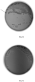
- the presence of a Shiga toxin-producing E. coli is confirmed by the presence of a halo around said Shiga toxin-producing E. coli present in the inhibition concentration gradient zone.
- This preferred embodiment has the advantage of being able to detect and isolate said STEC or EHEC directly on the culture medium. This allows an immediate result.
- the method allows for the detection of a STEC or an EHEC.
- the detected Shiga toxin-producing E. coli is an enterohemorrhagic E. coli .
- This method is particularly advantageous for samples loaded with additional flora, where the large quantity of colonies on the Petri dishes can lead to a risk of false negatives. Given the large number of colonies on the dish and the low representation of the target microorganism, the person collecting the colonies for confirmation purposes may never collect the target microorganism. The probability of isolating a STEC is then greatly increased in the presence of the gradient of the inhibitory compound.
- the present invention therefore provides a method for detecting and isolating STECs that avoids false negatives and saves considerable time.
- reaction medium means a medium comprising all the elements necessary for the expression of a metabolism, for the survival and/or growth of the microorganisms.
- This reaction medium can either be a microbiological culture medium or a microbiological revelation medium. In the latter case, the culture of the microorganisms can be carried out beforehand in another medium.
- the reaction medium can also be brought into contact with an agar culture medium. It can be placed under or on the culture medium which allows the growth of the target microorganisms.
- the reaction medium can be added after the incubation of the culture medium.
- the reaction medium is a microbiological culture medium comprising all the elements necessary for the growth of the microorganisms.
- the reaction medium is gelled. It is in solid or semi-solid form.
- Agar is the traditional gelling agent used in microbiology for the culture of microorganisms, but it is possible to use other gelling agents such as example, gelatin, agarose and other natural or artificial gelling agents.
- the conjugate is capable of forming a network with the target component in a gelled medium. This network is visually detectable by the formation of a halo without it being necessary to reduce the hardness of a conventional semi-solid reaction medium. Thus it is not necessary to modify the physicochemical properties of the reaction medium, such as the hardness, to allow on the one hand the diffusion of the targeted components and on the other hand the reaction with the conjugates.
- reaction medium can further comprise possible additives such as amino acids, peptones, one or more growth factors, carbohydrates, nucleotides, minerals, vitamins, one or more selective agents, inducers, toxin inducers, buffers, etc.
- selective agent is meant any compound capable of preventing or slowing the growth of a so-called “non-target” microorganism, i.e. other than the target microorganism(s).
- the term "inducer” refers to a compound capable of inducing the expression of a compound, such as an enzyme, a toxin, which would normally remain unexpressed.
- Said reaction medium may also comprise a dye.
- the following dyes may be mentioned: Evans blue, neutral red, sheep's blood, horse's blood, an opacifier such as titanium oxide, nitroaniline, malachite green, brilliant green.
- a chromogenic and/or fluorogenic substrate is preferably used.
- chromogenic and/or fluorogenic substrate is meant a substrate allowing the detection of an enzymatic or metabolic activity of the target/desired microorganisms using a detectable signal.
- the reaction medium according to the invention may additionally comprise a pH indicator, sensitive to the pH variation induced by the consumption of the substrate and revealing the metabolism of the target microorganisms.
- Said pH indicator may be a chromophore or a fluorophore.
- chromophores include bromocresol purple, bromothymol blue, neutral red, aniline blue, bromocresol blue.
- the person skilled in the art can also use a Petri dish divided into segments, such as a bi-dish, or a tri-dish, allowing several media to be easily compared, including different substrates or different selective mixtures, on which the same biological sample will have been deposited.
- microorganism has the same meaning as that generally accepted in microbiology and includes in particular gram-positive or gram-negative bacteria, yeasts, molds and more generally, single-celled organisms, invisible to the naked eye, which can be manipulated and multiplied in the laboratory.
- target microorganism at least one microorganism that it is desired to detect and/or identify.
- the microorganism is chosen from Shiga toxin-producing E. coli strains.
- Enterohemorrhagic E. coli are strains representing a subgroup of Shiga toxin-producing Escherichia coli , which have the eae gene or other genes for adhesion to cells such as the aggR gene and cause a hemolytic-uremic syndrome.
- the possession of these two virulence factors stx and eae simultaneously makes this pathovar very virulent for humans. And in particular when it comes to serogroups O26, O45, O80, O103, O111, 0121, O145 and O157.
- the target microorganism is an E. coli belonging to the groups of Shiga toxin-producing E. coli or typical enterohemorrhagic E. coli carrying the stx and eae genes or atypical enterohemorrhagic E. coli carrying the stx and aggR genes, for example.
- the non-target microorganisms are bacteria growing on the culture medium outside the concentration gradient zone of an inhibitory compound. Indeed, since the sample is polymicrobial, the STEC or EHEC potentially present are present among non-specific Gram-negative bacteria such as commensal E. coli .
- the reaction medium comprises at least one specific binding partner of STX1 and/or STX2, coupled to a nanoparticle.
- STX1 and STX2 are the toxins secreted by STEC strains. There are in fact two main types of shiga toxins: STX1 and STX2 which themselves have many variants. To date, STX1 has 4 subtypes STX1a, STX1c, STX1d, STX1e, and STX2 has 12: STX2a, STX2b STX2c, STX2d, STX2e, STX2f, STX2g, STX2h, STX2i, STX2j, STX2K, STX2l.
- the specific binding partner is selected from antibodies, all types of Fab fragments, recombinant proteins, phages, phage proteins, oligonucleotides, aptamers, affimers or any other ligand or anti-ligand well known to those skilled in the art.
- the binding partner is selected from antibodies or phage proteins.
- the antibody is a monoclonal antibody or a monoclonal antibody fragment.
- the binding partner is specific for a component of a microorganism. target.
- the component of the target microorganism is a component released by said microorganism.
- the component can thus be an element from the surface of the bacteria such as a protein, a Lipopolysaccharide (LPS), a flagellum.
- LPS Lipopolysaccharide
- the medium comprises at least one binding partner specific for the STX1 protein and/or at least one binding partner specific for the STX2 protein.
- the medium comprises at least one binding partner specific for a subtype of the STX1 protein and/or at least one binding partner specific for a subtype of the STX2 protein such as XTX2a or STX2d.
- the binding partner is coupled to a nanoparticle. This final complex is called a conjugate.
- conjugates having binding partners of different nature coupled to nanoparticles themselves of different nature.
- nanoparticle refers to particles of the order of magnitude of a nanometer.
- the nanoparticles can be chosen from gold, iron, silver, copper, carbon, latex, silicon, aluminum.
- the nanoparticles are colloidal nanoparticles having optical properties, namely their ability to distinguish themselves when a network is formed.
- the nanoparticle is chosen from gold, silver, copper.
- the nanoparticles change color, for example from red to gray when they form a network.
- the wavelength of the absorbed light is in the red around 530 nm.
- the absorbed wavelength changes from red to blue/gray around 600 to 700 nm.
- the networks thus formed allow the distinction of several components of the target microorganism.
- the nanoparticles have a size between 10 and 200 nm.
- the nanoparticles have a size between 20 and 90 nm, allowing better mobility of the conjugates in the reaction medium.
- nanoparticles make it possible to reduce the quantity of binding partners required for the formation of agglutination.
- concentration required in binding partners for the production of a reaction medium according to the invention requires 100 to 1000 times fewer binding partners than a medium without nanoparticles.
- the amount of binding partners required corresponds to the amount required to cover at least half of the surface of the nanoparticle, and even more preferably to cover between a third and a half of the surface of the nanoparticle. This proportion allows the agglutinating conjugate to form a network in the gelled reaction medium.
- nanoparticles can make it possible to visualize agglutination around a bacterial colony whose size does not yet allow it to be visible to the naked eye. Detection can thus be earlier.
- nanoparticles can make it possible to visualize agglutination around a bacterial colony whose translucent appearance does not allow detection by a reading machine. Detection can thus be facilitated.
- the coupling of the nanoparticle to the binding partner can be done either by direct attachment or by indirect attachment.
- Direct attachment means attachment by adsorption or by covalent bond.
- Indirect attachment means attachment by the interaction of ligands/anti-ligands such as biotin/strepatividin or other couples well known to those skilled in the art (Nicholas G. Weich et al, 2017). Depending on the type of bond chosen, those skilled in the art will adapt the physicochemical conditions of the reaction medium and in particular its pH.
- the conjugate is agglutinating, that is to say that it causes the formation of an agglutination network in the presence of a component of a target microorganism or a component from said microorganism.
- the component being multi-epitope, several conjugates will bind to this component and form an agglutination.
- agglutination we mean the result of an interaction between at least one component of a target microorganism or at least one component from said microorganism with binding partners coupled to a nanoparticle.
- Agglutination reactions include immunological reactions, such as antigen-antibody reactions or more generally specific interactions between two molecules. Through this interaction, components and conjugates aggregate, adhere to each other and form a network in the reaction medium.
- the network formed by said specific reaction is then detected either visually or automatically using an optical system.
- the colony of the target microorganism is thus identified.
- the network forms a halo in the gelled reaction medium detectable either visually or using an optical system. The network or the halo thus circumscribes the colony which can then be advantageously differentiated and/or identified within a population.
- the binding partner is an antibody in an amount sufficient to cover at least half of the surface of the nanoparticle and preferably at least a third of the surface of the nanoparticle.
- the nanoparticle is a gold nanoparticle of size between 20 and 90 nm and at a concentration between 10 10 and 10 12 nanoparticles/ml of reaction medium.
- the reaction medium according to the invention comprises a toxin inducer.
- the toxin inducer causes stress in the bacteria which will trigger the lytic cycle of the prophages in the bacteria and therefore stimulate the production of toxins which will be released.
- the toxin inducer is chosen from antibiotics or a physicochemical stress.
- antibiotics examples include trimethoprim, sulfamethoxazole, norfloxacin, azithromycin, gemtamicin, polymyxin B, chloramphenicol, streptomycin, chlortetracycline, oxytetracyline, tylosin, mitomycin C, carbodox, oliquindox, rifampincin, imipenem, ciprofloxacin, cotrimoxazole, penicillin G, linomycin.
- the medium according to the invention comprises ciprofloxacin at a concentration of between 0.005 and 0.030 mg/l.
- the medium according to the invention comprises mitomycin C at a concentration of between 0.10 and 0.5 mg/l.
- the toxin inducer can also be a physicochemical stress achieved, for example, by the addition of salt or EDTA or by a change in pH or by UV stress. Stress can also be, for example, induced by the addition of noradrenaline.
- sample is meant a small part or small quantity isolated from an entity for analysis.
- the sample may be of industrial origin, or, according to a non-exhaustive list, a air sample, a water sample, a sample taken from a surface, a part or a manufactured product, a product of food origin.
- the sample may also correspond to a sample of biological fluid (stools, urine, whole blood, serum, plasma, cerebrospinal fluid, organic secretion, etc.), an external sample (skin, nose, throat, etc.) or tissue or isolated cells.
- the sample may be used as is or, prior to analysis, undergo a preparation such as enrichment, extraction, concentration, purification, according to methods known to those skilled in the art.
- the reaction medium according to the invention may be all the more interesting when the sample is a polymicrobial sample or loaded with additional flora, such as for example food samples such as raw milk cheese or stools in clinical samples.
- the reaction medium comprises a concentration gradient of a compound inhibiting non-target bacteria.
- the inhibitory compound is chosen such that the minimum inhibitory concentrations (MICs) of non-target bacteria such as commensal enterobacteria are sufficiently lower than the MIC of STEC/EHEC.
- inhibitory compound means any compound that inhibits bacterial growth. It may be an antibiotic, a dye such as brilliant green, a salt such as lithium chloride, a bacteriophage endolysin. Preferably, the inhibitory compound is tellurite. According to the present invention, the inhibitory compound promotes the inhibition of non-STEC and non-EHEC strains.
- a “gradient” is defined as a non-homogeneous, increasing or decreasing concentration of an inhibitory compound.
- non-target microorganisms are microorganisms capable of growing on the medium without the concentration gradient of the inhibitory compound.
- Target microorganisms are microorganisms carrying the stx genes, i.e. STEC.
- non-target bacteria such as commensal Enterobacteria generally have a lower MIC than STEC and EHEC.
- the tellurite gradient on the medium is between 0 ⁇ g/ml and 100 ⁇ g/ml, preferably between 0 ⁇ g/ml and 50 ⁇ g/ml, even more preferably between 0 ⁇ g/ml and 30 ⁇ g/ml.
- the different Strains belonging to STEC and EHEC have different sensitivities to inhibitory compounds such as tellurite. Indeed, STEC and EHEC are mostly more resistant to tellurite than other enterobacteria.
- serogroups may have different susceptibility, for example, an E. coli 0157 will not have the same MIC to Cefixime-Tellurite as an E. coli 0103. There is also different susceptibility within a serogroup. Thus, an E. coli O111 strain will not have the same MIC as another strain of E. coli O111.
- the medium according to the invention makes it possible to isolate STEC or EHEC from a multitude of E. coli.
- the inhibitor compound is a mixture of cefixime and tellurite.
- the amount of cefixime in the deposited cefixime tellurite mixture is between 0.20 ⁇ g and 0.30 ⁇ g and the amount of tellurite is between 12 and 13 ⁇ g.
- the inhibitor compound deposited on the medium has a volume of between 5 ⁇ l to 50 ⁇ l.
- the inhibitor compound can be deposited by any means such as for example a pipette.
- the inhibitory compound is contained in at least one substrate capable of diffusing the inhibitory compound onto the gelled culture medium.
- This may be, for example, a blotting paper tablet containing the inhibitory compound and capable of diffusing it.
- the reaction medium is manufactured extemporaneously using a kit according to the invention.
- This allows the unitary use of a reaction medium, and to increase the stability thereof.
- the medium is used within 24 hours, even more preferably within 12 hours.
- the agglutinating conjugate is brought into contact with a supercooled gelling medium to form the reaction medium according to the invention.
- the gelled medium also comprises a toxin inducer. The whole is then homogenized and poured into the Petri dish.
- An inhibitory compound for non-target bacteria is deposited onto an area of the gelled culture medium.
- the inhibitory compound diffuses and forms an inhibition gradient around the deposition area; in another embodiment, the inhibitory compound is deposited after the sample is deposited.
- the inhibitory compound comprises a dye which makes it possible to locate its deposit on the culture medium and then allow the sample to be deposited at the level of the inhibitory compound.
- Another subject of the present invention relates to a method of in vitro microbiological culture, in which microorganisms likely to be present in a sample are seeded in or on a culture medium according to the invention.
- the seeded culture medium is incubated under appropriate conditions known to those skilled in the art.
- the seeding is carried out according to conventional microbiology techniques.
- the seeding is done by exhausting the polymicrobial sample on the culture medium.
- the sample is deposited at the level of the deposition of the inhibitory compound such that the sample with a maximum load of microorganisms is in contact with the inhibition concentration gradient. Indeed, in the first zone of the isolation, it is rare to be able to distinguish the target microorganisms from the non-target microorganisms due to a very high concentration of microorganisms.
- Seeding techniques are well known to those skilled in the art. This may involve seeding in quadrants.
- the quadrant method consists of dividing a Petri dish in two, then dividing one half in half again to obtain 3 quadrants of 50%, 25% and 25%. On the largest quadrant a small amount of inoculum is placed and then spread out. The dish is then turned by a quarter in order to spread the bacteria on a smaller quadrant, then again rotated by a quarter in order to inoculate the last small quadrant.
- the inhibitor compound and the sample are deposited in the same quadrant.
- It can also be a multiple streak exhaustion which consists of spreading the inoculum downwards in tight streaks and then again other streaks starting from the edges of the previous edge-to-edge streaks.
- detection is meant the detection by eye or by optical device of the existence of growth of the target microorganisms, preferably target bacteria.
- the detection may be carried out by optical device for fluorogenic substrates, or by eye or by optical device for chromogenic substrates.
- the detection may be carried out by eye or by optical device by observing the agglutination around the target microorganism.
- isolation we mean obtaining colonies spaced apart from each other.
- a gradient of an inhibitory compound improved the detection and isolation of a target bacterium such as a Shiga toxin-producing E. coli or an enterohemorrhagic E. coli among a multitude of E. coli.
- a selective medium for the growth of E. coli comprising a concentration gradient of a compound that inhibits non-target bacteria such as commensal enterobacteria.
- a concentration gradient of a compound that inhibits non-target bacteria such as commensal enterobacteria.
- Examples of mediums for the growth of E. coli include CHROMID® coli, CHROMID® EHEC, TBX or Rainbow agar. It is then appropriate to add a concentration gradient of a compound that inhibits non-target bacteria.
- the inhibitory compound is chosen in such a way that it makes it possible to discriminate within the group of E. coli producing shiga toxin from strains that are more or less sensitive to said inhibitory compound.
- the inhibitory compound can be deposited on the culture medium by any means such as a pipette. Once deposited, the inhibitory compound forms an inhibition concentration gradient around the deposition zone. It can also be a substrate diffusing the inhibitory compound.
- the inhibitory compound is deposited after sample deposition.
- a step of confirming the presence of the Shiga toxin-producing E. coli is carried out.
- This step can be carried out according to conventional confirmation methods such as a molecular biology method or an immunological method.
- the colony, present on the medium in contact with the gradient of the inhibitory compound is sampled.
- a latex test or a PCR test designed for the target microorganisms can be carried out. Examples include the SLIDEX ® E. coli test or the GENE-UP ® STEC top 6 PCR test or GENE-UP ® E.coli 0157:H7 or GENE-UP ® STEC stx and eae.
- the Shiga toxin-producing E. coli is an enterohemorrhagic E. coli .
- a step of incubation or enrichment of the sample is carried out before deposition on the gelled culture medium.
- This enrichment step requires not only an ad hoc culture medium but also an incubation of the assembly formed at least by the biological sample and the culture medium at an optimal temperature to allow the growth of the target microorganism(s).
- the incubation is generally carried out at a temperature ranging from 25 to 45°C for a predetermined period of time (for example from 6 hours to 48 hours).
- This enrichment phase requires the use of culture media, selective or not (depending on the desired goal), which aim to promote the growth of the target microorganisms in the samples, while limiting the growth of non-target flora.
- the culture media are frequently used in containers of the sterile plastic bag type, in which they are placed in contact with food, clinical or environmental samples, for the purpose of resuspension and enrichment of the microorganisms sought.
- this enrichment phase is necessary in particular in order to reveal the presence of at least one target microorganism in a very variable and possibly very large quantity of sample, for example from 25 grams (g) to 375 g diluted in a volume of culture medium of between 225 and 3375 milliliters (mL).
- an aliquot generally of a volume of between 5 microliters ( ⁇ L) and 5 mL
- ⁇ L microliters
- the sample is deposited and distributed over the medium by an exhaustion technique.
- reaction medium is incubated under appropriate conditions known to those skilled in the art.
- the culture medium is the culture medium according to the invention comprising at least one agglutinating conjugate and a gradient of an inhibitory compound.
- the presence of a Shiga toxin-producing E. coli is confirmed by the presence of a halo around said STEC present in the zone of the inhibition gradient.
- This preferred embodiment has the advantage of being able to detect and isolate said STEC or EHEC directly on the culture medium. This allows an immediate result.
- This invention is particularly interesting for facilitating the detection of Shiga toxin-producing E. coli in a polymicrobial sample. Indeed, without the present invention which makes it possible to locate the colony of interest, the ISO 16136 reference method specifies that it is necessary to test up to 50 colonies by molecular method to confirm the presence of a STEC or an EHEC. Given the large number of colonies on the dish and the low representation of the target microorganism, the person collecting the colonies for confirmation purposes may never collect the target microorganism. The probability of isolating a STEC is greatly increased in the presence of the gradient of the inhibitor compound at the location of the sample deposit. This method is particularly advantageous for samples loaded with additional flora, where the large quantity of colonies on the Petri dishes can lead to a risk of false negatives.
- Example 1 Preparation of samples, agar plates and inoculation
- the 40 nm nanoparticles are manufactured by reducing gold chloride with sodium citrate (method described by Turkevich and Frens in 1951).
- the 20 nm gold nanoparticles are manufactured using a solution of gold chloride diluted in distilled water, to which trisodium citrate is added and then brought to the boil.
- the presence of an absorbance peak at 517-519 nm ensures the correct particle size.
- 40 nm particles are synthesized.
- the 20 nm particles are diluted in distilled water and then brought to the boil with the addition of trisodium citrate and gold chloride.
- the absorbance peak is then observed at 524-526 nm when cold, indicating the increase in particle size.
- the antibodies used are the 13C4 antibody (hybridoma ref ATCC CRL-1794) directed against the STX1 toxin and the 9E4H11 antibody directed against the STX2 toxin.
- This mixture is adsorbed on the nanoparticles at a pH of 8
- the conjugate is added to the TBX agar supercooled at 50°C containing the toxin inducer, namely mitomycin C (Sigma M4287) at 250 ng/mL, in order to obtain a nanoparticle concentration of OD 3.
- the medium is then poured into the dishes and dried.
- Example 2 Isolation of the targeted strain ( E. coli O157:H7; STX1+ eae+, ATCC: 43890) within a minced steak and Reblochon matrix on a culture medium without gradient
- the culture medium, the “reblochon” matrix and the seeding are carried out according to example 1.
- the Reblochon matrix which has a very loaded additional flora
- Example 3 Isolation of the targeted strain ( E. coli O157:H7; STX1+ eae+, ATCC: 43890) within a minced steak matrix using the method according to the invention
- the culture medium, the “minced steak” matrix are made according to example 1.
- the seeding of the boxes is carried out according to example 1.
- the enriched minced steak sample is placed at the level of the CT supplement deposition zone.
- the CT drop has no negative effects.
- the method is therefore versatile and can be used regardless of the matrix.
- Example 4 Isolation of the targeted strain ( E. coli O157:H7; STX1+ eae+, ATCC: 43890) within a reblochon matrix using the method according to the invention
- the boxes are seeded according to example 1.
- the enriched reblochon sample is placed in the CT supplement deposition zone.
- a concentration gradient for inhibiting non-target bacteria allows, in the presence of a complex matrix loaded with additional flora, the inhibition of said non-target bacteria in order to isolate target colonies which are more resistant.
Landscapes
- Health & Medical Sciences (AREA)
- Life Sciences & Earth Sciences (AREA)
- Chemical & Material Sciences (AREA)
- Engineering & Computer Science (AREA)
- Organic Chemistry (AREA)
- Immunology (AREA)
- Molecular Biology (AREA)
- Zoology (AREA)
- Wood Science & Technology (AREA)
- Proteomics, Peptides & Aminoacids (AREA)
- Physics & Mathematics (AREA)
- Analytical Chemistry (AREA)
- Microbiology (AREA)
- General Health & Medical Sciences (AREA)
- Biotechnology (AREA)
- Biochemistry (AREA)
- Genetics & Genomics (AREA)
- Bioinformatics & Cheminformatics (AREA)
- General Engineering & Computer Science (AREA)
- Hematology (AREA)
- Biomedical Technology (AREA)
- Urology & Nephrology (AREA)
- Toxicology (AREA)
- Biophysics (AREA)
- Pathology (AREA)
- General Physics & Mathematics (AREA)
- Virology (AREA)
- Tropical Medicine & Parasitology (AREA)
- Cell Biology (AREA)
- Medicinal Chemistry (AREA)
- Food Science & Technology (AREA)
- Measuring Or Testing Involving Enzymes Or Micro-Organisms (AREA)
Priority Applications (4)
| Application Number | Priority Date | Filing Date | Title |
|---|---|---|---|
| EP23182495.4A EP4484569A1 (de) | 2023-06-29 | 2023-06-29 | Reaktionsmedium und verfahren zum nachweis von shigatoxin-produzierenden e. coli und/oder enterohämorrhagischer e. coli |
| AU2024307553A AU2024307553A1 (en) | 2023-06-29 | 2024-06-25 | Reaction medium and method for detecting shiga toxin-producing e. coli and/or enterohaemorrhagic e. coli |
| CN202480043250.5A CN121399274A (zh) | 2023-06-29 | 2024-06-25 | 用于检测产志贺毒素大肠杆菌和/或肠出血性大肠杆菌的反应培养基及方法 |
| PCT/EP2024/067778 WO2025003117A1 (fr) | 2023-06-29 | 2024-06-25 | Milieu réactionnel et méthode permettant la détection d'une e. coli productrice de shigatoxine et/ou d'une e. coli entérohémorragique. |
Applications Claiming Priority (1)
| Application Number | Priority Date | Filing Date | Title |
|---|---|---|---|
| EP23182495.4A EP4484569A1 (de) | 2023-06-29 | 2023-06-29 | Reaktionsmedium und verfahren zum nachweis von shigatoxin-produzierenden e. coli und/oder enterohämorrhagischer e. coli |
Publications (1)
| Publication Number | Publication Date |
|---|---|
| EP4484569A1 true EP4484569A1 (de) | 2025-01-01 |
Family
ID=87070923
Family Applications (1)
| Application Number | Title | Priority Date | Filing Date |
|---|---|---|---|
| EP23182495.4A Pending EP4484569A1 (de) | 2023-06-29 | 2023-06-29 | Reaktionsmedium und verfahren zum nachweis von shigatoxin-produzierenden e. coli und/oder enterohämorrhagischer e. coli |
Country Status (4)
| Country | Link |
|---|---|
| EP (1) | EP4484569A1 (de) |
| CN (1) | CN121399274A (de) |
| AU (1) | AU2024307553A1 (de) |
| WO (1) | WO2025003117A1 (de) |
Citations (2)
| Publication number | Priority date | Publication date | Assignee | Title |
|---|---|---|---|---|
| JPH11133029A (ja) * | 1997-10-31 | 1999-05-21 | Iatron Lab Inc | 大腸菌o157の分析方法及び分析用試薬 |
| US20140272945A1 (en) * | 2011-10-14 | 2014-09-18 | Gwangju Institute Of Science And Technology | Method for manufacturing multi-functional bio-material conjugate using two kinds of particle, and multi-functional bio-material conjugate manufactured by means of same |
-
2023
- 2023-06-29 EP EP23182495.4A patent/EP4484569A1/de active Pending
-
2024
- 2024-06-25 AU AU2024307553A patent/AU2024307553A1/en active Pending
- 2024-06-25 WO PCT/EP2024/067778 patent/WO2025003117A1/fr not_active Ceased
- 2024-06-25 CN CN202480043250.5A patent/CN121399274A/zh active Pending
Patent Citations (2)
| Publication number | Priority date | Publication date | Assignee | Title |
|---|---|---|---|---|
| JPH11133029A (ja) * | 1997-10-31 | 1999-05-21 | Iatron Lab Inc | 大腸菌o157の分析方法及び分析用試薬 |
| US20140272945A1 (en) * | 2011-10-14 | 2014-09-18 | Gwangju Institute Of Science And Technology | Method for manufacturing multi-functional bio-material conjugate using two kinds of particle, and multi-functional bio-material conjugate manufactured by means of same |
Non-Patent Citations (13)
| Title |
|---|
| BETTELHEIM K.A. ET AL: "Rapid laboratory identification and characterization of verocytotoxigenic (Shiga toxin producing) Escherichia coli (VTEC/STEC)", JOURNAL OF APPLIED MICROBIOLOGY, vol. 95, no. 2, 1 August 2003 (2003-08-01), GB, pages 205 - 217, XP093106152, ISSN: 1364-5072, DOI: 10.1046/j.1365-2672.2003.02031.x * |
| BEUTIN L ET AL: "Rapid detection and isolation of shiga-like toxin (verocytotoxin)-producing Escherichia coli by direct testing of individual enterohemolytic colonies from washed sheep blood agar plates in the VTEC-RPLA assay", JOURNAL OF CLINICAL MICROBIOLOGY, vol. 34, no. 11, 1 November 1996 (1996-11-01), US, pages 2812 - 2814, XP093106156, ISSN: 0095-1137, DOI: 10.1128/jcm.34.11.2812-2814.1996 * |
| BEUTIN LOTHAR ET AL: "Evaluation of the VTEC-Screen "Seiken" test for detection of different types of Shiga toxin (verotoxin)-producing Escherichia coli (STEC) in human stool samples", DIAGNOSTIC MICROBIOLOGY AND INFECTIOUS DISEASE, vol. 42, no. 1, 1 January 2002 (2002-01-01), AMSTERDAM, NL, pages 1 - 8, XP093106158, ISSN: 0732-8893, DOI: 10.1016/S0732-8893(01)00325-X * |
| KARCH H ET AL: "Growth of Escherichia coli in the presence of trimethoprim-sulfamethoxazole facilitates detection of Shiga-like toxin producing strains by colony blot assay", FEMS MICROBIOLOGY LETTERS, NO LONGER PUBLISHED BY ELSEVIER, vol. 35, no. 2-3, 1 July 1986 (1986-07-01), pages 141 - 145, XP025956550, ISSN: 0378-1097, [retrieved on 19860701], DOI: 10.1111/J.1574-6968.1986.TB01516.X * |
| KARMALI M A ET AL: "EVALUATION OF A MICROPLATE LATEX AGGLUTINATION METHOD (VEROTOX-F ASSAY) FOR DETECTING AND CHARACTERIZING VEROTOXINS (SHIGA TOXINS) IN ESCHERICHIA COLI", JOURNAL OF CLINICAL MICROBIOLOGY, AMERICAN SOCIETY FOR MICROBIOLOGY, US, vol. 37, no. 2, 1 February 1999 (1999-02-01), pages 396 - 399, XP000908814, ISSN: 0095-1137 * |
| KHATIWADA JANAK ET AL: "Antimicrobial Susceptibility Testing of Shiga Toxin-Producing Escherichia coli from Various Samples by Using a Spiral Gradient Endpoint Technique", FOODBORNE PATHOGENS AND DISEASE, vol. 9, no. 1, 1 January 2012 (2012-01-01), US, pages 20 - 26, XP093105233, ISSN: 1535-3141, DOI: 10.1089/fpd.2011.0943 * |
| LEVNER M ET AL: "Induction of Escherichia coli and Vibrio cholerae enterotoxins by an inhibitor of protein synthesis", INFECTION AND IMMUNITY, vol. 15, no. 1, 1 January 1977 (1977-01-01), US, pages 132 - 137, XP093106421, ISSN: 0019-9567, DOI: 10.1128/iai.15.1.132-137.1977 * |
| MEDINA MARJORIE B ET AL: "Latex Agglutination Assays for Detection of Non-O157 Shiga Toxin-Producing Escherichia coli Serogroups O26, O45, O103, O111, O121, and O145", JOURNAL OF FOOD PROTECTION, INTERNATIONAL ASSOCIATION FOR FOOD PROTECTION, US, vol. 75, no. 5, 1 May 2012 (2012-05-01), pages 819 - 826, XP008175736, ISSN: 0362-028X, DOI: 10.4315/0362-028X.JFP-11-430 * |
| NADA HANADY G. ET AL: "Detection of multidrug-resistant Shiga toxin-producing Escherichia coli in some food products and cattle faeces in Al-Sharkia, Egypt: one health menace", BMC MICROBIOLOGY, vol. 23, no. 1, 12 May 2023 (2023-05-12), XP093105213, DOI: 10.1186/s12866-023-02873-2 * |
| SHIMIZU TAKESHI ET AL: "Shiga Toxin 2 Is Specifically Released from Bacterial Cells by Two Different Mechanisms", INFECTION AND IMMUNITY, vol. 77, no. 7, 1 July 2009 (2009-07-01), US, pages 2813 - 2823, XP093105235, ISSN: 0019-9567, DOI: 10.1128/IAI.00060-09 * |
| VALLIÈRES EMILIE ET AL: "Comparison of Three Different Methods for Detection of Shiga Toxin-Producing Escherichia coli in a Tertiary Pediatric Care Center", JOURNAL OF CLINICAL MICROBIOLOGY, vol. 51, no. 2, 21 November 2012 (2012-11-21), US, pages 481 - 486, XP093106124, ISSN: 0095-1137, DOI: 10.1128/JCM.02219-12 * |
| VARSHNEY MADHUKAR ET AL: "Magnetic nanoparticle-antibody conjugates for the separation of Escherichia coli O157:H7 in ground beef", JOURNAL OF FOOD PROTECTION, INTERNATIONAL ASSOCIATION FOR FOOD PROTECTION, US, vol. 68, no. 9, 1 January 2005 (2005-01-01), pages 1804 - 1811, XP008153004, ISSN: 0362-028X * |
| YOKOYAMA KEIKO ET AL: "Production of Shiga toxin by Escherichia coli measured with reference to the membrane vesicle-associated toxins", FEMS MICROBIOLOGY LETTERS, vol. 192, no. 1, 1 November 2000 (2000-11-01), pages 139 - 144, XP093106125, ISSN: 0378-1097, DOI: 10.1111/j.1574-6968.2000.tb09372.x * |
Also Published As
| Publication number | Publication date |
|---|---|
| AU2024307553A1 (en) | 2026-02-12 |
| CN121399274A (zh) | 2026-01-23 |
| WO2025003117A1 (fr) | 2025-01-02 |
Similar Documents
| Publication | Publication Date | Title |
|---|---|---|
| EP1546366B1 (de) | Verfahren zur detektion und zählung von mikroorganismen in einer probe | |
| EP2465940B1 (de) | Milieus zur spezifischen Erkennung von resistenten Mikroorganismen | |
| EP2252892B1 (de) | Verfahren zum echtzeitnachweis von mikroorganismen in einem flüssigen kulturmedium durch agglutinierung | |
| CN116482357B (zh) | 一种用于检测单增李斯特菌的荧光淬灭型生物传感器及其制备方法 | |
| EP2916939A1 (de) | Verfahren zur behandlung von zumindest einer biologischen probe | |
| CN114214238A (zh) | 一种多重耐药印第安纳沙门菌及其应用 | |
| Wu et al. | Digital metabolic activity assay enables fast assessment of 2D materials bactericidal efficiency | |
| CN102286606A (zh) | 克罗诺杆菌肉汤培养基及检测方法 | |
| EP4484569A1 (de) | Reaktionsmedium und verfahren zum nachweis von shigatoxin-produzierenden e. coli und/oder enterohämorrhagischer e. coli | |
| WO2009071831A1 (fr) | Milieu réactionnel pour la détection et/ou l'identification de staphyloccocus aureus | |
| EP2802874A1 (de) | Verfahren zur erfassung und konzentration eines mikroorganismus in einer biologischen probe | |
| EP4484578A1 (de) | Verfahren zum nachweis und zur bestätigung von shigatoxin-produzierenden escherichia coli und/oder enterohämorrhagischer e. coli | |
| WO2014161864A1 (fr) | Utilisation d'au moins un substrat de phosphatase chromogène et/ou fluorogène pour la détection et/ou le dénombrement d'entérobactéries dans un échantillon | |
| WO2015097414A1 (fr) | Utilisation d'au moins un substrat de carboxylestérase et/ou de triacylglycérol-lipase pour la détection des bactéries du groupe bacillus cereus | |
| EP4514991A1 (de) | Reaktionsmedium und zugehöriges verfahren zum nachweis eines zielmikroorganismus | |
| CA2187406A1 (fr) | Temoin de revelation de contaminants et procede d'application a la realisation d'un antibiogramme directement effectue sur prelevement | |
| Saleem et al. | Antimicrobial Resistance Patterns among Bacterial Pathogens Isolated from Clinical Specimens in Sheikh Zaid Hospital, Lahore | |
| CA2713155C (fr) | Procede de detection et/ou de quantification et/ou d'identification in vitro de bacteries dans un materiau biologique | |
| FR2881754A1 (fr) | Milieux pour la detection specifique de micro-organismes resistants | |
| Arachchillaya | Development and evaluation of a paper based biochemical sensor for realtime detection of food pathogen | |
| FR2928655A1 (fr) | Procede de detection en temps reel de microorganismes dans un milieu de culture liquide par lyse cellulaire. | |
| FR2921669A1 (fr) | Milieu de culture de microorganismes comprenant un inhibiteur de bacteriocines. | |
| WO2004111263A1 (fr) | Milieu de culture pour la recherche des salmonelles | |
| Wang | Rapid and simultaneous detection of foodborne bacterial pathogens using multiplex assays |
Legal Events
| Date | Code | Title | Description |
|---|---|---|---|
| PUAI | Public reference made under article 153(3) epc to a published international application that has entered the european phase |
Free format text: ORIGINAL CODE: 0009012 |
|
| STAA | Information on the status of an ep patent application or granted ep patent |
Free format text: STATUS: REQUEST FOR EXAMINATION WAS MADE |
|
| 17P | Request for examination filed |
Effective date: 20230629 |
|
| AK | Designated contracting states |
Kind code of ref document: A1 Designated state(s): AL AT BE BG CH CY CZ DE DK EE ES FI FR GB GR HR HU IE IS IT LI LT LU LV MC ME MK MT NL NO PL PT RO RS SE SI SK SM TR |