EP4506366A1 - Protéine de liaison à l'antigène ciblant pd-l1 et cd40, sa préparation et son utilisation - Google Patents
Protéine de liaison à l'antigène ciblant pd-l1 et cd40, sa préparation et son utilisation Download PDFInfo
- Publication number
- EP4506366A1 EP4506366A1 EP23778477.2A EP23778477A EP4506366A1 EP 4506366 A1 EP4506366 A1 EP 4506366A1 EP 23778477 A EP23778477 A EP 23778477A EP 4506366 A1 EP4506366 A1 EP 4506366A1
- Authority
- EP
- European Patent Office
- Prior art keywords
- amino acid
- seq
- acid sequence
- set forth
- sequence set
- Prior art date
- Legal status (The legal status is an assumption and is not a legal conclusion. Google has not performed a legal analysis and makes no representation as to the accuracy of the status listed.)
- Pending
Links
Images


Classifications
-
- C—CHEMISTRY; METALLURGY
- C07—ORGANIC CHEMISTRY
- C07K—PEPTIDES
- C07K14/00—Peptides having more than 20 amino acids; Gastrins; Somatostatins; Melanotropins; Derivatives thereof
- C07K14/435—Peptides having more than 20 amino acids; Gastrins; Somatostatins; Melanotropins; Derivatives thereof from animals; from humans
- C07K14/705—Receptors; Cell surface antigens; Cell surface determinants
- C07K14/70503—Immunoglobulin superfamily
- C07K14/7051—T-cell receptor (TcR)-CD3 complex
-
- C—CHEMISTRY; METALLURGY
- C07—ORGANIC CHEMISTRY
- C07K—PEPTIDES
- C07K16/00—Immunoglobulins [IG], e.g. monoclonal or polyclonal antibodies
- C07K16/18—Immunoglobulins [IG], e.g. monoclonal or polyclonal antibodies against material from animals or humans
- C07K16/28—Immunoglobulins [IG], e.g. monoclonal or polyclonal antibodies against material from animals or humans against receptors, cell surface antigens or cell surface determinants
- C07K16/2803—Immunoglobulins [IG], e.g. monoclonal or polyclonal antibodies against material from animals or humans against receptors, cell surface antigens or cell surface determinants against the immunoglobulin superfamily
- C07K16/2827—Immunoglobulins [IG], e.g. monoclonal or polyclonal antibodies against material from animals or humans against receptors, cell surface antigens or cell surface determinants against the immunoglobulin superfamily against B7 molecules, e.g. CD80, CD86
-
- A—HUMAN NECESSITIES
- A61—MEDICAL OR VETERINARY SCIENCE; HYGIENE
- A61K—PREPARATIONS FOR MEDICAL, DENTAL OR TOILETRY PURPOSES
- A61K40/00—Cellular immunotherapy
- A61K40/10—Cellular immunotherapy characterised by the cell type used
- A61K40/11—T-cells, e.g. tumour infiltrating lymphocytes [TIL] or regulatory T [Treg] cells; Lymphokine-activated killer [LAK] cells
-
- A—HUMAN NECESSITIES
- A61—MEDICAL OR VETERINARY SCIENCE; HYGIENE
- A61K—PREPARATIONS FOR MEDICAL, DENTAL OR TOILETRY PURPOSES
- A61K40/00—Cellular immunotherapy
- A61K40/10—Cellular immunotherapy characterised by the cell type used
- A61K40/15—Natural-killer [NK] cells; Natural-killer T [NKT] cells
-
- A—HUMAN NECESSITIES
- A61—MEDICAL OR VETERINARY SCIENCE; HYGIENE
- A61K—PREPARATIONS FOR MEDICAL, DENTAL OR TOILETRY PURPOSES
- A61K40/00—Cellular immunotherapy
- A61K40/30—Cellular immunotherapy characterised by the recombinant expression of specific molecules in the cells of the immune system
- A61K40/31—Chimeric antigen receptors [CAR]
-
- A—HUMAN NECESSITIES
- A61—MEDICAL OR VETERINARY SCIENCE; HYGIENE
- A61K—PREPARATIONS FOR MEDICAL, DENTAL OR TOILETRY PURPOSES
- A61K40/00—Cellular immunotherapy
- A61K40/40—Cellular immunotherapy characterised by antigens that are targeted or presented by cells of the immune system
- A61K40/41—Vertebrate antigens
- A61K40/42—Cancer antigens
- A61K40/4202—Receptors, cell surface antigens or cell surface determinants
-
- A—HUMAN NECESSITIES
- A61—MEDICAL OR VETERINARY SCIENCE; HYGIENE
- A61K—PREPARATIONS FOR MEDICAL, DENTAL OR TOILETRY PURPOSES
- A61K47/00—Medicinal preparations characterised by the non-active ingredients used, e.g. carriers or inert additives; Targeting or modifying agents chemically bound to the active ingredient
- A61K47/50—Medicinal preparations characterised by the non-active ingredients used, e.g. carriers or inert additives; Targeting or modifying agents chemically bound to the active ingredient the non-active ingredient being chemically bound to the active ingredient, e.g. polymer-drug conjugates
- A61K47/51—Medicinal preparations characterised by the non-active ingredients used, e.g. carriers or inert additives; Targeting or modifying agents chemically bound to the active ingredient the non-active ingredient being chemically bound to the active ingredient, e.g. polymer-drug conjugates the non-active ingredient being a modifying agent
- A61K47/68—Medicinal preparations characterised by the non-active ingredients used, e.g. carriers or inert additives; Targeting or modifying agents chemically bound to the active ingredient the non-active ingredient being chemically bound to the active ingredient, e.g. polymer-drug conjugates the non-active ingredient being a modifying agent the modifying agent being an antibody, an immunoglobulin or a fragment thereof, e.g. an Fc-fragment
- A61K47/6801—Drug-antibody or immunoglobulin conjugates defined by the pharmacologically or therapeutically active agent
- A61K47/6803—Drugs conjugated to an antibody or immunoglobulin, e.g. cisplatin-antibody conjugates
-
- A—HUMAN NECESSITIES
- A61—MEDICAL OR VETERINARY SCIENCE; HYGIENE
- A61P—SPECIFIC THERAPEUTIC ACTIVITY OF CHEMICAL COMPOUNDS OR MEDICINAL PREPARATIONS
- A61P35/00—Antineoplastic agents
-
- C—CHEMISTRY; METALLURGY
- C07—ORGANIC CHEMISTRY
- C07K—PEPTIDES
- C07K16/00—Immunoglobulins [IG], e.g. monoclonal or polyclonal antibodies
- C07K16/18—Immunoglobulins [IG], e.g. monoclonal or polyclonal antibodies against material from animals or humans
- C07K16/28—Immunoglobulins [IG], e.g. monoclonal or polyclonal antibodies against material from animals or humans against receptors, cell surface antigens or cell surface determinants
- C07K16/2878—Immunoglobulins [IG], e.g. monoclonal or polyclonal antibodies against material from animals or humans against receptors, cell surface antigens or cell surface determinants against the NGF-receptor/TNF-receptor superfamily, e.g. CD27, CD30, CD40, CD95
-
- C—CHEMISTRY; METALLURGY
- C12—BIOCHEMISTRY; BEER; SPIRITS; WINE; VINEGAR; MICROBIOLOGY; ENZYMOLOGY; MUTATION OR GENETIC ENGINEERING
- C12N—MICROORGANISMS OR ENZYMES; COMPOSITIONS THEREOF; PROPAGATING, PRESERVING, OR MAINTAINING MICROORGANISMS; MUTATION OR GENETIC ENGINEERING; CULTURE MEDIA
- C12N15/00—Mutation or genetic engineering; DNA or RNA concerning genetic engineering, vectors, e.g. plasmids, or their isolation, preparation or purification; Use of hosts therefor
- C12N15/09—Recombinant DNA-technology
- C12N15/63—Introduction of foreign genetic material using vectors; Vectors; Use of hosts therefor; Regulation of expression
- C12N15/70—Vectors or expression systems specially adapted for E. coli
-
- C—CHEMISTRY; METALLURGY
- C12—BIOCHEMISTRY; BEER; SPIRITS; WINE; VINEGAR; MICROBIOLOGY; ENZYMOLOGY; MUTATION OR GENETIC ENGINEERING
- C12N—MICROORGANISMS OR ENZYMES; COMPOSITIONS THEREOF; PROPAGATING, PRESERVING, OR MAINTAINING MICROORGANISMS; MUTATION OR GENETIC ENGINEERING; CULTURE MEDIA
- C12N15/00—Mutation or genetic engineering; DNA or RNA concerning genetic engineering, vectors, e.g. plasmids, or their isolation, preparation or purification; Use of hosts therefor
- C12N15/09—Recombinant DNA-technology
- C12N15/63—Introduction of foreign genetic material using vectors; Vectors; Use of hosts therefor; Regulation of expression
- C12N15/79—Vectors or expression systems specially adapted for eukaryotic hosts
- C12N15/85—Vectors or expression systems specially adapted for eukaryotic hosts for animal cells
-
- C—CHEMISTRY; METALLURGY
- C12—BIOCHEMISTRY; BEER; SPIRITS; WINE; VINEGAR; MICROBIOLOGY; ENZYMOLOGY; MUTATION OR GENETIC ENGINEERING
- C12N—MICROORGANISMS OR ENZYMES; COMPOSITIONS THEREOF; PROPAGATING, PRESERVING, OR MAINTAINING MICROORGANISMS; MUTATION OR GENETIC ENGINEERING; CULTURE MEDIA
- C12N5/00—Undifferentiated human, animal or plant cells, e.g. cell lines; Tissues; Cultivation or maintenance thereof; Culture media therefor
- C12N5/06—Animal cells or tissues; Human cells or tissues
- C12N5/0602—Vertebrate cells
- C12N5/0634—Cells from the blood or the immune system
- C12N5/0635—B lymphocytes
-
- G—PHYSICS
- G01—MEASURING; TESTING
- G01N—INVESTIGATING OR ANALYSING MATERIALS BY DETERMINING THEIR CHEMICAL OR PHYSICAL PROPERTIES
- G01N33/00—Investigating or analysing materials by specific methods not covered by groups G01N1/00 - G01N31/00
- G01N33/48—Biological material, e.g. blood, urine; Haemocytometers
- G01N33/50—Chemical analysis of biological material, e.g. blood, urine; Testing involving biospecific ligand binding methods; Immunological testing
- G01N33/53—Immunoassay; Biospecific binding assay; Materials therefor
- G01N33/575—Immunoassay; Biospecific binding assay; Materials therefor for cancer
- G01N33/5758—Immunoassay; Biospecific binding assay; Materials therefor for cancer involving compounds serving as markers for tumours, cancers or neoplasias, e.g. cellular determinants, receptors, heat shock/stress proteins, A-protein, oligosaccharides or metabolites
- G01N33/5759—Immunoassay; Biospecific binding assay; Materials therefor for cancer involving compounds serving as markers for tumours, cancers or neoplasias, e.g. cellular determinants, receptors, heat shock/stress proteins, A-protein, oligosaccharides or metabolites involving compounds localised on the membrane of tumour or cancer cells
-
- A—HUMAN NECESSITIES
- A61—MEDICAL OR VETERINARY SCIENCE; HYGIENE
- A61K—PREPARATIONS FOR MEDICAL, DENTAL OR TOILETRY PURPOSES
- A61K39/00—Medicinal preparations containing antigens or antibodies
- A61K2039/505—Medicinal preparations containing antigens or antibodies comprising antibodies
-
- C—CHEMISTRY; METALLURGY
- C07—ORGANIC CHEMISTRY
- C07K—PEPTIDES
- C07K2317/00—Immunoglobulins specific features
- C07K2317/20—Immunoglobulins specific features characterized by taxonomic origin
- C07K2317/21—Immunoglobulins specific features characterized by taxonomic origin from primates, e.g. man
-
- C—CHEMISTRY; METALLURGY
- C07—ORGANIC CHEMISTRY
- C07K—PEPTIDES
- C07K2317/00—Immunoglobulins specific features
- C07K2317/30—Immunoglobulins specific features characterized by aspects of specificity or valency
- C07K2317/31—Immunoglobulins specific features characterized by aspects of specificity or valency multispecific
-
- C—CHEMISTRY; METALLURGY
- C07—ORGANIC CHEMISTRY
- C07K—PEPTIDES
- C07K2317/00—Immunoglobulins specific features
- C07K2317/30—Immunoglobulins specific features characterized by aspects of specificity or valency
- C07K2317/33—Crossreactivity, e.g. for species or epitope, or lack of said crossreactivity
-
- C—CHEMISTRY; METALLURGY
- C07—ORGANIC CHEMISTRY
- C07K—PEPTIDES
- C07K2317/00—Immunoglobulins specific features
- C07K2317/50—Immunoglobulins specific features characterized by immunoglobulin fragments
- C07K2317/52—Constant or Fc region; Isotype
-
- C—CHEMISTRY; METALLURGY
- C07—ORGANIC CHEMISTRY
- C07K—PEPTIDES
- C07K2317/00—Immunoglobulins specific features
- C07K2317/50—Immunoglobulins specific features characterized by immunoglobulin fragments
- C07K2317/56—Immunoglobulins specific features characterized by immunoglobulin fragments variable (Fv) region, i.e. VH and/or VL
- C07K2317/569—Single domain, e.g. dAb, sdAb, VHH, VNAR or nanobody®
-
- C—CHEMISTRY; METALLURGY
- C07—ORGANIC CHEMISTRY
- C07K—PEPTIDES
- C07K2317/00—Immunoglobulins specific features
- C07K2317/60—Immunoglobulins specific features characterized by non-natural combinations of immunoglobulin fragments
-
- C—CHEMISTRY; METALLURGY
- C07—ORGANIC CHEMISTRY
- C07K—PEPTIDES
- C07K2317/00—Immunoglobulins specific features
- C07K2317/60—Immunoglobulins specific features characterized by non-natural combinations of immunoglobulin fragments
- C07K2317/64—Immunoglobulins specific features characterized by non-natural combinations of immunoglobulin fragments comprising a combination of variable region and constant region components
-
- C—CHEMISTRY; METALLURGY
- C07—ORGANIC CHEMISTRY
- C07K—PEPTIDES
- C07K2317/00—Immunoglobulins specific features
- C07K2317/70—Immunoglobulins specific features characterized by effect upon binding to a cell or to an antigen
-
- C—CHEMISTRY; METALLURGY
- C07—ORGANIC CHEMISTRY
- C07K—PEPTIDES
- C07K2317/00—Immunoglobulins specific features
- C07K2317/70—Immunoglobulins specific features characterized by effect upon binding to a cell or to an antigen
- C07K2317/71—Decreased effector function due to an Fc-modification
-
- C—CHEMISTRY; METALLURGY
- C07—ORGANIC CHEMISTRY
- C07K—PEPTIDES
- C07K2317/00—Immunoglobulins specific features
- C07K2317/70—Immunoglobulins specific features characterized by effect upon binding to a cell or to an antigen
- C07K2317/75—Agonist effect on antigen
-
- C—CHEMISTRY; METALLURGY
- C07—ORGANIC CHEMISTRY
- C07K—PEPTIDES
- C07K2317/00—Immunoglobulins specific features
- C07K2317/70—Immunoglobulins specific features characterized by effect upon binding to a cell or to an antigen
- C07K2317/76—Antagonist effect on antigen, e.g. neutralization or inhibition of binding
-
- C—CHEMISTRY; METALLURGY
- C07—ORGANIC CHEMISTRY
- C07K—PEPTIDES
- C07K2317/00—Immunoglobulins specific features
- C07K2317/90—Immunoglobulins specific features characterized by (pharmaco)kinetic aspects or by stability of the immunoglobulin
- C07K2317/92—Affinity (KD), association rate (Ka), dissociation rate (Kd) or EC50 value
-
- C—CHEMISTRY; METALLURGY
- C07—ORGANIC CHEMISTRY
- C07K—PEPTIDES
- C07K2317/00—Immunoglobulins specific features
- C07K2317/90—Immunoglobulins specific features characterized by (pharmaco)kinetic aspects or by stability of the immunoglobulin
- C07K2317/94—Stability, e.g. half-life, pH, temperature or enzyme-resistance
-
- C—CHEMISTRY; METALLURGY
- C12—BIOCHEMISTRY; BEER; SPIRITS; WINE; VINEGAR; MICROBIOLOGY; ENZYMOLOGY; MUTATION OR GENETIC ENGINEERING
- C12N—MICROORGANISMS OR ENZYMES; COMPOSITIONS THEREOF; PROPAGATING, PRESERVING, OR MAINTAINING MICROORGANISMS; MUTATION OR GENETIC ENGINEERING; CULTURE MEDIA
- C12N2501/00—Active agents used in cell culture processes, e.g. differentation
- C12N2501/50—Cell markers; Cell surface determinants
- C12N2501/52—CD40, CD40-ligand (CD154)
-
- C—CHEMISTRY; METALLURGY
- C12—BIOCHEMISTRY; BEER; SPIRITS; WINE; VINEGAR; MICROBIOLOGY; ENZYMOLOGY; MUTATION OR GENETIC ENGINEERING
- C12N—MICROORGANISMS OR ENZYMES; COMPOSITIONS THEREOF; PROPAGATING, PRESERVING, OR MAINTAINING MICROORGANISMS; MUTATION OR GENETIC ENGINEERING; CULTURE MEDIA
- C12N2501/00—Active agents used in cell culture processes, e.g. differentation
- C12N2501/998—Proteins not provided for elsewhere
-
- C—CHEMISTRY; METALLURGY
- C12—BIOCHEMISTRY; BEER; SPIRITS; WINE; VINEGAR; MICROBIOLOGY; ENZYMOLOGY; MUTATION OR GENETIC ENGINEERING
- C12N—MICROORGANISMS OR ENZYMES; COMPOSITIONS THEREOF; PROPAGATING, PRESERVING, OR MAINTAINING MICROORGANISMS; MUTATION OR GENETIC ENGINEERING; CULTURE MEDIA
- C12N2510/00—Genetically modified cells
-
- G—PHYSICS
- G01—MEASURING; TESTING
- G01N—INVESTIGATING OR ANALYSING MATERIALS BY DETERMINING THEIR CHEMICAL OR PHYSICAL PROPERTIES
- G01N2333/00—Assays involving biological materials from specific organisms or of a specific nature
- G01N2333/435—Assays involving biological materials from specific organisms or of a specific nature from animals; from humans
- G01N2333/705—Assays involving receptors, cell surface antigens or cell surface determinants
- G01N2333/70578—NGF-receptor/TNF-receptor superfamily, e.g. CD27, CD30 CD40 or CD95
-
- G—PHYSICS
- G01—MEASURING; TESTING
- G01N—INVESTIGATING OR ANALYSING MATERIALS BY DETERMINING THEIR CHEMICAL OR PHYSICAL PROPERTIES
- G01N2333/00—Assays involving biological materials from specific organisms or of a specific nature
- G01N2333/435—Assays involving biological materials from specific organisms or of a specific nature from animals; from humans
- G01N2333/705—Assays involving receptors, cell surface antigens or cell surface determinants
- G01N2333/70596—Molecules with a "CD"-designation not provided for elsewhere in G01N2333/705
Definitions
- the present invention relates to the field of biopharmaceuticals, and in particular to a bispecific antigen-binding protein targeting PD-L1 and CD40, preparation therefor, and use thereof.
- CD40 is a glycosylated type I transmembrane protein and a member of the tumor necrosis factor receptor superfamily (TNFRSF), and also known as tumor necrosis factor receptor superfamily member 5 (TNFRSF5).
- TNFRSF tumor necrosis factor receptor superfamily
- TNFRSF5 tumor necrosis factor receptor superfamily member 5
- CD40 is expressed on the surface of a series of antigen-presenting cells (APCs), including a monocyte, a dendritic cell (DC), a B cell, and a macrophage.
- APCs antigen-presenting cells
- CD40L a ligand of CD40, is mainly expressed on the surface of lymphocytes including a T cell, a B cell, and an NK cell, and usually exists in the form of a trimer and a polymer.
- CD40 and CD40L are a pair of co-stimulatory molecules.
- CD40 downstream signaling pathway requires the crosslinking of CD40 to CD40L in the trimer and polymer form.
- CD40 and CD40L interact on the cell surface, causing CD40 to redistribute to membrane lipid rafts and undergo conformational changes.
- CD40 recruits TNFR-associated factor (TRAF) in the cytoplasm by the intracellular terminal domain to promote intracellular signal transduction, thereby activating different signaling pathways, such as classical and non-classical nuclear factor ⁇ B pathways, p38 mitogen-activated protein kinase, phosphatidylinositol-3 kinase (PI3K) and phospholipase C ⁇ pathways, and further regulating apoptosis, cell cycle progression, cytokine production and the expression of cell surface immune regulator via the genes targeted by these signaling pathways. Therefore, activation of CD40 can increase antigen presentation, promote cytokine secretion, activate lymphocyte, and simultaneously stimulate and activate the human innate immune system and acquired immune system, thereby producing a synergistic effect to resist the occurrence and development of cancers.
- TNF TNFR-associated factor
- CD40 is also widely expressed in tumor cells and is expressed in almost all B-cell malignancies and a wide range of solid tumors, including melanoma, lung cancer, breast cancer, colon cancer, prostate cancer, pancreatic cancer, kidney cancer, ovarian cancer, and head and neck cancer.
- CD40 expressed on the surface of tumor cells can mediate tumor cell death.
- CD40 expressed on the surface of a variety of tumor cells will mediate direct cytotoxic effects by crosslinking to CD40L.
- crosslinking of CD40 to CD40L has been shown to induce tumor cell apoptosis and inhibit the growth of solid tumor cells and B malignant tumor cells.
- activation of CD40 also mediates inhibitory effects on tumors.
- CD40-mediated tumor cell death can be dual, namely, stimulation of the immune system to kill tumor cells and direct tumor cytotoxicity, which can be synergistic in anti-tumor effects.
- An agonistic anti-CD40 antibody similar to CD40L, can crosslink and activate CD40 on the surface of immune cells and tumor cells, exerting a significant anti-tumor effect. This anti-tumor effect has been demonstrated in trials in preclinical animal models and clinical tumor patients.
- the anti-CD40 antibody can be combined with chemotherapeutic drugs, such as gemcitabine and paclitaxel, or with immunomodulatory drugs, such as a PD-1 antibody and a CTLA-4 antibody, to produce a synergistic anti-tumor effect.
- CD40 antibody drugs At present, in the field of anti-tumor, there are nearly 20 CD40 antibody drugs in the clinical trial stage, but the earliest product is only in Phase II clinical trials, and there is no commercially available product yet.
- the main problems of the CD40 antibody in clinical practice include low objective response rate, significant toxic side effects and low tolerated dose. These problems may be due to the weak activity of agonists, such as Celldex product CDX-1140.
- agonists such as Celldex product CDX-1140.
- Phase I clinical trial of CDX-1140 no complete or partial responses were observed in 42 patients treated with monotherapy.
- rhFLT3L only 1 of the 20 patients experienced partial response.
- This product has a weak activation effect on DC cells in vitro, and the agonist activity cannot be enhanced in the presence of crosslinking.
- CD40 antibody does not have selective activation property, resulting in systemic toxic side effects.
- Roche CD40 antibody, Selicrelumab caused significant toxic side effects such as cytokine release syndrome and liver toxicity in some patients at a dosage exceeded 0.2 mg/kg.
- Apexigen CD40 antibody, APX005M caused significant neutropenia, as well as further sepsis and septic shock in some patients at a dosage exceeded 0.3 mg/kg.
- existing clinical CD40 antibodies face the problem and challenge of a too-small therapeutic window between effectiveness and safety. Therefore, the development of other CD40 antibodies will provide the possibility for the treatment of various tumors and has high scientific and market value.
- PD-L1 programmed death ligand 1
- CD274 and B7H1 is a glycosylated type I transmembrane protein and one of the ligands of PD-1, programmed death protein 1.
- PD-L1 is widely expressed on antigen-presenting cells, such as a dendritic cell, a macrophage and a B cell, as well as other immune cells.
- antigen-presenting cells such as a dendritic cell, a macrophage and a B cell, as well as other immune cells.
- PD-L1 binds to the PD-1 receptor expressed on the surface of T cells, it inhibits the activation of T cells and the secretion of cytokines, and plays a key role in inducing the body's immune contraction and maintaining immune tolerance.
- PD-L1 is abundantly expressed on the surface of many cancer cells, including renal cell carcinoma, breast cancer, colorectal cancer, gastric cancer, non-small cell lung cancer, papillary thyroid carcinoma and testicular cancer. Binding of PD-L1 on tumor cells to PD-1 on tumor-infiltrating T cells (TILs) activates Src homology region 2 domain-containing phosphatases (SHPs), leading to inhibition of the T cell receptor (TCR) pathway and T cell activity. In addition, tumor cells interrupt immune surveillance and promote cancer cell survival by using the PD-L1/PD-1 signaling pathway.
- TILs tumor-infiltrating T cells
- SHPs Src homology region 2 domain-containing phosphatases
- TCR T cell receptor
- tumor cells interrupt immune surveillance and promote cancer cell survival by using the PD-L1/PD-1 signaling pathway.
- PD-L1/PD-1 signaling axis by an antibody can reactivate suppressed and exhausted immune cells such as T cells in the tumor microenvironment, thereby eliminating cancer cells by the immune cells and reestablishing immune balance.
- therapeutic PD-L1 antibodies e.g., atezolizumab, avelumab and durvalumab
- PD-1 antibodies e.g., nivolumab, pembrolizumab and cemiplimab
- anti-PD-L1/PD-1 therapy has shown impressive effects in tumor treatment, especially in solid tumor treatment, durable response occurs only in a small number of patients, and some patients who initially respond to treatment eventually develop acquired resistance. At the same time, there are still a large number of patients who do not respond to the anti-PD-L1/PD-1 therapy, that is, primary resistance. Therefore, it is urgent to develop more effective immunotherapies that can produce a synergistic anti-tumor effect with the anti-PD-L1/PD-1 therapy to overcome primary and acquired resistance. This direction has become the focus of tumor immunotherapy at the present stage.
- the present invention provides an antigen-binding protein targeting PD-L1 and CD40, preparation therefor, and use thereof.
- the antigen-binding protein targeting PD-L1 and CD40 by means of blocking a PD-1/PD-L1 inhibitory signaling pathway, acting on an activated CD40 receptor, and simultaneously activating an antigen-presenting cell and a lymphocyte, achieves a significant synergistic anti-tumor effect; meanwhile, the activation of the antigen-binding protein targeting PD-L1 and CD40 on an immune cell only occurs at a tumor microenvironment site, so that problems of systemic drug toxicity and side effects are significantly solved, which enables the antigen-binding protein targeting PD-L1 and CD40 to exert an excellent anti-tumor efficacy under very safe conditions.
- a first aspect of the present invention provides an antigen-binding protein targeting PD-L1 and CD40, which comprises a first protein functional region and a second protein functional region, wherein the first protein functional region comprises an antigen-binding protein targeting CD40, and the second protein functional region comprises an antigen-binding protein targeting PD-L1, wherein the antigen-binding protein targeting CD40 comprises a light chain variable region (VL) comprising LCDR1, LCDR2, and LCDR3 and a heavy chain variable region (VH) comprising HCDR1, HCDR2, and HCDR3,
- the LCDR1 comprises the amino acid sequence set forth in SEQ ID NO: 38 or a variant 1 with 3, 2 or 1 amino acid mutation in SEQ ID NO: 38
- the LCDR2 comprises the amino acid sequence set forth in SEQ ID NO: 43 or a variant 2 with 3, 2 or 1 amino acid mutation in SEQ ID NO: 43
- the LCDR3 comprises the amino acid sequence set forth in SEQ ID NO: 48 or a
- the antigen-binding protein targeting CD40 comprises a light chain variable region (VL) comprising LCDR1, LCDR2, and LCDR3 and a heavy chain variable region (VH) comprising HCDR1, HCDR2, and HCDR3,
- the LCDR1 comprises the amino acid sequence set forth in SEQ ID NO: 38
- the LCDR2 comprises the amino acid sequence set forth in SEQ ID NO: 43
- the LCDR3 comprises the amino acid sequence set forth in SEQ ID NO: 48 or a variant 3 with 3, 2 or 1 amino acid mutation in SEQ ID NO: 48
- the HCDR1 comprises the amino acid sequence set forth in SEQ ID NO: 8
- the HCDR2 comprises the amino acid sequence set forth in SEQ ID NO: 18
- the HCDR3 comprises the amino acid sequence set forth in SEQ ID NO: 27.
- the variant 3 comprises an amino acid sequence having a PTM site mutation in the amino acid sequence set forth in SEQ ID NO: 48, preferably comprises an amino acid sequence having an amino acid mutation at position 4 and/or position 5 of the amino acid sequence set forth in SEQ ID NO: 48, preferably the amino acid mutation is an amino acid substitution, more preferably a conservative amino acid substitution.
- the variant 3 is an amino acid sequence having an N4A/F/Y/V/N and/or S5N mutation in the amino acid sequence set forth in SEQ ID NO: 48. More preferably, the variant 3 is an amino acid sequence set forth in any one of SEQ ID NOs: 49-53.
- the LCDR1 comprises the amino acid sequence set forth in SEQ ID NO: 38
- the LCDR2 comprises the amino acid sequence set forth in SEQ ID NO: 43
- the LCDR3 comprises amino acid sequence 3 set forth in any one of SEQ ID NOs: 48-53
- the HCDR1 comprises the amino acid sequence set forth in SEQ ID NO: 8
- the HCDR2 comprises the amino acid sequence set forth in SEQ ID NO: 18
- the HCDR3 comprises the amino acid sequence set forth in SEQ ID NO: 27. See Table 1-1 for specific CDR combinations. Table 1-1.
- the VL comprises the amino acid sequence set forth in SEQ ID NO: 68 or an amino acid sequence having at least 80%, 85%, 90%, 92%, 94%, 95%, 96%, 97%, 98% or 99% identity to SEQ ID NO: 68
- the VH comprises the amino acid sequence set forth in SEQ ID NO: 60 or an amino acid sequence having at least 80%, 85%, 90%, 92%, 94%, 95%, 96%, 97%, 98% or 99% identity to SEQ ID NO: 60.
- the VL comprises the amino acid sequence set forth in any one of SEQ ID NOs: 68-73
- the VH comprises the amino acid sequence set forth in SEQ ID NO: 60. See Table 1-2 for specific VL and VH combinations. Table 1-2. Specific VL and VH combinations for the antigen-binding protein targeting CD40 SEQ ID NO: Molecular number VL VH PR003379 68 60 PR006495 69 60 PR006496 70 60 PR006497 71 60 PR006504 72 60 PR006511 73 60
- the antigen-binding protein targeting CD40 is a full-length antibody comprising a light chain and a heavy chain
- the light chain comprises a light chain constant region (CL)
- the light chain constant region is a light chain constant region of human antibody, more preferably the light chain constant region of the human antibody is a light chain constant region of ⁇ subtype
- the heavy chain comprises a heavy chain constant region (CH)
- the heavy chain constant region is preferably a heavy chain constant region of human antibody, more preferably a heavy chain constant region of hIgG1, hIgG2, hIgG3 or hIgG4 subtype, and further preferably a heavy chain constant region of hIgG1 subtype.
- the antigen-binding protein targeting CD40 is a full-length antibody comprising a light chain and a heavy chain
- the light chain comprises the amino acid sequence set forth in any one of SEQ ID NOs: 87-92
- the heavy chain comprises the amino acid sequence set forth in SEQ ID NO: 78 or 84.
- the light chain comprises the amino acid sequence set forth in SEQ ID NO: 87
- the heavy chain comprises the amino acid sequence set forth in SEQ ID NO: 78
- the light chain comprises the amino acid sequence set forth in any one of SEQ ID NOs: 88-92
- the heavy chain comprises the amino acid sequence set forth in SEQ ID NO: 84. See Table 1-3 for specific combinations of light chain and heavy chain.
- the antigen-binding protein targeting PD-L1 comprises a heavy chain variable region (VH) comprising HCDR1, HCDR2, and HCDR3,
- VH heavy chain variable region
- the HCDR1 comprises the amino acid sequence set forth in SEQ ID NO: 5 or a variant 7 with 3, 2 or 1 amino acid mutation in SEQ ID NO: 5
- the HCDR2 comprises the amino acid sequence set forth in SEQ ID NO: 16 or a variant 8 with 3, 2 or 1 amino acid mutation in SEQ ID NO: 16
- the HCDR3 comprises the amino acid sequence set forth in SEQ ID NO: 25 or a variant 9 with 3, 2 or 1 amino acid mutation in SEQ ID NO: 25.
- the variant 7 is an amino acid sequence having an amino acid mutation at position 3 and/or 6 of SEQ ID NO: 5
- the variant 8 is an amino acid sequence having an amino acid mutation at position 1, 3 and/or 6 of SEQ ID NO: 16
- the variant 9 is an amino acid sequence having an amino acid mutation at position 4, 8 and/or 11 of SEQ ID NO: 25
- the amino acid mutation is preferably an amino acid substitution, more preferably a conservative amino acid substitution.
- the variant 7 is an amino acid sequence having N3T/D and/or N5S mutation in SEQ ID NO: 5
- the variant 8 is an amino acid sequence having W1R, D3T and/or K5E mutation in SEQ ID NO: 16
- the variant 9 is an amino acid sequence having I4L, V8I and/or A11D mutation in SEQ ID NO: 25.
- the variant 7 is an amino acid sequence set forth in SEQ ID NO: 7 or 9
- the variant 8 is an amino acid sequence set forth in SEQ ID NO: 19
- the variant 9 is an amino acid sequence set forth in SEQ ID NO: 28, 29 or 30.
- the HCDR1 comprises the amino acid sequence set forth in SEQ ID NO: 5, 7 or 9
- the HCDR2 comprises the amino acid sequence set forth in SEQ ID NO: 16 or 19
- the HCDR3 comprises the amino acid sequence set forth in SEQ ID NO: 25, 28, 29 or 30.
- the HCDR1 comprises the amino acid sequence set forth in SEQ ID NO: 5
- the HCDR2 comprises the amino acid sequence set forth in SEQ ID NO: 16
- the HCDR3 comprises the amino acid sequence set forth in SEQ ID NO: 25
- the HCDR1 comprises the amino acid sequence set forth in SEQ ID NO: 7
- the HCDR2 comprises the amino acid sequence set forth in SEQ ID NO: 16
- the HCDR3 comprises the amino acid sequence set forth in SEQ ID NO: 25
- the HCDR1 comprises the amino acid sequence set forth in SEQ ID NO: 7
- the HCDR2 comprises the amino acid sequence set forth in SEQ ID NO: 19
- the HCDR3 comprises the amino acid sequence set forth in SEQ ID NO: 28
- the HCDR1 comprises the amino acid sequence set forth in SEQ ID NO: 9
- the VH comprises the amino acid sequence set forth in SEQ ID NO: 57 or an amino acid sequence having at least 85%, 90%, 92%, 94%, 95%, 96%, 97%, 98% or 99% identity to SEQ ID NO: 57. More preferably, the VH comprises the amino acid sequence set forth in any one of SEQ ID NOs: 57-65.
- the first protein functional region is an immunoglobulin comprising the antigen-binding protein targeting CD40
- the second protein functional region comprises one, two or more VHs of the antigen-binding protein targeting PD-L1.
- the VHs of the antigen-binding protein targeting PD-L1 are directly linked to the immunoglobulin; or, the VHs are linked to the immunoglobulin via a linker; or, when the number of the VHs is greater than 1, each of the VHs can be linked to the immunoglobulin directly or via a linker, respectively.
- the linker is selected from any one of the amino acid sequences set forth in GS, GGS and SEQ ID NOs: 100-106.
- the second protein functional region comprises two VHs of the antigen-binding protein targeting PD-L1.
- the two VHs comprised in the second protein functional region are identical; and/or, the C-termini of the two VHs are linked to the N-termini of the two heavy chains in the immunoglobulin via a linker set forth in GGS or SEQ ID NO: 5, respectively.
- the first protein functional region comprises two light chains comprising an amino acid sequence set forth in any one of SEQ ID NOs: 88-92, and two heavy chains comprising an amino acid sequence set forth in SEQ ID NO: 84
- the second protein functional region comprises two VHs comprising an amino acid sequence set forth in any one of SEQ ID NOs: 79-83
- the C-termini of the two VHs are linked to the N-termini of the two heavy chains in the first protein functional region via a linker set forth in GGS or SEQ ID NO: 5, respectively.
- the antigen-binding protein targeting PD-L1 and CD40 comprises a first polypeptide chain and a second polypeptide chain as shown below: a second polypeptide chain consisting of the amino acid sequence set forth in SEQ ID NO: 89, and a first polypeptide chain consisting of the amino acid sequence set forth in SEQ ID NO: 93; or, a second polypeptide chain consisting of the amino acid sequence set forth in SEQ ID NO: 88, and a first polypeptide chain consisting of the amino acid sequence set forth in SEQ ID NO: 94; or, a second polypeptide chain consisting of the amino acid sequence set forth in SEQ ID NO: 91, and a first polypeptide chain consisting of the amino acid sequence set forth in SEQ ID NO: 94; or, a second polypeptide chain consisting of the amino acid sequence set forth in SEQ ID NO: 90, and a first polypeptide chain consisting of the amino acid sequence set forth in SEQ ID NO: 93; or, a second polypeptide chain
- a second aspect of the present invention provides an antigen-binding protein targeting PD-L1 and CD40, which comprises a first protein functional region and a second protein functional region, wherein the first protein functional region comprises an antigen-binding protein targeting CD40, and the second protein functional region comprises an antigen-binding protein targeting PD-L1, wherein the antigen-binding protein targeting PD-L1 comprises a heavy chain variable region (VH) comprising HCDR1, HCDR2, and HCDR3, the HCDR1 comprises the amino acid sequence set forth in SEQ ID NO: 5 or a variant 7 with 3, 2 or 1 amino acid mutation in SEQ ID NO: 5, the HCDR2 comprises the amino acid sequence set forth in SEQ ID NO: 16 or a variant 8 with 3, 2 or 1 amino acid mutation in SEQ ID NO: 16, and the HCDR3 comprises the amino acid sequence set forth in SEQ ID NO: 25 or a variant 9 with 3, 2 or 1 amino acid mutation in SEQ ID NO: 25.
- VH heavy chain variable region
- the variant 7 is an amino acid sequence having an amino acid mutation at position 3 and/or 6 of SEQ ID NO: 5
- the variant 8 is an amino acid sequence having an amino acid mutation at position 1, 3 and/or 6 of SEQ ID NO: 16
- the variant 9 is an amino acid sequence having an amino acid mutation at position 4, 8 and/or 11 of SEQ ID NO: 25
- the amino acid mutation is preferably an amino acid substitution, more preferably a conservative amino acid substitution.
- the variant 7 is an amino acid sequence having N3T/D and/or N5S mutation in SEQ ID NO: 5
- the variant 8 is an amino acid sequence having W1R, D3T and/or K5E mutation in SEQ ID NO: 16
- the variant 9 is an amino acid sequence having I4L, V8I and/or A11D mutation in SEQ ID NO: 25.
- the variant 7 is an amino acid sequence set forth in SEQ ID NO: 7 or 9
- the variant 8 is an amino acid sequence set forth in SEQ ID NO: 19
- the variant 9 is an amino acid sequence set forth in SEQ ID NO: 28, 29 or 30.
- the HCDR1 comprises the amino acid sequence set forth in SEQ ID NO: 5, 7 or 9
- the HCDR2 comprises the amino acid sequence set forth in SEQ ID NO: 16 or 19
- the HCDR3 comprises the amino acid sequence set forth in SEQ ID NO: 25, 28, 29 or 30.
- the HCDR1 comprises the amino acid sequence set forth in SEQ ID NO: 5
- the HCDR2 comprises the amino acid sequence set forth in SEQ ID NO: 16
- the HCDR3 comprises the amino acid sequence set forth in SEQ ID NO: 25
- the HCDR1 comprises the amino acid sequence set forth in SEQ ID NO: 7
- the HCDR2 comprises the amino acid sequence set forth in SEQ ID NO: 16
- the HCDR3 comprises the amino acid sequence set forth in SEQ ID NO: 25
- the HCDR1 comprises the amino acid sequence set forth in SEQ ID NO: 7
- the HCDR2 comprises the amino acid sequence set forth in SEQ ID NO: 19
- the HCDR3 comprises the amino acid sequence set forth in SEQ ID NO: 28
- the HCDR1 comprises the amino acid sequence set forth in SEQ ID NO: 9
- the VH comprises the amino acid sequence set forth in SEQ ID NO: 57 or an amino acid sequence having at least 85%, 90%, 92%, 94%, 95%, 96%, 97%, 98% or 99% identity to SEQ ID NO: 57. More preferably, the VH comprises the amino acid sequence set forth in any one of SEQ ID NOs: 57-65.
- the antigen-binding protein targeting CD40 comprises a light chain variable region (VL) comprising LCDR1, LCDR2, and LCDR3 and a heavy chain variable region (VH) comprising HCDR1, HCDR2, and HCDR3,
- the LCDR1 comprises the amino acid sequence set forth in SEQ ID NO: 38 or a variant 1 with 3, 2 or 1 amino acid mutation in SEQ ID NO: 38
- the LCDR2 comprises the amino acid sequence set forth in SEQ ID NO: 43 or a variant 2 with 3, 2 or 1 amino acid mutation in SEQ ID NO: 43
- the LCDR3 comprises the amino acid sequence set forth in SEQ ID NO: 48 or a variant 3 with 3, 2 or 1 amino acid mutation in SEQ ID NO: 48
- the HCDR1 comprises the amino acid sequence set forth in SEQ ID NO: 8 or a variant 4 with 3, 2 or 1 amino acid mutation in SEQ ID NO: 8
- the HCDR2 comprises the amino acid sequence set forth in SEQ ID NO: 18 or a variant 5 with 3, 2 or 1 amino acid
- the antigen-binding protein targeting CD40 comprises a light chain variable region (VL) comprising LCDR1, LCDR2, and LCDR3 and a heavy chain variable region (VH) comprising HCDR1, HCDR2, and HCDR3,
- the LCDR1 comprises the amino acid sequence set forth in SEQ ID NO: 38
- the LCDR2 comprises the amino acid sequence set forth in SEQ ID NO: 43
- the LCDR3 comprises the amino acid sequence set forth in SEQ ID NO: 48 or a variant 3 with 3, 2 or 1 amino acid mutation in SEQ ID NO: 48
- the HCDR1 comprises the amino acid sequence set forth in SEQ ID NO: 8
- the HCDR2 comprises the amino acid sequence set forth in SEQ ID NO: 18
- the HCDR3 comprises the amino acid sequence set forth in SEQ ID NO: 27.
- the variant 3 comprises an amino acid sequence having a PTM site mutation in the amino acid sequence set forth in SEQ ID NO: 48, preferably comprises an amino acid sequence having an amino acid mutation at position 4 and/or position 5 of the amino acid sequence set forth in SEQ ID NO: 48, preferably the amino acid mutation is an amino acid substitution, more preferably a conservative amino acid substitution.
- the variant 3 is an amino acid sequence having an N4A/F/Y/V/N and/or S5N mutation in the amino acid sequence set forth in SEQ ID NO: 48. More preferably, the variant 3 is an amino acid sequence set forth in any one of SEQ ID NOs: 49-53.
- the LCDR1 comprises the amino acid sequence set forth in SEQ ID NO: 38
- the LCDR2 comprises the amino acid sequence set forth in SEQ ID NO: 43
- the LCDR3 comprises amino acid sequence 3 set forth in any one of SEQ ID NOs: 48-53
- the HCDR1 comprises the amino acid sequence set forth in SEQ ID NO: 8
- the HCDR2 comprises the amino acid sequence set forth in SEQ ID NO: 18
- the HCDR3 comprises the amino acid sequence set forth in SEQ ID NO: 27. See Table 1-1 for specific CDR combinations.
- the VL comprises the amino acid sequence set forth in SEQ ID NO: 68 or an amino acid sequence having at least 80%, 85%, 90%, 92%, 94%, 95%, 96%, 97%, 98% or 99% identity to SEQ ID NO: 68
- the VH comprises the amino acid sequence set forth in SEQ ID NO: 60 or an amino acid sequence having at least 80%, 85%, 90%, 92%, 94%, 95%, 96%, 97%, 98% or 99% identity to SEQ ID NO: 60; more preferably, the VL comprises the amino acid sequence set forth in any one of SEQ ID NOs: 68-73, and the VH comprises the amino acid sequence set forth in SEQ ID NO: 60. See Table 1-2 for specific VL and VH combinations.
- the antigen-binding protein targeting CD40 is a full-length antibody comprising a light chain and a heavy chain
- the light chain comprises a light chain constant region (CL)
- the light chain constant region is a light chain constant region of human antibody, more preferably the light chain constant region of the human antibody is a light chain constant region of ⁇ subtype
- the heavy chain comprises a heavy chain constant region (CH)
- the heavy chain constant region is preferably a heavy chain constant region of human antibody, more preferably a heavy chain constant region of hIgG1, hIgG2, hIgG3 or hIgG4 subtype, and further preferably a heavy chain constant region of hIgG1 subtype.
- the antigen-binding protein targeting CD40 is a full-length antibody comprising a light chain and a heavy chain
- the light chain comprises the amino acid sequence set forth in any one of SEQ ID NOs: 87-92
- the heavy chain comprises the amino acid sequence set forth in SEQ ID NO: 78 or 84.
- the light chain comprises the amino acid sequence set forth in SEQ ID NO: 87
- the heavy chain comprises the amino acid sequence set forth in SEQ ID NO: 78
- the light chain comprises the amino acid sequence set forth in any one of SEQ ID NOs: 88-92
- the heavy chain comprises the amino acid sequence set forth in SEQ ID NO: 84. See Table 1-3 for specific combinations of light chain and heavy chain.
- the first protein functional region is an immunoglobulin comprising the antigen-binding protein targeting CD40
- the second protein functional region comprises one, two or more VHs of the antigen-binding protein targeting PD-L1.
- the VHs of the antigen-binding protein targeting PD-L1 are directly linked to the immunoglobulin; or, the VHs are linked to the immunoglobulin via a linker; or, when the number of the VHs is greater than 1, each of the VHs can be linked to the immunoglobulin directly or via a linker, respectively.
- the linker is selected from any one of the amino acid sequences set forth in GS, GGS and SEQ ID NOs: 100-106.
- the second protein functional region comprises two VHs of the antigen-binding protein targeting PD-L1.
- the two VHs comprised in the second protein functional region are identical; and/or, the C-termini of the two VHs are linked to the N-termini of the two heavy chains in the immunoglobulin via a linker set forth in GGS or SEQ ID NO: 5, respectively.
- the first protein functional region comprises two light chains comprising the amino acid sequence set forth in any one of SEQ ID NOs: 88-92, and two heavy chains comprising the amino acid sequence set forth in SEQ ID NO: 84
- the second protein functional region comprises two VHs comprising the amino acid sequence set forth in any one of SEQ ID NOs: 79-83
- the C-termini of the two VHs are linked to the N-termini of the two heavy chains in the first protein functional region via a linker set forth in GGS or SEQ ID NO: 5, respectively.
- the antigen-binding protein targeting PD-L1 and CD40 comprises a first polypeptide chain and a second polypeptide chain as shown below: a second polypeptide chain consisting of the amino acid sequence set forth in SEQ ID NO: 89, and a first polypeptide chain consisting of the amino acid sequence set forth in SEQ ID NO: 93; or, a second polypeptide chain consisting of the amino acid sequence set forth in SEQ ID NO: 88, and a first polypeptide chain consisting of the amino acid sequence set forth in SEQ ID NO: 94; or, a second polypeptide chain consisting of the amino acid sequence set forth in SEQ ID NO: 91, and a first polypeptide chain consisting of the amino acid sequence set forth in SEQ ID NO: 94; or, a second polypeptide chain consisting of the amino acid sequence set forth in SEQ ID NO: 90, and a first polypeptide chain consisting of the amino acid sequence set forth in SEQ ID NO: 93; or, a second polypeptide chain
- the variant 1, variant 2, variant 3, variant 4, variant 5, variant 6, variant 7, variant 8 and variant 9 are only used to distinguish different variants
- the first protein functional region and the second protein functional region are only used to distinguish different protein functions
- the first polypeptide chain and the second polypeptide chain are only used to distinguish different polypeptide chains
- the numbers and serial numbers do not indicate the actual order they represent.
- amino acid sequence of the CDR in the present invention is determined according to rules of Chothia definition.
- another aspect of the present invention provides a chimeric antigen receptor (CAR) comprising the antigen-binding protein targeting PD-L1 and CD40 of the present invention.
- CAR chimeric antigen receptor
- another aspect of the present invention provides an isolated nucleic acid encoding the antigen-binding protein targeting PD-L1 and CD40 of the present invention.
- another aspect of the present invention provides a recombinant expression vector comprising the isolated nucleic acid of the present invention.
- the recombinant expression vector comprises a eukaryotic cell expression vector and/or a prokaryotic cell expression vector.
- another aspect of the present invention provides a transformant comprising the isolated nucleic acid of the present invention or the recombinant expression vector of the present invention.
- the host cell of the transformant is a prokaryotic cell and/or a eukaryotic cell
- the prokaryotic cell is preferably an E. coli cell such as a TG1 or a BL21
- the eukaryotic cell is preferably an HEK293 cell or a CHO cell.
- another aspect of the present invention provides a method for preparing the antigen-binding protein targeting PD-L1 and CD40 of the present invention, which comprises the following steps: culturing the transformant of the present invention, and obtaining the antigen-binding protein targeting PD-L1 and CD40 from the culture.
- an antibody-drug conjugate comprising the antigen-binding protein targeting PD-L1 and CD40 of the present invention, and a cytotoxic agent or a label.
- the cytotoxic agent is MMAF or MMAE
- the label is a fluorescent agent.
- another aspect of the present invention provides a genetically modified cell expressing the chimeric antigen receptor of the present invention.
- the genetically modified cell is a eukaryotic cell, preferably an isolated human cell, more preferably an immune cell such as T cell or NK cell.
- another aspect of the present invention provides a pharmaceutical composition
- a pharmaceutical composition comprising one or more of the antigen-binding protein targeting PD-L1 and CD40 of the present invention, the chimeric antigen receptor of the present invention, the isolated nucleic acid of the present invention, the recombinant expression vector of the present invention, the genetically modified cell of the present invention and the antibody-drug conjugate of the present invention, and/or a pharmaceutically acceptable carrier.
- the pharmaceutical composition further comprises other anti-tumor drugs as active ingredients.
- the anti-tumor drugs include, but are not limited to: a chemotherapeutic drug, a nucleotide drug, a small molecule targeted drug, a monoclonal antibody drug, a bi/multi-specific antibody drug, a recombinant protein drug, an immunomodulatory drug, a cell therapy drug and a gene therapy drug.
- another aspect of the present invention provides a detection reagent comprising the antigen-binding protein targeting PD-L1 and CD40 of the present invention and/or the antibody-drug conjugate of the present invention.
- the detection reagent is in a liquid dosage form, a gaseous dosage form, a solid dosage form and a semi-solid dosage form.
- the detection reagent further comprises a secondary antibody, CD40 or a derivative thereof, the secondary antibody is, for example, an anti-human IgG antibody conjugated to horseradish peroxidase and an anti-human IgG antibody conjugated to biotin.
- kit A containing one or more of the antigen-binding protein targeting PD-L1 and CD40 of the present invention, the chimeric antigen receptor of the present invention, the pharmaceutical composition of the present invention, the detection reagent of the present invention, the genetically modified cell of the present invention and the antibody-drug conjugate of the present invention.
- the kit of parts further comprises kit B containing other anti-tumor antibodies or a pharmaceutical composition comprising the other anti-tumor antibodies, and/or one or more selected from the group consisting of a hormone preparation, a targeting small molecule preparation, a proteasome inhibitor, an imaging agent, a diagnostic agent, a chemotherapeutic agent, an oncolytic drug, a cytotoxic agent, a cytokine, an activator of a costimulatory molecule, an inhibitor of an inhibitory molecule, and a vaccine.
- kit B containing other anti-tumor antibodies or a pharmaceutical composition comprising the other anti-tumor antibodies, and/or one or more selected from the group consisting of a hormone preparation, a targeting small molecule preparation, a proteasome inhibitor, an imaging agent, a diagnostic agent, a chemotherapeutic agent, an oncolytic drug, a cytotoxic agent, a cytokine, an activator of a costimulatory molecule, an inhibitor of an inhibitory molecule, and
- another aspect of the present invention provides the use of one or more of the antigen-binding protein targeting PD-L1 and CD40 of the present invention, the chimeric antigen receptor of the present invention, the isolated nucleic acid of the present invention, the recombinant expression vector of the present invention, the pharmaceutical composition of the present invention, the detection reagent of the present invention, the kit of parts of the present invention, the genetically modified cell of the present invention and the antibody-drug conjugate of the present invention in the preparation of a drug for diagnosing, preventing and/or treating PD-L1 and/or CD40 related diseases.
- the PD-L1 and/or CD40 related diseases are PD-L1 related tumors, CD40 related tumors or PD-L1 ⁇ CD40 related tumors.
- the CD40 related tumors include solid tumors and hematologic malignancies
- the PD-L1 related tumors include solid tumors and hematologic malignancies.
- the CD40 related tumors include B-cell NHL, chronic lymphocytic leukemia (CLL), hairy cell leukemia (HCL), Hodgkin's disease, multiple myeloma, bladder cancer, kidney cancer, ovarian cancer, cervical cancer, breast cancer, lung cancer, nasopharyngeal cancer, malignant melanoma, pancreatic cancer and colon cancer;
- the PD-L1 related tumors include squamous cell carcinoma, myeloma, small cell lung cancer, non-small cell lung cancer (NSCLC), head and neck squamous cell carcinoma (HNSCC), glioma, Hodgkin's lymphoma, non-Hodgkin's lymphoma, diffuse large B-cell lymphoma (DLBCL), follicular lymphoma, acute lymphoblastic leukemia (ALL), acute myeloid leukemia (AML), chronic lymphocytic leukemia (CLL), chronic myeloid leukemia (C
- another aspect of the present invention provides a method for detecting PD-L1 and/or CD40 in a sample, which comprises the step of contacting the sample with one or more of the antigen-binding protein targeting PD-L1 and CD40 of the present invention, the detection reagent of the present invention, and the antibody-drug conjugate of the present invention.
- the sample comprises, for example, a blood sample (e.g., a whole blood sample and a serum sample) and a reagent comprising PD-L1 and/or CD40.
- the method is for a non-therapeutic/diagnostic purpose, such as in scientific research, detecting the concentration of PD-L1 and/or CD40 standards, whether other reagents are contaminated by PD-L1 and/or CD40, etc.
- another aspect of the present invention provides a method for diagnosing, treating and/or preventing PD-L1 and/or CD40 related diseases, the method comprising administering to a patient in need thereof a therapeutically effective amount of one or more of the antigen-binding protein targeting PD-L1 and CD40 of the present invention, the chimeric antigen receptor of the present invention, the isolated nucleic acid of the present invention, the recombinant expression vector of the present invention, the pharmaceutical composition of the present invention, the detection reagent of the present invention, the genetically modified cell of the present invention, and the antibody-drug conjugate of the present invention, or using the kit of parts of the present invention to diagnose or treat the patient in need thereof.
- the PD-L1 and/or CD40 related diseases are PD-L1 related tumors, CD40 related tumors or PD-L1 ⁇ CD40 related tumors.
- the CD40 related tumors include solid tumors and hematologic malignancies
- the PD-L1 related tumors include solid tumors and hematologic malignancies.
- the CD40 related tumors include B-cell NHL, chronic lymphocytic leukemia (CLL), hairy cell leukemia (HCL), Hodgkin's disease, multiple myeloma, bladder cancer, kidney cancer, ovarian cancer, cervical cancer, breast cancer, lung cancer, nasopharyngeal cancer, malignant melanoma, pancreatic cancer and colon cancer;
- the PD-L1 related tumors include squamous cell carcinoma, myeloma, small cell lung cancer, non-small cell lung cancer (NSCLC), head and neck squamous cell carcinoma (HNSCC), glioma, Hodgkin's lymphoma, non-Hodgkin's lymphoma, diffuse large B-cell lymphoma (DLBCL), follicular lymphoma, acute lymphoblastic leukemia (ALL), acute myeloid leukemia (AML), chronic lymphocytic leukemia (CLL), chronic myeloid leukemia (C
- another aspect of the present invention provides the antigen-binding protein targeting PD-L1 and CD40 of the present invention, the chimeric antigen receptor of the present invention, the isolated nucleic acid of the present invention, the recombinant expression vector of the present invention, the pharmaceutical composition of the present invention, the detection reagent of the present invention, the kit of parts of the present invention, the genetically modified cell of the present invention and the antibody-drug conjugate of the present invention, for use in diagnosing, preventing and/or treating PD-L1 and/or CD40 related diseases.
- the PD-L1 and/or CD40 related diseases are PD-L1 related tumors, CD40 related tumors or PD-L1 ⁇ CD40 related tumors.
- the CD40 related tumors include solid tumors and hematologic malignancies
- the PD-L1 related tumors include solid tumors and hematologic malignancies.
- the CD40 related tumors include B-cell NHL, chronic lymphocytic leukemia (CLL), hairy cell leukemia (HCL), Hodgkin's disease, multiple myeloma, bladder cancer, kidney cancer, ovarian cancer, cervical cancer, breast cancer, lung cancer, nasopharyngeal cancer, malignant melanoma, pancreatic cancer and colon cancer;
- the PD-L1 related tumors include squamous cell carcinoma, myeloma, small cell lung cancer, non-small cell lung cancer (NSCLC), head and neck squamous cell carcinoma (HNSCC), glioma, Hodgkin's lymphoma, non-Hodgkin's lymphoma, diffuse large B-cell lymphoma (DLBCL), follicular lymphoma, acute lymphoblastic leukemia (ALL), acute myeloid leukemia (AML), chronic lymphocytic leukemia (CLL), chronic myeloid leukemia (C
- the term "with 3, 2 or 1 amino acid mutation” refers to including amino acid insertion, deletion or substitution occurring on the basis of the original amino acid sequence.
- the mutation of CDRs can comprise 3, 2 or 1 amino acid mutation, and the same or a different number of amino acid residues can optionally be selected for mutation between these CDRs, for example, it can be 1 amino acid mutation for CDR1, but no amino acid mutation for CDR2 and CDR3.
- the term "PTM site mutation” refers to an amino acid mutation at the PTM site in the sequence of a variant comparing to the original amino acid sequence.
- mutation design methods One method is to replace the "hot spot" amino acid (such as N or S in the NS pattern) with an amino acid having similar physicochemical properties (e.g., mutation of N to Q). If the PTM sequence pattern is derived from somatic hypermutation and is not present in the germline gene sequences, another method may be to replace the sequence pattern with the corresponding germline gene sequence.
- the VH, VL or the full-length antibody can comprise mutations on the basis of the defined sequence.
- the mutation is a deletion, substitution or addition of one or more amino acid residues occurring in the defined amino acid sequence, and the mutated amino acid sequence has at least 80% sequence identity to the defined amino acid sequence, and maintains or improves the binding activity of the antigen-binding protein comprising the mutated amino acid sequence;
- the at least 80% sequence identity is preferably at least 85% sequence identity or at least 90% sequence identity; more preferably at least 95% sequence identity; most preferably at least 99% sequence identity.
- Reagents and raw materials used in the present invention are all commercially available.
- the present invention has the positive improvement effects as follows.
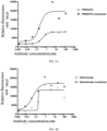
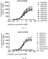
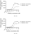
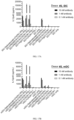
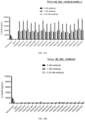
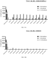
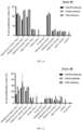
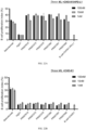
- the antigen-binding protein targeting CD40 of the present invention have properties of high affinity for CD40 and strong agonistic activity on signaling pathway, especially the enhanced agonistic activity after crosslinking, such that the antigen-binding protein targeting CD40 of the present invention has a greater treatment window and provides a good basis for significantly improving patient response rate under safe and tolerable dosage conditions in clinical trials.
- the antigen-binding protein targeting PD-L1 of the present invention has a high affinity for PD-L1 and can effectively inhibit the biological activity of PD-L1. On this basis, the antigen-binding protein targeting PD-L1 and CD40 of the present invention has excellent anti-tumor efficacy, good safety, and outstanding druggability.
- the antigen-binding protein targeting PD-L1 and CD40 of the present invention achieves a significant synergistic anti-tumor effect by means of blocking a PD-1/PD-L1 inhibitory signaling pathway, acting on an activated CD40 receptor, simultaneously activating an antigen-presenting cell and a lymphocyte, enhancing the presentation of tumor antigen by the antigen-presenting cell to the lymphocyte, and promoting the response of lymphocyte; meanwhile, the activation of an immune cell by the antigen-binding protein targeting PD-L1 and CD40 only occurs at a tumor microenvironment site, so that problems of systemic drug toxicity and side effects are significantly solved, which enables the antigen-binding protein to exert an excellent anti-tumor efficacy under very safe conditions.
- the present invention is expected to bring a new opportunity for the treatment of various tumors.
- binding protein or "antigen-binding protein” generally refers to a protein comprising a portion binding to an antigen and optionally a scaffold or framework portion that allows the antigen-binding portion to adopt a conformation that promotes binding of the antigen-binding protein to the antigen. It can typically comprise a light chain variable region (VL) of an antibody, a heavy chain variable region (VH) of an antibody, or both. VH and VL regions can be further divided into hypervariable regions called complementary determining regions (CDRs), which are scattered within more conserved regions called framework regions (FRs).
- CDRs complementary determining regions
- Each VH and VL can be composed of three CDRs and four FRs, which can be arranged from an amino terminus to a carboxyl terminus in the following order: FR-1, CDR1, FR-2, CDR2, FR-3, CDR3 and FR-4.
- the variable regions of the heavy and light chains contain binding domains that interact with antigens.
- Three CDRs of VH are denoted as HCDR1, HCDR2, and HCDR3, respectively, and can also be denoted as VH CDR1, VH CDR2, and VH CDR3; and three CDRs of VL are denoted as LCDR1, LCDR2, and LCDR3, respectively, and can also be denoted as VL CDR1, VL CDR2, and VL CDR3.
- antigen-binding protein examples include, but are not limited to, an antibody, an antigen-binding fragment (Fab, Fab', F(ab) 2 , Fv fragment, F(ab') 2 , scFv, di-scFv and/or dAb), an immunoconjugate, a multi-specific antibody (e.g., a bispecific antibody), an antibody fragment, an antibody derivative, an antibody analog, a fusion protein, and the like so long as they exhibit the desired antigen-binding activity.
- Fab antigen-binding fragment
- Fv fragment fragment
- F(ab') 2 fragment
- scFv di-scFv and/or dAb
- an immunoconjugate e.g., a multi-specific antibody (e.g., a bispecific antibody)
- an antibody fragment an antibody derivative, an antibody analog, a fusion protein, and the like so long as they exhibit the desired antigen-binding activity.
- the amino acid sequences of the CDRs are shown according to the rules of Chothia definition.
- the CDRs of an antibody can be defined in the art by a variety of methods, such as the rules of Kabat definition based on sequence variability (see, Kabat et al., Sequences of Proteins of Immunological Interest, fifth edition, National Institutes of Health, Bethesda, Maryland (1991 )), and the rules of Chothia definition based on the location of a structural loop region (see JMol Biol 273: 927-48, 1997 ).
- amino acid residues in variable domain sequences can also be determined according to the rules of Combined definition that incorporates both Kabat definition and Chothia definition.
- the rules of Combined definition refer to the combination of the ranges of Kabat definition and Chothia definition, based on which a larger scope is taken, see Table 2 below for details. It should be understood by those skilled in the art that unless otherwise specified, the terms "CDR" and "complementarity determining region" of a given antibody or region thereof (for example, a variable region) should be understood to encompass the complementarity determining region as defined by any of the above-mentioned known schemes as described in the present invention.
- Laa-Lbb may refer to an amino acid sequence from position aa (Chothia numbering) to position bb (Chothia numbering), starting from the N-terminus of an antibody light chain; and Haa-Hbb may refer to an amino acid sequence from position aa (Chothia numbering) to position bb (Chothia numbering), starting from the N-terminus of an antibody heavy chain.
- L24-L34 may refer to an amino acid sequence from position 24 to position 34 according to Chothia numbering, starting from the N-terminus of an antibody light chain;
- H26-H32 may refer to an amino acid sequence from position 26 to position 32 according to the Chothia coding rules, starting from the N-terminus of an antibody heavy chain. It should be known by those skilled in the art, when Chothia numbering scheme is used to number CDR, there may be cases where insertion sites are present at some locations (see http://bioinf.org.uk/abs/).
- the term "monoclonal antibody” generally refers to an antibody obtained from a population of substantially homogeneous antibodies, that is, individual antibodies in the population are the same, except for the possible small amounts of natural mutations.
- the monoclonal antibody is usually highly specific for a single antigenic site.
- each monoclonal antibody is for a single determinant on the antigen.
- the monoclonal antibody has the advantage that it can be synthesized by hybridoma culture and is not contaminated by other immunoglobulins.
- the modifier "monoclonal” indicates the characteristic of the antibody obtained from a population of substantially homogeneous antibodies, and is not to be construed as requiring production of the antibody by any particular method.
- the monoclonal antibody used according to the present invention can be prepared in hybridoma cells, or can be prepared by the recombinant DNA method.
- the term "fully human antibody” generally refers to an antibody expressed in a genetically engineered antibody gene-deficient animal, to which all human antibody-encoding genes are transferred. All parts of the antibody, including variable and constant regions of the antibody, are encoded by human-derived genes.
- the fully human antibody can greatly reduce the immune side effects caused by a heterologous antibody to the human body. Methods for obtaining the fully human antibody in the art can include phage display technique, transgenic mouse technique etc.
- the term “specific binding” generally refers to the binding of an antibody to an epitope by the antigen binding domain of the antibody, and the binding requires some complementarity between the antigen binding domain and the epitope.
- an antibody is said to "specifically bind” to an antigen when the antibody more likely to bind to an epitope by the antigen binding domain of the antibody than to bind to a random, unrelated epitope.
- epitope refers to a specific group of atoms (e.g., sugar side chain, phosphoryl and sulfonyl) or an amino acid on an antigen that binds to an antigen-binding protein (e.g., an antibody).
- Fab generally refers to the antigen-binding portion of a conventional antibody (e.g., IgG), including the heavy chain variable region VH, light chain variable region VL, heavy chain constant region domain CH1, and light chain constant region CL of the antibody.
- a conventional antibody e.g., IgG
- the C-terminus of VH is linked to the N-terminus of CH1 to form the heavy chain Fd fragment
- the C-terminus of VL is linked to the N-terminus of CL to form the light chain
- the C-terminus of CH1 is further linked to the hinge region and other constant region domains of the heavy chain to form the heavy chain.
- Fab also refers to the variant structure of Fab.
- the C-terminus of VH is linked to the N-terminus of CL to form one polypeptide chain
- the C-terminus of VL is linked to the N-terminus of CH1 to form another polypeptide chain, thereby forming a Fab (cross VH/VL) structure
- CH1 of Fab is not linked to the hinge region, but the C-terminus of CL is linked to the hinge region of the heavy chain to form a Fab (cross Fd/LC) structure.
- VH generally refers to the heavy chain variable region VH domain of an antibody, that is, VH can be the heavy chain variable region VH of a conventional antibody of human or other animals (H2L2 structure), can also be the heavy chain variable region VHH of a heavy chain antibody of animal, such as camelid (HCAb structure), or can be the heavy chain variable region VH of a fully human heavy chain antibody produced by Harbour HCAb transgenic mice (HCAb structure).
- the term "antigen-binding fragment” generally refers to any protein functional region that can specifically bind to an antigen, that is, the antigen-binding fragment can be "Fab", "VH”, or other antigen-binding forms (e.g., lipocalins, neural cell adhesion molecules (NCAM), fibronectin, ankyrin repeat proteins (DARPins), and other derived protein structures).
- tumor antigen can refer to either a tumor specific antigen (TSA) or a tumor associated antigen (TAA).
- TSA tumor specific antigen
- TAA tumor associated antigen
- the tumor specific antigen refers to an antigen that is unique to tumor cells and do not exist on normal cells or tissues.
- TAA tumor specific antigen
- TAA tumor associated antigen
- the tumor associated antigen is not unique to tumor cells and also exists in normal cells or tissues, but is highly expressed during tumor cell proliferation.
- target cell refers to cells that need to be eliminated, mainly tumor cells, but also immunosuppressive cells and the like.
- immune effector cell generally refers to an immune cell that participates in clearing foreign antigens and exercising effector functions in an immune response, such as a plasma cell, a cytotoxic T cell and an NK cell.
- co-stimulatory molecule refers to a cell surface molecule and a ligand thereof which provides a co-stimulatory signal for the full activation of an immune cell such as T or B cell, and positively regulates the activation of immune cell, such as, CD28, 4-1BB, ICOS, OX40 and CD40.
- co-inhibitory molecule or “co-inhibitory molecule antigen” refers to a class of cell surface molecules and ligands thereof which negatively regulate the function of immune cells, such as, CTLA-4, PD-L1 and PD-1.
- PD-L1 generally refers to programmed death ligand 1 protein, a functional variant thereof and/or a functional fragment thereof.
- PD-L1 is also known as cluster of differentiation 274 (CD274) or B7 homolog 1 (B7-H1), and is a protein encoded by CD274 gene (in human).
- CD274 cluster of differentiation 274
- B7-H1 B7 homolog 1
- the sequence of PD-L1 is known in the art.
- amino acid sequence of an exemplary full-length human PD-L1 protein can be found in NCBI under the accession number of NP_054862 or UniProt under the accession number of Q9NZQ7; and the sequence of an exemplary full-length cynomolgus monkey PD-L1 protein can be found in NCBI under the accession number of XP_005581836 or Uniprot under the accession number of G7PSE7.
- CD40 generally refers to tumor necrosis factor receptor superfamily member 5 protein, a functional variant thereof and/or a functional fragment thereof, also known as TNFRSF5.
- the sequence of CD40 is known in the art.
- the amino acid sequence of an exemplary human CD40 protein can be found in UniProt under the accession number of P25942; the sequence of an exemplary cynomolgus monkey CD40 protein can be found in Uniprot under the accession number of G7PG38; and the sequence of an exemplary mouse CD40 protein can be found in Uniprot under the accession number of P27512.
- CD40L is the natural trimeric ligand molecule of CD40.
- the present invention is further described below by way of examples; however, the present invention is not limited to the scope of the described examples.
- the examples do not include a detailed description of conventional methods, such as methods for constructing vectors and plasmids, methods for inserting protein-encoding genes into such vectors and plasmids, or methods for introducing plasmids into host cells. Such methods are well known to an ordinary person skilled in the art and are described in numerous publications. For the experimental methods in which no specific conditions are specified in the following examples, selections are made according to conventional methods and conditions or according to the product instructions.
- the general method for preparing antibodies using techniques such as mammalian host cells e.g., human embryonic kidney cells HEK293 or Chinese hamster ovary cells CHO and derivatives thereof
- transient transfection expression and affinity capture isolation was described.
- the present method is suitable for an antibody of interest containing an Fc region; the antibody of interest may be composed of one or more protein polypeptide chains; and the antibody of interest may be derived from one or more expression plasmids.
- the amino acid sequences of antibody polypeptide chains are converted into nucleotide sequences by codon optimization; and the nucleotide sequences for encoding are respectively synthesized and cloned onto expression vectors compatible with a host cell.
- the plasmids encoding the antibody polypeptide chains are simultaneously transfected into a mammalian host cell according to a particular ratio, and the recombinant antibodies having correct folding and polypeptide chain assembly can be obtained using conventional recombinant protein expression and purification techniques.
- FreeStyle TM 293-F cells (Thermo, # R79007) were subjected to scale-up culture in a FreeStyle TM F17 Expression Medium (Thermo, # A1383504).
- the cells were adjusted to a cell concentration of 6-8 ⁇ 10 5 cells/mL, and cultured in a shaker at 37°C and 8% CO 2 for 24 hours at a cell concentration of 1.2 ⁇ 10 6 cells/mL. 30 mL of the cultured cells was prepared.
- the plasmids encoding the antibody polypeptide chains were mixed according to a certain ratio, a total of 30 ⁇ g of plasmids (the ratio of the plasmids to the cells being 1 ⁇ g : 1 mL) were dissolved in 1.5 mL of Opti-MEM reduced serum medium (Thermo, # 31985088), and the resulting mixture was filtered through a 0.22 ⁇ m filter membrane for sterilization. Another 1.5 mL of Opti-MEM was taken and dissolved in 120 ⁇ L of 1 mg/mL PEI (Polysciences, # 23966-2), and the resulting mixture was left to stand for 5 minutes.
- Opti-MEM reduced serum medium Thermo, # 31985088
- the PEI was slowly added to the plasmids, the resulting mixture was incubated at room temperature for 10 minutes, the plasmid and PEI mixed solution was slowly dripped into the culture flask while shaking the culture flask, and cultured in a shaker at 37°C and 8% CO 2 for 5 days. The cell viability was observed after 5 days.
- the culture was collected, and centrifuged at a rotary speed of 3300 g for 10 minutes, and then the supernatant was taken; and the supernatant was then centrifuged at a high speed to remove impurities.
- a gravity column (Bio-Rad, #7311550) containing MabSelect TM (GE Healthcare, #71-5020-91) was equilibrated with a PBS buffer with pH 7.4, that is, the column was rinsed with 2-5 times the column volume of the buffer. The supernatant sample was loaded onto the column; and the column was rinsed with 5-10 times the column volume of PBS buffer, the protein of interest was eluted with 0.1 M glycine with pH 3.5, then adjusted with Tris-HCl with pH 8.0 until neutrality, and finally transferred to a PBS buffer or a buffer containing other components through concentration and liquid exchange by an ultrafiltration tube (Millipore, # UFC901024) to obtain a purified recombinant antibody solution. Finally, the concentration was determined using NanoDrop (Thermo, NanoDrop TM One), and the purified recombinant antibody solution was subpackaged and stored for later use.
- NanoDrop Thermo, NanoDrop TM One
- analytical size exclusion chromatography was used to analyze the purity and aggregate form of the protein samples.
- An analytical chromatographic column TSKgel G3000SWxl (Tosoh Bioscience, #08541, 5 ⁇ m, 7.8 mm ⁇ 30 cm) was connected to a high pressure liquid chromatograph HPLC (Agilent Technologies, Agilent 1260 Infinity II), and equilibrated with a PBS buffer (pH 7.4) for at least 1 hour at room temperature.
- Differential scanning fluorimetry is a common high-throughput method for determine the thermal stability of proteins.
- a real-time fluorescent quantitative PCR instrument is used to monitor the change in the fluorescence intensity of the dye bound to the unfolded protein molecule to reflect the process of protein denaturation, thereby reflecting the thermal stability of the protein molecule.
- Tm thermal denaturation temperature
- mice which could be mice, rats, rabbits, sheep, camels, etc.
- CD40 antigen to obtain antibody molecules that specifically bind to CD40.
- the resulting antibody molecules are of non-human origin.
- these molecules need to be humanized using antibody engineering techniques to reduce immunogenicity and improve druggability.
- the process of humanizing antibodies has its technical complexity, and molecules that have been humanized tend to have reduced affinity for antigens.
- transgenic technology allows the breeding of genetically engineered mice that carry the human immunoglobulin repertoire and in which their endogenous murine immunoglobulin repertoires are deleted.
- the antibody produced by this transgenic mouse has a fully human sequence, so no further humanization is required, greatly improving the efficiency of therapeutic antibody development.
- Harbour H2L2 mouse (Harbour antibodies BV) is a transgenic mouse carrying the human immunoglobulin repertoire, and the antibodies produced thereby have complete human antibody variable domains and rat constant domains.
- each mouse was immunized with an immunogen reagent prepared by mixing 25 ⁇ g of the antigen protein with Sigma Adjuvant System adjuvant (Sigma, #S6322).
- the interval between two rounds of booster immunization was at least two weeks, typically with no more than 5 rounds of booster immunization.
- the immunization times were days 0, 14, 28, 42, 56, and 70; and on days 49 and 77, the antibody titers in the sera of mice were measured.
- the final booster immunization was performed at a dose of 25 ⁇ g of the antigen protein per mouse.
- the mouse spleen cells were taken out and fused with the myeloma cell line to obtain hybridoma cells.
- the hybridoma expressing the CD40 monoclonal antibody molecule was isolated.
- the isolated hybridoma expressed the antibody molecule having heavy and light chains of complete human variable domains and rat constant domains.
- the above-mentioned monoclonal antibody was further identified, and several hybridoma clones were selected for sequencing based on parameters such as the binding ability to human CD40, the binding ability to cynomolgus monkey CD40, and the ability to activate CD40 downstream signaling pathway.
- Selicrelumab (also numbered PR001028 in the present application) was used as a positive control antibody.
- the nucleotide sequence encoding the variable domain of the antibody molecule and the corresponding amino acid sequence was obtained by conventional hybridoma sequencing means.
- the sequences of the variable domains of the CD40 monoclonal antibody molecule obtained from the immunized Harbour H2L2 mice were human antibody sequences.
- the CDR sequences of the antibody variable domains could be analyzed according to Kabat or Chothia or other CDR definition rules (Table-I).
- the light and heavy chain variable domain sequences of the encoding antibody molecule can be expressed in fusion with the light and heavy chain constant domain sequences of corresponding human antibody using conventional recombinant DNA techniques to obtain recombinant antibody molecules using the method described in 0.
- an antibody heavy chain variable domain (VH) sequence was obtained by gene synthesis and cloned into a mammalian cell expression plasmid vector encoding heavy chain constant domain sequences of human IgG1 or IgG2 antibody to encode and produce a full-length heavy chain of an IgG antibody.
- a light chain variable domain (VL) sequence of an antibody was obtained by gene synthesis and cloned into a mammalian cell expression plasmid vector encoding a ⁇ light chain constant domain sequence of human antibody Ig to encode and produce a full-length light chain of an antibody.
- VL variable domain
- the heavy chain variable domain sequence of the antibody is derived from events such as gene rearrangement of germline gene V, D and J gene fragments of the heavy chain gene group on chromosomes and somatic hypermutation; and the light chain variable domain sequence is derived from events such as gene rearrangement of germline gene V and J gene fragments of the light chain gene group and somatic hypermutation.
- Gene rearrangement and somatic hypermutation are main factors that increase antibody diversity.
- Antibodies derived from the same germline V gene fragment may also give rise to different sequences, but the overall similarity is high.
- the possible germline gene fragments in the event of gene rearrangement can be deduced from the variable domain sequence of the antibody using some algorithms, such as IMGT/DomainGapAlign (http://imgt.org/3Dstructure-DB/cgi/DomainGapAlign.cgi) or NCBI/IgBLAST (https://www.ncbi.nlm.nih.gov/igblast/).
- IMGT/DomainGapAlign http://imgt.org/3Dstructure-DB/cgi/DomainGapAlign.cgi
- NCBI/IgBLAST https://www.ncbi.nlm.nih.gov/igblast/).
- PTMs post-translational modifications
- a PTM is present in a variable domain, particularly an antigen-binding region (e.g., CDR) of an antibody
- the presence of such a PTM can have a greater impact on the binding to an antigen, and can also cause a change in the physicochemical properties of the antibody. For example, glycosylation, deamidation, isomerization, oxidation, etc. may increase the instability or heterogeneity of antibody molecules, thereby increasing the difficulty and risk of antibody development. Avoiding some potential PTMs is thus very important for the development of therapeutic antibodies.
- an N-linked glycosylation site can be predicted according the sequence pattern N-x-S/T (asparagine at the first position, any amino acid other than proline at the second position, and serine or threonine at the third position).
- Amino acid sequence patterns that cause PTMs may be derived from germline gene sequences, for example, pattern NST where human germline gene fragment IGHV3-33 naturally has a glycosylation modification in the FR3 region; and they may also be derived from somatic hypermutation.
- NGS or NLT may be a glycosylation site
- NS may be a deamidation site
- DG may cause isomerization of aspartic acid.
- the amino acid sequence patterns of PTMs can be disrupted by amino acid mutations, thereby reducing or removing the formation of particular PTMs. Depending on different antibody sequences and different PTM sequence patterns, there are different mutation design methods.
- One method is to replace the "hot spot" amino acid (such as N or S in the NS pattern) with an amino acid having similar physicochemical properties (e.g., mutation of N to Q).
- Another method uses a mutation library approach to randomly mutate the PTM sequence pattern. In actual operation, multiple mutation design methods may be used for the same PTM sequence pattern.
- Table 3 listed the anti-CD40 hybridoma clone and the recombinant antibody molecule (PR003379) obtained in this example, as well as the analyzed germline gene V gene segments and potential PTM sites of VH and VL.
- Table 4 listed the new antibody molecules obtained by amino acid mutation of CD40 antibody PR003379.
- Purified recombinant antibodies were obtained from all designed mutant molecules according to the method described in 0, and further verified in subsequent functional experiments.
- the method for obtaining the antibodies in Table 5 comprises: the heavy chain variable region (VH) of PR003379 was linked to the heavy chain constant domain sequence of an IgG1 subtype antibody containing L234A, L235A and G237A mutations and cloned into a mammalian cell expression plasmid to encode and produce a full-length heavy chain of IgG1 (L234A, L235A and G237A); a light chain variable domain (VL) sequence of an antibody with the PTM mutation site was obtained by gene synthesis and cloned into a mammalian cell expression plasmid vector encoding a ⁇ light chain constant domain sequence of human antibody Ig to encode and produce a full-length light chain of an antibody; and the two plasmids were co
- Table 5 listed the sequences of the CD40 antibodies of the present invention and the amino acid sequences of the CDRs defined according to the rules of Chothia definition. These CD40 antibodies include a positive control antibody, the CD40 antibody PR003379 of the present invention, and mutant molecules thereof. Table 3. Germline gene analysis and PTM site analysis of CD40 antibodies Clone No. VH germline V gene VL germline V gene VH PTM VL PTM Recombinant antibody Recombinant antibody subtype 18C4A12-1B1 IGHV1-2 IGKV1-9 NS (LCDR3) PR003379 Human IgG2 Table 4.
- flow cytometry was used to study the binding activity of the antibody or antigen-binding protein targeting CD40 to cells expressing CD40.
- FACS flow cytometry
- the CHO-K1 cell line highly expressing human CD40, CHO-K1/hCD40 (Shanghai ChemPartner), the CHO-K1 cell line highly expressing cynomolgus monkey CD40, CHO-K1/cyCD40 (Shanghai ChemPartner), and the lymphoma cell highly expressing human CD40, Raji (ATCC, #CCL-86) were used.
- the cells expressing CD40 were digested and resuspended with complete medium (F-12K for CHO-K1 cells; and RPMI-1640 for Raji cells); and the cells were adjusted to a cell density of 1 ⁇ 10 6 cells/mL.
- the cells were seeded in a 96-well V-bottom plate (Corning, #3894) at 100 ⁇ L/well, and the antibody to be tested at 2 ⁇ final concentration was then added at 100 ⁇ L/well and mixed evenly, with the highest final concentration of the antibody of 300 nM, a total of 8 concentrations, and 5-fold concentration gradient dilution.
- hIgG (Crownbio, #C0002) was used as a negative control and Selicrelumab was used as a positive control molecule, and the concentration settings were the same as the subsequent bispecific antibody to be tested.
- the cells were incubated at 4°C for 1 hours in the dark. After the incubation, the cells were rinsed twice by adding pre-chilled PBS at 100 ⁇ L/well, and centrifuged at 500 g at 4°C for 5 minutes, and the supernatant was discarded.
- a fluorescent secondary antibody (goat anti-human IgG (H+L) secondary antibody, Alexa Fluor ® 488 conjugate, Invitrogen, #A11013, 1 : 1000 dilution) was added at 100 ⁇ L/well, and the cells were incubated at 4°C for 30 minutes in the dark. The cells were washed twice with pre-chilled PBS at 200 ⁇ L/well, and centrifuged at 500 g at 4°C for 5 minutes, and the supernatant was discarded. Finally, the cells were resuspended in pre-chilled PBS at 200 ⁇ L/well. The fluorescence signal values were read using a BD FACS CANTOII flow cytometer or ACEA NovoCyte flow cytometer, and the data were processed and analyzed using FlowJo v10 software (FlowJo, LLC).
- FIG. 1A-FIG. 1D and Table 6 showed the activity of CD40 antibody PR003379 and the variant molecules thereof in binding to Raji cells.
- the results showed that PR003379 and the variant molecules thereof could strongly bind to Raji cells; and the binding activity of PR003379 was stronger than that of Selicrelumab. Table 6. Binding of CD40 antibody to Raji cells (corresponding to FIG. 1A-FIG.
- activation experiments of HEK293-hCD40-NF ⁇ B fluorescent reporter gene cells were used to study the activation effect of antibodies or antigen-binding proteins targeting CD40 in the presence of or in the absence of crosslinking on CD40.
- Cells highly expressing human CD32B were used to perform Fc-mediated crosslinking of the molecules to be tested, to study whether crosslinking could further enhance the ability to activate CD40.
- CHO-K1 cells expressing human CD32B (CHO-K1/hCD32B, GenScript, #M00600) and CHO-K1 cells were plated at 1 ⁇ 10 4 cells/well or 100 ⁇ L/well in a 96-well plate (PerkinElmer, #6005181) and incubated at 37°C in 5% CO 2 environment overnight. The supernatant was removed, and HEK293-hCD40-NF ⁇ B reporter gene cells expressing human CD40 and NF ⁇ B-luciferase reporter gene (BPS bioscience, #60626) were collected and added to a 96-well plate at 5 ⁇ 10 4 /well or 50 ⁇ L/well.
- crosslinked The sample in which the CD40 antibody was incubated with CHO-K1/hCD32B cells was referred to as "crosslinked”; and the sample in which the CD40 antibody was incubated with CHO-K1 cells was referred to as "un-crosslinked”.
- PR003379 had a more obvious "crosslinking enhancement" effect than the control antibody Selicrelumab, that is, PR003379 in the presence of CD32B-mediated antibody crosslinking could significantly amplify the signal of the fluorescent reporter gene, indicating that the activation effect of the CD40 molecule on the downstream signal molecule was significantly enhanced.
- FIG. 2C-FIG. 2D and Table 8 showed the enhancement fold of fluorescence intensity of the activation of reporter gene cells by the variant molecules of PR003379 in the presence of or in the absence of crosslinking mediated by cells expressing human CD32B.
- the results showed that the variant molecules of PR003379 in the presence of crosslinking could significantly enhance the activation on CD40 molecule, which was manifested in that the EC 50 value in the presence of crosslinking was significantly reduced compared with the EC 50 value in the absence of crosslinking (expressed as "fold (EC 50 in the absence of crosslinking/EC 50 in the presence of crosslinking)" > 1).
- Table 7 Table 7.
- HCAb mouse (Harbour Antibodies BV, WO 2010/109165 A2 ) is a transgenic mouse carrying a human immunoglobulin immune repertoire, and can produce a heavy-chain-only antibody (HCAb) which is only half the size of conventional IgG antibody.
- the antibody produced thereby has only heavy chain variable domain of human antibody and Fc constant domain of mouse. Due to the characteristic of not containing light chains, the antibody almost solves the problems of light chain mispairing and heterodimerization, so that this technique platform can develop products that are difficult to achieve with conventional antibody platforms.
- mice 6-8 week-old Harbour HCAb mice were immunized in multiple rounds with a soluble recombinant human PD-L1-mFc fusion protein (Novo Protein Inc., #CM06).
- a soluble recombinant human PD-L1-mFc fusion protein Novo Protein Inc., #CM06
- each mouse received a total injection dose of 100 ⁇ L by subcutaneous injection in the inguinal region or by intraperitoneal injection.
- an immunogen reagent prepared by mixing 50 ⁇ g of the antigen protein with a complete Freund's adjuvant (Sigma, #F5881) at a volume ratio of 1 : 1.
- each mouse was immunized with an immunogen reagent prepared by mixing 25 ⁇ g of the antigen protein with Ribi adjuvant (Sigma Adjuvant System, #S6322).
- the interval between two rounds of booster immunization was at least two weeks, typically with no more than 5 rounds of booster immunization.
- the immunization times were days 0, 14, 28, 42, 56, and 70; and on days 49 and 77, the antibody titers in the sera of mice were measured.
- the mouse spleen cells were removed to isolate B cells, and CD138-positive plasma cells and human PD-1 antigen-positive B cell populations were sorted using BD FACS AriaII Cell Sorter.
- the human VH gene was amplified from plasma cells and B cell populations by conventional molecular biological means, and the amplified human VH gene fragments were constructed into a mammalian cell expression plasmid pCAG vector encoding the Fc region sequence of the human IgG1 antibody heavy chain.
- the plasmid was transfected into mammalian host cells (such as human embryonic kidney cell HEK293) for expression to obtain the supernatant containing the fully human HCAb antibody.
- the supernatant containing expressed HCAb was screened using Mirrorball high-throughput no-wash antibody screening platform (SPT Labtech), that is, the binding of the supernatant to the CHO-K1/hPD-L1 (GenScript, #M00543) stable cell line overexpressing human PD-L1 was detected, and the positive antibody PR000151 (Atezolizumab analog) was used as a positive control.
- SPT Labtech Mirrorball high-throughput no-wash antibody screening platform
- CHO-K1/hPD-L1 cells were washed with serum-free F12K medium (Thermofisher, #21127022) and resuspended to 1 ⁇ 10 6 /mL.
- Draq5 fluorescent probe CTS, #4048L was added (1 ⁇ L of Draq5 being added to 1 mL of CHO-K1/hPD-L1 cells, 1 : 1000 dilution), and the cells were incubated for 30 minutes in the dark. After the cells were centrifuged, the cells were washed with culture medium and adjusted to a cell density of 1.0 ⁇ 10 5 cells/mL.
- the secondary antibody Alexa Fluor ® 488 AffiniPure Goat Anti-Human IgG,Fc ⁇ Fragment Specific, Jackson ImmunoResearch, #109-545-098
- the secondary antibody Alexa Fluor ® 488 AffiniPure Goat Anti-Human IgG,Fc ⁇ Fragment Specific, Jackson ImmunoResearch, #109-545-098
- the mixture was taken and added to a 384-well plate (Greiner, #781091) at 30 ⁇ L/well.
- 10 ⁇ L of positive control or the supernatant containing expressed HCAb was added to the 384-well plate, and incubation was performed for 2 hours. Fluorescence values were read on Mirrorball.
- the positive antibody supernatant was further identified.
- the binding to CHO-K1/hPD-L1 cell was detected by FACS and the cross-binding activity to cynomolgus monkey PD-L1 protein (Acro biosystems, #PD1-C52H4) was detected by ELISA.
- the screened HCAb was sequenced by conventional sequencing means to obtain the nucleotide sequence encoding corresponding VH and the corresponding amino acid sequence.
- the VH sequence of the HCAb antibody and the Fc sequence of the human IgG1 heavy chain were expressed in fusion to obtain the fully human recombinant HCAb antibody molecule.
- Table 9 listed the preferred anti-PD-L1 recombinant heavy chain antibody molecule PR000960 obtained in this example, as well as the analyzed VH germline gene V gene segment and potential post-translational modification (PTM) site.
- PR000960 has an N-glycosylation modification site in HCDR1, which may affect the drugability in later development. In order to remove the N-glycosylation modification site, PR000960 was subjected to amino acid mutation to obtain a new antibody molecule PR002082 (Table 10).
- the PR002082 molecule was subjected to in vitro affinity maturation engineering.
- a yeast display antibody mutation library was established by random mutation of the CDR region, and the mutation library of PR002082 molecular was screened by BD FACS AriaIII sorting platform.
- mutations were randomly introduced into the three CDRs of PR002082 to establish three yeast display mutation libraries (CDR1, CDR2 and CDR3); enrichment was performed using the MACS sorting method; and then multiple rounds of sorting were performed using flow cytometry FACS to enrich high-affinity mutant molecules.
- yeast cells with higher binding affinity were sorted using 0.1 nM biotinylated PD-L1-his protein; and then yeast cells were further cultured for induction.
- yeast cells with higher binding affinity were further sorted using 0.01 nM biotinylated PD-L1-his protein; and then yeast cells were further cultured for induction.
- the antigen concentration during sorting was further reduced, and the final round of sorting was performed using 0.003 nM biotinylated PD-L1-his protein.
- VHs of the mutant molecules obtained by affinity maturation were subjected to gene synthesis and cloned into a mammalian cell expression plasmid vector encoding the human IgG1 heavy chain Fc sequence to produce recombinant HCAb antibody molecules.
- Table 11 listed the variant molecules of PR002082 obtained by affinity maturation process.
- Table 12 listed the sequences of the PD-L1 antibodies of the present invention and the amino acid sequences of the CDRs defined according to the rules of Chothia definition. These PD-L1 antibodies include a positive control antibody, the PD-L1 antibody PR000960 of the present invention, and derived mutant molecules thereof. Table 9. Germline gene analysis and PTM site analysis of PD-L1 heavy chain antibodies Recombinant antibody Molecular structure IgG subtype VH germline V gene VH PTM PR000960 HCAb Human IgG1 IGHV3-33 NxS/T (HCDR1); DG (HCDR2) Table 10.
- Sequence numbering table of the sequences and CDR sequences (Chothia) for the PD-L1 antibodies of the present invention Molecular number Light chain Heavy chain VL VH LCDR1 LCDR2 LCDR3 HCDR1 HCDR2 HCDR3 PR000151 (Atezolizum ab) 85 74 66 56 36 41 46 4 15 24 PR000960 75 57 5 16 25 PR002082 77 59 7 16 25 PR005878 79 61 7 19 28 PR006245 80 62 9 19 25 PR006246 81 63 9 19 29 PR006247 82 64 9 19 30 PR006248 83 65 9 19 28
- flow cytometry was used to study the binding activity of the antibody or antigen-binding protein targeting PD-L1 to cells expressing PD-L1.
- the cells expressing PD-L1 were digested and resuspended in PBS buffer containing 2% BSA. The cells were adjusted to a cell density of 1 ⁇ 10 6 cells/mL, respectively. The cells were seeded in a 96-well V-bottom plate (Corning, #3894) at 100 ⁇ L/well, and the antibody to be tested at 2 ⁇ final concentration with 4-fold concentration gradient dilution was then added at 100 ⁇ L/well. The cells were incubated at 4°C for 2 hours in the dark.
- the cells were rinsed twice by adding pre-chilled PBS containing 2% BSA at 100 ⁇ L/well, and centrifuged at 500 g at 4°C for 5 minutes, and the supernatant was discarded.
- a fluorescent secondary antibody Alexa Fluor 488-conjugated AffiniPure Goat Anti-Human IgG, Fc ⁇ Fragment Specific, Jackson, #109-545-098, 1 : 500 dilution
- the cells were washed twice by adding pre-chilled PBS containing 2% BSA at 100 ⁇ L/well, and centrifuged at 500 g at 4°C for 5 minutes, and the supernatant was discarded. Finally, the cells were resuspended in pre-chilled PBS buffer containing 2% BSA at 200 ⁇ L/well.
- the fluorescence signal values were read using a BD FACS CANTOII flow cytometer or ACEA NovoCyte flow cytometer, and the data were processed and analyzed using FlowJo v10 software (FlowJo, LLC).
- FIG. 3A and FIG. 3B showed the binding curves of PD-L1 heavy chain antibodies PR000960 and PR002082, and the variant molecules thereof to CHO-K1/hPD-L1 cells, and Table 13 listed EC 50 values and the maximum MFI signal of the corresponding curves.
- Table 13 Binding of PD-L1 antibody to CHO-K1/hPD-L1 cells (corresponding to FIG. 3A-FIG.
- PD-L1 antibodies or antigen-binding proteins removed the inhibitory effect of PD-1 on T cells by blocking the interaction between PD-L1 and PD-1.
- the Jurkat-NFAT-hPD1 cells were adjusted to a cell density of 1 ⁇ 10 6 cells/mL, and 293T-OS8-hPDL1 cells were adjusted to a cell density of 5 ⁇ 10 5 cells/mL.
- FIG. 4A and FIG. 4B and Table 14 showed that in the fluorescent reporter gene experiments, PD-L1 heavy chain antibodies PR000960 and PR002082 and variant molecules thereof inhibited the PD-1 signaling pathway, and therefore restored the NFAT signal in Jurkat.
- Table 14 Inhibition of the PD-1 signaling pathway of Jurkat-NFAT-hPD1 by PD-L1 antibodies (corresponding to FIG. 4A and FIG.
- the CD40 antibody from 0 and the PD-L1 heavy chain antibody from 0 were used to construct a PD-L1 ⁇ CD40 bispecific antigen-binding protein with the structure shown in FIG. 5 .
- the Fab end is derived from conventional antibody A
- VH_A and VL_A are the heavy chain variable region and light chain variable region of antibody A, respectively.
- the VH end is derived from heavy chain antibody B
- VH_B is the heavy chain variable region of heavy chain antibody B.
- CL is the light chain constant region domain.
- CH1, CH2 and CH3 are the first, second and third domains of the heavy chain constant region, respectively.
- h is the hinge region of IgG antibody or a derived sequence
- L is a linker peptide.
- the binding protein having the shown structure comprises two different polypeptide chains: a light chain, from amino terminus to carboxyl terminus, comprising VL_A-CL; and a heavy chain, from amino terminus to carboxyl terminus, comprising VH _B-L-VH_A-CH1-h-CH2-CH3.
- VH_B of the heavy chain is directly fused to VH_A, i.e., the length of L is 0; and in other embodiments, VH_B of the heavy chain is linked to VH_A via a linker peptide L.
- L can be a sequence in Table 15. Table 15.
- Linker peptide sequence Linker peptide name Length Sequence SEQ ID NO GS_2 2 GS GS_3 3 GGS GS_4 4 GSGS 100 GS_5 5 GGGGS 101 GS_7 7 GGGGSGS 102 GS_15 15 GGGGSGGGGSGGGGS 103 H1_15 15 EPKSSDKTHTPPPPP 104 LH1 10 DKTHTCPPCP 105 G5-LH 15 GGGGGDKTHTCPPCP 106
- VH_A and VL_A in FIG. 5 were derived from the antigen-binding fragment Fab of the H2L2 (IgG) antibodies PR006495, PR006496, PR006497, PR006504 and PR006511 that bind to CD40; and VH_B was derived from the antigen-binding fragment VH of the heavy chain antibodies PR005878, PR006245, PR006246, PR006247 and PR006248 that bind to PD-L1. Based on these PD-L1 and/or CD40 antigen-binding fragments, several PD-L1 ⁇ CD40 bispecific antigen-binding proteins having the structure as shown in FIG. 5 (listed in Table 16) were constructed.
- PD-L1 ⁇ CD40 bispecific antigen-binding proteins of the present invention Bispecific antibody molecule CD40 antibody PD-L1 antibody from which VH was derived Fc type (mutation)
- Linker peptide PR006919 PR006496 PR006246 Human IgG1 (L234A/L235A/G237A)
- Sequence numbering table of polypeptide chains of the PD-L1 ⁇ CD40 bispecific antibody molecules of the present invention Molecular number Heavy chain Light chain PR006919 93 89 PR007070 94 88 PR007151 94 91 PR007268 93 90 PR007269 93 92 PR007273 95 88 PR007275 96 88 PR007276 97 88 PR007280 95 91 PR007281 93 91 PR007282 96 91 PR007556 97 89 PR007616 97 90 PR007617 98 88 PR007618 99 88 PR007619 98 91 PR007620 99 91
- 0 and the method described in 0 were used to test the binding ability of the PD-L1 ⁇ CD40 bispecific binding proteins to various cells expressing PD-L1 or CD40.
- These cells include: CHO-K1 cell that does not express PD-L1 or CD40, CHO-K1 cell line highly expressing human PD-L1, CHO-K1/hPD-L1 (Shanghai ChemPartner), CHO-K1 cell line highly expressing cynomolgus monkey PD-L1, CHO-K1/cyPD-L1 (Shanghai ChemPartner), CHO-K1 cell line highly expressing human CD40, CHO-K1/hCD40 (Shanghai ChemPartner), CHO-K1 cell line highly expressing cynomolgus monkey CD40, CHO-K1/cyCD40 (Shanghai ChemPartner), lymphoma cell highly expressing human CD40, Raji (ATCC, #CCL-86), lung cancer cell highly expressing
- Table 19 summarized the corresponding results of binding of PD-L1 ⁇ CD40 bispecific binding proteins and control molecule to various cells, corresponding figures showed the binding curves of the antigen-binding proteins to the target cell, and the corresponding tables listed the binding force parameters obtained by fitting the corresponding curves (EC 50 value and maximum MFI signal).
- FIG. 10A and FIG. 10B showed that PD-L1 ⁇ CD40 did not bind to the CHO-K1 cell.
- FIG. 6A, FIG. 6B and Table 20 showed that PD-L1 ⁇ CD40 bound to the CHO-K1/hPD-L1 cell.
- FIG. 7A, FIG. 7B and Table 21 showed that PD-L1 ⁇ CD40 bound to the cynomolgus monkey PD-L1 cell.
- FIG. 8A, FIG. 8B and Table 22 showed that PD-L1 ⁇ CD40 bound to the CHO-K1/hCD40 cell.
- FIG. 9A, FIG. 9B and Table 23 showed that PD-L1 ⁇ CD40 bound to the cynomolgus monkey CD40 cell.
- FIG. 11A, FIG. 11B and Table 24 showed that PD-L1 ⁇ CD40 bound to the NCI-H226 cell.
- FIG. 12A, FIG. 12B and Table 25 showed that PD-L1 ⁇ CD40 bound to the Raji cell.
- FIG. 13A, FIG. 13B and Table 26 showed that PD-L1 ⁇ CD40 bound to the MDA-MB-231 cell.
- PD-L1 ⁇ CD40 bispecific antibodies could efficiently bind to the cell lines expressing PD-L1, such as CHO-K1/hPD-L1 (EC 50 being about 1 nM), CHO-K1/cy PD-L1 (EC 50 being less than 1 nM) and NCI-226 (EC 50 being less than 1 nM), the binding activities thereof were comparable to that of the positive control Atezolizumab; the PD-L1 ⁇ CD40 bispecific antibodies did not bind to the CHO-K1 cell without expressing the target; the PD-L1 ⁇ CD40 bispecific antibodies also efficiently bound to the MDA-MB-231 cell (EC 50 being less than 1 nM), and the binding effects thereof were significantly stronger than that of the positive control Atezolizumab (having a higher MFI signal).
- PD-L1 such as CHO-K1/hPD-L1 (EC 50 being about 1 nM), CHO-K1/cy PD-L1 (EC 50 being less than 1 nM)
- all PD-L1 ⁇ CD40 bispecific antibodies could efficiently bind to CD40-expressing cell lines, such as CHO-K1/hCD40 (EC 50 being about 1-2 nM), CHO-K1/cyCD40 (EC 50 being about 1-4 nM) and Raji (EC 50 being about 1-2 nM), the binding activities thereof were stronger than or equivalent to that of the positive control Selicrelumab; the PD-L1 ⁇ CD40 bispecific antibodies also efficiently bound to the MDA-MB-231 cell, and the binding effects thereof were significantly stronger than that of the positive control Selicrelumab. Table 19.
- FIG. 12A-FIG. 12B Table 25 MDA-MB-231 Human PD-L1 Human CD40
- FIG. 13A-FIG. 13B Table 26 Table 20. Binding of PD-L1 ⁇ CD40 to CHO-K1/hPD-L1 cell (corresponding to FIG. 6A and FIG.
- Binding of PD-L1 ⁇ CD40 to CHO-K1/cyPD-L1 cell (corresponding to FIG. 7A and FIG. 7B)
- Antibody EC 50 (nM) Maximum MFI Antibody EC 50 (nM) Maximum MFI (A) PR006919 0.6221 4540 (B) PR007616 0.6867 4754 PR007268 0.6797 4553 PR007617 0.6351 4832 PR007269 0.7138 4672 PR007618 0.6904 4868 PR007273 0.8901 4621 PR007619 0.6292 4815 PR007275 0.8068 4617 PR007620 0.7572 4837 PR007276 0.656 4567 PR007070 0.7794 4873 PR007280 1.021 4827 PR007151 0.7538 4926 PR007281 0.6324 4611 Selicrelumab 94 PR007282 0.8344 4624 Atezolizumab 0.6486 4567 PR007556 0.6831 4716 Isotype 50 Table 22.
- the method described in 0 was used to test the inhibitory effect of the PD-L1 ⁇ CD40 bispecific binding proteins on the PD-1 signaling pathway.
- the results were as shown in FIG. 14A-FIG. 14D and Table 27.
- All PD-L1 ⁇ CD40 bispecific antibodies could effectively block PD-1/PD-L1 interaction and activate TCR and NFAT signals, and the activities were comparable to that of the positive control antibody Atezolizumab (EC 50 being about 0.1 - 0.3 nM).
- Table 27 Inhibition of the PD-1 signaling pathway of Jurkat-NFAT-hPD1 by PD-L1 ⁇ CD40 (corresponding to FIG. 14A-FIG.
- the method described in 0 was used to test the activation effect of PD-L1 ⁇ CD40 bispecific binding proteins in the presence of or in the absence of crosslinking mediated by PD-L1 on CD40.
- the cell highly expressing PD-L1, CHO-K1/hPD-L1 was used to perform crosslinking of the molecules to be tested, to study whether crosslinking could further enhance the ability to activate CD40.
- CHO-K1/hPD-L1 cell highly expressing PD-L1 could crosslink the PD-L1 ⁇ CD40 bispecific antibody molecules by using PD-L1, while the CHO-K1 cell that do not express PD-L1or CD40 could not bind to the molecules to be tested.
- the activations of HEK293-hCD40-NF ⁇ B reporter gene system by the molecules to be tested after incubation with CHO-K1/hPD-L1 cell or with CHO-K1 cell were tested respectively.
- PBMC-derived monocytes were induced into DCs which were used in activation experiments.
- monocytes were sorted from PBMCs using a human CD14 sorting kit (Miltenyi, #130-050-201), cultured with RPMI1640 complete medium, and induced with 50 ng/mL of IL-4 (R&D Systems, #204-IL) and 100 ng/mL of GM-CSF (R&D Systems, #215-GM) cytokine for six days to become immature dendritic cells (iDCs).
- Half of the iDCs were induced with 1 ⁇ g/mL of LPS (Sigma, #L2880-10MG) for 24 hours to become mature dendritic cells (mDCs).
- mDCs from donors #1 and #2 were used directly after 24 hours of LPS induction.
- mDCs from donor #4 were rested for 24 hours for use after LPS was washed out and RPMI1640 complete medium, 50 ng/mL of IL-4 and 100 ng/mL of GM-CSF cytokine were added again.
- CHO-K1 and CHO-K1/hPD-L1 cells were seeded in a 96-well flat-bottom plate (Corning, #3599) one day before mDC induction was completed.
- CHO-K1 and CHO-K1/hPD-L1 cells were collected, 50 ⁇ g/mL of mitomycin C (Selleckchem, #S8146) was added, and then reaction was carried out at 37°C for 1 hour to inhibit cell proliferation. After washing, the cells were adjusted to a cell density of 1 ⁇ 10 5 cells/mL using F-12K complete medium and added to a 96-well flat-bottom plate at 100 ⁇ L/well, and the plate was finally placed in a cell incubator overnight.
- the antibody to be tested was diluted to 2 ⁇ final concentration using RPMI1640 complete medium, and then subjected to 10-fold or 3-fold gradient dilution. Selicrelumab and hIgG1 were used as positive and negative controls, respectively.
- the culture media of CHO-K1 and CHO-K1/hPD-L1 cells were discarded and the antibody was added at 100 ⁇ L/well.
- iDCs and mDCs were then collected, adjusted to a cell density of 5 ⁇ 10 5 cells/mL, added to a 96-well plate at 100 ⁇ L/well and mixed with the above-mentioned antibodies to be tested, and the plate was finally placed in a cell incubator for culture.
- IL12p40 secretion level of IL12p40 was detected using a human IL-12/-23 (p40) ELISA kit (MABTECH, #3450-1H-20). Data processing and graphical analysis were performed using GraphPad Prism 8 software to show the IL12p40 secretion level related to antibody concentration.
- the PD-L1 ⁇ CD40 bispecific antibodies were tested multiple times using different types of DC cells from multiple donors, and the results are shown in FIG. 16A and FIG. 16B , FIG. 17A and FIG. 17B , FIG. 18A and FIG. 18B , and FIG. 19A-FIG. 19D .
- Table 29 summarized the important parameters of the experiments corresponding to each figure, such as DC source, target cell for crosslinking, and tested molecule.
- FIG. 17A Donor #2, iDC CHO-K1/hPD-L1 PR006919, PR007619, Selicrelumab CHO-K1 FIG. 17B Donor #2, mDC CHO-K1/hPD-L1 CHO-K1 FIG.
- FIG. 18A Donor #3 iDC CHO-K1/hPD-L1 Selicrelumab, PR006919, PR007070, PR007151, PR007268, PR007269, PR007273, PR007275, PR007276, PR007280, PR007281, PR007282, PR007556, PR007616, PR007617, PR007618, PR007619, PR007620
- CHO-K1 and CHO-K1/hPD-L1 cells were seeded in a 96-well flat-bottom plate (Corning, #3599) one day in advance.
- CHO-K1 and CHO-K1/hPD-L1 cells were collected, 50 ⁇ g/mL of mitomycin C (Selleckchem, #S8146) was added, and then reaction was carried out at 37°C for 1 hour to inhibit cell proliferation.
- the cells were adjusted to a cell density of 1 ⁇ 10 5 cells/mL using F-12K complete medium and added to a 96-well flat-bottom plate at 100 ⁇ L/well, and the plate was placed in a cell incubator overnight.
- the antibody to be tested was diluted to 2 ⁇ final concentration using RPMI1640 complete medium, and then subjected to 10-fold gradient dilution. Selicrelumab and hIgG1 were used as positive and negative controls, respectively.
- the working concentrations of the antibodies to be tested were 100 nM, 10 nM, and 1 nM.
- the culture media of CHO-K1 and CHO-K1/hPD-L1 cells were discarded and the antibody was added at 100 ⁇ L/well.
- B cells were then sorted from donor PBMCs using a human CD19 sorting kit (Miltenyi, #130-050-301); and 1 ⁇ M CFSE (Invitrogen, #34554) was added for labeling at 37°C for 20 minutes. After washing twice, the cells were adjusted to a cell density of 1 ⁇ 10 6 cells/mL, added to a 96-well plate at 100 ⁇ L/well and mixed with the above-mentioned antibodies to be tested, and the plate was finally placed in a cell incubator for culture. Six days later, the cells were transferred to a 96-well U-bottom plate (Corning, #3799) and centrifuged at 500 g for 4 min at 4°C.
- a human CD19 sorting kit Miltenyi, #130-050-301
- 1 ⁇ M CFSE Invitrogen, #34554
- the cells were washed twice with 2% FBS PBS at 200 ⁇ L/well, and finally resuspended with 100 ⁇ L of 2% FBS PBS.
- the fluorescence signal values were read using a BD FACS CANTOII flow cytometer. Data processing and graphical analysis were performed using GraphPad Prism 8 software.
- the PD-L1 ⁇ CD40 bispecific antibodies were tested multiple times using B cells from multiple donors, and the results are shown in FIG. 20A and FIG. 20B, FIG. 21A and FIG. 21B, FIG. 22A and FIG. 22B , FIG. 23A and FIG. 23B , and FIG. 24A and FIG. 24B .
- Table 30 summarized the important parameters of the experiments corresponding to each figure, such as B cell source, target cell for crosslinking, and tested molecule.
- the property of this selective activation effect of the PD-L1 ⁇ CD40 bispecific antibodies is not available to Selicrelumab, and also provides a basis for bispecific antibody molecules to exert efficacy efficiently and safely.
- FIG. 20 Donor #1 CHO-K1/hPD-L1 Selicrelumab, PR006919, PR007619 CHO-K1
- FIG. 21 Donor #2 CHO-K1/hPD-L1 PR006919, PR007070, PR007151 CHO-K1
- FIG. 20 Donor #1 CHO-K1/hPD-L1 Selicrelumab, PR006919, PR007619 CHO-K1
- FIG. 21 Donor #2 CHO-K1/hPD-L1 PR006919, PR007070, PR007151 CHO-K1
- FIG. 20 Donor #1 CHO-K1/hPD-L1 Selicrelumab, PR006919, PR007619 CHO-K1
- FIG. 21 Donor #2 CHO-K1/hPD-L1 PR006919, PR007070, PR007151 CHO-K1
- FIG. 22A Donor #3 CHO-K1/hPD-L1 Selicrelumab, PR007619, PR007275, PR007276, PR007280, PR007281, PR007556
- FIG. 22B Donor #3 CHO-K1
- FIG. 23A Donor #4 CHO-K1/hPD-L1 Selicrelumab, PR007556, PR007282, PR007616, PR007617, PR007618, PR007620, PR007281
- FIG. 23B Donor #4 CHO-K1 FIG.
- FIG. 24A Donor #5 CHO-K1/hPD-L1 Selicrelumab, PR007619, PR006919, PR007070, PR007151, PR007268, PR007269, PR007273
- FIG. 24B Donor #5 CHO-K1
- MC38-hPDL1 cell is an MC38 mouse colon cancer cell overexpressing human PD-L1.
- hPD1 ⁇ hCD40-C57BL/6J mouse is a PD1/CD40-humanized transgenic mouse obtained by knocking human PD-1 and human CD40 genes into C57BL/6J mouse (provided by Shanghai Model Organisms Center, Inc.).
- MC38-hPDL1 cells were cultured in an incubator at 37°C and 5% CO 2 , where the medium was DMEM medium containing 10% inactivated fetal bovine serum. The cells grew to fill the culture dish every 3 to 4 days, and then were split to separate dish(s) for subculture. MC38-hPDL1 in the logarithmic growth phase was taken, resuspended in PBS, counted, and adjusted to a cell concentration of 1.0 ⁇ 10 7 /mL.
- the cell suspension was inoculated subcutaneously in the right lateral thorax of hPD1 ⁇ hCD40-C57BL/6J mice at 100 ⁇ L/mouse using a 1 mL syringe, with about 1.0 ⁇ 10 6 tumor cells inoculated per mouse.
- mice with a moderate tumor volume were picked and randomly divided to each experimental group according to tumor volumes, with N mice per group.
- the mice were administrated from the day of grouping, twice a week (BIW), for a total of 7 times, by intraperitoneal injection. On the day of administration, the tumor volume was measured and the mouse body weight was recorded.
- mice with a moderate tumor volume were picked, and grouped and administrated according to the schemes in Table 31.
- Table 31. Experimental protocol design and tumor inhibition rate of each administration group Group Number of mice (N) Drug, dosage Administration frequency Tumor inhibition rate (%) Group 1 10 Isotype, 3 mg/kg BIW, 7 times Group 2 10 Selicrelumab, 3 mg/kg BIW, 7 times 93.89 Group 3 10 Atezolizumab, 3 mg/kg BIW, 7 times 30.81 Group 4 10 [Selicrelumab, 3 mg/kg] + [Atezolizumab, 3 mg/kg] BIW, 7 times 102.78 Group 5 10 PR007070, 3.6 mg/kg BIW, 7 times 74.41 Group 6 10 PR007151, 3.6 mg/kg BIW, 7 times 81.10
- the results are shown in FIG. 25A and FIG. 25B .
- the average tumor volume of the control group Group 1 was 2210.35 ⁇ 487.58 mm 3 .
- the tumor volumes of administration groups Group 2 (Selicrelumab), Group 4 (Selicrelumab+Atezolizumab), Group 5 (PR007070) and Group 6 (PR007151) were 239.07 ⁇ 73.25 mm 3 , 52.33 ⁇ 18.99 mm 3 , 647.92 ⁇ 176.36 mm 3 and 507.58 ⁇ 116.91 mm 3 , respectively, and the corresponding tumor inhibition rates were 93.89%, 102.78%, 74.41% and 81.10%, respectively, which has a significant statistical difference compared with that of Group 1 (Isotype) (P ⁇ 0.01).
- the tumor volume was 1563.33 ⁇ 325.77 mm 3 and the tumor inhibition rate was 30.81%.
- mice in Group 2 (Selicrelumab) and Group 4 (Selicrelumab + Atezolizumab) were decreased significantly (nearly 10%), while the body weight of the mice in Group 3 (Atezolizumab) was normal. This suggested that Selicrelumab had side effects and toxicity that significantly cause weight loss in mice.
- the body weights of mice in PD-L1 ⁇ CD40 bispecific antibody groups Group 5 (PR007070) and Group 6 (PR007151) were normal, indicating good safety.
- the PD-L1 ⁇ CD40 bispecific antibodies PR007070 and PR007151 had significant anti-tumor activity and are significantly better than Atezolizumab.
- the safety of PD-L1 ⁇ CD40 bispecific antibodies PR007070 and PR007151 was significantly better than that of Selicrelumab and the combination of Selicrelumab and Atezolizumab.
- mice with a moderate tumor volume were picked, and grouped and administrated according to the schemes in Table 32.
- Table 32. Experimental protocol design and tumor inhibition rate of each administration group Group Number of mice (N) Drug, dosage Administration frequency Tumor inhibition rate (%) Group 1 7 hIgG1, 10 mg/kg BIW, 6 times Group 2 7 PR007619, 12 mg/kg BIW, 6 times 82.73 Group 3 7 PR007619, 6 mg/kg BIW, 6 times 72.05 Group 4 7 PR007281, 12 mg/kg BIW, 6 times 68.75 Group 5 7 PR007281, 6 mg/kg BIW, 6 times 96.44 Group 6 7 PR007556, 12 mg/kg BIW, 6 times 81.97 Group 7 7 PR007556, 6 mg/kg BIW, 6 times 86.59 Group 8 7 PR007276, 12 mg/kg BIW, 6 times 88.57 Group 9 7 PR007276,
- the results are shown in FIG. 26A and FIG. 26B .
- the average tumor volume of the control group Group 1 was 1927.77 ⁇ 200.44 mm 3 .
- the tumor volumes of Group 2 (PR007619, 12 mg/kg) and Group 3 (PR007619, 6 mg/kg) were 333.00 ⁇ 103.06 mm 3 and 538.85 ⁇ 224.35 mm 3 , respectively, and the corresponding tumor inhibition rates were 82.73% and 72.05%, respectively.
- the tumor volumes of Group 4 (PR007281, 12 mg/kg) and Group 5 (PR007281, 6 mg/kg) were 602.40 ⁇ 278.52 mm 3 and 68.66 ⁇ 11.68 mm 3 , respectively, and the corresponding tumor inhibition rates were 68.75% and 96.44%, respectively.
- the tumor volumes of Group 6 (PR007556, 12 mg/kg) and Group 7 (PR007556, 6 mg/kg) were 347.56 ⁇ 167.80 mm 3 and 258.51 ⁇ 102.12 mm 3 , respectively, and the corresponding tumor inhibition rates were 81.97% and 86.59%, respectively.
- the tumor volumes of Group 8 (PR007276, 12 mg/kg) and Group 9 (PR007276, 6 mg/kg) were 200.34 ⁇ 59.12 mm 3 and 58.33 ⁇ 42.67 mm 3 , respectively, and the corresponding tumor inhibition rates were 88.57% and 96.97%, respectively.
- the body weight of the mice in the administration group was normal, and the drug had no significant effect on the mice condition.
- mice with a moderate tumor volume were picked, and grouped and administrated according to the schemes in Table 33.
- mice in each group 24 hours after the last administration, the serum of mice in each group (36 samples in total) was collected for blood biochemical indicator test. The levels of ALT and AST (alanine aminotransferase and aspartate aminotransferase) in mouse serum were detected.
- liver tissues of 36 mice were collected and embedded in paraffin. Each liver tissue was serially sliced 6 times, and the sections were subjected to H&E staining. Finally, three stained sections were selected for scanning and microscopic analysis. Table 33.
- mice Group Number of mice (N) Drug, dosage Administration frequency Tumor inhibition rate (%) Group 1 6 hIgG1, 3.6 mg/kg BIW, 6 times Group 2 6 PR007619, 1.8 mg/kg BIW, 6 times 36.86 Group 3 6 PR007619, 3.6 mg/kg BIW, 6 times 55.55 Group 4 6 PR007619, 6.0 mg/kg BIW, 6 times 62.00 Group 5 6 PR007619, 10.8 mg/kg BIW, 6 times 70.94 Group 6 6 Selicrelumab, 5 mg/kg BIW, 6 times 81.79
- the anti-tumor efficacy results are shown in FIG. 27A and FIG. 27B .
- the tumor volume of the control group Group 1 (hIgG1, 3.6 mg/kg) was 1865.24 ⁇ 125.47 mm 3 .
- the tumor volumes of Group 2 (PR007619, 1.8 mg/kg), Group 3 (PR007619, 3.6 mg/kg), Group 4 (PR007619, 6 mg/kg), and Group 5 (PR007619, 10.8 mg/kg) were 1177.74 ⁇ 198.47 mm 3 , 829.01 ⁇ 164.87 mm 3 , 708.84 ⁇ 208.76 mm 3 and 541.99 ⁇ 41.2 mm 3 , respectively; and the corresponding tumor inhibition rates were 36.86%, 55.55%, 62% and 70.94%, respectively.
- the tumor volume of Group 6 (Selicrelumab, 5 mg/kg) was 339.68 ⁇ 72.9 mm 3 and the corresponding tumor inhibition rate was 81.79%.
- mice in the hIgG1 control group and the PR007619 administration group were normal, and the drug had no significant effect on the mice condition.
- mice in the Selicrelumab administration group experienced a certain degree of weight loss, suggesting that Selicrelumab had certain side effects and toxicity.
- the results of blood biochemical indicator test are shown in FIG. 28 .
- the corresponding average ALT values of Group 1 - Group 6 were 34 ⁇ 8.83 U/L, 46 ⁇ 24.64 U/L, 34.5 ⁇ 7.04 U/L, 32.5 ⁇ 9.38 U/L, 31.5 ⁇ 1.64 U/L and 128.7 ⁇ 76.2 U/L, respectively.
- the corresponding average AST values of Group 1 - Group 6 were 180 ⁇ 40.3 U/L, 197.5 ⁇ 95.9 U/L, 120.5 ⁇ 41.2 U/L, 120 ⁇ 61.3 U/L, 123.5 ⁇ 16.69 U/L and 326.17 ⁇ 153.6 U/L, respectively.
- the results of tissue sections are shown in FIG. 29 .
- the liver tissue of mice in Group 6 (Selicrelumab, 5 mg/kg) showed obvious damages, specifically such as moderate multifocal infiltration of mononuclear cells and lymphocytes in the portal vein; moderate multifocal Kupffer cell proliferation in sinusoid; significant mononuclear cell proliferation and dispersion in sinusoid; and significant multifocal hepatocellular degeneration and necrosis.
- no obvious pathological changes were observed in the liver tissues of mice in Group 1 (control group) and Group 2 - Group 5 (administration groups with different doses of PR007619).
- the fourth experiment aimed to study the effect of PD-L1 ⁇ CD40 bispecific antibody on mice, including the inhibition of tumor growth, changes in mouse body weight, changes in serum cytokine levels, and changes in blood biochemical indicators of hepatotoxicity.
- mice with a moderate tumor volume were picked, and grouped and administrated according to the schemes in Table 34.
- 10 mice in each group (5 female mice and 5 male mice) were administered from the day of grouping, twice a week (BIW), for a total of 4 times (days 0, 3, 7, and 10), by intraperitoneal injection. Tumor volumes were measured during the period (days 0, 2, 5, 9, and 11).
- serum was collected for testing cytokine (IL-6, TNF- ⁇ , IFN- ⁇ and IL12p40) levels.
- serum was collected for blood biochemical tests of hepatotoxicity indicators (ALT and AST).
- liver tissues of mice were collected (50 samples in total) and embedded in paraffin. Each liver tissue was serially sliced 6 times, and the sections were subjected to H&E staining. Finally, three stained sections were selected for scanning and microscopic analysis. In addition to the liver, the heart, spleen, lung, and kidney of the mice were collected, embedded in paraffin and subjected to section staining analysis. Table 34.
- mice Group Number of mice (N) Drug, dosage Administration frequency Tumor inhibition rate (%) Group 1 10 (5F, 5M) hIgG1, 10 mg/kg BIW, 4 times Group 2 10 (5F, 5M) PR007619, 12 mg/kg BIW, 4 times 66.56 Group 3 10 (5F, 5M) Selicrelumab, 1 mg/kg BIW, 4 times 67.21 Group 4 10 (5F, 5M) Selicrelumab, 2.5 mg/kg BIW, 4 times 78.80 Group 5 10 (5F, 5M) Selicrelumab, 5 mg/kg BIW, 4 times 83.51
- the anti-tumor efficacy results are shown in FIG. 30A and FIG. 30B .
- the average tumor volume of the control group Group 1 (hIgG1, 10 mg/kg) was 1653.68 ⁇ 153.10 mm 3 .
- the tumor volumes of Group 2 (PR007619, 12 mg/kg), Group 3 (Selicrelumab, 1 mg/kg), Group 4 (Selicrelumab, 2.5 mg/kg), and Group 5 (Selicrelumab, 5 mg/kg) were 552.96 ⁇ 123.59 mm 3 , 542.23 ⁇ 79.83 mm 3 , 350.60 ⁇ 61.35 mm 3 and 272.65 ⁇ 45.48 mm 3 , respectively, and the corresponding tumor inhibition rates were 66.56%, 67.21%, 78.80% and 83.51%, respectively. There were statistical differences in each administration group compared with Group 1 (P > 0.05).
- the results of blood biochemical indicator test are shown in FIG. 31 and Table 35.
- the corresponding ALT values of Group 1 - Group 5 were 168.3 ⁇ 79.8 U/L, 107.6 ⁇ 54.2 U/L, 105.1 ⁇ 44.8 U/L, 310 ⁇ 189.3 U/L, and 507.6 ⁇ 150.0 U/L, respectively;
- the corresponding AST values of Group 1 - Group 5 were 51.6 ⁇ 69.1 U/L, 19.7 ⁇ 7.26 U/L, 31 ⁇ 19.1/L, 96.5 ⁇ 86.1 U/L and 147.7 ⁇ 73.1 U/L, respectively.
- Group 2 PR007619, 12 mg/kg
- Group 1 control group there was no significant difference in ALT and AST between Group 2 (PR007619, 12 mg/kg) and Group 1 control group. This indicated that Selicrelumab caused a significant increase in the blood biochemical indicators of hepatotoxicity, ALT and AST, while the PD-L1 ⁇ CD40 bispecific antibody did not cause an increase in ALT and AST levels in the serum of mice. Table 36.
- the results of peripheral blood cytokine test are shown in FIG. 32 , FIG. 33 and Table 36.
- the corresponding cytokine IL-6 values of Group 1 - Group 5 were 16.3 ⁇ 8.74 pg/mL, 24.8 ⁇ 11.7 pg/mL, 1343.8 ⁇ 529.5 pg/mL, 1428.2 ⁇ 569.5 pg/mL, and 2038.0 ⁇ 747.2 pg/mL, respectively.
- the corresponding cytokine IFN- ⁇ values of Group 1 - Group 5 were 0.54 ⁇ 0.19 pg/mL, 1.48 ⁇ 0.98 pg/mL, 159.0 ⁇ 90.9 pg/mL, 138.9 ⁇ 91.7 pg/mL, and 147.4 ⁇ 79.44 pg/mL, respectively.
- the corresponding cytokine TNF- ⁇ values of Group 1 - Group 5 were 14.1 ⁇ 3.79 pg/mL, 17.3 ⁇ 8.08 pg/mL, 1186.1 ⁇ 255.7 pg/mL, 1274.7 ⁇ 392.3 pg/mL and 1311.5 ⁇ 179.9 pg/mL, respectively.
- the corresponding cytokine IL12p40 values of Group 1 - Group 5 were 1157.1 ⁇ 125.8 pg/mL, 1786.8 ⁇ 369.5 pg/mL, 92119.7 ⁇ 30959.2 pg/mL, 99263.0 ⁇ 24254.9 pg/mL, and 110786.1 ⁇ 21607.5 pg/mL, respectively.
- each dose group of Selicrelumab (Group 3 - 5) could cause a significant increase in cytokine levels, and had an extremely significant difference compared with the Group 1 control group (P ⁇ 0.0001), however, the PD-L1 ⁇ CD40 bispecific antibody (Group 2) group did not cause an increase in cytokine levels, and did not have significant difference compared with the Group 1 control group (P > 0.05).
- liver tissue sections are shown in FIG. 34 .
- Group 1 the livers of all 10 animals were within the normal range.
- Group 2 only one animal, B5784, had a small amount of multifocal perivascular lymphocytic infiltration, and the livers of the other 9 animals were within normal range.
- Group 3 the livers of all 10 animals had minimal, mild, or mildest multifocal perivascular lymphocytic infiltration.
- the liver of animal A5757 had moderate multifocal perivascular lymphocytic infiltration, and the livers of other animals had moderate multifocal granulomatous inflammation (mononuclear cell infiltration with fibrosis, mostly lymphocytes and macrophages) around the blood vessels or in venous sinuses.
- the livers of all animals had moderate and significant multifocal granulomatous inflammation (mononuclear cell infiltration with fibrosis, mostly lymphocytes and macrophages) around the blood vessels or in venous sinuses.
- Selicrelumab caused severe cytokine release syndrome, increased serum indicators of hepatotoxicity, and damage to liver and other organ tissues in mice.
- the PD-L1 ⁇ CD40 bispecific antibody had a strong anti-tumor efficacy and excellent safety, and did not cause cytokine release syndrome, increased serum indicators of hepatotoxicity, and damage to liver and other organ tissues in mice.
- test binding protein molecule 6 female C57BL/6J mice, weighing 18-22 grams, were selected and administered with the test binding protein molecule at a dose of 6 mg/kg by intravenous injection.
- Whole blood was collected from one group of 3 mice at 5 minutes, 24 hours (day 1), day 4, and day 10 after administration, and whole blood was collected from another group of 3 mice before administration and at 5 hours, day 2, day 7, and day 14 after administration.
- Whole blood was left to stand to clot, and then centrifuged. The separated serum samples were frozen at -80°C until analysis.
- ELISA method 1 i.e., Fc-end detection method: goat anti-human Fc polyclonal antibody (ROCKLAND, #609-101-017) coated on a 96-well plate was used to capture an antibody containing human Fc in mouse serum, and then HRP-labeled goat anti-human Fc secondary antibody (Jackson, #109-035-098) was added for detection.
- Fc-end detection method goat anti-human Fc polyclonal antibody (ROCKLAND, #609-101-017) coated on a 96-well plate was used to capture an antibody containing human Fc in mouse serum, and then HRP-labeled goat anti-human Fc secondary antibody (Jackson, #109-035-098) was added for detection.
- ELISA method 2 i.e., the functional domain detection method: the PD-L1 antigen protein (Novoprotein, #C315) coated on a 96-well plate was used to capture an antibody specifically recognizing the antigen in mouse serum, and then HRP-labeled goat anti-human Fc secondary antibody (Jackson, #109-035-098) was added for detection.
- HRP-labeled goat anti-human Fc secondary antibody Jackson, #109-035-098
- the bispecific antibody molecule PR007556 had a serum half-life t1/2 value similar to that of conventional IgG antibody, and the PD-L1 end detection method showed that the t1/2 value thereof was 10.3 days.
- the bispecific antibodies PR007619, PR007281 and PR007276 had similar pharmacokinetics to conventional IgG antibody.
- the Fc-end detection method showed that serum half-life t 1/2 values of PR007619, PR007281 and PR007276 in mice were about 13.6 days, 15.1 days, and 15.7 days, respectively.
- the PD-L1 detection method showed that serum half-life t1/2 values of PR007619, PR007281 and PR007276 in mice were about 13.5 days, 18.1 days, and 13.7 days, respectively.
- PK parameters Fc-end detection PD-L1-end detection Fc-end detection PD-L1-end detection Fc-end detection PD-L1-end detection Fc-end detection PD-L1-end detection T 1/2 (day) 13.6 13.5 20.3 10.3 15.1 18.1 15.7 13.7 V d (mL/kg) 125 137 100 93.6 113 119 108 105 AUC( ⁇ g*hour/mL ) 11600 10500 15800 13700 12900 12700 13700 13600 Cl (mL/hour/kg) 0.269 0.298 0.143 0.258 0.213 0.186 0.197 0.223 C 0 ( ⁇ g/mL) 99.9 97.5 130 143 138 132 140 140 140
Landscapes
- Health & Medical Sciences (AREA)
- Chemical & Material Sciences (AREA)
- Life Sciences & Earth Sciences (AREA)
- Immunology (AREA)
- Organic Chemistry (AREA)
- Genetics & Genomics (AREA)
- General Health & Medical Sciences (AREA)
- Engineering & Computer Science (AREA)
- Molecular Biology (AREA)
- Medicinal Chemistry (AREA)
- Biochemistry (AREA)
- Biomedical Technology (AREA)
- Biophysics (AREA)
- Bioinformatics & Cheminformatics (AREA)
- Zoology (AREA)
- Biotechnology (AREA)
- Proteomics, Peptides & Aminoacids (AREA)
- Wood Science & Technology (AREA)
- Public Health (AREA)
- Veterinary Medicine (AREA)
- Animal Behavior & Ethology (AREA)
- General Engineering & Computer Science (AREA)
- Microbiology (AREA)
- Physics & Mathematics (AREA)
- Epidemiology (AREA)
- Cell Biology (AREA)
- Hematology (AREA)
- Pharmacology & Pharmacy (AREA)
- Urology & Nephrology (AREA)
- General Chemical & Material Sciences (AREA)
- Chemical Kinetics & Catalysis (AREA)
- Nuclear Medicine, Radiotherapy & Molecular Imaging (AREA)
- Plant Pathology (AREA)
- Gastroenterology & Hepatology (AREA)
- Toxicology (AREA)
- Pathology (AREA)
- General Physics & Mathematics (AREA)
- Food Science & Technology (AREA)
- Analytical Chemistry (AREA)
- Peptides Or Proteins (AREA)
Applications Claiming Priority (2)
| Application Number | Priority Date | Filing Date | Title |
|---|---|---|---|
| CN202210351186 | 2022-04-02 | ||
| PCT/CN2023/085540 WO2023186113A1 (fr) | 2022-04-02 | 2023-03-31 | Protéine de liaison à l'antigène ciblant pd-l1 et cd40, sa préparation et son utilisation |
Publications (2)
| Publication Number | Publication Date |
|---|---|
| EP4506366A1 true EP4506366A1 (fr) | 2025-02-12 |
| EP4506366A4 EP4506366A4 (fr) | 2026-04-01 |
Family
ID=88199488
Family Applications (1)
| Application Number | Title | Priority Date | Filing Date |
|---|---|---|---|
| EP23778477.2A Pending EP4506366A4 (fr) | 2022-04-02 | 2023-03-31 | Protéine de liaison à l'antigène ciblant pd-l1 et cd40, sa préparation et son utilisation |
Country Status (7)
| Country | Link |
|---|---|
| US (1) | US20250215089A1 (fr) |
| EP (1) | EP4506366A4 (fr) |
| JP (1) | JP2025511356A (fr) |
| KR (1) | KR20240166575A (fr) |
| CN (1) | CN119013309A (fr) |
| TW (1) | TW202346366A (fr) |
| WO (1) | WO2023186113A1 (fr) |
Families Citing this family (1)
| Publication number | Priority date | Publication date | Assignee | Title |
|---|---|---|---|---|
| WO2025185656A1 (fr) * | 2024-03-06 | 2025-09-12 | 信达生物制药(苏州)有限公司 | Anticorps bispécifique anti-pd-l1 et cd40 et son utilisation |
Family Cites Families (9)
| Publication number | Priority date | Publication date | Assignee | Title |
|---|---|---|---|---|
| GB0905023D0 (en) | 2009-03-24 | 2009-05-06 | Univ Erasmus Medical Ct | Binding molecules |
| US12297266B2 (en) * | 2016-04-18 | 2025-05-13 | Celldex Therapeutics, Inc. | Agonistic antibodies that bind human CD40 and uses thereof |
| UA126905C2 (uk) * | 2016-06-13 | 2023-02-22 | Ай-Маб Байофарма Юес Лімітед | Антитіло до pd-l1 та його застосування |
| RU2769282C2 (ru) * | 2016-06-20 | 2022-03-30 | Кимаб Лимитед | Анти-PD-L1 и IL-2 цитокины |
| CN106519034B (zh) * | 2016-12-22 | 2020-09-18 | 鲁南制药集团股份有限公司 | 抗pd-1抗体及其用途 |
| PE20210377A1 (es) * | 2018-03-29 | 2021-03-02 | I Mab Biopharma Us Ltd | Anticuerpos anti-pd-l1 y usos de los mismos |
| JP7575100B2 (ja) * | 2018-11-13 | 2024-10-29 | ジェイエヌ バイオサイエンシーズ エルエルシー | 免疫細胞活性化のための二重特異性抗体 |
| CN111454364B (zh) * | 2019-03-04 | 2021-03-02 | 北京天广实生物技术股份有限公司 | 结合cd40的抗体及其用途 |
| KR20220103957A (ko) * | 2019-10-23 | 2022-07-25 | 리브젠 바이오파마 홀딩스 리미티드 | 항-cd40 결합 분자 및 이를 포함하는 이중-특이적 항체 |
-
2023
- 2023-03-31 WO PCT/CN2023/085540 patent/WO2023186113A1/fr not_active Ceased
- 2023-03-31 EP EP23778477.2A patent/EP4506366A4/fr active Pending
- 2023-03-31 JP JP2024558466A patent/JP2025511356A/ja active Pending
- 2023-03-31 CN CN202380030975.6A patent/CN119013309A/zh active Pending
- 2023-03-31 US US18/853,132 patent/US20250215089A1/en active Pending
- 2023-03-31 KR KR1020247036451A patent/KR20240166575A/ko active Pending
- 2023-03-31 TW TW112112642A patent/TW202346366A/zh unknown
Also Published As
| Publication number | Publication date |
|---|---|
| WO2023186113A9 (fr) | 2024-01-04 |
| EP4506366A4 (fr) | 2026-04-01 |
| KR20240166575A (ko) | 2024-11-26 |
| JP2025511356A (ja) | 2025-04-15 |
| CN119013309A (zh) | 2024-11-22 |
| TW202346366A (zh) | 2023-12-01 |
| WO2023186113A1 (fr) | 2023-10-05 |
| US20250215089A1 (en) | 2025-07-03 |
Similar Documents
| Publication | Publication Date | Title |
|---|---|---|
| TWI793325B (zh) | 對cd3具特異性之抗體及其用途 | |
| WO2018205985A1 (fr) | PROTÉINE DE FUSION CONTENANT UN RÉCEPTEUR DE TGF-β ET UTILISATIONS MÉDICALES ASSOCIÉES | |
| JP2022514693A (ja) | Muc18に特異的な抗体 | |
| CN113728008B (zh) | 抗cd40抗体及其用途 | |
| CN116059341A (zh) | 药物组合物及用途 | |
| US20230295324A1 (en) | Ox40-targeted antibody, and preparation method therefor and application thereof | |
| JP2022514786A (ja) | Muc18に特異的な抗体 | |
| TW201916890A (zh) | 抗pd-1抗體和抗lag-3抗體聯合在製備治療腫瘤的藥物中的用途 | |
| CN110343180B (zh) | 抗ctla-4抗体及其应用 | |
| JP2019522624A (ja) | 抗pd−l1−抗tim−3二重特異性抗体 | |
| CN114790241B (zh) | 抗tigit抗体及其应用 | |
| CN117177770A (zh) | 使用抗tigit抗体与抗pd1抗体组合治疗癌症的方法 | |
| EP4506366A1 (fr) | Protéine de liaison à l'antigène ciblant pd-l1 et cd40, sa préparation et son utilisation | |
| US20210179726A1 (en) | Anti-cd27 antibodies and use thereof | |
| US20230287143A1 (en) | Binding protein in fab-hcab structure | |
| EP4467568A1 (fr) | Anticorps ciblant cd25, son procédé de préparation et son utilisation | |
| WO2023052541A1 (fr) | Combinaison d'un anticorps d'activation anti-btn3a et d'un agoniste il-2 en vue d'une utilisation en thérapie | |
| WO2022247826A1 (fr) | Protéine de liaison spécifique ciblant pd-l1 et cd73 | |
| EP4506364A1 (fr) | Protéine de liaison à l'antigène ciblant cd40, sa préparation et son utilisation | |
| CN118215680A (zh) | 在治疗疾病中应用的多特异性抗体 | |
| TW202229355A (zh) | 抗tnfr2抗體及其應用 | |
| TWI833227B (zh) | 靶向pd-l1和cd73的特異性結合蛋白及其應用 | |
| WO2022228545A1 (fr) | Anticorps et variants de ceux-ci contre la 4-1bb humaine | |
| HK40046552A (en) | Antibodies specific for cd3 and uses thereof | |
| HK40046552B (zh) | 对cd3特异性的抗体及其用途 |
Legal Events
| Date | Code | Title | Description |
|---|---|---|---|
| STAA | Information on the status of an ep patent application or granted ep patent |
Free format text: STATUS: THE INTERNATIONAL PUBLICATION HAS BEEN MADE |
|
| PUAI | Public reference made under article 153(3) epc to a published international application that has entered the european phase |
Free format text: ORIGINAL CODE: 0009012 |
|
| STAA | Information on the status of an ep patent application or granted ep patent |
Free format text: STATUS: REQUEST FOR EXAMINATION WAS MADE |
|
| 17P | Request for examination filed |
Effective date: 20241031 |
|
| AK | Designated contracting states |
Kind code of ref document: A1 Designated state(s): AL AT BE BG CH CY CZ DE DK EE ES FI FR GB GR HR HU IE IS IT LI LT LU LV MC ME MK MT NL NO PL PT RO RS SE SI SK SM TR |
|
| DAV | Request for validation of the european patent (deleted) | ||
| DAX | Request for extension of the european patent (deleted) | ||
| REG | Reference to a national code |
Ref country code: DE Ref legal event code: R079 Free format text: PREVIOUS MAIN CLASS: C07K0016460000 Ipc: C07K0016280000 |
|
| A4 | Supplementary search report drawn up and despatched |
Effective date: 20260303 |
|
| RIC1 | Information provided on ipc code assigned before grant |
Ipc: C07K 16/28 20060101AFI20260226BHEP Ipc: C07K 16/46 20060101ALI20260226BHEP Ipc: C07K 19/00 20060101ALI20260226BHEP Ipc: A61K 39/395 20060101ALI20260226BHEP Ipc: A61P 35/00 20060101ALI20260226BHEP Ipc: C12N 15/13 20060101ALI20260226BHEP Ipc: C12N 15/867 20060101ALI20260226BHEP Ipc: C12N 5/10 20060101ALI20260226BHEP |