EP4516295A1 - Nanovecteurs lipidiques à commutation de structure inductible par le ph, vésicules extracellulaires semi-synthétiques, leurs procédés de fabrication et leurs utilisations - Google Patents
Nanovecteurs lipidiques à commutation de structure inductible par le ph, vésicules extracellulaires semi-synthétiques, leurs procédés de fabrication et leurs utilisations Download PDFInfo
- Publication number
- EP4516295A1 EP4516295A1 EP23195151.8A EP23195151A EP4516295A1 EP 4516295 A1 EP4516295 A1 EP 4516295A1 EP 23195151 A EP23195151 A EP 23195151A EP 4516295 A1 EP4516295 A1 EP 4516295A1
- Authority
- EP
- European Patent Office
- Prior art keywords
- lnvs
- lnv
- loaded
- sirna
- evs
- Prior art date
- Legal status (The legal status is an assumption and is not a legal conclusion. Google has not performed a legal analysis and makes no representation as to the accuracy of the status listed.)
- Withdrawn
Links
Images










Classifications
-
- A—HUMAN NECESSITIES
- A61—MEDICAL OR VETERINARY SCIENCE; HYGIENE
- A61K—PREPARATIONS FOR MEDICAL, DENTAL OR TOILETRY PURPOSES
- A61K9/00—Medicinal preparations characterised by special physical form
- A61K9/48—Preparations in capsules, e.g. of gelatin, of chocolate
- A61K9/50—Microcapsules having a gas, liquid or semi-solid filling; Solid microparticles or pellets surrounded by a distinct coating layer, e.g. coated microspheres, coated drug crystals
- A61K9/51—Nanocapsules; Nanoparticles
- A61K9/5107—Excipients; Inactive ingredients
- A61K9/5123—Organic compounds, e.g. fats, sugars
-
- A—HUMAN NECESSITIES
- A61—MEDICAL OR VETERINARY SCIENCE; HYGIENE
- A61K—PREPARATIONS FOR MEDICAL, DENTAL OR TOILETRY PURPOSES
- A61K47/00—Medicinal preparations characterised by the non-active ingredients used, e.g. carriers or inert additives; Targeting or modifying agents chemically bound to the active ingredient
- A61K47/06—Organic compounds, e.g. natural or synthetic hydrocarbons, polyolefins, mineral oil, petrolatum or ozokerite
- A61K47/08—Organic compounds, e.g. natural or synthetic hydrocarbons, polyolefins, mineral oil, petrolatum or ozokerite containing oxygen, e.g. ethers, acetals, ketones, quinones, aldehydes, peroxides
- A61K47/14—Esters of carboxylic acids, e.g. fatty acid monoglycerides, medium-chain triglycerides, parabens or PEG fatty acid esters
-
- A—HUMAN NECESSITIES
- A61—MEDICAL OR VETERINARY SCIENCE; HYGIENE
- A61K—PREPARATIONS FOR MEDICAL, DENTAL OR TOILETRY PURPOSES
- A61K47/00—Medicinal preparations characterised by the non-active ingredients used, e.g. carriers or inert additives; Targeting or modifying agents chemically bound to the active ingredient
- A61K47/06—Organic compounds, e.g. natural or synthetic hydrocarbons, polyolefins, mineral oil, petrolatum or ozokerite
- A61K47/24—Organic compounds, e.g. natural or synthetic hydrocarbons, polyolefins, mineral oil, petrolatum or ozokerite containing atoms other than carbon, hydrogen, oxygen, halogen, nitrogen or sulfur, e.g. cyclomethicone or phospholipids
-
- A—HUMAN NECESSITIES
- A61—MEDICAL OR VETERINARY SCIENCE; HYGIENE
- A61K—PREPARATIONS FOR MEDICAL, DENTAL OR TOILETRY PURPOSES
- A61K47/00—Medicinal preparations characterised by the non-active ingredients used, e.g. carriers or inert additives; Targeting or modifying agents chemically bound to the active ingredient
- A61K47/06—Organic compounds, e.g. natural or synthetic hydrocarbons, polyolefins, mineral oil, petrolatum or ozokerite
- A61K47/26—Carbohydrates, e.g. sugar alcohols, amino sugars, nucleic acids, mono-, di- or oligo-saccharides; Derivatives thereof, e.g. polysorbates, sorbitan fatty acid esters or glycyrrhizin
-
- A—HUMAN NECESSITIES
- A61—MEDICAL OR VETERINARY SCIENCE; HYGIENE
- A61K—PREPARATIONS FOR MEDICAL, DENTAL OR TOILETRY PURPOSES
- A61K9/00—Medicinal preparations characterised by special physical form
- A61K9/48—Preparations in capsules, e.g. of gelatin, of chocolate
- A61K9/50—Microcapsules having a gas, liquid or semi-solid filling; Solid microparticles or pellets surrounded by a distinct coating layer, e.g. coated microspheres, coated drug crystals
- A61K9/51—Nanocapsules; Nanoparticles
- A61K9/5192—Processes
Definitions
- the present disclosure relates to pH-inducible structure-switching lipid nanovectors, semi-synthetic extracellular vesicle, methods of making same and uses thereof. More specifically, the present disclosure is concerned with lipid nanovectors having the ability to spontaneously fuse with extracellular vesicles.
- Extracellular vesicles are cell-derived vesicular structures and important intercellular communicators that transport macromolecules, such as messenger ribonucleic acids (mRNA) or proteins, through the extracellular space (O'Brien et al., 2020).
- mRNA messenger ribonucleic acids
- mRNA messenger ribonucleic acids
- mRNA messenger ribonucleic acids
- mRNA messenger ribonucleic acids
- proteins extracellular space
- the present disclosure provides a lipid nanostructure (i.e., lipid nanovector (LNV)) able to spontaneously hybridize/fuse with EVs.
- LNV lipid nanovector
- This passive process does not need further external stimuli (i.e., sonication, electroporation, specific buffer components) and occurs at very mild conditions (e.g., physiological conditions at pH 7.4, 37°C) within minutes.
- the LNVs promote fusion stalk in EVs leading to semi-synthetic EV particles (ssEVs) without compromising the EVs biological activity.
- ssEVs semi-synthetic EV particles
- These versatile particles may be loaded with various molecular and/or macromolecular therapeutics.
- the present disclosure also provides a method of making the LNVs and ssEVs.
- the intrinsic fusogenic property of the LNVs of the present disclosure is based on their non-lamellar lyotropic liquid crystal (LLC)-based structure (e.g., sponge-based structure).
- LLC non-lamellar lyotropic liquid crystal
- the present disclosure provides pH-inducible structure-switching non-lamellar LNVs.
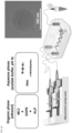
- LNVs of the present disclosure are pH-inducible structure-switching in that they are able to transition from a hexagonal phase at an acidic pH (e.g., lower than about 6) to a sponge phase at a neutral pH (e.g., about 6.5 or more) and vice-versa.
- the LNVs of the present disclosure comprise at least one ionizable cationic lipid, at least one phospholipid displaying a CPP > 1 and at least one non-ionic surfactant displaying a CPP value ⁇ 1, these components conferring to the LNVs their non-lamellar structure and their pH-inducible structure-switching properties.
- the sponge-based structure of LNVs enables a successful fusion with EVs, forming ssEVs displaying biological activity retention.
- the LNVs of the present disclosure are not vesicles.
- the LNVs' average size (d h ) is between about 20 nm and about 200 nm.
- ionizable cationic lipids i.e. lipids that switch their charge depending on the pH
- a negatively charged drug e.g., nucleic acids such as RNAs
- a pH-inducible structure-switching non-lamellar LNV together with the phospholipid having a CPP > 1 (e.g., 1,2-Dioleoyl- sn -glycero-3-phosphoethanolamine (DOPE)) and the non-ionic surfactant displaying a CPP value ⁇ 1 (e.g., KLP); and 3) to support the fusion stalk process with EVs.
- DOPE 1,2-Dioleoyl- sn -glycero-3-phosphoethanolamine
- ionizable cationic lipids of the present disclosure have an apparent acid dissociation constant (pKa) (prior to incorporation in LNVs) between 5 and 7.5 (or between 5.3 and 7.4; between 5.4 and 7.4; between 5.5 and 7.4; or between 5.6 and 7.4, or between 5.7 and 7.4, or between 5.8 and 7.4, or between 5.9 and 7.4, or between 6 and 7.4; or between 5.3 and 7.3; between 5.4 and 7.3; between 5.5 and 7.3; or between 5.6 and 7.3, or between 5.3 and 7.3, or between 5.8 and 7.3, or between 5.9 and 7.3, or between 6 and 7.3; or between 5.3 and 7.2; between 5.4 and 7.2; between 5.5 and 7.2; or between 5.6 and 7.2, or between 5.3 and 7.2, or between 5.8 and 7.2, or between 5.9 and 7.2, or between 6 and 7.2; or between 5.3 and 7.1; between 5.4 and 7.1; between 5.5 and 7.1; or between 5.6 and 7.2,
- ionizable lipids of the present disclosure are (DMA)butanoate ionizable cationic lipid derivatives.
- the pKa value of ionizable cationic lipids can be measured by commercial software such as that from ACD/Labs.
- phospholipids having a CPP > 1 e.g ., DOPE
- the function of phospholipids having a CPP > 1 (e.g ., DOPE) in the LNVs of the present invention is to promote the non-lamellar structures of LNVs and to contribute to the stability and delivery efficacy of nanoparticles.
- Phospholipids of the present disclosure are those displaying a critical packing parameter (CPP) value > 1 (i.e., an intrinsic curvature of ⁇ 0, inverse-cone shaped lipids) that usually promote non-lamellar phases (i.e. , hexagonal (HII) and/or sponge (L3) phases depending on the pH) in LNVs and subsequent fusion stalk in EVs.
- CPP critical packing parameter
- phospholipids having a CPP > 1 can be used in accordance with the present disclosure: phosphatidylethanolamine (PE)-based lipids having a CPP > 1, monogalactosyldiacylglycerol (MGDG)-based lipids having a CPP > 1, monoglucosyldiacylglycerol (MGIcDG)-based lipids having a CPP > 1, diphosphatidylglycerol (DPG)-based lipids having a CPP > 1, phosphatidylserine (PS)- and phosphatidic acid (PA)-based lipids having a CPP > 1.
- PE phosphatidylethanolamine
- MGDG monogalactosyldiacylglycerol
- MMIcDG monoglucosyldiacylglycerol
- DPG diphosphatidylglycerol
- PS phosphatidylserine
- PA phosphatidic acid
- the at least one phospholipid having a CPP > 1 used in LNVs of the present disclosure is a phosphatidylethanolamine (PE)-based lipid having a CPP value > 1, in a more specific embodiment it is DOPE.
- PE phosphatidylethanolamine
- non-ionic surfactant displaying a CPP value ⁇ 1 e.g., KLP
- the non-ionic surfactant acts as a membrane permeation enhancer.
- Non-ionic surfactants displaying an intrinsic curvature of > 0 and a CPP value ⁇ 1 can be used in LNVs of the present disclosure.
- polyethylene glycol esters of fatty acids e.g., polyethylene glycol 12-hydoxystearate (e.g ., a mixture of polyglycol mono- and di-esters of 12-hydroxystearic acid such as Kolliphor ® HS15 (KLP))
- polysorbates e.g., Tween ® 20 and 80
- polyethylene glycol aliphatic ethers such as those commonly known as Brij ® 35 and 58
- polyethylene glycol aliphatic esters such as PEG stearates
- sorbitans e.g., Span ® 60
- a polyethylene glycol esters of fatty acid such as the non-ionic surfactant KLP is used.
- the volume difference between head group and tail blocks can be thought of as a measure of the lipid spontaneous curvature.
- the spontaneous curvature of a monolayer is dictated by a packing of the lipids in the membrane, i.e., the interaction of a lipid with its surrounding molecules.
- the spontaneous curvature of a monolayer may slightly differ in a bilayer membrane and a monolayer at an oil-water interface because the oil chains may differently interdigitate into the hydrophobic part of the monolayer than in the case of a bilayer.
- Lipids with large head groups and small tails (cone-like shaped, positive curvature, CPP ⁇ 1) will prefer to self-assemble into convex structures, such as micelles, whereas lipids with comparable volumes of their head group and tail blocks (cylindrical lipids, CPP ⁇ 1) will form planar bilayers or large vesicles, and lipids with small head groups and large tails (inverted cone-like, negative curvature, CPP > 1) will yield inverted aggregates, such as the inverted hexagonal phase.
- Different approaches i.e., X-ray diffraction and osmotic stress
- These methods and techniques are disclosed in e.g., Sodt et al., 2013; Barragan Vidal et al., 2017; Rand et al., 1990; and Hamai et al., 2006.
- the LNV of the present disclosure comprises a molar concentration of about 30 % to about 70 % ( e.g., about 35 % to about 70 %, about 35 % to about 65 %, about 35 % to about 60 %, about 40 % to about 60 %, about 40 % to about 65 %, about 40 % to about 70 %) of the at least one ionizable cationic lipid; of about 10 % to about 50 % ( e .
- the molar concentration is selected so as to enable useful loading efficiencies of drug or diagnostic agent (e.g., nucleic acid) in the LNVs.
- drug or diagnostic agent e.g., nucleic acid
- the LNV of the present disclosure comprise only the at least one ionizable cationic lipid, the at least one phospholipid having a CPP > 1; and the at least one non-ionic surfactant displaying a CPP value ⁇ 1, the sum of the molar concentrations of each of these components amounts to 100%.
- Drug(s) as used herein refers to any pharmacologically active molecule or mixture of molecules (including non approved active pharmaceutical ingredients (APIs)). As used herein, mixtures of molecules display pharmacological activity. Without being so limited, biological extracts such as plant or animal extracts are encompassed by the term mixture of molecules, and thereby by the term drug.
- Drugs that can be incorporated into the device of the present disclosure can be low molecular weight APIs ( ⁇ 1000 Da) or large molecules (> 1000 Da) such as polysaccharides, peptides, proteins, nucleic acids (e.g., RNA (such as siRNA, mRNA, etc.), DNA), proteolysis-targeting chimeras (PROTACs), antibody-drug conjugates, and vaccine antigens.
- Diagnostic agents as used herein refer to any pharmaceutical product used as part of a diagnostic test, such as radiopharmaceuticals, contrast agents for use in imaging techniques including X-rays, magnetic resonance imaging (MRI) and ultrasound (US), compounds used in diagnostic tests that do not involve radioisotopes or imaging techniques, various stains/markers ( i.e., stains used in detection of malignant cells), antibodies ( i.e., monoclonal antibodies) and nucleic acid-based probes.
- the diagnostic agent is a negatively charged molecule (e.g., nucleic acid containing diagnostic agent) or a hydrophobic molecule.
- LNVs of the disclosure may be used to entrap drugs or diagnostic agents.
- the drug or diagnostic agent is a nucleic acid
- the proportion of ionizable cationic lipid in the LNV vs. the nucleic acid drug or diagnostic agent in the nucleic acid-loaded LNV of the present disclosure i.e., N/P ratio
- N/P ratio is ⁇ than about 1 ( e.g., ⁇ 2, ⁇ 3, ⁇ 4, ⁇ 5, between 6 and 300, or between 6 and 200, or between 6 and 150 or between 6 and 100 or between 6 and 50).
- LNVs are prepared by mixing an aqueous phase (e.g., saline, pure water) at an acidic pH and an organic medium (e.g., ethanol). Hydrophilic drugs will be dissolved in the aqueous phase prior to the entrapment into LNVs, whereas hydrophobic drugs will be dissolved in the organic phase prior to the loading into LNVs.
- aqueous phase e.g., saline, pure water
- organic medium e.g., ethanol
- the term "subject” is meant to refer to any animal, such as a mammal, including a human, dog, cat, pig, cow, monkey, cattle, horse, etc. In a particular embodiment, it refers to a human, in specific embodiments to an adult subject.
- LNVs of the present disclosure are prepared by mixing (i) an organic phase at least one ionizable cationic lipid, at least one phospholipid having a CPP > 1, at least one non-ionic surfactant displaying a CPP value ⁇ 1 dissolved in a solvent; and (ii) an aqueous phase.
- the aqueous-to-organic solvent volumetric flow rate ratio is between 0.5:1 and 5:1. In a more specific embodiment it is of 1.5:1.
- the hydrophobic drug (or diagnostic agent) When producing a hydrophobic drug (or diagnostic agent) loaded LNV, the hydrophobic drug (or diagnostic agent) is included in the organic phase, and when producing a hydrophilic drug (or diagnostic agent) loaded LNV (e.g., nucleic acid), the hydrophilic drug (or diagnostic agent) is included in the aqueous phase.
- a hydrophilic drug (or diagnostic agent) loaded LNV e.g., nucleic acid
- Mixing methods used to produce LNVs of the present disclosure include any microfluidic devices. Without being so limited, it includes any microfluidic chip designs, including micromixer, hydrodynamic flow focusing chip, T-junction chip, chaotic micromixer, bifurcating mixer and baffle mixer. In a particular embodiment, it is a chip formed by a Y-shaped inlet design. In specific embodiments, the mixing device used is a microfluidic device employing a high-precision syringe pump.
- the microfluidic device has a total flow rate between 0.001 mL/min and 100 mL/min, preferably, the flow rate is of about 0.05 mL/min; and/or the microfluidic device has a volumetric flow rate ratio of between 0.5:1 and 5:1; and/or the microfluidic device has a microfluidic channel depth of between 1 ⁇ m and 500 ⁇ m, preferably about 100 ⁇ m, channel inlets width between 1 ⁇ m and 500 ⁇ m, preferably about 100/200 ⁇ m, channel mixer width of between 1 ⁇ m and 500 ⁇ m, preferably about 200 ⁇ m and channel outlet width between 1 ⁇ m and 500 ⁇ m, preferably about 200 ⁇ m.
- Mixing methods also include ethanol injection, thin film evaporation, reversed-phase evaporation, high-pressure homogenization (HPH), high shear homogenization, sonication, microemulsion formation, and any other approach that leads to the production of lipid-based particles.
- HPH high-pressure homogenization
- HSP high shear homogenization
- sonication sonication
- microemulsion formation and any other approach that leads to the production of lipid-based particles.
- pH-inducible structure-switching non-lamellar LNVs of size 20 to 200 nm, in specific embodiments about ⁇ 100 nm, formed by hexagonal (HII) phases at acidic pH and sponge (L3) phases at neutral pH are produced ( FIGs. 1A , 1G-H , 2A ).
- the organic solvent used in the present disclosure is any biocompatible solvent in which the ionizable cationic lipid, and phospholipid having a CPP > 1 are soluble. Without being so limited, it includes alcohol (e.g., ethanol), oils (e.g., fluorinated), and chloroform. In specific embodiments, the organic phase solvent is ethanol.
- the concentration of organic solvent used is sufficient to dissolve the lipids. In specific embodiments, the concentration of lipids in the solvent is between 10 ⁇ g of lipids per mL of solvent to 1 g lipids per mL of solvent.
- the concentration of lipids in the solvent is between 1 mg of lipids per mL of solvent to 20 mg of lipids per mL of solvent., or between 5 mg of lipids per mL of solvent to 15 mg of lipids per mL of solvent, or between10 mg of lipids per mL of solvent to 15 mg of lipids per mL of solvent.
- aqueous medium is used in the production of the LNVs (aqueous phase) and in the hybridization step between the LNVs and the EVs.
- the aqueous medium used in the present disclosure is any aqueous medium, including pure water or saline.
- the aqueous medium may be of diverse compositions, pH and osmotic strength.
- the pH of the aqueous medium used in the production of LNVs may have a pH ranging from about 4 to about 6, e.g., 4, 4.5, 5, 5.5, 6, and preferably between about 4 and 6.
- sodium acetate buffer pH 4 is used.
- the pH may be achieved by adding buffer components (e.g., acetate buffer, sodium acetate, citrate buffer, sodium citrate or sodium citrate phosphate buffer), and/or an acid or base (e.g., hydrochloric acid and/or sodium hydroxide) for pH adjustments.
- buffer components e.g., acetate buffer, sodium acetate, citrate buffer, sodium citrate or sodium citrate phosphate buffer
- an acid or base e.g., hydrochloric acid and/or sodium hydroxide
- the pH of the aqueous medium may also be higher than 6 and up to 10.
- the aqueous solution may be neutralized to a pH 7.4 with e.g., a phosphate buffer (such as PBS) or HEPES-buffered saline (HEBS) or a Tris buffer.
- the aqueous solution containing the LNVs can be formulated for storage for future use or adapted for the step of hybridization with the EVs by increasing the pH to about 7 or more, and up to about 10. If the aqueous solution containing the LNVs has already been neutralized (e.g., with PBS), it may be more precisely adapted with an acid or base (e.g., hydrochloric acid and/or sodium hydroxide).
- an acid or base e.g., hydrochloric acid and/or sodium hydroxide
- the aqueous medium used for the hybridization step may further contain osmotic agents to achieve an isotonic solution with the EVs.
- EVs will generally have an osmolarity of 200-500 mmOsm/kg.
- Osmotic agents such as sodium chloride, glucose, gelatin, xylitol, sorbitol, mannitol, glucose polymer ( e.g., icodextrin), or amino acids may be used.
- the pH of the aqueous medium used in the hybridization step is higher than 6 and up to about 10, higher than 7, higher than 7.1, higher than 7.2, higher than 7.3, or between about 7.4 and about 10.
- EVs describes herein native EVs and synthetic or bioengineered EVs prepared to mimic native EVs (see e.g., exoIL-12 TM , exoSTING TM , exoASO TM -STAT6, AGLE-102 TM , ExoAAV, OmniSome, ILB-202)).
- Native EVs can be defined as heterogeneous populations of membranous vesicular structures originating from any eukaryotic or prokaryotic cell or tissue.
- small EVs ⁇ 200 nm
- medium or large EVs > 200 nm
- EVs further refers to a fraction of EVs as described above and/or to an extruded/homogenized form of native EVs and to an extruded form of whole cells.
- Extrusion/homogenization is conducted by any known method e.g., extrusion through nano-/micro-sized filter pores using high-pressure extruders or centrifugation setups, as well as mechanical homogenization/disruption of cells/EVs e.g., via sonication, nitrogen cavitation, freeze-thaw cycles or Dounce homogenizer.
- ssEVs of the present disclosure retain at least a portion of EV surface/membrane protein functionality, EV inherent bioactivity (e . g ., regenerative activity, anti-inflammatory activity, angiogenesis, cardioprotective, and homing/targeting properties ( i.e., ability to recognize specific tissues/cells).
- EV inherent bioactivity e. g ., regenerative activity, anti-inflammatory activity, angiogenesis, cardioprotective, and homing/targeting properties (i.e., ability to recognize specific tissues/cells).
- EVs may be isolated from biological samples with known methods such as centrifugation-based techniques (e.g., ultracentrifugation and density gradient centrifugation), affinity-based techniques (e.g., protein-based affinity (such as antibody capture or MagCapture TM kit) or membrane-based affinity (such as ExoEasy TM kit)), precipitation-based techniques (e.g., ExoQuick TM kit), microfluidic-based techniques, filtration-based techniques (e.g., tangential flow filtration and ultrafiltration), and size exclusion chromatography as described in Wu et al., 2023.
- centrifugation-based techniques e.g., ultracentrifugation and density gradient centrifugation
- affinity-based techniques e.g., protein-based affinity (such as antibody capture or MagCapture TM kit) or membrane-based affinity (such as ExoEasy TM kit)
- precipitation-based techniques e.g., ExoQuick TM kit
- biological sample from which EVs for use in the present disclosure may be isolated includes any cells and tissues derived from eukaryotes and prokaryotes. It includes cultured animal (mammalian such as human) cell line or primary cells belonging to any tissue/organ (e.g., lung, brain, pancreas, tumor/cancer, liver, bone marrow, blood, etc.). Any human/mammalian biofluid (e.g., blood, CSF, serum, urine, (breast) milk, saliva, synovial fluid, bile, ascites fluid, amniotic fluid semen, excrement). Any cultured bacterial strain (gram-positive and gram-negative). Any EV from fungi and plants.
- tissue/organ e.g., lung, brain, pancreas, tumor/cancer, liver, bone marrow, blood, etc.
- Any human/mammalian biofluid e.g., blood, CSF, serum, urine, (breast
- the LNVs are mixed (e.g., incubated) with EVs in an aqueous medium (see above, e.g., (PBS)) with the EVs at a temperature between 4 and 60 °C, and at a pH between larger than 6 and up to 10, between 6.1 and 10, between 6.2 and 10, between 6.3 and 10, between 6.4 and 10, between 6.5 and 10, between 6.6 and 10, between 6.7 and 10, between 6.8 and 10, between 6.9 and 10, between 7 and 10, between 7.1 and 10, between 7.2 and 10, between 7.3 and 10, between 7.4 and 10, between 7.5 and 10, between 6.1 and 9, between 6.2 and 9, between 6.3 and 9, between 6.4 and 9, between 6.5 and 9, between 6.6 and 9, between 6.7 and 9, between 6.8 and 9, between 6.9 and 9, between 7 and 9, between 7.1 and 9, between 7.2 and 9, between 7.3 and 9, between 7.4 and 9, and between 7.5 and 9.
- PBS aqueous medium
- the mixing is conducted for a duration of between 1 second and 48 hours (e.g., 5, 10, 15, 20, 30, 40, 50, 55 seconds; 1, 2, 3, 4, 5, 6, 10, 15, 20, 25, 30, 55 minutes, 1, 2, 3, 4, 5, 6, 7, 8, 9, 10, 11, 12, 13, 14, 15, 20, 24, 30, 36 or 42 hours, and any range thereof).
- the ssEVs are the result of the LNVs and EV fusion (see e.g., FIGs. 2C-E , 2G-I , FIG. 3E and 3G ). In specific embodiments they are produced by the method of the present disclosure. In other specific embodiments, they contain essentially all of the components, partially or completely mixed, of an LNV of the present disclosure and of EVs as described herein.
- the terms "essentially all of the components” is meant to denote that while a large majority (at least 70%, at least 75%, at least 80%, at least 90%, at least 95%, 96%, 97%, 98% or 99%) of the components of each of the LNV and of the EVs are in the ssEVs, they do not necessarily contain all of them ( e.g., because of the method of producing them).
- the terms “partially or completely mixed” is meant to encompass the ssEVs as it exists during all phases of the fusion (see e.g., FIGs. 2C-2E and 3F ).
- the average size of ssEVs in accordance with the present disclosure is between about 20 nm and 2000 nm (dependent on e.g., EV source, extrusion, etc.). In specific embodiments the average size is about 20 nm ⁇ 500 nm, 20 nm ⁇ 450 nm, 20 nm ⁇ 400 nm, 20 nm ⁇ 350 nm, 20 nm ⁇ 300 nm, 20 nm ⁇ 250 nm, about 50 nm ⁇ 250 nm, about 75 nm ⁇ 250nm, about 100 nm ⁇ 250 nm, about 125 nm ⁇ 250 nm, about 130 nm ⁇ 250 nm, about 140 nm ⁇ 250 nm, about 150 nm ⁇ 250 nm, about 160 nm ⁇ 250 nm, about 20 nm ⁇ 225 nm, about 50 nm ⁇ 225 nm, about 75 nm ⁇
- the present method allows for the in situ (i.e., at the point of care) production of ssEVs, before administration to a subject in need thereof.
- compositions include one or more of cryoprotectants (e.g., sucrose, dextrose, trehalose, or synthetic polymers such as poloxamers and polyvinylpyrrolidone (PVP), etc.), osmotic agents (e.g., dextrose, lactulose, sorbitol, magnesium hydroxide, magnesium citrate, sodium chloride, potassium chloride, polyethylene glycol (PEG), etc.), salt(s) (e.g., sodium phosphate, potassium phosphate, tris, sodium citrate, etc.).
- cryoprotectants e.g., sucrose, dextrose, trehalose, or synthetic polymers such as poloxamers and polyvinylpyrrolidone (PVP), etc.
- osmotic agents e.g., dextrose, lactulose, sorbitol, magnesium hydroxide, magnesium citrate, sodium chloride, potassium chloride, polyethylene glycol (PEG), etc.
- LNVs can be mixed with one or more pharmaceutically acceptable excipients to form a composition (liquid or solid) and e .g., stored for future use. For example, they may be freeze-dried in the presence of at least one of (i) one or more cryoprotectants (e.g., sucrose, dextrose, trehalose, or synthetic polymers such as poloxamers and PVP), (ii) one or more osmotic agents such as e.g., dextrose, PEG, lactulose, sorbitol, magnesium hydroxide, magnesium citrate, sodium chloride and potassium chloride), and (iii) one or more salt(s) (e.g., sodium phosphate, potassium phosphate, tris, sodium citrates) before resuspension with an aqueous medium, or stored as an aqueous suspension at a temperature ranging from - 80 to 8°C.
- cryoprotectants e.g., sucrose, dextrose,
- ssEVs can be mixed with one or more pharmaceutically acceptable excipients to form a composition (liquid or solid) and e.g., stored for future use.
- a composition liquid or solid
- they may be freeze-dried in the presence of cryoprotectants (e.g., sucrose, dextrose, trehalose, or synthetic polymers such as poloxamers and PVP) and salt compositions (e.g., sodium phosphate, potassium phosphate, tris, sodium citrates, sodium chloride and potassium chloride) before resuspension with an aqueous medium, or stored as an aqueous suspension at a temperature ranging from - 80 to + 8 °C.
- cryoprotectants e.g., sucrose, dextrose, trehalose, or synthetic polymers such as poloxamers and PVP
- salt compositions e.g., sodium phosphate, potassium phosphate, tris, sodium citrates, sodium chloride and potassium chloride
- polystyrene foams can also be embedded in polymer matrices (e.g., as gels, implants or devices) or classical pharmaceutical formulations (e.g., ointments, capsules, pre-filled syringes, etc.).
- polymer matrices e.g., as gels, implants or devices
- classical pharmaceutical formulations e.g., ointments, capsules, pre-filled syringes, etc.
- ssEVs may alternatively be formed in situ (at point of care location) before injection to a subject in need thereof.
- kits comprising (a) at least one LNV of the present disclosure or a composition (liquid or solid e.g., LNV powder ( e.g., lyophilized)) comprising at least one LNV and at least one pharmaceutically acceptable excipient; and (b) (i) a solution for hydrating the at least one LNV or composition thereof ( e.g., prior to its use), optionally with at least one pharmaceutically acceptable excipient; (ii) a drug or diagnostic agent (e.g., for loading in LNV or for use as control); (iii) a composition (liquid or solid) comprising EVs and at least one pharmaceutically acceptable excipient; (iv) instructions for using the at least one LNV or composition thereof (e.g., instructions for the production of ssEVs from LNVs and/or instructions for administration of ssEVs); or (v) a combination of at least two of (i) to (iv).
- a composition liquid or solid e.g., LNV powder (
- the kit comprises (a) at least one LNV (unloaded or loaded with drug or diagnostic agent) in a solid form (e.g., lyophilized) or a composition in a solid form ( e.g., lyophilized) comprising the at least one LNV and at least one pharmaceutically acceptable excipient (e.g., at least one cryoprotectant); and (b) (i) a solution for hydrating the at least one LNV or composition, the solution containing a least one pharmaceutically acceptable excipient (e.g., at least one of osmotic agent and salt, to provide isotonic environment to the LNVs).
- the kit further comprises (b) (ii) a drug or diagnostic agent (e.g., for loading in LNV or for use as control).
- kits comprising (a) at least one ssEV of the present disclosure, or a composition (liquid or solid) comprising the at least one ssEV and at least one pharmaceutically acceptable excipient; and (b) (i) a solution for hydrating the at least one ssEV or composition thereof (prior to its use); (ii) instructions for the use of these ingredients (e.g., instructions for administration of at least one ssEV or composition thereof); or (iii) a combination of (i) and (ii).
- Kits typically include a label indicating the intended use of the contents of the kit.
- the term label includes any writing, or recorded material supplied on or with the kit, or which otherwise accompanies the kit.
- the kit may further comprise one or more container(s), reagent(s), administration device(s).
- An example of such a kit comprises (i) a first composition (liquid or solid) comprising LNVs contained in a first container or in a first compartment of the first container; and (ii) a second composition (liquid or solid) comprising EVs and at least one pharmaceutically acceptable excipient in a second container or in a second compartment of the first container.
- the first and second compositions Prior to use, the first and second compositions could be mixed by allowing the content of the first and second compartments to be joined by e.g., breaking a wall separating these compartments. After the formation of the ssEVs in the mixture, the resulting solution can be administered to a subject in need thereof.
- the LNVs e.g., drug or diagnostic agent loaded
- the LNVs can be included in a commercial kit for use to produce the ssEVs.
- Such ssEVs can be used in in vitro and in vivo studies or for clinical ( e.g., therapeutic or diagnostic) purposes.
- the ssEVs of the present disclosure can be administered parenterally, topically or orally.
- the terms "subject” or “subject in need thereof” refer to a subject who would benefit from receiving an effective amount of the ssEVs or composition thereof of the present invention. It refers to an animal, a mammal and to a human in a specific embodiment.
- the ssEVs and compositions of the present invention may also be used for veterinary applications and be used in pets or other animals (e.g., pets such as cats, dogs, horses, etc.; and cattle, fishes, swine, poultry, etc.).
- LNVs and nanoparticles were prepared by mixing an organic phase comprising the components listed in Table I below at the molar concentrations (mol %) listed in Table I below, in an ethanol solvent with an aqueous phase comprising an acetate buffer at pH 4 (see FIG. 1A for LNVs of Examples 2-9).
- the method for making the nanoparticles of Examples 10-12 is as presented in FIG. 1A except that the organic phase contains the components listed in each of the corresponding columns of Table I.
- Table I Components of LNVs and nanoparticles used in Examples herein Components Concentration (mol %) MC3 40 50 60 60 60 DOPE 18 18 18 18 18 DOPC 18 KLP 42 32 22 22 22 22 DOTMA 60 MPPC 22 Examples Nos. 2 2 2-9 10 11 12
- the mixing was performed by introducing each phase in a microfluidic device through separate syringes (see FIG. 1A ).
- the aqueous phase-to-organic phase volumetric flow rate ratio was set to 1.5:1 and a high-precision syringe pump was used with a total flow rate of 0.05 mL/min.
- MWCO 20 kDa
- HEBS 20 mM HEPES, 150 mM NaCl
- siRNA-loaded LNVs were prepared as were the unloaded LNVs except that siRNA was added to the aqueous phase after pH adjustment.
- GFP-siRNA was custom synthesized by Microsynth AG (Switzerland).
- FAM-labeled siRNA MISSION ® siRNA Fluorescent Universal Negative Control, 6-FAM, Sigma-Aldrich, USA
- MW 13854 g/mol
- the control siRNA was custom synthesized by Microsynth AG (Switzerland).
- the LNVs were prepared as described above except that mRNA was added to the aqueous phase after pH adjustment.
- mRNA Firefly luciferasemRNA (FLuc-mRNA (TriLink BioTechnologies, USA); MW ⁇ 637715.0 g/mol or Cy-5-labeled firefly luciferase mRNA (Cy5-FLuc-mRNA (Cellerna Bioscience, Germany)); MW ⁇ 785980.0 g/mol) at the concentration of 0.25 ⁇ M to obtain an N/P ratio of 25.
- Nanoparticles were prepared by using the same protocol as LNVs, as described above, except that the organic phase contained DOPE and KLP at the molar concentrations of 48 % and 52 %.
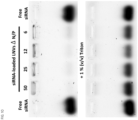
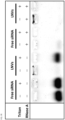
- EMSA experiments 0.1 ⁇ g of GFP-siRNA loaded in LNVs or free GFP-siRNA were mixed with or without 1 % (v/v) Triton TM X-100 and loaded on a 0.5 % (w/v) agarose gel. The gel was run for 30 min at 70 V in 0.5 ⁇ TAE buffer (Tris-base 20 mM, 10 mM acetic acid and 0.5 mM EDTA), stained for 45 min with SYBR TM Gold Nucleic Acid Gel Stain (Thermo Fischer Scientific, USA) and imaged with a ChemiDoc TM MP Imaging System (Bio-Rad, USA).
- EMSA experiments were performed after preincubation with 5 ⁇ g/mL RNase A for 1 h at 37 °C (with/without Triton TM X-100) to assess the protection of the siRNA cargo from nuclease-mediated degradation.
- the bone marrow-derived mesenchymal stem cell (MSC) line HS-5 was cultured on microcarriers in a 3D culture setup. Therefore, 118 ⁇ 10 6 HS-5 cells were seeded on 4.5 g of Corning TM Enhanced Attachment microcarriers (Corning, USA) in a total volume of 300 mL serum-containing DEMEM inside 500 mL Corning TM Reusable Glass Spinner Flasks (Corning, USA). An intermitted stirring protocol (5 min on at 30 rpm and 60 min off) was applied before the impeller speed was set to 35 rpm in continuous mode.
- Corning TM Enhanced Attachment microcarriers Corning, USA
- An intermitted stirring protocol (5 min on at 30 rpm and 60 min off) was applied before the impeller speed was set to 35 rpm in continuous mode.
- microcarriers were washed twice with PBS and 400 mL fresh, serum- and phenol red-free medium was added to the spinner flasks. Cell culture supernatant was harvested after 48 h of culture in serum-free medium.
- EVs were isolated from cell culture supernatant using a combination of centrifugation, microfiltration and ultracentrifugation steps. First, the supernatant was centrifuged at 500 and 2,000 ⁇ g for 5 min each and 10,000 ⁇ g for 20 min using a Sorvall TM LYNX 6000 centrifuge equipped with a Fiberlite TM F12-6 ⁇ 500 LEX Fixed Angle Rotor (both Thermo Fisher Scientific, USA).
- the clarified cell culture supernatant was then filtered through a 0.2- ⁇ m membrane before an ultracentrifugation step (70 min at 4 °C and 100,000 ⁇ g) was performed using an Optima TM XE-90 ultracentrifuge (UC) (Beckman Coulter, USA) and a Type 45 Ti Fixed-Angle Titanium Rotor. After the first ultracentrifugation run, EV pellets were washed with PBS or HEBS and pooled together. Another centrifugation step with identical conditions was performed and the EV pellet was suspended in ice-cold PBS/HEBS and stored at -20 °C.
- UC Optima TM XE-90 ultracentrifuge
- RNA-loaded LNVs i.e., siRNA and mRNA as described above
- MSC-derived EVs in an aqueous medium (either PBS or HEBS at pH 5, 6, 7.4 or 9) at different EV: LNV particle number ratios (4:1, 1:1 and 1:4), time points (5 and 30 min, 1 and 4 h), temperatures (4, 25 and 37°C) and pH values (5, 6, 7.4 and 9).
- the pH of the LNVs was carefully adjusted by the addition of hydrochloric acid and/or sodium hydroxide, if necessary, and checked with the pH meter. More particularly, a 62.5 mM hydrochloric acid or sodium hydroxide solution was used to respectively decrease or increase the pH. The pH of the samples was carefully checked each time using a pH electrode InLab Micro TM (Mettler Toledo, USA).
- Cell viability was determined using the CellTiter 96 ® AQueous One Solution Cell Proliferation Assay (MTS) (Promega, Switzerland).
- MTS CellTiter 96 ® AQueous One Solution Cell Proliferation Assay
- HeLa cells 2,500 cells per well in a total volume of 200 ⁇ L complete DMEM
- As a negative control cells received 2 ⁇ L PBS.
- ssEV, LNV or EV suspensions in PBS were added on glow-discharged (30 s in a Emitech TM K100X glow discharge system, Quorum Technologies Ltd., UK) 300-mesh lacey carbon-coated copper grids.
- a Vitrobot TM Mark II was used to vitrify the samples in a mixture of liquid ethane and propane.
- SAXS Small-angle X-ray scattering
- a volume of 20 ⁇ L of samples was carefully added to borosilicate glass capillary tubes.
- the capillary tubes with a length of 80 mm and a wall thickness of 0.01 mm, were loaded using hypodermic stainless-steel needles.
- the upper part of each capillary was meticulously closed using ultra-high vacuum 2-K-epoxy resin adhesive (1-part resin to 1-part hardener, v/v, UHU plus, Germany) over the course of two consecutive one-hour sessions. Subsequently, the capillaries were inserted into a specialized sample holder before being loaded into the instrument for further analysis.
- SAXS measurements were performed using an in-house SAXS setup (Xeuss TM 3.0, Xenocs, Grenoble, France) equipped with a Genix TM 3D source (Xenocs).
- the beam size on the sample was ca. 0.5 ⁇ 0.5 mm 2 FWHM.
- the sample to-detector distance, calibrated using a silver behenate standard, was 500 mm and the scattered signal was collected on a Dectris Eiger 1 M detector (1028 ⁇ 1062 pixels, pixel size 75 ⁇ 75 ⁇ m 2 Dectris Ltd., Baden, Switzerland).
- SAXS data are shown in the q-range from 0.016 to 0.69 ⁇ -1, corresponding to real length scales from 1.0466 to 39.25 nm.
- Detector data were masked, azimuthally integrated, normalized to absolute intensity (cm -1 ) and background-subtracted using XSACT (Xenocs) 2.6 software package.
- the hydrodynamic size (d h in nm) and zeta potential ( ⁇ -pot in mV) of LNVs were measured with DLS using a Malvern Zetasizer TM Advance Pro instrument (Malvern Panalytical, UK) operated at a scattering angle of 179 ° at 25 °C.
- DLS and SLS time course experiments were performed with a DynaPro TM Plate Reader III (Wyatt Technology, USA) under temperature-controlled conditions (4°C, 25 °C and 37 °C) in sealed 384-well microplates (Aurora Microplates, USA).
- the microplate was pre-incubated with 30 ⁇ L of LNV suspensions ( ⁇ 8 ⁇ 10 8 particles/mL) for ⁇ 30 min until a stable reading of the particle concentration was reached. An equal particle amount (except for ratio experiments) of EVs was then added and the measurement (particle concentration and d h ) was continued for a time course of 1 h.
- EVs were either subjected to a treatment with LNVs to form ssEVs of the present disclosure or transfected with the Exo-Fect TM Exosome Transfection Kit (System Biosciences, USA) prior to measuring the enzymatic activity.
- an absorbance-based malachite green assay was employed for measuring the activity of the EV membrane protein CD73. Therefore, 0.1 ⁇ g of EV proteins from the differently tested conditions (in HEBS, pH 7.4) were suspended in 60 ⁇ L of 10 mM Tris buffer (pH 7.4) and 30 ⁇ L of 800 ⁇ M AMP stock solution in 10 mM Tris buffer were added to reach a final concentration of 240 ⁇ M.
- the mixture was then incubated for 10 min at 25 °C and the enzymatic reaction was stopped by adding 40 ⁇ L color reagent (0.034 % (w/v) malachite green, 1.55 % (w/v) ammonium molybdate tetrahydrate, 0.0625 % (v/v) polysorbate 20) to the wells.
- the plate was incubated for 1 h at 25 °C and the absorbance was measured at 620 nm with the Infinite TM M200 plate reader.
- the background subtracted absorbance values were normalized to the values obtained from EVs that were subjected to identical hybridization conditions.
- DPPIV-Glo TM Protease Assay Kit for assessing the DPP4 enzymatic activity after hybridization, a commercially available DPPIV-Glo TM Protease Assay Kit (Promega, Switzerland) was used according to manufacturer instructions. Briefly, 0.08 ⁇ g of EV proteins in 25 uL of HEBS (pH 7.4) were mixed with an equal amount of DPPIV-Glo TM Reagent and incubated for 30 min at 25 °C. Luminescence was measured with an Infinite TM M200 Pro plate reader (Tecan, Switzerland) and the values were normalized with the values obtained from EVs that were subjected to identical hybridization conditions.
- EVs were labeled with either CellTrace TM Far Red (Thermo Fisher Scientific, USA) or with 5(6)-carboxyfluorescein diacetate N-succinimidyl ester (CFDA-SE) (Chemodex AG, Switzerland) dye according to manufacturer instructions with certain modifications. Briefly, EVs were mixed with 5 ⁇ M of the respective dyes and incubated overnight at 4 °C. Subsequently, the mixtures were incubated at 37 °C for 15 min to allow the dye to undergo acetate hydrolysis.
- CellTrace TM Far Red Thermo Fisher Scientific, USA
- CFDA-SE 5(6)-carboxyfluorescein diacetate N-succinimidyl ester
- Unbound dye was subsequently removed by dialysis against a 1000-fold access of PBS at 4 °C overnight using the Pur-A-Lyzer TM Mini 6000 Dialysis kit (Sigma-Aldrich, USA). Fluorescently labeled LNVs (FAM-siRNA, Cy5-FLuc-mRNA and Atto 488 DOPE) and labeled EVs where then mixed at a particle ratio of 1:1 at 4 °C or 37 °C (30 min), and ssEV formation was assessed using a Flow NanoAnalyzer TM N30 (NanoFCM Co., Ltd, UK) instrument.
- the blue 488 nm laser was set 10/50 mW and the red 638 nm laser was set 40/100 mW with a SS decay of 0.2 %.
- the 488 nm laser was changed to 20/50 mW and the 638 nm laser to 20/100 mW.
- Most samples were diluted 1 to 50 (v/v) with PBS before measurement except for the Exo-Fect TM transfected samples which were diluted 1 to 80 (v/v) with PBS.
- the samples were processed using the NanoFCM TM Professional Suite software (version 1.8, NanoFCM Co., Ltd, UK) and the data was analyzed with the FlowJo TM v10 software (BD Biosciences, USA)
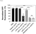
- 30,000 HeLa-GFP cells (Cell Biolabs, Inc., USA) were seeded per well inside a 24-well plate. After 24 h of incubation at 37 °C and 5 % CO 2 the cells were treated with LNVs, or ssEVs that were loaded with either the GFP-siRNA (siGFP) or a scrambled, control siRNA (siCTRL).
- LNVs LNVs
- siRNA siRNA
- Lipofectamine TM RNAiMAX Thermo Fisher Scientific, USA
- GFP-siRNA and naked GFP-siRNA served as a positive and negative control, respectively.
- 5,000 HeLa cells were seeded per well in a 96-well plate one day prior to transfection. After 24 h of incubation at 37 °C and 5 % CO 2 the culture medium was replaced with 90 ⁇ L of FBS. The cells were then treated with different concentrations of FLuc-mRNA-loaded LNVs and ssEVs. Lipofectamine TM MessengerMAX TM (Thermo Fisher Scientific, USA) with FLuc-mRNA and naked FLuc-mRNA served as positive and negative controls, respectively.
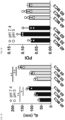
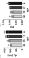
- the hydrodynamic diameter (d h ) of the unloaded LNVs and siRNA-loaded LNVs increased in LNVs containing a higher molar concentration of MC3 or prepared with a more alkaline pH ( FIG. 1B (unloaded LNVs), FIG. 1D (siRNA-loaded LNVs)).
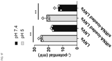
- the ⁇ -pot decreased in unloaded LNVs, and siRNA loaded LNVs prepared with a more alkaline pH ( FIG. 1F ).
- the polydispersity index (PDI) did not significantly vary with MC3 percentage or pH in the unloaded LNVs ( FIG. 1C (unloaded LNVs)) or the siRNA loaded LNVs ( FIG. 1E (siRNA-loaded LNVs)).
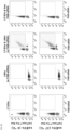
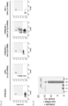
- FIG. 2G CellTrace TM Far Red (CT)-labeled EVs were mixed with either FAM-siRNA-loaded LNVs ( FIG. 2G ) or Atto 488 DOPE-labeled LNVs ( FIG. 2I ) using LNVs (60% MC3; N/P 25) at 37 °C and 4 °C, pH 7.4 for 30 min, in accordance with the method described in Example 1.
- FIGs. 2G-I The double positive population (in the square insert) appears upon mixing LNVs and EVs and confirm the formation of ssEVs. This population was not present when, for instance, only naked FAM-siRNA was added to EVs ( FIG.
- FIG. 2G shows that the loading of FAM-siRNA into ssEVs by using LNVs is significantly more efficient than that achieved with the commercial Exo-Fect TM Kit.
- FIGs. 2I shows that the double positive population appears also (in the square inset) when Atto 488 DOPE-labeled LNVs are mixed with CellTrace TM -labeled EVs, confirming the ssEV formation.
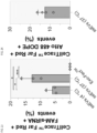
- FIG. 2K illustrates the percentage of the double positive ssEV population over the total particle population, hence the transfer of Atto 488 DOPE from LNVs to EVs, confirming the ssEV formation.
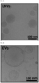
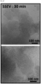
- LNVs spontaneously and instantaneously produced a fusion stalk in EVs during the hybridization step at pH 7.4 when the LNVs exist as sponges ( FIGs. 1H and 1L ), generating hybrid systems referred to herein as semi-synthetic extracellular vesicles (ssEVs) as shown in FIGs. 2A-E (number ratio siRNA-loaded LNVs to EVs of 4:1, LNVs having been prepared with a N/P ratio of 25 and 60 mol% MC3).
- the interaction was also confirmed by nanoparticle flow cytometry (NanoFCM) measurements ( FIGs. 2G-I ).
- nanoFCM nanoparticle flow cytometry
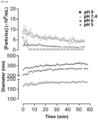
- FIG 2L shows that the different number ratios do not affect the ssEV size population and reaction kinetics as determined by an increase in the hydrodynamic diameter and the particle concentration over time (1 h).
- FIG. 2M shows that the velocity of the hybridization process depends on the temperature, being quicker at 37 °C than at 4 °C, whereas FIG. 2N shows that ssEV size increases when the pH is decreased. Further, at pH 5 the particle concentration could not be detected after 5 min, possibly due to presence of multiple particle species including larger aggregates.
- Unloaded LNVs and mRNA-loaded LNVs were prepared at pH values of 5 and 7.4 as described in Example 1 and used to generate ssEVs.
- the produced mRNA-loaded LNVs (N/P 25 and 60 mol % MC3) had a mean hydrodynamic diameter of ⁇ 130 nm ( FIG. 3A ), a low polydispersity index (PDI) value of about 0.11 ( FIG. 3B ).
- Data also shows that the ⁇ -pot of the LNVs slightly decreases once mRNA is loaded into the particles, both at acidic and neutral pH ( FIG. 3C ).
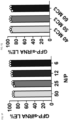
- mRNA was entrapped into LNVs with a loading efficiency (LE) of 94 % ⁇ 5 ( FIG. 3D ).
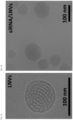
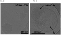
- the mRNA-loaded LNVs' structure is shown in cryo-TEM micrographs (number ratio mRNA-loaded LNVs : EVs of 4:1, LNVs having been prepared with a N/P ratio of 25 and 60 mol% MC3)( FIG. 3E ).
- FIG. 3F The loading of the mRNA in the ssEVs is confirmed in FIGs. 3G-H .
- CFSE-labeled EVs were incubated with either Cy5-FLuc-mRNA-loaded LNVs or with FLuc-mRNA-loaded LNVs.
- a double positive population appears upon mixing Cy5-mRNA-loaded LNVs and CFSE-labeled EVs and suggesting the formation of mRNA-loaded ssEVs. This population was not present when, for instance, only naked Cy5-FLuc-mRNA was added to EVs or when the unlabeled Fluc-mRNA was encapsulated within LNVs and mixed with unlabeled EVs.
- EXAMPLE 5 Impact on viability of HeLa cells treated with LNVs, and siRNA-loaded LNVs in an in vitro model
- HeLa cells were cultured at 37 °C and their viability was tested with the method described in Example 1.
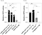
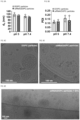
- the enzymatic activity of EV membrane proteins CD73 and DPP4 was determined as described in Example 1. It was shown not to be altered after hybridization with siRNA-loaded LNVs at 4 °C and 37 °C ( FIGs. 5A-B ). By contrast, at pH 5, the enzymatic activity of CD73 was significantly reduced after hybridization with siRNA-loaded LNVs ( FIG. 5A ). The Exo-Fect TM reagent also significantly reduced the enzymatic activity of both enzymes (CD73 and DPP4) after hybridization compared to ssEVs (4 °C and 37 °C at pH 7.4) ( FIGs. 5A-B ).
- EXAMPLE 7 Transfection efficacy of siRNA-loaded ssEVs in GFP-expressing HeLa cells
- Untreated cells that received only the medium
- PBS free GFP-siRNA represent the negative controls
- cells treated with Lipofectamine TM RNAiMAX with GFP-siRNA represent the positive control, respectively.
- the transfection efficacy of GFP-siRNA-loaded LNVs vs GFP-siRNA-loaded ssEVs was assessed in HeLa-GFP cells in the presence of ⁇ 8 % (v/v) FBS.
- EXAMPLE 8 Transfection efficacy of mRNA-loaded ssEVs in HeLa cells
- Nanostructures similar to multilamellar liposomes were produced with DOPE and KLP but without MC3, as shown in FIG. 8 .
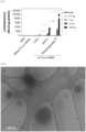
- Nanoparticles were produced with MC3, DOPC, a phospholipid displaying a CPP ⁇ 1 and KLP and their d h and PDI were evaluated with DLS at pH values of 5 and 7.4 ( FIGs. 9A and 9B ).
- Cryo-TEM images shows DOPC-based particles ( FIG. 9C ) and siRNA/DOPC-based particles ( FIG. 9D ) having a multivesicular structure.
- Such a result reveals the fundamental role of phospholipids displaying a CPP value > 1 (e.g., DOPE) in forming non-lamellar-based particles.
- FIG. 9E suggests that siRNA/DOPC-based nanoparticles do not form ssEVs with MSC-derived EVs.
- Nanoparticles were produced with MC3, DOPE and MPPC, a lysolipid with CPP ⁇ 1.
- the nanoparticles size distribution was evaluated with a DLS instrument at pH 7.4 ( FIG. 10 ). Particles with a broad size distribution comprising more than one particle population were detected, demonstrating the key role of the non-ionic surfactant (e.g., KLP) in forming particles with appropriate size and PDI values.
- the non-ionic surfactant e.g., KLP
- Nanoparticles were produced with DOTMA, a fixed cationic lipid, DOPE and KLP and their d h and PDI were evaluated with the DLS at pH 5 and 7.4 ( FIGs. 11A and 11B ).
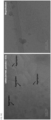
- Cryo-TEM images show that siRNA-loaded DOTMA particles form massive aggregates in the presence of MSC-derived EVs.
Landscapes
- Health & Medical Sciences (AREA)
- Chemical & Material Sciences (AREA)
- Engineering & Computer Science (AREA)
- Life Sciences & Earth Sciences (AREA)
- Bioinformatics & Cheminformatics (AREA)
- Animal Behavior & Ethology (AREA)
- Public Health (AREA)
- Veterinary Medicine (AREA)
- General Health & Medical Sciences (AREA)
- Medicinal Chemistry (AREA)
- Pharmacology & Pharmacy (AREA)
- Epidemiology (AREA)
- Physics & Mathematics (AREA)
- Biomedical Technology (AREA)
- Nanotechnology (AREA)
- Optics & Photonics (AREA)
- Oil, Petroleum & Natural Gas (AREA)
- Chemical Kinetics & Catalysis (AREA)
- General Chemical & Material Sciences (AREA)
- Molecular Biology (AREA)
- Biochemistry (AREA)
- Biophysics (AREA)
- Medicinal Preparation (AREA)
- Medicines Containing Antibodies Or Antigens For Use As Internal Diagnostic Agents (AREA)
Priority Applications (8)
| Application Number | Priority Date | Filing Date | Title |
|---|---|---|---|
| EP23195151.8A EP4516295A1 (fr) | 2023-09-04 | 2023-09-04 | Nanovecteurs lipidiques à commutation de structure inductible par le ph, vésicules extracellulaires semi-synthétiques, leurs procédés de fabrication et leurs utilisations |
| CN202480056120.5A CN121752257A (zh) | 2023-09-04 | 2024-09-03 | pH诱导的结构转换脂质纳米载体、半合成细胞外囊泡、其制备方法及应用 |
| EP24782351.1A EP4773946A1 (fr) | 2023-09-04 | 2024-09-03 | Nanovecteurs lipidiques à changement de structure inductibles par ph, vésicules extracellulaires semi-synthétiques, leurs procédés de fabrication et leurs utilisations |
| AU2024336703A AU2024336703A1 (en) | 2023-09-04 | 2024-09-03 | Ph-inducible structure-switching lipid nanovectors, semi-synthetic extracellular vesicles, methods of making same and uses thereof |
| KR1020267006640A KR20260066058A (ko) | 2023-09-04 | 2024-09-03 | pH-유도성 구조-전환 지질 나노벡터, 반합성 세포외 소포, 이의 제조 방법 및 이의 용도 |
| IL326493A IL326493A (en) | 2023-09-04 | 2024-09-03 | Ph-inducible structure-switching lipid nanovectors, semi-synthetic extracellular vesicles, methods of making same and uses thereof |
| PCT/IB2024/058555 WO2025052244A1 (fr) | 2023-09-04 | 2024-09-03 | Nanovecteurs lipidiques à changement de structure inductibles par ph, vésicules extracellulaires semi-synthétiques, leurs procédés de fabrication et leurs utilisations |
| MX2026002472A MX2026002472A (es) | 2023-09-04 | 2026-02-27 | Nanovectores lipidicos con conmutacion de estructura inducible por ph, vesiculas extracelulares semisinteticas, metodos para su elaboracion y usos de estos |
Applications Claiming Priority (1)
| Application Number | Priority Date | Filing Date | Title |
|---|---|---|---|
| EP23195151.8A EP4516295A1 (fr) | 2023-09-04 | 2023-09-04 | Nanovecteurs lipidiques à commutation de structure inductible par le ph, vésicules extracellulaires semi-synthétiques, leurs procédés de fabrication et leurs utilisations |
Publications (1)
| Publication Number | Publication Date |
|---|---|
| EP4516295A1 true EP4516295A1 (fr) | 2025-03-05 |
Family
ID=88017592
Family Applications (2)
| Application Number | Title | Priority Date | Filing Date |
|---|---|---|---|
| EP23195151.8A Withdrawn EP4516295A1 (fr) | 2023-09-04 | 2023-09-04 | Nanovecteurs lipidiques à commutation de structure inductible par le ph, vésicules extracellulaires semi-synthétiques, leurs procédés de fabrication et leurs utilisations |
| EP24782351.1A Pending EP4773946A1 (fr) | 2023-09-04 | 2024-09-03 | Nanovecteurs lipidiques à changement de structure inductibles par ph, vésicules extracellulaires semi-synthétiques, leurs procédés de fabrication et leurs utilisations |
Family Applications After (1)
| Application Number | Title | Priority Date | Filing Date |
|---|---|---|---|
| EP24782351.1A Pending EP4773946A1 (fr) | 2023-09-04 | 2024-09-03 | Nanovecteurs lipidiques à changement de structure inductibles par ph, vésicules extracellulaires semi-synthétiques, leurs procédés de fabrication et leurs utilisations |
Country Status (7)
| Country | Link |
|---|---|
| EP (2) | EP4516295A1 (fr) |
| KR (1) | KR20260066058A (fr) |
| CN (1) | CN121752257A (fr) |
| AU (1) | AU2024336703A1 (fr) |
| IL (1) | IL326493A (fr) |
| MX (1) | MX2026002472A (fr) |
| WO (1) | WO2025052244A1 (fr) |
Citations (5)
| Publication number | Priority date | Publication date | Assignee | Title |
|---|---|---|---|---|
| WO2014172045A1 (fr) * | 2013-03-15 | 2014-10-23 | The University Of British Columbia | Nanoparticules lipidiques pour la transfection et procédés associés |
| US20170151333A1 (en) | 2010-05-12 | 2017-06-01 | Protiva Biotherapeutics, Inc. | Novel cationic lipids and methods of use thereof |
| WO2018232357A1 (fr) * | 2017-06-15 | 2018-12-20 | Modernatx, Inc. | Formulations d'arn |
| WO2020061457A1 (fr) * | 2018-09-20 | 2020-03-26 | Modernatx, Inc. | Préparation de nanoparticules lipidiques et leurs méthodes d'administration |
| WO2022069577A1 (fr) * | 2020-09-29 | 2022-04-07 | Evox Therapeutics Ltd | Vésicules extracellulaires modifiées présentant une pharmacocinétique améliorée |
-
2023
- 2023-09-04 EP EP23195151.8A patent/EP4516295A1/fr not_active Withdrawn
-
2024
- 2024-09-03 CN CN202480056120.5A patent/CN121752257A/zh active Pending
- 2024-09-03 AU AU2024336703A patent/AU2024336703A1/en active Pending
- 2024-09-03 KR KR1020267006640A patent/KR20260066058A/ko active Pending
- 2024-09-03 IL IL326493A patent/IL326493A/en unknown
- 2024-09-03 EP EP24782351.1A patent/EP4773946A1/fr active Pending
- 2024-09-03 WO PCT/IB2024/058555 patent/WO2025052244A1/fr not_active Ceased
-
2026
- 2026-02-27 MX MX2026002472A patent/MX2026002472A/es unknown
Patent Citations (5)
| Publication number | Priority date | Publication date | Assignee | Title |
|---|---|---|---|---|
| US20170151333A1 (en) | 2010-05-12 | 2017-06-01 | Protiva Biotherapeutics, Inc. | Novel cationic lipids and methods of use thereof |
| WO2014172045A1 (fr) * | 2013-03-15 | 2014-10-23 | The University Of British Columbia | Nanoparticules lipidiques pour la transfection et procédés associés |
| WO2018232357A1 (fr) * | 2017-06-15 | 2018-12-20 | Modernatx, Inc. | Formulations d'arn |
| WO2020061457A1 (fr) * | 2018-09-20 | 2020-03-26 | Modernatx, Inc. | Préparation de nanoparticules lipidiques et leurs méthodes d'administration |
| WO2022069577A1 (fr) * | 2020-09-29 | 2022-04-07 | Evox Therapeutics Ltd | Vésicules extracellulaires modifiées présentant une pharmacocinétique améliorée |
Non-Patent Citations (10)
| Title |
|---|
| ALVAREZ-ERVITI, L., SEOW, Y., YIN, H.: "Delivery of siRNA to the mouse brain by systemic injection of targeted exosomes.", NAT. BIOTECHNOL., vol. 29, 2011, pages 341 - 345, XP055554900, DOI: 10.1038/nbt.1807 |
| BARRAGAN VIDAL, I.A., MULLER, M.: "Generalization of the swelling method to measure the intrinsic curvature of lipids", CHEM. PHYS., vol. 147, 2017, pages 224902 |
| HAMAI, C., YANG, T., KATAOKA, S., CREMER, P.S., MUSSER, S. M.: "Effect of Average Phospholipid Curvature on Supported Bilayer Formation on Glass by Vesicle Fusion. ", BIOPHYS. J., vol. 90, 2006, pages 1241 |
| JOSEPHA ROERIG ET AL: "Standardization Approaches for Extracellular Vesicle Loading with Oligonucleotides and Biologics", SMALL, WILEY, HOBOKEN, USA, vol. 19, no. 40, 7 June 2023 (2023-06-07), pages n/a, XP072511490, ISSN: 1613-6810, DOI: 10.1002/SMLL.202301763 * |
| LAMICHHANE, T. N., JEYARAM, A., PATEL, D. B.: "Oncogene Knockdown via Active Loading of Small RNAs into Extracellular Vesicles by Sonication.", CELL. MOL. BIOENG., vol. 9, 2016, pages 315 - 324 |
| O'BRIEN, K., BREYNE, K., UGHETTO, S., LAURENT, L.C.: "RNA delivery by extracellular vesicles in mammalian cells and its applications.", NAT. REV., vol. 21, 2020, pages 585 - 606, XP037249795, DOI: 10.1038/s41580-020-0251-y |
| RAND, R. P., FULLER, N. L., GRUNER S. M. AND PARSEGIAN, V. A.: "Membrane Curvature, Lipid Segregation, and Structural Transitions for Phospholipids under Dual-Solvent Stress,", BIOCHEMISTRY, vol. 29, 1990, pages 76 |
| SODT, J.PASTOR, R. W.: "Bending Free Energy from Simulation: Correspondence of Planar and Inverse Hexagonal Lipid Phases", BIOPHYS. J, vol. 104, no. 10, 2013, pages 2202 - 11, XP028550202, DOI: 10.1016/j.bpj.2013.03.048 |
| THERY ET AL.: "Minimal information for studies of extracellular vesicles", J EXTRACELLULAR VESICLES, vol. 7, no. 1, 2018, pages 1535750, XP055790152, DOI: 10.1080/20013078.2018.1535750 |
| WU ET AL.: "Advances in Extracellular Vesicle Nanotechnology for Precision Theranostics", ADV. SCI., vol. 10, 2023, pages 2204814 |
Also Published As
| Publication number | Publication date |
|---|---|
| KR20260066058A (ko) | 2026-05-12 |
| EP4773946A1 (fr) | 2026-07-15 |
| IL326493A (en) | 2026-04-01 |
| MX2026002472A (es) | 2026-05-04 |
| AU2024336703A1 (en) | 2026-04-16 |
| CN121752257A (zh) | 2026-03-27 |
| WO2025052244A1 (fr) | 2025-03-13 |
Similar Documents
| Publication | Publication Date | Title |
|---|---|---|
| Yang et al. | Chitosan mediated solid lipid nanoparticles for enhanced liver delivery of zedoary turmeric oil in vivo | |
| Leamon et al. | Folate-liposome-mediated antisense oligodeoxynucleotide targeting to cancer cells: evaluation in vitro and in vivo | |
| Giger et al. | Gene delivery with bisphosphonate-stabilized calcium phosphate nanoparticles | |
| Guarro et al. | Efficient extracellular vesicles freeze-dry method for direct formulations preparation and use | |
| Fu et al. | Tumor‐targeted paclitaxel delivery and enhanced penetration using tat‐decorated liposomes comprising redox‐responsive poly (ethylene glycol) | |
| Gaspar et al. | Targeted delivery of paromomycin in murine infectious diseases through association to nano lipid systems | |
| Khatri et al. | Development and characterization of siRNA lipoplexes: effect of different lipids, in vitro evaluation in cancerous cell lines and in vivo toxicity study | |
| US12384740B2 (en) | Cationic lipids and transfection methods | |
| US20210230636A1 (en) | Nucleic acid delivery carrier, nucleic acid delivery carrier set, nucleic acid delivery composition, and nucleic acid delivery method | |
| EP2460516B1 (fr) | Nanoparticules lipidiques pour thérapie génique | |
| KR20070037444A (ko) | 고체 미립자성 치료제의 생체외 적용방법 | |
| Tang et al. | Effect of size and pH-sensitivity of liposomes on cellular uptake pathways and pharmacokinetics of encapsulated gemcitabine | |
| Kuo et al. | Carmustine-loaded catanionic solid lipid nanoparticles with serotonergic 1B receptor subtype antagonist for in vitro targeted delivery to inhibit brain cancer growth | |
| KR101916941B1 (ko) | 플라스미드 디엔에이 전달용 고분자 나노입자 조성물 및 그의 제조방법 | |
| CN114848831A (zh) | 包裹型纳米制剂及其载体的制备方法和应用 | |
| EP2474306B1 (fr) | Composition particulaire et composition médicamenteuse comprenant celle-ci | |
| Streiber et al. | Extrahepatic Gene Editing In Vivo Using Organic Solvent‐Free Lipid Nanoparticles | |
| EP4691476A1 (fr) | Procédé de production de nanoparticules lipidiques encapsulées dans un acide nucléique | |
| WO2025052244A1 (fr) | Nanovecteurs lipidiques à changement de structure inductibles par ph, vésicules extracellulaires semi-synthétiques, leurs procédés de fabrication et leurs utilisations | |
| Qian et al. | Synthesis of novel cholesterol-based ionizable lipids for mRNA delivery | |
| EP4674431A1 (fr) | Nanoparticules lipidiques, composition pharmaceutique et procédé de production de nanoparticules lipidiques | |
| Riazanski et al. | Functional interaction of hybrid extracellular vesicle-liposome nanoparticles with target cells: absence of toxicity | |
| Jhan | Engineering Extracellular Vesicles for Gene-Modulating Combinatorial Therapies | |
| CA3287902A1 (fr) | Procédé de production de nanoparticules lipidiques encapsulées dans un acide nucléique | |
| CN121950934A (zh) | 一种核酸递送载体、组合物及其制备方法 |
Legal Events
| Date | Code | Title | Description |
|---|---|---|---|
| PUAI | Public reference made under article 153(3) epc to a published international application that has entered the european phase |
Free format text: ORIGINAL CODE: 0009012 |
|
| STAA | Information on the status of an ep patent application or granted ep patent |
Free format text: STATUS: THE APPLICATION HAS BEEN PUBLISHED |
|
| AK | Designated contracting states |
Kind code of ref document: A1 Designated state(s): AL AT BE BG CH CY CZ DE DK EE ES FI FR GB GR HR HU IE IS IT LI LT LU LV MC ME MK MT NL NO PL PT RO RS SE SI SK SM TR |
|
| STAA | Information on the status of an ep patent application or granted ep patent |
Free format text: STATUS: REQUEST FOR EXAMINATION WAS MADE |
|
| 17P | Request for examination filed |
Effective date: 20250905 |
|
| RAX | Requested extension states of the european patent have changed |
Extension state: BA Payment date: 20250905 |
|
| STAA | Information on the status of an ep patent application or granted ep patent |
Free format text: STATUS: THE APPLICATION IS DEEMED TO BE WITHDRAWN |
|
| 18D | Application deemed to be withdrawn |
Effective date: 20250906 |