EP4528353A2 - Système de mise au point automatique pour un dispositif de mesure ou de microscopie optique, procédé de réglage de mise au point dans un dispositif de mesure optique ou de microscopie et dispositif de mesure optique - Google Patents
Système de mise au point automatique pour un dispositif de mesure ou de microscopie optique, procédé de réglage de mise au point dans un dispositif de mesure optique ou de microscopie et dispositif de mesure optique Download PDFInfo
- Publication number
- EP4528353A2 EP4528353A2 EP25157090.9A EP25157090A EP4528353A2 EP 4528353 A2 EP4528353 A2 EP 4528353A2 EP 25157090 A EP25157090 A EP 25157090A EP 4528353 A2 EP4528353 A2 EP 4528353A2
- Authority
- EP
- European Patent Office
- Prior art keywords
- autofocus
- microscopy
- intensity
- specimen slide
- optical measuring
- Prior art date
- Legal status (The legal status is an assumption and is not a legal conclusion. Google has not performed a legal analysis and makes no representation as to the accuracy of the status listed.)
- Pending
Links
Images





Classifications
-
- G—PHYSICS
- G02—OPTICS
- G02B—OPTICAL ELEMENTS, SYSTEMS OR APPARATUS
- G02B21/00—Microscopes
- G02B21/24—Base structure
- G02B21/241—Devices for focusing
- G02B21/245—Devices for focusing using auxiliary sources, detectors
-
- G—PHYSICS
- G02—OPTICS
- G02B—OPTICAL ELEMENTS, SYSTEMS OR APPARATUS
- G02B21/00—Microscopes
- G02B21/24—Base structure
- G02B21/241—Devices for focusing
- G02B21/244—Devices for focusing using image analysis techniques
-
- G—PHYSICS
- G02—OPTICS
- G02B—OPTICAL ELEMENTS, SYSTEMS OR APPARATUS
- G02B21/00—Microscopes
- G02B21/06—Means for illuminating specimens
- G02B21/08—Condensers
- G02B21/082—Condensers for incident illumination only
-
- G—PHYSICS
- G02—OPTICS
- G02B—OPTICAL ELEMENTS, SYSTEMS OR APPARATUS
- G02B21/00—Microscopes
- G02B21/16—Microscopes adapted for ultraviolet illumination ; Fluorescence microscopes
-
- G—PHYSICS
- G02—OPTICS
- G02B—OPTICAL ELEMENTS, SYSTEMS OR APPARATUS
- G02B21/00—Microscopes
- G02B21/36—Microscopes arranged for photographic purposes or projection purposes or digital imaging or video purposes including associated control and data processing arrangements
Definitions
- the present invention relates to an autofocus system for an optical measuring or microscopy device, in particular a microscope or a microplate reader, in which a distance between an optical imaging unit of the optical measuring or microscopy device, in particular an objective lens, and a specimen slide is adjustable for focus adjustment.
- an autofocus system comprises an autofocus light source and an intensity detector that is spatially resolving in at least one direction.
- the autofocus light source generates an autofocus light beam that is coupled into the beam path of the optical measuring or microscopy device in such a way that the autofocus light beam passes through the optical imaging unit onto the specimen slide and creates a light spot there.
- the intensity detector of the autofocus system is arranged in such a way that a light component of the autofocus light beam reflected by the specimen slide and coupled out in the reverse direction from the beam path of the optical measuring or microscopy device strikes it and creates a reflected light pattern there. Furthermore, the autofocus system has an evaluation unit connected to the intensity detector, which generates an autofocus value from a shift of the reflected light pattern relative to a reference pattern determined by a reference measurement, wherein the autofocus value serves as a control signal for focus adjustment. Furthermore, the present invention relates to an optical measuring and microscopy system according to claim 14.
- a focus change can occur due to thermal influences or unevenness of the slide.
- the distance between the optical imaging unit of the microscope and the slide can change during the long-term measurement, which can lead to a loss of image sharpness.
- Microscopy images are altered and subsequent image processing and evaluation is difficult or even impossible.
- a microplate with 96 wells can be used as a slide, for example.
- a well is a sample well in the microplate into which the sample is introduced.
- a bottom of the microplate facing the optical imaging unit may have a production-related unevenness, which particularly affects neighboring wells. If microscopy images of samples are acquired in different wells during long-term measurements, a refocus adjustment may be necessary when switching from a first to a second well in order to obtain a microscopy image with high image sharpness for the second well.
- focus adjustment or “focusing” generally refers to the adjustment of the distance between the optical imaging unit and the slide, or between the optical imaging unit and a sample mounted on the slide, in order to improve the sharpness of the microscopic image.
- the distance is selected such that the microscopic image of the sample area under investigation has high image sharpness or that this sample area is clearly visible when viewed through a microscope eyepiece.
- the methods for automatic focus adjustment also referred to as autofocusing, can be divided into two categories: hardware-based and image-based autofocusing.
- image-based autofocusing microscopic images are captured with a microscope camera at different distances between the specimen slide and the optical imaging system. With an evaluation unit of the microscope, each microscopy image is processed and evaluated in order to determine the microscopy image with the highest image sharpness and from this the corresponding distance between the slide and the optical imaging unit is determined.
- Image-based autofocusing has the disadvantage that each individual microscopy image must first be processed and then evaluated by the evaluation unit. This image processing and evaluation is time-consuming. If autofocusing is required between each individual microscopy image of the long-term microscopic measurement in order to correct the focus change, then the time-consuming image-based autofocusing limits the maximum possible number of microscopy images per unit of time.
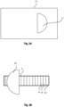
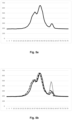
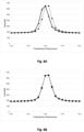
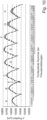
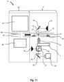
- the recalibration process works as follows: using image-based focus determination, a new focus point is determined for the test pattern, typically resulting in a changed Z position. Using the initially determined distance between the focus point for the test pattern and the user-specified Z position at which the original reference measurement was performed, a new Z position can be calculated from the new Z position for the focus point of the test pattern. A new reference measurement (Z position and intensity profile) is recorded at this new Z position, which is used for the hardware-based autofocus system until a subsequent recalibration.
- intensity detector 4 determines new reference patterns at specified intervals for the subsequent, much faster, hardware-based determination of the autofocus value.
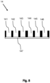
- the image-based determination of the focus setting is particularly robust, i.e., less prone to errors, due to the selected quality and durability of the test pattern.
- the test pattern allows for many other quality checks, including quantitative ones, in all areas of operation and the instrument's life cycle.
Landscapes
- Physics & Mathematics (AREA)
- Chemical & Material Sciences (AREA)
- Analytical Chemistry (AREA)
- General Physics & Mathematics (AREA)
- Optics & Photonics (AREA)
- Engineering & Computer Science (AREA)
- Computer Vision & Pattern Recognition (AREA)
- Automatic Focus Adjustment (AREA)
- Microscoopes, Condenser (AREA)
Applications Claiming Priority (2)
| Application Number | Priority Date | Filing Date | Title |
|---|---|---|---|
| DE102020122856 | 2020-09-01 | ||
| EP21193949.1A EP3961287B1 (fr) | 2020-09-01 | 2021-08-31 | Système de mise au point automatique pour un dispositif optique de mesure ou de microscopie, procédé de réglage de la mise au point dans un dispositif optique de mesure ou de microscopie, ainsi que dispositif optique de mesure ou de microscopie |
Related Parent Applications (1)
| Application Number | Title | Priority Date | Filing Date |
|---|---|---|---|
| EP21193949.1A Division EP3961287B1 (fr) | 2020-09-01 | 2021-08-31 | Système de mise au point automatique pour un dispositif optique de mesure ou de microscopie, procédé de réglage de la mise au point dans un dispositif optique de mesure ou de microscopie, ainsi que dispositif optique de mesure ou de microscopie |
Publications (2)
| Publication Number | Publication Date |
|---|---|
| EP4528353A2 true EP4528353A2 (fr) | 2025-03-26 |
| EP4528353A3 EP4528353A3 (fr) | 2025-09-17 |
Family
ID=77563952
Family Applications (2)
| Application Number | Title | Priority Date | Filing Date |
|---|---|---|---|
| EP25157090.9A Pending EP4528353A3 (fr) | 2020-09-01 | 2021-08-31 | Système de mise au point automatique pour un dispositif de mesure ou de microscopie optique, procédé de réglage de mise au point dans un dispositif de mesure optique ou de microscopie et dispositif de mesure optique |
| EP21193949.1A Active EP3961287B1 (fr) | 2020-09-01 | 2021-08-31 | Système de mise au point automatique pour un dispositif optique de mesure ou de microscopie, procédé de réglage de la mise au point dans un dispositif optique de mesure ou de microscopie, ainsi que dispositif optique de mesure ou de microscopie |
Family Applications After (1)
| Application Number | Title | Priority Date | Filing Date |
|---|---|---|---|
| EP21193949.1A Active EP3961287B1 (fr) | 2020-09-01 | 2021-08-31 | Système de mise au point automatique pour un dispositif optique de mesure ou de microscopie, procédé de réglage de la mise au point dans un dispositif optique de mesure ou de microscopie, ainsi que dispositif optique de mesure ou de microscopie |
Country Status (1)
| Country | Link |
|---|---|
| EP (2) | EP4528353A3 (fr) |
Families Citing this family (5)
| Publication number | Priority date | Publication date | Assignee | Title |
|---|---|---|---|---|
| DE102022125286A1 (de) * | 2022-09-30 | 2024-04-04 | Leica Microsystems Cms Gmbh | Vorrichtungen für ein Mikroskopsystem, Mikroskopsystem, Verfahren und Computerprogramm |
| CN115790377B (zh) * | 2022-11-29 | 2025-08-26 | 东莞市兆丰精密仪器有限公司 | 一种全自动影像仪的z轴归零位标定方法及测量系统 |
| CN116921882B (zh) * | 2023-08-01 | 2024-07-16 | 广州市博泰光学科技有限公司 | 一种高清镜片切割预加工系统及其加工方式 |
| CN118112777B (zh) * | 2024-04-30 | 2024-07-09 | 深圳市生强科技有限公司 | 基于病理玻片的多段交叉对焦处理方法及其应用 |
| CN119270491B (zh) * | 2024-11-22 | 2025-09-23 | 南京波长光电科技股份有限公司 | 一种适用于荧光显微镜的快速自动对焦模组及方法 |
Citations (2)
| Publication number | Priority date | Publication date | Assignee | Title |
|---|---|---|---|---|
| US20110317260A1 (en) | 2010-06-23 | 2011-12-29 | Leica Microsystems Cms Gmbh | Autofocusing Device for Microscopes and Suitable Autofocus Aperture Stops |
| US20170199367A1 (en) | 2014-06-23 | 2017-07-13 | Perkinelmer Cellular Technologies Germany Gmbh | Method for Autofocusing a Microscope at a Correct Autofocus Position in a Sample |
Family Cites Families (8)
| Publication number | Priority date | Publication date | Assignee | Title |
|---|---|---|---|---|
| US5932872A (en) * | 1994-07-01 | 1999-08-03 | Jeffrey H. Price | Autofocus system for scanning microscopy having a volume image formation |
| JP4370554B2 (ja) * | 2002-06-14 | 2009-11-25 | 株式会社ニコン | オートフォーカス装置およびオートフォーカス付き顕微鏡 |
| DE102011003807A1 (de) * | 2011-02-08 | 2012-08-09 | Leica Microsystems Cms Gmbh | Mikroskop mit Autofokuseinrichtung und Verfahren zur Autofokussierung bei Mikroskopen |
| EP2877834B1 (fr) * | 2012-07-25 | 2018-09-05 | Theranos, Inc. | Procédé d'analyse d'un échantillon |
| DE102014109687B4 (de) * | 2014-07-10 | 2020-03-19 | Carl Zeiss Microscopy Gmbh | Positionsbestimmung eines Objekts im Strahlengang einer optischen Vorrichtung |
| WO2017099700A1 (fr) * | 2015-12-06 | 2017-06-15 | Swanson Dorothy | Cuillère pour aliments marinés jetable |
| JP6659133B2 (ja) * | 2015-12-10 | 2020-03-04 | キヤノン株式会社 | スライドおよびスライドとカバーガラスのセット、並びに顕微鏡システム |
| DE102017115658A1 (de) * | 2017-07-12 | 2019-01-17 | Carl Zeiss Microscopy Gmbh | Flackern bei Winkel-variabler Beleuchtung |
-
2021
- 2021-08-31 EP EP25157090.9A patent/EP4528353A3/fr active Pending
- 2021-08-31 EP EP21193949.1A patent/EP3961287B1/fr active Active
Patent Citations (2)
| Publication number | Priority date | Publication date | Assignee | Title |
|---|---|---|---|---|
| US20110317260A1 (en) | 2010-06-23 | 2011-12-29 | Leica Microsystems Cms Gmbh | Autofocusing Device for Microscopes and Suitable Autofocus Aperture Stops |
| US20170199367A1 (en) | 2014-06-23 | 2017-07-13 | Perkinelmer Cellular Technologies Germany Gmbh | Method for Autofocusing a Microscope at a Correct Autofocus Position in a Sample |
Also Published As
| Publication number | Publication date |
|---|---|
| EP3961287A2 (fr) | 2022-03-02 |
| EP3961287A3 (fr) | 2022-06-29 |
| EP4528353A3 (fr) | 2025-09-17 |
| EP3961287C0 (fr) | 2025-02-12 |
| EP3961287B1 (fr) | 2025-02-12 |
Similar Documents
| Publication | Publication Date | Title |
|---|---|---|
| EP3961287B1 (fr) | Système de mise au point automatique pour un dispositif optique de mesure ou de microscopie, procédé de réglage de la mise au point dans un dispositif optique de mesure ou de microscopie, ainsi que dispositif optique de mesure ou de microscopie | |
| EP2219815B1 (fr) | Usinage par faisceau laser | |
| DE102016124191B4 (de) | Messvorrichtung optischer Eigenschaften und optisches System | |
| EP2406679B1 (fr) | Procédé d'autofocus et système d'autofocus | |
| EP3058414B1 (fr) | Microscope à balayage et procédé de détermination de la fonction d'étalement ponctuel d'un microscope à balayage | |
| DE102012223128B4 (de) | Autofokusverfahren für Mikroskop und Mikroskop mit Autofokuseinrichtung | |
| EP4325208B1 (fr) | Procédé et dispositif de détection de déplacements d'un échantillon par rapport à un objectif | |
| DE102013218795A1 (de) | Laserscanningmikroskop und Verfahren zur Korrektur von Abbildungsfehlern insbesondere in der hochauflösenden Scanning-Mikroskopie | |
| EP2038690A2 (fr) | Procédé et dispositif destinés à produire une image d'une couche mince d'un objet | |
| WO2019175441A1 (fr) | Procédé et appareil pour manipuler un trajet de faisceau dans un microscope, procédé et appareil pour enregistrer des piles d'images dans un microscope | |
| DE102011003807A1 (de) | Mikroskop mit Autofokuseinrichtung und Verfahren zur Autofokussierung bei Mikroskopen | |
| DE102017223014A1 (de) | Verfahren zur Bestimmung der Dicke einer Probenhalterung im Strahlengang eines Mikroskops | |
| EP0144732B1 (fr) | Dispositif pour mettre au point automatiquement des appareils optiques | |
| DE102015116452A1 (de) | Mikroskop | |
| DE102007038579A1 (de) | Mikroskop mit Innenfokussierung | |
| WO2021069743A1 (fr) | Procédé et dispositif de correction d'aberration dans un microscope à fluorescence | |
| DE102019109832B3 (de) | Lichtblattmikroskop und Verfahren zum Erfassen einer Messgröße | |
| WO2014147257A1 (fr) | Procédé par microscopie optique de localisation d'objets ponctuels | |
| DE102021104871A1 (de) | Verfahren und Vorrichtung zur lichtblattmikroskopischen Untersuchung einer Probe | |
| DE10333445B4 (de) | Konfokales Rastermikroskop | |
| DE102018126009B4 (de) | Verfahren und Mikroskop zur Bestimmung der Dicke eines Deck- oder Tragglases | |
| EP1543369B1 (fr) | Dispositif et procede pour l'analyse optique d'objet ou pour la detection ou l'analyse de couches | |
| DE10330716A1 (de) | Verfahren und Anordnung zur Eliminierung von Falschlicht | |
| DE202009003288U1 (de) | Autofokuseinrichtung | |
| CH672376A5 (fr) |
Legal Events
| Date | Code | Title | Description |
|---|---|---|---|
| PUAI | Public reference made under article 153(3) epc to a published international application that has entered the european phase |
Free format text: ORIGINAL CODE: 0009012 |
|
| STAA | Information on the status of an ep patent application or granted ep patent |
Free format text: STATUS: THE APPLICATION HAS BEEN PUBLISHED |
|
| AC | Divisional application: reference to earlier application |
Ref document number: 3961287 Country of ref document: EP Kind code of ref document: P |
|
| AK | Designated contracting states |
Kind code of ref document: A2 Designated state(s): AL AT BE BG CH CY CZ DE DK EE ES FI FR GB GR HR HU IE IS IT LI LT LU LV MC MK MT NL NO PL PT RO RS SE SI SK SM TR |
|
| REG | Reference to a national code |
Ref country code: DE Ref legal event code: R079 Free format text: PREVIOUS MAIN CLASS: G02B0021360000 Ipc: G02B0021240000 |
|
| RIC1 | Information provided on ipc code assigned before grant |
Ipc: G02B 21/16 20060101ALN20250522BHEP Ipc: G02B 21/36 20060101ALN20250522BHEP Ipc: G02B 21/08 20060101ALN20250522BHEP Ipc: G02B 21/24 20060101AFI20250522BHEP |
|
| PUAL | Search report despatched |
Free format text: ORIGINAL CODE: 0009013 |
|
| AK | Designated contracting states |
Kind code of ref document: A3 Designated state(s): AL AT BE BG CH CY CZ DE DK EE ES FI FR GB GR HR HU IE IS IT LI LT LU LV MC MK MT NL NO PL PT RO RS SE SI SK SM TR |
|
| RIC1 | Information provided on ipc code assigned before grant |
Ipc: G02B 21/24 20060101AFI20250814BHEP Ipc: G02B 21/08 20060101ALN20250814BHEP Ipc: G02B 21/36 20060101ALN20250814BHEP Ipc: G02B 21/16 20060101ALN20250814BHEP |
|
| STAA | Information on the status of an ep patent application or granted ep patent |
Free format text: STATUS: REQUEST FOR EXAMINATION WAS MADE |
|
| 17P | Request for examination filed |
Effective date: 20260317 |
|
| STAA | Information on the status of an ep patent application or granted ep patent |
Free format text: STATUS: EXAMINATION IS IN PROGRESS |