EP4529839A2 - Inserts de doigt pour dispositif d'imagerie de pliage d'ongles - Google Patents
Inserts de doigt pour dispositif d'imagerie de pliage d'ongles Download PDFInfo
- Publication number
- EP4529839A2 EP4529839A2 EP24218887.8A EP24218887A EP4529839A2 EP 4529839 A2 EP4529839 A2 EP 4529839A2 EP 24218887 A EP24218887 A EP 24218887A EP 4529839 A2 EP4529839 A2 EP 4529839A2
- Authority
- EP
- European Patent Office
- Prior art keywords
- finger
- nailfold
- insert
- wall
- wall portion
- Prior art date
- Legal status (The legal status is an assumption and is not a legal conclusion. Google has not performed a legal analysis and makes no representation as to the accuracy of the status listed.)
- Pending
Links
Images









Classifications
-
- A—HUMAN NECESSITIES
- A61—MEDICAL OR VETERINARY SCIENCE; HYGIENE
- A61B—DIAGNOSIS; SURGERY; IDENTIFICATION
- A61B5/00—Measuring for diagnostic purposes; Identification of persons
- A61B5/0059—Measuring for diagnostic purposes; Identification of persons using light, e.g. diagnosis by transillumination, diascopy, fluorescence
- A61B5/0077—Devices for viewing the surface of the body, e.g. camera, magnifying lens
-
- A—HUMAN NECESSITIES
- A61—MEDICAL OR VETERINARY SCIENCE; HYGIENE
- A61B—DIAGNOSIS; SURGERY; IDENTIFICATION
- A61B5/00—Measuring for diagnostic purposes; Identification of persons
- A61B5/0059—Measuring for diagnostic purposes; Identification of persons using light, e.g. diagnosis by transillumination, diascopy, fluorescence
- A61B5/0082—Measuring for diagnostic purposes; Identification of persons using light, e.g. diagnosis by transillumination, diascopy, fluorescence adapted for particular medical purposes
-
- A—HUMAN NECESSITIES
- A61—MEDICAL OR VETERINARY SCIENCE; HYGIENE
- A61B—DIAGNOSIS; SURGERY; IDENTIFICATION
- A61B5/00—Measuring for diagnostic purposes; Identification of persons
- A61B5/02—Detecting, measuring or recording for evaluating the cardiovascular system, e.g. pulse, heart rate, blood pressure or blood flow
- A61B5/021—Measuring pressure in heart or blood vessels
- A61B5/022—Measuring pressure in heart or blood vessels by applying pressure to close blood vessels, e.g. against the skin; Ophthalmodynamometers
- A61B5/02233—Occluders specially adapted therefor
- A61B5/02241—Occluders specially adapted therefor of small dimensions, e.g. adapted to fingers
-
- A—HUMAN NECESSITIES
- A61—MEDICAL OR VETERINARY SCIENCE; HYGIENE
- A61B—DIAGNOSIS; SURGERY; IDENTIFICATION
- A61B5/00—Measuring for diagnostic purposes; Identification of persons
- A61B5/02—Detecting, measuring or recording for evaluating the cardiovascular system, e.g. pulse, heart rate, blood pressure or blood flow
- A61B5/026—Measuring blood flow
- A61B5/0261—Measuring blood flow using optical means, e.g. infrared light
-
- A—HUMAN NECESSITIES
- A61—MEDICAL OR VETERINARY SCIENCE; HYGIENE
- A61B—DIAGNOSIS; SURGERY; IDENTIFICATION
- A61B5/00—Measuring for diagnostic purposes; Identification of persons
- A61B5/145—Measuring characteristics of blood in vivo, e.g. gas concentration or pH-value ; Measuring characteristics of body fluids or tissues, e.g. interstitial fluid or cerebral tissue
- A61B5/1455—Measuring characteristics of blood in vivo, e.g. gas concentration or pH-value ; Measuring characteristics of body fluids or tissues, e.g. interstitial fluid or cerebral tissue using optical sensors, e.g. spectral photometrical oximeters
-
- A—HUMAN NECESSITIES
- A61—MEDICAL OR VETERINARY SCIENCE; HYGIENE
- A61B—DIAGNOSIS; SURGERY; IDENTIFICATION
- A61B5/00—Measuring for diagnostic purposes; Identification of persons
- A61B5/44—Detecting, measuring or recording for evaluating the integumentary system, e.g. skin, hair or nails
- A61B5/449—Nail evaluation, e.g. for nail disorder diagnosis
-
- A—HUMAN NECESSITIES
- A61—MEDICAL OR VETERINARY SCIENCE; HYGIENE
- A61B—DIAGNOSIS; SURGERY; IDENTIFICATION
- A61B5/00—Measuring for diagnostic purposes; Identification of persons
- A61B5/68—Arrangements of detecting, measuring or recording means, e.g. sensors, in relation to patient
- A61B5/6801—Arrangements of detecting, measuring or recording means, e.g. sensors, in relation to patient specially adapted to be attached to or worn on the body surface
- A61B5/6813—Specially adapted to be attached to a specific body part
- A61B5/6825—Hand
- A61B5/6826—Finger
-
- A—HUMAN NECESSITIES
- A61—MEDICAL OR VETERINARY SCIENCE; HYGIENE
- A61B—DIAGNOSIS; SURGERY; IDENTIFICATION
- A61B5/00—Measuring for diagnostic purposes; Identification of persons
- A61B5/70—Means for positioning the patient in relation to the detecting, measuring or recording means
- A61B5/702—Posture restraints
-
- A—HUMAN NECESSITIES
- A61—MEDICAL OR VETERINARY SCIENCE; HYGIENE
- A61B—DIAGNOSIS; SURGERY; IDENTIFICATION
- A61B2562/00—Details of sensors; Constructional details of sensor housings or probes; Accessories for sensors
- A61B2562/14—Coupling media or elements to improve sensor contact with skin or tissue
- A61B2562/146—Coupling media or elements to improve sensor contact with skin or tissue for optical coupling
-
- A—HUMAN NECESSITIES
- A61—MEDICAL OR VETERINARY SCIENCE; HYGIENE
- A61B—DIAGNOSIS; SURGERY; IDENTIFICATION
- A61B2562/00—Details of sensors; Constructional details of sensor housings or probes; Accessories for sensors
- A61B2562/16—Details of sensor housings or probes; Details of structural supports for sensors
-
- A—HUMAN NECESSITIES
- A61—MEDICAL OR VETERINARY SCIENCE; HYGIENE
- A61B—DIAGNOSIS; SURGERY; IDENTIFICATION
- A61B2562/00—Details of sensors; Constructional details of sensor housings or probes; Accessories for sensors
- A61B2562/16—Details of sensor housings or probes; Details of structural supports for sensors
- A61B2562/164—Details of sensor housings or probes; Details of structural supports for sensors the sensor is mounted in or on a conformable substrate or carrier
-
- A—HUMAN NECESSITIES
- A61—MEDICAL OR VETERINARY SCIENCE; HYGIENE
- A61B—DIAGNOSIS; SURGERY; IDENTIFICATION
- A61B2562/00—Details of sensors; Constructional details of sensor housings or probes; Accessories for sensors
- A61B2562/16—Details of sensor housings or probes; Details of structural supports for sensors
- A61B2562/168—Fluid filled sensor housings
-
- A—HUMAN NECESSITIES
- A61—MEDICAL OR VETERINARY SCIENCE; HYGIENE
- A61B—DIAGNOSIS; SURGERY; IDENTIFICATION
- A61B5/00—Measuring for diagnostic purposes; Identification of persons
- A61B5/145—Measuring characteristics of blood in vivo, e.g. gas concentration or pH-value ; Measuring characteristics of body fluids or tissues, e.g. interstitial fluid or cerebral tissue
- A61B5/14546—Measuring characteristics of blood in vivo, e.g. gas concentration or pH-value ; Measuring characteristics of body fluids or tissues, e.g. interstitial fluid or cerebral tissue for measuring analytes not otherwise provided for, e.g. ions, cytochromes
-
- G—PHYSICS
- G06—COMPUTING OR CALCULATING; COUNTING
- G06V—IMAGE OR VIDEO RECOGNITION OR UNDERSTANDING
- G06V40/00—Recognition of biometric, human-related or animal-related patterns in image or video data
- G06V40/10—Human or animal bodies, e.g. vehicle occupants or pedestrians; Body parts, e.g. hands
- G06V40/12—Fingerprints or palmprints
- G06V40/1341—Sensing with light passing through the finger
Definitions
- WBCs White blood cells
- leukocytes or leucocytes are cells of the immune system that are involved in protecting the body against both infectious disease and foreign invaders.
- WBCs can exist not only in the blood, but also in the lymphatic system and tissues. Some conditions can trigger a response in the immune system and cause an increase in the number of WBCs (also referred to as WBC count). Other conditions can affect the production of WBCs by the bone marrow or the survival of existing WBCs in the circulation system. As these examples illustrate, various conditions can cause a change (either an increase or a decrease) of the number of circulating WBCs.
- WBC count can be a relevant physiological parameter for the diagnosis, monitoring, and/or treatment of various conditions including, but not limited to, bacterial and viral infections (e.g., pneumonia or meningitis), bone marrow functionality associated with chemotherapy toxicity, and hematologic proliferative processes such as leukemia.
- bacterial and viral infections e.g., pneumonia or meningitis
- bone marrow functionality associated with chemotherapy toxicity
- hematologic proliferative processes such as leukemia.
- the finger well 110 of the imaging device 100 accommodates the user's finger 120 in the imaging device 100 and contains optical immersion oil.
- the finger well 110 also includes a flat optical window 130, to allow for illumination and for time-lapse microscopic imaging of the nailfold region of the user's finger 120 through the optical window 130.
- the rigid housing that forms the finger well 110 can provide enough space to accommodate different finger sizes, but can leave a gap between the finger and the housing, allowing space for the finger to move easily.
- a finger insert for a finger imaging device includes a housing an opening to receive a finger of a subject.
- the housing defines a landing region abuts against a distal phalange of the finger of the subject when the finger is placed into the finger insert via the opening.
- the housing holds a liquid to facilitate imaging of a nailfold of the finger of the subject, such that at least the distal phalange of the finger is immersed in the liquid when the liquid is present in the finger insert and as the finger is placed into the finger insert via the opening.
- the housing includes a first wall and a second wall, with the second wall being optically transparent to facilitate imaging of the nailfold of the finger.
- the finger insert further includes a deformable pad positioned on at least a portion of the first wall, to form an open-pore structure that fills a gap between the first wall and the finger of the user when the finger is inserted into the finger insert, and to reduce trapped air in the liquid when the liquid is present in the finger insert, during insertion and movement of the finger in the finger insert
- a system includes a finger imaging device including a light source, a detector, and a receptacle including an imaging window.
- the light source and the detector are optically coupled to the imaging window.
- the system also includes a finger insert, the finger insert being disposable in the receptacle.
- the finger insert includes a housing defining an opening an opening to receive a finger of a subject, and further defining a landing region that abuts against a distal phalange of the finger of the subject when the finger is placed into the finger insert via the opening, to hold a liquid to facilitate imaging of a nailfold of the finger of the subject. At least the distal phalange of the finger is immersed in the liquid when the liquid is present in the finger insert and as the finger is placed into the finger insert via the opening.
- the housing includes a first wall and a second wall, the second wall being optically transparent to facilitate imaging of the nailfold of the finger via the light source and detector.
- the finger insert further includes a deformable pad positioned on at least a portion of the first wall, to form an open-pore structure that fills a gap between the first wall and the finger of the user when the finger is inserted into the finger insert, and to reduce trapped air in the liquid when the liquid is present in the finger insert, during insertion and movement of the finger in the finger insert.
- a kit includes a finger imaging device including a light source, a detector, and a receptacle including an imaging window. The light source and the detector are optically coupled to the imaging window.
- the kit also includes a set of finger inserts, each finger insert of the set of finger inserts being disposable in the receptacle such that at least a section of the second wall of that finger insert is in optical communication with the imaging window when that finger insert is disposed in the receptacle.
- a first finger insert of the set of finger inserts is different from a finger insert apparatus of the set of finger inserts in one or more of a length of the housing along its longitudinal axis, and an average cross-sectional area of a curved portion of the first wall.
- a method includes receiving a finger of a user in a finger insert disposed in a finger imaging device.
- the finger insert includes a housing defining an opening an opening to receive a finger of a subject, and further defining a landing region abuts against a distal phalange of the finger of the subject when the finger is placed into the finger insert via the opening, to hold a liquid to facilitate imaging of a nailfold of the finger of the subject.
- At least the distal phalange of the finger is immersed in the liquid when the liquid is present in the finger insert and as the finger is placed into the finger insert via the opening.
- the housing including a first wall and a second wall, the second wall being optically transparent to facilitate imaging of the nailfold of the finger.
- the finger insert also includes a deformable pad positioned on at least a portion of the first wall, to form an open-pore structure that fills a gap between the first wall and the finger of the user when the finger is inserted into the finger insert, and to reduce trapped air in the liquid when the liquid is present in the finger insert, during insertion and movement of the finger in the finger insert.
- the method further includes imaging a nailfold portion of the finger via the wall portion of the finger insert using the finger imaging device.
- a finger insert as described herein can include and/or encompass a removable piece that is inserted into a nailfold imaging device before a measurement (e.g., imaging, video recording) starts, and is removed afterwards.
- the finger insert may be single-use and disposable, or reusable.
- the finger insert can be designed to engage with the nailfold imaging device ergonomically and securely, such that it provides a sturdy yet comfortable support for the finger while in use.
- the finger insert can include an incorporated optical window to ensure that the optical path between the imaging and illumination optics is clean and transparent each time.
- Immersion oil can be pre-filled into the finger insert to prevent reuse and contamination of immersion oils with particulates.
- the insert accommodates different finger sizes through variable internal geometry, with different sized inserts available (e.g., small-long, small-short, medium-long, medium-short, large-long, large-short).
- the finger insert includes one or more flexible spacers to effectively and comfortably fill a gap that may be present between the rigid body of the finger insert and the finger of the subject.
- the flexible spacers may be implemented as rubber cylinders extruded from (and extending from) the walls of the body of the finger insert.
- the open-pore structure of the rubber cylinders significantly mitigates the trapping of air which would otherwise occur with semi-closed-pore structures like sponges, that would produce air bubbles in the immersion oil under the typical compression that occurs during inserting and movement of the finger.
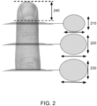
- FIG. 2 illustrates aspects of finger geometry that can be considered when designing inventive finger inserts according to the present disclosure to accommodate various finger sizes.
- cross-sectional area and shape along the length of the finger e.g., see the cross-sectional profiles 210, 220, 230 along the length of the finger
- Excessive force on the finger can result in restriction of blood flow (that could in turn impede measurement made on the nailfold capillaries of that finger), and too little support or restriction would reduce the necessary constraint for minimizing involuntary movement.
- a second example aspect can be the length of the finger from fingertip to nailfold (e.g., the length 240), which is typically the same or similar to the fingernail length of that finger.
- the finger insert is designed such that the fingertip registers with the bottom of the insert, so that the distance between the end of the finger and the nailfold affects how high the nailfold sits in the well of the finger insert. This can be significant since the nailfold would need to fall within a region where the nailfold imaging device is able to image the nailfold region.
- FIG. 3 illustrates an example finger insert 310.
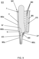
- the insert 310 can include a housing 312, a plate 330 (that may or may not be integrally formed with the housing), and multiple spacers 340 that are deformable by a user's finger when inserted into the insert 310.
- the housing 312 and the plate 330 can be made from a substantially rigid, inelastic material such as a transparent thermoplastic (e.g. Poly(methyl methacrylate), or PMMA).
- the spacers can be made from an elastic, deformable material such as a silicone. In some cases, the material of the spacers can be optically transparent, while it can be absorbing in some other cases.
- the housing can include a top end 314 that has an opening to receive the user's finger, and a bottom end 316.
- a body 318 of the housing 312 is disposed and/or otherwise formed between the top end 314 and the bottom end 316, and can hold an immersion oil.
- the immersion oil can be selected to have a refractive index (e.g., a RI of about 1.51) that is similar to that of the housing and/or the dermis of the finger, to facilitate the nailfold imaging.
- the housing 312 can be sized to hold enough immersion oil such that at least the distal phalange of the finger is fully immersed in it.
- the housing 312 can include a curved portion 320 and a wall portion 322.
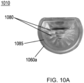
- the wall portion 322 can be optically transparent and substantially flat to prevent spurious reflections that can arise due to the illumination. In some cases the wall portion 322 can be curved, or another suitable form to conform to the nailfold imaging device during use.
- the curved portion 320 can have a cross-sectional area (CSA) that (in at least a portion of the curved portion 320) continuously or discontinuously changes from the top end 314 to the bottom end 316. As explained above in connection with FIG. 2 , such a CSA profile can accommodate for the typical changes in CSA of a user's finger along its length, towards the nailfold region.
- the curved portion 320 can be optically transparent, or absorbing to prevent reflection of a light beam from the light source of the nailfold imaging device.
- the CSA can vary from about 3 cm 2 to about 1 cm 2 from the top end 314 to the bottom end 316.
- the depth of the housing from the top end 314 to the bottom end 316 can vary from about 10 mm to about 20 mm.
- the plate 330 is disposed, attached, coupled and/or otherwise present at the bottom end 316 of the housing 312 and can abut against a distal phalange of the finger of the subject to position the finger for imaging.
- the plate 330 and the bottom end 316 can form a fluid-tight seal to prevent the immersion oil from leaking.
- multiple, deformable spacers 340 can be positioned, attached, formed, and/or otherwise disposed on the curved portion 320.
- the number, size, shape, and/or other aspects of the spacers 340 can be useful for forming an open-pore structure that can effectively and significantly fill a gap between the curved portion 320 and the finger of the user when inserted.
- the spacers 340 can be designed to accommodate a variety of finger geometries, with the goal of providing support to minimize unintentional movement during the measurement.
- the open-pore structure of the spacers 340 even when deformed due to the pressure from the user's finger, significantly reduces or eliminates trapped air in the immersion oil when the user's finger is inserted into the insert, or moved around within the insert.
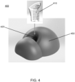
- FIG. 4 illustrates a system 400 that includes a finger insert 410, which can be structurally and/or functionally similar to the insert 310.
- the system also includes a nailfold imaging device 450, which can be similar to such devices disclosed in the related U.S. patent publication nos. 2016/0148038 and 2019/0139221 , the disclosures of which are incorporated herein by reference in their entirety.
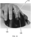
- the insert 410 is inserted into a receptacle 420 of the device 450 to achieve a firm mating to allow for rigid mechanical coupling of the insert 410 and device 450, and to achieve a stable optical alignment for imaging the nailfold region of a finger in the insert 410.
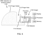
- FIG. 5 generally illustrates an example nailfold imaging device 1100 (also referred to as a "WBC detection and analysis system") that includes a finger holder 1110 with a receptacle 1112 (also referred to as a "finger hold").
- An imaging window 1114 is formed within the receptacle 1112 can be in optical communication with the interior of the insert 410 via its wall portion to permit nailfold imaging of the user's finger in the inert.
- the device 1100 can include an imager/imaging setup 1120 that includes a light source (not shown) to illuminate the user's nailfold region within the insert 410 via the window 1114.
- the imager 1120 includes a focusing optic 1122 to collect light reflected or scattered from the finger and detector 1124 to receive the reflected or scattered light so as to form images of the finger.
- the device 1100 further includes a processor 1130 operably coupled to the imager 1120 and a memory 1140 operably coupled to the processor 1130.
- the memory 1140 is encoded with processor-executable instructions, which, when executed by processor 1130, may perform the methods described in the '038 and/or the '221 publications to analyze images received from the imager 1120.
- the device 1100 also includes a display 1150, which can display the images or videos taken by the imager 1120 and/or data associated with WBC events detected by the processor 1130.
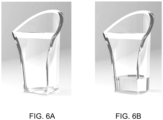
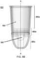
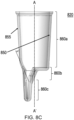
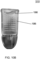
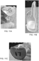
- FIG. 6A shows the insert 310 without the spacers and without any immersion oil, and illustrates the optical clarity of the insert 310.
- FIG. 6B illustrates the insert of FIG. 3A with immersion oil added.
Landscapes
- Health & Medical Sciences (AREA)
- Life Sciences & Earth Sciences (AREA)
- Physics & Mathematics (AREA)
- Heart & Thoracic Surgery (AREA)
- Molecular Biology (AREA)
- Veterinary Medicine (AREA)
- Public Health (AREA)
- General Health & Medical Sciences (AREA)
- Biophysics (AREA)
- Pathology (AREA)
- Engineering & Computer Science (AREA)
- Biomedical Technology (AREA)
- Animal Behavior & Ethology (AREA)
- Medical Informatics (AREA)
- Surgery (AREA)
- Cardiology (AREA)
- Vascular Medicine (AREA)
- Physiology (AREA)
- Dermatology (AREA)
- Physical Education & Sports Medicine (AREA)
- Dentistry (AREA)
- Ophthalmology & Optometry (AREA)
- Hematology (AREA)
- Spectroscopy & Molecular Physics (AREA)
- Optics & Photonics (AREA)
- Measurement Of The Respiration, Hearing Ability, Form, And Blood Characteristics Of Living Organisms (AREA)
- Measuring Pulse, Heart Rate, Blood Pressure Or Blood Flow (AREA)
Applications Claiming Priority (3)
| Application Number | Priority Date | Filing Date | Title |
|---|---|---|---|
| US201962878011P | 2019-07-24 | 2019-07-24 | |
| EP20843851.5A EP4003167B1 (fr) | 2019-07-24 | 2020-05-26 | Inserts pour doigt pour un dispositif d'imagerie du sillon latéral de l'ongle |
| PCT/US2020/034483 WO2021015843A1 (fr) | 2019-07-24 | 2020-05-26 | Inserts pour doigt pour un dispositif d'imagerie du sillon latéral de l'ongle |
Related Parent Applications (1)
| Application Number | Title | Priority Date | Filing Date |
|---|---|---|---|
| EP20843851.5A Division EP4003167B1 (fr) | 2019-07-24 | 2020-05-26 | Inserts pour doigt pour un dispositif d'imagerie du sillon latéral de l'ongle |
Publications (2)
| Publication Number | Publication Date |
|---|---|
| EP4529839A2 true EP4529839A2 (fr) | 2025-04-02 |
| EP4529839A3 EP4529839A3 (fr) | 2025-04-16 |
Family
ID=74189551
Family Applications (2)
| Application Number | Title | Priority Date | Filing Date |
|---|---|---|---|
| EP24218887.8A Pending EP4529839A3 (fr) | 2019-07-24 | 2020-05-26 | Inserts de doigt pour dispositif d'imagerie de pliage d'ongles |
| EP20843851.5A Active EP4003167B1 (fr) | 2019-07-24 | 2020-05-26 | Inserts pour doigt pour un dispositif d'imagerie du sillon latéral de l'ongle |
Family Applications After (1)
| Application Number | Title | Priority Date | Filing Date |
|---|---|---|---|
| EP20843851.5A Active EP4003167B1 (fr) | 2019-07-24 | 2020-05-26 | Inserts pour doigt pour un dispositif d'imagerie du sillon latéral de l'ongle |
Country Status (4)
| Country | Link |
|---|---|
| US (3) | US11160492B2 (fr) |
| EP (2) | EP4529839A3 (fr) |
| CN (2) | CN116211296A (fr) |
| WO (1) | WO2021015843A1 (fr) |
Families Citing this family (5)
| Publication number | Priority date | Publication date | Assignee | Title |
|---|---|---|---|---|
| EP3697294B1 (fr) | 2017-10-16 | 2023-12-06 | Massachusetts Institute Of Technology | Système et procédé de mesures hématologiques non invasives |
| EP4529839A3 (fr) | 2019-07-24 | 2025-04-16 | Massachusetts Institute of Technology | Inserts de doigt pour dispositif d'imagerie de pliage d'ongles |
| WO2021242983A1 (fr) | 2020-05-28 | 2021-12-02 | Leuko Labs, Inc. | Procédé de détection de globules blancs et/ou de sous-types de globules blancs à partir de vidéos capillaires non invasives |
| JP7828109B2 (ja) | 2021-08-02 | 2026-03-11 | レウコ・ラボズ・インコーポレイテッド | 毛細血管床における1つまたは複数の毛細血管の画像を取得するための自動化システムおよび方法 |
| CN119014823B (zh) * | 2024-10-28 | 2025-01-24 | 达州市中心医院(达州市人民医院) | 用于甲襞微循环成像的环形光源检测与调整方法和系统 |
Citations (2)
| Publication number | Priority date | Publication date | Assignee | Title |
|---|---|---|---|---|
| US20160148038A1 (en) | 2014-11-24 | 2016-05-26 | Massachusetts Institute Of Technology | Systems, apparatus, and methods for analyzing blood cell dynamics |
| US20190139221A1 (en) | 2017-10-16 | 2019-05-09 | Carlos CASTRO-GONZALEZ | Systems, devices and methods for non-invasive hematological measurements |
Family Cites Families (116)
| Publication number | Priority date | Publication date | Assignee | Title |
|---|---|---|---|---|
| US534211A (en) * | 1895-02-12 | Charles h | ||
| US2588528A (en) * | 1948-07-27 | 1952-03-11 | California Thimble Corp | Finger shaped thimble |
| US2548378A (en) * | 1949-12-06 | 1951-04-10 | Ewald H Kleinfeld | Finger splint |
| US4425924A (en) | 1981-09-29 | 1984-01-17 | Riva Charles E | Blood velocity measurement in retinal capillaries utilizing the blue field entoptic phenomenon |
| US4998533A (en) | 1986-07-15 | 1991-03-12 | Winkelman James W | Apparatus and method for in vivo analysis of red and white blood cell indices |
| US4694843A (en) * | 1986-10-07 | 1987-09-22 | Casenhiser Elaine J | Fingertip cover |
| US5596987A (en) | 1988-11-02 | 1997-01-28 | Noninvasive Technology, Inc. | Optical coupler for in vivo examination of biological tissue |
| US5365066A (en) | 1989-01-19 | 1994-11-15 | Futrex, Inc. | Low cost means for increasing measurement sensitivity in LED/IRED near-infrared instruments |
| US5077476A (en) | 1990-06-27 | 1991-12-31 | Futrex, Inc. | Instrument for non-invasive measurement of blood glucose |
| US5218207A (en) | 1989-01-19 | 1993-06-08 | Futrex, Inc. | Using led harmonic wavelengths for near-infrared quantitative |
| US5068536A (en) | 1989-01-19 | 1991-11-26 | Futrex, Inc. | Method for providing custom calibration for near infrared instruments for measurement of blood glucose |
| US5237178A (en) | 1990-06-27 | 1993-08-17 | Rosenthal Robert D | Non-invasive near-infrared quantitative measurement instrument |
| US5204532A (en) | 1989-01-19 | 1993-04-20 | Futrex, Inc. | Method for providing general calibration for near infrared instruments for measurement of blood glucose |
| US5086229A (en) | 1989-01-19 | 1992-02-04 | Futrex, Inc. | Non-invasive measurement of blood glucose |
| US5362966A (en) | 1990-06-27 | 1994-11-08 | Rosenthal Robert D | Measurement of finger temperature in near-infrared quantitative measurement instrument |
| US5436455A (en) | 1990-06-27 | 1995-07-25 | Futrex Inc. | Non-invasive near-infrared quantitative measurement instrument |
| US5638818A (en) | 1991-03-21 | 1997-06-17 | Masimo Corporation | Low noise optical probe |
| IL107396A (en) | 1992-11-09 | 1997-02-18 | Boehringer Mannheim Gmbh | Method and apparatus for analytical determination of glucose in a biological matrix |
| US5452717A (en) * | 1993-07-14 | 1995-09-26 | Masimo Corporation | Finger-cot probe |
| US5598842A (en) | 1993-09-03 | 1997-02-04 | Toa Medical Electronics Co., Ltd. | Non-invasive blood analyzer and method using the same |
| US5791345A (en) | 1993-09-03 | 1998-08-11 | Toa Medical Electronics Co., Ltd. | Non-invasive blood analyzer |
| JP3364323B2 (ja) | 1994-05-17 | 2003-01-08 | 謙 石原 | 非侵襲血液分析装置 |
| US5582705A (en) | 1995-05-19 | 1996-12-10 | Iowa State University Research Foundation, Inc. | Multiplexed capillary electrophoresis system |
| JP3267625B2 (ja) | 1995-10-23 | 2002-03-18 | サイトメトリクス インコーポレイテッド | 反射画像分析の方法および装置 |
| US6041247A (en) * | 1995-11-29 | 2000-03-21 | Instrumentarium Corp | Non-invasive optical measuring sensor and measuring method |
| IL120881A (en) * | 1996-07-30 | 2002-09-12 | It M R Medic L Cm 1997 Ltd | Method and device for continuous and non-invasive monitoring of peripheral arterial tone |
| JP4136017B2 (ja) | 1996-09-19 | 2008-08-20 | シスメックス株式会社 | 粒子分析装置 |
| MY117121A (en) * | 1997-01-09 | 2004-05-31 | Nec Corp | Finger fixing apparatus. |
| US5855212A (en) * | 1997-10-23 | 1999-01-05 | Walker; Alvin Miller | Thumb and finger nail polish remover device |
| US6179159B1 (en) * | 1998-01-26 | 2001-01-30 | Mariruth D. Gurley | Communicable disease barrier digit cover and dispensing package therefor |
| US6424851B1 (en) | 1998-10-13 | 2002-07-23 | Medoptix, Inc. | Infrared ATR glucose measurement system (II) |
| US6438396B1 (en) | 1998-11-05 | 2002-08-20 | Cytometrics, Inc. | Method and apparatus for providing high contrast imaging |
| US6358208B1 (en) | 1998-11-21 | 2002-03-19 | Philipp Lang | Assessment of cardiovascular performance using ultrasound methods and devices that interrogate interstitial fluid |
| US6154285A (en) * | 1998-12-21 | 2000-11-28 | Secugen Corporation | Surface treatment for optical image capturing system |
| CA2385849A1 (fr) | 1999-09-23 | 2001-03-29 | Richard G. Nadeau | Applications medicales de formations d'images spectrales par polarisation croisee |
| US6213952B1 (en) | 1999-09-28 | 2001-04-10 | Orsense Ltd. | Optical device for non-invasive measurement of blood related signals utilizing a finger holder |
| US6687521B2 (en) * | 2000-02-03 | 2004-02-03 | Hamamatsu Photonics K.K. | Noninvasion biological optical measuring instrument, measured portion holding device, and method for manufacturing the same |
| US20090018417A1 (en) * | 2001-01-19 | 2009-01-15 | Wei-Kung Wang | Apparatus monitoring signal in situ |
| WO2002007276A1 (fr) | 2000-07-19 | 2002-01-24 | Hamamatsu Photonics K.K. | Circuit d'attaque de dispositif photoemetteur |
| US6634367B2 (en) * | 2002-03-25 | 2003-10-21 | Kathleen Hunter Abraham | Sealing enclosure for finger tips |
| CN1166951C (zh) | 2002-08-28 | 2004-09-15 | 彭黎明 | 无创性血细胞参数定量测定方法 |
| GB0225791D0 (en) | 2002-11-05 | 2002-12-11 | Kratos Analytical Ltd | Charged particle spectrometer and detector therefor |
| NZ546918A (en) | 2003-10-03 | 2009-01-31 | Amc Amsterdam | System and method for imaging the reflectance of a substrate |
| CN1882279B (zh) | 2003-11-18 | 2011-01-26 | 索尼株式会社 | 输入装置及输入方法以及电子设备 |
| US7333186B2 (en) | 2004-03-17 | 2008-02-19 | Matsushita Electric Industrial Co., Ltd. | Method and device for measuring biological information |
| US7251836B2 (en) * | 2004-06-08 | 2007-08-07 | Wanda Santiago | Finger cover |
| CA2634466A1 (fr) | 2004-12-22 | 2006-06-29 | Bio-Tree Systems, Inc. | Procedes et appareil d'imagerie medicale pour diagnostic et surveillance de maladies et utilisations correspondantes |
| US20060161063A1 (en) | 2005-01-19 | 2006-07-20 | Yio-Wha Shau | System and method for real-time microcirculation diagnosis |
| US20070092115A1 (en) | 2005-10-26 | 2007-04-26 | Usher David B | Method and system for detecting biometric liveness |
| US7991210B2 (en) | 2005-11-23 | 2011-08-02 | Vital Images, Inc. | Automatic aortic detection and segmentation in three-dimensional image data |
| JP5095986B2 (ja) | 2005-11-30 | 2012-12-12 | 学校法人慶應義塾 | 経爪無侵襲血中物質測定装置及び爪甲蒸散装置 |
| JP2007215951A (ja) | 2006-02-20 | 2007-08-30 | Sony Ericsson Mobilecommunications Japan Inc | 静脈認証補助パネル |
| WO2007105198A2 (fr) | 2006-03-10 | 2007-09-20 | Hadas Lewy | Échantillonnage et analyse automatiques au moyen d'un échantilloneur personnel |
| US7477924B2 (en) * | 2006-05-02 | 2009-01-13 | Nellcor Puritan Bennett Llc | Medical sensor and technique for using the same |
| US9646415B2 (en) | 2006-05-16 | 2017-05-09 | Underground Imaging Technologies, Inc. | System and method for visualizing multiple-sensor subsurface imaging data |
| US8366652B2 (en) | 2007-08-17 | 2013-02-05 | The Invention Science Fund I, Llc | Systems, devices, and methods including infection-fighting and monitoring shunts |
| EP3229010A3 (fr) | 2007-10-25 | 2018-01-10 | Washington University in St. Louis | Microscopie photo-acoustique confocale présentant une résolution latérale optique |
| US20090204044A1 (en) * | 2008-02-07 | 2009-08-13 | Catherine Benison | Finger splint assembly and method of treating mallet finger |
| US8842900B2 (en) | 2008-10-28 | 2014-09-23 | Sysmex Corporation | Specimen processing system and blood cell image classifying apparatus |
| WO2010058872A1 (fr) * | 2008-11-24 | 2010-05-27 | Young-Tae Lee | Dé et gants de travail |
| US10489673B2 (en) | 2009-04-03 | 2019-11-26 | Siemens Healthcare Gmbh | System and method for detecting landmarks in a three-dimensional image volume |
| WO2010117576A2 (fr) | 2009-04-07 | 2010-10-14 | Virginia Commonwealth University | Traitement des images et apprentissage automatique utilisés dans l'analyse diagnostique de la microcirculation |
| US8423117B2 (en) | 2009-06-22 | 2013-04-16 | General Electric Company | System and method to process an acquired image of a subject anatomy to differentiate a portion of subject anatomy to protect relative to a portion to receive treatment |
| JP5767775B2 (ja) | 2009-07-06 | 2015-08-19 | 富士フイルム株式会社 | 内視鏡装置 |
| US20110081056A1 (en) | 2009-10-05 | 2011-04-07 | Salafia Carolyn M | Automated placental measurement |
| CN104330346B (zh) | 2009-12-04 | 2017-04-12 | 生命技术公司 | 用于声学流式细胞计量术的方法 |
| US9706138B2 (en) | 2010-04-23 | 2017-07-11 | Flir Systems, Inc. | Hybrid infrared sensor array having heterogeneous infrared sensors |
| EP2385474A1 (fr) | 2010-05-07 | 2011-11-09 | TomTec Imaging Systems GmbH | Procédé d'analyse de données médicales |
| US8731262B2 (en) | 2010-06-03 | 2014-05-20 | Siemens Medical Solutions Usa, Inc. | Medical image and vessel characteristic data processing system |
| US8977013B2 (en) * | 2010-07-12 | 2015-03-10 | The Institute For Diagnostic Imaging Research, University Of Windsor | Biometric sensor and method for generating a three-dimensional representation of a portion of a finger |
| CA2814198A1 (fr) | 2010-10-25 | 2012-05-10 | Steven Verdooner | Appareil et procede de detection d'amyloide dans une retine dans un diagnostic, un avancement et un pronostic de la maladie d'alzheimer, d'une lesion cerebrale traumatique, d'une degenerescence maculaire et d'une pluralite de maladies neurodegeneratives, et de maladies oculaires |
| JP5702991B2 (ja) | 2010-11-19 | 2015-04-15 | キヤノン株式会社 | 画像処理装置及び画像処理方法 |
| MX338778B (es) | 2011-02-15 | 2016-05-02 | Microbix Biosystems Inc | Metodos, sistemas y aparato para realizar citometrias de flujo. |
| US9202127B2 (en) | 2011-07-08 | 2015-12-01 | Qualcomm Incorporated | Parallel processing method and apparatus for determining text information from an image |
| KR101273692B1 (ko) | 2011-09-09 | 2013-06-17 | 스타링포스 주식회사 | 조갑주름모세혈관 영상분석방법 |
| US8848996B2 (en) | 2012-02-17 | 2014-09-30 | Siemens Medical Solutions Usa, Inc. | System for suppressing vascular structure in medical images |
| JP2013169296A (ja) | 2012-02-20 | 2013-09-02 | Canon Inc | 画像処理装置及び画像処理方法 |
| US8908797B2 (en) | 2012-03-07 | 2014-12-09 | Samsung Electronics Co., Ltd. | Apparatus and method for time alignment of an envelope tracking power amplifier |
| US9357916B2 (en) | 2012-05-10 | 2016-06-07 | Carl Zeiss Meditec, Inc. | Analysis and visualization of OCT angiography data |
| WO2013173446A1 (fr) | 2012-05-15 | 2013-11-21 | 1087 Systems, Inc. | Système cytométrique à fonction de mesure interférométrique |
| US8855430B1 (en) | 2012-05-30 | 2014-10-07 | Google Inc. | Refining image annotations |
| KR101452576B1 (ko) | 2012-07-03 | 2014-10-21 | 곽명수 | 인체 접사촬영 보조장치 및 핸드폰을 이용한 건강을 검진하는 방법 |
| US9855009B2 (en) | 2012-07-15 | 2018-01-02 | Cnoga Medical Ltd. | Apparatus for measuring blood characteristics for deployment on a host device having a digital sensor |
| KR102090776B1 (ko) | 2012-07-25 | 2020-03-18 | 테라노스, 인코포레이티드 | 생체시료의 이미지 분석 및 측정 |
| JP2014045868A (ja) | 2012-08-30 | 2014-03-17 | Canon Inc | インタラクティブ制御装置 |
| US8957374B2 (en) | 2012-09-28 | 2015-02-17 | Corning Incorporated | Systems and methods for measuring birefringence in glass and glass-ceramics |
| US9217714B2 (en) | 2012-12-06 | 2015-12-22 | Seagate Technology Llc | Reflective surfaces for surface features of an article |
| ES2949276T3 (es) | 2013-01-28 | 2023-09-27 | Univ Oslo Hf | Evaluación de la insuficiencia circulatoria |
| US9445057B2 (en) | 2013-02-20 | 2016-09-13 | Magna Electronics Inc. | Vehicle vision system with dirt detection |
| JP6200168B2 (ja) | 2013-02-28 | 2017-09-20 | キヤノン株式会社 | 画像処理装置及び画像処理方法 |
| US9424395B2 (en) | 2013-03-04 | 2016-08-23 | Heartflow, Inc. | Method and system for sensitivity analysis in modeling blood flow characteristics |
| KR102095617B1 (ko) | 2013-03-15 | 2020-03-31 | 아이리스 인터내셔널 인크. | 혈액 샘플에서의 입자 분석을 위한 오토포커스 시스템 및 방법 |
| US20150141766A1 (en) | 2013-09-30 | 2015-05-21 | Flfi-Tfch Ltd. | Apparatus and method for optical measurement of cardiovascular recovery and/or repiration rate |
| JP6608141B2 (ja) | 2014-01-24 | 2019-11-20 | 国立大学法人九州工業大学 | 健康状態評価支援システム |
| WO2015161286A1 (fr) | 2014-04-17 | 2015-10-22 | The Trustees Of Columbia University In The City Of New York | Technologies pour diagnostiquer des maladies neurologiques ou psychiatriques |
| US10284486B2 (en) | 2014-07-10 | 2019-05-07 | Oracle International Corporation | System and method for resource isolation and consumption in a multitenant application server environment |
| JP2016086842A (ja) | 2014-10-29 | 2016-05-23 | キヤノン株式会社 | 画像処理装置及び画像処理方法 |
| EP3232909B1 (fr) | 2014-12-15 | 2022-09-28 | Koninklijke Philips N.V. | Approche pour mesurer le temps de remplissage capillaire |
| JP6551729B2 (ja) | 2015-04-20 | 2019-07-31 | あっと株式会社 | 毛細血管の画像処理方法および画像処理プログラム、並びに毛細血管分析診断装置 |
| WO2016194718A1 (fr) | 2015-06-05 | 2016-12-08 | ソニー株式会社 | Appareil de traitement d'image, procédé de traitement d'image et système chirurgical |
| US10952622B2 (en) | 2015-11-01 | 2021-03-23 | Elfi-Tech Ltd. | Method and apparatus for hemodynamically characterizing a neurological or fitness state by dynamic light scattering (DLS) |
| US9772282B2 (en) | 2015-11-12 | 2017-09-26 | Massachusetts Institute Of Technology | System for wide field-of-view, highly oblique illumination microscopy for scatter-based discrimination of cells |
| JP2017097711A (ja) | 2015-11-26 | 2017-06-01 | 国立大学法人九州工業大学 | 頭皮毛細血管の数値化システム及び頭皮毛細血管の数値化プログラム、並びに、頭皮評価システム及び頭皮評価プログラム |
| CA3014288A1 (fr) | 2016-01-21 | 2017-07-27 | Indiana University Research And Technology Corporation | Traitement et prevention d'une maladie vasculaire retinienne par photocoagulation |
| EP3452825A4 (fr) | 2016-05-04 | 2020-01-08 | Labthroughput LLC | Système et procédé pour distinguer des composants sanguins |
| US20170367924A1 (en) * | 2016-06-28 | 2017-12-28 | Nanma Manufacturing Co., Ltd. | Systems and methods for a male massage apparatus |
| JP6938863B2 (ja) * | 2016-06-28 | 2021-09-22 | カシオ計算機株式会社 | 描画装置 |
| US20180012359A1 (en) | 2016-07-06 | 2018-01-11 | Marinko Venci Sarunic | Systems and Methods for Automated Image Classification and Segmentation |
| EP3500964B1 (fr) | 2016-08-22 | 2025-07-09 | Iris International, Inc. | Système et procédé de classement de particules biologiques |
| CN106803247B (zh) | 2016-12-13 | 2021-01-22 | 上海交通大学 | 一种基于多级筛选卷积神经网络的微血管瘤图像识别方法 |
| US10485309B1 (en) * | 2016-12-16 | 2019-11-26 | Mark Bonner | Finger mountable implement |
| US20180211380A1 (en) | 2017-01-25 | 2018-07-26 | Athelas Inc. | Classifying biological samples using automated image analysis |
| CN107115099A (zh) * | 2017-03-29 | 2017-09-01 | 广州医软智能科技有限公司 | 一种甲襞微循环成像装置 |
| US11690540B2 (en) | 2019-01-25 | 2023-07-04 | The General Hospital Corporation | Systems, methods, and apparatus for differential phase contrast microscopy by transobjective differential EPI-detection of forward scattered light |
| EP4529839A3 (fr) | 2019-07-24 | 2025-04-16 | Massachusetts Institute of Technology | Inserts de doigt pour dispositif d'imagerie de pliage d'ongles |
| WO2021242983A1 (fr) | 2020-05-28 | 2021-12-02 | Leuko Labs, Inc. | Procédé de détection de globules blancs et/ou de sous-types de globules blancs à partir de vidéos capillaires non invasives |
-
2020
- 2020-05-26 EP EP24218887.8A patent/EP4529839A3/fr active Pending
- 2020-05-26 CN CN202211658856.9A patent/CN116211296A/zh active Pending
- 2020-05-26 WO PCT/US2020/034483 patent/WO2021015843A1/fr not_active Ceased
- 2020-05-26 CN CN202080065336.XA patent/CN114401669B/zh active Active
- 2020-05-26 EP EP20843851.5A patent/EP4003167B1/fr active Active
- 2020-05-26 US US16/882,966 patent/US11160492B2/en active Active
-
2021
- 2021-11-01 US US17/516,109 patent/US12402831B2/en active Active
-
2025
- 2025-08-05 US US19/290,464 patent/US20250352131A1/en active Pending
Patent Citations (2)
| Publication number | Priority date | Publication date | Assignee | Title |
|---|---|---|---|---|
| US20160148038A1 (en) | 2014-11-24 | 2016-05-26 | Massachusetts Institute Of Technology | Systems, apparatus, and methods for analyzing blood cell dynamics |
| US20190139221A1 (en) | 2017-10-16 | 2019-05-09 | Carlos CASTRO-GONZALEZ | Systems, devices and methods for non-invasive hematological measurements |
Also Published As
| Publication number | Publication date |
|---|---|
| CN114401669B (zh) | 2023-01-17 |
| CN116211296A (zh) | 2023-06-06 |
| US20210022665A1 (en) | 2021-01-28 |
| EP4003167A4 (fr) | 2023-05-10 |
| WO2021015843A1 (fr) | 2021-01-28 |
| US20250352131A1 (en) | 2025-11-20 |
| EP4003167A1 (fr) | 2022-06-01 |
| EP4003167B1 (fr) | 2024-12-11 |
| US20220192589A1 (en) | 2022-06-23 |
| EP4529839A3 (fr) | 2025-04-16 |
| CN114401669A (zh) | 2022-04-26 |
| US12402831B2 (en) | 2025-09-02 |
| US11160492B2 (en) | 2021-11-02 |
Similar Documents
| Publication | Publication Date | Title |
|---|---|---|
| EP4003167B1 (fr) | Inserts pour doigt pour un dispositif d'imagerie du sillon latéral de l'ongle | |
| US9901296B2 (en) | Blood lancet with hygienic tip protection | |
| US20100113981A1 (en) | Skin incision instrument and method for incising skin with the same | |
| JP2019193868A (ja) | 体液サンプル収集のためのシステム,機器、および方法 | |
| EP2745779A2 (fr) | Alvéole capillaire ayant un double moyen de collecte | |
| US20040133127A1 (en) | Capillary tube tip design to assist blood flow | |
| CN1494396A (zh) | 用于穿刺装置的盖件 | |
| JP2016516494A (ja) | 体液サンプル収集のためのシステム、機器、及び方法 | |
| RU2744488C1 (ru) | Картридж анализатора с капиллярным очистителем | |
| KR0177489B1 (ko) | 임상기구 및 핸들 | |
| AU2023202958B2 (en) | Fluid collection unit and related devices and methods | |
| US20220142618A1 (en) | Device for Positioning an Absorptive Matrix Element within a Body Cavity and Kit for Collecting Biological Secretions | |
| US20130150675A1 (en) | Transparent Speculum Apparatus and System | |
| KR20210029588A (ko) | 체액 검사용 챔버 및 이를 이용하는 체액 검사장치 | |
| US20240299017A1 (en) | Examination Scope With Tissue Sample Collection Brush | |
| CN115192080A (zh) | 有弯角的采样拭子 | |
| US8097013B2 (en) | Skin incision instrument and method for incising skin with the same | |
| US20250064432A1 (en) | Device of collection of biological samples, container for this device and method of collection of biological samples | |
| KR20200016344A (ko) | 포도당 테스트 배열체 및 방법 | |
| KR102907393B1 (ko) | 부분 교체형 내시경 생체 검사용 기구 | |
| CN211381373U (zh) | 检验科用多功能采血装置 | |
| CN223055660U (zh) | 一种可便于更换tip的负压吸引器的吸引头 | |
| US20250058322A1 (en) | Device of collection of biological samples, container for this device and method of collection of biological samples | |
| RU66177U1 (ru) | Устройство для взятия проб жидкости из ротовой полости | |
| WO2025015428A1 (fr) | Dispositif et procédé de collecte et de transfert d'échantillon de biofluides |
Legal Events
| Date | Code | Title | Description |
|---|---|---|---|
| PUAI | Public reference made under article 153(3) epc to a published international application that has entered the european phase |
Free format text: ORIGINAL CODE: 0009012 |
|
| STAA | Information on the status of an ep patent application or granted ep patent |
Free format text: STATUS: THE APPLICATION HAS BEEN PUBLISHED |
|
| REG | Reference to a national code |
Ref country code: DE Ref legal event code: R079 Free format text: PREVIOUS MAIN CLASS: A61B0005145000 Ipc: A61B0005145500 |
|
| PUAL | Search report despatched |
Free format text: ORIGINAL CODE: 0009013 |
|
| AC | Divisional application: reference to earlier application |
Ref document number: 4003167 Country of ref document: EP Kind code of ref document: P |
|
| AK | Designated contracting states |
Kind code of ref document: A2 Designated state(s): AL AT BE BG CH CY CZ DE DK EE ES FI FR GB GR HR HU IE IS IT LI LT LU LV MC MK MT NL NO PL PT RO RS SE SI SK SM TR |
|
| AK | Designated contracting states |
Kind code of ref document: A3 Designated state(s): AL AT BE BG CH CY CZ DE DK EE ES FI FR GB GR HR HU IE IS IT LI LT LU LV MC MK MT NL NO PL PT RO RS SE SI SK SM TR |
|
| RIC1 | Information provided on ipc code assigned before grant |
Ipc: A61B 5/145 20060101ALI20250312BHEP Ipc: A61B 5/026 20060101ALI20250312BHEP Ipc: A61B 5/00 20060101ALI20250312BHEP Ipc: A61B 5/1455 20060101AFI20250312BHEP |
|
| STAA | Information on the status of an ep patent application or granted ep patent |
Free format text: STATUS: REQUEST FOR EXAMINATION WAS MADE |
|
| 17P | Request for examination filed |
Effective date: 20251014 |